Management of acute myocardial infarction in patients presenting ...
Management of acute myocardial infarction in patients presenting ...
Management of acute myocardial infarction in patients presenting ...

You also want an ePaper? Increase the reach of your titles
YUMPU automatically turns print PDFs into web optimized ePapers that Google loves.
European Heart Journal (2008) 29, 2909–2945<br />
doi:10.1093/eurheartj/ehn416<br />
ESC GUIDELINES<br />
<strong>Management</strong> <strong>of</strong> <strong>acute</strong> <strong>myocardial</strong> <strong><strong>in</strong>farction</strong><br />
<strong>in</strong> <strong>patients</strong> present<strong>in</strong>g with persistent<br />
ST-segment elevation<br />
The Task Force on the management <strong>of</strong> ST-segment elevation <strong>acute</strong><br />
<strong>myocardial</strong> <strong><strong>in</strong>farction</strong> <strong>of</strong> the European Society <strong>of</strong> Cardiology:<br />
Authors/Task Force Members: Frans Van de Werf, Chairperson (Belgium)*,<br />
Jeroen Bax (The Netherlands), Amadeo Betriu (Spa<strong>in</strong>),<br />
Car<strong>in</strong>a Blomstrom-Lundqvist (Sweden), Filippo Crea (Italy), Volkmar Falk<br />
(Germany), Gerasimos Filippatos (Greece), Keith Fox (UK), Kurt Huber (Austria),<br />
Adnan Kastrati (Germany), Annika Rosengren (Sweden), P. Gabriel Steg (France),<br />
Marco Tubaro (Italy), Freek Verheugt (The Netherlands), Franz Weid<strong>in</strong>ger<br />
(Austria), Michael Weis (Germany)<br />
ESC Committee for Practice Guidel<strong>in</strong>es (CPG): Alec Vahanian, Chairperson (France), John Camm (UK),<br />
Raffaele De Cater<strong>in</strong>a (Italy), Veronica Dean (France), Kenneth Dickste<strong>in</strong> (Norway), Gerasimos Filippatos (Greece),<br />
Christian Funck-Brentano (France), Irene Hellemans (The Netherlands), Steen Dalby Kristensen (Denmark),<br />
Keith McGregor (France), Udo Sechtem (Germany), Sigmund Silber (Germany), Michal Tendera (Poland),<br />
Petr Widimsky (Czech Republic), José Luis Zamorano (Spa<strong>in</strong>)<br />
Document Reviewers: Sigmund Silber (CPG Review Coord<strong>in</strong>ator) (Germany), Frank V. Aguirre (USA),<br />
Nawwar Al-Attar (France), Eduardo Alegria (Spa<strong>in</strong>), Felicita Andreotti (Italy), Werner Benzer (Austria),<br />
Ole Breithardt (Germany), Nicholas Danch<strong>in</strong> (France), Carlo Di Mario (UK), Dariusz Dudek (Poland),<br />
Dietrich Gulba (Germany), Sigrun Halvorsen (Norway), Philipp Kaufmann (Switzerland), Ran Kornowski (Israel),<br />
Gregory Y. H. Lip (UK), Frans Rutten (The Netherlands)<br />
Keywords<br />
Acute <strong>myocardial</strong> <strong><strong>in</strong>farction</strong> † ST-segment elevation † Ischaemic heart disease † Reperfusion therapy<br />
† Secondary prevention<br />
* Correspond<strong>in</strong>g author. Pr<strong>of</strong>essor Dr F. Van de Werf, Department <strong>of</strong> Cardiology, University Hospitals Leuven, Herestraat 49, B-3000 Leuven, Belgium.<br />
Email: frans.vandewerf@uzleuven.be<br />
The content <strong>of</strong> these European Society <strong>of</strong> Cardiology (ESC) Guidel<strong>in</strong>es has been published for personal and educational use only. No commercial use is authorized. No part <strong>of</strong> the<br />
ESC Guidel<strong>in</strong>es may be translated or reproduced <strong>in</strong> any form without written permission from the ESC. Permission can be obta<strong>in</strong>ed upon submission <strong>of</strong> a written request to Oxford<br />
University Press, the publisher <strong>of</strong> the European Heart Journal and the party authorized to handle such permissions on behalf <strong>of</strong> the ESC.<br />
Disclaimer. The ESC Guidel<strong>in</strong>es represent the views <strong>of</strong> the ESC and were arrived at after careful consideration <strong>of</strong> the available evidence at the time they were written. Health<br />
pr<strong>of</strong>essionals are encouraged to take them fully <strong>in</strong>to account when exercis<strong>in</strong>g their cl<strong>in</strong>ical judgement. The guidel<strong>in</strong>es do not, however, override the <strong>in</strong>dividual responsibility <strong>of</strong> health<br />
pr<strong>of</strong>essionals to make appropriate decisions <strong>in</strong> the circumstances <strong>of</strong> the <strong>in</strong>dividual <strong>patients</strong>, <strong>in</strong> consultation with that patient, and where appropriate and necessary the patient’s<br />
guardian or carer. It is also the health pr<strong>of</strong>essional’s responsibility to verify the rules and regulations applicable to drugs and devices at the time <strong>of</strong> prescription.<br />
& The European Society <strong>of</strong> Cardiology 2008. All rights reserved. For permissions please email: journals.permissions@oxfordjournals.org.
2910<br />
ESC Guidel<strong>in</strong>es<br />
Table <strong>of</strong> contents<br />
A. Preamble . . . . . . . . . . . . . . . . . . . . . . . . . . . . . . . . . 2911<br />
B. Introduction . . . . . . . . . . . . . . . . . . . . . . . . . . . . . . . . 2912<br />
1. The def<strong>in</strong>ition <strong>of</strong> <strong>acute</strong> <strong>myocardial</strong> <strong><strong>in</strong>farction</strong> . . . . . . . 2912<br />
2. The pathogenesis <strong>of</strong> ST-segment elevation <strong>acute</strong><br />
<strong>myocardial</strong> <strong><strong>in</strong>farction</strong> . . . . . . . . . . . . . . . . . . . . . . . 2913<br />
3. The natural history <strong>of</strong> STEMI . . . . . . . . . . . . . . . . . 2913<br />
C. First medical contact and emergency care flow . . . . . . . . 2913<br />
1. Initial diagnosis and early risk stratification . . . . . . . . . 2914<br />
2. Relief <strong>of</strong> pa<strong>in</strong>, breathlessness, and anxiety . . . . . . . . . 2914<br />
3. Cardiac arrest . . . . . . . . . . . . . . . . . . . . . . . . . . . 2915<br />
D. Pre-hospital or early <strong>in</strong>-hospital care . . . . . . . . . . . . . . . 2915<br />
1. Restor<strong>in</strong>g coronary flow and <strong>myocardial</strong> tissue<br />
reperfusion . . . . . . . . . . . . . . . . . . . . . . . . . . . . . 2915<br />
a. Percutaneous coronary <strong>in</strong>terventions . . . . . . . . . . 2917<br />
b. Fibr<strong>in</strong>olytic treatment . . . . . . . . . . . . . . . . . . . . 2919<br />
c. Antithrombotic therapy without reperfusion therapy 2922<br />
d. Prevention and treatment <strong>of</strong> microvascular<br />
obstruction and reperfusion <strong>in</strong>jury . . . . . . . . . . . . 2922<br />
e. Coronary bypass surgery . . . . . . . . . . . . . . . . . . 2923<br />
2. Pump failure and shock . . . . . . . . . . . . . . . . . . . . . 2923<br />
a. Cl<strong>in</strong>ical features . . . . . . . . . . . . . . . . . . . . . . . . 2923<br />
b. Mild heart failure (Killip class II) . . . . . . . . . . . . . 2923<br />
c. Severe heart failure and shock (Killip class III and IV) 2923<br />
3. Mechanical complications: cardiac rupture and mitral<br />
regurgitation . . . . . . . . . . . . . . . . . . . . . . . . . . . . 2924<br />
a. Cardiac rupture . . . . . . . . . . . . . . . . . . . . . . . . 2924<br />
b. Mitral regurgitation . . . . . . . . . . . . . . . . . . . . . . 2925<br />
4. Arrhythmias and conduction disturbances <strong>in</strong> the <strong>acute</strong><br />
phase . . . . . . . . . . . . . . . . . . . . . . . . . . . . . . . . . 2925<br />
a. Ventricular arrhythmias . . . . . . . . . . . . . . . . . . . 2925<br />
b. Supraventricular arrhythmias . . . . . . . . . . . . . . . 2926<br />
c. S<strong>in</strong>us bradycardia and heart block . . . . . . . . . . . . 2926<br />
5. Rout<strong>in</strong>e prophylactic therapies <strong>in</strong> the <strong>acute</strong> phase . . . . 2927<br />
a. Antithrombotic agents: aspir<strong>in</strong>, clopidogrel,<br />
and antithromb<strong>in</strong>s . . . . . . . . . . . . . . . . . . . . . . . 2927<br />
b. Antiarrhythmic drugs . . . . . . . . . . . . . . . . . . . . 2927<br />
c. b-Blockers . . . . . . . . . . . . . . . . . . . . . . . . . . . 2927<br />
d. Nitrates . . . . . . . . . . . . . . . . . . . . . . . . . . . . . 2927<br />
e. Calcium antagonists . . . . . . . . . . . . . . . . . . . . . 2927<br />
f. Angiotens<strong>in</strong>-convert<strong>in</strong>g enzyme <strong>in</strong>hibitors and<br />
angiotens<strong>in</strong> receptor blockers . . . . . . . . . . . . . . . 2928<br />
g. Magnesium . . . . . . . . . . . . . . . . . . . . . . . . . . . 2928<br />
h. Glucose–<strong>in</strong>sul<strong>in</strong>–potassium . . . . . . . . . . . . . . . . 2928<br />
6. <strong>Management</strong> <strong>of</strong> specific types <strong>of</strong> <strong><strong>in</strong>farction</strong> . . . . . . . . . 2928<br />
a. Right ventricular <strong><strong>in</strong>farction</strong> . . . . . . . . . . . . . . . . . 2928<br />
b. Myocardial <strong><strong>in</strong>farction</strong> <strong>in</strong> diabetic <strong>patients</strong> . . . . . . . . 2928<br />
c. Patients with renal dysfunction . . . . . . . . . . . . . . 2929<br />
E. <strong>Management</strong> <strong>of</strong> the later <strong>in</strong>-hospital course . . . . . . . . . . . 2929<br />
1. Ambulation . . . . . . . . . . . . . . . . . . . . . . . . . . . . . 2929<br />
2. <strong>Management</strong> <strong>of</strong> specific <strong>in</strong>-hospital complications . . . . 2929<br />
a. Deep ve<strong>in</strong> thrombosis and pulmonary embolism . . 2929<br />
b. Intraventricular thrombus and systemic emboli . . . . 2929<br />
c. Pericarditis . . . . . . . . . . . . . . . . . . . . . . . . . . . 2929<br />
d. Late ventricular arrhythmias . . . . . . . . . . . . . . . . 2929<br />
e. Post-<strong><strong>in</strong>farction</strong> ang<strong>in</strong>a and ischaemia . . . . . . . . . . . 2930<br />
F. Risk assessment . . . . . . . . . . . . . . . . . . . . . . . . . . . . . 2930<br />
1. Indications and tim<strong>in</strong>g . . . . . . . . . . . . . . . . . . . . . . 2930<br />
2. Assessment <strong>of</strong> <strong>myocardial</strong> viability . . . . . . . . . . . . . . 2930<br />
3. Evaluation <strong>of</strong> risk <strong>of</strong> arrhythmia for prevention <strong>of</strong><br />
sudden death . . . . . . . . . . . . . . . . . . . . . . . . . . . . 2931<br />
G. Rehabilitation and pre-discharge advice . . . . . . . . . . . . . . 2931<br />
1. Psychological and socio-economic aspects . . . . . . . . . 2931<br />
2. Lifestyle advice . . . . . . . . . . . . . . . . . . . . . . . . . . . 2931<br />
3. Physical activity . . . . . . . . . . . . . . . . . . . . . . . . . . . 2931<br />
H. Secondary prevention . . . . . . . . . . . . . . . . . . . . . . . . . 2931<br />
1. Smok<strong>in</strong>g cessation . . . . . . . . . . . . . . . . . . . . . . . . . 2932<br />
2. Diet, dietary supplements, and weight control . . . . . . 2932<br />
3. Physical activity . . . . . . . . . . . . . . . . . . . . . . . . . . . 2934<br />
4. Antiplatelet and anticoagulant treatment . . . . . . . . . . 2934<br />
5. b-Blockers . . . . . . . . . . . . . . . . . . . . . . . . . . . . . . 2934<br />
6. Calcium antagonists . . . . . . . . . . . . . . . . . . . . . . . . 2934<br />
7. Nitrates . . . . . . . . . . . . . . . . . . . . . . . . . . . . . . . 2934<br />
8. Angiotens<strong>in</strong>-convert<strong>in</strong>g enzyme <strong>in</strong>hibitors and<br />
angiotens<strong>in</strong> receptor blockers . . . . . . . . . . . . . . . . . 2935<br />
9. Aldosterone blockade . . . . . . . . . . . . . . . . . . . . . . 2935<br />
10. Blood pressure control . . . . . . . . . . . . . . . . . . . . 2935<br />
11. <strong>Management</strong> <strong>of</strong> diabetes . . . . . . . . . . . . . . . . . . . . 2935<br />
12. Interventions on lipid pr<strong>of</strong>ile . . . . . . . . . . . . . . . . . 2935<br />
13. Influenza vacc<strong>in</strong>ation . . . . . . . . . . . . . . . . . . . . . . 2936<br />
14. Cardiac resynchronization therapy . . . . . . . . . . . . . 2936<br />
15. Prophylactic implantation <strong>of</strong> an implantable<br />
cardioverter–defibrillator . . . . . . . . . . . . . . . . . . . 2936<br />
I. Logistics <strong>of</strong> care . . . . . . . . . . . . . . . . . . . . . . . . . . . . . . 2936<br />
1. Pre-hospital care . . . . . . . . . . . . . . . . . . . . . . . . . . 2936<br />
a. Patient delay . . . . . . . . . . . . . . . . . . . . . . . . . . 2936<br />
b. Emergency medical system . . . . . . . . . . . . . . . . . 2936<br />
c. Public education <strong>in</strong> cardiopulmonary resuscitation . 2936<br />
d. The ambulance service . . . . . . . . . . . . . . . . . . . 2936<br />
e. Networks . . . . . . . . . . . . . . . . . . . . . . . . . . . . 2937<br />
f. General practitioners . . . . . . . . . . . . . . . . . . . . . 2937<br />
g. Admission procedures . . . . . . . . . . . . . . . . . . . . 2937<br />
2. The Intensive Cardiac Care Unit . . . . . . . . . . . . . . . 2937<br />
a. Non-<strong>in</strong>vasive monitor<strong>in</strong>g . . . . . . . . . . . . . . . . . . 2937<br />
b. Invasive monitor<strong>in</strong>g . . . . . . . . . . . . . . . . . . . . . . 2937<br />
3. The post-discharge period . . . . . . . . . . . . . . . . . . . 2937<br />
J. Gaps <strong>in</strong> evidence . . . . . . . . . . . . . . . . . . . . . . . . . . . . . 2937<br />
K. Procedures <strong>of</strong> the Task Force . . . . . . . . . . . . . . . . . . . . 2937<br />
L. References . . . . . . . . . . . . . . . . . . . . . . . . . . . . . . . . . 2938<br />
Abbreviations<br />
ACE<br />
ACT<br />
AF<br />
APTT<br />
ARB<br />
AV<br />
BMI<br />
bpm<br />
CABG<br />
angiotens<strong>in</strong>-convert<strong>in</strong>g enzyme<br />
activated clott<strong>in</strong>g time<br />
atrial fibrillation<br />
activated partial prothromb<strong>in</strong> time<br />
angiotens<strong>in</strong> receptor blocker<br />
atrio-ventricular<br />
body mass <strong>in</strong>dex<br />
beats per m<strong>in</strong>ute<br />
coronary artery bypass graft
ESC Guidel<strong>in</strong>es 2911<br />
CI confidence <strong>in</strong>terval<br />
COX cyclo-oxygenase<br />
CPG Committee for Practice Guidel<strong>in</strong>es<br />
CRP C-reactive prote<strong>in</strong><br />
CRT cardiac resynchronization therapy<br />
ECG electrocardiographic/electrocardiogram<br />
EF ejection fraction<br />
EMS emergency medical system<br />
ESC European Society <strong>of</strong> Cardiology<br />
FMC first medical contact<br />
GP glycoprote<strong>in</strong><br />
h hour<br />
HDL high-density lipoprote<strong>in</strong><br />
IABP <strong>in</strong>tra-aortic balloon pump<br />
ICCU Intensive Cardiac Care Unit<br />
ICD implantable cardioverter–defibrillator<br />
INR <strong>in</strong>ternational normalized ratio<br />
i.v. <strong>in</strong>travenous<br />
LDL low-density lipoprote<strong>in</strong><br />
LMWH low-molecular-weight hepar<strong>in</strong><br />
LV left ventricular<br />
m<strong>in</strong> m<strong>in</strong>ute<br />
MBG <strong>myocardial</strong> blush grade<br />
MRI magnetic resonance imag<strong>in</strong>g<br />
NSAID non-steroidal anti-<strong>in</strong>flammatory drug<br />
NYHA New York Heart Association<br />
OR odds ratio<br />
PCI percutaneous coronary <strong>in</strong>tervention<br />
PDA personal digital assistant<br />
PET positron emission tomography<br />
s seconds<br />
s.c. subcutaneous<br />
SCD sudden cardiac death<br />
SPECT s<strong>in</strong>gle-photon emission computed tomography<br />
STEMI <strong>acute</strong> ST-segment elevation <strong>myocardial</strong> <strong><strong>in</strong>farction</strong><br />
TIMI thrombolysis <strong>in</strong> <strong>myocardial</strong> <strong><strong>in</strong>farction</strong><br />
t-PA tissue plasm<strong>in</strong>ogen activator<br />
VF ventricular fibrillation<br />
VT ventricular tachycardia<br />
A. Preamble<br />
Guidel<strong>in</strong>es and Expert Consensus Documents summarize and<br />
evaluate all currently available evidence on a particular issue with<br />
the aim <strong>of</strong> assist<strong>in</strong>g physicians <strong>in</strong> select<strong>in</strong>g the best management<br />
strategies for a typical patient, suffer<strong>in</strong>g from a given condition,<br />
tak<strong>in</strong>g <strong>in</strong>to account the impact on outcome, as well as the risk/<br />
benefit ratio <strong>of</strong> particular diagnostic or therapeutic means. Guidel<strong>in</strong>es<br />
are no substitutes for textbooks. The legal implications <strong>of</strong><br />
medical guidel<strong>in</strong>es have been discussed previously.<br />
A great number <strong>of</strong> Guidel<strong>in</strong>es and Expert Consensus Documents<br />
have been issued <strong>in</strong> recent years by the European Society<br />
<strong>of</strong> Cardiology (ESC) as well as by other societies and organizations.<br />
Because <strong>of</strong> the impact on cl<strong>in</strong>ical practice, quality criteria for the<br />
development <strong>of</strong> guidel<strong>in</strong>es have been established <strong>in</strong> order to<br />
make all decisions transparent to the user. The recommendations<br />
for formulat<strong>in</strong>g and issu<strong>in</strong>g ESC Guidel<strong>in</strong>es and Expert Consensus<br />
Documents can be found on the ESC website (http://www.<br />
escardio.org/knowledge/guidel<strong>in</strong>es/rules).<br />
In brief, experts <strong>in</strong> the field are selected and undertake a<br />
comprehensive review <strong>of</strong> the published evidence for management<br />
and/or prevention <strong>of</strong> a given condition. Unpublished cl<strong>in</strong>ical trial<br />
results have not been taken <strong>in</strong>to account. A critical evaluation <strong>of</strong><br />
diagnostic and therapeutic procedures is performed <strong>in</strong>clud<strong>in</strong>g<br />
assessment <strong>of</strong> the risk/benefit ratio. Estimates <strong>of</strong> expected health<br />
outcomes for larger societies are <strong>in</strong>cluded, where data exist. The<br />
level <strong>of</strong> evidence and the strength <strong>of</strong> recommendation <strong>of</strong> particular<br />
treatment options are weighed and graded accord<strong>in</strong>g to predef<strong>in</strong>ed<br />
scales, as outl<strong>in</strong>ed <strong>in</strong> Tables 1 and 2.<br />
The experts <strong>of</strong> the writ<strong>in</strong>g panels have provided disclosure statements<br />
<strong>of</strong> all relationships they may have which might be perceived<br />
as real or potential sources <strong>of</strong> conflicts <strong>of</strong> <strong>in</strong>terest. These disclosure<br />
forms are kept on file at the European Heart House, headquarters<br />
<strong>of</strong> the ESC. Any changes <strong>in</strong> conflict <strong>of</strong> <strong>in</strong>terest that arise dur<strong>in</strong>g the<br />
writ<strong>in</strong>g period must be notified to the ESC. The Task Force report<br />
was entirely supported f<strong>in</strong>ancially by the ESC and was developed<br />
without any <strong>in</strong>volvement <strong>of</strong> the <strong>in</strong>dustry.<br />
The ESC Committee for Practice Guidel<strong>in</strong>es (CPG) supervises<br />
and coord<strong>in</strong>ates the preparation <strong>of</strong> new Guidel<strong>in</strong>es and Expert<br />
Consensus Documents produced by Task Forces, expert groups,<br />
or consensus panels. The Committee is also responsible for the<br />
endorsement process <strong>of</strong> these Guidel<strong>in</strong>es and Expert Consensus<br />
Documents or statements. Once the document has been f<strong>in</strong>alized<br />
and approved by all the experts <strong>in</strong>volved <strong>in</strong> the Task Force, it is<br />
submitted to outside specialists for review. The document is<br />
revised, and f<strong>in</strong>ally approved by the CPG and subsequently<br />
published.<br />
After publication, dissem<strong>in</strong>ation <strong>of</strong> the message is <strong>of</strong> paramount<br />
importance. Pocket-sized versions and personal digital assistant<br />
(PDA)-downloadable versions are useful at the po<strong>in</strong>t <strong>of</strong> care.<br />
Some surveys have shown that the <strong>in</strong>tended end-users are sometimes<br />
not aware <strong>of</strong> the existence <strong>of</strong> guidel<strong>in</strong>es, or simply do not<br />
translate them <strong>in</strong>to practice, so this is why implementation<br />
programmes for new guidel<strong>in</strong>es form an important component<br />
<strong>of</strong> the dissem<strong>in</strong>ation <strong>of</strong> knowledge. Meet<strong>in</strong>gs are organized by<br />
the ESC, and directed towards its member National Societies<br />
and key op<strong>in</strong>ion leaders <strong>in</strong> Europe. Implementation meet<strong>in</strong>gs can<br />
also be undertaken at national levels, once the guidel<strong>in</strong>es have<br />
been endorsed by the ESC member societies, and translated <strong>in</strong>to<br />
the national language. Implementation programmes are needed<br />
because it has been shown that the outcome <strong>of</strong> disease may be<br />
favourably <strong>in</strong>fluenced by the thorough application <strong>of</strong> cl<strong>in</strong>ical<br />
recommendations.<br />
Thus, the task <strong>of</strong> writ<strong>in</strong>g Guidel<strong>in</strong>es or Expert Consensus documents<br />
covers not only the <strong>in</strong>tegration <strong>of</strong> the most recent research,<br />
but also the creation <strong>of</strong> educational tools and implementation<br />
programmes for the recommendations. The loop between cl<strong>in</strong>ical<br />
research, writ<strong>in</strong>g <strong>of</strong> guidel<strong>in</strong>es, and implement<strong>in</strong>g them <strong>in</strong> cl<strong>in</strong>ical<br />
practice can then only be completed if surveys and registries<br />
are performed to verify that real-life daily practice is <strong>in</strong> keep<strong>in</strong>g<br />
with what is recommended <strong>in</strong> the guidel<strong>in</strong>es. Such surveys<br />
and registries also make it possible to evaluate the impact <strong>of</strong><br />
implementation <strong>of</strong> the guidel<strong>in</strong>es on patient outcomes. Guidel<strong>in</strong>es
2912<br />
ESC Guidel<strong>in</strong>es<br />
Table 1 Classes <strong>of</strong> recommendations<br />
Table 2 Levels <strong>of</strong> evidence<br />
and recommendations should help the physicians to make<br />
decisions <strong>in</strong> their daily practice; however, the ultimate judgement<br />
regard<strong>in</strong>g the care <strong>of</strong> an <strong>in</strong>dividual patient must be made by the<br />
physician <strong>in</strong> charge <strong>of</strong> his/her care.<br />
In order to keep this document surveyable and useful for the<br />
practis<strong>in</strong>g physician, the results <strong>of</strong> the studies on which the guidel<strong>in</strong>es<br />
are based are not discussed <strong>in</strong> detail, especially those that<br />
have been published some time ago. For details, readers are<br />
referred to the publications <strong>in</strong> the reference list.<br />
It must be recognized that even when excellent cl<strong>in</strong>ical trials<br />
have been undertaken, their results are open to <strong>in</strong>terpretation,<br />
and that treatment options may be limited by resources. The<br />
Task Force realizes that the recommended diagnostic exam<strong>in</strong>ations<br />
and treatment options may not be available or affordable <strong>in</strong> all<br />
countries. Even <strong>in</strong> rich countries cost-effectiveness is becom<strong>in</strong>g<br />
an <strong>in</strong>creas<strong>in</strong>gly important issue when decid<strong>in</strong>g upon therapeutic<br />
strategies. As always with guidel<strong>in</strong>es, they are not prescriptive.<br />
Patients vary so much from one another that <strong>in</strong>dividual care is<br />
paramount, and there is still an important place for cl<strong>in</strong>ical judgement,<br />
experience, and common sense.<br />
When compared with the 2003 guidel<strong>in</strong>es, the most significant<br />
changes made <strong>in</strong> the current document relate to antithrombotic<br />
therapies and the choice between mechanical vs. pharmacological<br />
reperfusion.<br />
B. Introduction<br />
1. The def<strong>in</strong>ition <strong>of</strong> <strong>acute</strong> <strong>myocardial</strong><br />
<strong><strong>in</strong>farction</strong><br />
Acute <strong>myocardial</strong> <strong><strong>in</strong>farction</strong> can be def<strong>in</strong>ed from a number <strong>of</strong> different<br />
perspectives related to cl<strong>in</strong>ical, electrocardiographic (ECG), biochemical,<br />
and pathological characteristics. 1 The present guidel<strong>in</strong>es<br />
perta<strong>in</strong> to <strong>patients</strong> present<strong>in</strong>g with ischaemic symptoms and persistent<br />
ST-segment elevation on the ECG (STEMI). The great majority<br />
<strong>of</strong> these <strong>patients</strong> will show a typical rise <strong>of</strong> biomarkers <strong>of</strong> <strong>myocardial</strong><br />
necrosis and progress to Q-wave <strong>myocardial</strong> <strong><strong>in</strong>farction</strong>. Separate<br />
guidel<strong>in</strong>es 2 have been developed by another Task Force <strong>of</strong> the<br />
ESC for <strong>patients</strong> present<strong>in</strong>g with ischaemic symptoms but without<br />
persistent ST-segment elevation.
ESC Guidel<strong>in</strong>es 2913<br />
2. The pathogenesis <strong>of</strong> ST-segment<br />
elevation <strong>acute</strong> <strong>myocardial</strong> <strong><strong>in</strong>farction</strong><br />
Most cases <strong>of</strong> STEMI are caused by an occlusion <strong>of</strong> a major coronary<br />
artery. Coronary occlusion and reduction <strong>in</strong> coronary blood<br />
flow are usually due to physical disruption <strong>of</strong> an atherosclerotic<br />
plaque with subsequent formation <strong>of</strong> an occlud<strong>in</strong>g thrombus.<br />
Concomitant coronary vasoconstriction and microembolization<br />
may be <strong>in</strong>volved to some extent. Less commonly a thrombus<br />
may form from a superficial erosion <strong>of</strong> the endothelial surface.<br />
The risk <strong>of</strong> plaque disruption depends on plaque composition<br />
and vulnerability (plaque type) and degree <strong>of</strong> stenosis (plaque<br />
size). 3 As many as three-quarters <strong>of</strong> all <strong>in</strong>farct-related thrombi<br />
appear to evolve over plaques caus<strong>in</strong>g only mild to moderate stenosis.<br />
Even portions <strong>of</strong> the coronary arterial tree that appear<br />
normal by angiographic criteria <strong>of</strong>ten harbour a substantial<br />
burden <strong>of</strong> atherosclerosis. In particular, plaques with substantial<br />
outward remodell<strong>in</strong>g, or ‘compensatory enlargement’, can have<br />
th<strong>in</strong>, fibrous caps and large lipid pools without encroachment <strong>of</strong><br />
the lumen. 4 However, severe stenoses are as likely to undergo<br />
plaque events lead<strong>in</strong>g to <strong><strong>in</strong>farction</strong> as mild ones. 5 There is frequently<br />
a delay (up to 2 weeks) between the rupture <strong>of</strong> a<br />
plaque and its cl<strong>in</strong>ical consequences. 6 Inflammation plays an<br />
important role <strong>in</strong> plaque <strong>in</strong>stability, and therefore <strong>in</strong> the pathogenesis<br />
<strong>of</strong> <strong>acute</strong> coronary syndromes. Circulat<strong>in</strong>g levels <strong>of</strong> <strong>in</strong>flammatory<br />
markers such as C-reactive prote<strong>in</strong> (CRP) and <strong>in</strong>terleuk<strong>in</strong>-6<br />
are correlat<strong>in</strong>g with the cl<strong>in</strong>ical course and outcome <strong>of</strong> an <strong>acute</strong><br />
coronary syndrome. 7–9<br />
The circadian variation <strong>of</strong> STEMI with a higher <strong>in</strong>cidence <strong>in</strong> the<br />
early morn<strong>in</strong>g hours can be expla<strong>in</strong>ed by the comb<strong>in</strong>ation <strong>of</strong><br />
b-adrenergic stimulation (<strong>in</strong>creased vascular tone and blood<br />
pressure), hypercoagulability <strong>of</strong> the blood, and hyper-reactivity <strong>of</strong><br />
platelets. Activities associated with <strong>in</strong>creased sympathetic stimulation<br />
and vasoconstriction, such as physical or emotional stress,<br />
may also trigger plaque disruption and coronary thrombosis. 10<br />
Myocardial necrosis caused by complete coronary artery occlusion<br />
beg<strong>in</strong>s to develop after 15–30 m<strong>in</strong> <strong>of</strong> severe ischaemia (no<br />
forward or collateral flow) and progresses from the subendocardium<br />
to the subepicardium <strong>in</strong> a time-dependent fashion (‘the wavefront<br />
phenomenon’). Reperfusion, <strong>in</strong>clud<strong>in</strong>g recruitment <strong>of</strong> collaterals,<br />
may save myocardium at risk from undergo<strong>in</strong>g necrosis,<br />
and subcritical but persistent forward flow may extend the time<br />
w<strong>in</strong>dow for achiev<strong>in</strong>g <strong>myocardial</strong> salvage.<br />
The thrombotic response to plaque disruption is dynamic: thrombosis<br />
and clot lysis, <strong>of</strong>ten associated with vasospasm, occur simultaneously,<br />
and may cause <strong>in</strong>termittent flow obstruction and distal<br />
embolization. 11 The absence <strong>of</strong> complete heal<strong>in</strong>g <strong>of</strong> an age<strong>in</strong>g<br />
plaque (<strong>in</strong>complete re-endothelialization) and thrombus formation<br />
play an important role <strong>in</strong> the occurrence <strong>of</strong> sudden occlusive<br />
coronary thrombosis. In 25–30% <strong>of</strong> <strong>patients</strong> undergo<strong>in</strong>g primary<br />
percutaneous <strong>in</strong>tervention (PCI), <strong>in</strong>itial angiography shows a<br />
patent <strong>in</strong>farct-related artery. 12 In these <strong>patients</strong>, it is presumed that<br />
spontaneous, endogenous lysis occurred before angiography.<br />
Both platelets and fibr<strong>in</strong> are <strong>in</strong>volved <strong>in</strong> the evolution <strong>of</strong> a persist<strong>in</strong>g<br />
coronary thrombus. Whereas platelet adhesion and aggregation<br />
<strong>in</strong>itiate mural thrombus formation, fibr<strong>in</strong> is important for the<br />
subsequent stabilization <strong>of</strong> the early and fragile platelet thrombus.<br />
3. The natural history <strong>of</strong> STEMI<br />
The true natural history <strong>of</strong> STEMI is hard to establish for a<br />
number <strong>of</strong> reasons: the common occurrence <strong>of</strong> silent <strong><strong>in</strong>farction</strong>,<br />
the frequency <strong>of</strong> sudden death outside the hospital, and the<br />
vary<strong>in</strong>g methods and def<strong>in</strong>itions used <strong>in</strong> the diagnosis <strong>of</strong> the<br />
condition. Community studies have consistently shown that the<br />
overall case fatality rate <strong>of</strong> <strong>patients</strong> with presumed <strong>myocardial</strong><br />
<strong><strong>in</strong>farction</strong> or <strong>acute</strong> coronary syndrome <strong>in</strong> the first month is<br />
50%, and <strong>of</strong> these deaths about half occur with<strong>in</strong> the first<br />
2h. 13 This high <strong>in</strong>itial mortality seems to have altered little<br />
over the last years <strong>in</strong> contrast to hospital mortality. 14 In contrast<br />
to community mortality, there has been a pr<strong>of</strong>ound fall <strong>in</strong> the<br />
fatality <strong>of</strong> <strong>patients</strong> treated <strong>in</strong> hospital. Prior to the <strong>in</strong>troduction<br />
<strong>of</strong> coronary care units <strong>in</strong> the 1960s, the <strong>in</strong>-hospital mortality<br />
seems to have averaged 25–30%. A systematic review <strong>of</strong> mortality<br />
studies <strong>in</strong> the pre-reperfusion era <strong>of</strong> the mid-1980s<br />
showed an average <strong>in</strong>-hospital fatality <strong>of</strong> 16%. With the widespread<br />
use <strong>of</strong> coronary <strong>in</strong>terventions, fibr<strong>in</strong>olytic agents, antithrombotic<br />
therapy, and secondary prevention, the overall<br />
1-month mortality has s<strong>in</strong>ce been reduced to 4–6%, at least<br />
<strong>in</strong> those who participated <strong>in</strong> the latest randomized large-scale<br />
trials and qualified for fibr<strong>in</strong>olysis and/or coronary <strong>in</strong>terventions.<br />
15,16 However, mortality rates <strong>in</strong> registry studies are<br />
much higher, suggest<strong>in</strong>g that the <strong>patients</strong> <strong>in</strong>cluded <strong>in</strong> the randomized<br />
studies 17 are at a lower risk when compared with<br />
those seen <strong>in</strong> the real world.<br />
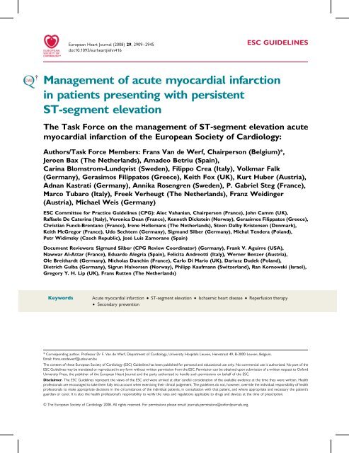
C. First medical contact<br />
and emergency care flow<br />
Optimal treatment <strong>of</strong> STEMI should be based on the implementation<br />
<strong>of</strong> an emergency medical system (EMS) supervis<strong>in</strong>g a<br />
network between hospitals with various levels <strong>of</strong> technology, connected<br />
by an efficient ambulance (or helicopter) service (Figure 1).<br />
Figure 1 Pre-hospital management. EMS ¼ emergency medical<br />
system; STEMI ¼ <strong>acute</strong> ST-segment elevation <strong>myocardial</strong> <strong><strong>in</strong>farction</strong>;<br />
GP ¼ general practitioner; PCI ¼ percutaneous coronary<br />
<strong>in</strong>tervention. Thick arrows ¼ preferred patient flow; dotted<br />
l<strong>in</strong>e ¼ to be avoided.
2914<br />
ESC Guidel<strong>in</strong>es<br />
The ma<strong>in</strong> features <strong>of</strong> such a network are: clear def<strong>in</strong>ition <strong>of</strong> geographical<br />
areas <strong>of</strong> <strong>in</strong>terest, shared protocols based on risk stratification,<br />
and transportation with appropriately equipped and staffed<br />
ambulances (or helicopters). The logistics <strong>of</strong> such a network are<br />
discussed <strong>in</strong> section I. A well-function<strong>in</strong>g regional system <strong>of</strong> care<br />
based on pre-hospital diagnosis and triage and fast transport to<br />
the most appropriate facility is key to the success <strong>of</strong> the treatment,<br />
and significantly improves outcome. 18,19<br />
For selection <strong>of</strong> the reperfusion strategy see Figure 2.<br />
1. Initial diagnosis and early<br />
risk stratification<br />
Rapid diagnosis and early risk stratification <strong>of</strong> <strong>patients</strong> present<strong>in</strong>g<br />
with <strong>acute</strong> chest pa<strong>in</strong> are important to identify <strong>patients</strong> <strong>in</strong> whom<br />
early <strong>in</strong>terventions can improve outcome. On the other hand,<br />
when the diagnosis <strong>of</strong> STEMI has been ruled out, attention can<br />
be focused on the detection <strong>of</strong> other cardiac or non-cardiac<br />
causes <strong>of</strong> the present<strong>in</strong>g symptoms such as aortic dissection,<br />
pulmonary embolism, and pericarditis. A work<strong>in</strong>g diagnosis <strong>of</strong><br />
STEMI must first be made (Table 3). This is usually based on the<br />
history <strong>of</strong> chest pa<strong>in</strong>/discomfort last<strong>in</strong>g for 10–20 m<strong>in</strong> or more<br />
(not respond<strong>in</strong>g fully to nitroglycer<strong>in</strong>e). Other locations such as<br />
epigastric or <strong>in</strong>terscapular are possible. Important clues are a previous<br />
history <strong>of</strong> coronary artery disease and radiation <strong>of</strong> the pa<strong>in</strong> to<br />
the neck, lower jaw, or left arm. The pa<strong>in</strong> may not be severe and, <strong>in</strong><br />
the elderly particularly, other presentations such as fatigue, dyspnoea,<br />
fa<strong>in</strong>tness, or syncope are common. There are no <strong>in</strong>dividual<br />
physical signs diagnostic <strong>of</strong> STEMI, but many <strong>patients</strong> have evidence<br />
<strong>of</strong> autonomic nervous system activation (pallor, sweat<strong>in</strong>g) and<br />
either hypotension or a narrow pulse pressure. Features may<br />
also <strong>in</strong>clude irregularities <strong>of</strong> the pulse, bradycardia or tachycardia,<br />
a third heart sound, and basal rales. An ECG should be obta<strong>in</strong>ed as<br />
soon as possible. Even at an early stage, the ECG is seldom normal.<br />
In the case <strong>of</strong> STEMI or new or presumed new left bundle-branch<br />
block, reperfusion therapy needs to be given, and measures to<br />
<strong>in</strong>itiate this treatment must be taken as soon as possible.<br />
However, the ECG can be equivocal <strong>in</strong> the early hours, and<br />
even <strong>in</strong> proven <strong><strong>in</strong>farction</strong> it may never show the classical features<br />
<strong>of</strong> ST-segment elevation and new Q-waves. Repeated ECG record<strong>in</strong>gs<br />
should be obta<strong>in</strong>ed and, when possible, the current ECG<br />
should be compared with previous records. Additional record<strong>in</strong>gs<br />
<strong>of</strong> lead V 7 –V 8 or V 4R are helpful to make the diagnosis <strong>in</strong> selected<br />
cases (true posterior <strong><strong>in</strong>farction</strong> or right ventricular <strong><strong>in</strong>farction</strong>,<br />
respectively). ECG monitor<strong>in</strong>g should be <strong>in</strong>itiated as soon as possible<br />
<strong>in</strong> all <strong>patients</strong> to detect life-threaten<strong>in</strong>g arrhythmias. In <strong>patients</strong><br />
with slowly evolv<strong>in</strong>g or stutter<strong>in</strong>g <strong>myocardial</strong> <strong><strong>in</strong>farction</strong>, serial<br />
ECGs should be taken to detect evolv<strong>in</strong>g <strong><strong>in</strong>farction</strong>. Blood<br />
sampl<strong>in</strong>g for serum markers <strong>of</strong> necrosis is rout<strong>in</strong>ely done <strong>in</strong> the<br />
<strong>acute</strong> phase, but one should not wait for the results to <strong>in</strong>itiate<br />
reperfusion treatment. The f<strong>in</strong>d<strong>in</strong>g <strong>of</strong> elevated markers <strong>of</strong> necrosis<br />
may sometimes be helpful <strong>in</strong> decid<strong>in</strong>g to perform coronary<br />
angiography (e.g. <strong>in</strong> <strong>patients</strong> with left bundle-branch block).<br />
Two-dimensional echocardiography has become a useful<br />
bedside technique <strong>in</strong> the triage <strong>of</strong> <strong>patients</strong> with <strong>acute</strong> chest pa<strong>in</strong>.<br />
Regional wall motion abnormalities occur with<strong>in</strong> seconds after<br />
coronary occlusion, well before necrosis. However, wall motion<br />
abnormalities are not specific for STEMI and may be due to ischaemia<br />
or an old <strong><strong>in</strong>farction</strong>. Two-dimensional echocardiography is <strong>of</strong><br />
particular value when the diagnosis <strong>of</strong> STEMI is uncerta<strong>in</strong>, and<br />
other causes <strong>of</strong> chest pa<strong>in</strong> such as <strong>acute</strong> aortic dissection, pericardial<br />
effusion, or pulmonary embolism are be<strong>in</strong>g considered. The<br />
performance <strong>of</strong> echocardiography should not delay the <strong>in</strong>itiation<br />
<strong>of</strong> treatment. The absence <strong>of</strong> wall motion abnormalities excludes<br />
major <strong>myocardial</strong> ischaemia.<br />
Older age, higher Killip class, elevated heart rate, lower systolic<br />
blood pressure, and anterior location <strong>of</strong> the <strong>in</strong>farct have been<br />
identified as the most important <strong>in</strong>dependent predictors <strong>of</strong> early<br />
mortality <strong>in</strong> cl<strong>in</strong>ical trials 20 and registries. 17,21 These characteristics<br />
conta<strong>in</strong> most <strong>of</strong> the prognostic <strong>in</strong>formation <strong>in</strong> the cl<strong>in</strong>ical data<br />
available at the time <strong>of</strong> the first medical contact. Other <strong>in</strong>dependent<br />
predictors are previous <strong><strong>in</strong>farction</strong>, height, time to treatment,<br />
diabetes, weight, and smok<strong>in</strong>g status 20 .<br />
2. Relief <strong>of</strong> pa<strong>in</strong>, breathlessness,<br />
and anxiety<br />
Relief <strong>of</strong> pa<strong>in</strong> is <strong>of</strong> paramount importance, not only for humane<br />
reasons but also because the pa<strong>in</strong> is associated with sympathetic<br />
activation, which causes vasoconstriction and <strong>in</strong>creases the workload<br />
<strong>of</strong> the heart. I.v. opioids are the analgesics most commonly<br />
used <strong>in</strong> this context (e.g. 4–8 mg <strong>of</strong> morph<strong>in</strong>e with additional<br />
doses <strong>of</strong> 2 mg at <strong>in</strong>tervals <strong>of</strong> 5–15 m<strong>in</strong> until the pa<strong>in</strong> is relieved);<br />
<strong>in</strong>tramuscular <strong>in</strong>jections should be avoided (Table 4). Side effects<br />
<strong>in</strong>clude nausea and vomit<strong>in</strong>g, hypotension with bradycardia, and<br />
respiratory depression. Antiemetics (e.g. metoclopramide<br />
5–10 mg i.v.) may be adm<strong>in</strong>istered concurrently with opioids.<br />
Table 3 Initial diagnosis<br />
History <strong>of</strong> chest pa<strong>in</strong>/discomfort<br />
Persistent ST-segment elevation or (presumed) new left<br />
bundle-branch block. Repeated ECG record<strong>in</strong>gs <strong>of</strong>ten needed.<br />
Elevated markers <strong>of</strong> <strong>myocardial</strong> necrosis (CK-MB, tropon<strong>in</strong>s). One<br />
should not wait for the results to <strong>in</strong>itiate reperfusion treatment.<br />
2-D echocardiography to rule out major <strong>acute</strong> <strong>myocardial</strong> ischaemia<br />
or other causes <strong>of</strong> chest pa<strong>in</strong>/discomfort.<br />
CK-MB ¼ creat<strong>in</strong>e k<strong>in</strong>ase MB form.<br />
Table 4 Relief <strong>of</strong> pa<strong>in</strong>, breathlessness, and anxiety<br />
Recommendations Class a Level b<br />
................................................................................<br />
I.v. opioids (4–8 mg morph<strong>in</strong>e) with additional I C<br />
doses <strong>of</strong> 2 mg at 5–15 m<strong>in</strong> <strong>in</strong>tervals<br />
O 2 (2–4 L/m<strong>in</strong>) if breathlessness or other signs I C<br />
<strong>of</strong> heart failure<br />
Tranquillizer—<strong>in</strong> very anxious <strong>patients</strong> IIa C<br />
a Class <strong>of</strong> recommendation.<br />
b Level <strong>of</strong> evidence.
ESC Guidel<strong>in</strong>es 2915<br />
The hypotension and bradycardia will usually respond to atrop<strong>in</strong>e<br />
(0.5–1 mg i.v., up to a total dose <strong>of</strong> 2 mg), and respiratory<br />
depression may require ventilatory support. Oxygen (2–4 L/m<strong>in</strong><br />
by mask or nasal prongs) should be adm<strong>in</strong>istered to those who<br />
are breathless or who have any features <strong>of</strong> heart failure or shock<br />
(see also Table 15). Non-<strong>in</strong>vasive monitor<strong>in</strong>g <strong>of</strong> blood oxygen<br />
saturation greatly helps <strong>in</strong> decid<strong>in</strong>g on the need for oxygen adm<strong>in</strong>istration<br />
or, <strong>in</strong> severe cases, ventilatory support. Non-steroidal<br />
anti-<strong>in</strong>flammatory drugs (NSAIDs) should not be given for pa<strong>in</strong><br />
relief because <strong>of</strong> possible prothrombotic effects.<br />
Anxiety is a natural response to the pa<strong>in</strong> and to the circumstances<br />
surround<strong>in</strong>g a heart attack. Reassurance <strong>of</strong> <strong>patients</strong> and<br />
those closely associated with them is <strong>of</strong> great importance. If the<br />
patient becomes excessively disturbed, it may be appropriate to<br />
adm<strong>in</strong>ister a tranquillizer, but opioids are all that is required <strong>in</strong><br />
many cases.<br />
3. Cardiac arrest<br />
Many deaths occur <strong>in</strong> the very first hours after STEMI due to<br />
ventricular fibrillation (VF). The implementation <strong>of</strong> an organization<br />
to cope with out-<strong>of</strong>-hospital cardiac arrest is pivotal to provide<br />
prompt cardiopulmonary resuscitation, early defibrillation if<br />
needed, and effective advanced cardiac life support. Availability<br />
<strong>of</strong> automated external defibrillators is a key factor <strong>in</strong> <strong>in</strong>creas<strong>in</strong>g<br />
survival. Readers are referred to the latest guidel<strong>in</strong>es on cardiopulmonary<br />
resuscitation provided by the European Resuscitation<br />
Council. 22<br />
D. Pre-hospital or early<br />
<strong>in</strong>-hospital care<br />
1. Restor<strong>in</strong>g coronary flow and<br />
<strong>myocardial</strong> tissue reperfusion<br />
For <strong>patients</strong> with the cl<strong>in</strong>ical presentation <strong>of</strong> STEMI with<strong>in</strong> 12 h<br />
after symptom onset and with persistent ST-segment elevation<br />
or new or presumed new left bundle-branch block, early mechanical<br />
(PCI) or pharmacological reperfusion should be performed.<br />
There is general agreement that reperfusion therapy (primary<br />
PCI) should be considered if there is cl<strong>in</strong>ical and/or electrocardiographic<br />
evidence <strong>of</strong> ongo<strong>in</strong>g ischaemia, even if, accord<strong>in</strong>g to the<br />
patient, symptoms started .12 h before as the exact onset <strong>of</strong><br />
symptoms is <strong>of</strong>ten unclear. However, there is no consensus as to<br />
whether PCI is also beneficial <strong>in</strong> <strong>patients</strong> present<strong>in</strong>g .12 h from<br />
symptom onset <strong>in</strong> the absence <strong>of</strong> cl<strong>in</strong>ical and/or electrocardiographic<br />
evidence <strong>of</strong> ongo<strong>in</strong>g ischaemia. In a randomized study <strong>in</strong><br />
STEMI <strong>patients</strong> present<strong>in</strong>g without persist<strong>in</strong>g symptoms between<br />
12 and 48 h after symptom onset (n ¼ 347), PCI was associated<br />
with significant <strong>myocardial</strong> salvage, lend<strong>in</strong>g some support to an<br />
<strong>in</strong>vasive strategy <strong>in</strong> these <strong>patients</strong>, but cl<strong>in</strong>ical outcomes were not<br />
better. 23 In the OAT trial <strong>in</strong>clud<strong>in</strong>g 2166 stable <strong>patients</strong> with an<br />
occluded <strong>in</strong>farct-related vessel 3 to 28 calendar days after<br />
symptom onset, PCI did not improve cl<strong>in</strong>ical outcome, 24 <strong>in</strong>clud<strong>in</strong>g<br />
<strong>in</strong> the subgroup <strong>of</strong> 331 <strong>patients</strong> randomized between 24 and 72 h<br />
after onset <strong>of</strong> <strong><strong>in</strong>farction</strong>. 25 No firm recommendations can be<br />
made given the limited data currently available (Table 5).<br />
Figure 2 Reperfusion strategies. The thick arrow <strong>in</strong>dicates the preferred strategy.
2916<br />
ESC Guidel<strong>in</strong>es<br />
Table 5 Reperfusion therapy<br />
Recommendations Class a Level b<br />
.............................................................................................................................................................................<br />
Reperfusion therapy is <strong>in</strong>dicated <strong>in</strong> all <strong>patients</strong> with history <strong>of</strong> chest pa<strong>in</strong>/discomfort <strong>of</strong> ,12 h and with persistent<br />
I A<br />
ST-segment elevation or (presumed) new left bundle-branch block<br />
Reperfusion therapy should be considered if there is cl<strong>in</strong>ical and/or ECG evidence <strong>of</strong> ongo<strong>in</strong>g ischaemia even if, accord<strong>in</strong>g IIa C<br />
to patient, symptoms started .12 h before<br />
Reperfusion us<strong>in</strong>g PCI may be considered <strong>in</strong> stable <strong>patients</strong> present<strong>in</strong>g .12 to 24 h after symptom onset IIb B<br />
PCI <strong>of</strong> a totally occluded <strong>in</strong>farct artery .24 h after symptom onset <strong>in</strong> stable <strong>patients</strong> without signs <strong>of</strong> ischaemia III B<br />
.............................................................................................................................................................................<br />
Primary PCI<br />
Preferred treatment if performed by an experienced team as soon as possible after FMC I A<br />
Time from FMC to balloon <strong>in</strong>flation should be ,2 h <strong>in</strong> any case and ,90 m<strong>in</strong> <strong>in</strong> <strong>patients</strong> present<strong>in</strong>g early (e.g. ,2 h) I B<br />
with large <strong>in</strong>farct and low bleed<strong>in</strong>g risk<br />
Indicated for <strong>patients</strong> <strong>in</strong> shock and those with contra<strong>in</strong>dications to fibr<strong>in</strong>olytic therapy irrespective <strong>of</strong> time delay I B<br />
Antiplatelet co-therapy c<br />
Aspir<strong>in</strong> I B<br />
NSAID and COX-2 selective <strong>in</strong>hibitors III B<br />
Clopidogrel load<strong>in</strong>g dose I C<br />
GPIIb/IIIa antagonist<br />
Abciximab IIa A<br />
Tir<strong>of</strong>iban IIb B<br />
Eptifibatide IIb C<br />
Antithromb<strong>in</strong> therapy c<br />
Hepar<strong>in</strong> I C<br />
Bivalirud<strong>in</strong> IIa B<br />
Fondapar<strong>in</strong>ux III B<br />
Adjunctive devices<br />
Thrombus aspiration IIb B<br />
.............................................................................................................................................................................<br />
Rescue PCI<br />
After failed fibr<strong>in</strong>olysis <strong>in</strong> <strong>patients</strong> with large <strong>in</strong>farcts if performed with<strong>in</strong> 12 h after onset IIa A<br />
.............................................................................................................................................................................<br />
Fibr<strong>in</strong>olytic therapy c<br />
In the absence <strong>of</strong> contra<strong>in</strong>dications (see Table 7) and if primary PCI cannot be performed with<strong>in</strong> the recommended time I A<br />
(see above and Figure 2)<br />
A fibr<strong>in</strong>-specific agent should be given I B<br />
Pre-hospital <strong>in</strong>itiation <strong>of</strong> fibr<strong>in</strong>olytic therapy IIa A<br />
Antiplatelet co-therapy c<br />
if not already on aspir<strong>in</strong> oral (soluble or chewable/non-enteric-coated) or i.v. dose <strong>of</strong> aspir<strong>in</strong> plus I B<br />
clopidogrel oral load<strong>in</strong>g dose if age 75 years I B<br />
if age .75 years start with ma<strong>in</strong>tenance dose IIa B<br />
Antithromb<strong>in</strong> co-therapy c<br />
with alteplase, reteplase, and tenecteplase:<br />
enoxapar<strong>in</strong> i.v. bolus followed 15 m<strong>in</strong> later by first s.c. dose; if age .75 years no i.v. bolus and start with reduced I A<br />
first s.c. dose<br />
if enoxapar<strong>in</strong> is not available: a weight-adjusted bolus <strong>of</strong> i.v. hepar<strong>in</strong> followed by a weight-adjusted i.v. <strong>in</strong>fusion I A<br />
with first aPTT control after 3 h<br />
with streptok<strong>in</strong>ase:<br />
an i.v. bolus <strong>of</strong> fondapar<strong>in</strong>ux followed by an s.c. dose 24 h later or IIa B<br />
enoxapar<strong>in</strong> i.v. bolus followed 15 m<strong>in</strong> later by first s.c. dose; if age .75 years no i.v. bolus and start with reduced IIa B<br />
first s.c. dose<br />
or a weight-adjusted dose <strong>of</strong> i.v. hepar<strong>in</strong> followed by a weight-adjusted <strong>in</strong>fusion IIa C<br />
a Class <strong>of</strong> recommendation.<br />
b Level <strong>of</strong> evidence.<br />
c For doses see Tables 8, 9, and 10.
ESC Guidel<strong>in</strong>es 2917<br />
Different reperfusion strategies are depicted <strong>in</strong> Figure 2. In this<br />
figure the first medical contact is the place (ambulance or hospital)<br />
where, at least <strong>in</strong> pr<strong>in</strong>ciple, reperfusion therapy could be given. The<br />
(<strong>in</strong>creas<strong>in</strong>g) time limits for the different reperfusion strategies are<br />
also depicted schematically.<br />
a. Percutaneous coronary <strong>in</strong>terventions<br />
The role <strong>of</strong> PCIs dur<strong>in</strong>g the early hours <strong>of</strong> STEMI can be divided<br />
<strong>in</strong>to primary PCI, PCI comb<strong>in</strong>ed with pharmacological reperfusion<br />
therapy (facilitated PCI), and ‘rescue PCI’ after failed pharmacological<br />
reperfusion. Separate ESC Guidel<strong>in</strong>es cover<strong>in</strong>g all <strong>in</strong>dications<br />
for PCI have been published before. 26<br />
Primary PCI and delay times<br />
Primary PCI is def<strong>in</strong>ed as angioplasty and/or stent<strong>in</strong>g without prior<br />
or concomitant fibr<strong>in</strong>olytic therapy, and is the preferred therapeutic<br />
option when it can be performed expeditiously by an<br />
experienced team (Table 5). An experienced team <strong>in</strong>cludes not<br />
only <strong>in</strong>terventional cardiologists but also skilled support<strong>in</strong>g staff.<br />
This means that only hospitals with an established <strong>in</strong>terventional<br />
cardiology programme (24 h/7 days) should use primary PCI as a<br />
rout<strong>in</strong>e treatment option for <strong>patients</strong> present<strong>in</strong>g with the symptoms<br />
and signs <strong>of</strong> STEMI. Lower mortality rates among <strong>patients</strong><br />
undergo<strong>in</strong>g primary PCI are observed <strong>in</strong> centres with a high<br />
volume <strong>of</strong> PCI procedures. 27,28 Primary PCI is effective <strong>in</strong> secur<strong>in</strong>g<br />
and ma<strong>in</strong>ta<strong>in</strong><strong>in</strong>g coronary artery patency and avoids some <strong>of</strong> the<br />
bleed<strong>in</strong>g risks <strong>of</strong> fibr<strong>in</strong>olysis. Randomized cl<strong>in</strong>ical trials compar<strong>in</strong>g<br />
timely performed primary PCI with <strong>in</strong>-hospital fibr<strong>in</strong>olytic<br />
therapy <strong>in</strong> high-volume, experienced centres have shown more<br />
effective restoration <strong>of</strong> patency, less reocclusion, improved residual<br />
left ventricular (LV), function and better cl<strong>in</strong>ical outcome with<br />
primary PCI. 29 Rout<strong>in</strong>e coronary stent implantation <strong>in</strong> <strong>patients</strong><br />
with STEMI decreases the need for target vessel revascularization<br />
but is not associated with significant reductions <strong>in</strong> death or re<strong><strong>in</strong>farction</strong><br />
rates 30,31 when compared with primary angioplasty. In<br />
addition, several randomized cl<strong>in</strong>ical trials with medium-term<br />
follow-up, <strong>in</strong>clud<strong>in</strong>g <strong>patients</strong> with STEMI, have shown that<br />
drug-elut<strong>in</strong>g stents reduce the risk <strong>of</strong> re<strong>in</strong>tervention compared<br />
with bare metal stents, without hav<strong>in</strong>g a significant impact on the<br />
risk <strong>of</strong> stent thrombosis, recurrent <strong>myocardial</strong> <strong><strong>in</strong>farction</strong>, and<br />
death. 32 – 34 As for other cl<strong>in</strong>ical presentations <strong>of</strong> coronary artery<br />
disease, long-term data on the efficacy and safety <strong>of</strong> drug-elut<strong>in</strong>g<br />
stents <strong>in</strong> <strong>patients</strong> with STEMI are still needed.<br />
Both randomized studies and registries have <strong>in</strong>dicated that long<br />
delay times to primary PCI are associated with a worse cl<strong>in</strong>ical<br />
outcome. 35,36 Several delay times can be def<strong>in</strong>ed: time from<br />
symptom onset to first medical contact (FMC), time from FMC<br />
to arrival <strong>in</strong> cath lab, time from FMC to sheath <strong>in</strong>sertion, time<br />
from FMC to balloon <strong>in</strong>flation. The ‘PCI-related delay time’ is<br />
the theoretical difference between the time <strong>of</strong> FMC to balloon<br />
<strong>in</strong>flation m<strong>in</strong>us the time from FMC to start <strong>of</strong> fibr<strong>in</strong>olytic therapy<br />
(¼ ‘door-to-balloon’ m<strong>in</strong>us ‘door-to-needle’). The extent to<br />
which the PCI-related time delay dim<strong>in</strong>ishes the advantages <strong>of</strong><br />
PCI over fibr<strong>in</strong>olysis has been the subject <strong>of</strong> many analyses and<br />
debates. Because no specifically designed study has addressed<br />
this issue, caution is needed when <strong>in</strong>terpret<strong>in</strong>g the results <strong>of</strong><br />
these post hoc analyses. From randomized trials it was calculated<br />
that the PCI-related time delay that may mitigate the benefit <strong>of</strong><br />
the mechanical <strong>in</strong>tervention varies between 60 37 and 110 m<strong>in</strong> 38<br />
depend<strong>in</strong>g on the fibr<strong>in</strong>olytic used. 39 In another analysis <strong>of</strong> these<br />
trials, a benefit <strong>of</strong> primary PCI over fibr<strong>in</strong>olytic therapy up to a<br />
PCI-related delay <strong>of</strong> 120 m<strong>in</strong> was calculated. 40 In 192 509 <strong>patients</strong><br />
<strong>in</strong>cluded <strong>in</strong> the NRMI 2-4 registry, 41 the mean PCI-related time<br />
delay where mortality rates <strong>of</strong> the two reperfusion strategies<br />
were equal was calculated at 114 m<strong>in</strong>. This study also <strong>in</strong>dicated<br />
that this time delay varied considerably accord<strong>in</strong>g to age,<br />
symptom duration, and <strong>in</strong>farct location: from ,1 h for an anterior<br />
<strong><strong>in</strong>farction</strong> <strong>in</strong> a patient ,65 years present<strong>in</strong>g ,2 h after symptom<br />
onset, to almost 3 h for a non-anterior <strong><strong>in</strong>farction</strong> <strong>in</strong> a patient<br />
.65 years present<strong>in</strong>g .2 h after symptom onset. Although<br />
these results were derived from a post hoc analysis <strong>of</strong> a registry<br />
and reported delay times are sometimes <strong>in</strong>accurate, this study<br />
suggests that an <strong>in</strong>dividualized rather than a uniform approach<br />
for select<strong>in</strong>g the optimal reperfusion modality could be more<br />
appropriate when PCI cannot be performed with<strong>in</strong> a short delay.<br />
Tak<strong>in</strong>g <strong>in</strong>to account the studies and registries mentioned above,<br />
primary PCI (balloon <strong>in</strong>flation) should be performed with<strong>in</strong> 2 h<br />
after FMC <strong>in</strong> all cases. In <strong>patients</strong> present<strong>in</strong>g early with a large<br />
amount <strong>of</strong> myocardium at risk, the delay should be shorter.<br />
Although no specific studies have been performed, a maximum<br />
delay <strong>of</strong> only 90 m<strong>in</strong> after FMC seems to be a reasonable recommendation<br />
<strong>in</strong> these <strong>patients</strong>.<br />
Patients with contra<strong>in</strong>dications to fibr<strong>in</strong>olytic therapy have a<br />
higher morbidity and mortality than those eligible for this<br />
therapy. Primary PCI can be performed with success <strong>in</strong> these<br />
<strong>patients</strong>. 42 Primary PCI is the preferred treatment for <strong>patients</strong> <strong>in</strong><br />
shock. 43 Except for <strong>patients</strong> <strong>in</strong> cardiogenic shock, only the<br />
culprit lesion should be dilated <strong>in</strong> the <strong>acute</strong> sett<strong>in</strong>g. Complete<br />
revascularization <strong>of</strong> the non-culprit lesions may be performed at<br />
a later time po<strong>in</strong>t depend<strong>in</strong>g on the rema<strong>in</strong><strong>in</strong>g ischaemia.<br />
Facilitated PCI<br />
Facilitated PCI is def<strong>in</strong>ed as a pharmacological reperfusion treatment<br />
delivered prior to a planned PCI, <strong>in</strong> order to bridge the<br />
PCI-related time delay. Full-dose lytic therapy, half-dose lytic<br />
therapy with a glycoprote<strong>in</strong> (GP)IIb/IIIa <strong>in</strong>hibitor and GPIIb/IIIa<br />
<strong>in</strong>hibitor alone have been tested for this <strong>in</strong>dication. There is no evidence<br />
<strong>of</strong> a significant cl<strong>in</strong>ical benefit with any <strong>of</strong> these<br />
agents. 16,12,44,45 In spite <strong>of</strong> the fact that pre-PCI patency rates<br />
were higher with lytic-based treatments, no mortality benefit but<br />
more bleed<strong>in</strong>g complications were observed. The pre-PCI<br />
patency rates with upfront abciximab or high-bolus dose tir<strong>of</strong>iban<br />
alone were not higher than with placebo. Facilitated PCI as it has<br />
been tested <strong>in</strong> these trials cannot be recommended.<br />
Rescue PCI<br />
Rescue PCI is def<strong>in</strong>ed as PCI performed on a coronary artery which<br />
rema<strong>in</strong>s occluded despite fibr<strong>in</strong>olytic therapy. The non-<strong>in</strong>vasive<br />
identification <strong>of</strong> failed fibr<strong>in</strong>olysis rema<strong>in</strong>s a challeng<strong>in</strong>g issue, but<br />
,50% ST-segment resolution <strong>in</strong> the lead(s) with the highest<br />
ST-segment elevations 60–90 m<strong>in</strong> after start <strong>of</strong> fibr<strong>in</strong>olytic<br />
therapy has <strong>in</strong>creas<strong>in</strong>gly been used as a surrogate. Rescue PCI has<br />
been shown to be feasible and relatively safe. In a randomized<br />
study <strong>of</strong> 427 <strong>patients</strong> (REACT), the event-free survival at
2918<br />
ESC Guidel<strong>in</strong>es<br />
6 months after failed fibr<strong>in</strong>olysis was significantly higher with rescue<br />
PCI than with repeated adm<strong>in</strong>istration <strong>of</strong> a fibr<strong>in</strong>olytic agent or conservative<br />
treatment. 46 A recent meta-analysis, <strong>in</strong>clud<strong>in</strong>g REACT,<br />
showed that rescue PCI is associated with a significant reduction<br />
<strong>in</strong> heart failure and re<strong><strong>in</strong>farction</strong> and a trend towards lower all-cause<br />
mortality when compared with a conservative strategy, at the cost,<br />
however, <strong>of</strong> an <strong>in</strong>creased risk <strong>of</strong> stroke and bleed<strong>in</strong>g complications.<br />
47 Rescue PCI should be considered when there is evidence<br />
<strong>of</strong> failed fibr<strong>in</strong>olysis based on cl<strong>in</strong>ical signs and <strong>in</strong>sufficient<br />
ST-segment resolution (,50%), if there is cl<strong>in</strong>ical or ECG evidence<br />
<strong>of</strong> a large <strong>in</strong>farct, and if the procedure can be performed with<strong>in</strong> a<br />
reasonable time delay (up to 12 h after onset <strong>of</strong> symptoms).<br />
Adjunctive antithrombotic treatment and devices (Tables 6 and 9)<br />
Aspir<strong>in</strong>, NSAID, COX-2 <strong>in</strong>hibitors. Aspir<strong>in</strong> should be given to all<br />
<strong>patients</strong> with a STEMI as soon as possible after the diagnosis is<br />
deemed probable. There are few contra<strong>in</strong>dications to the use <strong>of</strong><br />
aspir<strong>in</strong>, but it should not be given to those with a known hypersensitivity,<br />
active gastro<strong>in</strong>test<strong>in</strong>al bleed<strong>in</strong>g, known clott<strong>in</strong>g disorders,<br />
or severe hepatic disease. Aspir<strong>in</strong> may occasionally trigger bronchospasm<br />
<strong>in</strong> asthmatic <strong>patients</strong>. Aspir<strong>in</strong> should be started at a dose <strong>of</strong><br />
150–325 mg <strong>in</strong> a chewable form (enteric-coated aspir<strong>in</strong> should not<br />
be given because <strong>of</strong> slow onset <strong>of</strong> action). An alternative approach,<br />
especially if oral <strong>in</strong>gestion is not possible, is i.v. adm<strong>in</strong>istration <strong>of</strong><br />
aspir<strong>in</strong> at a dose <strong>of</strong> 250–500 mg, although no specific data are<br />
available on the relative merits <strong>of</strong> this strategy. A lower dose<br />
(75–160 mg) is given orally daily thereafter for life.<br />
NSAIDs (apart from aspir<strong>in</strong>) and selective cyclo-oxygenase<br />
(COX-2) <strong>in</strong>hibitors have been demonstrated to <strong>in</strong>crease the risk<br />
<strong>of</strong> death, re<strong><strong>in</strong>farction</strong>, cardiac rupture, and other complications <strong>in</strong><br />
STEMI <strong>patients</strong>: discont<strong>in</strong>uation <strong>of</strong> these drugs is <strong>in</strong>dicated at the<br />
time <strong>of</strong> STEMI. 48,49<br />
Clopidogrel. Although clopidogrel is less studied <strong>in</strong> <strong>patients</strong> with<br />
STEMI treated with primary PCI, there is abundant evidence on<br />
Table 6 Antithrombotic treatment without<br />
reperfusion therapy<br />
Recommendations Class a Level b<br />
................................................................................<br />
Antiplatelet co-therapy c<br />
If not already on aspir<strong>in</strong> oral (soluble or chewable/ I A<br />
non-enteric-coated) or i.v. dose <strong>of</strong> aspir<strong>in</strong> if<br />
oral <strong>in</strong>gestion is not feasible<br />
Oral dose <strong>of</strong> clopidogrel I B<br />
................................................................................<br />
Antithromb<strong>in</strong> co-therapy<br />
I.v. bolus <strong>of</strong> fondapar<strong>in</strong>ux followed 24 h later by an I B<br />
s.c. dose<br />
If fondapar<strong>in</strong>ux is not available: enoxapar<strong>in</strong> i.v. I B<br />
bolus followed 15 m<strong>in</strong> later by first s.c. dose; if<br />
age .75 years no i.v. bolus and start with<br />
reduced s.c. dose or<br />
I.v. hepar<strong>in</strong> followed by a weight-adjusted i.v.<br />
<strong>in</strong>fusion with first aPTT control after 3 h<br />
a Class <strong>of</strong> recommendation.<br />
b Level <strong>of</strong> evidence.<br />
c For doses see Tables 9 and 10.<br />
I<br />
B<br />
Table 7 Contra<strong>in</strong>dications to fibr<strong>in</strong>olytic therapy<br />
Absolute contra<strong>in</strong>dications<br />
Haemorrhagic stroke or stroke <strong>of</strong> unknown orig<strong>in</strong> at any time<br />
Ischaemic stroke <strong>in</strong> preced<strong>in</strong>g 6 months<br />
Central nervous system trauma or neoplasms<br />
Recent major trauma/surgery/head <strong>in</strong>jury (with<strong>in</strong> preced<strong>in</strong>g 3 weeks)<br />
Gastro<strong>in</strong>test<strong>in</strong>al bleed<strong>in</strong>g with<strong>in</strong> the last month<br />
Known bleed<strong>in</strong>g disorder<br />
Aortic dissection<br />
Non-compressible punctures (e.g. liver biopsy, lumbar puncture)<br />
................................................................................<br />
Relative contra<strong>in</strong>dications<br />
Transient ischaemic attack <strong>in</strong> preced<strong>in</strong>g 6 months<br />
Oral anticoagulant therapy<br />
Pregnancy or with<strong>in</strong> 1 week post-partum<br />
Refractory hypertension (systolic blood pressure .180 mmHg and/or<br />
diastolic blood pressure .110 mmHg)<br />
Advanced liver disease<br />
Infective endocarditis<br />
Active peptic ulcer<br />
Refractory resuscitation<br />
its usefulness as an adjunctive antiplatelet therapy on top <strong>of</strong><br />
aspir<strong>in</strong> <strong>in</strong> <strong>patients</strong> undergo<strong>in</strong>g PCI. 50 – 52 Based on these data, clopidogrel<br />
should be given as soon as possible to all <strong>patients</strong> with<br />
STEMI undergo<strong>in</strong>g PCI. It is started with a load<strong>in</strong>g dose <strong>of</strong> at<br />
least 300 mg, but a 600 mg load<strong>in</strong>g dose achieves a more rapid<br />
and stronger <strong>in</strong>hibition <strong>of</strong> platelet aggregation. 53,54 This should<br />
be followed by a daily dose <strong>of</strong> 75 mg.<br />
GPIIb/IIIa antagonists. Platelet GPIIb/IIIa <strong>in</strong>hibitors block the f<strong>in</strong>al<br />
pathway <strong>of</strong> platelet aggregation. Most <strong>of</strong> the studies on the role<br />
<strong>of</strong> GPIIb/IIIa antagonists <strong>in</strong> STEMI have focused on abciximab<br />
rather than on the other two members <strong>of</strong> the family, tir<strong>of</strong>iban<br />
and eptifibatide. Several randomized trials have assessed the<br />
value <strong>of</strong> periprocedural adm<strong>in</strong>istration <strong>of</strong> i.v. abciximab <strong>in</strong> addition<br />
to aspir<strong>in</strong> and hepar<strong>in</strong> <strong>in</strong> this sett<strong>in</strong>g. A systematic review <strong>of</strong> these<br />
trials showed that abciximab reduced 30-day mortality by 32%<br />
without affect<strong>in</strong>g the risk <strong>of</strong> haemorrhagic stroke and major bleed<strong>in</strong>g.<br />
55 Abciximab did not have a significant impact on the patency <strong>of</strong><br />
<strong>in</strong>farct-related vessels, and its adm<strong>in</strong>istration upstream <strong>of</strong> a planned<br />
PCI procedure did not <strong>of</strong>fer advantages compared with the adm<strong>in</strong>istration<br />
<strong>in</strong> the cath lab. 44 Abciximab is given i.v. as a bolus <strong>of</strong><br />
0.25 mg/kg bolus, 0.125 mg/kg/m<strong>in</strong> <strong>in</strong>fusion (maximum 10 mg/m<strong>in</strong><br />
for 12 h). However, it rema<strong>in</strong>s to be elucidated whether abciximab<br />
provides an additional benefit to STEMI <strong>patients</strong> who receive an<br />
optimal clopidogrel treatment prior to PCI. In the On-TIME 2<br />
trial (n ¼ 984) pre-hospital <strong>in</strong>itiation <strong>of</strong> high-bolus dose tir<strong>of</strong>iban<br />
<strong>in</strong> association with aspir<strong>in</strong>, clopidogrel (600 mg), and hepar<strong>in</strong><br />
improved ST-segment resolution but was not associated with<br />
more patency <strong>of</strong> the <strong>in</strong>farct vessel or a significant net cl<strong>in</strong>ical<br />
benefit when compared with placebo. 45<br />
Hepar<strong>in</strong>. Hepar<strong>in</strong> is standard anticoagulant therapy dur<strong>in</strong>g PCI. The<br />
lack <strong>of</strong> randomized cl<strong>in</strong>ical trials <strong>of</strong> hepar<strong>in</strong> vs. placebo dur<strong>in</strong>g PCI<br />
<strong>in</strong> STEMI is due to the strong belief that anticoagulation therapy is a<br />
requirement dur<strong>in</strong>g the procedure. Hepar<strong>in</strong> is given as an i.v. bolus<br />
at a usual start<strong>in</strong>g dose <strong>of</strong> 100 U/kg weight (60 U/kg if GPIIb/IIIa
ESC Guidel<strong>in</strong>es 2919<br />
Table 8 Doses <strong>of</strong> fibr<strong>in</strong>olytic agents<br />
Initial treatment<br />
Specific contra<strong>in</strong>dications<br />
...............................................................................................................................................................................<br />
Streptok<strong>in</strong>ase (SK) 1.5 million units over 30–60 m<strong>in</strong> i.v. Prior SK or anistreplase<br />
Alteplase (t-PA)<br />
15 mg i.v. bolus<br />
0.75 mg/kg over 30 m<strong>in</strong> then 0.5 mg/kg over 60 m<strong>in</strong> i.v.<br />
Total dosage not to exceed 100 mg<br />
Reteplase (r-PA)<br />
10 U þ 10 U i.v. bolus given 30 m<strong>in</strong> apart<br />
Tenecteplase (TNK-tPA)<br />
S<strong>in</strong>gle i.v. bolus<br />
30 mg if ,60 kg<br />
35 mg if 60 to ,70 kg<br />
40 mg if 70 to ,80 kg<br />
45 mg if 80 to ,90 kg<br />
50 mg if 90 kg<br />
Table 9 Doses <strong>of</strong> antiplatelet co-therapies<br />
With primary PCI<br />
Aspir<strong>in</strong> Oral dose <strong>of</strong> 150–325 mg or i.v. dose <strong>of</strong> 250–<br />
500 mg if oral <strong>in</strong>gestion is not possible<br />
Clopidogrel Oral load<strong>in</strong>g dose <strong>of</strong> at least 300 mg, preferably<br />
600 mg<br />
GPIIb/IIIa Abciximab: i.v. bolus <strong>of</strong> 0.25 mg/kg bolus followed<br />
<strong>in</strong>hibitors by 0.125 mg/kg per m<strong>in</strong> <strong>in</strong>fusion (maximum<br />
10 mg/m<strong>in</strong> for 12 h)<br />
................................................................................<br />
With fibr<strong>in</strong>olytic treatment<br />
Aspir<strong>in</strong> Oral dose <strong>of</strong> 150–325 mg or i.v. dose <strong>of</strong> 250 mg if<br />
oral <strong>in</strong>gestion is not possible<br />
Clopidogrel Load<strong>in</strong>g dose <strong>of</strong> 300 mg if age 75 years; 75 mg if<br />
age .75<br />
................................................................................<br />
Without reperfusion therapy<br />
Aspir<strong>in</strong> Oral dose <strong>of</strong> 150–325 mg<br />
Clopidogrel Oral dose <strong>of</strong> 75 mg<br />
antagonists are used). It is recommended to perform the procedure<br />
under activated clott<strong>in</strong>g time (ACT) guidance: hepar<strong>in</strong><br />
should be given at a dose able to ma<strong>in</strong>ta<strong>in</strong> an ACT <strong>of</strong> 250–350 s<br />
(200–250 s if GPIIb/IIIa antagonists are used).<br />
Low-molecular-weight hepar<strong>in</strong>s (LMWHs) have been studied <strong>in</strong><br />
a limited number <strong>of</strong> STEMI <strong>patients</strong> undergo<strong>in</strong>g primary PCI. Thus,<br />
there is little evidence to support their use <strong>in</strong>stead <strong>of</strong> hepar<strong>in</strong> <strong>in</strong><br />
this sett<strong>in</strong>g.<br />
Bivalirud<strong>in</strong>. The direct thromb<strong>in</strong> <strong>in</strong>hibitor, bivalirud<strong>in</strong>, has been<br />
<strong>in</strong>vestigated as an adjunct antithrombotic therapy <strong>in</strong> <strong>patients</strong><br />
undergo<strong>in</strong>g PCI. In the Harmoniz<strong>in</strong>g Outcomes With Revascularization<br />
and Stents <strong>in</strong> Acute Myocardial Infarction<br />
(HORIZONS-AMI) trial, 3602 <strong>patients</strong> undergo<strong>in</strong>g PCI were randomly<br />
assigned <strong>in</strong> an unbl<strong>in</strong>ded fashion to receive either bivalirud<strong>in</strong><br />
with provisional use <strong>of</strong> GPIIb/IIIa <strong>in</strong>hibitor or hepar<strong>in</strong> (or enoxapar<strong>in</strong>)<br />
plus a GPIIb/IIIa <strong>in</strong>hibitor. 56 The primary end-po<strong>in</strong>t, the composite<br />
<strong>of</strong> 30-day <strong>in</strong>cidence <strong>of</strong> major adverse cardiac events or<br />
major bleed<strong>in</strong>g, was significantly reduced by bivalirud<strong>in</strong> due to a<br />
40% reduction <strong>in</strong> major bleed<strong>in</strong>g (P ,0.001). All-cause mortality<br />
at 30 days was 1% lower (P ,0.0047), but <strong>acute</strong> stent thrombosis<br />
occurred more frequently (P ,0.001). Bivalirud<strong>in</strong> is given as an i.v.<br />
bolus <strong>of</strong> 0.75 mg/kg followed by an <strong>in</strong>fusion <strong>of</strong> 1.75 mg/kg/h not<br />
titrated to ACT and usually term<strong>in</strong>ated at the end <strong>of</strong> the<br />
procedure.<br />
Fondapar<strong>in</strong>ux. Fondapar<strong>in</strong>ux, a factor Xa <strong>in</strong>hibitor, has been compared<br />
with hepar<strong>in</strong> or placebo <strong>in</strong> 12 092 STEMI <strong>patients</strong> treated<br />
with fibr<strong>in</strong>olytic agents or PCI, or no reperfusion therapy. 57 In<br />
the PCI subset, fondapar<strong>in</strong>ux was associated with a non-significant<br />
1% higher <strong>in</strong>cidence <strong>of</strong> death or recurrent <strong><strong>in</strong>farction</strong> at 30 days.<br />
These f<strong>in</strong>d<strong>in</strong>gs together with the occurrence <strong>of</strong> catheter thrombosis<br />
do not lend support to the use <strong>of</strong> fondapar<strong>in</strong>ux as the sole<br />
anticoagulant <strong>in</strong> <strong>patients</strong> undergo<strong>in</strong>g primary PCI.<br />
Adjunctive devices. Adjunctive devices aim<strong>in</strong>g at the prevention <strong>of</strong><br />
distal embolization have been <strong>in</strong>vestigated <strong>in</strong> several randomized<br />
studies. Formal meta-analyses <strong>of</strong> these studies show heterogeneous<br />
results with no overall cl<strong>in</strong>ical benefit despite a lower<br />
rate <strong>of</strong> distal embolization angiographically. 58 In a recent randomized<br />
study <strong>in</strong> 1071 <strong>patients</strong>, aspiration <strong>of</strong> thrombus prior to PCI<br />
was associated with improved tissue reperfusion [<strong>myocardial</strong><br />
blush grades (MBGs)] and improved survival at 1 year when compared<br />
with conventional PCI. 59,60 See also section D.1.d. and<br />
Table 13.<br />
b. Fibr<strong>in</strong>olytic treatment<br />
The evidence for benefit<br />
The benefit <strong>of</strong> fibr<strong>in</strong>olytic therapy is well established: 61 approximately<br />
30 early deaths are prevented per 1000 <strong>patients</strong> treated,<br />
with 20 deaths prevented per 1000 <strong>patients</strong> treated between 7<br />
and 12 h after symptom onset. Overall, the largest absolute<br />
benefit is seen among <strong>patients</strong> with the highest risk, even though<br />
the proportional benefit may be similar. In a subgroup <strong>of</strong> 3300<br />
<strong>patients</strong> over the age <strong>of</strong> 75 present<strong>in</strong>g with<strong>in</strong> 12 h <strong>of</strong> symptom<br />
onset and with either STEMI or bundle-branch block, mortality<br />
rates were significantly reduced by fibr<strong>in</strong>olytic therapy. 62<br />
Time to treatment<br />
Analysis <strong>of</strong> studies <strong>in</strong> which .6000 <strong>patients</strong> were randomized to<br />
pre-hospital or <strong>in</strong>-hospital fibr<strong>in</strong>olysis has shown a significant<br />
reduction (17%) <strong>in</strong> early mortality with pre-hospital treatment. 63<br />
In a meta-analysis <strong>of</strong> 22 trials, 64 a much larger mortality reduction
2920<br />
ESC Guidel<strong>in</strong>es<br />
Table 10 Doses <strong>of</strong> antithromb<strong>in</strong> co-therapies<br />
With primary PCI<br />
Hepar<strong>in</strong> I.v. bolus at a usual start<strong>in</strong>g dose <strong>of</strong> 100 U/kg weight (60 U/kg if GPIIb/IIIa antagonists are used). If the procedure is be<strong>in</strong>g performed<br />
under activated clott<strong>in</strong>g time (ACT) guidance, hepar<strong>in</strong> is given at a dose able to ma<strong>in</strong>ta<strong>in</strong> an ACT <strong>of</strong> 250–350 s (200–250 s if GPIIb/IIIa<br />
antagonists are used). Infusion should be stopped at the end <strong>of</strong> the procedure.<br />
Bivalirud<strong>in</strong> I.v. bolus <strong>of</strong> 0.75 mg/kg followed by an <strong>in</strong>fusion <strong>of</strong> 1.75 mg/kg/h not titrated to ACT and usually term<strong>in</strong>ated at the end <strong>of</strong> the procedure.<br />
.............................................................................................................................................................................<br />
With fibr<strong>in</strong>olytic treatment<br />
Enoxapar<strong>in</strong> In <strong>patients</strong> ,75 years and creat<strong>in</strong><strong>in</strong>e levels 2.5 mg/mL or 221 mmol/L (men) or 2 mg/mL or 177 mmol/L (women): i.v. bolus <strong>of</strong><br />
30 mg followed 15 m<strong>in</strong> later by s.c. dose <strong>of</strong> 1 mg/kg every 12 h until hospital discharge for a maximum <strong>of</strong> 8 days. The first two s.c.<br />
doses should not exceed 100 mg.<br />
In <strong>patients</strong> .75 years: no i.v. bolus; start with first s.c. dose <strong>of</strong> 0.75 mg/kg with a maximum <strong>of</strong> 75 mg for the first two s.c. doses. In<br />
<strong>patients</strong> with creat<strong>in</strong><strong>in</strong>e clearance <strong>of</strong> ,30 mL/m<strong>in</strong>, regardless <strong>of</strong> age, the s.c. doses are repeated every 24 h<br />
Hepar<strong>in</strong> I.v. bolus <strong>of</strong> 60 U/kg with a maximum <strong>of</strong> 4000 U followed by an i.v. <strong>in</strong>fusion <strong>of</strong> 12 U/kg with a maximum <strong>of</strong> 1000 U/h for 24–48 h. Target<br />
aPTT: 50–70 s to be monitored at 3, 6, 12, and 24 h<br />
Fondapar<strong>in</strong>ux 2.5 mg i.v. bolus followed by an s.c. dose <strong>of</strong> 2.5 mg once daily up to 8 days or hospital discharge if creat<strong>in</strong><strong>in</strong>e 3 mg/mL or 265 mmol/L<br />
.............................................................................................................................................................................<br />
Without reperfusion therapy<br />
Fondapar<strong>in</strong>ux Same dose as with fibr<strong>in</strong>olytics<br />
Enoxapar<strong>in</strong> Same dose as with fibr<strong>in</strong>olytics<br />
Hepar<strong>in</strong> Same dose as with fibr<strong>in</strong>olytics<br />
was found <strong>in</strong> <strong>patients</strong> treated with<strong>in</strong> the first 2 h than <strong>in</strong> those<br />
treated later. These data support pre-hospital <strong>in</strong>itiation <strong>of</strong> fibr<strong>in</strong>olytic<br />
treatment if this reperfusion strategy is <strong>in</strong>dicated. More recent<br />
post hoc analyses <strong>of</strong> several randomized trials and data from registries<br />
have confirmed the cl<strong>in</strong>ical usefulness <strong>of</strong> pre-hospital fibr<strong>in</strong>olysis.<br />
16,65 – 67 Most <strong>of</strong> these studies reported outcome data similar<br />
to those <strong>of</strong> primary PCI, provided early angiography and PCI<br />
were performed <strong>in</strong> those who needed <strong>in</strong>tervention. However,<br />
whether pre-hospital fibr<strong>in</strong>olysis is associated with a similar or<br />
better cl<strong>in</strong>ical outcome than primary PCI <strong>in</strong> early present<strong>in</strong>g<br />
<strong>patients</strong> has not been studied prospectively <strong>in</strong> an adequately<br />
sized randomized fashion.<br />
Hazards <strong>of</strong> fibr<strong>in</strong>olysis<br />
Fibr<strong>in</strong>olytic therapy is associated with a small but significant excess<br />
<strong>of</strong> strokes, 61 with all <strong>of</strong> the excess hazard appear<strong>in</strong>g on the first day<br />
after treatment. The early strokes are largely attributable to cerebral<br />
haemorrhage; later strokes are more frequently thrombotic<br />
or embolic. Advanced age, lower weight, female gender, prior cerebrovascular<br />
disease, and systolic and diastolic hypertension on<br />
admission are significant predictors <strong>of</strong> <strong>in</strong>tracranial haemorrhage. 68<br />
In the latest trials, <strong>in</strong>tracranial bleed<strong>in</strong>g occurred <strong>in</strong> 0.9–1.0% <strong>of</strong> the<br />
total population studied. 69,70 Major non-cerebral bleeds (bleed<strong>in</strong>g<br />
complications requir<strong>in</strong>g blood transfusion or that are lifethreaten<strong>in</strong>g)<br />
can occur <strong>in</strong> 4–13% <strong>of</strong> the <strong>patients</strong> treated. 69,71 The<br />
most common sources <strong>of</strong> bleed<strong>in</strong>g are procedure related. Independent<br />
predictors <strong>of</strong> non-cerebral bleed<strong>in</strong>g are older age, lower<br />
weight, and female gender, also <strong>in</strong> <strong>patients</strong> not undergo<strong>in</strong>g PCI.<br />
Adm<strong>in</strong>istration <strong>of</strong> streptok<strong>in</strong>ase may be associated with hypotension,<br />
but severe allergic reactions are rare. Rout<strong>in</strong>e adm<strong>in</strong>istration<br />
<strong>of</strong> hydrocortisone is not <strong>in</strong>dicated. When hypotension<br />
occurs, it should be managed by temporarily halt<strong>in</strong>g the <strong>in</strong>fusion,<br />
lay<strong>in</strong>g the patient flat and, elevat<strong>in</strong>g the feet. Occasionally atrop<strong>in</strong>e<br />
or <strong>in</strong>travascular volume expansion may be required. Streptok<strong>in</strong>ase<br />
should never be readm<strong>in</strong>istered because <strong>of</strong> antibodies which can<br />
impair its activity and because <strong>of</strong> the risk <strong>of</strong> allergic reactions.<br />
Comparison <strong>of</strong> fibr<strong>in</strong>olytic agents<br />
In the GUSTO Trial, 72 an accelerated <strong>in</strong>fusion <strong>of</strong> the fibr<strong>in</strong>-specific<br />
agent t-PA (tissue plasm<strong>in</strong>ogen activator; alteplase) with concomitant<br />
aPTT- (activated partial thromboplast<strong>in</strong> time) adjusted i.v.<br />
hepar<strong>in</strong> resulted <strong>in</strong> 10 fewer deaths per 1000 <strong>patients</strong> treated<br />
when compared with streptok<strong>in</strong>ase, at the cost <strong>of</strong> three additional<br />
strokes. In assess<strong>in</strong>g the net cl<strong>in</strong>ical benefit <strong>of</strong> t-PA (survival<br />
without a neurological deficit), one must take <strong>in</strong>to account that<br />
only one <strong>of</strong> these additional strokes survived with a residual neurological<br />
deficit. Several variants <strong>of</strong> t-PA have been studied. Doublebolus<br />
r-PA (reteplase) does not <strong>of</strong>fer any advantage over accelerated<br />
t-PA except for its ease <strong>of</strong> adm<strong>in</strong>istration. 70 S<strong>in</strong>gle-bolus<br />
weight-adjusted TNK-tPA (tenecteplase) is equivalent to accelerated<br />
t-PA for 30-day mortality and associated with a significantly<br />
lower rate <strong>of</strong> non-cerebral bleed<strong>in</strong>gs and less need for blood transfusion.<br />
69 Bolus fibr<strong>in</strong>olytic therapy is easier to use <strong>in</strong> the prehospital<br />
sett<strong>in</strong>g.<br />
Cl<strong>in</strong>ical implications<br />
Where appropriate facilities exist, with tra<strong>in</strong>ed medical or paramedical<br />
staff able to analyse on-site or to transmit the ECG to the<br />
hospital for supervision, pre-hospital fibr<strong>in</strong>olysis is recommended<br />
provided that fibr<strong>in</strong>olytic therapy is the most appropriate reperfusion<br />
strategy. The aim is to start fibr<strong>in</strong>olytic therapy with<strong>in</strong> 30 m<strong>in</strong><br />
<strong>of</strong> arrival <strong>of</strong> the ambulance (Table 5). For <strong>patients</strong> arriv<strong>in</strong>g at the<br />
hospital, a realistic aim is to <strong>in</strong>itiate fibr<strong>in</strong>olysis with<strong>in</strong> 30 m<strong>in</strong><br />
(door-to-needle time).<br />
Contra<strong>in</strong>dications to fibr<strong>in</strong>olytic therapy (Table 7)<br />
Absolute and relative contra<strong>in</strong>dications to fibr<strong>in</strong>olytic therapy are<br />
shown <strong>in</strong> Table 7. Diabetes (more particularly diabetic ret<strong>in</strong>opathy)
ESC Guidel<strong>in</strong>es 2921<br />
and successful resuscitation are no contra<strong>in</strong>dication to fibr<strong>in</strong>olytic<br />
therapy. Fibr<strong>in</strong>olytic therapy should not be given to <strong>patients</strong> refractory<br />
to resuscitation. 73<br />
Readm<strong>in</strong>istration <strong>of</strong> a fibr<strong>in</strong>olytic agent<br />
If there is evidence <strong>of</strong> persistent occlusion, re-occlusion, or re<strong><strong>in</strong>farction</strong><br />
with recurrence <strong>of</strong> ST-segment elevation the patient<br />
should be immediately transferred to a hospital with PCI capabilities.<br />
If rescue PCI is not available, a second adm<strong>in</strong>istration <strong>of</strong> a nonimmunogenic<br />
fibr<strong>in</strong>olytic agent may be considered, if there is a<br />
large <strong>in</strong>farct and if the risk <strong>of</strong> bleed<strong>in</strong>g is not high, 74 although <strong>in</strong><br />
the REACT trial readm<strong>in</strong>istration <strong>of</strong> a fibr<strong>in</strong>olytic agent was not<br />
better than a conservative therapy. 46<br />
Fibr<strong>in</strong>olytic regimens (Tables 8, 9 and 10)<br />
Angiography after fibr<strong>in</strong>olytic therapy (Table 11)<br />
If it is likely that fibr<strong>in</strong>olysis was successful (ST-segment resolution<br />
<strong>of</strong> .50% at 60–90 m<strong>in</strong>, typical reperfusion arrhythmia, disappearance<br />
<strong>of</strong> chest pa<strong>in</strong>) angiography is recommended if there are no<br />
contra<strong>in</strong>dications. In the CARESS trial, a more conservative strategy<br />
with send<strong>in</strong>g <strong>patients</strong> for angiography only <strong>in</strong> the case <strong>of</strong><br />
failed fibr<strong>in</strong>olysis was associated with a worse cl<strong>in</strong>ical outcome<br />
when compared with a strategy <strong>of</strong> referr<strong>in</strong>g all <strong>patients</strong> for angiography<br />
and (if <strong>in</strong>dicated) PCI. 75 In order to avoid an early PCI<br />
dur<strong>in</strong>g the prothrombotic period follow<strong>in</strong>g fibr<strong>in</strong>olysis, on the<br />
one hand, and to m<strong>in</strong>imize the risk <strong>of</strong> reocclusion, on the other<br />
hand, a time w<strong>in</strong>dow <strong>of</strong> 3–24 h follow<strong>in</strong>g successful fibr<strong>in</strong>olysis<br />
16,76 – 78<br />
is recommended.<br />
Adjunctive anticoagulant and antiplatelet therapy (Tables 5, 9 and 10)<br />
Conv<strong>in</strong>c<strong>in</strong>g evidence <strong>of</strong> the effectiveness <strong>of</strong> aspir<strong>in</strong> was demonstrated<br />
by the ISIS-2 trial, 79 <strong>in</strong> which the benefits <strong>of</strong> aspir<strong>in</strong> and<br />
streptok<strong>in</strong>ase were additive. The first dose <strong>of</strong> 150–325 mg<br />
should be chewed (no enteric-coated aspir<strong>in</strong> because <strong>of</strong> slow<br />
onset <strong>of</strong> action) and a lower dose (75–100 mg) given orally daily<br />
thereafter. If oral <strong>in</strong>gestion is not possible, aspir<strong>in</strong> can be given<br />
i.v. (250–500 mg). In the CLARITY trial, <strong>patients</strong> 75 years<br />
were treated with a standard fibr<strong>in</strong>olytic regimen and randomized<br />
Table 11 Angiography dur<strong>in</strong>g hospital stay after<br />
fibr<strong>in</strong>olytic therapy and <strong>in</strong> <strong>patients</strong> who did not receive<br />
reperfusion therapy<br />
Recommendations Class a Level b<br />
................................................................................<br />
Evidence <strong>of</strong> failed fibr<strong>in</strong>olysis or uncerta<strong>in</strong>ty about IIa B<br />
success: immediate<br />
Recurrent ischaemia, reocclusion after <strong>in</strong>itial I B<br />
successful fibr<strong>in</strong>olysis: immediate<br />
Evidence <strong>of</strong> successful fibr<strong>in</strong>olysis: with<strong>in</strong> 3–24 h IIa A<br />
after start <strong>of</strong> fibr<strong>in</strong>olytic therapy<br />
In unstable <strong>patients</strong> who did not receive I C<br />
reperfusion therapy: immediate<br />
In stable <strong>patients</strong> who did not receive reperfusion IIb C<br />
therapy: before discharge<br />
a Class <strong>of</strong> recommendation.<br />
b Level <strong>of</strong> evidence.<br />
to 300 mg clopidogrel load<strong>in</strong>g dose followed by 75 mg per day or<br />
placebo on top <strong>of</strong> aspir<strong>in</strong> up to and <strong>in</strong>clud<strong>in</strong>g the day <strong>of</strong> angiography<br />
with a maximum <strong>of</strong> 8 days (mean duration 3 days). By 30 days,<br />
clopidogrel therapy reduced the odds <strong>of</strong> the composite end-po<strong>in</strong>t<br />
<strong>of</strong> death from cardiovascular causes, recurrent <strong>myocardial</strong> <strong><strong>in</strong>farction</strong>,<br />
or recurrent ischaemia, lead<strong>in</strong>g to a reduction <strong>of</strong> the need<br />
for urgent revascularization <strong>of</strong> 20%. The rates <strong>of</strong> major bleed<strong>in</strong>g<br />
and <strong>in</strong>tracranial haemorrhage were similar <strong>in</strong> the two groups. 52<br />
In the COMMIT study, 80 45 852 Ch<strong>in</strong>ese <strong>patients</strong> <strong>of</strong> any age<br />
(but ,1000 <strong>patients</strong> .75 years) with suspected <strong>myocardial</strong> <strong><strong>in</strong>farction</strong><br />
(93% with STEMI) were randomized to clopidogrel 75 mg (no<br />
load<strong>in</strong>g dose) or placebo <strong>in</strong> addition to aspir<strong>in</strong>. Clopidogrel significantly<br />
reduced the odds <strong>of</strong> the composite <strong>of</strong> death, <strong>myocardial</strong><br />
<strong><strong>in</strong>farction</strong>, or stroke, correspond<strong>in</strong>g to n<strong>in</strong>e fewer events per<br />
1000 <strong>patients</strong> treated for 2 weeks. Accord<strong>in</strong>gly, there is a<br />
good case for the rout<strong>in</strong>e use <strong>of</strong> clopidogrel <strong>in</strong> the <strong>acute</strong> phase.<br />
A comb<strong>in</strong>ation <strong>of</strong> half-dose fibr<strong>in</strong>olytic therapy and full-dose<br />
abciximab did not reduce mortality but was associated with an<br />
<strong>in</strong>creased risk <strong>of</strong> bleed<strong>in</strong>g complications, especially <strong>in</strong> elderly<br />
<strong>patients</strong> when compared with full dosis lytic therapy <strong>in</strong> two large<br />
randomized trials. 81,82<br />
Hepar<strong>in</strong> has been extensively used dur<strong>in</strong>g and after fibr<strong>in</strong>olysis,<br />
especially with alteplase. Hepar<strong>in</strong> does not improve immediate clot<br />
lysis, but coronary patency evaluated <strong>in</strong> the hours or days follow<strong>in</strong>g<br />
fibr<strong>in</strong>olytic therapy with alteplase appears to be better with i.v.<br />
hepar<strong>in</strong>. 83 No difference <strong>in</strong> patency was apparent <strong>in</strong> <strong>patients</strong><br />
treated with either s.c. or i.v. hepar<strong>in</strong> and streptok<strong>in</strong>ase. 84 I.v.<br />
hepar<strong>in</strong> adm<strong>in</strong>istration until discharge has not been shown to<br />
prevent reocclusion after angiographically proven successful coronary<br />
fibr<strong>in</strong>olysis. 85 Hepar<strong>in</strong> <strong>in</strong>fusion after fibr<strong>in</strong>olytic therapy<br />
may be discont<strong>in</strong>ued after 24–48 h. Close monitor<strong>in</strong>g <strong>of</strong> i.v.<br />
hepar<strong>in</strong> therapy is mandatory; aPTT values .70 are associated<br />
with higher likelihood <strong>of</strong> mortality, bleed<strong>in</strong>g, and re<strong><strong>in</strong>farction</strong>. 86<br />
A full weight adjustment <strong>of</strong> the hepar<strong>in</strong> dose may decrease the<br />
risk <strong>of</strong> non-cerebral bleed<strong>in</strong>g complications. 82<br />
In the ASSENT-3 trial (n ¼ 6095), a standard dose <strong>of</strong> the<br />
LMWH enoxapar<strong>in</strong> given <strong>in</strong> association with tenecteplase for a<br />
maximum <strong>of</strong> 7 days 82 reduced the risk <strong>of</strong> <strong>in</strong>-hospital re<strong><strong>in</strong>farction</strong><br />
or <strong>in</strong>-hospital refractory ischaemia when compared with hepar<strong>in</strong>.<br />
However, <strong>in</strong> the ASSENT-3 PLUS (n ¼ 1639) trial, 87 pre-hospital<br />
adm<strong>in</strong>istration <strong>of</strong> the same dose <strong>of</strong> enoxapar<strong>in</strong> resulted <strong>in</strong> a significant<br />
<strong>in</strong>crease <strong>in</strong> <strong>in</strong>tracranial haemorrhage rate <strong>in</strong> elderly <strong>patients</strong>. In<br />
the large ExTRACT trial (n ¼ 20 506), a lower dose <strong>of</strong> enoxapar<strong>in</strong><br />
was given to <strong>patients</strong> .75 years and to those with impaired renal<br />
function (estimated creat<strong>in</strong><strong>in</strong>e clearance ,30 mL/m<strong>in</strong>). Enoxapar<strong>in</strong><br />
treatment was associated with a significant reduction <strong>in</strong> the risk <strong>of</strong><br />
death and re<strong><strong>in</strong>farction</strong> at 30 days when compared with a weightadjusted<br />
hepar<strong>in</strong> dose, however at the cost <strong>of</strong> a significant <strong>in</strong>crease<br />
<strong>in</strong> non-cerebral bleed<strong>in</strong>g complications. The net cl<strong>in</strong>ical benefit<br />
(absence <strong>of</strong> death, non-fatal <strong><strong>in</strong>farction</strong>, or <strong>in</strong>tracranial haemorrhage)<br />
favoured enoxapar<strong>in</strong>. Benefit was observed regardless <strong>of</strong><br />
the type <strong>of</strong> fibr<strong>in</strong>olytic agent and the age <strong>of</strong> the patient. 88,89<br />
In the large OASIS-6 trial, a low dose <strong>of</strong> fondapar<strong>in</strong>ux, a synthetic<br />
<strong>in</strong>direct anti-Xa agent, was superior to placebo or hepar<strong>in</strong><br />
<strong>in</strong> prevent<strong>in</strong>g death and re<strong><strong>in</strong>farction</strong> <strong>in</strong> 5436 <strong>patients</strong> who received<br />
fibr<strong>in</strong>olytic therapy. 57 In the subgroup <strong>of</strong> 1021 <strong>patients</strong> <strong>in</strong> whom<br />
concomitant hepar<strong>in</strong> was felt to be <strong>in</strong>dicated, fondapar<strong>in</strong>ux was
2922<br />
ESC Guidel<strong>in</strong>es<br />
not superior to hepar<strong>in</strong> <strong>in</strong> prevent<strong>in</strong>g death, re<strong><strong>in</strong>farction</strong>, or major<br />
bleed<strong>in</strong>g complications. 90<br />
In a large trial with streptok<strong>in</strong>ase, 91 no mortality reduction at 30<br />
days but significantly fewer re<strong><strong>in</strong>farction</strong>s were seen with a direct<br />
antithromb<strong>in</strong>, bivalirud<strong>in</strong>, given for 48 h, as compared with<br />
hepar<strong>in</strong>, however at the cost <strong>of</strong> a modest and non-significant<br />
<strong>in</strong>crease <strong>in</strong> non-cerebral bleed<strong>in</strong>g complications. Bivalirud<strong>in</strong> has<br />
not been studied with fibr<strong>in</strong>-specific agents. Direct thromb<strong>in</strong><br />
<strong>in</strong>hibitors are not recommended as an adjunct to fibr<strong>in</strong>olysis.<br />
c. Antithrombotic therapy without reperfusion therapy<br />
In <strong>patients</strong> present<strong>in</strong>g with<strong>in</strong> 12 h after symptom onset and <strong>in</strong><br />
whom reperfusion therapy was not given, or <strong>in</strong> <strong>patients</strong> present<strong>in</strong>g<br />
after 12 h aspir<strong>in</strong>, clopidogrel 80 and an antithromb<strong>in</strong> agent<br />
(hepar<strong>in</strong>, enoxapar<strong>in</strong>, or fondapar<strong>in</strong>ux) should be given as soon<br />
as possible 92 – 94 (Table 8). In OASIS-6, fondapar<strong>in</strong>ux was superior<br />
to hepar<strong>in</strong> <strong>in</strong> a subgroup <strong>of</strong> 1641 <strong>patients</strong> and might be the preferred<br />
antithromb<strong>in</strong> for this <strong>in</strong>dication. 95 If coronary angiography/<br />
PCI is needed <strong>in</strong> a patient on fondapar<strong>in</strong>ux. an i.v. bolus <strong>of</strong> 5000<br />
U <strong>of</strong> hepar<strong>in</strong> is recommended to avoid catheter thrombosis.<br />
Recommended doses are given <strong>in</strong> Tables 9 and 10.<br />
In most <strong>patients</strong> who did not receive reperfusion therapy, angiography<br />
before hospital discharge is recommended, similar to<br />
<strong>patients</strong> after successful fibr<strong>in</strong>olysis (Table 11) if no contra<strong>in</strong>dications<br />
are present.<br />
No-reflow<br />
d. Prevention and treatment <strong>of</strong> microvascular obstruction<br />
and reperfusion <strong>in</strong>jury<br />
The ‘no-reflow’ phenomenon <strong>in</strong> STEMI <strong>patients</strong> is characterized by<br />
<strong>in</strong>adequate <strong>myocardial</strong> reperfusion after successful re-open<strong>in</strong>g <strong>of</strong><br />
the epicardial <strong>in</strong>farct-related artery.<br />
Depend<strong>in</strong>g on the technique used, 10–40% <strong>of</strong> <strong>patients</strong> undergo<strong>in</strong>g<br />
reperfusion therapy for STEMI may show evidence <strong>of</strong><br />
96 – 99<br />
no-reflow.<br />
may occur as a consequence <strong>of</strong> downstream microvascular<br />
embolization <strong>of</strong> thrombotic or atheromatous (lipid-rich)<br />
debris, reperfusion <strong>in</strong>jury, microvascular disruption, endothelial<br />
dysfunction, <strong>in</strong>flammation, and <strong>myocardial</strong> oedema. 100,101<br />
No-reflow can cause prolonged <strong>myocardial</strong> ischaemia, may<br />
result <strong>in</strong> severe arrhythmia and critical haemodynamic deterioration,<br />
and is associated with a significantly <strong>in</strong>creased risk <strong>of</strong> cl<strong>in</strong>ical<br />
complications. 97,102 Revers<strong>in</strong>g no-reflow is associated with a<br />
favourable effect on LV remodell<strong>in</strong>g even <strong>in</strong> the absence <strong>of</strong> significant<br />
improvement <strong>in</strong> regional contractile function. 103<br />
Diagnostic techniques 104 to detect ‘no-reflow’ after PCI <strong>in</strong>clude<br />
grad<strong>in</strong>g <strong>of</strong> flow <strong>in</strong> the <strong>in</strong>farct vessel and <strong>of</strong> ‘<strong>myocardial</strong> blush’ by<br />
angiography (see Table 12), and coronary flow velocity measurement<br />
with a Doppler wire 105 (rapid deceleration <strong>of</strong> diastolic flow<br />
velocity). Non-<strong>in</strong>vasive techniques that have been used are<br />
ST-segment resolution analysis, contrast echocardiography, s<strong>in</strong>glephoton<br />
emission tomography, positron emission tomography<br />
(PET), and contrast-enhanced magnetic resonance imag<strong>in</strong>g (MRI).<br />
The diagnosis <strong>of</strong> no-reflow is usually made when post-procedural<br />
thrombolysis <strong>in</strong> <strong>myocardial</strong> <strong><strong>in</strong>farction</strong> (TIMI) flow is ,3 or<strong>in</strong>the<br />
case <strong>of</strong> a TIMI flow 3 when MBG is 0 or 1 or when ST resolution<br />
with<strong>in</strong> 4 h <strong>of</strong> the procedure is ,70%. 102<br />
Intracoronary adm<strong>in</strong>istration <strong>of</strong> vasodilators such as adenos<strong>in</strong>e,<br />
verapamil, nicorandil, papaver<strong>in</strong>e, and nitroprusside dur<strong>in</strong>g and<br />
after primary PCI has been shown to improve flow <strong>in</strong> the<br />
<strong>in</strong>farct-related coronary artery and <strong>myocardial</strong> perfusion, and/or<br />
to reduce <strong>in</strong>farct size, but large prospective randomized trials<br />
with hard cl<strong>in</strong>ical outcomes are miss<strong>in</strong>g. 104,108 High-dose i.v. <strong>in</strong>fusion<br />
<strong>of</strong> adenos<strong>in</strong>e was also associated with a reduction <strong>in</strong> <strong>in</strong>farct<br />
size, but cl<strong>in</strong>ical outcomes were not significantly improved<br />
(Table 13). 109<br />
The GPIIb/IIIa receptor antagonist abciximab was found to<br />
improve tissue perfusion 110,111 and is recommended as antithrombotic<br />
co-therapy with primary PCI (see section D.1.a.). The use <strong>of</strong><br />
Table 12 Grad<strong>in</strong>g <strong>of</strong> coronary flow and <strong>myocardial</strong> blush<br />
TIMI 0<br />
TIMI 1<br />
TIMI 2<br />
TIMI 3<br />
MBG 0<br />
MBG 1<br />
MBG 2<br />
MBG 3<br />
There is no antegrade flow beyond the po<strong>in</strong>t <strong>of</strong> occlusion.<br />
The contrast material passes beyond the area <strong>of</strong> obstruction, but ‘hangs up’ and fails to opacify the entire coronary bed distal to the obstruction<br />
for duration <strong>of</strong> the c<strong>in</strong>e run.<br />
The contrast material passes across the obstruction and opacifies the coronary bed distal to the obstruction. However, the rate <strong>of</strong> entry <strong>of</strong><br />
contrast material <strong>in</strong>to the vessel distal to the obstruction or its rate <strong>of</strong> clearance from the distal bed (or both) are perceptibly slower than its<br />
entry <strong>in</strong>to or clearance from comparable areas not perfused by the previously occluded vessel, e.g. the opposite coronary artery or the<br />
coronary bed proximal to the obstruction.<br />
Antegrade flow <strong>in</strong>to the bed distal to the obstruction occurs as promptly as antegrade flow <strong>in</strong>to the bed proximal to the obstruction, and<br />
clearance <strong>of</strong> contrast material from the <strong>in</strong>volved bed is as rapid as clearance from an un<strong>in</strong>volved bed <strong>in</strong> the same vessel or the opposite<br />
artery.<br />
No <strong>myocardial</strong> blush or sta<strong>in</strong><strong>in</strong>g <strong>of</strong> blush (due to leakage <strong>of</strong> dye <strong>in</strong>to the extravascular space)<br />
M<strong>in</strong>imal <strong>myocardial</strong> blush<br />
Moderate <strong>myocardial</strong> blush, less than that obta<strong>in</strong>ed dur<strong>in</strong>g angiography <strong>of</strong> a contralateral or ipsilateral non-<strong>in</strong>farct-related artery<br />
Normal <strong>myocardial</strong> blush, comparable with that obta<strong>in</strong>ed dur<strong>in</strong>g angiography <strong>of</strong> a contralateral or ipsilateral non-<strong>in</strong>farct-related artery<br />
Flow grades <strong>in</strong> the <strong>in</strong>farct-related vessel by the TIMI group. 106<br />
Myocardial blush grade (MBG) is a densitometric, semi-quantitative parameter which depends on the tissue phase <strong>of</strong> <strong>myocardial</strong> perfusion that appears as a ‘blush’ or a<br />
‘ground-glass’ after a sufficiently long, 25 frames/s, X-ray acquisition. MBG is measured on <strong>patients</strong> with TIMI 3 flow and is based on the pr<strong>in</strong>ciple that a functionally preserved<br />
microvascular bed allows the <strong>in</strong>jected contrast to pass easily from the arterial to the venous side <strong>of</strong> coronary circulation, show<strong>in</strong>g an appreciable ‘blush’ at the <strong>myocardial</strong><br />
level. 107
ESC Guidel<strong>in</strong>es 2923<br />
Table 13 Recommendations for prevention and<br />
treatment <strong>of</strong> no-reflow<br />
Recommendations Class a Level b<br />
................................................................................<br />
Prevention<br />
Thrombus aspiration IIa B<br />
Abciximab 0.25 mg/kg bolus and 0.125 mg/kg/m<strong>in</strong> IIa B<br />
<strong>in</strong>fusion for 12–24 h<br />
................................................................................<br />
Treatment<br />
Adenos<strong>in</strong>e: 70 mg/kg/m<strong>in</strong> i.v. over 3 h dur<strong>in</strong>g and IIb B<br />
after PCI<br />
Adenos<strong>in</strong>e: <strong>in</strong>tracoronary bolus <strong>of</strong> 30–60 mg IIb C<br />
dur<strong>in</strong>g PCI<br />
Verapamil: <strong>in</strong>tracoronary bolus <strong>of</strong> 0.5–1 mg IIb C<br />
dur<strong>in</strong>g PCI<br />
a Class <strong>of</strong> recommendation.<br />
b Level <strong>of</strong> evidence.<br />
adjunctive devices for prevent<strong>in</strong>g distal embolization is discussed <strong>in</strong><br />
section D.1.d.<br />
e. Coronary bypass surgery<br />
The number <strong>of</strong> <strong>patients</strong> who need a coronary artery bypass graft<br />
(CABG) <strong>in</strong> the <strong>acute</strong> phase is limited, but CABG may be <strong>in</strong>dicated<br />
after failed PCI, coronary occlusion not amenable for PCI, presence<br />
<strong>of</strong> refractory symptoms after PCI, cardiogenic shock, or<br />
mechanical complications such as ventricular rupture, <strong>acute</strong><br />
mitral regurgitation, or ventricular septal defect. 112,113<br />
If a patient requires emergency stent<strong>in</strong>g <strong>of</strong> a culprit lesion <strong>in</strong> the<br />
sett<strong>in</strong>g <strong>of</strong> a STEMI but further surgical revascularization is already<br />
predictable <strong>in</strong> the near future, the use <strong>of</strong> bare metal stents<br />
<strong>in</strong>stead <strong>of</strong> drug-elut<strong>in</strong>g stents should be recommended to avoid<br />
the problem <strong>of</strong> <strong>acute</strong> perioperative stent thrombosis. In <strong>patients</strong><br />
with an <strong>in</strong>dication for CABG, e.g. multivessel disease, it is recommended<br />
to treat the <strong>in</strong>farct-related lesion by PCI and to perform<br />
CABG later <strong>in</strong> more stable conditions.<br />
2. Pump failure and shock<br />
a. Cl<strong>in</strong>ical features<br />
Heart failure is usually due to <strong>myocardial</strong> damage but may also be<br />
the consequence <strong>of</strong> arrhythmia or mechanical complications such<br />
as mitral regurgitation or ventricular septal defect. Heart failure<br />
dur<strong>in</strong>g the <strong>acute</strong> phase <strong>of</strong> STEMI is associated with a poor shortand<br />
long-term prognosis. 114 The cl<strong>in</strong>ical features are those <strong>of</strong><br />
breathlessness, s<strong>in</strong>us tachycardia, a third heart sound, and pulmonary<br />
rales, which are basal, but may extend throughout both lung<br />
fields. The degree <strong>of</strong> failure may be categorized accord<strong>in</strong>g to the<br />
Killip classification: class 1, no rales or third heart sound; class 2,<br />
pulmonary congestion with rales over ,50% <strong>of</strong> the lung fields<br />
or third heart sound; class 3, pulmonary oedema with rales over<br />
50% <strong>of</strong> the lung fields; class 4, shock. Haemodynamic states that<br />
can occur after STEMI are given <strong>in</strong> Table 14.<br />
General measures <strong>in</strong>clude monitor<strong>in</strong>g for arrhythmias, check<strong>in</strong>g<br />
for electrolyte abnormalities and for the presence <strong>of</strong> concomitant<br />
Table 14 Haemodynamic states<br />
Normal Normal blood pressure, heart and respiration<br />
rates, good peripheral circulation<br />
................................................................................<br />
Hyperdynamic Tachycardia, loud heart sounds, good peripheral<br />
state<br />
circulation<br />
................................................................................<br />
Hypotension<br />
Bradycardia ‘Warm hypotension’, bradycardia, venodilatation,<br />
normal jugular venous pressure, decreased<br />
tissue perfusion. Usually <strong>in</strong> <strong>in</strong>ferior <strong><strong>in</strong>farction</strong>,<br />
but may be provoked by opiates. Responds to<br />
atrop<strong>in</strong>e or pac<strong>in</strong>g<br />
Right ventricular<br />
<strong><strong>in</strong>farction</strong><br />
High jugular venous pressure, poor tissue<br />
perfusion or shock, bradycardia, hypotension<br />
Hypovolaemia Venoconstriction, low jugular venous pressure,<br />
poor tissue perfusion. Responds to fluid<br />
<strong>in</strong>fusion<br />
................................................................................<br />
Pump failure<br />
Pulmonary Tachycardia, tachypnoea, basal rales<br />
congestion<br />
Pulmonary Tachycardia, tachypnoea, rales over 50% <strong>of</strong> lung<br />
oedema<br />
fields<br />
................................................................................<br />
Cardiogenic Cl<strong>in</strong>ical signs <strong>of</strong> poor tissue perfusion (oliguria,<br />
shock<br />
decreased mentation), hypotension, small<br />
pulse pressure, tachycardia, pulmonary<br />
oedema<br />
conditions such as valvular dysfunction or pulmonary disease. Pulmonary<br />
congestion can be assessed by portable chest X-rays. Echocardiography<br />
is the key diagnostic tool and should be performed to<br />
assess the extent <strong>of</strong> <strong>myocardial</strong> damage and possible complications,<br />
such as mitral regurgitation and ventricular septal defect.<br />
b. Mild heart failure (Killip class II)<br />
Oxygen should be adm<strong>in</strong>istered early by mask or <strong>in</strong>tranasally, but<br />
caution is necessary <strong>in</strong> the presence <strong>of</strong> chronic pulmonary disease.<br />
Monitor<strong>in</strong>g blood oxygen saturation is <strong>in</strong>dicated.<br />
M<strong>in</strong>or degrees <strong>of</strong> failure <strong>of</strong>ten respond quickly to nitrates and<br />
diuretics, such as furosemide 20–40 mg given slowly i.v., repeated<br />
at 1–4 hourly <strong>in</strong>tervals, if necessary. Higher doses may be required<br />
<strong>in</strong> <strong>patients</strong> with renal failure or chronic use <strong>of</strong> diuretics. If there is<br />
no hypotension, i.v. nitrates are <strong>in</strong>dicated. The dose <strong>of</strong> nitrates<br />
should be titrated while monitor<strong>in</strong>g blood pressure to avoid hypotension.<br />
Angiotens<strong>in</strong>-convert<strong>in</strong>g enzyme (ACE) <strong>in</strong>hibitors [or an<br />
angiotens<strong>in</strong> receptor blocker (ARB) if ACE-<strong>in</strong>hibitors are not tolerated]<br />
should be <strong>in</strong>itiated with<strong>in</strong> 24 h <strong>in</strong> the absence <strong>of</strong> hypotension,<br />
hypovolaemia, or significant renal failure (Table 15). See also<br />
section D.5.<br />
c. Severe heart failure and shock (Killip class III and IV)<br />
Oxygen should be adm<strong>in</strong>istered and pulse oximetry is <strong>in</strong>dicated for<br />
monitor<strong>in</strong>g <strong>of</strong> oxygen saturation. Blood gases should be checked<br />
regularly, and cont<strong>in</strong>uous positive airway pressure or endotracheal<br />
<strong>in</strong>tubation with ventilatory support may be required. Non-<strong>in</strong>vasive<br />
ventilation should be considered as early as possible <strong>in</strong> every
2924<br />
ESC Guidel<strong>in</strong>es<br />
Table 15 Treatment <strong>of</strong> pump failure and cardiogenic<br />
shock<br />
Recommendations Class a Level b<br />
................................................................................<br />
Treatment <strong>of</strong> mild heart failure<br />
(Killip class II)<br />
O 2 I C<br />
Loop diuretics, i.e. furosemide: 20–40 mg i.v. I C<br />
repeated at 1–4 hourly <strong>in</strong>tervals if necessary<br />
Nitrates if no hypotension I C<br />
ACE-<strong>in</strong>hibitor <strong>in</strong> the absence <strong>of</strong> hypotension, I A<br />
hypovolaemia or renal failure<br />
ARB (valsartan) if ACE-<strong>in</strong>hibitor is not tolerated I B<br />
................................................................................<br />
Treatment <strong>of</strong> severe heart failure<br />
(Killip class III)<br />
O 2 I C<br />
Ventilatory support accord<strong>in</strong>g to blood gasses I C<br />
Furosemide: see above I C<br />
Nitrates if no hypotension I C<br />
Inotropic agents: dopam<strong>in</strong>e IIb C<br />
and/or dobutam<strong>in</strong>e IIa B<br />
Haemodynamic assessment with balloon float<strong>in</strong>g IIb B<br />
catheter<br />
Early revascularization I C<br />
................................................................................<br />
Treatment <strong>of</strong> shock (Killip class IV)<br />
O 2 I C<br />
Mechanical ventilatory support accord<strong>in</strong>g to I C<br />
blood gasses<br />
Haemodynamic assessment with balloon float<strong>in</strong>g IIb C<br />
catheter<br />
Inotropic agents: dopam<strong>in</strong>e IIb B<br />
and dobutam<strong>in</strong>e IIa C<br />
Intra-aortic balloon pump I C<br />
LV assist devices IIa C<br />
Early revascularization I B<br />
a Class <strong>of</strong> recommendation.<br />
b Level <strong>of</strong> evidence.<br />
patient with <strong>acute</strong> cardiogenic pulmonary oedema. Intubation and<br />
mechanical ventilation should be restricted to <strong>patients</strong> <strong>in</strong> whom<br />
oxygenation is not adequate by oxygen mask or non-<strong>in</strong>vasive ventilation<br />
and to <strong>patients</strong> with respiratory exhaustion as assessed by<br />
hypercapnia. 115<br />
Unless the patient is hypotensive, i.v. nitroglycer<strong>in</strong>e should be<br />
given, start<strong>in</strong>g with 0.25 mg/kg/m<strong>in</strong>, and <strong>in</strong>creas<strong>in</strong>g every 5 m<strong>in</strong><br />
until a fall <strong>in</strong> systolic blood pressure <strong>of</strong> 30 mmHg is observed<br />
or until the systolic blood pressure falls to ,90 mmHg. Inotropic<br />
agents may be <strong>of</strong> value if there is hypotension. Dopam<strong>in</strong>e is preferred<br />
when the blood pressure is very low <strong>in</strong> a dosage <strong>of</strong> 5–<br />
15 mg/kg/m<strong>in</strong>. If signs <strong>of</strong> renal hypoperfusion are present, dopam<strong>in</strong>e<br />
<strong>in</strong> a dosage ,3.0 mg/kg/m<strong>in</strong> may be considered. Evidence<br />
from cl<strong>in</strong>ical trials is limited (Table 15).<br />
Pulmonary artery catheterization may be considered <strong>in</strong> <strong>patients</strong><br />
not respond<strong>in</strong>g to treatment.<br />
Patients with <strong>acute</strong> heart failure may have stunned (reperfused<br />
but with delayed contractile recovery) or hypoperfused, viable<br />
myocardium. Identification <strong>of</strong> viable myocardium followed by<br />
revascularization can lead to improved LV function.<br />
Cardiogenic shock<br />
Cardiogenic shock is a cl<strong>in</strong>ical state <strong>of</strong> hypoperfusion characterized<br />
by a systolic pressure ,90 mmHg and a central fill<strong>in</strong>g pressure<br />
(wedge pressure) .20 mmHg, or a cardiac <strong>in</strong>dex ,1.8 L/m<strong>in</strong>/m 2<br />
and caused by extensive loss <strong>of</strong> viable <strong>myocardial</strong> tissue. Shock is<br />
also considered present if i.v. <strong>in</strong>otropes and/or an <strong>in</strong>tra-aortic<br />
balloon pump (IABP) are needed to ma<strong>in</strong>ta<strong>in</strong> a systolic blood<br />
pressure .90 mmHg and a cardiac <strong>in</strong>dex <strong>of</strong> .1.8 L/m<strong>in</strong>/m 2 .<br />
The diagnosis <strong>of</strong> cardiogenic shock should be made when other<br />
causes <strong>of</strong> hypotension have been excluded, such as hypovolaemia,<br />
vasovagal reactions, electrolyte disturbances, pharmacological side<br />
effects, tamponade, or arrhythmias. It is usually associated with<br />
extensive LV damage, but may occur <strong>in</strong> right ventricular <strong><strong>in</strong>farction</strong><br />
(see above). LV function and associated mechanical complications<br />
should be evaluated urgently by two-dimensional Doppler echocardiography.<br />
Haemodynamic assessment with a balloon float<strong>in</strong>g<br />
catheter should be considered. A fill<strong>in</strong>g pressure (pulmonary<br />
wedge) <strong>of</strong> at least 15 mmHg should be aimed for, with a cardiac<br />
<strong>in</strong>dex <strong>of</strong> .2 L/kg/m<strong>in</strong>. In some cases <strong>of</strong> cardiogenic shock, <strong>in</strong>otropic<br />
agents may stabilize <strong>patients</strong> at risk <strong>of</strong> progressive haemodynamic<br />
collapse or serve as a life-susta<strong>in</strong><strong>in</strong>g bridge to more<br />
def<strong>in</strong>itive therapy. Dopam<strong>in</strong>e ,3 mg/kg/m<strong>in</strong> may be given to<br />
improve renal function. Dopam<strong>in</strong>e at higher doses or dobutam<strong>in</strong>e<br />
5–20 mg/kg/m<strong>in</strong> may be given to improve or stabilize the haemodynamic<br />
state.<br />
Supportive treatment with a balloon pump is recommended as a<br />
bridge to mechanical <strong>in</strong>terventions.<br />
Emergency PCI or surgery may be life-sav<strong>in</strong>g and should be considered<br />
at an early stage. 43,116 If neither <strong>of</strong> these are available or<br />
can only be provided after a long delay, fibr<strong>in</strong>olytic therapy<br />
should be given.<br />
LV assist devices have been used <strong>in</strong> <strong>patients</strong> not respond<strong>in</strong>g to<br />
standard treatment <strong>in</strong>clud<strong>in</strong>g IABP and as a bridge to transplantation<br />
but the experience is<br />
117 – 119<br />
limited.<br />
3. Mechanical complications: cardiac<br />
rupture and mitral regurgitation<br />
a. Cardiac rupture<br />
Acute free wall rupture<br />
This is characterized by cardiovascular collapse with electromechanical<br />
dissociation, i.e. cont<strong>in</strong>u<strong>in</strong>g electrical activity with a loss <strong>of</strong><br />
cardiac output and pulse. It is usually fatal with<strong>in</strong> a few m<strong>in</strong>utes,<br />
and does not respond to standard cardiopulmonary resuscitation.<br />
Only very rarely is there time to br<strong>in</strong>g the patient to surgery.<br />
Sub<strong>acute</strong> free wall rupture<br />
In 25% <strong>of</strong> cases the presentation is sub<strong>acute</strong> (thrombus or adhesions<br />
seal the rupture), giv<strong>in</strong>g time for <strong>in</strong>tervention. The cl<strong>in</strong>ical<br />
picture may simulate re<strong><strong>in</strong>farction</strong> because <strong>of</strong> the recurrence <strong>of</strong><br />
pa<strong>in</strong> and re-elevation <strong>of</strong> ST-segments, but more frequently there<br />
is sudden haemodynamic deterioration with transient or susta<strong>in</strong>ed<br />
hypotension. The classical signs <strong>of</strong> cardiac tamponade occur and
ESC Guidel<strong>in</strong>es 2925<br />
can be confirmed by echocardiography. Although echocardiography<br />
is not always able to show the site <strong>of</strong> rupture, it can demonstrate<br />
pericardial fluid with or without signs <strong>of</strong> tamponade. The<br />
presence <strong>of</strong> pericardial fluid alone is not sufficient to diagnose a<br />
sub<strong>acute</strong> free wall rupture, because it is relatively common after<br />
<strong>acute</strong> <strong>myocardial</strong> <strong><strong>in</strong>farction</strong>. The typical f<strong>in</strong>d<strong>in</strong>g is an echo-dense<br />
mass <strong>in</strong> the pericardial space consistent with clot (haemopericardium).<br />
Immediate surgery should be considered.<br />
Ventricular septal rupture<br />
The diagnosis <strong>of</strong> ventricular septal rupture, first suspected because<br />
<strong>of</strong> sudden severe cl<strong>in</strong>ical deterioration, is confirmed by the occurrence<br />
<strong>of</strong> a loud systolic murmur, by echocardiography and/or by<br />
detect<strong>in</strong>g an oxygen step-up <strong>in</strong> the right ventricle. Echocardiography<br />
reveals the location and size <strong>of</strong> the ventricular septal defect;<br />
the left-to-right shunt can be depicted by colour Doppler and<br />
further quantified by pulsed Doppler technique. Pharmacological<br />
treatment with vasodilators, such as i.v. nitroglycer<strong>in</strong>e, may<br />
produce some improvement if there is no cardiogenic shock, but<br />
<strong>in</strong>tra-aortic balloon counter pulsation is the most effective<br />
method <strong>of</strong> provid<strong>in</strong>g circulatory support while prepar<strong>in</strong>g for<br />
surgery. Urgent surgery <strong>of</strong>fers the only chance <strong>of</strong> survival for<br />
large post-<strong><strong>in</strong>farction</strong> ventricular septal defect with cardiogenic<br />
shock. 120,121 Even if there is no haemodynamic <strong>in</strong>stability, early<br />
surgery is usually <strong>in</strong>dicated, also because the defect may<br />
<strong>in</strong>crease. 122 However, there is still no consensus on the optimal<br />
tim<strong>in</strong>g <strong>of</strong> surgery as early surgical repair is difficult because <strong>of</strong><br />
friable necrotic tissue. Successful percutaneous closure <strong>of</strong> the<br />
defect has been reported, but more experience is needed before<br />
it can be recommended.<br />
b. Mitral regurgitation<br />
Mitral regurgitation is common and it occurs usually after 2–7<br />
days. There are three mechanisms <strong>of</strong> <strong>acute</strong> mitral regurgitation <strong>in</strong><br />
this sett<strong>in</strong>g: (i) mitral valve annulus dilatation due to LV dilatation<br />
and dysfunction; (ii) papillary muscle dysfunction usually due to<br />
<strong>in</strong>ferior <strong>myocardial</strong> <strong><strong>in</strong>farction</strong>; and (iii) rupture <strong>of</strong> the trunk or tip<br />
<strong>of</strong> the papillary muscle. In most <strong>patients</strong>, <strong>acute</strong> mitral regurgitation<br />
is secondary to papillary muscle dysfunction rather than rupture.<br />
The most frequent cause <strong>of</strong> partial or total papillary muscle<br />
rupture is a small <strong>in</strong>farct <strong>of</strong> the posteromedial papillary muscle <strong>in</strong><br />
the right or circumflex coronary artery distribution. 123 Papillary<br />
muscle rupture typically presents as a sudden haemodynamic<br />
deterioration. Due to the abrupt and severe elevation <strong>of</strong> left atrial<br />
pressure, the murmur is <strong>of</strong>ten <strong>of</strong> low <strong>in</strong>tensity. Chest radiography<br />
shows pulmonary congestion (this may be unilateral). The presence<br />
and severity <strong>of</strong> mitral regurgitation are best assessed by colour<br />
Doppler-echocardiography. Initially, a hyperdynamic left ventricle<br />
can be found. The left atrium is usually <strong>of</strong> normal size or slightly<br />
enlarged. In some <strong>patients</strong>, transoesophageal echocardiography<br />
may be necessary to establish the diagnosis clearly. A pulmonary<br />
artery catheter can be used to guide patient management; the pulmonary<br />
capillary wedge pressure trac<strong>in</strong>g may show large V-waves.<br />
Most <strong>patients</strong> with <strong>acute</strong> mitral regurgitation should be operated<br />
early because they may deteriorate suddenly. Cardiogenic shock<br />
and pulmonary oedema with severe mitral regurgitation require<br />
emergency surgery. Most <strong>patients</strong> need IABP placement dur<strong>in</strong>g<br />
preparation for coronary angiography and surgery.<br />
Valve replacement is the procedure <strong>of</strong> choice for rupture <strong>of</strong> the<br />
papillary muscle, although repair can be attempted <strong>in</strong> selected<br />
cases. 124<br />
4. Arrhythmias and conduction<br />
disturbances <strong>in</strong> the <strong>acute</strong> phase<br />
A life-threaten<strong>in</strong>g arrhythmia, such as ventricular tachycardia (VT), VF,<br />
and total atrio-ventricular (AV) block, may be the first manifestation <strong>of</strong><br />
ischaemia and requires immediate correction. These arrhythmias may<br />
cause many <strong>of</strong> the reported sudden cardiac deaths (SCDs) <strong>in</strong> <strong>patients</strong><br />
with <strong>acute</strong> ischaemic syndromes. VF or susta<strong>in</strong>ed VT has been<br />
reported <strong>in</strong> up to 20% <strong>of</strong> <strong>patients</strong> who present with STEMI. 125<br />
The mechanisms <strong>of</strong> arrhythmias dur<strong>in</strong>g <strong>acute</strong> ischaemia may be<br />
different from those seen <strong>in</strong> chronic stable ischaemic heart disease.<br />
Often arrhythmias are a manifestation <strong>of</strong> a serious underly<strong>in</strong>g disorder,<br />
such as cont<strong>in</strong>u<strong>in</strong>g ischaemia, pump failure, or endogenous<br />
factors such as abnormal potassium levels, autonomic imbalances,<br />
hypoxia, and acid–base disturbances, that may require corrective<br />
measures. The necessity for arrhythmia treatment and its<br />
urgency depend ma<strong>in</strong>ly upon the haemodynamic consequences<br />
<strong>of</strong> the rhythm disorder. Recommendations are given <strong>in</strong> Tables 16<br />
and 17.<br />
a. Ventricular arrhythmias<br />
The <strong>in</strong>cidence <strong>of</strong> VF occurr<strong>in</strong>g with<strong>in</strong> 48 h <strong>of</strong> the onset <strong>of</strong> STEMI<br />
may be decreas<strong>in</strong>g ow<strong>in</strong>g to <strong>in</strong>creased use <strong>of</strong> reperfusion treatment<br />
and b-blockers. 126 VF occurr<strong>in</strong>g early after STEMI has been<br />
associated with an <strong>in</strong>crease <strong>in</strong> <strong>in</strong>-hospital mortality, but not with<br />
<strong>in</strong>creased long-term mortality. The major determ<strong>in</strong>ants <strong>of</strong> risk <strong>of</strong><br />
sudden death are related more to the severity <strong>of</strong> the cardiac<br />
disease and less to the frequency or classification <strong>of</strong> ventricular<br />
arrhythmias. 127,128<br />
Use <strong>of</strong> prophylactic b-blockers <strong>in</strong> the sett<strong>in</strong>g <strong>of</strong> STEMI reduces<br />
the <strong>in</strong>cidence <strong>of</strong> VF. 129 Similarly, correction <strong>of</strong> hypomagnesaemia<br />
and hypokalaemia is encouraged because <strong>of</strong> the potential contribution<br />
<strong>of</strong> electrolyte disturbances to VF. Prophylaxis with lidoca<strong>in</strong>e<br />
may reduce the <strong>in</strong>cidence <strong>of</strong> VF but appears to be associated with<br />
<strong>in</strong>creased mortality probably ow<strong>in</strong>g to bradycardia and asystole,<br />
and has therefore been abandoned. In general, treatment is <strong>in</strong>dicated<br />
to prevent potential morbidity or reduce the risk <strong>of</strong><br />
sudden death. There is no reason to treat asymptomatic ventricular<br />
arrhythmias <strong>in</strong> the absence <strong>of</strong> such potential benefit.<br />
Ventricular ectopic rhythms<br />
Ventricular ectopic beats are common dur<strong>in</strong>g the <strong>in</strong>itial phase. Irrespective<br />
<strong>of</strong> their complexity (multiform QRS complex beats, short<br />
runs <strong>of</strong> ventricular beats, or the R-on-T phenomenon) their value<br />
as predictors <strong>of</strong> VF is questionable. No specific therapy is required.<br />
Ventricular tachycardia and ventricular fibrillation<br />
Neither non-susta<strong>in</strong>ed VT (last<strong>in</strong>g ,30 s) nor accelerated idioventricular<br />
rhythm (usually a harmless consequence <strong>of</strong> reperfusion<br />
with a ventricular rate ,120 bpm) occurr<strong>in</strong>g <strong>in</strong> the sett<strong>in</strong>g <strong>of</strong><br />
STEMI serves as a reliably predictive marker <strong>of</strong> early VF. As such,<br />
these arrhythmias do not require prophylactic antiarrhythmic
2926<br />
ESC Guidel<strong>in</strong>es<br />
Table 16 <strong>Management</strong> <strong>of</strong> arrhythmias and conduction<br />
disturbances <strong>in</strong> the <strong>acute</strong> phase<br />
Recommendations Class a Level b<br />
................................................................................<br />
Haemodynamically unstable VT and VF<br />
DC cardioversion I C<br />
................................................................................<br />
Haemodynamically unstable, susta<strong>in</strong>ed<br />
monomorphic VT refractory to DC<br />
cardioversion<br />
I.v. amiodarone IIa B<br />
I.v. lidoca<strong>in</strong>e or sotalol * IIa C<br />
Transvenous catheter pace term<strong>in</strong>ation if IIa C<br />
refractory to cardioversion or frequently<br />
recurrent despite antiarrhythmic medication<br />
................................................................................<br />
Repetitive symptomatic salvoes <strong>of</strong><br />
non-susta<strong>in</strong>ed monomorphic VT<br />
I.v. amiodarone, sotalol * or other b-blocker * IIa C<br />
................................................................................<br />
Polymorphic VT<br />
If basel<strong>in</strong>e QT is normal<br />
I.v. sotalol * or other b-blocker * , amiodarone I C<br />
or lidoca<strong>in</strong>e<br />
If basel<strong>in</strong>e QT is prolonged<br />
Correct electrolytes, consider magnesium, I C<br />
overdrive pac<strong>in</strong>g, isoproterenol, or lidoca<strong>in</strong>e<br />
Urgent angiography should be considered I C<br />
................................................................................<br />
Rate control <strong>of</strong> atrial fibrillation<br />
I.v. b-blockers or non-dihydropyrid<strong>in</strong>e, calcium I C<br />
antagonists (e.g. diltiazem, verapamil) # .Ifno<br />
cl<strong>in</strong>ical signs <strong>of</strong> heart failure, bronchospasm<br />
(only for b-blockers), or AV block<br />
I.v. amiodarone to slow a rapid ventricular<br />
response and improve LV function<br />
I.v. digitalis if severe LV dysfunction and/or heart<br />
failure<br />
Electrical cardioversion if severe haemodynamic I C<br />
compromise or <strong>in</strong>tractable ischaemia, or when<br />
adequate rate control cannot be achieved with<br />
pharmacological agents<br />
................................................................................<br />
Anticoagulation for atrial fibrillation<br />
I.v. adm<strong>in</strong>istration <strong>of</strong> a therapeutic dose <strong>of</strong> hepar<strong>in</strong> I C<br />
or a LMWH<br />
................................................................................<br />
S<strong>in</strong>us bradycardia associated with<br />
hypotension<br />
I.v. atrop<strong>in</strong>e I C<br />
Temporary pac<strong>in</strong>g if failed response to atrop<strong>in</strong>e I C<br />
................................................................................<br />
AV block II (Mobitz 2) or AV block III with<br />
bradycardia that causes hypotension or<br />
heart failure<br />
I.v. atrop<strong>in</strong>e I C<br />
Temporary pac<strong>in</strong>g if atrop<strong>in</strong>e fails I C<br />
Recommended doses <strong>of</strong> antiarrhythmic agents are given <strong>in</strong> Table 17.<br />
a Class <strong>of</strong> recommendation.<br />
b Level <strong>of</strong> evidence.<br />
*I.v. sotalol or other b-blockers should not be given if EF is low.<br />
# These calcium antagonists should be used cautiously or avoided <strong>in</strong> <strong>patients</strong><br />
with heart failure because <strong>of</strong> their negative <strong>in</strong>otropic effects.<br />
AV ¼ atrio-ventricular; DC ¼ direct current; i.v. ¼ <strong>in</strong>travenous; LMWH ¼<br />
low-molecular-weight hepar<strong>in</strong>; LV ¼ left ventricular; VT ¼ ventricular tachycardia.<br />
I<br />
IIb<br />
C<br />
C<br />
therapy. Susta<strong>in</strong>ed and/or haemodynamically compromis<strong>in</strong>g VT<br />
(occurr<strong>in</strong>g <strong>in</strong> 3%) requires suppressive therapy, summarized <strong>in</strong><br />
Table 16 and outl<strong>in</strong>ed <strong>in</strong> the guidel<strong>in</strong>es for ventricular arrhythmias.<br />
130 Pulseless VT and VF should be managed accord<strong>in</strong>g to<br />
the resuscitation guidel<strong>in</strong>es. 22 Prophylactic drug <strong>in</strong>fusion with<br />
amiodarone plus a b-blocker may be cont<strong>in</strong>ued after resuscitation.<br />
b. Supraventricular arrhythmias<br />
Atrial fibrillation (AF), which complicates 10–20% <strong>of</strong> STEMIs, is<br />
more prevalent <strong>in</strong> older <strong>patients</strong> and <strong>in</strong> those with severe LV<br />
damage and heart failure. Stroke rates are higher <strong>in</strong> <strong>patients</strong> with<br />
STEMI and AF compared with those without AF. AF is associated<br />
with <strong>in</strong>creased <strong>in</strong>-hospital mortality. 131 Specific recommendations<br />
for management <strong>of</strong> <strong>patients</strong> with AF <strong>in</strong> the sett<strong>in</strong>g <strong>of</strong> STEMI are<br />
based primarily on consensus. 132<br />
In many cases the arrhythmia is well tolerated and no treatment<br />
is required. In other <strong>in</strong>stances, the fast rate contributes to heart<br />
failure and prompt treatment is needed (Table 16). Class IC antiarrhythmic<br />
drugs should not be used. Adm<strong>in</strong>istration <strong>of</strong> an anticoagulant<br />
is <strong>in</strong>dicated if the patient is not already on such therapy.<br />
Other supraventricular tachycardias are rare and usually selflimited.<br />
They may respond to carotid s<strong>in</strong>us pressure. b-Blockers<br />
may be effective, if not contra<strong>in</strong>dicated. I.v. adenos<strong>in</strong>e may be considered<br />
if the haemodynamic status is stable; the ECG should be<br />
monitored dur<strong>in</strong>g adm<strong>in</strong>istration.<br />
c. S<strong>in</strong>us bradycardia and heart block<br />
S<strong>in</strong>us bradycardia<br />
S<strong>in</strong>us bradycardia is common (9–25%) <strong>in</strong> the first hour, especially <strong>in</strong><br />
<strong>in</strong>ferior <strong><strong>in</strong>farction</strong>. 133 In some cases, opioids are responsible. If associated<br />
with hemodynamic compromise it should be treated (Table 16).<br />
AV block<br />
Data from four large, randomized trials suggest that AV block<br />
occurs <strong>in</strong> almost 7% 134 and persistent bundle-branch block <strong>in</strong> up<br />
to 5.3% <strong>of</strong> cases <strong>of</strong> STEMI. 135 Patients with peri-<strong><strong>in</strong>farction</strong> AV<br />
block have a higher <strong>in</strong>-hospital and late mortality than those with<br />
preserved AV conduction. 134 The <strong>in</strong>creased mortality is related<br />
to the extensive <strong>myocardial</strong> damage required to develop heart<br />
block rather than to the heart block itself.<br />
While pac<strong>in</strong>g has not been shown to <strong>in</strong>crease long-term survival,<br />
it may still be <strong>in</strong>dicated <strong>in</strong> symptomatic bradyarrhythmias associated<br />
with STEMI. 136<br />
First-degree AV block needs no treatment. AV block associated with<br />
<strong>in</strong>ferior wall <strong><strong>in</strong>farction</strong> is usually transient, with a narrow QRS<br />
escape rhythm above 40 bpm and low mortality, whereas AV<br />
block related to anterior wall <strong><strong>in</strong>farction</strong> is more <strong>of</strong>ten located<br />
below the AV node and associated with an unstable, wide QRS<br />
escape rhythm due to the extensive <strong>myocardial</strong> necrosis.<br />
A new left bundle-branch block usually <strong>in</strong>dicates extensive<br />
anterior <strong><strong>in</strong>farction</strong> with a high likelihood for develop<strong>in</strong>g complete<br />
AV block and pump failure. The preventive placement <strong>of</strong> a temporary<br />
pac<strong>in</strong>g electrode may be warranted. The subclavian route<br />
should be avoided follow<strong>in</strong>g fibr<strong>in</strong>olysis or <strong>in</strong> the presence <strong>of</strong><br />
antithromb<strong>in</strong> therapy.<br />
Recommendations for permanent cardiac pac<strong>in</strong>g for persistent<br />
conduction disturbances (14 days) due to STEMI are outl<strong>in</strong>ed<br />
<strong>in</strong> the ESC Guidel<strong>in</strong>es for cardiac pac<strong>in</strong>g. 137
ESC Guidel<strong>in</strong>es 2927<br />
Table 17 Intravenous doses <strong>of</strong> recommended antiarrhythmic/antibradycardia medications<br />
Drug Bolus Ma<strong>in</strong>tenance <strong>in</strong>fusion<br />
.............................................................................................................................................................................<br />
Amiodarone 150 mg over 10 m<strong>in</strong>. Supplemental boluses <strong>of</strong> 150 mg may be given over 10–30 m<strong>in</strong> 1 mg/m<strong>in</strong> for 6 h and then 0.5 mg/m<strong>in</strong> may be<br />
for recurrent arrhythmias, but limited to 6–8 supplemental boluses <strong>in</strong> any 24-h necessary after <strong>in</strong>itial bolus dose<br />
period<br />
Esmolol 500 mg/kg over 1 m<strong>in</strong>, followed by 50 mg/kg/m<strong>in</strong> over 4 m<strong>in</strong> 60–200 mg/kg/m<strong>in</strong><br />
Metoprolol 2.5–5 mg over 2 m<strong>in</strong>; up to three doses –<br />
Atenolol 5–10 mg (1 mg/m<strong>in</strong>) –<br />
Propranolol 0.15 mg/kg –<br />
Digox<strong>in</strong> 0.25 mg each 2 h, up to 1.5 mg –<br />
Lidoca<strong>in</strong>e 0.5–0.75 mg/kg –<br />
Sotalol 20–120 mg over 10 m<strong>in</strong> (0.5–1.5 mg/kg). May be repeated after 6 h (maximum –<br />
640 mg/24 h)<br />
Verapamil 0.075–0.15 mg/kg over 2 m<strong>in</strong> –<br />
Diltiazem 0.25 mg/kg over 2 m<strong>in</strong> –<br />
Atrop<strong>in</strong>e Rapid bolus <strong>of</strong> at least 0.5 mg, repeated up to a total dose <strong>of</strong> 1.5–2.0 mg (0.04 mg/kg) –<br />
Isoproterenol 0.05–0.1 mg/kg/m<strong>in</strong>, up to 2 mg/kg/m<strong>in</strong>. Dosage adjusted to heart rate and rhythm –<br />
5. Rout<strong>in</strong>e prophylactic therapies<br />
<strong>in</strong> the <strong>acute</strong> phase<br />
Recommendations are summarized <strong>in</strong> Table 18.<br />
a. Antithrombotic agents: aspir<strong>in</strong>, clopidogrel,<br />
and antithromb<strong>in</strong>s<br />
See reperfusion treatments (Table 5).<br />
b. Antiarrhythmic drugs<br />
Rout<strong>in</strong>e prophylactic use is not justified.<br />
Table 18 Rout<strong>in</strong>e prophylactic therapies <strong>in</strong> the <strong>acute</strong><br />
phase <strong>of</strong> STEMI<br />
Recommendations Class a Level b<br />
................................................................................<br />
Aspir<strong>in</strong>: ma<strong>in</strong>tenance dose <strong>of</strong> 75–100 mg I A<br />
Clopidogrel: ma<strong>in</strong>tenance dose <strong>of</strong> 75 mg I A<br />
Non-selective and selective COX-2 agents III C<br />
I.v. b-blocker IIb A<br />
Oral b-blocker I A<br />
ACE-<strong>in</strong>hibitor: oral formulation on first day<br />
for all <strong>patients</strong> <strong>in</strong> whom it is not contra<strong>in</strong>dicated IIa A<br />
for high-risk <strong>patients</strong> I A<br />
Nitrates IIb A<br />
Calcium antagonists III B<br />
Magnesium III A<br />
Lidoca<strong>in</strong>e III B<br />
Glucose–<strong>in</strong>sul<strong>in</strong>–potassium <strong>in</strong>fusion III B<br />
a Class <strong>of</strong> recommendation.<br />
b Level <strong>of</strong> evidence.<br />
c. b-Blockers<br />
The benefit <strong>of</strong> long-term b-blockers after STEMI is well established<br />
(see below); the role <strong>of</strong> rout<strong>in</strong>e early i.v. adm<strong>in</strong>istration is less<br />
firmly established. Two randomized trials <strong>of</strong> i.v. b-blockade <strong>in</strong><br />
<strong>patients</strong> receiv<strong>in</strong>g fibr<strong>in</strong>olysis 138,139 were too small to allow conclusions<br />
to be drawn. A post hoc analysis <strong>of</strong> the use <strong>of</strong> atenolol<br />
<strong>in</strong> the GUSTO-I trial and a systematic review did not support<br />
the rout<strong>in</strong>e early i.v. use <strong>of</strong> b-blockers. 140,141<br />
In the COMMIT CCS 2 trial, i.v. metoprolol followed by oral<br />
adm<strong>in</strong>istration until discharge or up to 4 weeks <strong>in</strong> 45 852 <strong>patients</strong><br />
with suspected <strong><strong>in</strong>farction</strong> 142 did not improve survival when compared<br />
with placebo. Fewer <strong>patients</strong> had re<strong><strong>in</strong>farction</strong> or VF with<br />
metoprolol, but this was counterbalanced by a significant <strong>in</strong>crease<br />
<strong>in</strong> cardiogenic shock. Early i.v. use <strong>of</strong> b-blockers is clearly contra<strong>in</strong>dicated<br />
<strong>in</strong> <strong>patients</strong> with cl<strong>in</strong>ical signs <strong>of</strong> hypotension or congestive<br />
heart failure. Early use may be associated with a modest benefit <strong>in</strong><br />
low-risk, haemodynamically stable <strong>patients</strong>. In most <strong>patients</strong>,<br />
however, it is prudent to wait for the patient to stabilize before<br />
start<strong>in</strong>g an oral b-blocker.<br />
d. Nitrates<br />
The GISSI-3 143 trial tested a strategy <strong>of</strong> rout<strong>in</strong>e transdermal use <strong>of</strong><br />
nitrates vs. selected adm<strong>in</strong>istration because <strong>of</strong> ongo<strong>in</strong>g ischaemia <strong>in</strong><br />
19 394 <strong>patients</strong>. No significant reduction <strong>in</strong> mortality was observed<br />
with the rout<strong>in</strong>e adm<strong>in</strong>istration. The ISIS-4 trial, 144 <strong>in</strong> which oral<br />
mononitrate was adm<strong>in</strong>istered <strong>acute</strong>ly and cont<strong>in</strong>ued for 1<br />
month, also failed to show a benefit. The rout<strong>in</strong>e use <strong>of</strong> nitrates<br />
<strong>in</strong> the <strong>in</strong>itial phase <strong>of</strong> a STEMI has not been shown conv<strong>in</strong>c<strong>in</strong>gly<br />
to be <strong>of</strong> value and is, therefore, not recommended.<br />
e. Calcium antagonists<br />
A meta-analysis <strong>of</strong> trials <strong>in</strong>volv<strong>in</strong>g calcium antagonists early <strong>in</strong> the<br />
course <strong>of</strong> a STEMI showed a non-significant adverse trend. 145<br />
There is no case for us<strong>in</strong>g calcium antagonists for prophylactic purposes<br />
<strong>in</strong> the <strong>acute</strong> phase.
2928<br />
ESC Guidel<strong>in</strong>es<br />
Table 19 Dosages <strong>of</strong> <strong>in</strong>hibitors <strong>of</strong> the ren<strong>in</strong>–angiotens<strong>in</strong>–aldosterone system <strong>in</strong> trials after <strong>myocardial</strong> <strong><strong>in</strong>farction</strong><br />
Initial dosage<br />
Target dosage<br />
.............................................................................................................................................................................<br />
GISSI-3 143 lis<strong>in</strong>opril 5 mg <strong>in</strong>itially Up to 10 mg daily<br />
ISIS-4 144 captopril 6.25 mg <strong>in</strong>itially, 12.5 mg <strong>in</strong> 2 h, 25 mg at 10–12 h Up to 50 mg b.i.d.<br />
CHINESE 146 captopril 6.25 mg <strong>in</strong>itially, 12.5 mg 2 h later if tolerated Up to 12.5 mg t.i.d.<br />
SMILE 214 z<strong>of</strong>enopril 7.5 mg <strong>in</strong>itially, repeated after 12 h and repeatedly doubled if tolerated Up to 30 mg b.i.d.<br />
AIRE 213 ramipril 2.5 mg b.i.d. <strong>in</strong>creased to 5 mg b.i.d. if tolerated Up to 5 mg b.i.d.<br />
SAVE 212 captopril Test <strong>of</strong> 6.25 mg, <strong>in</strong>creased if tolerated to 25 mg t.i.d. Up to 50 mg t.i.d.<br />
TRACE 215 trandolapril Test <strong>of</strong> 0.5 mg Up to 4 mg daily<br />
VALIANT 221 valsartan 20 mg <strong>in</strong>itially uptitrated <strong>in</strong> four steps Up to 160 mg b.i.d.<br />
OPTIMAAL 220 losartan 12.5 mg Up to 50 mg daily<br />
EPHESUS 222 eplerone 25 mg <strong>in</strong>tially Up to 50 mg daily<br />
b.i.d ¼ twice daily; t.i.d ¼ three times daily.<br />
f. Angiotens<strong>in</strong>-convert<strong>in</strong>g enzyme <strong>in</strong>hibitors<br />
and angiotens<strong>in</strong> receptor blockers<br />
It is now well established that ACE-<strong>in</strong>hibitors should be given to<br />
<strong>patients</strong> who have an impaired ejection fraction (EF; 40%) or<br />
who have experienced heart failure <strong>in</strong> the early phase. The<br />
GISSI-3, 143 ISIS-4, 144 and the Ch<strong>in</strong>ese Study 146 have shown that<br />
ACE-<strong>in</strong>hibitors started on the first day reduce mortality <strong>in</strong> the succeed<strong>in</strong>g<br />
4–6 weeks by a small but significant amount. A systematic<br />
overview <strong>of</strong> trials <strong>of</strong> ACE-<strong>in</strong>hibition early <strong>in</strong> STEMI <strong>in</strong>dicated that<br />
this therapy is safe, well tolerated, and associated with a small<br />
but significant reduction <strong>in</strong> 30-day mortality, with most <strong>of</strong> the<br />
benefit observed <strong>in</strong> the first week. 144 ACE-<strong>in</strong>hibitors should be<br />
started <strong>in</strong> the first 24 h if no contra<strong>in</strong>dications are present. 147<br />
Op<strong>in</strong>ions still differ as to whether to give ACE-<strong>in</strong>hibitors to all<br />
<strong>patients</strong> or to high-risk <strong>patients</strong> only. Patients who do not tolerate<br />
an ACE-<strong>in</strong>hibitor should be given an ARB (see Section H). Dosages<br />
are given <strong>in</strong> Table 19.<br />
g. Magnesium<br />
The large ISIS-4 trial 144 does not support the use <strong>of</strong> magnesium,<br />
although it has been argued that the magnesium regimen was<br />
not optimal. The large MAGIC trial confirmed that there is no <strong>in</strong>dication<br />
for the rout<strong>in</strong>e adm<strong>in</strong>istration <strong>of</strong> i.v. magnesium <strong>in</strong> <strong>patients</strong><br />
with STEMI. 148<br />
h. Glucose–<strong>in</strong>sul<strong>in</strong>–potassium<br />
Although smaller studies have shown a favourable effect on the<br />
metabolism <strong>of</strong> the ischaemic myocardium, a high-dose glucose–<br />
<strong>in</strong>sul<strong>in</strong>–potassium <strong>in</strong>fusion had a neutral effect on mortality,<br />
cardiac arrest, and cardiogenic shock <strong>in</strong> .20 000 <strong>patients</strong><br />
studied <strong>in</strong> the CREATE-ECLA trial. 94 Therefore, there is no <strong>in</strong>dication<br />
for this therapy <strong>in</strong> STEMI.<br />
6. <strong>Management</strong> <strong>of</strong> specific types<br />
<strong>of</strong> <strong><strong>in</strong>farction</strong><br />
a. Right ventricular <strong><strong>in</strong>farction</strong><br />
The recognition <strong>of</strong> right ventricular <strong><strong>in</strong>farction</strong> is important because<br />
it may manifest itself as cardiogenic shock, but the appropriate<br />
treatment strategy is quite different from that for shock due to<br />
severe LV dysfunction.<br />
Right ventricular <strong><strong>in</strong>farction</strong> may be suspected by the specific,<br />
but <strong>in</strong>sensitive, cl<strong>in</strong>ical triad <strong>of</strong> hypotension, clear lung fields, and<br />
raised jugular venous pressure <strong>in</strong> a patient with <strong>in</strong>ferior STEMI.<br />
ST-segment elevation <strong>in</strong> V 4 R is very suggestive <strong>of</strong> the diagnosis;<br />
this lead should certa<strong>in</strong>ly be recorded <strong>in</strong> all cases <strong>of</strong> <strong>in</strong>ferior<br />
STEMI and shock, if not done as a rout<strong>in</strong>e. Q-waves and<br />
ST-segment elevation <strong>in</strong> V 1–3 also suggest right ventricular <strong><strong>in</strong>farction</strong>.<br />
Echocardiography may confirm the diagnosis. Various<br />
degrees <strong>of</strong> right ventricular <strong>in</strong>volvement <strong>in</strong> <strong>in</strong>ferior STEMI can<br />
be found.<br />
When right ventricular <strong><strong>in</strong>farction</strong> can be implicated <strong>in</strong> hypotension<br />
or shock, it is important to ma<strong>in</strong>ta<strong>in</strong> right ventricular preload.<br />
It is desirable to avoid (if possible) vasodilator drugs such as the<br />
opioids, nitrates, diuretics, and ACE-<strong>in</strong>hibitors/ARBs. I.v. fluid<br />
load<strong>in</strong>g is effective <strong>in</strong> many cases: <strong>in</strong>itially, it should be adm<strong>in</strong>istered<br />
rapidly. Careful haemodynamic monitor<strong>in</strong>g is required dur<strong>in</strong>g i.v.<br />
fluid load<strong>in</strong>g. Right ventricular <strong><strong>in</strong>farction</strong> is <strong>of</strong>ten complicated by<br />
AF. This should be corrected promptly as the atrial contribution<br />
to right ventricular fill<strong>in</strong>g is important <strong>in</strong> this context. Likewise, if<br />
heart block develops, dual chamber pac<strong>in</strong>g should be undertaken.<br />
Direct PCI should be performed as soon as possible as it may<br />
result <strong>in</strong> rapid haemodynamic improvement. 149 There has been<br />
some question <strong>of</strong> the effectiveness <strong>of</strong> fibr<strong>in</strong>olytic therapy <strong>in</strong> right<br />
ventricular <strong><strong>in</strong>farction</strong>, 150 but it certa<strong>in</strong>ly seems appropriate <strong>in</strong> the<br />
hypotensive patient if PCI is not available.<br />
b. Myocardial <strong><strong>in</strong>farction</strong> <strong>in</strong> diabetic <strong>patients</strong><br />
Up to 20% <strong>of</strong> all <strong>patients</strong> with an <strong><strong>in</strong>farction</strong> have diabetes, and this<br />
figure is expected to <strong>in</strong>crease. 151–153 Importantly, <strong>patients</strong> with diabetes<br />
may present with atypical symptoms, and heart failure is a<br />
common complication. Diabetic <strong>patients</strong> who susta<strong>in</strong> a STEMI still<br />
have doubled mortality compared with non-diabetic <strong>patients</strong>. 154,155<br />
Despite this, <strong>patients</strong> with diabetes do not receive the same extensive<br />
treatment as non-diabetic <strong>patients</strong>. This has been shown to be associated<br />
with poorer outcome, and is presumably triggered by fear <strong>of</strong><br />
treatment-related complications. 156,157 Fibr<strong>in</strong>olysis should not be<br />
withheld <strong>in</strong> <strong>patients</strong> with diabetes when <strong>in</strong>dicated, even <strong>in</strong> the
ESC Guidel<strong>in</strong>es 2929<br />
presence <strong>of</strong> ret<strong>in</strong>opathy. 158 Furthermore, treatment with stat<strong>in</strong>s,<br />
b-blockers, and ACE-<strong>in</strong>hibitors seems to be at least as effective and<br />
safe <strong>in</strong> diabetic <strong>patients</strong> as <strong>in</strong> non-diabetic <strong>patients</strong>. 157,159–161<br />
Deterioration <strong>of</strong> the glucometabolic state <strong>of</strong> diabetic <strong>patients</strong> at<br />
the time <strong>of</strong> admission for an <strong>acute</strong> coronary event, reflect<strong>in</strong>g an<br />
<strong>acute</strong> stress response to sudden impairment <strong>of</strong> LV function,<br />
appears to have an effect on outcome. Higher glucose levels on<br />
admission are <strong>in</strong>deed associated with <strong>in</strong>creased mortality rates <strong>in</strong><br />
diabetic <strong>patients</strong> present<strong>in</strong>g with STEMI. 162,163 Strict attention to<br />
the glycaemic control by use <strong>of</strong> <strong>in</strong>sul<strong>in</strong> <strong>in</strong>fusion followed by<br />
multiple-dose <strong>in</strong>sul<strong>in</strong> treatment has been shown to reduce longterm<br />
mortality as compared with rout<strong>in</strong>e oral antidiabetic<br />
therapy <strong>in</strong> diabetic <strong>patients</strong>. 164 – 166 In the more recent DIGAMI-2<br />
study (n ¼ 1253 <strong>patients</strong>), however, mortality did not differ significantly<br />
between diabetic <strong>patients</strong> randomized to either <strong>acute</strong> <strong>in</strong>sul<strong>in</strong><br />
<strong>in</strong>fusion followed by <strong>in</strong>sul<strong>in</strong>-based long-term glucose control,<br />
<strong>in</strong>sul<strong>in</strong> <strong>in</strong>fusion followed by standard glucose control, or standard<br />
glucometabolic management, probably reflect<strong>in</strong>g a lack <strong>of</strong> difference<br />
<strong>in</strong> glucose control among the three groups, 167 Because<br />
hyperglycaemia rema<strong>in</strong>ed one <strong>of</strong> the most important predictors<br />
<strong>of</strong> outcome <strong>in</strong> this study, however, it appears to be reasonable<br />
to keep glucose levels with<strong>in</strong> normal ranges <strong>in</strong> diabetic <strong>patients</strong>.<br />
Target glucose levels between 90 and 140 mg/dL (5 and<br />
7.8 mmol/L) have been suggested. 168 Care needs to be taken to<br />
avoid blood glucose levels below 80–90 mg/dL (4.4–5 mmol/L),<br />
however, as hypoglycaemia-<strong>in</strong>duced ischaemia might also affect<br />
outcome <strong>in</strong> diabetic <strong>patients</strong> with <strong>acute</strong> coronary syndromes. 169<br />
c. Patients with renal dysfunction<br />
The 2-year mortality rate among STEMI <strong>patients</strong> with end-stage<br />
renal disease (creat<strong>in</strong><strong>in</strong>e clearance ,30 mL/m<strong>in</strong>) is much higher<br />
than <strong>in</strong> the general population, 170 which might be expla<strong>in</strong>ed on<br />
the one hand by a higher proportion <strong>of</strong> cardiovascular risk<br />
factors and on the other hand by the fact that <strong>acute</strong> reperfusion<br />
strategies are <strong>of</strong>fered to these <strong>patients</strong> less frequently because <strong>of</strong><br />
fear <strong>of</strong> higher bleed<strong>in</strong>g rates and contrast medium-<strong>in</strong>duced renal<br />
failure. 171,172<br />
Although recommendations for STEMI <strong>patients</strong> with renal dysfunction<br />
are essentially the same as for <strong>patients</strong> without renal<br />
disease, the risk <strong>of</strong> a further detoriation <strong>of</strong> renal function must<br />
be taken <strong>in</strong>to account when adm<strong>in</strong>ister<strong>in</strong>g contrast dye dur<strong>in</strong>g<br />
primary PCI and prescrib<strong>in</strong>g drugs such as ACE-<strong>in</strong>hibitors, ARBs,<br />
and diuretics.<br />
E. <strong>Management</strong> <strong>of</strong> the later<br />
<strong>in</strong>-hospital course<br />
<strong>Management</strong> <strong>of</strong> the later <strong>in</strong>-hospital phase will be determ<strong>in</strong>ed by<br />
the amount <strong>of</strong> <strong>myocardial</strong> necrosis, the demographic characteristics<br />
<strong>of</strong> the <strong>patients</strong>, and the presence or absence <strong>of</strong> co-morbidity.<br />
While the patient who has become asymptomatic and with<br />
m<strong>in</strong>imum <strong>myocardial</strong> damage may go home after a few days, particularly<br />
after a successful PCI, <strong>patients</strong> with significant LV dysfunction<br />
or those who are at risk <strong>of</strong> new events may require a longer<br />
hospitalization.<br />
1. Ambulation<br />
Patients with significant LV damage should rest <strong>in</strong> bed for the first<br />
12–24 h, by which time it will be apparent whether the <strong><strong>in</strong>farction</strong><br />
is go<strong>in</strong>g to be complicated. In uncomplicated cases, the patient can<br />
sit out <strong>of</strong> bed late on the first day, be allowed to use a commode,<br />
and undertake self-care and self-feed<strong>in</strong>g. Ambulation can start the<br />
next day, and such <strong>patients</strong> can be walk<strong>in</strong>g up to 200 m on the flat,<br />
and walk<strong>in</strong>g up stairs with<strong>in</strong> a few days. Those who have experienced<br />
heart failure, shock, or serious arrhythmias should be<br />
kept <strong>in</strong> bed longer, and their physical activity <strong>in</strong>creased slowly,<br />
dependent upon their symptoms and the extent <strong>of</strong> <strong>myocardial</strong><br />
damage.<br />
2. <strong>Management</strong> <strong>of</strong> specific <strong>in</strong>-hospital<br />
complications<br />
a. Deep ve<strong>in</strong> thrombosis and pulmonary embolism<br />
These complications are now relatively uncommon after <strong><strong>in</strong>farction</strong>,<br />
except <strong>in</strong> <strong>patients</strong> kept <strong>in</strong> bed because <strong>of</strong> heart failure. In such<br />
<strong>patients</strong>, they can be prevented by prophylactic doses <strong>of</strong> a<br />
LMWH and the application <strong>of</strong> compression stock<strong>in</strong>gs. When<br />
they occur, they should be treated with therapeutic doses <strong>of</strong> a<br />
LMWH, followed by oral anticoagulation for 3–6 months.<br />
b. Intraventricular thrombus and systemic emboli<br />
Echocardiography may reveal <strong>in</strong>traventricular thrombi, especially <strong>in</strong><br />
<strong>patients</strong> with large anterior <strong><strong>in</strong>farction</strong>s. If the thrombi are mobile or<br />
protuberant, they should be treated <strong>in</strong>itially with i.v. unfractionated<br />
hepar<strong>in</strong> or LMWH, and subsequently with oral anticoagulants for<br />
at least 3–6 months.<br />
c. Pericarditis<br />
Acute pericarditis may complicate STEMI with transmural necrosis.<br />
It gives rise to chest pa<strong>in</strong> that may be mis<strong>in</strong>terpreted as recurrent<br />
<strong><strong>in</strong>farction</strong> or ang<strong>in</strong>a. The pa<strong>in</strong> is, however, dist<strong>in</strong>guished by its sharp<br />
nature, and its relationship to posture and respiration. The diagnosis<br />
may be confirmed by a pericardial rub. If the pa<strong>in</strong> is troublesome,<br />
it may be treated by high-dose i.v. aspir<strong>in</strong> (1000 mg/24 h)<br />
or NSAIDs. A haemorrhagic effusion with tamponade is uncommon<br />
and is particularly associated with antithromb<strong>in</strong> treatment.<br />
It can usually be recognized echocardiographically. Treatment is<br />
by pericardiocentesis if haemodynamic compromise occurs.<br />
Antithromb<strong>in</strong> therapy must be <strong>in</strong>terrupted unless there is an<br />
absolute <strong>in</strong>dication for its cont<strong>in</strong>uous use.<br />
d. Late ventricular arrhythmias<br />
VT and VF occurr<strong>in</strong>g dur<strong>in</strong>g the first 24–48 h have a low predictive<br />
value for recurr<strong>in</strong>g risk <strong>of</strong> arrhythmias over time. Arrhythmias<br />
develop<strong>in</strong>g later are liable to recur and are associated with an<br />
<strong>in</strong>creased risk <strong>of</strong> sudden death. 173<br />
Aggressive attempts should be made to treat heart failure and to<br />
search for and correct <strong>myocardial</strong> ischaemia <strong>in</strong> <strong>patients</strong> with ventricular<br />
tachyarrhythmias. Myocardial revascularization should be performed,<br />
when appropriate, to reduce the risk <strong>of</strong> sudden death <strong>in</strong> <strong>patients</strong> experienc<strong>in</strong>g<br />
VF or polymorphic VT. 130 No controlled trials, however, have<br />
evaluated the effects <strong>of</strong> <strong>myocardial</strong> revascularization on VT or VF<br />
after STEMI. Observational studies suggest that revascularization is<br />
unlikely to prevent recurrent cardiac arrest <strong>in</strong> <strong>patients</strong> with markedly
2930<br />
ESC Guidel<strong>in</strong>es<br />
abnormal LV function or susta<strong>in</strong>ed monomorphic VT, even if the<br />
orig<strong>in</strong>al arrhythmia appeared to result from transient ischaemia. 174,175<br />
Several prospective multicentre cl<strong>in</strong>ical trials have documented<br />
improved survival with implantable cardioverter–defibrillator<br />
(ICD) therapy <strong>in</strong> high-risk <strong>patients</strong> with LV dysfunction (EF<br />
,40%) due to prior <strong><strong>in</strong>farction</strong>. 176 – 178 Compared with conventional<br />
antiarrhythmic drug therapy, ICD therapy was associated with<br />
mortality reductions from 23 to 55% depend<strong>in</strong>g on the risk<br />
group studied. The ICD is therefore the primary therapy to<br />
reduce mortality <strong>in</strong> <strong>patients</strong> with significant LV dysfunction, who<br />
present with haemodynamically unstable susta<strong>in</strong>ed VT or who<br />
are resuscitated from VF that does not occur with<strong>in</strong> the first<br />
24–48 h. 130 Electrophysiological test<strong>in</strong>g with catheter ablation<br />
may occasionally be <strong>of</strong> benefit if curable arrhythmias, such as<br />
bundle-branch re-entry, are revealed.<br />
Patients with susta<strong>in</strong>ed monomorphic VT without haemodynamic<br />
<strong>in</strong>stability are usually, but not always, at relatively low<br />
risk for sudden death (2% yearly). 179 If episodes are relatively <strong>in</strong>frequent,<br />
the ICD alone may be the most appropriate <strong>in</strong>itial therapy,<br />
<strong>in</strong> order to avoid the relative <strong>in</strong>effectiveness and adverse complications<br />
<strong>of</strong> drug therapy. ICD implantation is <strong>in</strong> this context also<br />
reasonable for treatment <strong>of</strong> recurrent susta<strong>in</strong>ed VT <strong>in</strong> <strong>patients</strong><br />
with normal or near normal LV function. Electrophysiologically<br />
guided drug test<strong>in</strong>g for assess<strong>in</strong>g antiarrhythmic drug efficacy has<br />
largely been abandoned.<br />
S<strong>in</strong>ce there is no evidence that suppression <strong>of</strong> asymptomatic nonsusta<strong>in</strong>ed<br />
VT prolongs life there is no <strong>in</strong>dication to treat non-susta<strong>in</strong>ed<br />
VT, unless it is associated with haemodynamic <strong>in</strong>stability. Sotalol or<br />
amiodarone would then be most appropriate if symptomatic nonsusta<strong>in</strong>ed<br />
VT is unresponsive to b-adrenergic block<strong>in</strong>g agents.<br />
With the exception <strong>of</strong> b-blockers, antiarrhythmic drugs have<br />
not been shown <strong>in</strong> randomized cl<strong>in</strong>ical trials to be effective <strong>in</strong><br />
the primary management <strong>of</strong> <strong>patients</strong> with life-threaten<strong>in</strong>g ventricular<br />
arrhythmias or <strong>in</strong> the prevention <strong>of</strong> sudden death, and should<br />
not be used as primary therapy for such purpose. Amiodarone<br />
therapy may be considered <strong>in</strong> special situations. The SCD-HeFT<br />
study showed no benefit <strong>of</strong> amiodarone <strong>in</strong> <strong>patients</strong> with<br />
New York Heart Association (NYHA) functional class II heart<br />
failure and potential harm <strong>in</strong> <strong>patients</strong> with NYHA functional class<br />
III heart failure and EF 35%. 176<br />
e. Post-<strong><strong>in</strong>farction</strong> ang<strong>in</strong>a and ischaemia<br />
Ang<strong>in</strong>a or recurrent ischaemia or re<strong><strong>in</strong>farction</strong> <strong>in</strong> the early post<strong><strong>in</strong>farction</strong><br />
phase follow<strong>in</strong>g either successful fibr<strong>in</strong>olysis or PCI is<br />
an absolute <strong>in</strong>dication for urgent (repeated) coronary angiography<br />
and, if <strong>in</strong>dicated, (repeated) PCI or CABG.<br />
Although analyses from several trials have identified a patent<br />
<strong>in</strong>farct-related vessel as a marker for good long-term outcome, it<br />
has not been shown that late PCI with the sole aim <strong>of</strong> restor<strong>in</strong>g<br />
patency is beneficial. In the OAT trial, PCI <strong>of</strong> the occluded<br />
<strong>in</strong>farct-related artery 3–28 calendar days after the <strong>acute</strong> event<br />
<strong>in</strong> 2166 stable <strong>patients</strong> (no chest pa<strong>in</strong> or signs <strong>of</strong> ongo<strong>in</strong>g ischaemia)<br />
did not reduce death, re<strong><strong>in</strong>farction</strong>, or heart failure, and was<br />
associated with an excess re<strong><strong>in</strong>farction</strong> dur<strong>in</strong>g 4 years <strong>of</strong><br />
follow-up. 24<br />
Coronary artery bypass surgery may be <strong>in</strong>dicated if symptoms<br />
are not controlled by other means or if coronary angiography<br />
demonstrates lesions, such as left ma<strong>in</strong> stenosis or three-vessel<br />
disease with poor LV function.<br />
F. Risk assessment<br />
1. Indications and tim<strong>in</strong>g<br />
Several risk scores have been developed based on readily identifiable<br />
parameters <strong>in</strong> the <strong>acute</strong> phase before reperfusion. 21,20,180 After<br />
reperfusion treatment it is important to identify <strong>patients</strong> at high<br />
risk <strong>of</strong> further events such as re<strong><strong>in</strong>farction</strong> or death, and hopefully<br />
to <strong>in</strong>tervene <strong>in</strong> order to prevent these events. Because the risk <strong>of</strong><br />
events decreases with time, early risk assessment is <strong>in</strong>dicated. If<br />
not performed by LV angiography <strong>in</strong> the <strong>acute</strong> phase, assessment<br />
<strong>of</strong> <strong>in</strong>farct size and rest<strong>in</strong>g LV function by echocardiography will be<br />
undertaken with<strong>in</strong> the first 24–48 h. The tim<strong>in</strong>g <strong>of</strong> further <strong>in</strong>vestigations<br />
will depend on local facilities and on whether or not angiography<br />
and PCI have been performed. With the <strong>in</strong>creas<strong>in</strong>g use <strong>of</strong><br />
primary PCI, risk assessment before discharge has become less<br />
important s<strong>in</strong>ce it can be assumed that the <strong>in</strong>farct-related coronary<br />
lesion has been treated and the presence or absence <strong>of</strong> significant<br />
lesions <strong>in</strong> other arteries has been assessed.<br />
If <strong>in</strong> spite <strong>of</strong> angiography performed <strong>in</strong> the <strong>acute</strong> phase at the time<br />
<strong>of</strong> PCI there are concerns about <strong>in</strong>ducible ischaemia <strong>in</strong> the <strong>in</strong>farct or<br />
non-<strong>in</strong>farct area, outpatient exercise test<strong>in</strong>g (bicycle or treadmill) or<br />
stress imag<strong>in</strong>g (us<strong>in</strong>g sc<strong>in</strong>tigraphy, echocardiography, or MRI) with<strong>in</strong><br />
4–6 weeks is appropriate (Table 20). The relative advantages or disadvantages<br />
<strong>of</strong> these stress tests <strong>in</strong> a post-STEMI population are not<br />
well established. If the ma<strong>in</strong> concern is arrhythmia, additional electrophysiological<br />
test<strong>in</strong>g may be needed before discharge (see<br />
below).<br />
All <strong>patients</strong> should have their metabolic risk markers measured<br />
<strong>in</strong>clud<strong>in</strong>g total cholesterol, low-density lipoprote<strong>in</strong> (LDL) cholesterol,<br />
and high-density lipoprote<strong>in</strong> (HDL) cholesterol, fast<strong>in</strong>g triglyceride,<br />
and plasma glucose, as well as renal function. It has been<br />
shown that mean lipid levels vary little <strong>in</strong> the 4 days after an<br />
<strong>acute</strong> coronary syndrome and can be used for cl<strong>in</strong>ical decisions<br />
about further therapy. 181<br />
2. Assessment <strong>of</strong> <strong>myocardial</strong> viability<br />
LV dysfunction after STEMI may be due to necrosis, to stunn<strong>in</strong>g <strong>of</strong><br />
viable myocardium rema<strong>in</strong><strong>in</strong>g <strong>in</strong> the <strong>in</strong>farct territory, to hibernation<br />
<strong>of</strong> viable myocardium, or to a comb<strong>in</strong>ation <strong>of</strong> all three. 181b Simple<br />
stunn<strong>in</strong>g should usually recover with<strong>in</strong> 2 weeks <strong>of</strong> the <strong>acute</strong> ischaemic<br />
<strong>in</strong>sult if reperfusion has been established, but, if ischaemic episodes<br />
persist, recurrent stunn<strong>in</strong>g may become hibernation and<br />
requires revascularization for recovery <strong>of</strong> function. These concepts<br />
are <strong>of</strong> most relevance <strong>in</strong> the patient with severely impaired LV<br />
function after STEMI when the need for revascularization to<br />
improve function is considered.<br />
Several diagnostic techniques can detect <strong>myocardial</strong> viability. Of<br />
these, conventional <strong>myocardial</strong> perfusion sc<strong>in</strong>tigraphy (with<br />
thallium-201- or technetium-99 m-labelled agents) or stress echocardiography<br />
(usually with dobutam<strong>in</strong>e) are most widely available,<br />
whereas MRI and PET are less available.
ESC Guidel<strong>in</strong>es 2931<br />
Table 20 Imag<strong>in</strong>g modalities: tim<strong>in</strong>g and usefulness<br />
At presentation With<strong>in</strong> 48 h Before or after discharge<br />
.............................................................................................................................................................................<br />
Echo at rest<br />
If required for For LV function and presence <strong>of</strong> For LV function, heart failure, shock, or new<br />
diagnosis<br />
thrombus<br />
murmur<br />
Stress ECG<br />
For ischaemia<br />
Stress perfusion SPECT<br />
For viability and ischaemia, <strong>in</strong>farct size<br />
Stress echo<br />
For viability and ischaemia<br />
PET (rest)<br />
For viability<br />
MRI (rest, stress,<br />
contrast-enhanced)<br />
For LV function, <strong>in</strong>farct size, viability, and<br />
ischaemia<br />
Echo ¼ transthoracic echocardiography or transoesophageal if required; LV ¼ left ventricular; MRI ¼ magnetic resonance imag<strong>in</strong>g; PET ¼ positron emission tomography;<br />
SPECT ¼ s<strong>in</strong>gle-photon emission computed tomography.<br />
3. Evaluation <strong>of</strong> risk <strong>of</strong> arrhythmia<br />
for prevention <strong>of</strong> sudden death<br />
Primary prevention (prophylaxis) refers to treatment <strong>of</strong> subjects<br />
who are at at risk but have not yet had a life-threaten<strong>in</strong>g ventricular<br />
arrhythmia or SCD episode.<br />
Patients without symptomatic arrhythmias and those with EF<br />
40% are at such low risk <strong>of</strong> SCD that further test<strong>in</strong>g or prophylactic<br />
therapy is not <strong>in</strong>dicated.<br />
Factors that <strong>in</strong> addition to reduced EF have been demonstrated<br />
to contribute to the risk for SCD <strong>in</strong>clude the presence <strong>of</strong> nonsusta<strong>in</strong>ed<br />
VT, symptomatic heart failure, and susta<strong>in</strong>ed monomorphic<br />
VT <strong>in</strong>ducible by electrophysiological test<strong>in</strong>g. It is important to<br />
stress that the cl<strong>in</strong>ician’s ability to stratify <strong>patients</strong> us<strong>in</strong>g risk<br />
markers other than the ones mentioned is limited related to the<br />
lack <strong>of</strong> large prospective studies. Although T-wave alternans 182<br />
and other ECG techniques (heart rate variability/turbulence, QT<br />
dispersion, baroreflex sensitivity, and signal-averaged ECG) may<br />
be useful, additional studies are needed to further clarify their<br />
role <strong>in</strong> assess<strong>in</strong>g the risk <strong>of</strong> SCD <strong>in</strong> differ<strong>in</strong>g cl<strong>in</strong>ical sett<strong>in</strong>gs.<br />
G. Rehabilitation and<br />
pre-discharge advice<br />
Rehabilitation is aimed at restor<strong>in</strong>g the patient to as full a life as<br />
possible, <strong>in</strong>clud<strong>in</strong>g return to work. It must take <strong>in</strong>to account physical,<br />
psychological, and socio-economic factors. Rehabilitation<br />
should be <strong>of</strong>fered to all <strong>patients</strong> after STEMI. The process should<br />
start as soon as possible after hospital admission, and be cont<strong>in</strong>ued<br />
<strong>in</strong> the succeed<strong>in</strong>g weeks and months. Rehabilitation programmes<br />
should be multidiscipl<strong>in</strong>ary and aimed at reduc<strong>in</strong>g risk factors for<br />
coronary artery disease (see also Section H 183 ). Home- and<br />
hospital-based rehabilitation seem to be similarly beneficial. 184<br />
The details <strong>of</strong> the rehabilitation programmes are dealt with <strong>in</strong><br />
the position paper <strong>of</strong> the Work<strong>in</strong>g Group on cardiac rehabilitation<br />
and exercise physiology <strong>of</strong> the ESC. 185<br />
1. Psychological and socio-economic<br />
aspects<br />
Anxiety is almost <strong>in</strong>evitable, <strong>in</strong> both <strong>patients</strong> and their associates,<br />
so that reassurance and explanation <strong>of</strong> the nature <strong>of</strong> the illness<br />
is <strong>of</strong> great importance and must be handled sensitively. It is also<br />
necessary to warn <strong>of</strong> the frequent occurrence <strong>of</strong> depression and<br />
irritability that more frequently occurs after return home. It must<br />
also be recognized that denial is common; while this may have a<br />
protective effect <strong>in</strong> the <strong>acute</strong> stage, it may make subsequent acceptance<br />
<strong>of</strong> the diagnosis more difficult. Large studies suggest a role<br />
for psychosocial factors as prognostic factors <strong>in</strong> cardiovascular<br />
disease, 186 with the strongest evidence for depression as a negative<br />
factor <strong>in</strong> post-<strong><strong>in</strong>farction</strong> <strong>patients</strong>. However, whether depression is<br />
an <strong>in</strong>dependent risk (after adjustment for conventional risk factors)<br />
is still unclear and there is, so far, little evidence that any <strong>in</strong>tervention<br />
target<strong>in</strong>g these factors improves prognosis. 187,188<br />
The question <strong>of</strong> return to work and resum<strong>in</strong>g other activities<br />
should be discussed prior to hospital discharge.<br />
2. Lifestyle advice<br />
The possible causes <strong>of</strong> coronary disease should be discussed with<br />
<strong>patients</strong> and their partners dur<strong>in</strong>g hospitalization, and <strong>in</strong>dividualized<br />
advice on a healthy diet, weight control, smok<strong>in</strong>g, and exercise<br />
given (see Section H). 183<br />
3. Physical activity<br />
All <strong>patients</strong> should be given advice with regard to physical activity<br />
based upon their recovery from the <strong>acute</strong> event, tak<strong>in</strong>g <strong>in</strong>to<br />
account their age, their pre-<strong><strong>in</strong>farction</strong> level <strong>of</strong> activity, and their<br />
physical limitations. In selected cases assessment is aided by a predischarge<br />
exercise test, which not only provides useful cl<strong>in</strong>ical<br />
<strong>in</strong>formation but can be reassur<strong>in</strong>g to the overanxious patient.<br />
A meta-analysis <strong>of</strong> rehabilitation programmes performed <strong>in</strong> the<br />
pre-reperfusion era which <strong>in</strong>cluded exercise suggested a significant<br />
reduction <strong>in</strong> mortality, 189 f<strong>in</strong>d<strong>in</strong>gs recently confirmed <strong>in</strong> another<br />
meta-analysis updated with studies conducted until 2003. 190<br />
H. Secondary prevention<br />
Coronary heart disease is a chronic condition, and <strong>patients</strong> who<br />
have recovered from a STEMI are at high risk for new events<br />
and premature death. Eight to 10% <strong>of</strong> post-<strong><strong>in</strong>farction</strong> <strong>patients</strong><br />
have a recurrent <strong><strong>in</strong>farction</strong> with<strong>in</strong> a year after discharge, 191 and<br />
mortality after discharge rema<strong>in</strong>s much higher than <strong>in</strong> the general<br />
population.
2932<br />
ESC Guidel<strong>in</strong>es<br />
Several evidence-based <strong>in</strong>terventions can improve prognosis.<br />
Even though long-term management <strong>of</strong> this large group <strong>of</strong> <strong>patients</strong><br />
will be the responsibility <strong>of</strong> the general practitioner, these <strong>in</strong>terventions<br />
will have a higher chance <strong>of</strong> be<strong>in</strong>g implemented if <strong>in</strong>itiated<br />
dur<strong>in</strong>g hospital stay. In addition, lifestyle changes should be expla<strong>in</strong>ed<br />
and proposed to the patient before discharge. However, habits <strong>of</strong><br />
a lifetime are not easily changed, and the implementation and<br />
follow-up <strong>of</strong> these changes are a long-term undertak<strong>in</strong>g. In this<br />
regard, a close collaboration between the cardiologist and the<br />
general practitioner is critically important. Recommendations are<br />
given <strong>in</strong> Tables 21 and 22.<br />
1. Smok<strong>in</strong>g cessation<br />
Unselected <strong>acute</strong> coronary syndrome <strong>patients</strong> who are smokers<br />
are twice as likely to present as STEMI, compared with nonsmokers,<br />
192 <strong>in</strong>dicat<strong>in</strong>g a strong prothrombotic effect <strong>of</strong> smok<strong>in</strong>g.<br />
Evidence from observational studies shows that those who stop<br />
smok<strong>in</strong>g reduce their mortality <strong>in</strong> the succeed<strong>in</strong>g years by at<br />
least one-third compared with those who cont<strong>in</strong>ue to smoke. 193<br />
Stopp<strong>in</strong>g smok<strong>in</strong>g is potentially the most effective <strong>of</strong> all secondary<br />
prevention measures, and much effort should be devoted to this<br />
end. Patients do not smoke dur<strong>in</strong>g the <strong>acute</strong> phase <strong>of</strong> a STEMI,<br />
and the convalescent period is ideal for health pr<strong>of</strong>essionals to<br />
help smokers to quit. However, resumption <strong>of</strong> smok<strong>in</strong>g is<br />
common after return<strong>in</strong>g home, and cont<strong>in</strong>ued support and<br />
advice is needed dur<strong>in</strong>g rehabilitation. Nicot<strong>in</strong>e replacement,<br />
buproprione, and antidepressants may be useful. 183 Nicot<strong>in</strong>e<br />
patches have been demonstrated to be safe <strong>in</strong> <strong>acute</strong> coronary syndrome<br />
<strong>patients</strong>. 194 A randomized study has also demonstrated the<br />
effectiveness <strong>of</strong> a nurse-directed programme. 195 A smok<strong>in</strong>g cessation<br />
protocol should be adopted by each hospital.<br />
2. Diet, dietary supplements,<br />
and weight control<br />
Evidence from systematic reviews <strong>of</strong> randomized controlled trials<br />
on food and nutrition <strong>in</strong> secondary prevention has recently been<br />
published. 196 Current guidel<strong>in</strong>es on prevention 183 recommend<br />
(i) to eat a wide variety <strong>of</strong> foods; (ii) adjustment <strong>of</strong> calorie <strong>in</strong>take<br />
to avoid overweight; (iii) <strong>in</strong>creased consumption <strong>of</strong> fruit and vegetables,<br />
along with wholegra<strong>in</strong> cereals and bread, fish (especially<br />
oily), lean meat, and low-fat dairy products; (iv) to replace saturated<br />
and trans fats with monounsaturated and polyunsaturated<br />
fats from vegetable and mar<strong>in</strong>e sources and to reduce total fats<br />
to ,30% <strong>of</strong> total calorie <strong>in</strong>take, <strong>of</strong> which less than one-third<br />
should be saturated; and (v) to reduce salt <strong>in</strong>take if blood pressure<br />
Table 21 Long-term medical treatment after STEMI<br />
Recommendations Class a Level b<br />
.............................................................................................................................................................................<br />
Antiplatelets/anticoagulants<br />
Aspir<strong>in</strong> for ever (75–100 mg daily) <strong>in</strong> all <strong>patients</strong> without allergy I A<br />
Clopidogrel (75 mg daily) for 12 months <strong>in</strong> all <strong>patients</strong> irrespective <strong>of</strong> the <strong>acute</strong> treatment IIa C<br />
Clopidogrel (75 mg daily) <strong>in</strong> all <strong>patients</strong> with contra<strong>in</strong>dication to aspir<strong>in</strong> I B<br />
Oral anticoagulant at INR 2–3 <strong>in</strong> <strong>patients</strong> who do not tolerate aspir<strong>in</strong> and clopidogrel IIa B<br />
Oral anticoagulant at recommended INR when cl<strong>in</strong>ically <strong>in</strong>dicated (e.g. atrial fibrillation, LV thrombus, mechanical valve) I A<br />
Oral anticoagulant (at INR 2–3) <strong>in</strong> addition to low-dose aspir<strong>in</strong> (75–100 mg) <strong>in</strong> <strong>patients</strong> at high risk <strong>of</strong> thromboembolic events IIa B<br />
Oral anticoagulant <strong>in</strong> addition to aspir<strong>in</strong> and clopidogrel (recent stent placement plus <strong>in</strong>dication for oral anticoagulation) c IIb C<br />
Oral anticoagulant <strong>in</strong> addition to clopidogrel or aspir<strong>in</strong> (recent stent placement plus <strong>in</strong>dication for oral anticoagulation<br />
IIb C<br />
and <strong>in</strong>creased risk <strong>of</strong> bleed<strong>in</strong>g)<br />
.............................................................................................................................................................................<br />
b-Blockers<br />
Oral b-blockers <strong>in</strong> all <strong>patients</strong> who tolerate these medications and without contra<strong>in</strong>dications, regardless <strong>of</strong> blood pressure or LV I A<br />
function<br />
.............................................................................................................................................................................<br />
ACE-<strong>in</strong>hibitor and ARB<br />
ACE-<strong>in</strong>hibitor should be considered <strong>in</strong> all <strong>patients</strong> without contra<strong>in</strong>dications, regardless <strong>of</strong> blood pressure or LV function IIa A<br />
ARB <strong>in</strong> all <strong>patients</strong> without contra<strong>in</strong>dications who do not tolerate ACE-<strong>in</strong>hibitors, regardless <strong>of</strong> blood pressure or LV function IIa C<br />
.............................................................................................................................................................................<br />
Stat<strong>in</strong>s<br />
Stat<strong>in</strong>s <strong>in</strong> all <strong>patients</strong>, <strong>in</strong> the absence <strong>of</strong> contra<strong>in</strong>dications, irrespective <strong>of</strong> cholesterol levels, <strong>in</strong>itiated as soon as possible to achieve I A<br />
LDL cholesterol ,100 mg/dL (2.5 mmol/L) (see also Table 22)<br />
.............................................................................................................................................................................<br />
Influenza immunization<br />
In all <strong>patients</strong> I B<br />
a Class <strong>of</strong> recommendation.<br />
b Level <strong>of</strong> evidence.<br />
c If long-term oral anticoagulation is required, use <strong>of</strong> a bare metal stent rather than a drug-elut<strong>in</strong>g stent will expose the patient to a shorter duration <strong>of</strong> triple therapy and hence a<br />
lower bleed<strong>in</strong>g risk.
ESC Guidel<strong>in</strong>es 2933<br />
Table 22 Long-term management <strong>of</strong> specific coronary risk factors and LV dysfunction<br />
Recommendations Class a Level b<br />
.............................................................................................................................................................................<br />
Smok<strong>in</strong>g cessation<br />
Assess smok<strong>in</strong>g status and advise to quit and to avoid passive smok<strong>in</strong>g at each visit I B<br />
Bupropione and nicot<strong>in</strong>e treatment <strong>in</strong> <strong>patients</strong> who keep smok<strong>in</strong>g at follow-up I B<br />
Antidepressants IIa C<br />
.............................................................................................................................................................................<br />
Physical activity<br />
Exercise test-guided moderate <strong>in</strong>tensity aerobic exercise at least five times per week I B<br />
Medically supervised rehabilitation programmes for high-risk <strong>patients</strong> I B<br />
.............................................................................................................................................................................<br />
Diabetes management<br />
Lifestyle changes and pharmacotherapy to achieve HbA1c ,6.5% I B<br />
Intensive modification <strong>of</strong> other risk factors (hypertension, obesity, dyslipidaemia) I B<br />
Coord<strong>in</strong>ation with a physician specialized <strong>in</strong> diabetes I C<br />
.............................................................................................................................................................................<br />
Diet and weight reduction<br />
Weight reduction is recommended when BMI is 30 kg/m 2 and when waist circumference is .102/88 cm (men/women) I B<br />
Diet based on low <strong>in</strong>take <strong>of</strong> salt and saturated fats, and regular <strong>in</strong>take <strong>of</strong> fruit, vegetables, and fish I B<br />
Increased consumption <strong>of</strong> omega-3 fatty acid (oily fish) IIb B<br />
Supplementation with 1 g <strong>of</strong> fish oil <strong>in</strong> <strong>patients</strong> with a low <strong>in</strong>take <strong>of</strong> oily fish IIa B<br />
Moderate alcohol consumption should not be discouraged I B<br />
.............................................................................................................................................................................<br />
Blood pressure control<br />
Lifestyle changes and pharmacotherapy to achieve BP ,130/80 mmHg I A<br />
.............................................................................................................................................................................<br />
Lipid management<br />
Stat<strong>in</strong>s <strong>in</strong> all <strong>patients</strong>, <strong>in</strong> the absence <strong>of</strong> contra<strong>in</strong>dications, irrespective <strong>of</strong> cholesterol levels, <strong>in</strong>itiated as soon as possible to achieve I A<br />
LDL cholesterol ,100 mg/dL (2.5 mmol/L)<br />
Further reduction <strong>of</strong> LDL cholesterol to achieve ,80 mg/dL (2.0 mmol/L) should be considered <strong>in</strong> high-risk <strong>patients</strong> IIa A<br />
Lifestyle change emphasized if TG .150 mg/dL (1.7 mmol/L) and/or HDL cholesterol ,40 mg/dL (1.0 mmol/L) I B<br />
Fibrates and omega-3 supplements should be considered <strong>in</strong> <strong>patients</strong> who do not tolerate stat<strong>in</strong>s, especially if TG .150 mg/dL IIa B<br />
(1.7 mmol/L) and/or HDL cholesterol ,40 mg/ dL (1.0 mmol/L)<br />
.............................................................................................................................................................................<br />
<strong>Management</strong> <strong>of</strong> heart failure or LV dysfunction<br />
Oral b-blockers <strong>in</strong> all <strong>patients</strong> without contra<strong>in</strong>dications I A<br />
ACE-<strong>in</strong>hibitors <strong>in</strong> all <strong>patients</strong> without contra<strong>in</strong>dications I A<br />
ARB (valsartan) <strong>in</strong> all <strong>patients</strong> without contra<strong>in</strong>dications who do not tolerate ACE-<strong>in</strong>hibitors I B<br />
Aldosterone antagonists if EF 40% and signs <strong>of</strong> heart failure or diabetes if creat<strong>in</strong><strong>in</strong>e is ,2.5 mg/dL <strong>in</strong> men and ,2.0 mg/dL I B<br />
<strong>in</strong> women and potassium is ,5.0 mmol/L<br />
CRT <strong>in</strong> <strong>patients</strong> with EF 35% and QRS duration <strong>of</strong> 120ms who rema<strong>in</strong> <strong>in</strong> NYHA class III–VI <strong>in</strong> spite <strong>of</strong> optimal medical I A<br />
therapy if stunn<strong>in</strong>g can be excluded<br />
.............................................................................................................................................................................<br />
Prevention <strong>of</strong> sudden death<br />
ICD if EF 30–40% and NYHA II or III at least 40 days after STEMI I A<br />
ICD if EF 30–35% and NYHA I at least 40 days after STEMI IIa B<br />
a Class <strong>of</strong> recommendation.<br />
b Level <strong>of</strong> evidence.<br />
TG ¼ triglyceride.<br />
is raised. Many processed and prepared foods are high <strong>in</strong> salt, and<br />
<strong>in</strong> fat <strong>of</strong> doubtful quality.<br />
There is no evidence for the use <strong>of</strong> antioxidant supplements<br />
<strong>of</strong> antioxidants, low glycaemic <strong>in</strong>dex diets, or homocyste<strong>in</strong>elower<strong>in</strong>g<br />
therapies post-STEMI. The role <strong>of</strong> omega-3 fatty acid<br />
supplements for secondary prevention is unclear. 183 In the only<br />
(open-label) randomized study <strong>in</strong> <strong>patients</strong> post-<strong>myocardial</strong> <strong><strong>in</strong>farction</strong>,<br />
the GISSI prevenzione trial, 1 g daily <strong>of</strong> fish oil on top <strong>of</strong> a<br />
Mediterranean diet significantly reduced total and cardiovascular<br />
mortality. 197 However a meta-analysis, <strong>in</strong>clud<strong>in</strong>g GISSI prevenzione,<br />
showed no effect on mortality or cardiovascular events 198<br />
and no evidence that the source or dose affect outcome.<br />
Obesity is an <strong>in</strong>creas<strong>in</strong>g problem <strong>in</strong> <strong>patients</strong> with STEMI. At least<br />
one-third <strong>of</strong> European women and one <strong>in</strong> four men with <strong>acute</strong><br />
coronary syndromes below the age <strong>of</strong> 65 have a body mass<br />
<strong>in</strong>dex (BMI) <strong>of</strong> 30 kg/m 2 . 199 Current ESC Guidel<strong>in</strong>es 183 def<strong>in</strong>e a
2934<br />
ESC Guidel<strong>in</strong>es<br />
BMI ,25 kg/m 2 as optimal and recommend weight reduction when<br />
BMI is 30 kg/m 2 , or more, and when waist circumference is .102/<br />
88 cm (men/women) because weight loss can improve many<br />
obesity-related risk factors. However, it has not been established<br />
that weight reduction per se reduces mortality.<br />
3. Physical activity<br />
Exercise therapy has long been used for rehabilitation purposes<br />
follow<strong>in</strong>g STEMI, and the benefit <strong>of</strong> regular physical exercise <strong>in</strong><br />
stable coronary artery disease <strong>patients</strong> is also well established.<br />
Four mechanisms are considered to be important mediators <strong>of</strong> a<br />
reduced cardiac event rate: (i) improvement <strong>of</strong> endothelial function;<br />
(ii) reduced progression <strong>of</strong> coronary lesions; (iii) reduced<br />
thrombogenic risk; and (iv) improved collateralization. In a large<br />
meta-analysis, exercise tra<strong>in</strong><strong>in</strong>g as part <strong>of</strong> coronary rehabilitation<br />
programmes was associated with a 26% reduction <strong>in</strong> cardiac mortality<br />
rate <strong>in</strong> <strong>patients</strong> with coronary artery disease. 200 It should be<br />
appreciated that apart from its <strong>in</strong>fluence on mortality, exercise<br />
rehabilitation can have other beneficial effects. Exercise capacity,<br />
cardiorespiratory fitness, and perception <strong>of</strong> well-be<strong>in</strong>g have also<br />
been reported to improve, at least dur<strong>in</strong>g the actual tra<strong>in</strong><strong>in</strong>g<br />
period, even <strong>in</strong> elderly <strong>patients</strong>. Thirty m<strong>in</strong>utes <strong>of</strong> moderate <strong>in</strong>tensity<br />
aerobic exercise at least five times per week is recommended.<br />
183 Each s<strong>in</strong>gle-stage <strong>in</strong>crease <strong>in</strong> physical work capacity is<br />
associated with a reduction <strong>in</strong> all-cause mortality risk <strong>in</strong> the<br />
range <strong>of</strong> 8–14%.<br />
4. Antiplatelet and anticoagulant<br />
treatment<br />
The Antiplatelet Trialists Collaboration 201 meta-analysis demonstrated<br />
about a 25% reduction <strong>in</strong> re<strong><strong>in</strong>farction</strong> and death <strong>in</strong> post<strong><strong>in</strong>farction</strong><br />
<strong>patients</strong>. In the trials analysed, aspir<strong>in</strong> dosages ranged<br />
from 75 to 325 mg daily. There is evidence that the lower<br />
dosages are effective, with fewer side effects. 201 Cl<strong>in</strong>ical trials<br />
undertaken before the widespread use <strong>of</strong> aspir<strong>in</strong> showed that<br />
oral anticoagulants (vitam<strong>in</strong> K antagonists) are effective <strong>in</strong> prevent<strong>in</strong>g<br />
re<strong><strong>in</strong>farction</strong> and death <strong>in</strong> <strong>in</strong>farct survivors. 202,203 Aspir<strong>in</strong> can be<br />
replaced by oral anticoagulants at the recommended <strong>in</strong>ternational<br />
normalized ratio (INR) if there is an <strong>in</strong>dication for oral anticoagulation<br />
(e.g. atrial fibrillation, LV thrombus, mechanical valves). In a<br />
large meta-analysis <strong>of</strong> <strong>patients</strong> with <strong>acute</strong> coronary syndromes followed<br />
for up to 5 years (<strong>in</strong>clud<strong>in</strong>g .10 000 <strong>patients</strong> with <strong><strong>in</strong>farction</strong>),<br />
the comb<strong>in</strong>ation <strong>of</strong> aspir<strong>in</strong> and oral anticoagulation at INR<br />
2–3 prevented three major adverse events and caused one<br />
major bleed per 100 <strong>patients</strong> treated compared with aspir<strong>in</strong><br />
alone. 204 This comb<strong>in</strong>ation therefore seems to be a reasonable<br />
treatment <strong>in</strong> STEMI survivors who have a high risk <strong>of</strong> thromboembolic<br />
events. In some <strong>patients</strong>, there is an <strong>in</strong>dication for dual antiplatelet<br />
therapy and oral anticoagulation (e.g. stent placement and<br />
AF). In the absence <strong>of</strong> prospective randomized studies, no firm<br />
recommendations can be given. 205 – 207 Triple therapy seems to<br />
have an acceptable risk–benefit ratio provided clopidogrel<br />
co-therapy is kept short and the bleed<strong>in</strong>g risk is low. 205,206 Oral<br />
anticoagulants plus a short course <strong>of</strong> clopidogrel might be an<br />
alternative <strong>in</strong> <strong>patients</strong> with a higher risk <strong>of</strong> bleed<strong>in</strong>g. 205 Most<br />
importantly, drug-elut<strong>in</strong>g stents should be avoided <strong>in</strong> <strong>patients</strong><br />
who need oral anticoagulation. Oral anticoagulants may also be<br />
considered <strong>in</strong> <strong>patients</strong> who do not tolerate aspir<strong>in</strong> or clopidogrel.<br />
Clopidogrel (given on top <strong>of</strong> aspir<strong>in</strong> for 3–12 months, median 9<br />
months) has been studied for secondary prevention <strong>in</strong> 12 562<br />
<strong>patients</strong> after an <strong>acute</strong> coronary syndrome without persistent<br />
ST-segment elevation. 208 There was a relative risk reduction <strong>of</strong><br />
20% <strong>in</strong> the composite end-po<strong>in</strong>t <strong>of</strong> death from cardiovascular<br />
causes, non-fatal <strong>myocardial</strong> <strong><strong>in</strong>farction</strong>, or stroke at 12 months.<br />
However, there were significantly more <strong>patients</strong> with major bleed<strong>in</strong>g<br />
<strong>in</strong> the clopidogrel group although episodes <strong>of</strong> life-threaten<strong>in</strong>g<br />
bleed<strong>in</strong>g or haemorrhagic strokes were similar <strong>in</strong> the two<br />
groups. The use <strong>of</strong> clopidogrel for primary PCI and <strong>in</strong> conjunction<br />
with fibr<strong>in</strong>olytic therapy has been described above (see reperfusion<br />
therapy section D.1.). The optimal duration <strong>of</strong> clopidogrel<br />
treatment after STEMI has not been determ<strong>in</strong>ed. Consider<strong>in</strong>g<br />
the long-term effect <strong>of</strong> clopidogrel <strong>in</strong> <strong>patients</strong> after a<br />
non-ST-segment <strong>acute</strong> coronary syndrome <strong>in</strong> the CURE trial<br />
and tak<strong>in</strong>g <strong>in</strong>to account the current recommendation for non-<br />
STEMI <strong>patients</strong>, 2 a treatment duration <strong>of</strong> 12 months is recommended<br />
whether or not a stent has been placed. 50,208 Patients<br />
who received a drug-elut<strong>in</strong>g stent might need a longer duration<br />
<strong>of</strong> thienopyrid<strong>in</strong>e therapy, although this issue is still not resolved<br />
by specific studies.<br />
5. b-Blockers<br />
Several trials and meta-analyses have demonstrated that<br />
b-blockers reduce mortality and re<strong><strong>in</strong>farction</strong> by 20–25% <strong>in</strong><br />
those who have recovered from an <strong><strong>in</strong>farction</strong>. Most <strong>of</strong> these<br />
trials have been performed <strong>in</strong> the pre-reperfusion era. A metaanalysis<br />
<strong>of</strong> 82 randomized trials provides strong evidence for longterm<br />
use <strong>of</strong> b-blockers to reduce morbidity and mortality after<br />
STEMI even if ACE-<strong>in</strong>hibitors are co-adm<strong>in</strong>istered. 141 The significant<br />
mortality reductions observed with b-blockers <strong>in</strong> heart<br />
failure <strong>in</strong> general further support the use <strong>of</strong> these agents after<br />
STEMI. Evidence from all available studies suggests that b-blockers<br />
should be used <strong>in</strong>def<strong>in</strong>itely <strong>in</strong> all <strong>patients</strong> who recovered from a<br />
STEMI and do not have a contra<strong>in</strong>dication. 141<br />
6. Calcium antagonists<br />
Trials with verapamil 209 and diltiazem 210 have suggested that they<br />
may prevent re<strong><strong>in</strong>farction</strong> and death. In a trial <strong>in</strong> 874 <strong>patients</strong> with<br />
STEMI treated with fibr<strong>in</strong>olytic agents but without heart failure,<br />
the 6-month use <strong>of</strong> diltiazem (300 mg daily) reduced the rate <strong>of</strong><br />
coronary <strong>in</strong>terventions. 211 The use <strong>of</strong> verapamil and diltiazem<br />
may be appropriate when b-blockers are contra<strong>in</strong>dicated,<br />
especially <strong>in</strong> obstructive airways disease. Caution must be exercised<br />
<strong>in</strong> the presence <strong>of</strong> impaired LV function. Trials with dihydropyrid<strong>in</strong>es<br />
have failed to show a benefit <strong>in</strong> terms <strong>of</strong> improved<br />
prognosis; they should, therefore, only be prescribed for clear cl<strong>in</strong>ical<br />
<strong>in</strong>dications such as hypertension or ang<strong>in</strong>a. 145<br />
7. Nitrates<br />
There is no evidence that oral or transdermal nitrates improve<br />
prognosis. The ISIS-4 144 and GISSI-3 143 trials failed to show a<br />
benefit at 4–6 weeks after the event. Nitrates cont<strong>in</strong>ue to be firstl<strong>in</strong>e<br />
therapy for ang<strong>in</strong>a pectoris.
ESC Guidel<strong>in</strong>es 2935<br />
8. Angiotens<strong>in</strong>-convert<strong>in</strong>g enzyme<br />
<strong>in</strong>hibitors and angiotens<strong>in</strong> receptor<br />
blockers<br />
Several trials have established that ACE-<strong>in</strong>hibitors reduce mortality<br />
212 – 215<br />
after STEMI with reduced residual LV function (,40%).<br />
There is a strong case for adm<strong>in</strong>ister<strong>in</strong>g ACE-<strong>in</strong>hibitors to <strong>patients</strong><br />
who have experienced heart failure <strong>in</strong> the <strong>acute</strong> phase, even if no<br />
features <strong>of</strong> this persist, who have an EF <strong>of</strong> 40%, or a wall motion<br />
<strong>in</strong>dex <strong>of</strong> 1.2, provided there are no contra<strong>in</strong>dications. As discussed<br />
above, there is also a case for adm<strong>in</strong>ister<strong>in</strong>g ACE-<strong>in</strong>hibitors<br />
to all <strong>patients</strong> with STEMI from admission, provided there are no<br />
contra<strong>in</strong>dications. 143,144,216 Aga<strong>in</strong>st such a policy is the <strong>in</strong>creased<br />
<strong>in</strong>cidence <strong>of</strong> hypotension and renal failure <strong>in</strong> those receiv<strong>in</strong>g<br />
ACE-<strong>in</strong>hibitors <strong>in</strong> the <strong>acute</strong> phase, and the small benefit <strong>in</strong> those<br />
at relatively low risk, such as <strong>patients</strong> with small <strong>in</strong>ferior <strong><strong>in</strong>farction</strong>s.<br />
In favour are observations from studies <strong>in</strong> populations with stable<br />
cardiovascular disease but without LV dysfunction show<strong>in</strong>g beneficial<br />
effects, <strong>in</strong>clud<strong>in</strong>g a reduction <strong>in</strong> mortality and stroke. 217–219<br />
Use <strong>of</strong> ACE-<strong>in</strong>hibitors should be considered <strong>in</strong> all <strong>patients</strong> with<br />
atherosclerosis, but, given the relatively modest effect, their longterm<br />
use cannot be considered to be mandatory <strong>in</strong> post-STEMI<br />
<strong>patients</strong> who are normotensive, without heart failure or compromised<br />
systolic LV function.<br />
Two trials have evaluated ARBs <strong>in</strong> the context <strong>of</strong> STEMI as<br />
alternatives to ACE-<strong>in</strong>hibitors: the OPTIMAAL trial with losartan<br />
(50 mg) failed to show superiority or non-<strong>in</strong>feriority over captopril<br />
(50 mg three times daily). 220 Conversely, the VALIANT trial compared<br />
valsartan alone (160 mg twice daily), full-dose captopril<br />
(50 mg three times daily), or both (80 mg twice daily and 50 mg<br />
three times daily). Mortality was similar <strong>in</strong> the three groups, but<br />
discont<strong>in</strong>uations were more frequent <strong>in</strong> the groups receiv<strong>in</strong>g captopril.<br />
221 Therefore, valsartan <strong>in</strong> dosages as used <strong>in</strong> the trial<br />
represents an alternative to ACE-<strong>in</strong>hibitors <strong>in</strong> <strong>patients</strong> who do<br />
not tolerate ACE-<strong>in</strong>hibitors and have cl<strong>in</strong>ical signs <strong>of</strong> heart failure<br />
and/or an EF 40%.<br />
9. Aldosterone blockade<br />
The EPHESUS trial randomized 6642 post-STEMI <strong>patients</strong> with LV<br />
dysfunction (EF 40%) and heart failure or diabetes to eplerenone,<br />
a selective aldosterone blocker, or placebo. After a mean<br />
follow-up <strong>of</strong> 16 months, there was a 15% relative reduction <strong>in</strong><br />
total mortality and a 13% reduction <strong>in</strong> the composite <strong>of</strong> death<br />
and hospitalization for cardiovascular events. 222 However,<br />
serious hyperkalaemia was more frequent <strong>in</strong> the group receiv<strong>in</strong>g<br />
eplerenone. The results suggest that aldosterone blockade may<br />
be considered for post-STEMI <strong>patients</strong> with an EF ,40% and<br />
heart failure or diabetes provided that creat<strong>in</strong><strong>in</strong>e is ,2.5 mg/dL<br />
<strong>in</strong> men and 2.0 mg/dL <strong>in</strong> women, and potassium is 5.0 mEq/L.<br />
Rout<strong>in</strong>e monitor<strong>in</strong>g <strong>of</strong> serum potassium is warranted and should<br />
be particularly careful when other potential potassium-spar<strong>in</strong>g<br />
agents are used.<br />
10. Blood pressure control<br />
Accord<strong>in</strong>g to the ESC Guidel<strong>in</strong>es for the management <strong>of</strong> arterial<br />
hypertension the goal is to achieve a blood pressure ,130/<br />
80 mmHg <strong>in</strong> <strong>patients</strong> with stroke, <strong>myocardial</strong> <strong><strong>in</strong>farction</strong>, renal<br />
disease, and diabetes. 223 Pharmacotherapy recommended post-<br />
STEMI (b-blockers, ACE-<strong>in</strong>hibitors, or ARBs) will help to achieve<br />
these goals, <strong>in</strong> addition to lifestyle modification with respect to<br />
physical activity and weight loss. Additional pharmacotherapy<br />
may be needed.<br />
11. <strong>Management</strong> <strong>of</strong> diabetes<br />
Glucometabolic disturbances are common <strong>in</strong> <strong>patients</strong> with coronary<br />
disease and should be actively searched for. S<strong>in</strong>ce an abnormal<br />
glucose tolerance test is a significant risk factor for future cardiovascular<br />
events after <strong>myocardial</strong> <strong><strong>in</strong>farction</strong>, 224 it seems mean<strong>in</strong>gful<br />
to perform such a test before or shortly after discharge. 225<br />
In <strong>patients</strong> with established diabetes, the aim is to achieve<br />
HbA1c levels 6.5%. This calls for <strong>in</strong>tensive modification <strong>of</strong> lifestyle<br />
(diet, physical activity, weight reduction), usually <strong>in</strong> addition<br />
to pharmacotherapy. Coord<strong>in</strong>ation with a physician specialized <strong>in</strong><br />
diabetes is advisable. In <strong>patients</strong> with impaired fast<strong>in</strong>g glucose<br />
level or impaired glucose tolerance, only lifestyle changes are currently<br />
recommended. 225<br />
12. Interventions on lipid pr<strong>of</strong>ile<br />
Several trials have unequivocally demonstrated the benefits <strong>of</strong><br />
long-term use <strong>of</strong> stat<strong>in</strong>s <strong>in</strong> the prevention <strong>of</strong> new ischaemic<br />
events and mortality <strong>in</strong> <strong>patients</strong> with coronary heart disease. The<br />
targets established by the Fourth Jo<strong>in</strong>t Task Force <strong>of</strong> the ESC<br />
and other societies <strong>in</strong> <strong>patients</strong> after <strong><strong>in</strong>farction</strong> are: for total cholesterol,<br />
175 mg/dL (4.5 mmol/L) with an option <strong>of</strong> 155 mg/dL<br />
(4.0 mmol/L) if feasible, and for lower LDL cholesterol 100 mg/<br />
dL (2.5 mmol/L) with an option <strong>of</strong> 80 mg/dL (2.0 mmol/L) if feasible.<br />
183 Although pharmacological treatment is highly efficient <strong>in</strong><br />
treat<strong>in</strong>g dyslipidaemia <strong>in</strong> heart disease, diet rema<strong>in</strong>s a basic requirement<br />
for all <strong>patients</strong> with coronary heart disease. The most recent<br />
controversy on lipid-lower<strong>in</strong>g treatment has been focused on<br />
<strong>in</strong>tensive, vs. standard lipid-lower<strong>in</strong>g therapy. A recent<br />
meta-analysis <strong>of</strong> randomized controlled trials that compared different<br />
<strong>in</strong>tensities <strong>of</strong> stat<strong>in</strong> therapy identified a total <strong>of</strong> seven trials,<br />
with a total <strong>of</strong> 29 395 <strong>patients</strong> with coronary artery disease. 226<br />
Compared with less <strong>in</strong>tensive stat<strong>in</strong> regimens, more <strong>in</strong>tensive regimens<br />
further reduced LDL cholesterol levels and reduced the risk<br />
<strong>of</strong> <strong>myocardial</strong> <strong><strong>in</strong>farction</strong> and stroke. Although there was no effect<br />
on mortality among <strong>patients</strong> with chronic coronary artery disease<br />
[odds ratio (OR) 0.96, 95% confidence <strong>in</strong>terval (CI) 0.80–1.14], allcause<br />
mortality was reduced among <strong>patients</strong> with <strong>acute</strong> coronary<br />
syndromes treated with more <strong>in</strong>tensive stat<strong>in</strong> regimens (OR<br />
0.75, 95% CI 0.61–0.93). All seven trials reported events by randomization<br />
arm rather than by LDL cholesterol level achieved.<br />
About half <strong>of</strong> the <strong>patients</strong> treated with more <strong>in</strong>tensive stat<strong>in</strong><br />
therapy did not achieve an LDL cholesterol level <strong>of</strong> ,80 mg/dL<br />
(2.0 mmol/L), and none <strong>of</strong> the trials tested comb<strong>in</strong>ation therapies.<br />
The analysis supports the use <strong>of</strong> more <strong>in</strong>tensive stat<strong>in</strong> regimens <strong>in</strong><br />
<strong>patients</strong> with established coronary artery disease. There is <strong>in</strong>sufficient<br />
evidence to advocate treat<strong>in</strong>g to particular LDL cholesterol<br />
targets, us<strong>in</strong>g comb<strong>in</strong>ation lipid-lower<strong>in</strong>g therapy to achieve<br />
these targets.<br />
In <strong>patients</strong> who do not tolerate stat<strong>in</strong>s, or who have contra<strong>in</strong>dications,<br />
other lipid-lower<strong>in</strong>g therapy may be warranted. In a study<br />
with gemfibrozil (a fibrate), 227 <strong>patients</strong> with HDL cholesterol levels
2936<br />
ESC Guidel<strong>in</strong>es<br />
40 mg/dL (1.04 mmol/L) but LDL cholesterol levels 140 mg/dL<br />
(3.6 mmol/L) and triglycerides 300 mg/dL (7.7 mmol/L), and with<br />
a previous <strong><strong>in</strong>farction</strong> benefited from gemfibrozil, with a 24%<br />
reduction <strong>in</strong> the comb<strong>in</strong>ed end-po<strong>in</strong>t <strong>of</strong> death from coronary<br />
artery disease, non-fatal <strong><strong>in</strong>farction</strong>, and stroke. In the BIP study,<br />
bezafibrate given to <strong>patients</strong> with a previous <strong><strong>in</strong>farction</strong> or stable<br />
ang<strong>in</strong>a and with low [45 mg/dL (1.2 mmol/L)] HDL cholesterol<br />
levels was associated with a non-significant 7.3% reduction <strong>in</strong> the<br />
<strong>in</strong>cidence <strong>of</strong> fatal or non-fatal (re)<strong><strong>in</strong>farction</strong> or sudden death.<br />
A larger benefit was seen for this end-po<strong>in</strong>t <strong>in</strong> <strong>patients</strong> with high<br />
triglycerides at basel<strong>in</strong>e. 228<br />
Ezetimibe, a compound which reduces cholesterol uptake from<br />
the <strong>in</strong>test<strong>in</strong>e, decreases LDL cholesterol (and CRP), but there are<br />
no cl<strong>in</strong>ical data to support its current use <strong>in</strong> STEMI survivors.<br />
13. Influenza vacc<strong>in</strong>ation<br />
Influenza immunization is <strong>in</strong>dicated <strong>in</strong> all <strong>patients</strong> with coronary<br />
artery disease and thus also <strong>in</strong> those surviv<strong>in</strong>g a STEMI. 229,230<br />
14. Cardiac resynchronization therapy<br />
In heart failure <strong>patients</strong> who rema<strong>in</strong> symptomatic <strong>in</strong> NYHA classes<br />
III–IV despite optimal medical therapy, with an EF 35%, LV dilatation,<br />
normal s<strong>in</strong>us rhythm, and wide QRS complex (120 ms),<br />
cardiac resynchronization therapy (CRT) is an acceptable treatment<br />
option for <strong>patients</strong> who are expected to survive <strong>in</strong> a reasonable<br />
functional state for .1 year. 137 Patients may be evaluated for<br />
CRT treatment whenever stunn<strong>in</strong>g <strong>of</strong> viable myocardium can be<br />
excluded.<br />
15. Prophylactic implantation <strong>of</strong> an<br />
implantable cardioverter–defibrillator<br />
The ICD is the only specific antiarrhythmic treatment proved consistently<br />
effective to reduce the risk <strong>of</strong> both sudden death and total<br />
mortality. Primary preventive ICD therapy has been shown to<br />
reduce the risk <strong>of</strong> sudden death <strong>in</strong> two patient groups: (i) <strong>patients</strong><br />
whose EF is 40% and who have spontaneous non-susta<strong>in</strong>ed VT<br />
and susta<strong>in</strong>ed monomorphic VT <strong>in</strong>ducible by electrophysiological<br />
test<strong>in</strong>g; 231 and (ii) <strong>patients</strong> whose EF is 30% as a result <strong>of</strong> an<br />
<strong><strong>in</strong>farction</strong> that occurred at least 40 days earlier when heart<br />
failure (NYHA functional class II or III symptoms) is present.<br />
176,232 – 234 In view <strong>of</strong> the above, ICD therapy after STEMI is reasonable<br />
<strong>in</strong> <strong>patients</strong> with an EF 30% to 35%, and who are <strong>in</strong> NYHA<br />
functional class I on chronic optimal medical therapy. In general,<br />
ICD implantation should be deferred until at least 40 days after<br />
the <strong>acute</strong> event. Evaluation <strong>of</strong> the need for an ICD and implantation<br />
should be deferred until at least 3 months after revascularization<br />
procedures to allow adequate time for recovery <strong>of</strong> LV<br />
function. Prophylactic antiarrhythmic drug therapy is not <strong>in</strong>dicated<br />
to reduce mortality.<br />
I. Logistics <strong>of</strong> care<br />
1. Pre-hospital care<br />
a. Patient delay<br />
The most critical time <strong>of</strong> a STEMI is the very early phase, dur<strong>in</strong>g<br />
which the patient is <strong>of</strong>ten <strong>in</strong> severe pa<strong>in</strong> and liable to cardiac<br />
arrest. Furthermore, the earlier that some treatments, notably<br />
reperfusion therapy, are given, the greater the beneficial effect<br />
(‘time is muscle’). Yet, it is <strong>of</strong>ten an hour or more after the<br />
onset <strong>of</strong> symptoms before medical aid is requested. Older <strong>patients</strong>,<br />
female, diabetic, and congestive heart failure <strong>patients</strong> are more<br />
likely to delay seek<strong>in</strong>g care.<br />
It should be a normal part <strong>of</strong> the care <strong>of</strong> <strong>patients</strong> with known<br />
coronary heart disease to <strong>in</strong>form their partners and family about<br />
the symptoms <strong>of</strong> a heart attack and how to respond to it. The<br />
benefit <strong>of</strong> education <strong>of</strong> the general public for reduc<strong>in</strong>g delay<br />
times is uncerta<strong>in</strong>. The public must at least be aware <strong>of</strong> how to<br />
call the EMS.<br />
b. Emergency medical system<br />
An EMS with a well-known unique telephone number for medical<br />
emergencies only is important to avoid further delays. 235 Dispatchers<br />
have variable degrees <strong>of</strong> medical tra<strong>in</strong><strong>in</strong>g. A tele-consultation<br />
with a reference cardiological centre is ideal but available <strong>in</strong> a<br />
limited number <strong>of</strong> countries only. An updated and shared<br />
written management protocol is critically important. 236 Although<br />
the use <strong>of</strong> an EMS decreases delay time, 237 it is underutilized 238<br />
<strong>in</strong> many countries.<br />
c. Public education <strong>in</strong> cardiopulmonary resuscitation<br />
The techniques <strong>of</strong> basic life support should be part <strong>of</strong> the school<br />
curriculum. Those most likely to encounter cardiac arrest while<br />
at work, such as the police and fire service personnel, should be<br />
pr<strong>of</strong>icient <strong>in</strong> advanced cardiopulmonary resuscitation.<br />
d. The ambulance service<br />
The ambulance (helicopter) service has a critical role <strong>in</strong> the management<br />
<strong>of</strong> a STEMI, 239 and should be considered not only a mode<br />
<strong>of</strong> transport but a place for <strong>in</strong>itial diagnosis, triage, and treatment.<br />
240 Ambulances should be able to reach the great majority<br />
<strong>of</strong> chest pa<strong>in</strong> <strong>patients</strong> with<strong>in</strong> 15 m<strong>in</strong> <strong>of</strong> the call. The quality <strong>of</strong><br />
the care given depends on the tra<strong>in</strong><strong>in</strong>g <strong>of</strong> the staff concerned. At<br />
the most simple level, all ambulance personnel should be tra<strong>in</strong>ed<br />
to recognize the symptoms <strong>of</strong> a STEMI, adm<strong>in</strong>ister oxygen,<br />
relieve pa<strong>in</strong>, and provide basic life support. All emergency ambulances<br />
(helicopters) should be equipped with 12-lead ECG recorders<br />
and defibrillators, and at least one person on board should be<br />
tra<strong>in</strong>ed <strong>in</strong> advanced life support.<br />
Ambulance staff should be able to record an ECG for diagnostic<br />
purposes and either <strong>in</strong>terpret it or transmit it so that it can be<br />
reviewed by experienced staff <strong>in</strong> an Intensive Cardiac Care Unit<br />
(ICCU) or elsewhere. The record<strong>in</strong>g <strong>of</strong> an ECG prior to hospital<br />
admission can greatly accelerate <strong>in</strong>-hospital management 241,242<br />
and <strong>in</strong>crease the probability <strong>of</strong> reperfusion therapy. 243,244<br />
Physician-manned ambulances, available <strong>in</strong> only a few countries,<br />
can provide more advanced diagnostic and therapeutic services,<br />
<strong>in</strong>clud<strong>in</strong>g adm<strong>in</strong>istration <strong>of</strong> opioids and fibr<strong>in</strong>olytic drugs. S<strong>in</strong>ce prehospital<br />
adm<strong>in</strong>istration <strong>of</strong> fibr<strong>in</strong>olytic therapy is the most effective<br />
way to shorten delay times for this type <strong>of</strong> reperfusion therapy, 245<br />
tra<strong>in</strong><strong>in</strong>g <strong>of</strong> paramedics to undertake these functions is recommended.<br />
246 In specific regions, air ambulance systems further<br />
improve transportation delays and outcomes. 247
ESC Guidel<strong>in</strong>es 2937<br />
e. Networks<br />
As <strong>in</strong>dicated above, the implementation <strong>of</strong> a network <strong>of</strong> hospitals<br />
connected by an efficient ambulance (helicopter) service and us<strong>in</strong>g<br />
a common protocol is key for an optimal management <strong>of</strong> <strong>patients</strong><br />
with STEMI.<br />
With such a network <strong>in</strong> place, target delay times should be:<br />
,10 m<strong>in</strong> for ECG transmission; 5 m<strong>in</strong> for tele-consultation;<br />
,30 m<strong>in</strong> for ambulance arrival to start fibr<strong>in</strong>olytic therapy; and<br />
120 m<strong>in</strong> for ambulance arrival to first balloon <strong>in</strong>flation. Quality<br />
<strong>of</strong> care, appropriateness <strong>of</strong> reperfusion therapy, delay times, and<br />
patient outcomes should be measured and compared at regular<br />
times, and appropriate measures for improvement should be taken.<br />
f. General practitioners<br />
In many countries, general practitioners still play a major role <strong>in</strong> the<br />
early care <strong>of</strong> STEMI. In these countries they are <strong>of</strong>ten the first to<br />
be called by <strong>patients</strong>. If they respond quickly, they can be very<br />
effective s<strong>in</strong>ce they usually know the <strong>in</strong>dividual patient and can<br />
take and <strong>in</strong>terpret the ECG, are able to adm<strong>in</strong>ister opioids, to<br />
call the ambulance service, and undertake defibrillation if<br />
needed. 242,248 In other circumstances, consultation with a general<br />
practitioner is one <strong>of</strong> the reasons for an <strong>in</strong>creased pre-hospital<br />
delay. 249,250<br />
g. Admission procedures<br />
The process<strong>in</strong>g <strong>of</strong> <strong>patients</strong> once they arrive <strong>in</strong> hospital must be<br />
speedy, particularly with regard to the diagnosis and adm<strong>in</strong>istration<br />
<strong>of</strong> fibr<strong>in</strong>olytic agents or the performance <strong>of</strong> a primary PCI, if <strong>in</strong>dicated.<br />
Candidates for primary PCI must be admitted directly to the<br />
cath lab, bypass<strong>in</strong>g the emergency room and/or ICCU, while<br />
<strong>patients</strong> candidate for fibr<strong>in</strong>olysis must be treated directly <strong>in</strong> the<br />
emergency room. 251<br />
2. The Intensive Cardiac Care Unit<br />
STEMI <strong>patients</strong> should be admitted to ICCUs after the <strong>in</strong>itial reperfusion<br />
therapy, which is given <strong>in</strong> the ambulance, <strong>in</strong> the emergency<br />
room, or <strong>in</strong> the cath lab. ICCUs should be equipped adequately<br />
and staffed with dedicated and properly tra<strong>in</strong>ed physicians and<br />
nurses, due to the <strong>in</strong>creased complexity <strong>of</strong> older and sicker<br />
<strong>patients</strong>.<br />
a. Non-<strong>in</strong>vasive monitor<strong>in</strong>g<br />
ECG monitor<strong>in</strong>g for arrhythmias and ST-segment deviations<br />
should be started immediately <strong>in</strong> any patient suspected <strong>of</strong> hav<strong>in</strong>g<br />
susta<strong>in</strong>ed a STEMI. This should be cont<strong>in</strong>ued for at least 24 h.<br />
Further ECG monitor<strong>in</strong>g for arrhythmias is dependent upon the<br />
perceived risk and upon the equipment available. When a patient<br />
leaves the ICCU, monitor<strong>in</strong>g <strong>of</strong> rhythm may be cont<strong>in</strong>ued, if<br />
necessary, by telemetry. A more prolonged stay at the ICCU is<br />
appropriate for those who have susta<strong>in</strong>ed heart failure, shock, or<br />
serious arrhythmias <strong>in</strong> the <strong>acute</strong> phase, as the risk <strong>of</strong> further<br />
events is high.<br />
b. Invasive monitor<strong>in</strong>g<br />
All ICCUs should have the skills and equipment to undertake<br />
<strong>in</strong>vasive monitor<strong>in</strong>g <strong>of</strong> the arterial and pulmonary artery<br />
pressures. Arterial pressure monitor<strong>in</strong>g should be undertaken<br />
<strong>in</strong> <strong>patients</strong> with cardiogenic shock. Pulmonary artery catheters<br />
have been used for a long time <strong>in</strong> ICCUs <strong>in</strong> haemodynamically<br />
unstable <strong>patients</strong>. However, recent studies 252–254 did not<br />
show a benefit <strong>of</strong> a rout<strong>in</strong>e use <strong>of</strong> these procedures on<br />
mortality or on the length <strong>of</strong> the hospital stay. A restricted use<br />
is recommended.<br />
3. The post-discharge period<br />
Multidiscipl<strong>in</strong>ary rehabilitation services should be available, and<br />
follow-up <strong>of</strong> the secondary prevention programme should be<br />
organized before discharge.<br />
J. Gaps <strong>in</strong> evidence<br />
There is limited experience with PCI <strong>in</strong> STEMI <strong>patients</strong> present<strong>in</strong>g<br />
more than 12 h after onset <strong>of</strong> symptoms. Transport<strong>in</strong>g <strong>patients</strong><br />
from a community to a PCI-capable hospital for primary PCI<br />
rema<strong>in</strong>s a challenge. Even <strong>in</strong> the best networks many <strong>patients</strong> are<br />
treated with PCI outside the recommended time w<strong>in</strong>dow. It is<br />
unknown whether pre-hospital fibr<strong>in</strong>olysis dur<strong>in</strong>g transport to a<br />
PCI-capable hospital <strong>in</strong> <strong>patients</strong> present<strong>in</strong>g early to the EMS is<br />
beneficial if the <strong>in</strong>tervention cannot be performed with<strong>in</strong> the<br />
recommended time w<strong>in</strong>dow. Cardiologists <strong>in</strong> community hospitals<br />
are still uncerta<strong>in</strong> which pharmacological treatment to start before<br />
transport. A number <strong>of</strong> <strong>patients</strong> need oral anticoagulation after<br />
primary PCI with stent<strong>in</strong>g. Whether aspir<strong>in</strong> and/or an ADP antagonist<br />
added to coumar<strong>in</strong>s are effective and safe <strong>in</strong> all <strong>patients</strong> is<br />
unknown as is the optimal duration <strong>of</strong> this antithrombotic<br />
regimen. Randomized studies <strong>in</strong> <strong>patients</strong> with mechanical complications<br />
are lack<strong>in</strong>g.<br />
K. Procedures <strong>of</strong> the Task Force<br />
This Task Force was created by the ESC <strong>in</strong> 2006. Individual<br />
members were <strong>in</strong>vited to update sections <strong>of</strong> the 2003 guidel<strong>in</strong>es<br />
<strong>in</strong> their area <strong>of</strong> expertise. These were discussed at meet<strong>in</strong>gs <strong>in</strong><br />
Frankfurt on March 16, 2007 and January 8, 2008. After several<br />
revisions, the f<strong>in</strong>al document was submitted to the Committee<br />
for Practice Guidel<strong>in</strong>es for approval on August 19, 2008. Invaluable<br />
assistance <strong>in</strong> process<strong>in</strong>g the document was provided by Veronica<br />
Dean, Kar<strong>in</strong>e Piellard (ESC), Krista Bogaert, Anita Meuris, and<br />
Roos Struyven (University <strong>of</strong> Leuven). The guidel<strong>in</strong>es were<br />
developed without any <strong>in</strong>volvement <strong>of</strong> the <strong>in</strong>dustry.<br />
Recommendations <strong>of</strong> guidel<strong>in</strong>es have <strong>of</strong>ten not been<br />
implemented <strong>in</strong> practice, and treatments which have been shown<br />
to be <strong>of</strong> little or no value cont<strong>in</strong>ue to be used widely. For <strong>in</strong>stance,<br />
large registries have demonstrated that 30% <strong>of</strong> all STEMI <strong>patients</strong><br />
did not receive reperfusion therapy. 255 – 257 There is a great need<br />
both for cont<strong>in</strong>u<strong>in</strong>g medical education and for ongo<strong>in</strong>g audit to<br />
ensure the implementation <strong>of</strong> guidel<strong>in</strong>es. Task Forces should play<br />
an active role <strong>in</strong> this effort.<br />
The electronic version <strong>of</strong> this document is available on the<br />
website <strong>of</strong> the European Society <strong>of</strong> Cardiology: www.escardio.<br />
org <strong>in</strong> the section ‘Scientific <strong>in</strong>formation’, Guidel<strong>in</strong>es.
2938<br />
ESC Guidel<strong>in</strong>es<br />
The CME text ‘<strong>Management</strong> <strong>of</strong> <strong>acute</strong> <strong>myocardial</strong> <strong><strong>in</strong>farction</strong> <strong>in</strong> <strong>patients</strong> present<strong>in</strong>g with persistent ST-segment elevation’ is accredited by the European Board for Accreditation <strong>in</strong><br />
Cardiology (EBAC) for ‘5’ hours <strong>of</strong> External CME credits. Each participant should claim only those hours <strong>of</strong> credit that have actually been spent <strong>in</strong> the educational activity. EBAC<br />
works <strong>in</strong> cooperation with the European Accreditation Council for Cont<strong>in</strong>u<strong>in</strong>g Medical Education (EACCME), which is an <strong>in</strong>stitution <strong>of</strong> the European Union <strong>of</strong> Medical Specialists<br />
(UEMS). In compliance with EBAC/EACCME guidel<strong>in</strong>es, all authors participat<strong>in</strong>g <strong>in</strong> this programme have disclosed potential conflicts <strong>of</strong> <strong>in</strong>terest that might cause a bias <strong>in</strong> the article.<br />
The Organiz<strong>in</strong>g Committee is responsible for ensur<strong>in</strong>g that all potential conflicts <strong>of</strong> <strong>in</strong>terest relevant to the programme are declared to the participants prior to the CME activities.<br />
CME questions for this article are available at the web sites <strong>of</strong> the European Heart Journal (http://cme.oxfordjournals.org/cgi/hierarchy/oupcme_node;ehj) and the European Society <strong>of</strong><br />
Cardiology (http://www.escardio.org/knowledge/guidel<strong>in</strong>es).<br />
L. References<br />
1. Thygesen K, Alpert JS, White HD. Universal def<strong>in</strong>ition <strong>of</strong> <strong>myocardial</strong> <strong><strong>in</strong>farction</strong>.<br />
Eur Heart J 2007;28:2525–2538.<br />
2. Bassand JP, Hamm CW, Ardiss<strong>in</strong>o D, Boersma E, Budaj A, Fernandez-Aviles F,<br />
Fox KA, Hasdai D, Ohman EM, Wallent<strong>in</strong> L, Wijns W. Guidel<strong>in</strong>es for the diagnosis<br />
and treatment <strong>of</strong> non-ST-segment elevation <strong>acute</strong> coronary syndromes.<br />
Eur Heart J 2007;28:1598–1660.<br />
3. Fuster V, Moreno PR, Fayad ZA, Corti R, Badimon JJ. Atherothrombosis and<br />
high-risk plaque: part I: evolv<strong>in</strong>g concepts. J Am Coll Cardiol 2005;46:937–954.<br />
4. Rodriguez-Granillo GA, Garcia-Garcia HM, Valgimigli M, Va<strong>in</strong>a S,<br />
van Mieghem C, van Geuns RJ, van der Ent M, Regar E, de Jaegere P, van der<br />
Giessen W, de Feyter P, Serruys PW. Global characterization <strong>of</strong> coronary<br />
plaque rupture phenotype us<strong>in</strong>g three-vessel <strong>in</strong>travascular ultrasound radi<strong>of</strong>requency<br />
data analysis. Eur Heart J 2006;27:1921–1927.<br />
5. Detrano R, Guerci AD, Carr JJ, Bild DE, Burke G, Folsom AR, Liu K, Shea S,<br />
Szklo M, Bluemke DA, O’Leary DH, Tracy R, Watson K, Wong ND,<br />
Kronmal RA. Coronary calcium as a predictor <strong>of</strong> coronary events <strong>in</strong> four<br />
racial or ethnic groups. N Engl J Med 2008;358:1336–1345.<br />
6. Rittersma SZ, van der Wal AC, Koch KT, Piek JJ, Henriques JP, Mulder KJ,<br />
Ploegmakers JP, Meesterman M, de W<strong>in</strong>ter RJ. Plaque <strong>in</strong>stability frequently<br />
occurs days or weeks before occlusive coronary thrombosis: a pathological<br />
thrombectomy study <strong>in</strong> primary percutaneous coronary <strong>in</strong>tervention. Circulation<br />
2005;111:1160–1165.<br />
7. Hansson GK. Inflammation, atherosclerosis, and coronary artery disease. N Engl J<br />
Med 2005;352:1685–1695.<br />
8. Libby P, Theroux P. Pathophysiology <strong>of</strong> coronary artery disease. Circulation 2005;<br />
111:3481–3488.<br />
9. Lee KW, Lip GY, Tayebjee M, Foster W, Blann AD. Circulat<strong>in</strong>g endothelial cells,<br />
von Willebrand factor, <strong>in</strong>terleuk<strong>in</strong>-6, and prognosis <strong>in</strong> <strong>patients</strong> with <strong>acute</strong> coronary<br />
syndromes. Blood 2005;105:526–532.<br />
10. Stone PH. Trigger<strong>in</strong>g <strong>myocardial</strong> <strong><strong>in</strong>farction</strong>. N Engl J Med 2004;351:<br />
1716–1718.<br />
11. Davies MJ. The pathophysiology <strong>of</strong> <strong>acute</strong> coronary syndromes. Heart 2000;83:<br />
361–366.<br />
12. Keeley EC, Boura JA, Gr<strong>in</strong>es CL. Comparison <strong>of</strong> primary and facilitated percutaneous<br />
coronary <strong>in</strong>terventions for ST-elevation <strong>myocardial</strong> <strong><strong>in</strong>farction</strong>: quantitative<br />
review <strong>of</strong> randomised trials. Lancet 2006;367:579–588.<br />
13. Tunstall-Pedoe H, Kuulasmaa K, Mähönen M, Tolonen H, Ruokokoski E,<br />
Amouyel P. Contribution <strong>of</strong> trends <strong>in</strong> survival and coronary-event rates to<br />
changes <strong>in</strong> coronary heart disease mortality: 10-year results from 37 WHO<br />
MONICA project populations. Monitor<strong>in</strong>g trends and determ<strong>in</strong>ants <strong>in</strong> cardiovascular<br />
disease. Lancet 1999;353:1547–1557.<br />
14. Goldberg RJ, Glatfelter K, Burbank-Schmidt E, Lessard D, Gore JM. Trends <strong>in</strong><br />
community mortality due to coronary heart disease. Am Heart J 2006;151:<br />
501–507.<br />
15. Armstrong PW, Granger CB, Adams PX, Hamm C, Holmes D Jr, O’Neill WW,<br />
Todaro TG, Vahanian A, Van de Werf F. Pexelizumab for <strong>acute</strong> ST-elevation<br />
<strong>myocardial</strong> <strong><strong>in</strong>farction</strong> <strong>in</strong> <strong>patients</strong> undergo<strong>in</strong>g primary percutaneous coronary<br />
<strong>in</strong>tervention: a randomized controlled trial. JAMA 2007;297:43–51.<br />
16. Primary versus tenecteplase-facilitated percutaneous coronary <strong>in</strong>tervention <strong>in</strong><br />
<strong>patients</strong> with ST-segment elevation <strong>acute</strong> <strong>myocardial</strong> <strong><strong>in</strong>farction</strong> (ASSENT-4<br />
PCI): randomised trial. Lancet 2006;367:569–578.<br />
17. Fox KA, Dabbous OH, Goldberg RJ, Pieper KS, Eagle KA, Van de Werf F,<br />
Avezum A, Goodman SG, Flather MD, Anderson FA Jr, Granger CB. Prediction<br />
<strong>of</strong> risk <strong>of</strong> death and <strong>myocardial</strong> <strong><strong>in</strong>farction</strong> <strong>in</strong> the six months after presentation<br />
with <strong>acute</strong> coronary syndrome: prospective mult<strong>in</strong>ational observational study<br />
(GRACE). BMJ 2006;333:1091–1094.<br />
18. Le May MR, So DY, Dionne R, Glover CA, Froeschl MP, Wells GA, Davies RF,<br />
Sherrard HL, Maloney J, Marquis JF, O’Brien ER, Trickett J, Poirier P, Ryan SC,<br />
Ha A, Joseph PG, Lab<strong>in</strong>az M. A citywide protocol for primary PCI <strong>in</strong> ST-segment<br />
elevation <strong>myocardial</strong> <strong><strong>in</strong>farction</strong>. N Engl J Med 2008;358:231–240.<br />
19. Bassand JP, Danch<strong>in</strong> N, Filippatos G, Gitt A, Hamm C, Silber S, Tubaro M,<br />
Weid<strong>in</strong>ger F. Implementation <strong>of</strong> reperfusion therapy <strong>in</strong> <strong>acute</strong> <strong>myocardial</strong> <strong><strong>in</strong>farction</strong>.<br />
A policy statement from the European Society <strong>of</strong> Cardiology. Eur Heart J<br />
2005;26:2733–2741.<br />
20. Lee KL, Woodlief LH, Topol EJ, Weaver WD, Betriu A, Col J, Simoons M,<br />
Aylward P, Van de Werf F, Califf RM. Predictors <strong>of</strong> 30-day mortality <strong>in</strong> the<br />
era <strong>of</strong> reperfusion for <strong>acute</strong> <strong>myocardial</strong> <strong><strong>in</strong>farction</strong>. Results from an <strong>in</strong>ternational<br />
trial <strong>of</strong> 41,021 <strong>patients</strong>. GUSTO-I Investigators. Circulation 1995;91:1659–1668.<br />
21. Morrow DA, Antman EM, Charlesworth A, Cairns R, Murphy SA, de Lemos JA,<br />
Giugliano RP, McCabe CH, Braunwald E. TIMI risk score for ST-elevation <strong>myocardial</strong><br />
<strong><strong>in</strong>farction</strong>: a convenient, bedside, cl<strong>in</strong>ical score for risk assessment at<br />
presentation: an <strong>in</strong>travenous nPA for treatment <strong>of</strong> <strong>in</strong>farct<strong>in</strong>g myocardium early<br />
II trial substudy. Circulation 2000;102:2031–2037.<br />
22. European Resuscitation Council. European Resuscitation Council guidel<strong>in</strong>es for<br />
resuscitation 2005. Resuscitation 2005;67 Suppl 1:S3–S189.<br />
23. Schomig A, Mehilli J, Antoniucci D, Ndrepepa G, Markwardt C, Di Pede F,<br />
Nekolla SG, Schlotterbeck K, Schühlen H, Pache J, Seyfarth M, Mart<strong>in</strong><strong>of</strong>f S,<br />
Benzer W, Schmitt C, Dirsch<strong>in</strong>ger J, Schwaiger M, Kastrati A; Beyond 12 h<br />
Reperfusion AlternatiVe Evaluation (BRAVE-2) Trial Investigators. Mechanical<br />
reperfusion <strong>in</strong> <strong>patients</strong> with <strong>acute</strong> <strong>myocardial</strong> <strong><strong>in</strong>farction</strong> present<strong>in</strong>g more than<br />
12 hours from symptom onset: a randomized controlled trial. JAMA 2005;293:<br />
2865–2872.<br />
24. Hochman JS, Lamas GA, Buller CE, Dzavik V, Reynolds HR, Abramsky SJ,<br />
Forman S, Ruzyllo W, Maggioni AP, White H, Sadowski Z, Carvalho AC,<br />
Rank<strong>in</strong> JM, Renk<strong>in</strong> JP, Steg PG, Mascette AM, Sopko G, Pfisterer ME, Leor J,<br />
Fridrich V, Mark DB, Knatterud GL. Coronary <strong>in</strong>tervention for persistent occlusion<br />
after <strong>myocardial</strong> <strong><strong>in</strong>farction</strong>. N Engl J Med 2006;355:2395–2407.<br />
25. Menon V, Pearte CA, Buller CE, Steg PG, Forman SA, White HD, Mar<strong>in</strong>o P,<br />
Katritsis DG, Caramori P, Lasevitch R, Loboz-Grudzien K, Zurakowski A,<br />
Lamas GA, Hochman JS. Lack <strong>of</strong> benefit from percutaneous <strong>in</strong>tervention <strong>of</strong> persistently<br />
occluded <strong>in</strong>farct arteries after the <strong>acute</strong> phase <strong>of</strong> <strong>myocardial</strong> <strong><strong>in</strong>farction</strong><br />
is time <strong>in</strong>dependent: <strong>in</strong>sights from Occluded Artery Trial. Eur Heart J 2008, <strong>in</strong><br />
press.<br />
26. Silber S, Albertsson P, Avilés FF, Camici PG, Colombo A, Hamm C, Jørgensen E,<br />
Marco J, Nordrehaug JE, Ruzyllo W, Urban P, Stone GW, Wijns W; Task Force<br />
for Percutaneous Coronary Interventions <strong>of</strong> the European Society <strong>of</strong> Cardiology.<br />
Guidel<strong>in</strong>es for percutaneous coronary <strong>in</strong>terventions. The Task Force for<br />
Percutaneous Coronary Interventions <strong>of</strong> the European Society <strong>of</strong> Cardiology.<br />
Eur Heart J 2005;26:804–647.<br />
27. Canto JG, Every NR, Magid DJ, Rogers WJ, Malmgren JA, Frederick PD,<br />
French WJ, Tiefenbrunn AJ, Misra VK, Kiefe CI, Barron HV. The volume <strong>of</strong><br />
primary angioplasty procedures and survival after <strong>acute</strong> <strong>myocardial</strong> <strong><strong>in</strong>farction</strong>.<br />
National Registry <strong>of</strong> Myocardial Infarction 2 Investigators. N Engl J Med 2000;<br />
342:1573–1580.<br />
28. Spauld<strong>in</strong>g C, Morice MC, Lancel<strong>in</strong> B, El Haddad S, Lepage E, Bataille S, Tresca JP,<br />
Mouranche X, Fosse S, Monchi M, de Vernejoul N. Is the volume-outcome<br />
relation still an issue <strong>in</strong> the era <strong>of</strong> PCI with systematic stent<strong>in</strong>g? Results <strong>of</strong> the<br />
greater Paris area PCI registry. Eur Heart J 2006;27:1054–1060.<br />
29. Keeley EC, Boura JA, Gr<strong>in</strong>es CL. Primary angioplasty versus <strong>in</strong>travenous thrombolytic<br />
therapy for <strong>acute</strong> <strong>myocardial</strong> <strong><strong>in</strong>farction</strong>: a quantitative review <strong>of</strong> 23 randomised<br />
trials. Lancet 2003;361:13–20.<br />
30. Gr<strong>in</strong>es CL, Cox DA, Stone GW, Garcia E, Mattos LA, Giambartolomei A,<br />
Brodie BR, Madonna O, Eijgelshoven M, Lansky AJ, O’Neill WW, Morice MC.<br />
Coronary angioplasty with or without stent implantation for <strong>acute</strong> <strong>myocardial</strong><br />
<strong><strong>in</strong>farction</strong>. Stent Primary Angioplasty <strong>in</strong> Myocardial Infarction Study Group.<br />
N Engl J Med 1999;341:1949–1956.<br />
31. Stone GW, Gr<strong>in</strong>es CL, Cox DA, Garcia E, Tcheng JE, Griff<strong>in</strong> JJ, Guagliumi G,<br />
Stuckey T, Turco M, Carroll JD, Rutherford BD, Lansky AJ. Comparison <strong>of</strong> angioplasty<br />
with stent<strong>in</strong>g, with or without abciximab, <strong>in</strong> <strong>acute</strong> <strong>myocardial</strong> <strong><strong>in</strong>farction</strong>.<br />
N Engl J Med 2002;346:957–966.<br />
32. Spauld<strong>in</strong>g C, Henry P, Teiger E, Beatt K, Bramucci E, Carrie D, Slama MS,<br />
Merkely B, Erglis A, Margheri M, Varenne O, Cebrian A, Stoll HP, Snead DB,<br />
Bode C. Sirolimus-elut<strong>in</strong>g versus uncoated stents <strong>in</strong> <strong>acute</strong> <strong>myocardial</strong> <strong><strong>in</strong>farction</strong>.<br />
N Engl J Med 2006;355:1093–1104.<br />
33. Laarman GJ, Suttorp MJ, Dirksen MT, van Heerebeek L, Kiemeneij F,<br />
Slagboom T, van der Wieken LR, Tijssen JG, Rens<strong>in</strong>g BJ, Patterson M.<br />
Paclitaxel-elut<strong>in</strong>g versus uncoated stents <strong>in</strong> primary percutaneous coronary<br />
<strong>in</strong>tervention. N Engl J Med 2006;355:1105–1113.<br />
34. Kastrati A, Dibra A, Spauld<strong>in</strong>g C, Laarman GJ, Menichelli M, Valgimigli M, Di<br />
Lorenzo E, Kaiser C, Tierala I, Mehilli J, Seyfarth M, Varenne O, Dirksen MT,
ESC Guidel<strong>in</strong>es 2939<br />
Percoco G, Varricchio A, Pittl U, Syvanne M, Suttorp MJ, Viol<strong>in</strong>i R, Schomig A.<br />
Meta-analysis <strong>of</strong> randomized trials on drug-elut<strong>in</strong>g stents vs. bare-metal stents <strong>in</strong><br />
<strong>patients</strong> with <strong>acute</strong> <strong>myocardial</strong> <strong><strong>in</strong>farction</strong>. Eur Heart J 2007;28:2706–2713.<br />
35. De Luca G, Suryapranata H, Zijlstra F, van ‘t H<strong>of</strong> AW, Hoorntje JC,<br />
Gossel<strong>in</strong>k AT, Dambr<strong>in</strong>k JH, de Boer MJ. Symptom-onset-to-balloon time and<br />
mortality <strong>in</strong> <strong>patients</strong> with <strong>acute</strong> <strong>myocardial</strong> <strong><strong>in</strong>farction</strong> treated by primary angioplasty.<br />
J Am Coll Cardiol 2003;42:991–997.<br />
36. Nallamothu B, Fox KA, Kennelly BM, Van de Werf F, Gore JM, Steg PG,<br />
Granger CB, Dabbous OH, Kl<strong>in</strong>e-Rogers E, Eagle KA. Relationship <strong>of</strong> treatment<br />
delays and mortality <strong>in</strong> <strong>patients</strong> undergo<strong>in</strong>g fibr<strong>in</strong>olysis and primary percutaneous<br />
coronary <strong>in</strong>tervention. The Global Registry <strong>of</strong> Acute Coronary Events.<br />
Heart 2007;93:1552–1555.<br />
37. Nallamothu BK, Bates ER. Percutaneous coronary <strong>in</strong>tervention versus fibr<strong>in</strong>olytic<br />
therapy <strong>in</strong> <strong>acute</strong> <strong>myocardial</strong> <strong><strong>in</strong>farction</strong>: is tim<strong>in</strong>g (almost) everyth<strong>in</strong>g? Am J<br />
Cardiol 2003;92:824–826.<br />
38. Betriu A, Masotti M. Comparison <strong>of</strong> mortality rates <strong>in</strong> <strong>acute</strong> <strong>myocardial</strong> <strong><strong>in</strong>farction</strong><br />
treated by percutaneous coronary <strong>in</strong>tervention versus fibr<strong>in</strong>olysis. Am J<br />
Cardiol 2005;95:100–101.<br />
39. Nallamothu BK, Antman EM, Bates ER. Primary percutaneous coronary <strong>in</strong>tervention<br />
versus fibr<strong>in</strong>olytic therapy <strong>in</strong> <strong>acute</strong> <strong>myocardial</strong> <strong><strong>in</strong>farction</strong>: does the<br />
choice <strong>of</strong> fibr<strong>in</strong>olytic agent impact on the importance <strong>of</strong> time-to-treatment?<br />
Am J Cardiol 2004;94:772–774.<br />
40. Boersma E. Does time matter? A pooled analysis <strong>of</strong> randomized cl<strong>in</strong>ical trials<br />
compar<strong>in</strong>g primary percutaneous coronary <strong>in</strong>tervention and <strong>in</strong>-hospital fibr<strong>in</strong>olysis<br />
<strong>in</strong> <strong>acute</strong> <strong>myocardial</strong> <strong><strong>in</strong>farction</strong> <strong>patients</strong>. Eur Heart J 2006;27:779–788.<br />
41. P<strong>in</strong>to DS, Kirtane AJ, Nallamothu BK, Murphy SA, Cohen DJ, Laham RJ,<br />
Cutlip DE, Bates ER, Frederick PD, Miller DP, Carrozza JP Jr, Antman EM,<br />
Cannon CP, Gibson CM. Hospital delays <strong>in</strong> reperfusion for ST-elevation <strong>myocardial</strong><br />
<strong><strong>in</strong>farction</strong>: implications when select<strong>in</strong>g a reperfusion strategy. Circulation<br />
2006;114:2019–2025.<br />
42. Kastrati A, Mehilli J, Nekolla S, Bollwe<strong>in</strong> H, Mart<strong>in</strong><strong>of</strong>f S, Pache J, Schuhlen H,<br />
Seyfarth M, Gawaz M, Neumann FJ, Dirsch<strong>in</strong>ger J, Schwaiger M, Schomig A.<br />
A randomized trial compar<strong>in</strong>g <strong>myocardial</strong> salvage achieved by coronary stent<strong>in</strong>g<br />
versus balloon angioplasty <strong>in</strong> <strong>patients</strong> with <strong>acute</strong> <strong>myocardial</strong> <strong><strong>in</strong>farction</strong> considered<br />
<strong>in</strong>eligible for reperfusion therapy. J Am Coll Cardiol 2004;43:734–741.<br />
43. Hochman JS, Sleeper LA, Webb JG, Sanborn TA, White HD, Talley JD,<br />
Buller CE, Jacobs AK, Slater JN, Col J, McK<strong>in</strong>lay SM, LeJemtel TH. Early revascularization<br />
<strong>in</strong> <strong>acute</strong> <strong>myocardial</strong> <strong><strong>in</strong>farction</strong> complicated by cardiogenic shock.<br />
SHOCK Investigators. Should we emergently revascularize occluded coronaries<br />
for cardiogenic shock. N Engl J Med 1999;341:625–634.<br />
44. Ellis SG, Tendera M, de Belder MA, van Boven AJ, Widimsky P, Janssens L,<br />
Andersen HR, Betriu A, Savonitto S, Adamus J, Peruga JZ, Kosmider M,<br />
Katz O, Neunteufl T, Jorgova J, Dorobantu M, Gr<strong>in</strong>feld L, Armstrong P,<br />
Brodie BR, Herrmann HC, Montalescot G, Neumann FJ, Effron MB,<br />
Barnathan ES, Topol EJ; FINESSE Investigators. Facilitated PCI <strong>in</strong> <strong>patients</strong> with<br />
ST-elevation <strong>myocardial</strong> <strong><strong>in</strong>farction</strong>. N Engl J Med 2008;358:2205–2217.<br />
45. Van’t H<strong>of</strong> AW, Ten Berg J, Heestermans T, Dill T, Funck RC, van Werkum W,<br />
Dambr<strong>in</strong>k JH, Suryapranata H, van Houwel<strong>in</strong>gen G, Ottervanger JP, Stella P,<br />
Giannitsis E, Hamm C; Ongo<strong>in</strong>g Tir<strong>of</strong>iban In Myocardial <strong><strong>in</strong>farction</strong> Evaluation<br />
(On-TIME) 2 study group. Prehospital <strong>in</strong>itiation <strong>of</strong> tir<strong>of</strong>iban <strong>in</strong> <strong>patients</strong> with<br />
ST-elevation <strong>myocardial</strong> <strong><strong>in</strong>farction</strong> undergo<strong>in</strong>g primary angioplasty (On-TIME<br />
2): a multicentre, double-bl<strong>in</strong>d, randomised controlled trial. Lancet 2008;372:<br />
537–546.<br />
46. Gershlick AH, Stephens-Lloyd A, Hughes S, Abrams KR, Stevens SE, Uren NG,<br />
de Belder A, Davis J, Pitt M, Bann<strong>in</strong>g A, Baumbach A, Shiu MF, Sch<strong>of</strong>ield P,<br />
Dawk<strong>in</strong>s KD, Henderson RA, Oldroyd KG, Wilcox R. Rescue angioplasty after<br />
failed thrombolytic therapy for <strong>acute</strong> <strong>myocardial</strong> <strong><strong>in</strong>farction</strong>. N Engl J Med<br />
2005;353:2758–2768.<br />
47. Wijeysundera HC, Vijayaraghavan R, Nallamothu BK, Foody JM, Krumholz HM,<br />
Phillips CO, Kashani A, You JJ, Tu JV, Ko DT. Rescue angioplasty or repeat fibr<strong>in</strong>olysis<br />
after failed fibr<strong>in</strong>olytic therapy for ST-segment <strong>myocardial</strong> <strong><strong>in</strong>farction</strong>: a<br />
meta-analysis <strong>of</strong> randomized trials. J Am Coll Cardiol 2007;49:422–430.<br />
48. Gislason GH, Jacobsen S, Rasmussen JN, Rasmussen S, Buch P, Friberg J,<br />
Schramm TK, Abildstrom SZ, Kober L, Madsen M, Torp-Pedersen C. Risk <strong>of</strong><br />
death or re<strong><strong>in</strong>farction</strong> associated with the use <strong>of</strong> selective cyclooxygenase-2<br />
<strong>in</strong>hibitors and nonselective nosteroidal anti<strong>in</strong>flammatory drugs after <strong>acute</strong> <strong>myocardial</strong><br />
<strong><strong>in</strong>farction</strong>. Circulation 2006;113:2906–2913.<br />
49. Antman EM, Bennett JS, Daugherty A, Furberg C, Roberts H, Taubert KA. Use <strong>of</strong><br />
nonsteroidal anti<strong>in</strong>flammatory drugs: an update for cl<strong>in</strong>icians. A scientific statement<br />
from the American Heart Association. Circulation 2007;115:326–332.<br />
50. Fox KA, Mehta SR, Peters R, Zhao F, Lakkis N, Gersh BJ, Yusuf S. Benefits and<br />
risks <strong>of</strong> the comb<strong>in</strong>ation <strong>of</strong> clopidogrel and aspir<strong>in</strong> <strong>in</strong> <strong>patients</strong> undergo<strong>in</strong>g surgical<br />
revascularization for non-ST-elevation <strong>acute</strong> coronary syndrome: the Clopidogrel<br />
<strong>in</strong> Unstable ang<strong>in</strong>a to prevent Recurrent ischemic Events (CURE) Trial.<br />
Circulation 2004;110:1202–1208.<br />
51. Be<strong>in</strong>art SC, Kolm P, Veledar E, Zhang Z, Mahoney EM, Bou<strong>in</strong> O, Gabriel S,<br />
Jackson J, Chen R, Caro J, Ste<strong>in</strong>hubl S, Topol E, We<strong>in</strong>traub WS. Long-term<br />
cost effectiveness <strong>of</strong> early and susta<strong>in</strong>ed dual oral antiplatelet therapy with clopidogrel<br />
given for up to one year after percutaneous coronary <strong>in</strong>tervention<br />
results: from the Clopidogrel for the Reduction <strong>of</strong> Events Dur<strong>in</strong>g Observation<br />
(CREDO) trial. J Am Coll Cardiol 2005;46:761–769.<br />
52. Sabat<strong>in</strong>e MS, Cannon CP, Gibson CM, Lopez-Sendon JL, Montalescot G,<br />
Theroux P, Claeys MJ, Cools F, Hill KA, Skene AM, McCabe CH,<br />
Braunwald E. Addition <strong>of</strong> clopidogrel to aspir<strong>in</strong> and fibr<strong>in</strong>olytic therapy for <strong>myocardial</strong><br />
<strong><strong>in</strong>farction</strong> with ST-segment elevation. N Engl J Med 2005;352:1179–1189.<br />
53. von Beckerath N, Taubert D, Pogatsa-Murray G, Schomig E, Kastrati A,<br />
Schomig A. Absorption, metabolization, and antiplatelet effects <strong>of</strong> 300-, 600-,<br />
and 900-mg load<strong>in</strong>g doses <strong>of</strong> clopidogrel: results <strong>of</strong> the ISAR-CHOICE (Intracoronary<br />
Stent<strong>in</strong>g and Antithrombotic Regimen: Choose Between 3 High Oral<br />
Doses for Immediate Clopidogrel Effect) Trial. Circulation 2005;112:2946–2950.<br />
54. Montalescot G, Sideris G, Meuleman C, Bal-dit-Sollier C, Lellouche N, Steg PG,<br />
Slama M, Milleron O, Collet JP, Henry P, Beygui F, Drouet L. A randomized comparison<br />
<strong>of</strong> high clopidogrel load<strong>in</strong>g doses <strong>in</strong> <strong>patients</strong> with non-ST-segment<br />
elevation <strong>acute</strong> coronary syndromes: the ALBION (Assessment <strong>of</strong> the Best<br />
Load<strong>in</strong>g Dose <strong>of</strong> Clopidogrel to Blunt Platelet Activation, Inflammation and<br />
Ongo<strong>in</strong>g Necrosis) trial. J Am Coll Cardiol 2006;48:931–938.<br />
55. De Luca G, Suryapranata H, Stone GW, Antoniucci D, Tcheng JE, Neumann FJ,<br />
Van de Werf F, Antman EM, Topol EJ. Abciximab as adjunctive therapy to reperfusion<br />
<strong>in</strong> <strong>acute</strong> ST-segment elevation <strong>myocardial</strong> <strong><strong>in</strong>farction</strong>: a meta-analysis <strong>of</strong><br />
randomized trials. JAMA 2005;293:1759–1765.<br />
56. Stone GW, Witzenbichler B, Guagliumi G, Peruga JZ, Brodie BR, Dudek D,<br />
Kornowski R, Hartmann F, Gersh BJ, Pocock SJ, Dangas G, Wong SC,<br />
Kirtane AJ, Parise H, Mehran R; HORIZONS-AMI Trial Investigators. Bivalirud<strong>in</strong><br />
dur<strong>in</strong>g primary PCI <strong>in</strong> <strong>acute</strong> <strong>myocardial</strong> <strong><strong>in</strong>farction</strong>. N Engl J Med 2008;358:<br />
2218–2230.<br />
57. Yusuf S, Mehta SR, Chrolavicius S, Afzal R, Pogue J, Granger CB, Budaj A,<br />
Peters RJ, Bassand JP, Wallent<strong>in</strong> L, Joyner C, Fox KA. Effects <strong>of</strong> fondapar<strong>in</strong>ux<br />
on mortality and re<strong><strong>in</strong>farction</strong> <strong>in</strong> <strong>patients</strong> with <strong>acute</strong> ST-segment elevation <strong>myocardial</strong><br />
<strong><strong>in</strong>farction</strong>: the OASIS-6 randomized trial. JAMA 2006;295:1519–1530.<br />
58. Burzotta F, Testa L, Giannico F, Biondi-Zoccai GG, Trani C, Romagnoli E,<br />
Mazzari M, Mongiardo R, Siviglia M, Niccoli G, De Vita M, Porto I,<br />
Schiavoni G, Crea F. Adjunctive devices <strong>in</strong> primary or rescue PCI: a meta-analysis<br />
<strong>of</strong> randomized trials. Int J Cardiol 2008;123:313–321.<br />
59. Svilaas T, Vlaar PJ, van der Horst IC, Diercks GF, de Smet BJ, van den Heuvel AF,<br />
Anthonio RL, Jessurun GA, Tan ES, Suurmeijer AJ, Zijlstra F. Thrombus aspiration<br />
dur<strong>in</strong>g primary percutaneous coronary <strong>in</strong>tervention. N Engl J Med 2008;<br />
358:557–67.<br />
60. Vlaar PJ, Svilaas T, van der Horst IC, Diercks GF, Fokkema ML, de Smet BJ,<br />
van den Heuvel AF, Anthonio RL, Jessurun GA, Tan ES, Suurmeijer AJ,<br />
Zijlstra F. Cardiac death and re<strong><strong>in</strong>farction</strong> after 1 year <strong>in</strong> the Thrombus Aspiration<br />
dur<strong>in</strong>g Percutaneous coronary <strong>in</strong>tervention <strong>in</strong> Acute <strong>myocardial</strong> <strong><strong>in</strong>farction</strong> Study<br />
(TAPAS): a 1-year follow-up study. Lancet 2008;371:1915–20.<br />
61. Fibr<strong>in</strong>olytic Therapy Trialists’ (FTT) Collaborative Group. Indications for fibr<strong>in</strong>olytic<br />
therapy <strong>in</strong> suspected <strong>acute</strong> <strong>myocardial</strong> <strong><strong>in</strong>farction</strong>: collaborative overview <strong>of</strong><br />
early mortality and major morbidity results from all randomised trials <strong>of</strong> more<br />
than 1000 <strong>patients</strong>. Lancet 1994;343:311–322.<br />
62. White H. Thrombolytic therapy <strong>in</strong> the elderly. Lancet 2000;356:2028–2030.<br />
63. Morrison LJ, Verbeek PR, McDonald AC, Sawadsky BV, Cook DJ. Mortality and<br />
prehospital thrombolysis for <strong>acute</strong> <strong>myocardial</strong> <strong><strong>in</strong>farction</strong>: a meta-analysis. JAMA<br />
2000;283:2686–2692.<br />
64. Boersma H, Maas AC, Deckers JW, Simoons ML. Early thrombolytic treatment<br />
<strong>in</strong> <strong>acute</strong> <strong>myocardial</strong> <strong><strong>in</strong>farction</strong>: reappraisal <strong>of</strong> the golden hour. Lancet 1996;348:<br />
771–775.<br />
65. Steg PG, Bonnefoy E, Chabaud S, Lapostolle F, Dubien PY, Crist<strong>of</strong><strong>in</strong>i P,<br />
Leizorovicz A, Touboul P. Impact <strong>of</strong> time to treatment on mortality after prehospital<br />
fibr<strong>in</strong>olysis or primary angioplasty: data from the CAPTIM randomized<br />
cl<strong>in</strong>ical trial. Circulation 2003;108:2851–2856.<br />
66. Danch<strong>in</strong> N, Coste P, Ferrières J, Steg P-G, Cott<strong>in</strong> Y, Blanchard D, Belle L, Ritz B,<br />
Kirkorian G, Angioi M, Sans P, Charbonnier B, Eltchan<strong>in</strong><strong>of</strong>f H, Guéret P,<br />
Khalife K, Asseman P, Puel J, Goldste<strong>in</strong> P, Cambou J-P, Simon T, the FAST-MI<br />
Investigators. Comparison <strong>of</strong> thrombolysis followed by broad use <strong>of</strong> percutaneous<br />
coronary <strong>in</strong>tervention with primary percutaneous coronary <strong>in</strong>tervention<br />
for ST-segment-elevation <strong>acute</strong> <strong>myocardial</strong> <strong><strong>in</strong>farction</strong>: data from the French Registry<br />
on Acute ST-Elevation Myocardial Infarction (FAST-MI). Circulation 2008;<br />
118: 268–276.<br />
67. Kalla K, Christ G, Karnik R, Malzer R, Norman G, Prachar H, Schreiber W,<br />
Unger G, Glogar HD, Kaff A, Laggner AN, Maurer G, Mlczoch J, Slany J,<br />
Weber HS, Huber K. Implementation <strong>of</strong> guidel<strong>in</strong>es improves the standard <strong>of</strong><br />
care: the Viennese registry on reperfusion strategies <strong>in</strong> ST-elevation <strong>myocardial</strong><br />
<strong><strong>in</strong>farction</strong> (Vienna STEMI registry). Circulation 2006;113:2398–2405.
2940<br />
ESC Guidel<strong>in</strong>es<br />
68. Gore JM, Granger CB, Simoons ML, Sloan MA, Weaver WD, White HD,<br />
Barbash GI, Van de Werf F, Aylward PE, Topol EJ, Califf RM. Stroke after thrombolysis.<br />
Mortality and functional outcomes <strong>in</strong> the GUSTO-I trial. Global Use <strong>of</strong><br />
Strategies to Open Occluded Coronary Arteries. Circulation 1995;92:<br />
2811–2818.<br />
69. Assessment <strong>of</strong> the Safety and Efficacy <strong>of</strong> a New Thrombolytic (ASSENT-2)<br />
Investigators, Van de Werf F, Adgey J, Ardiss<strong>in</strong>o D, Armstrong PW,<br />
Aylward P, Barbash G, Betriu A, B<strong>in</strong>brek AS, Califf R, Diaz R, Fanebust R,<br />
Fox K, Granger C, Heikkilä J, Husted S, Jansky P, Langer A, Lupi E, Maseri A,<br />
Meyer J, Mlczoch J, Mocceti D, Myburgh D, Oto A, Paolasso E, Pehrsson K,<br />
Seabra-Gomes R, Soares-Piegas L, Sùgrue D, Tendera M, Topol E,<br />
Toutouzas P, Vahanian A, Verheugt F, Wallent<strong>in</strong> L, White H. S<strong>in</strong>gle-bolus tenecteplase<br />
compared with front-loaded alteplase <strong>in</strong> <strong>acute</strong> <strong>myocardial</strong> <strong><strong>in</strong>farction</strong>: the<br />
ASSENT-2 double-bl<strong>in</strong>d randomised trial. Lancet 1999;354:716–722.<br />
70. The Global Use <strong>of</strong> Strategies to Open Occluded Coronary Arteries (GUSTO III)<br />
Investigators. A comparsion <strong>of</strong> reteplase with alteplase for <strong>acute</strong> <strong>myocardial</strong><br />
<strong><strong>in</strong>farction</strong>. N Engl J Med 1997;337:1118–1123.<br />
71. Berkowitz SD, Granger CB, Pieper KS, Lee KL, Gore JM, Simoons M,<br />
Armstrong PW, Topol EJ, Califf RM. Incidence and predictors <strong>of</strong> bleed<strong>in</strong>g after<br />
contemporary thrombolytic therapy for <strong>myocardial</strong> <strong><strong>in</strong>farction</strong>. The Global Utilization<br />
<strong>of</strong> Streptok<strong>in</strong>ase and Tissue Plasm<strong>in</strong>ogen activator for Occluded coronary<br />
arteries (GUSTO) I Investigators. Circulation 1997;95:2508–2516.<br />
72. The GUSTO Investigators. An <strong>in</strong>ternational randomized trial compar<strong>in</strong>g four<br />
thrombolytic strategies for <strong>acute</strong> <strong>myocardial</strong> <strong><strong>in</strong>farction</strong>. N Engl J Med 1993;<br />
329:673–682.<br />
73. Böttiger BW, Arntz HR, Chamberla<strong>in</strong> DA, Bluhmki E, Belmans A, Danays T,<br />
Carli PA, Adgey JA, Bode C, Wenzel V for the Thrombolysis <strong>in</strong> Cardiac<br />
Arrest (TROICA)-Investigators and the European Resuscitation Council (ERC)<br />
Study Group. Thrombolysis dur<strong>in</strong>g resuscitation for out-<strong>of</strong>-hospital cardiac<br />
arrest. N Engl J Med 2008, <strong>in</strong> press.<br />
74. Barbash GI, Birnbaum Y, Bogaerts K, Hudson MP, Lesaffre E, Fu Y, Goodman S,<br />
Houbracken K, Munsters K, Granger CB, Pieper KS, Califf RM, Topol EJ,<br />
Van de Werf F. Treatment <strong>of</strong> re<strong><strong>in</strong>farction</strong> after thrombolytic therapy for <strong>acute</strong><br />
<strong>myocardial</strong> <strong><strong>in</strong>farction</strong>: an analysis <strong>of</strong> outcome and treatment choices <strong>in</strong> the<br />
global utilization <strong>of</strong> streptok<strong>in</strong>ase and tissue plasm<strong>in</strong>ogen activator for occluded<br />
coronary arteries (GUSTO I) and assessment <strong>of</strong> the safety <strong>of</strong> a new thrombolytic<br />
(ASSENT 2) studies. Circulation 2001;103:954–960.<br />
75. Di Mario C, Dudek D, Piscione F, Mielecki W, Savonitto S, Murena E,<br />
Dimopoulos K, Manari A, Gaspardone A, Ochala A, Zmudka K, Bolognese L,<br />
Steg PG, Flather M. Immediate angioplasty versus standard therapy with<br />
rescue angioplasty after thrombolysis <strong>in</strong> the Comb<strong>in</strong>ed Abciximab REteplase<br />
Stent Study <strong>in</strong> Acute Myocardial Infarction (CARESS-<strong>in</strong>-AMI): an open, prospective,<br />
randomised, multicentre trial. Lancet 2008;371:559–568.<br />
76. Fernandez-Aviles F, Alonso JJ, Pena G, Blanco J, Alonso-Briales J, Lopez-Mesa J,<br />
Fernandez-Vazquez F, Moreu J, Hernandez RA, Castro-Beiras A, Gabriel R,<br />
Gibson CM, Sanchez PL. Primary angioplasty vs. early rout<strong>in</strong>e post-fibr<strong>in</strong>olysis<br />
angioplasty for <strong>acute</strong> <strong>myocardial</strong> <strong><strong>in</strong>farction</strong> with ST-segment elevation: the<br />
GRACIA-2 non-<strong>in</strong>feriority, randomized, controlled trial. Eur Heart J 2007;28:<br />
949–960.<br />
77. Le May MR, Wells GA, Lab<strong>in</strong>az M, Davies RF, Turek M, Leddy D, Maloney J,<br />
McKibb<strong>in</strong> T, Qu<strong>in</strong>n B, Beanlands RS, Glover C, Marquis JF, O’Brien ER,<br />
Williams WL, Higg<strong>in</strong>son LA. Comb<strong>in</strong>ed angioplasty and pharmacological <strong>in</strong>tervention<br />
versus thrombolysis alone <strong>in</strong> <strong>acute</strong> <strong>myocardial</strong> <strong><strong>in</strong>farction</strong> (CAPITAL<br />
AMI study). J Am Coll Cardiol 2005;46:417–424.<br />
78. Fernandez-Aviles F, Alonso JJ, Castro-Beiras A, Vazquez N, Blanco J,<br />
Alonso-Briales J, Lopez-Mesa J, Fernandez-Vazquez F, Calvo I, Mart<strong>in</strong>ez-<br />
Elbal L, San Roman JA, Ramos B. Rout<strong>in</strong>e <strong>in</strong>vasive strategy with<strong>in</strong> 24 hours <strong>of</strong><br />
thrombolysis versus ischaemia-guided conservative approach for <strong>acute</strong> <strong>myocardial</strong><br />
<strong><strong>in</strong>farction</strong> with ST-segment elevation (GRACIA-1): a randomised controlled<br />
trial. Lancet 2004;364:1045–1053.<br />
79. ISIS-2 (Second International Study <strong>of</strong> Infarct Survival) Collaborative Group. Randomised<br />
trial <strong>of</strong> <strong>in</strong>travenous streptok<strong>in</strong>ase, oral aspir<strong>in</strong>, both, or neither among<br />
17,187 cases <strong>of</strong> suspected <strong>acute</strong> <strong>myocardial</strong> <strong><strong>in</strong>farction</strong>: ISIS-2. Lancet 1988;ii:<br />
349–360.<br />
80. COMMIT (ClOpidogrel and Metoprolol <strong>in</strong> Myocardial Infarction Trial) collaborative<br />
group. Addition <strong>of</strong> clopidogrel to aspir<strong>in</strong> <strong>in</strong> 45 852 <strong>patients</strong> with<br />
<strong>acute</strong> <strong>myocardial</strong> <strong><strong>in</strong>farction</strong>: randomised placebo-controlled trial, Lancet 2005;<br />
366:1607–1621.<br />
81. Topol EJ for The GUSTO V <strong>in</strong>vestigators. Reperfusion therapy for <strong>acute</strong> <strong>myocardial</strong><br />
<strong><strong>in</strong>farction</strong> with fibr<strong>in</strong>olytic therapy or comb<strong>in</strong>ation reduced fibr<strong>in</strong>olytic<br />
therapy and platelet glycoprote<strong>in</strong> IIb/IIIa <strong>in</strong>hibition: the GUSTO V randomised<br />
trial. Lancet 2001;357:1905–1914.<br />
82. The Assessment <strong>of</strong> the Safety and Efficacy <strong>of</strong> a New Thrombolytic Regimen<br />
(ASSENT)-3 Investigators. Efficacy and safety <strong>of</strong> tenecteplase <strong>in</strong> comb<strong>in</strong>ation<br />
with enoxapar<strong>in</strong>, abciximab, or unfractionated hepar<strong>in</strong>: the ASSENT-3 randomised<br />
trial <strong>in</strong> <strong>acute</strong> <strong>myocardial</strong> <strong><strong>in</strong>farction</strong>. Lancet 2001;358:605–613.<br />
83. de Bono D, Simoons ML, Tijssen J, Arnold AE, Betriu A, Burgersdijk C, López<br />
Bescos L, Mueller E, Pfisterer M, Van de Werf F. Effect <strong>of</strong> early <strong>in</strong>travenous<br />
hepar<strong>in</strong> on coronary patency, <strong>in</strong>farct size, and bleed<strong>in</strong>g complications after alteplase<br />
thrombolysis: results <strong>of</strong> a randomized double bl<strong>in</strong>d European Cooperative<br />
Study Group trial. Br Heart J 1992;67:122–128.<br />
84. The GUSTO Angiographic Investigators. The effects <strong>of</strong> tissue plasm<strong>in</strong>ogen activator,<br />
streptok<strong>in</strong>ase, or both on coronary artery patency, ventricular function,<br />
and survival after <strong>acute</strong> <strong>myocardial</strong> <strong><strong>in</strong>farction</strong>. N Engl J Med 1993;329:<br />
1615–1622.<br />
85. Thompson PL, Aylward PE, Federman J, Giles RW, Harris PJ, Hodge RL,<br />
Nelson GI, Thomson A, Tonk<strong>in</strong> AM, Walsh WF. A randomized comparison <strong>of</strong><br />
<strong>in</strong>travenous hepar<strong>in</strong> with oral aspir<strong>in</strong> and dipyridamole 24 h after recomb<strong>in</strong>ant<br />
tissue-type plasm<strong>in</strong>ogen activator for <strong>acute</strong> <strong>myocardial</strong> <strong><strong>in</strong>farction</strong>. National<br />
Heart Foundation <strong>of</strong> Australia Coronary Thrombolysis Group. Circulation<br />
1991;83:1534–1542.<br />
86. Granger CB, Hirsch J, Califf RM, Col J, White HD, Betriu A, Woodlief LH,<br />
Lee KL, Bovill EG, Simes RJ, Topol EJ. Activated partial thromboplast<strong>in</strong> time<br />
and outcome after thrombolytic therapy for <strong>acute</strong> <strong>myocardial</strong> <strong><strong>in</strong>farction</strong>:<br />
results from the GUSTO-I trial. Circulation 1996;93:870–878.<br />
87. Wallent<strong>in</strong> L, Goldste<strong>in</strong> P, Armstrong PW, Granger CB, Adgey AA, Arntz HR,<br />
Bogaerts K, Danays T, L<strong>in</strong>dahl B, Makijarvi M, Verheugt F, Van de Werf F. Efficacy<br />
and safety <strong>of</strong> tenecteplase <strong>in</strong> comb<strong>in</strong>ation with the low-molecular-weight<br />
hepar<strong>in</strong> enoxapar<strong>in</strong> or unfractionated hepar<strong>in</strong> <strong>in</strong> the prehospital sett<strong>in</strong>g: the<br />
Assessment <strong>of</strong> the Safety and Efficacy <strong>of</strong> a New Thrombolytic Regimen<br />
(ASSENT)-3 PLUS randomized trial <strong>in</strong> <strong>acute</strong> <strong>myocardial</strong> <strong><strong>in</strong>farction</strong>. Circulation<br />
2003;108:135–142.<br />
88. Giraldez RR, Nicolau JC, Corbalan R, Gurf<strong>in</strong>kel EP, Juarez U, Lopez-Sendon J,<br />
Parkhomenko A, Molhoek P, Mohanavelu S, Morrow DA, Antman EM. Enoxapar<strong>in</strong><br />
is superior to unfractionated hepar<strong>in</strong> <strong>in</strong> <strong>patients</strong> with ST elevation <strong>myocardial</strong><br />
<strong><strong>in</strong>farction</strong> undergo<strong>in</strong>g fibr<strong>in</strong>olysis regardless <strong>of</strong> the choice <strong>of</strong> lytic: an<br />
ExTRACT-TIMI 25 analysis. Eur Heart J 2007;28:1566–1573.<br />
89. White HD, Braunwald E, Murphy SA, Jacob AJ, Gotcheva N, Polonetsky L,<br />
Antman EM. Enoxapar<strong>in</strong> vs. unfractionated hepar<strong>in</strong> with fibr<strong>in</strong>olysis for<br />
ST-elevation <strong>myocardial</strong> <strong><strong>in</strong>farction</strong> <strong>in</strong> elderly and younger <strong>patients</strong>: results<br />
from ExTRACT-TIMI 25. Eur Heart J 2007;28:1066–1071.<br />
90. Peters RJ, Joyner C, Bassand JP, Afzal R, Chrolavicius S, Mehta SR, Oldgren J,<br />
Wallent<strong>in</strong> L, Budaj A, Fox KA, Yusuf S; for the OASIS-6 Investigators. The<br />
role <strong>of</strong> fondapar<strong>in</strong>ux as an adjunct to thrombolytic therapy <strong>in</strong> <strong>acute</strong> <strong>myocardial</strong><br />
<strong><strong>in</strong>farction</strong>: a subgroup analysis <strong>of</strong> the OASIS-6 trial. Eur Heart J 2008;29:<br />
324–331.<br />
91. The Hirulog and Early Reperfusion or Occlusion (HERO)-2 Trial Investigators.<br />
Thromb<strong>in</strong>-specific anticoagulation with bivalirud<strong>in</strong> versus hepar<strong>in</strong> <strong>in</strong> <strong>patients</strong><br />
receiv<strong>in</strong>g flbr<strong>in</strong>olytic therapy for <strong>acute</strong> <strong>myocardial</strong> <strong><strong>in</strong>farction</strong>: the HERO-2 randomised<br />
trial. Lancet 2001;358:1855–1863.<br />
92. Cohen M, Gens<strong>in</strong>i GF, Maritz F, Gurf<strong>in</strong>kel EP, Huber K, Timerman A,<br />
Krzem<strong>in</strong>ska-Pakula M, Danch<strong>in</strong> N, White HD, Santop<strong>in</strong>to J, Bigonzi F,<br />
Hecquet C, Vittori L. The safety and efficacy <strong>of</strong> subcutaneous enoxapar<strong>in</strong><br />
versus <strong>in</strong>travenous unfractionated hepar<strong>in</strong> and tir<strong>of</strong>iban versus placebo <strong>in</strong> the<br />
treatment <strong>of</strong> <strong>acute</strong> ST-segment elevation <strong>myocardial</strong> <strong><strong>in</strong>farction</strong> <strong>patients</strong> <strong>in</strong>eligible<br />
for reperfusion (TETAMI): a randomized trial. J Am Coll Cardiol 2003;42:<br />
1348–1356.<br />
93. Oldgren J, Wallent<strong>in</strong> L, Afzal R, Bassand JP, Budaj A, Chrolavicius S, Fox KA,<br />
Granger CB, Mehta SR, Pais P, Peters RJ, Xavier D, Zhu J, Yusuf S. Effects <strong>of</strong> fondapar<strong>in</strong>ux<br />
<strong>in</strong> <strong>patients</strong> with ST-segment elevation <strong>acute</strong> <strong>myocardial</strong> <strong><strong>in</strong>farction</strong> not<br />
receiv<strong>in</strong>g reperfusion treatment. Eur Heart J 2007;29:315–323.<br />
94. Mehta SR, Yusuf S, Diaz R, Zhu J, Pais P, Xavier D, Paolasso E, Ahmed R, Xie C,<br />
Kazmi K, Tai J, Orland<strong>in</strong>i A, Pogue J, Liu L. Effect <strong>of</strong> glucose–<strong>in</strong>sul<strong>in</strong>–potassium<br />
<strong>in</strong>fusion on mortality <strong>in</strong> <strong>patients</strong> with <strong>acute</strong> ST-segment elevation <strong>myocardial</strong><br />
<strong><strong>in</strong>farction</strong>: the CREATE-ECLA randomized controlled trial. JAMA 2005;293:<br />
437–446.<br />
95. Oldgren J, Wallent<strong>in</strong> L, Afzal R, Bassand JP, Budaj A, Chrolavicius S, Fox KA,<br />
Granger CB, Mehta SR, Pais P, Peters RJ, Xavier D, Zhu J, Yusuf S; for the<br />
OASIS-6 Investigators. Effects <strong>of</strong> fondapar<strong>in</strong>ux <strong>in</strong> <strong>patients</strong> with ST-segment<br />
elevation <strong>acute</strong> <strong>myocardial</strong> <strong><strong>in</strong>farction</strong> not receiv<strong>in</strong>g reperfusion treatment. Eur<br />
Heart J 2008;29:315–323.<br />
96. Ito H, Tomooka T, Sakai N, Yu H, Higash<strong>in</strong>o Y, Fujii K, Masuyama T,<br />
Kitabatake A, M<strong>in</strong>am<strong>in</strong>o T. Lack <strong>of</strong> <strong>myocardial</strong> perfusion immediately after successful<br />
thrombolysis. A predictor <strong>of</strong> poor recovery <strong>of</strong> left ventricular function <strong>in</strong><br />
anterior <strong>myocardial</strong> <strong><strong>in</strong>farction</strong>. Circulation 1992;85:1699–1705.<br />
97. Ito H, Maruyama A, Iwakura K, Takiuchi S, Masuyama T, Hori M, Higash<strong>in</strong>o Y,<br />
Fujii K, M<strong>in</strong>am<strong>in</strong>o T. Cl<strong>in</strong>ical implications <strong>of</strong> the ‘no reflow’ phenomenon. A predictor<br />
<strong>of</strong> complications and left ventricular remodel<strong>in</strong>g <strong>in</strong> reperfused anterior<br />
wall <strong>myocardial</strong> <strong><strong>in</strong>farction</strong>. Circulation 1996;93:223–228.
ESC Guidel<strong>in</strong>es 2941<br />
98. Kelly RV, Cohen MG, Runge MS, Stouffer GA. The no-reflow phenomenon <strong>in</strong><br />
coronary arteries. J Thromb Haemost 2004;2:1903–1907.<br />
99. Maes A, Van de Werf F, Nuyts J, Bormans G, Desmet W, Mortelmans L.<br />
Impaired <strong>myocardial</strong> tissue perfusion early after successful thrombolysis.<br />
Impact on <strong>myocardial</strong> flow, metabolism, and function at late follow-up. Circulation<br />
1995;92:2072–2078.<br />
100. Kaul S, Ito H. Microvasculature <strong>in</strong> <strong>acute</strong> <strong>myocardial</strong> ischemia: part II: evolv<strong>in</strong>g<br />
concepts <strong>in</strong> pathophysiology, diagnosis, and treatment. Circulation 2004;109:<br />
310–315.<br />
101. Yellon DM, Hausenloy DJ. Myocardial reperfusion <strong>in</strong>jury. N Engl J Med 2007;357:<br />
1121–1135.<br />
102. Sorajja P, Gersh BJ, Costant<strong>in</strong>i C, McLaughl<strong>in</strong> MG, Zimetbaum P, Cox DA,<br />
Garcia E, Tcheng JE, Mehran R, Lansky AJ, Kandzari DE, Gr<strong>in</strong>es CL,<br />
Stone GW. Comb<strong>in</strong>ed prognostic utility <strong>of</strong> ST-segment recovery and <strong>myocardial</strong><br />
blush after primary percutaneous coronary <strong>in</strong>tervention <strong>in</strong> <strong>acute</strong> <strong>myocardial</strong><br />
<strong><strong>in</strong>farction</strong>. Eur Heart J 2005;26:667–674.<br />
103. Galiuto L, Barchetta S, Palad<strong>in</strong>i S, Lanza G, Rebuzzi AG, Marzilli M, Crea F. Functional<br />
and structural correlates <strong>of</strong> persistent ST elevation after <strong>acute</strong> <strong>myocardial</strong><br />
<strong><strong>in</strong>farction</strong> successfully treated by percutaneous coronary <strong>in</strong>tervention. Heart<br />
2007;93:1376–1380.<br />
104. Ito H. No-reflow phenomenon and prognosis <strong>in</strong> <strong>patients</strong> with <strong>acute</strong> <strong>myocardial</strong><br />
<strong><strong>in</strong>farction</strong>. Nat Cl<strong>in</strong> Pract Cardiovas Med 2006;3:499–506.<br />
105. Iwakura K, Ito H, Takiuchi S, Taniyama Y, Nakatsuchi Y, Negoro S, Higash<strong>in</strong>o Y,<br />
Okamura A, Masuyama T, Hori M, Fujii K, M<strong>in</strong>am<strong>in</strong>o T. Alternation <strong>in</strong> the coronary<br />
blood flow velocity pattern <strong>in</strong> <strong>patients</strong> with no reflow and reperfused<br />
<strong>acute</strong> <strong>myocardial</strong> <strong><strong>in</strong>farction</strong>. Circulation 1996;94:1269–1275.<br />
106. Sheehan FH, Braunwald E, Canner P, Dodge HT, Gore J, Van Natta P,<br />
Passamani ER, Williams DO, Zaret B. The effect <strong>of</strong> <strong>in</strong>travenous thrombolytic<br />
therapy on left ventricular function: a report on tissue-type plasm<strong>in</strong>ogen activator<br />
and streptok<strong>in</strong>ase from the Thrombolysis <strong>in</strong> Myocardial Infarction (TIMI<br />
Phase I) trial. Circulation 1987;75:817–829.<br />
107. van ‘t H<strong>of</strong> AW, Liem A, Suryapranata H, Hoorntje JC, de Boer MJ, Zijlstra F.<br />
Angiographic assessment <strong>of</strong> <strong>myocardial</strong> reperfusion <strong>in</strong> <strong>patients</strong> treated with<br />
primary angioplasty for <strong>acute</strong> <strong>myocardial</strong> <strong><strong>in</strong>farction</strong>: <strong>myocardial</strong> blush grade.<br />
Zwolle Myocardial Infarction Study Group. Circulation 1998;97:2302–2306.<br />
108. Eeckhout E, Kern MJ. The coronary no-reflow phenomenon: a review <strong>of</strong> mechanisms<br />
and therapies. Eur Heart J 2001;22:729–739.<br />
109. Ross AM, Gibbons RJ, Stone GW, Kloner RA, Alexander RW. A randomized,<br />
double-bl<strong>in</strong>ded, placebo-controlled multicenter trial <strong>of</strong> adenos<strong>in</strong>e as an<br />
adjunct to reperfusion <strong>in</strong> the treatment <strong>of</strong> <strong>acute</strong> <strong>myocardial</strong> <strong><strong>in</strong>farction</strong><br />
(AMISTAD-II). J Am Coll Cardiol 2005;45:1775–1780.<br />
110. de Lemos JA, Antman EM, Gibson CM, McCabe CH, Giugliano RP, Murphy SA,<br />
Coulter SA, Anderson K, Scherer J, Frey MJ, Van Der Wieken R, Van de Werf F,<br />
Braunwald E. Abciximab improves both epicardial flow and <strong>myocardial</strong> reperfusion<br />
<strong>in</strong> ST-elevation <strong>myocardial</strong> <strong><strong>in</strong>farction</strong>. Observations from the TIMI 14 trial.<br />
Circulation 2000;101:239–243.<br />
111. Petronio AS, Rovai D, Musumeci G, Bagl<strong>in</strong>i R, Nardi C, Limbruno U, Palagi C,<br />
Volterrani D, Mariani M. Effects <strong>of</strong> abciximab on microvascular <strong>in</strong>tegrity and<br />
left ventricular functional recovery <strong>in</strong> <strong>patients</strong> with <strong>acute</strong> <strong><strong>in</strong>farction</strong> treated by<br />
primary coronary angioplasty. Eur Heart J 2003;24:67–76.<br />
112. Thielmann M, Massoudy P, Neuhauser M, Tsagakis K, Marggraf G, Kamler M,<br />
Mann K, Erbel R, Jakob H. Prognostic value <strong>of</strong> preoperative cardiac tropon<strong>in</strong> I<br />
<strong>in</strong> <strong>patients</strong> undergo<strong>in</strong>g emergency coronary artery bypass surgery with<br />
non-ST-elevation or ST-elevation <strong>acute</strong> coronary syndromes. Circulation 2006;<br />
114(Suppl I):I448–I453.<br />
113. Lee DC, Oz MC, We<strong>in</strong>berg AD, T<strong>in</strong>g W. Appropriate tim<strong>in</strong>g <strong>of</strong> surgical <strong>in</strong>tervention<br />
after transmural <strong>acute</strong> <strong>myocardial</strong> <strong><strong>in</strong>farction</strong>. J Thorac Cardiovasc Surg<br />
2003;125:115–119.<br />
114. Nicod P Jr., Gilp<strong>in</strong> E, Dittrich H, Chappuis F, Ahnve S, Engler R, Henn<strong>in</strong>g H,<br />
Ross J Jr. Influence on prognosis and morbidity <strong>of</strong> left ventricular ejection fraction<br />
with and without signs <strong>of</strong> left ventricular failure after <strong>acute</strong> <strong>myocardial</strong><br />
<strong><strong>in</strong>farction</strong>. Am J Cardiol 1988;61:1165–1171.<br />
115. Dickste<strong>in</strong> K, Cohen-Solal A, Filippatos G, McMurray JJV, Ponikowski P,<br />
Poole-Wilson PA, Strömberg A, van Veldhuisen DJ, Atar D, Hoes AW,<br />
Keren A, Mebazaa A, Niem<strong>in</strong>en M, Priori SG, Swedberg K. ESC Guidel<strong>in</strong>es for<br />
the diagnosis and treatment <strong>of</strong> <strong>acute</strong> and chronic heart failure 2008. Eur Heart<br />
J 2008;29: 2388–2442.<br />
116. Hochman JS, Sleeper LA, White HD, Dzavik V, Wong SC, Menon V, Webb JG,<br />
Ste<strong>in</strong>gart R, Picard MH, Menegus MA, Boland J, Sanborn T, Buller CE, Modur S,<br />
Forman R, Desvigne-Nickens P, Jacobs AK, Slater JN, LeJemtel TH. One-year<br />
survival follow<strong>in</strong>g early revascularization for cardiogenic shock. JAMA 2001;<br />
285:190–192.<br />
117. Leshnower BG, Gleason TG, O’Hara ML, Pochett<strong>in</strong>o A, Woo YJ, Morris RJ,<br />
Gardner TJ, Acker MA. Safety and efficacy <strong>of</strong> left ventricular assist device<br />
support <strong>in</strong> post<strong>myocardial</strong> <strong><strong>in</strong>farction</strong> cardiogenic shock. Ann Thorac Surg 2006;<br />
81:1365–1370; discussion 1370–1371.<br />
118. Tayara W, Starl<strong>in</strong>g RC, Yamani MH, Wazni O, Jubran F, Smedira N. Improved<br />
survival after <strong>acute</strong> <strong>myocardial</strong> <strong><strong>in</strong>farction</strong> complicated by cardiogenic shock<br />
with circulatory support and transplantation: compar<strong>in</strong>g aggressive <strong>in</strong>tervention<br />
with conservative treatment. J Heart Lung Transplant 2006;25:504–509.<br />
119. Thiele H, Small<strong>in</strong>g RW, Schuler GC. Percutaneous left ventricular assist devices<br />
<strong>in</strong> <strong>acute</strong> <strong>myocardial</strong> <strong><strong>in</strong>farction</strong> complicated by cardiogenic shock. Eur Heart J<br />
2007;28:2057–2063.<br />
120. Cumm<strong>in</strong>gs RG, Califf R, Jones RN, Reimer KA, Kong Y-H, Lowe JE. Correlates <strong>of</strong><br />
survival <strong>in</strong> <strong>patients</strong> with post<strong><strong>in</strong>farction</strong> ventricular septal defect. Ann Thorac Surg<br />
1989;47:824–830.<br />
121. Lemery R, Smith HC, Giuliani ER, Gersh BJ. Prognosis <strong>in</strong> rupture <strong>of</strong> the ventricular<br />
septum after <strong>acute</strong> <strong>myocardial</strong> <strong><strong>in</strong>farction</strong> and role <strong>of</strong> early surgical <strong>in</strong>tervention.<br />
Am J Cardiol 1992;70:147–151.<br />
122. Topaz O, Taylor AL. Interventricular septal rupture complicat<strong>in</strong>g <strong>acute</strong> <strong>myocardial</strong><br />
<strong><strong>in</strong>farction</strong>: from pathophysiologic features to the role <strong>of</strong> <strong>in</strong>vasive and non<strong>in</strong>vasive<br />
diagnostic modalities <strong>in</strong> current management. Am J Med 1992;93:<br />
683–688.<br />
123. Chevalier P, Burri H, Fahrat F, Cucherat M, Jegaden O, Obadia JF, Kirkorian G,<br />
Touboul P. Perioperative outcome and long-term survival <strong>of</strong> surgery for <strong>acute</strong><br />
post-<strong><strong>in</strong>farction</strong> mitral regurgitation. Eur J Cardiothorac Surg 2004;26:330–335.<br />
124. Chen Q, Darlymple-Hay MJ, Alexiou C, Ohri SK, Haw MP, Livesey SA, Monro JL.<br />
Mitral valve surgery for <strong>acute</strong> papillary muscle rupture follow<strong>in</strong>g <strong>myocardial</strong><br />
<strong><strong>in</strong>farction</strong>. Heart Valve Dis 2002;11:27–31.<br />
125. Newby KH, Thompson T, Stebb<strong>in</strong>s A, Topol EJ, Califf RM, Natale A. Susta<strong>in</strong>ed<br />
ventricular arrhythmias <strong>in</strong> <strong>patients</strong> receiv<strong>in</strong>g thrombolytic therapy: <strong>in</strong>cidence and<br />
outcomes. The GUSTO Investigators. Circulation 1998;98:2567–2573.<br />
126. Henkel DM, Witt BJ, Gersh BJ, Jacobsen SJ, Weston SA, Meverden RA,<br />
Roger VL. Ventricular arrhythmias after <strong>acute</strong> <strong>myocardial</strong> <strong><strong>in</strong>farction</strong>: a 20-year<br />
community study. Am Heart J 2006;151:806–812.<br />
127. Huikuri H, Castellanos A, Myerburg R. Sudden death due to cardiac arrhythmias.<br />
N Engl J Med 2001;345:1473–1482.<br />
128. Gardner RA, Kruyer WB, Pickard JS, Celio PV. Nonsusta<strong>in</strong>ed ventricular tachycardia<br />
<strong>in</strong> 193 U.S. military aviators: long-term follow-up. Aviat Space Environ Med<br />
2000;71:783–790.<br />
129. Hjalmarson A. Effects <strong>of</strong> beta blockade on sudden cardiac death dur<strong>in</strong>g <strong>acute</strong><br />
<strong>myocardial</strong> <strong><strong>in</strong>farction</strong> and the post<strong><strong>in</strong>farction</strong> period. Am J Cardiol 1997;80:<br />
35J–39J.<br />
130. Zipes DP, Camm AJ, Borggrefe M, Buxton AE, Chaitman B, Fromer M,<br />
Gregoratos G, Kle<strong>in</strong> G, Moss AJ, Myerburg RJ, Priori SG, Qu<strong>in</strong>ones MA,<br />
Roden DM, Silka MJ, Tracy C, Blanc JJ, Budaj A, Dean V, Deckers JW,<br />
Despres C, Dickste<strong>in</strong> K, Lekakis J, McGregor K, Metra M, Morais J,<br />
Osterspey A, Tamargo JL, Zamorano JL, Smith SC Jr, Jacobs AK, Adams CD,<br />
Antman EM, Anderson JL, Hunt SA, Halper<strong>in</strong> JL, Nishimura R, Ornato JP,<br />
Page RL, Riegel B. ACC/AHA/ESC 2006 guidel<strong>in</strong>es for management <strong>of</strong> <strong>patients</strong><br />
with ventricular arrhythmias and the prevention <strong>of</strong> sudden cardiac death—<br />
executive summary: a report <strong>of</strong> the American College <strong>of</strong> Cardiology/American<br />
Heart Association Task Force and the European Society <strong>of</strong> Cardiology Committee<br />
for Practice Guidel<strong>in</strong>es (Writ<strong>in</strong>g Committee to Develop Guidel<strong>in</strong>es for <strong>Management</strong><br />
<strong>of</strong> Patients with Ventricular Arrhythmias and the Prevention <strong>of</strong> Sudden<br />
Cardiac Death) developed <strong>in</strong> collaboration with the European Heart Rhythm<br />
Association and the Heart Rhythm Society. Eur Heart J 2006;27:2099–2140.<br />
131. Rathore SS, Berger AK, We<strong>in</strong>furt KP, Schulman KA, Oetgen WJ, Gersh BJ,<br />
Solomon AJ. Acute <strong>myocardial</strong> <strong><strong>in</strong>farction</strong> complicated by atrial fibrillation <strong>in</strong><br />
the elderly: prevalence and outcomes. Circulation 2000;101:969–974.<br />
132. Fuster V, Ryden LE, Cannom DS, Crijns HJ, Curtis AB, Ellenbogen KA,<br />
Halper<strong>in</strong> JL, Le Heuzey JY, Kay GN, Lowe JE, Olsson SB, Prystowsky EN,<br />
Tamargo JL, Wann S, Smith SC Jr, Jacobs AK, Adams CD, Anderson JL,<br />
Antman EM, Halper<strong>in</strong> JL, Hunt SA, Nishimura R, Ornato JP, Page RL, Riegel B,<br />
Priori SG, Blanc JJ, Budaj A, Camm AJ, Dean V, Deckers JW, Despres C,<br />
Dickste<strong>in</strong> K, Lekakis J, McGregor K, Metra M, Morais J, Osterspey A,<br />
Tamargo JL, Zamorano JL. ACC/AHA/ESC 2006 Guidel<strong>in</strong>es for the <strong>Management</strong><br />
<strong>of</strong> Patients with Atrial Fibrillation: a report <strong>of</strong> the American College <strong>of</strong> Cardiology/American<br />
Heart Association Task Force on Practice Guidel<strong>in</strong>es and the<br />
European Society <strong>of</strong> Cardiology Committee for Practice Guidel<strong>in</strong>es (Writ<strong>in</strong>g<br />
Committee to Revise the 2001 Guidel<strong>in</strong>es for the <strong>Management</strong> <strong>of</strong> Patients<br />
With Atrial Fibrillation): developed <strong>in</strong> collaboration with the European Heart<br />
Rhythm Association and the Heart Rhythm Society. Circulation 2006;114:<br />
e257–e354.<br />
133. Goldste<strong>in</strong> JA, Lee DT, Pica MC, Dixon SR, O’Neill WW. Patterns <strong>of</strong> coronary<br />
compromise lead<strong>in</strong>g to bradyarrhythmias and hypotension <strong>in</strong> <strong>in</strong>ferior <strong>myocardial</strong><br />
<strong><strong>in</strong>farction</strong>. Coron Artery Dis 2005;16:265–274.<br />
134. Me<strong>in</strong>e TJ, Al-Khatib SM, Alexander JH, Granger CB, White HD, Kilaru R,<br />
Williams K, Ohman EM, Topol E, Califf RM. Incidence, predictors, and outcomes
2942<br />
ESC Guidel<strong>in</strong>es<br />
<strong>of</strong> high-degree atrioventricular block complicat<strong>in</strong>g <strong>acute</strong> <strong>myocardial</strong> <strong><strong>in</strong>farction</strong><br />
treated with thrombolytic therapy. Am Heart J 2005;149:670–674.<br />
135. Newby KH, Pisano E, Kruc<strong>of</strong>f MW, Green C, Natale A. Incidence and cl<strong>in</strong>ical<br />
relevance <strong>of</strong> the occurrence <strong>of</strong> bundle-branch block <strong>in</strong> <strong>patients</strong> treated with<br />
thrombolytic therapy. Circulation 1996;94:2424–2428.<br />
136. Gregoratos G Jr., Abrams J, Epste<strong>in</strong> AE, Freedman RA, Hayes DL, Hlatky MA,<br />
Kerber RE, Naccarelli GV, Schoenfeld MH, Silka MJ, W<strong>in</strong>ters SL, Gibbons RJ,<br />
Antman EM, Alpert JS, Gregoratos G, Hiratzka LF, Faxon DP, Jacobs AK,<br />
Fuster V, Smith SC Jr. ACC/AHA/NASPE 2002 guidel<strong>in</strong>e update for implantation<br />
<strong>of</strong> cardiac pacemakers and antiarrhythmia devices: summary article: a<br />
report <strong>of</strong> the American College <strong>of</strong> Cardiology/American Heart Association<br />
Task Force on Practice Guidel<strong>in</strong>es (ACC/AHA/NASPE Committee to Update<br />
the 1998 Pacemaker Guidel<strong>in</strong>es). Circulation 2002;106:2145–2161.<br />
137. Vardas PE, Auricchio A, Blanc JJ, Daubert JC, Drexler H, Ector H, Gaspar<strong>in</strong>i M,<br />
L<strong>in</strong>de C, Morgado FB, Oto A, Sutton R, Trusz-Gluza M. Guidel<strong>in</strong>es for cardiac<br />
pac<strong>in</strong>g and cardiac resynchronization therapy: the task force for cardiac pac<strong>in</strong>g<br />
and cardiac resynchronization therapy <strong>of</strong> the European Society <strong>of</strong> Cardiology.<br />
Developed <strong>in</strong> collaboration with the European Heart Rhythm Association. Eur<br />
Heart J 2007;28:2256–2295.<br />
138. Roberts R, Rogers WJ, Mueller HS, Lambrew CT, Diver DJ, Smith HC,<br />
Willerson JT, Knatterud GL, Forman S, Passamani E, Zaret BL, Wackers FJT,<br />
Braunwald E for the TIMI Investigators. Immediate versus deferred betablockade<br />
follow<strong>in</strong>g thrombolytic therapy <strong>in</strong> <strong>patients</strong> with <strong>acute</strong> <strong>myocardial</strong><br />
<strong><strong>in</strong>farction</strong>. Results <strong>of</strong> the Thrombolysis <strong>in</strong> Myocardial Infarction (TIMI) II-B<br />
Study. Circulation 1991;83:422–437.<br />
139. Van de Werf F, Janssens L, Brzostek T, Mortelmans L, Wackers FJ, Willems GM,<br />
Heidbuchel H, Lesaffre E, Scheys I, Collen D, De Geest H. Short-term effects <strong>of</strong><br />
early <strong>in</strong>travenous treatment with a beta-adrenergic block<strong>in</strong>g agent or a specific<br />
bradycardiac agent <strong>in</strong> <strong>patients</strong> with <strong>acute</strong> <strong>myocardial</strong> <strong><strong>in</strong>farction</strong> receiv<strong>in</strong>g thrombolytic<br />
therapy. J Am Coll Cardiol 1993;22:407–416.<br />
140. Pfisterer M, Cox JL, Granger CB, Brener SJ, Naylor CD, Califf RM,<br />
Van de Werf F, Stebb<strong>in</strong>s AL, Lee KL, Topol EJ, Armstrong PW. Atenolol use<br />
and cl<strong>in</strong>ical outcomes after thrombolysis for <strong>acute</strong> <strong>myocardial</strong> <strong><strong>in</strong>farction</strong>: the<br />
GUSTO-I experience. Global Utilization <strong>of</strong> Streptok<strong>in</strong>ase and TPA (alteplase)<br />
for Occluded Coronary Arteries. J Am Coll Cardiol 1998;32:634–640.<br />
141. Freemantle N, Cleland J, Young P, Mason J, Harrison J. Beta blockade after <strong>myocardial</strong><br />
<strong><strong>in</strong>farction</strong>: systematic review and meta regression analysis. BMJ 1999;318:<br />
1730–1737.<br />
142. COMMIT (ClOpidogrel and Metoprolol <strong>in</strong> Myocardial Infarction Trial) collaborative<br />
group. Early <strong>in</strong>travenous then oral metoprolol <strong>in</strong> 45,852 <strong>patients</strong> with<br />
<strong>acute</strong> <strong>myocardial</strong> <strong><strong>in</strong>farction</strong>: randomised placebo-controlled trial. Lancet 2005;<br />
366:1622–1632.<br />
143. GISSI-3. Effects <strong>of</strong> lis<strong>in</strong>opril and transdermal glyceryl tr<strong>in</strong>itrate s<strong>in</strong>gly and<br />
together on 6-week mortality and ventricular function after <strong>acute</strong> <strong>myocardial</strong><br />
<strong><strong>in</strong>farction</strong>. Gruppo Italiano per lo Studio della Sopravvivenza nell’<strong>in</strong>farto Miocardico.<br />
Lancet 1994;343:1115–1122.<br />
144. ISIS-4. A randomised factorial trial assess<strong>in</strong>g early oral captopril, oral mononitrate,<br />
and <strong>in</strong>travenous magnesium <strong>in</strong> 58,050 <strong>patients</strong> with suspected <strong>acute</strong> <strong>myocardial</strong><br />
<strong><strong>in</strong>farction</strong>. ISIS-4 (Fourth International Study <strong>of</strong> Infarct Survival)<br />
Collaborative Group. Lancet 1995;345:669–685.<br />
145. Yusuf S, Held P, Furberg C. Update <strong>of</strong> effects <strong>of</strong> calcium antagonists <strong>in</strong> <strong>myocardial</strong><br />
<strong><strong>in</strong>farction</strong> or ang<strong>in</strong>a <strong>in</strong> light <strong>of</strong> the second Danish Verapamil Infarction Trial<br />
(DAVIT-II) and other recent studies. Am J Cardiol 1991;67:1295–1297.<br />
146. Ch<strong>in</strong>ese Cardiac Study Collaborative Group. Oral captopril versus placebo<br />
among 13,634 <strong>patients</strong> with suspected <strong>myocardial</strong> <strong><strong>in</strong>farction</strong>: <strong>in</strong>terim report<br />
from the Ch<strong>in</strong>ese Cardiac study (CCS-1). Lancet 1995;345:686–687.<br />
147. Pfeffer MA, Hennekens CH. When a question has an answer: rationale for our<br />
early term<strong>in</strong>ation <strong>of</strong> the HEART trial. Am J Cardiol 1995;75:1173–1175.<br />
148. Antman E. The MAGIC trial. Presented at the XXIVth Scientific Sessions <strong>of</strong> the European<br />
Society <strong>of</strong> Cardiology <strong>in</strong> Berl<strong>in</strong>, September 2002.<br />
149. Brodie BR, Stuckey TD, Hansen C, Bradshaw BH, Downey WE, Pulsipher MW.<br />
Comparison <strong>of</strong> late survival <strong>in</strong> <strong>patients</strong> with cardiogenic shock due to right ventricular<br />
<strong><strong>in</strong>farction</strong> versus left ventricular pump failure follow<strong>in</strong>g primary percutaneous<br />
coronary <strong>in</strong>tervention for ST-elevation <strong>acute</strong> <strong>myocardial</strong> <strong><strong>in</strong>farction</strong>. Am J<br />
Cardiol 2007;99:431–435.<br />
150. Zeymer U, Neuhaus KL, Wegscheider K, Tebbe U, Molhoek P, Schroder R.<br />
Effects <strong>of</strong> thrombolytic therapy <strong>in</strong> <strong>acute</strong> <strong>in</strong>ferior <strong>myocardial</strong> <strong><strong>in</strong>farction</strong> with or<br />
without right ventricular <strong>in</strong>volvement. HIT-4 Trial Group. Hirud<strong>in</strong> for Improvement<br />
<strong>of</strong> Thrombolysis. J Am Coll Cardiol 1998;32:876–881.<br />
151. Donahoe SM, Stewart GC, McCabe CH, Mohanavelu S, Murphy SA, Cannon CP,<br />
Antman EM. Diabetes and mortality follow<strong>in</strong>g <strong>acute</strong> coronary syndromes. JAMA<br />
2007;298:765–775.<br />
152. Mak KH, Moliterno DJ, Granger CB, Miller DP, White HD, Wilcox RG,<br />
Califf RM, Topol EJ. Influence <strong>of</strong> diabetes mellitus on cl<strong>in</strong>ical outcome <strong>in</strong> the<br />
thrombolytic era <strong>of</strong> <strong>acute</strong> <strong>myocardial</strong> <strong><strong>in</strong>farction</strong>. GUSTO-I Investigators.<br />
Global Utilization <strong>of</strong> Streptok<strong>in</strong>ase and Tissue Plasm<strong>in</strong>ogen Activator for<br />
Occluded Coronary Arteries. J Am Coll Cardiol 1997;30:171–179.<br />
153. McGuire DK, Emanuelsson H, Granger CB, Magnus Ohman E, Moliterno DJ,<br />
White HD, Ardiss<strong>in</strong>o D, Box JW, Califf RM, Topol EJ. Influence <strong>of</strong> diabetes mellitus<br />
on cl<strong>in</strong>ical outcomes across the spectrum <strong>of</strong> <strong>acute</strong> coronary syndromes.<br />
F<strong>in</strong>d<strong>in</strong>gs from the GUSTO-IIb study. GUSTO IIb Investigators. Eur Heart J<br />
2000;21:1750–1758.<br />
154. Haffner SM, Lehto S, Ronnemaa T, Pyorala K, Laakso M. Mortality from coronary<br />
heart disease <strong>in</strong> subjects with type 2 diabetes and <strong>in</strong> nondiabetic subjects with<br />
and without prior <strong>myocardial</strong> <strong><strong>in</strong>farction</strong>. N Engl J Med 1998;339:229–234.<br />
155. Laakso M. Cardiovascular disease <strong>in</strong> type 2 diabetes: challenge for treatment and<br />
prevention. J Intern Med 2001;249:225–235.<br />
156. Roe MT, Peterson ED, Newby LK, Chen AY, Pollack CV Jr, Br<strong>in</strong>dis RG,<br />
Harr<strong>in</strong>gton RA, Christenson RH, Smith SC Jr, Califf RM, Braunwald E,<br />
Gibler WB, Ohman EM. The <strong>in</strong>fluence <strong>of</strong> risk status on guidel<strong>in</strong>e adherence<br />
for <strong>patients</strong> with non-ST-segment elevation <strong>acute</strong> coronary syndromes. Am<br />
Heart J 2006;151:1205–1213.<br />
157. Norhammar A, Malmberg K, Ryden L, Tornvall P, Stenestrand U, Wallent<strong>in</strong> L.<br />
Under utilisation <strong>of</strong> evidence-based treatment partially expla<strong>in</strong>s the unfavourable<br />
prognosis <strong>in</strong> diabetic <strong>patients</strong> with <strong>acute</strong> <strong>myocardial</strong> <strong><strong>in</strong>farction</strong>. Eur Heart J 2003;<br />
24:838–844.<br />
158. Mahaffey KW, Granger CB, Toth CA, White HD, Stebb<strong>in</strong>s AL, Barbash GI,<br />
Vahanian A, Topol EJ, Califf RM. Diabetic ret<strong>in</strong>opathy should not be a contra<strong>in</strong>dication<br />
to thrombolytic therapy for <strong>acute</strong> <strong>myocardial</strong> <strong><strong>in</strong>farction</strong>: review <strong>of</strong><br />
ocular hemorrhage <strong>in</strong>cidence and location <strong>in</strong> the GUSTO-I trial. Global Utilization<br />
<strong>of</strong> Streptok<strong>in</strong>ase and t-PA for Occluded Coronary Arteries. J Am Coll<br />
Cardiol 1997;30:1606–1610.<br />
159. Anselm<strong>in</strong>o M, Bartnik M, Malmberg K, Ryden L. <strong>Management</strong> <strong>of</strong> coronary artery<br />
disease <strong>in</strong> <strong>patients</strong> with and without diabetes mellitus. Acute management<br />
reasonable but secondary prevention unacceptably poor: a report from the<br />
Euro Heart Survey on Diabetes and the Heart. Eur J Cardiovasc Prev Rehabil<br />
2007;14:28–36.<br />
160. Malmberg K, Herlitz J, Hjalmarson A, Ryden L. Effects <strong>of</strong> metoprolol on mortality<br />
and late <strong><strong>in</strong>farction</strong> <strong>in</strong> diabetics with suspected <strong>acute</strong> <strong>myocardial</strong> <strong><strong>in</strong>farction</strong>.<br />
Retrospective data from two large studies. Eur Heart J 1989;10:423–428.<br />
161. Stenestrand U, Wallent<strong>in</strong> L. Early stat<strong>in</strong> treatment follow<strong>in</strong>g <strong>acute</strong> <strong>myocardial</strong><br />
<strong><strong>in</strong>farction</strong> and 1-year survival. JAMA 2001;285:430–436.<br />
162. Cao JJ, Hudson M, Jankowski M, Whitehouse F, Weaver WD. Relation <strong>of</strong> chronic<br />
and <strong>acute</strong> glycemic control on mortality <strong>in</strong> <strong>acute</strong> <strong>myocardial</strong> <strong><strong>in</strong>farction</strong> with diabetes<br />
mellitus. Am J Cardiol 2005;96:183–186.<br />
163. Malmberg K, Norhammar A, Wedel H, Ryden L. Glycometabolic state at admission:<br />
important risk marker <strong>of</strong> mortality <strong>in</strong> conventionally treated <strong>patients</strong> with<br />
diabetes mellitus and <strong>acute</strong> <strong>myocardial</strong> <strong><strong>in</strong>farction</strong>: long-term results from the<br />
Diabetes and Insul<strong>in</strong>-Glucose Infusion <strong>in</strong> Acute Myocardial Infarction<br />
(DIGAMI) study. Circulation 1999;99:2626–2632.<br />
164. Malmberg K, Ryden L, Efendic S, Herlitz J, Nicol P, Waldenstrom A, Wedel H,<br />
Wel<strong>in</strong> L. Randomized trial <strong>of</strong> <strong>in</strong>sul<strong>in</strong>–glucose <strong>in</strong>fusion followed by subcutaneous<br />
<strong>in</strong>sul<strong>in</strong> treatment <strong>in</strong> diabetic <strong>patients</strong> with <strong>acute</strong> <strong>myocardial</strong> <strong><strong>in</strong>farction</strong> (DIGAMI<br />
study): effects on mortality at 1 year. J Am Coll Cardiol 1995;26:57–65.<br />
165. Malmberg K, Ryden L, Hamsten A, Herlitz J, Waldenstrom A, Wedel H. Effects<br />
<strong>of</strong> <strong>in</strong>sul<strong>in</strong> treatment on cause-specific one-year mortality and morbidity <strong>in</strong> diabetic<br />
<strong>patients</strong> with <strong>acute</strong> <strong>myocardial</strong> <strong><strong>in</strong>farction</strong>. DIGAMI Study Group. Diabetes<br />
Insul<strong>in</strong>-Glucose <strong>in</strong> Acute Myocardial Infarction. Eur Heart J 1996;17:1337–1344.<br />
166. Malmberg K. Prospective randomised study <strong>of</strong> <strong>in</strong>tensive <strong>in</strong>sul<strong>in</strong> treatment on<br />
long term survival after <strong>acute</strong> <strong>myocardial</strong> <strong><strong>in</strong>farction</strong> <strong>in</strong> <strong>patients</strong> with diabetes mellitus.<br />
DIGAMI (Diabetes Mellitus, Insul<strong>in</strong> Glucose Infusion <strong>in</strong> Acute Myocardial<br />
Infarction) Study Group. BMJ 1997;314:1512–1515.<br />
167. Malmberg K, Ryden L, Wedel H, Birkeland K, Bootsma A, Dickste<strong>in</strong> K, Efendic S,<br />
Fisher M, Hamsten A, Herlitz J, Hildebrandt P, MacLeod K, Laakso M,<br />
Torp-Pedersen C, Waldenstrom A. Intense metabolic control by means <strong>of</strong><br />
<strong>in</strong>sul<strong>in</strong> <strong>in</strong> <strong>patients</strong> with diabetes mellitus and <strong>acute</strong> <strong>myocardial</strong> <strong><strong>in</strong>farction</strong><br />
(DIGAMI 2): effects on mortality and morbidity. Eur Heart J 2005;26:650–661.<br />
168. Deedwania P, Kosiborod M, Barrett E, Ceriello A, Isley W, Mazzone T, Rask<strong>in</strong> P.<br />
Hyperglycemia and <strong>acute</strong> coronary syndrome. A Scientific Statement From the<br />
American Heart Association Diabetes Committee <strong>of</strong> the Council on Nutrition,<br />
Physical Activity, and Metabolism. Circulation 2008;117:1610–1619.<br />
169. Svensson AM, McGuire DK, Abrahamsson P, Dellborg M. Association between<br />
hyper- and hypoglycaemia and 2 year all-cause mortality risk <strong>in</strong> diabetic <strong>patients</strong><br />
with <strong>acute</strong> coronary events. Eur Heart J 2005;26:1245–1248.<br />
170. Sarnak MJ, Levey AS, Schoolwerth AC, Coresh J, Culleton B, Hamm LL,<br />
McCullough PA, Kasiske BL, Kelepouris E, Klag MJ, Parfrey P, Pfeffer M, Raij L,<br />
Sp<strong>in</strong>osa DJ, Wilson PW. Kidney disease as a risk factor for development <strong>of</strong> cardiovascular<br />
disease: a statement from the American Heart Association Councils<br />
on Kidney <strong>in</strong> Cardiovascular Disease, High Blood Pressure Research, Cl<strong>in</strong>ical
ESC Guidel<strong>in</strong>es 2943<br />
Cardiology, and Epidemiology and Prevention. Hypertension 2003;42:<br />
1050–1065.<br />
171. Anavekar NS, McMurray JJ, Velazquez EJ, Solomon SD, Kober L, Rouleau JL,<br />
White HD, Nordlander R, Maggioni A, Dickste<strong>in</strong> K, Zelenk<strong>of</strong>ske S,<br />
Leimberger JD, Califf RM, Pfeffer MA. Relation between renal dysfunction and<br />
cardiovascular outcomes after <strong>myocardial</strong> <strong><strong>in</strong>farction</strong>. N Engl J Med 2004;351:<br />
1285–1295.<br />
172. Assali AR, Brosh D, Ben-Dor I, Solodky A, Fuchs S, Teplitsky I, Kornowski R. The<br />
impact <strong>of</strong> renal <strong>in</strong>sufficiency on <strong>patients</strong>’ outcomes <strong>in</strong> emergent angioplasty for<br />
<strong>acute</strong> <strong>myocardial</strong> <strong><strong>in</strong>farction</strong>. Catheter Cardiovasc Interv 2007;69:395–400.<br />
173. Behar S, Kishon Y, Reicher-Reiss H, Zion M, Kapl<strong>in</strong>sky E, Ab<strong>in</strong>ader E, Agmon J,<br />
Friedman Y, Barzilai J, Kauli N, Palant A, Peled B, Reis<strong>in</strong> L, Schles<strong>in</strong>ger Z, Zahavi I,<br />
Goldbourt U. Prognosis <strong>of</strong> early versus late ventricular fibrillation complicat<strong>in</strong>g<br />
<strong>acute</strong> <strong>myocardial</strong> <strong><strong>in</strong>farction</strong>. Int J Cardiol 1994;45:191–198.<br />
174. Brugada J, Agu<strong>in</strong>aga L, Mont L, Betriu A, Mulet J, Sanz G. Coronary artery revascularization<br />
<strong>in</strong> <strong>patients</strong> with susta<strong>in</strong>ed ventricular arrhythmias <strong>in</strong> the chronic<br />
phase <strong>of</strong> a <strong>myocardial</strong> <strong><strong>in</strong>farction</strong>: effects on the electrophysiologic substrate<br />
and outcome. J Am Coll Cardiol 2001;37:529–533.<br />
175. Natale A, Sra J, Axtell K, Maglio C, Dhala A, Blanck Z, Deshpande S, Jazayeri M,<br />
Akhtar M. Ventricular fibrillation and polymorphic ventricular tachycardia with<br />
critical coronary artery stenosis: does bypass surgery suffice? J Cardiovasc Electrophysiol<br />
1994;5:988–994.<br />
176. Bardy GH, Lee KL, Mark DB, Poole JE, Packer DL, Bo<strong>in</strong>eau R, Domanski M,<br />
Troutman C, Anderson J, Johnson G, McNulty SE, Clapp-Chann<strong>in</strong>g N,<br />
Davidson-Ray LD, Fraulo ES, Fishbe<strong>in</strong> DP, Luceri RM, Ip JH. Amiodarone or<br />
an implantable cardioverter-defibrillator for congestive heart failure. NEnglJ<br />
Med 2005;352:225–237.<br />
177. Lee DS, Green LD, Liu PP, Dorian P, Newman DM, Grant FC, Tu JV, Alter DA.<br />
Effectiveness <strong>of</strong> implantable defibrillators for prevent<strong>in</strong>g arrhythmic events and<br />
death: a meta-analysis. J Am Coll Cardiol 2003;41:1573–1582.<br />
178. Ezekowitz J, Armstrong P, McAlister F. Implantable cardioverter defibrillators <strong>in</strong><br />
primary and secondary prevention: a systematic review <strong>of</strong> randomized, controlled<br />
trials. Ann Intern Med 2003;138:445–452.<br />
179. Sarter BH, F<strong>in</strong>kle JK, Gerszten RE, Buxton AE. What is the risk <strong>of</strong> sudden cardiac<br />
death <strong>in</strong> <strong>patients</strong> present<strong>in</strong>g with hemodynamically stable susta<strong>in</strong>ed ventricular<br />
tachycardia after <strong>myocardial</strong> <strong><strong>in</strong>farction</strong>? J Am Coll Cardiol 1996;28:122–129.<br />
180. Fox KA, Anderson FA Jr, Dabbous OH, Steg PG, Lopez-Sendon J,<br />
Van de Werf F, Budaj A, Gurf<strong>in</strong>kel EP, Goodman SG, Brieger D; GRACE<br />
Investigators. Intervention <strong>in</strong> <strong>acute</strong> coronary syndromes: do <strong>patients</strong> undergo<br />
<strong>in</strong>tervention on the basis <strong>of</strong> their risk characteristics? The Global Registry <strong>of</strong><br />
Acute Coronary Events (GRACE). Heart 2007;93:177–182.<br />
181. Sitt B, Loscalzo J, Ycas J, Raichlen JS. Lipid levels after <strong>acute</strong> coronary syndromes.<br />
J Am Coll Cardiol 2008;51:1440–1445.<br />
181b.Sch<strong>in</strong>kel AF, Bax JJ, Poldermans D, Elhendy A, Ferrari R, Rahimtoola SH.<br />
Hibernat<strong>in</strong>g myocardium: diagnosis and patient outcomes. Curr Probl Cardiol<br />
2007;32:375–410.<br />
182. Chow T, Kereiakes DJ, Bartone C, Booth T, Schloss EJ, Waller T, Chung ES,<br />
Menon S, Nallamothu BK, Chan PS. Prognostic utility <strong>of</strong> microvolt T-wave alternans<br />
<strong>in</strong> risk stratification <strong>of</strong> <strong>patients</strong> with ischemic cardiomyopathy. J Am Coll<br />
Cardiol 2006;47:1820–1827.<br />
183. Graham I, Atar D, Borch-Johnsen K, Boysen G, Burell G, Cifkova R,<br />
Dallongeville J, De Backer G, Ebrahim S, Gjelsvik B, Herrmann-L<strong>in</strong>gen C,<br />
Hoes A, Humphries S, Knapton M, Perk J, Priori SG, Pyorala K, Re<strong>in</strong>er Z,<br />
Ruilope L, Sans-Menendez S, Scholte op Reimer W, Weissberg P, Wood D,<br />
Yarnell J, Zamorano JL, Walma E, Fitzgerald T, Cooney MT, Dud<strong>in</strong>a A,<br />
Vahanian A, Camm J, De Cater<strong>in</strong>a R, Dean V, Dickste<strong>in</strong> K, Funck-Brentano C,<br />
Filippatos G, Hellemans I, Kristensen SD, McGregor K, Sechtem U, Silber S,<br />
Tendera M, Widimsky P, Zamorano JL, Hellemans I, Alt<strong>in</strong>er A, Bonora E,<br />
Durr<strong>in</strong>gton PN, Fagard R, Giampaoli S, Hem<strong>in</strong>gway H, Hakansson J,<br />
Kjeldsen SE, Larsen ML, Mancia G, Manolis AJ, Orth-Gomer K, Pedersen T,<br />
Rayner M, Ryden L, Sammut M, Schneiderman N, Stalenhoef AF,<br />
Tokgozoglu L, Wiklund O, Zampelas A. European guidel<strong>in</strong>es on cardiovascular<br />
disease prevention <strong>in</strong> cl<strong>in</strong>ical practice: executive summary. Eur Heart J 2007;<br />
28:2375–2414.<br />
184. Jolly K, Lip GY, Taylor RS, Raftery JP, Mant JW, Lane D, Greenfield S, Stevens A.<br />
The Birm<strong>in</strong>gham Rehabilitation Uptake Maximisation study (BRUM): a randomised<br />
controlled trial compar<strong>in</strong>g home-based with centre-based cardiac rehabilitation.<br />
Heart 2008 Mar 10 [Epub ahead <strong>of</strong> pr<strong>in</strong>t].<br />
185. Giannuzzi P, Mezzani A, Saner H, Björnstad H, Fioretti P, Mendes M,<br />
Cohen-Solal A, Dugmore L, Hambrecht R, Hellemans I, McGee H, Perk J,<br />
Vanhees L, Veress G; Work<strong>in</strong>g Group on Cardiac Rehabilitation and Exercise<br />
Physiology. European Society <strong>of</strong> Cardiology. Physical activity for primary and<br />
secondary prevention. Position paper <strong>of</strong> the Work<strong>in</strong>g Group on Cardiac Rehabilitation<br />
and Exercise Physiology <strong>of</strong> the European Society <strong>of</strong> Cardiology. Eur<br />
Heart J 2003;24:1273–1278.<br />
186. Rozanski A, Blumenthal JA, Davidson KW, Saab PG, Kubzansky L. The epidemiology,<br />
pathophysiology, and management <strong>of</strong> psychosocial risk factors <strong>in</strong><br />
cardiac practice: the emerg<strong>in</strong>g field <strong>of</strong> behavioral cardiology. J Am Coll Cardiol<br />
2005;45:637–651.<br />
187. Rees K, Bennett P, West R, Davey SG, Ebrahim S. Psychological <strong>in</strong>terventions for<br />
coronary heart disease. Cochrane Database Syst Rev 2004;(2):CD002902.<br />
188. Nicholson A, Kuper H, Hem<strong>in</strong>gway H. Depression as an aetiologic and prognostic<br />
factor <strong>in</strong> coronary heart disease: a meta-analysis <strong>of</strong> 6362 events among 146<br />
538 participants <strong>in</strong> 54 observational studies. Eur Heart J 2006;27:2763–2774.<br />
189. O’Connor GT, Bur<strong>in</strong>g JE, Yusuf S, Goldhaber SZ, Olmstead EM, Paffenbarger RS Jr,<br />
Hennekens CH. An overview <strong>of</strong> randomized trials <strong>of</strong> rehabilitation with exercise<br />
after <strong>myocardial</strong> <strong><strong>in</strong>farction</strong>. Circulation 1989;80:234–244.<br />
190. Taylor RS, Brown A, Ebrahim S, Jolliffe J, Noorani H, Rees K, Skidmore B,<br />
Stone JA, Thompson DR, Oldridge N. Exercise-based rehabilitation for <strong>patients</strong><br />
with coronary heart disease: systematic review and meta-analysis <strong>of</strong> randomized<br />
controlled trials. Am J Med 2004;116:682–692.<br />
191. Buch P, Rasmussen S, Gislason GH, Rasmussen JN, Kober L, Gadsboll N,<br />
Stender S, Madsen M, Torp-Pedersen C, Abildstrom SZ. Temporal decl<strong>in</strong>e <strong>in</strong><br />
the prognostic impact <strong>of</strong> a recurrent <strong>acute</strong> <strong>myocardial</strong> <strong><strong>in</strong>farction</strong> 1985 to<br />
2002. Heart 2007;93:210–215.<br />
192. Rosengren A, Wallent<strong>in</strong> L, Simoons M, Gitt AK, Behar S, Battler A, Hasdai D.<br />
Cardiovascular risk factors and cl<strong>in</strong>ical presentation <strong>in</strong> <strong>acute</strong> coronary syndromes.<br />
Heart 2005;91:1141–1147.<br />
193. Aberg A, Bergstrand R, Johansson S, Ulvenstam G, Ved<strong>in</strong> A, Wedel H,<br />
Wilhelmsson C, Wilhelmsen L. Cessation <strong>of</strong> smok<strong>in</strong>g after <strong>myocardial</strong> <strong><strong>in</strong>farction</strong>.<br />
Effects on mortality after 10 years. Br Heart J 1983;49:416–422.<br />
194. Me<strong>in</strong>e TJ, Patel MR, Washam JB, Pappas PA, Jollis JG. Safety and effectiveness <strong>of</strong><br />
transdermal nicot<strong>in</strong>e patch <strong>in</strong> smokers admitted with <strong>acute</strong> coronary syndromes.<br />
Am J Cardiol 2005;95:976–978.<br />
195. Taylor CB, Houston-Miller N, Killen JD, DeBusk RF. Smok<strong>in</strong>g cessation after<br />
<strong>acute</strong> <strong>myocardial</strong> <strong><strong>in</strong>farction</strong>: effects <strong>of</strong> a nurse-managed <strong>in</strong>tervention. Ann<br />
Intern Med 1990;113:118–123.<br />
196. Mead A, Atk<strong>in</strong>son G, Alb<strong>in</strong> D, Alphey D, Baic S, Boyd O, Cadigan L, Clutton L,<br />
Craig L, Flanagan C, Greene P, Griffiths E, Lee NJ, Li M, McKechnie L, Ottaway J,<br />
Paterson K, Perr<strong>in</strong> L, Rigby P, Stone D, V<strong>in</strong>e R, Whitehead J, Wray L, Hooper L.<br />
Dietetic guidel<strong>in</strong>es on food and nutrition <strong>in</strong> the secondary prevention <strong>of</strong> cardiovascular<br />
disease—evidence from systematic reviews <strong>of</strong> randomized controlled<br />
trials (second update, January 2006). J Hum Nutr Diet 2006;19:401–419.<br />
197. GISSI Prevenzione Investigators. Dietary supplementation with n-3 polyunsaturated<br />
fatty acids and vitam<strong>in</strong> E after <strong>myocardial</strong> <strong><strong>in</strong>farction</strong>: results from the GISSI-<br />
Prevenzione trial. Gruppo Italiano per lo Studio della Sopravivvenza nell’Infarcto<br />
miocardico. Lancet 1999; 354:447–455.<br />
198. Hooper L, Thompson RL, Harrison RA, Summerbell CD, Ness AR, Moore HJ,<br />
Worth<strong>in</strong>gton HV, Durr<strong>in</strong>gton PN, Higg<strong>in</strong>s JP, Capps NE, Riemersma RA,<br />
Ebrahim SB, Davey Smith G. Risks and benefits <strong>of</strong> omega 3 fats for mortality,<br />
cardiovascular disease, and cancer: systematic review. BMJ 2006;332:752–760.<br />
199. Rosengren A, Wallent<strong>in</strong> L, A KG, Behar S, Battler A, Hasdai D. Sex, age, and<br />
cl<strong>in</strong>ical presentation <strong>of</strong> <strong>acute</strong> coronary syndromes. Eur Heart J 2004;25:663–670.<br />
200. Taylor RS, Brown A, Ebrahim S, Jolliffe J, Noorani H, Rees K, Skidmore B,<br />
Stone JA, Thompson DR, Oldridge N. Exercise-based rehabilitation for <strong>patients</strong><br />
with coronary heart disease: systematic review and meta-analysis <strong>of</strong> randomized<br />
controlled trials. Am J Med 2004;116:682–692.<br />
201. Antithrombotic Trialists’ Collaboration. Collaborative meta-analysis <strong>of</strong> randomised<br />
trials <strong>of</strong> antiplatelet therapy for prevention <strong>of</strong> death, <strong>myocardial</strong> <strong><strong>in</strong>farction</strong>,<br />
and stroke <strong>in</strong> high risk <strong>patients</strong>. BMJ 2002;324:71–86.<br />
202. Smith P, Arnesen H, Holme I. The effect <strong>of</strong> warfar<strong>in</strong> on mortality and re<strong><strong>in</strong>farction</strong><br />
after <strong>myocardial</strong> <strong><strong>in</strong>farction</strong>. N Engl J Med 1990;323:147–152.<br />
203. Anticoagulants <strong>in</strong> the Secondary Prevention <strong>of</strong> Events <strong>in</strong> Coronary Thrombosis<br />
(ASPECT) Research Group. Effect <strong>of</strong> longterm oral anticoagulant treatment on<br />
mortality and cardiovascular morbidity after <strong>myocardial</strong> <strong>in</strong>fraction. Lancet 1994;<br />
343:499–503.<br />
204. Andreotti F, Testa L, Biondi-Zoccai GG, Crea F. Aspir<strong>in</strong> plus warfar<strong>in</strong> compared<br />
to aspir<strong>in</strong> alone after <strong>acute</strong> coronary syndromes: an updated and comprehensive<br />
meta-analysis <strong>of</strong> 25,307 <strong>patients</strong>. Eur Heart J 2006;27:519–526.<br />
205. Karjala<strong>in</strong>en PP, Porela P, Ylitalo A, Vikman S, Nyman K, Vaitt<strong>in</strong>en MA,<br />
Airaks<strong>in</strong>en TJ, Niemelä M, Vahlberg T, Airaks<strong>in</strong>en KE. Safety and efficacy <strong>of</strong> comb<strong>in</strong>ed<br />
antiplatelet–warfar<strong>in</strong> therapy after coronary stent<strong>in</strong>g. Eur Heart J 2007;28:<br />
726–732.<br />
206. Rubboli A, Milandri M, Castelvetri C, Cosmi B. Meta-analysis <strong>of</strong> trials compar<strong>in</strong>g<br />
oral anticoagulation and aspir<strong>in</strong> versus dual antiplatelet therapy after coronary<br />
stent<strong>in</strong>g. Clues for the management <strong>of</strong> <strong>patients</strong> with an <strong>in</strong>dication for long-term<br />
anticoagulation undergo<strong>in</strong>g coronary stent<strong>in</strong>g. Cardiology 2005;104:101–106.<br />
207. Nguyen MC, Lim YL, Walton A, Lefkovits J, Agnelli G, Goodman SG, Budaj A,<br />
Gulba DC, Allegrone J, Brieger D for the GRACE Investigators. Comb<strong>in</strong><strong>in</strong>g warfar<strong>in</strong><br />
and antiplatelet therapy after coronary stent<strong>in</strong>g <strong>in</strong> the Global Registry <strong>of</strong>
2944<br />
ESC Guidel<strong>in</strong>es<br />
Acute Coronary Events: is it safe and effective to use just one antiplatelet agent?<br />
Eur Heart J 2007;28:1717–1722.<br />
208. Yusuf S, Zhao F, Mehta SR, Chrolavicius S, Tognoni G, Fox KK; Clopidogrel<br />
<strong>in</strong> Unstable Ang<strong>in</strong>a to Prevent Recurrent Events Trail Investigators. Effects <strong>of</strong><br />
clopidogrel <strong>in</strong> addition to aspir<strong>in</strong> <strong>in</strong> <strong>patients</strong> with <strong>acute</strong> coronary syndromes<br />
without ST-segment elevation. N Engl J Med 2001;345:494–502.<br />
209. The Danish Study Group on Verapamil <strong>in</strong> Myocardial Infarction. Effect <strong>of</strong><br />
verapamil on mortality and major events after <strong>myocardial</strong> <strong><strong>in</strong>farction</strong> (the<br />
Danish Verapamil Infarction Trial II-DAVIT II). Am J Cardiol 1990;66:779–785.<br />
210. The Multicenter Diltiazem Post<strong><strong>in</strong>farction</strong> Trial Research Group. The effect <strong>of</strong><br />
diltiazem on mortality and re<strong><strong>in</strong>farction</strong> after <strong>myocardial</strong> <strong><strong>in</strong>farction</strong>. N Engl J<br />
Med 1988;319:385–392.<br />
211. Boden WE, van Gilst WH, Scheldewaert RG, Starkey IR, Carlier MF, Julian DG,<br />
Whitehead A, Bertrand ME, Col JJ, Pedersen OL, Lie KI, Santoni JP, Fox KM.<br />
Diltiazem <strong>in</strong> <strong>acute</strong> <strong>myocardial</strong> <strong><strong>in</strong>farction</strong> treated with thrombolytic agents: a<br />
randomised placebo-controlled trial. Incomplete Infarction Trial <strong>of</strong> European<br />
Research Collaborators Evaluat<strong>in</strong>g Prognosis post-Thrombolysis (INTERCEPT).<br />
Lancet 2000;355:1751–1756.<br />
212. Pfeffer MA, Braunwald E, Moye LA, Basta L, Brown EJ Jr, Cuddy TE, Davis BR,<br />
Geltman EM, Goldman S, Flaker GC, Kle<strong>in</strong> M, Lamas GA, Packer M, Rouleau J,<br />
Rouleau JL, Rutherford J, Wertheimer JH, Hawk<strong>in</strong>s CM. Effect <strong>of</strong> captopril on<br />
mortality and morbidity <strong>in</strong> <strong>patients</strong> with left ventricular dysfunction after <strong>myocardial</strong><br />
<strong><strong>in</strong>farction</strong>. Results <strong>of</strong> the survival and ventricular enlargement trial. The<br />
SAVE Investigators. N Engl J Med 1992;327:669–677.<br />
213. The Acute Infarction Ramipril Efficacy (AIRE) Study Investigators. Effect <strong>of</strong> ramipril<br />
on mortality and morbidity <strong>of</strong> survivors <strong>of</strong> <strong>acute</strong> <strong>myocardial</strong> <strong><strong>in</strong>farction</strong> with<br />
cl<strong>in</strong>ical evidence <strong>of</strong> heart failure. Lancet 1993;342:821–828.<br />
214. Ambrosioni E, Borghi C, Magnani B. The effect <strong>of</strong> the angiotens<strong>in</strong>convert<strong>in</strong>g-enzyme<br />
<strong>in</strong>hibitor z<strong>of</strong>enopril on mortality and morbidity after anterior<br />
<strong>myocardial</strong> <strong><strong>in</strong>farction</strong>. The Survival <strong>of</strong> Myocardial Infarction Long-term Evaluation<br />
(SMILE) Study Investigators. N Engl J Med 1995;332:80–85.<br />
215. Kober L, Torp-Pedersen C, Carlsen JE, Bagger H, Eliasen P, Lyngborg K,<br />
Videbaek J, Cole DS, Auclert L, Pauly NC. A cl<strong>in</strong>ical trial <strong>of</strong> the<br />
angiotens<strong>in</strong>-convert<strong>in</strong>g-enzyme <strong>in</strong>hibitor trandolapril <strong>in</strong> <strong>patients</strong> with left ventricular<br />
dysfunction after <strong>myocardial</strong> <strong><strong>in</strong>farction</strong>. Trandolapril Cardiac Evaluation<br />
(TRACE) Study Group. N Engl J Med 1995;333:1670–1676.<br />
216. ACE-Inhibitor Myocardial Infarction Collaborative Group. Indications for ACE<strong>in</strong>hibitors<br />
<strong>in</strong> the early treatment <strong>of</strong> <strong>acute</strong> <strong>myocardial</strong> <strong><strong>in</strong>farction</strong>: systematic overview<br />
<strong>of</strong> <strong>in</strong>dividual data from 100,000 <strong>patients</strong> <strong>in</strong> randomised trials. Circulation<br />
1998;97:2202–2212.<br />
217. Dagenais GR, Pogue J, Fox K, Simoons ML, Yusuf S. Angiotens<strong>in</strong>-convert<strong>in</strong>genzyme<br />
<strong>in</strong>hibitors <strong>in</strong> stable vascular disease without left ventricular systolic<br />
dysfunction or heart failure: a comb<strong>in</strong>ed analysis <strong>of</strong> three trials. Lancet 2006;<br />
368:581–588.<br />
218. Al-Mallah MH, Tleyjeh IM, Abdel-Latif AA, Weaver WD. Angiotens<strong>in</strong>-convert<strong>in</strong>g<br />
enzyme <strong>in</strong>hibitors <strong>in</strong> coronary artery disease and preserved left ventricular systolic<br />
function: a systematic review and meta-analysis <strong>of</strong> randomized controlled<br />
trials. J Am Coll Cardiol 2006;47:1576–1583.<br />
219. Danch<strong>in</strong> N, Cucherat M, Thuillez C, Durand E, Kadri Z, Steg PG. Angiotens<strong>in</strong>convert<strong>in</strong>g<br />
enzyme <strong>in</strong>hibitors <strong>in</strong> <strong>patients</strong> with coronary artery disease and<br />
absence <strong>of</strong> heart failure or left ventricular systolic dysfunction: an overview <strong>of</strong><br />
long-term randomized controlled trials. Arch Intern Med 2006;166:787–796.<br />
220. Dickste<strong>in</strong> K, Kjekshus J. Effects <strong>of</strong> losartan and captopril on mortality and morbidity<br />
<strong>in</strong> high-risk <strong>patients</strong> after <strong>acute</strong> <strong>myocardial</strong> <strong><strong>in</strong>farction</strong>: the OPTIMAAL randomised<br />
trial. Optimal Trial <strong>in</strong> Myocardial Infarction with Angiotens<strong>in</strong> II<br />
Antagonist Losartan. Lancet 2002;360:752–760.<br />
221. Pfeffer MA, McMurray JJ, Velazquez EJ, Rouleau JL, Kober L, Maggioni AP,<br />
Solomon SD, Swedberg K, Van de Werf F, White H, Leimberger JD, Henis M,<br />
Edwards S, Zelenk<strong>of</strong>ske S, Sellers MA, Califf RM. Valsartan, captopril, or both<br />
<strong>in</strong> <strong>myocardial</strong> <strong><strong>in</strong>farction</strong> complicated by heart failure, left ventricular dysfunction,<br />
or both. N Engl J Med 2003;349:1893–1906.<br />
222. Pitt B, Remme W, Zannad F, Neaton J, Mart<strong>in</strong>ez F, Roniker B, Bittman R,<br />
Hurley S, Kleiman J, Gatl<strong>in</strong> M. Eplerenone, a selective aldosterone blocker, <strong>in</strong><br />
<strong>patients</strong> with left ventricular dysfunction after <strong>myocardial</strong> <strong><strong>in</strong>farction</strong>. N Engl J<br />
Med 2003;348:1309–1321.<br />
223. Mancia G, De Backer G, Dom<strong>in</strong>iczak A, Cifkova R, Fagard R, Germano G,<br />
Grassi G, Heagerty AM, Kjeldsen SE, Laurent S, Narkiewicz K, Ruilope L,<br />
Rynkiewicz A, Schmieder RE, Struijker Boudier HA, Zanchetti A, Vahanian A,<br />
Camm J, De Cater<strong>in</strong>a R, Dean V, Dickste<strong>in</strong> K, Filippatos G, Funck-Brentano C,<br />
Hellemans I, Kristensen SD, McGregor K, Sechtem U, Silber S, Tendera M,<br />
Widimsky P, Zamorano JL, Kjeldsen SE, Erd<strong>in</strong>e S, Narkiewicz K, Kiowski W,<br />
Agabiti-Rosei E, Ambrosioni E, Cifkova R, Dom<strong>in</strong>iczak A, Fagard R,<br />
Heagerty AM, Laurent S, L<strong>in</strong>dholm LH, Mancia G, Manolis A, Nilsson PM,<br />
Redon J, Schmieder RE, Struijker-Boudier HA, Viigimaa M, Filippatos G,<br />
Adamopoulos S, Agabiti-Rosei E, Ambrosioni E, Bertomeu V, Clement D,<br />
Erd<strong>in</strong>e S, Farsang C, Gaita D, Kiowski W, Lip G, Mallion JM, Manolis AJ,<br />
Nilsson PM, O’Brien E, Ponikowski P, Redon J, Ruschitzka F, Tamargo J,<br />
van Zwieten P, Viigimaa M, Waeber B, Williams B, Zamorano JL, The task<br />
force for the management <strong>of</strong> arterial hypertension <strong>of</strong> the European Society <strong>of</strong><br />
Hypertension, The task force for the management <strong>of</strong> arterial hypertension <strong>of</strong><br />
the European Society <strong>of</strong> Cardiology. 2007 Guidel<strong>in</strong>es for the management <strong>of</strong><br />
arterial hypertension: The Task Force for the <strong>Management</strong> <strong>of</strong> Arterial Hypertension<br />
<strong>of</strong> the European Society <strong>of</strong> Hypertension (ESH) and <strong>of</strong> the European<br />
Society <strong>of</strong> Cardiology (ESC). Eur Heart J 2007;28:1462–1536.<br />
224. Bartnik M, Malmberg K, Norhammar A, Tenerz A, Ohrvik J, Rydén L. Newly<br />
detected abnormal glucose tolerance: an important predictor <strong>of</strong> long-term<br />
outcome after <strong>myocardial</strong> <strong><strong>in</strong>farction</strong>. Eur Heart J 2004;25:1990–1997.<br />
225. Ryden L, Standl E, Bartnik M, Van den Berghe G, Betteridge J, de Boer MJ,<br />
Cosent<strong>in</strong>o F, Jonsson B, Laakso M, Malmberg K, Priori S, Ostergren J,<br />
Tuomilehto J, Thra<strong>in</strong>sdottir I, Vanhorebeek I, Stramba-Badiale M, L<strong>in</strong>dgren P,<br />
Qiao Q, Priori SG, Blanc JJ, Budaj A, Camm J, Dean V, Deckers J, Dickste<strong>in</strong> K,<br />
Lekakis J, McGregor K, Metra M, Morais J, Osterspey A, Tamargo J,<br />
Zamorano JL, Deckers JW, Bertrand M, Charbonnel B, Erdmann E,<br />
Ferrann<strong>in</strong>i E, Flyvbjerg A, Gohlke H, Juanatey JR, Graham I, Monteiro PF,<br />
Parh<strong>of</strong>er K, Pyorala K, Raz I, Schernthaner G, Volpe M, Wood D. Guidel<strong>in</strong>es<br />
on diabetes, pre-diabetes, and cardiovascular diseases: executive summary.<br />
The Task Force on Diabetes and Cardiovascular Diseases <strong>of</strong> the European<br />
Society <strong>of</strong> Cardiology (ESC) and <strong>of</strong> the European Association for the Study <strong>of</strong><br />
Diabetes (EASD). Eur Heart J 2007;28:88–136.<br />
226. Josan K, Majumdar SR, McAlister FA. The efficacy and safety <strong>of</strong> <strong>in</strong>tensive stat<strong>in</strong><br />
therapy: a meta-analysis <strong>of</strong> randomized trials. CMAJ 2008;178:576–84.<br />
227. Rub<strong>in</strong>s HB, Rob<strong>in</strong>s SJ, Coll<strong>in</strong>s D, Fye CL, Anderson JW, Elam MB, Faas FH,<br />
L<strong>in</strong>ares E, Schaefer EJ, Schectman G, Wilt TJ, Wittes J. Gemfibrozil for the secondary<br />
prevention <strong>of</strong> coronary heart disease <strong>in</strong> men with low levels <strong>of</strong> highdensity<br />
lipoprote<strong>in</strong> cholesterol. Veterans Affairs High-Density Lipoprote<strong>in</strong><br />
Cholesterol Intervention Trial Study Group. N Engl J Med 1999;341:410–418.<br />
228. The BIP Study Group. Secondary prevention by rais<strong>in</strong>g HDL cholesterol and<br />
reduc<strong>in</strong>g triglycerides <strong>in</strong> <strong>patients</strong> with coronary artery disease. The Bezafibrate<br />
Infarction Prevention (BIP) Study. Circulation 2000;102:21–27.<br />
229. Gurf<strong>in</strong>kel EP, Leon de la Fuente R, Mendiz O, Mautner B. Flu vacc<strong>in</strong>ation <strong>in</strong> <strong>acute</strong><br />
coronary syndromes and planned percutaneous coronary <strong>in</strong>terventions<br />
(FLUVACS) Study; Eur Heart J 2004;25:25–31.<br />
230. Ciszewski A, Bil<strong>in</strong>ska ZT, Brydak LB, Kepka C, Kruk M, Romanowska M,<br />
Ksiezycka E, Przyluski J, Piotrowski W, Maczynska R, Ruzyllo W. Influenza vacc<strong>in</strong>ation<br />
<strong>in</strong> secondary prevention from coronary ischaemic events <strong>in</strong> coronary<br />
artery disease: FLUCAD study. Eur Heart J 2008;29:1350–1358.<br />
231. Buxton AE, Lee KL, Fisher JD, Josephson ME, Prystowsky EN, Hafley G. A randomized<br />
study <strong>of</strong> the prevention <strong>of</strong> sudden death <strong>in</strong> <strong>patients</strong> with coronary<br />
artery disease. Multicenter Unsusta<strong>in</strong>ed Tachycardia Trial Investigators. N Engl<br />
J Med 1999;341:1882–1890.<br />
232. Swedberg K, Cleland J, Dargie H, Drexler H, Follath F, Komajda M, Tavazzi L,<br />
Smiseth OA, Gavazzi A, Haverich A, Hoes A, Jaarsma T, Korewicki J, Levy S,<br />
L<strong>in</strong>de C, Lopez-Sendon JL, Niem<strong>in</strong>en MS, Pierard L, Remme WJ. Guidel<strong>in</strong>es<br />
for the diagnosis and treatment <strong>of</strong> chronic heart failure: executive summary<br />
(update 2005): The Task Force for the Diagnosis and Treatment <strong>of</strong> Chronic<br />
Heart Failure <strong>of</strong> the European Society <strong>of</strong> Cardiology. Eur Heart J 2005;26:<br />
1115–1140.<br />
233. Cleland JG, Ghosh J, Freemantle N, Kaye GC, Nasir M, Clark AL, Coletta AP.<br />
Cl<strong>in</strong>ical trials update and cumulative meta-analyses from the American College<br />
<strong>of</strong> Cardiology: WATCH, SCD-HeFT, DINAMIT, CASINO, INSPIRE,<br />
STRATUS-US, RIO-Lipids and cardiac resynchronisation therapy <strong>in</strong> heart<br />
failure. Eur J Heart Fail 2004;6:501–508.<br />
234. Moss AJ, Zareba W, Hall WJ, Kle<strong>in</strong> H, Wilber DJ, Cannom DS, Daubert JP,<br />
Higg<strong>in</strong>s SL, Brown MW, Andrews ML. Prophylactic implantation <strong>of</strong> a defibrillator<br />
<strong>in</strong> <strong>patients</strong> with <strong>myocardial</strong> <strong><strong>in</strong>farction</strong> and reduced ejection fraction. N Engl J Med<br />
2002;346:877–883.<br />
235. Chevalier V, Alauze C, Soland V, Cuny J, Goldste<strong>in</strong> P. Impact <strong>of</strong> a public-directed<br />
media campaign on emergency call to a mobile <strong>in</strong>tensive care units center for<br />
<strong>acute</strong> chest pa<strong>in</strong>. Ann Cardiol Angeiol (Paris) 2003;52:150–158.<br />
236. Terkelsen CJ, Norgaard BL, Lassen JF, Andersen HR. Prehospital evaluation <strong>in</strong><br />
ST-elevation <strong>myocardial</strong> <strong><strong>in</strong>farction</strong> <strong>patients</strong> treated with primary percutaneous<br />
coronary <strong>in</strong>tervention. J Electrocardiol 2005;38:187–192.<br />
237. Hutch<strong>in</strong>gs CB, Mann NC, Daya M, Jui J, Goldberg R, Cooper L, G<strong>of</strong>f DC Jr,<br />
Cornell C. Patients with chest pa<strong>in</strong> call<strong>in</strong>g 9-1-1 or self-transport<strong>in</strong>g to reach<br />
def<strong>in</strong>itive care: which mode is quicker? Am Heart J 2004;147:35–41.<br />
238. Cabrita B, Bouyer-Dalloz F, L’Huillier I, Dentan G, Zeller M, Laurent Y, Bril A,<br />
Jolak M, Jan<strong>in</strong>-Manificat L, Beer JC, Yeguiayan JM, Cott<strong>in</strong> Y, Wolf JE, Freysz M.<br />
Beneficial effects <strong>of</strong> direct call to emergency medical services <strong>in</strong> <strong>acute</strong> <strong>myocardial</strong><br />
<strong><strong>in</strong>farction</strong>. Eur J Emerg Med 2004;11:12–18.
ESC Guidel<strong>in</strong>es 2945<br />
239. Fukuoka Y, Dracup K, Ohno M, Kobayashi F, Hirayama H. Symptom severity as a<br />
predictor <strong>of</strong> reported differences <strong>of</strong> prehospital delay between medical records<br />
and structured <strong>in</strong>terviews among <strong>patients</strong> with AMI. Eur J Cardiovasc Nurs 2005;4:<br />
171–176.<br />
240. Johansson I, Stromberg A, Swahn E. Ambulance use <strong>in</strong> <strong>patients</strong> with <strong>acute</strong> <strong>myocardial</strong><br />
<strong><strong>in</strong>farction</strong>. J Cardiovasc Nurs 2004;19:5–12.<br />
241. Weaver WD, Cerqueira M, Hallstrom AP, Litw<strong>in</strong> PE, Mart<strong>in</strong> JS, Kudenchuk PJ,<br />
Eisenberg M. Prehospital-<strong>in</strong>itiated vs hospital-<strong>in</strong>itiated thrombolytic therapy.<br />
The Myocardial Infarction Triage and Intervention Trial. JAMA 1993;270:<br />
1211–1216.<br />
242. GREAT Group. Feasibility, safety and efficacy <strong>of</strong> domiciliary thrombolysis by<br />
general practitioners. Grampian region early anistreplase trial. BMJ 1992;305:<br />
548–553.<br />
243. Canto JG, Rogers WJ, Bowlby LJ, French WJ, Pearce DJ, Weaver WD. The prehospital<br />
electrocardiogram <strong>in</strong> <strong>acute</strong> <strong>myocardial</strong> <strong><strong>in</strong>farction</strong>: is its full potential<br />
be<strong>in</strong>g realized? National Registry <strong>of</strong> Myocardial Infarction 2 Investigators. JAm<br />
Coll Cardiol 1997;29:498–505.<br />
244. Le May MR, Davies RF, Dionne R, Maloney J, Trickett J, So D, Ha A, Sherrard H,<br />
Glover C, Marquis JF, O’Brien ER, Stiell IG, Poirier P, Lab<strong>in</strong>az M. Comparison <strong>of</strong><br />
early mortality <strong>of</strong> paramedic-diagnosed ST-segment elevation <strong>myocardial</strong> <strong><strong>in</strong>farction</strong><br />
with immediate transport to a designated primary percutaneous coronary<br />
<strong>in</strong>tervention center to that <strong>of</strong> similar <strong>patients</strong> transported to the nearest hospital.<br />
Am J Cardiol 2006;98:1329–1333.<br />
245. Bouten MJ, Simoons ML, Hartman JA, van Miltenburg AJ, van der Does E, Pool J.<br />
Prehospital thrombolysis with alteplase (rt-PA) <strong>in</strong> <strong>acute</strong> <strong>myocardial</strong> <strong><strong>in</strong>farction</strong>.<br />
Eur Heart J 1992;13:925–931.<br />
246. Grijseels EW, Deckers JW, Hoes AW, Hartman JA, Van der Does E, Van<br />
Loenen E, Simoons ML. Pre-hospital triage <strong>of</strong> <strong>patients</strong> with suspected <strong>myocardial</strong><br />
<strong><strong>in</strong>farction</strong>. Evaluation <strong>of</strong> previously developed algorithms and new proposals. Eur<br />
Heart J 1995;16:325–332.<br />
247. Hata N, Kobayashi N, Imaizumi T, Yokoyama S, Sh<strong>in</strong>ada T, Tanabe J, Shiiba K,<br />
Suzuki Y, Matsumoto H, Mashiko K. Use <strong>of</strong> an air ambulance system improves<br />
time to treatment <strong>of</strong> <strong>patients</strong> with <strong>acute</strong> <strong>myocardial</strong> <strong><strong>in</strong>farction</strong>. Intern Med<br />
2006;45:45–50.<br />
248. Colquhoun MC, Julian DG. Treatable arrhythmias <strong>in</strong> cardiac arrests seen outside<br />
hospital. Lancet 1992;339:1167.<br />
249. Moser DK, Kimble LP, Alberts MJ, Alonzo A, Cr<strong>of</strong>t JB, Dracup K, Evenson KR,<br />
Go AS, Hand MM, Kothari RU, Mensah GA, Morris DL, Pancioli AM, Riegel B,<br />
Zerwic JJ. Reduc<strong>in</strong>g delay <strong>in</strong> seek<strong>in</strong>g treatment by <strong>patients</strong> with <strong>acute</strong> coronary<br />
syndrome and stroke: a scientific statement from the American Heart Association<br />
Council on cardiovascular nurs<strong>in</strong>g and stroke council. Circulation 2006;<br />
114:168–182.<br />
250. Leslie WS, Urie A, Hooper J, Morrison CE. Delay <strong>in</strong> call<strong>in</strong>g for help dur<strong>in</strong>g <strong>myocardial</strong><br />
<strong><strong>in</strong>farction</strong>: reasons for the delay and subsequent pattern <strong>of</strong> access<strong>in</strong>g care.<br />
Heart 2000;84:137–141.<br />
251. Rokos IC, Larson DM, Henry TD, Koenig WJ, Eckste<strong>in</strong> M, French WJ,<br />
Granger CB, Roe MT. Rationale for establish<strong>in</strong>g regional ST-elevation<br />
<strong>myocardial</strong> <strong><strong>in</strong>farction</strong> receiv<strong>in</strong>g center (SRC) networks. Am Heart J 2006;152:<br />
661–667.<br />
252. Harvey S, Harrison DA, S<strong>in</strong>ger M, Ashcr<strong>of</strong>t J, Jones CM, Elbourne D,<br />
Brampton W, Williams D, Young D, Rowan K. PAC-Man study collaboration.<br />
Assessment <strong>of</strong> the cl<strong>in</strong>ical effectiveness <strong>of</strong> pulmonary artery catheters <strong>in</strong> management<br />
<strong>of</strong> <strong>patients</strong> <strong>in</strong> <strong>in</strong>tensive care (PAC-Man): a randomised controlled<br />
trial. Lancet 2005;366:472–477.<br />
253. The ESCAPE Investigators and ESCAPE Study Coord<strong>in</strong>ators. Evaluation study <strong>of</strong><br />
congestive heart failure and pulmonary artery catheterization effectiveness. The<br />
ESCAPE Trial. JAMA 2005;294:1625–1633.<br />
254. Shah MR, Hasselblad V, Stevenson LW, B<strong>in</strong>anay C, O’Conor CM, Sopko G,<br />
Califf RM. Impact <strong>of</strong> the pulmonary artery catheter <strong>in</strong> critically ill <strong>patients</strong>.<br />
Meta-analysis <strong>of</strong> randomized cl<strong>in</strong>ical trials. JAMA 2005;294:1664–1670.<br />
255. Fox KA, Cokk<strong>in</strong>os DV, Deckers J, Keil U, Maggioni A, Steg G. The ENACT study:<br />
a pan-European survey <strong>of</strong> <strong>acute</strong> coronary syndromes. European Network for<br />
Acute Coronary Treatment. Eur Heart J 2000;21:1440–1449.<br />
256. The GRACE Investigators. Rationale and design <strong>of</strong> the GRACE (Global Registry<br />
<strong>of</strong> Acute Coronary Events) Project: a mult<strong>in</strong>ational registry <strong>of</strong> <strong>patients</strong> hospitalized<br />
with <strong>acute</strong> coronary syndromes. Am Heart J 2001;141:190–199.<br />
257. Eagle KA, Nallamothu BK, Mehta RH, Granger CB, Steg PG, Van de Werf F,<br />
López-Sendón J, Goodman SG, Quill A, Fox KAA, for the Global Registry <strong>of</strong><br />
Acute Coronary Events (GRACE) Investigators. Trends <strong>in</strong> <strong>acute</strong> reperfusion<br />
therapy for ST-segment elevation <strong>myocardial</strong> <strong><strong>in</strong>farction</strong> from 1999 to 2006:<br />
we are gett<strong>in</strong>g better but we have got a long way to go. Eur Heart J 2008;29:<br />
609–617.