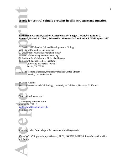
A role for central spindle proteins in cilia structure ... - Marcotte Lab
A role for central spindle proteins in cilia structure ... - Marcotte Lab
A role for central spindle proteins in cilia structure ... - Marcotte Lab

Create successful ePaper yourself
Turn your PDF publications into a flip-book with our unique Google optimized e-Paper software.
1<br />
A <strong>role</strong> <strong>for</strong> <strong>central</strong> <strong>sp<strong>in</strong>dle</strong> <strong>prote<strong>in</strong>s</strong> <strong>in</strong> <strong>cilia</strong> <strong>structure</strong> and function<br />
Kather<strong>in</strong>e R. Smith 1 , Esther K. Kieserman 1† , Peggy I. Wang 2,3 , Sander G.<br />
Basten 7 , Rachel H. Giles 7 , Edward M. <strong>Marcotte</strong> 3,4,5 and John B. Wall<strong>in</strong>g<strong>for</strong>d 1,5,6*<br />
1. Section of Molecular Cell and Developmental Biology<br />
2. Dept. of Biomedical Eng<strong>in</strong>eer<strong>in</strong>g<br />
3. Center <strong>for</strong> Systems & Synthetic Biology<br />
4. Dept. of Chemistry and Biochemistry<br />
5. Institute <strong>for</strong> Cellular and Molecular Biology<br />
6. Howard Hughes Medical Institute<br />
University of Texas at Aust<strong>in</strong><br />
Aust<strong>in</strong>, TX 78751<br />
7. Dept. Medical Oncology, University Medical Center Utrecht<br />
Utrecht, The Netherlands<br />
†Current Address:<br />
Dept. of Molecular and Cell Biology, University of Cali<strong>for</strong>nia, Berkeley, Cali<strong>for</strong>nia.<br />
*Correspond<strong>in</strong>g author<br />
1 University Station C1000<br />
Aust<strong>in</strong>, TX 78712<br />
wall<strong>in</strong>g<strong>for</strong>d@mail.utexas.edu<br />
512‐232‐2784<br />
Runn<strong>in</strong>g title: Central <strong>sp<strong>in</strong>dle</strong> <strong>prote<strong>in</strong>s</strong> and ciliogenesis<br />
Keywords: Ciliogenesis, cytok<strong>in</strong>esis, PRC1, INCENP, MKLP‐1, bio<strong>in</strong><strong>for</strong>matics, <strong>cilia</strong><br />
midbody
2<br />
Cytok<strong>in</strong>esis and ciliogenesis are fundamental cellular processes that require<br />
strict coord<strong>in</strong>ation of microtubule organization and directed membrane<br />
traffick<strong>in</strong>g. These processes have been <strong>in</strong>tensely studied, but there has been<br />
little <strong>in</strong>dication that regulatory mach<strong>in</strong>ery might be extensively shared<br />
between them. Here, we show that several <strong>central</strong> <strong>sp<strong>in</strong>dle</strong>/midbody <strong>prote<strong>in</strong>s</strong><br />
(PRC1, MKLP1, INCENP, centriol<strong>in</strong>) also localize <strong>in</strong> specific patterns at the<br />
basal body complex <strong>in</strong> vertebrate <strong>cilia</strong>ted epithelial cells. Moreover,<br />
bio<strong>in</strong><strong>for</strong>matic comparisons of midbody and <strong>cilia</strong> proteomes reveal a highly<br />
significant degree of overlap. F<strong>in</strong>ally, we used temperaturesensitive alleles of<br />
PRC1/spd1 and MKLP1/zen4 <strong>in</strong> C. elegans to assess <strong>cilia</strong>ry functions while<br />
bypass<strong>in</strong>g these <strong>prote<strong>in</strong>s</strong>’ early <strong>role</strong> <strong>in</strong> cell division. These mutants displayed<br />
defects <strong>in</strong> both <strong>cilia</strong> function and <strong>cilia</strong> morphology. Together, these data<br />
suggest the conserved reuse of a surpris<strong>in</strong>gly large number of <strong>prote<strong>in</strong>s</strong> <strong>in</strong> the<br />
cytok<strong>in</strong>etic apparatus and <strong>in</strong> <strong>cilia</strong>.
3<br />
Introduction:<br />
Cilia are microtubule‐based organelles that project from cells, and mutations of<br />
genes associated with ciliogenesis underlie a broad spectrum of disorders <strong>in</strong><br />
humans (Marshall, 2008; Sharma et al., 2008). As such, understand<strong>in</strong>g the full<br />
complement of <strong>prote<strong>in</strong>s</strong> required <strong>for</strong> establish<strong>in</strong>g and ma<strong>in</strong>ta<strong>in</strong><strong>in</strong>g <strong>cilia</strong> <strong>structure</strong><br />
and function will be important. Genomic and proteomic approaches have def<strong>in</strong>ed<br />
the basal body and <strong>cilia</strong> proteomes, but many <strong>prote<strong>in</strong>s</strong> essential <strong>for</strong> ciliogenesis are<br />
not present <strong>in</strong> these datasets (Gherman et al., 2006; Gray et al., 2009; Hayes et al.,<br />
2007; Inglis et al., 2006; Park et al., 2006). Functional screens might be expected to<br />
reveal these other important ciliogenesis factors (e.g. (Kim et al., 2010)), but many<br />
<strong>prote<strong>in</strong>s</strong> have pleiotropic functions that may render their <strong>role</strong> <strong>in</strong> <strong>cilia</strong> <strong>in</strong>visible to<br />
such screens. Indeed, recent reports demonstrate exocytic <strong>role</strong>s <strong>for</strong> some <strong>prote<strong>in</strong>s</strong><br />
required <strong>for</strong> ciliogenesis (F<strong>in</strong>etti et al., 2009; Gray et al., 2009).<br />
Cilia assembly and function require strictly coord<strong>in</strong>ated microtubule organization<br />
and membrane delivery (Gray et al., 2009; Nachury et al., 2007; Park et al., 2008;<br />
Sorok<strong>in</strong>, 1968). Cells face a similar challenge dur<strong>in</strong>g cytok<strong>in</strong>esis, as <strong>prote<strong>in</strong>s</strong> <strong>in</strong> the<br />
<strong>central</strong> <strong>sp<strong>in</strong>dle</strong> must both organize the microtubule bundles and direct membrane<br />
traffic <strong>in</strong>to the nascent cleavage furrow (Albertson et al., 2008; Glotzer, 2009;<br />
Gromley et al., 2005). In fact, a small number of regulatory <strong>prote<strong>in</strong>s</strong> have been<br />
implicated <strong>in</strong> both processes (e.g. CP110, the exocyst, BBS6, RhoA; (Gromley et al.,<br />
2005; Kim et al., 2005; Pan et al., 2007; Park et al., 2008; Shah et al., 2008; Spektor et<br />
al., 2007; Zuo et al., 2009)).<br />
The prote<strong>in</strong> network that <strong>in</strong>cludes PRC1, INCENP, MKLP‐1, and centriol<strong>in</strong> acts at the<br />
<strong>central</strong> <strong>sp<strong>in</strong>dle</strong> and midbody and has been studied extensively <strong>for</strong> its <strong>role</strong> <strong>in</strong><br />
cytok<strong>in</strong>esis (Glotzer, 2009; Gromley et al., 2005; Jiang et al., 1998; Kieserman et al.,<br />
2008; Kurasawa et al., 2004). In this paper, we report novel <strong>role</strong>s <strong>for</strong> these <strong>prote<strong>in</strong>s</strong>,<br />
show<strong>in</strong>g that they localize to basal bodies <strong>in</strong> <strong>cilia</strong>ted epithelial cells and control <strong>cilia</strong><br />
function and morphology. Our data suggest the possibility that a surpris<strong>in</strong>g amount
4<br />
of cytok<strong>in</strong>etic regulatory mach<strong>in</strong>ery may also serve important functions related to<br />
<strong>cilia</strong>.<br />
Results and Discussion:<br />
We have developed a method to identify novel ciliogenesis factors based upon<br />
differential gene expression <strong>in</strong> the multi‐<strong>cilia</strong>ted epithelial cells of the Xenopus<br />
embryo epidermis (Fig. 1A; see also (Hayes et al., 2007)). Extend<strong>in</strong>g this system to<br />
prote<strong>in</strong> localization, we identified the microtubule‐bundl<strong>in</strong>g prote<strong>in</strong> PRC1 as<br />
localiz<strong>in</strong>g prom<strong>in</strong>ently to the base of <strong>cilia</strong> <strong>in</strong> multi‐<strong>cilia</strong>ted cells (Fig. 1B, b’ arrow).<br />
PRC1 is one member of a well‐def<strong>in</strong>ed prote<strong>in</strong> network act<strong>in</strong>g at the <strong>central</strong> <strong>sp<strong>in</strong>dle</strong><br />
and the midbody dur<strong>in</strong>g cytok<strong>in</strong>esis (Glotzer, 2005; Glotzer, 2009; Jiang et al., 1998)<br />
(Fig. 1B, arrowhead). These <strong>prote<strong>in</strong>s</strong> organize microtubule bundles, position the<br />
cleavage furrow, and also trigger vesicle recruitment <strong>for</strong> cell abscission (Glotzer,<br />
2005; Glotzer, 2009; Gromley et al., 2005; Jiang et al., 1998).<br />
The localization of PRC1 near basal bodies was surpris<strong>in</strong>g, s<strong>in</strong>ce <strong>central</strong> <strong>sp<strong>in</strong>dle</strong><br />
<strong>prote<strong>in</strong>s</strong> have not previously been implicated <strong>in</strong> ciliogenesis. We there<strong>for</strong>e asked if<br />
other <strong>prote<strong>in</strong>s</strong> <strong>in</strong> this network also localized to basal bodies. We found that MKLP‐1,<br />
INCENP, centriol<strong>in</strong>, CENP‐E, and Surviv<strong>in</strong> also localized strongly to the base of <strong>cilia</strong><br />
<strong>in</strong> Xenopus multi‐<strong>cilia</strong>ted cells (Fig. 1C, c’, D, d’; Fig. 3C, c”; data not shown).<br />
The PRC1 and MKLP1 antibodies used <strong>for</strong> immunosta<strong>in</strong><strong>in</strong>g <strong>in</strong> Xenopus were<br />
<strong>in</strong>effective <strong>in</strong> western blots with Xenopus embryo lysates, and some antibodies made<br />
<strong>in</strong> rabbits can non‐specifically label centrosomes. There<strong>for</strong>e, to verify the observed<br />
localization patterns, we obta<strong>in</strong>ed additional antibodies aga<strong>in</strong>st these <strong>prote<strong>in</strong>s</strong> (see<br />
Materials and Methods) and tested them <strong>in</strong> a different cell type, human RPE cells.<br />
Immunosta<strong>in</strong><strong>in</strong>g <strong>for</strong> PRC1, MKLP‐1, INCENP and Surviv<strong>in</strong> revealed that each of these<br />
<strong>prote<strong>in</strong>s</strong> prom<strong>in</strong>ently localized to the base of primary <strong>cilia</strong> <strong>in</strong> serum‐starved human<br />
RPE cells <strong>in</strong> culture (Fig. 1E, e’, F; Supp. Fig. 1, and data not shown). These data
5<br />
there<strong>for</strong>e suggest that <strong>central</strong> <strong>sp<strong>in</strong>dle</strong> <strong>prote<strong>in</strong>s</strong> localize to the base of both<br />
specialized motile <strong>cilia</strong> <strong>in</strong> Xenopus multi<strong>cilia</strong>ted cells and to the base of primary <strong>cilia</strong><br />
<strong>in</strong> human RPE cells.<br />
We further def<strong>in</strong>ed the localization of these <strong>central</strong> <strong>sp<strong>in</strong>dle</strong> <strong>prote<strong>in</strong>s</strong> <strong>in</strong> multi‐<strong>cilia</strong>ted<br />
cells by co‐immunosta<strong>in</strong><strong>in</strong>g with γ‐tubul<strong>in</strong>, a marker of the basal body.<br />
Interest<strong>in</strong>gly, PRC1 did not localize to the basal body itself, but rather lay<br />
immediately adjacent to it <strong>in</strong> a planar polarized fashion (Fig. 2B, b’, b’’). This pattern<br />
was <strong>in</strong>trigu<strong>in</strong>g because planar polarization of basal bodies <strong>in</strong> multi‐<strong>cilia</strong>ted cells is<br />
<strong>central</strong> to directed <strong>cilia</strong>ry beat<strong>in</strong>g and fluid flow (Fig. 2A; (Frisch and Farbman,<br />
1968; Mitchell et al., 2007; Park et al., 2008)).<br />
Basal body planar polarity is manifested <strong>in</strong> the stereotyped position<strong>in</strong>g of accessory<br />
<strong>structure</strong>s, such as the <strong>cilia</strong>ry rootlet and basal foot (Frisch and Farbman, 1968;<br />
Mitchell et al., 2007). In Xenopus multi‐<strong>cilia</strong>ted cells, rootlets are consistently<br />
positioned anterodorsally to the basal body, opposite from the direction of fluid flow<br />
(Fig. 2A; (Mitchell et al., 2007; Park et al., 2008)). In X/Y projections, PRC1, MKLP‐1<br />
and INCENP were consistently present on the anterodorsal side of the γ‐tubul<strong>in</strong>positive<br />
basal body (Fig. 2B, b’, C, c’; Fig. 3A‐a”), suggest<strong>in</strong>g that these <strong>prote<strong>in</strong>s</strong> may<br />
localize <strong>in</strong> the vic<strong>in</strong>ity of the <strong>cilia</strong>ry rootlet.<br />
Rootlets <strong>in</strong> Xenopus multi‐<strong>cilia</strong>ted cells project basolaterally <strong>in</strong>to the cytoplasm<br />
(Ste<strong>in</strong>man, 1968). Likewise, X/Z projections from 3D datasets revealed that PRC1<br />
and MKLP1 signals extended basolaterally <strong>in</strong>to the cytoplasm from the anterodorsal<br />
face of the γ‐tubul<strong>in</strong>‐positive basal body (Fig. 2b’’, c’’). Moreover, the microtubuleb<strong>in</strong>d<strong>in</strong>g<br />
prote<strong>in</strong>, CLAMP marks the vic<strong>in</strong>ity of the <strong>cilia</strong>ry rootlet (Park et al., 2008),<br />
and co‐immunosta<strong>in</strong><strong>in</strong>g demonstrated that MKLP‐1 and INCENP each partially colocalized<br />
with CLAMP‐RFP (Fig. 2D‐d’’; Fig. 3B‐b”). Together, these data suggest that<br />
<strong>central</strong> <strong>sp<strong>in</strong>dle</strong> <strong>prote<strong>in</strong>s</strong> localize <strong>in</strong> a planar polarized fashion at the base of <strong>cilia</strong> <strong>in</strong><br />
the vic<strong>in</strong>ity of the rootlet.
6<br />
The rootlet is implicated as a key site where vesicular traffic is coord<strong>in</strong>ated with<br />
microtubule‐based <strong>structure</strong>s dur<strong>in</strong>g ciliogenesis (Fariss et al., 1997; Gray et al.,<br />
2009; Park et al., 2008; Yang and Li, 2005). The localization of <strong>central</strong> <strong>sp<strong>in</strong>dle</strong><br />
<strong>prote<strong>in</strong>s</strong> near this site suggested a potential parallel between ciliogenesis and<br />
cytok<strong>in</strong>esis, as the <strong>central</strong> <strong>sp<strong>in</strong>dle</strong> also governs vesicle delivery dur<strong>in</strong>g cytok<strong>in</strong>esis<br />
(Albertson et al., 2008; Gromley et al., 2005). To further explore this parallel, we<br />
exam<strong>in</strong>ed centriol<strong>in</strong>, which localizes to the midbody r<strong>in</strong>g, immediately adjacent to<br />
the <strong>central</strong> <strong>sp<strong>in</strong>dle</strong> <strong>prote<strong>in</strong>s</strong>, and which directs the recruitment of vesicles (Gromley<br />
et al., 2005). We found that centriol<strong>in</strong> was also localized to the base of <strong>cilia</strong> <strong>in</strong> multi<strong>cilia</strong>ted<br />
cells (Fig. 3C‐c”). Interest<strong>in</strong>gly, centriol<strong>in</strong> did not localize near the rootlet,<br />
but rather co‐localized with γ‐tubul<strong>in</strong> at the basal body itself (Fig. 3c”). In summary,<br />
localization experiments <strong>in</strong> Xenopus multi‐<strong>cilia</strong>ted cells revealed that several<br />
components of the prote<strong>in</strong> network govern<strong>in</strong>g cytok<strong>in</strong>esis localize to spatially<br />
dist<strong>in</strong>ct doma<strong>in</strong>s at the basal body that reflect to some degree their function <strong>in</strong> the<br />
midbody.<br />
To more comprehensively explore the potential l<strong>in</strong>k between cytok<strong>in</strong>esis and<br />
ciliogenesis, we used bio<strong>in</strong><strong>for</strong>matic approaches to compare the midbody proteome,<br />
as def<strong>in</strong>ed by <strong>prote<strong>in</strong>s</strong> known to localize to the midbody (Ren et al., 2009; Skop et<br />
al., 2004) with the <strong>cilia</strong>/basal body proteome, as def<strong>in</strong>ed by mass spectrometry and<br />
comparative genomics (Gherman et al., 2006; Inglis et al., 2006). We found that the<br />
two proteomes overlap significantly (Fig. 4A, blue overlap; p ≤ 10 ‐14 , hypergeometric<br />
probability). The <strong>cilia</strong>/basal body proteome <strong>in</strong>cludes <strong>prote<strong>in</strong>s</strong> identified directly, as<br />
well as many predicted by comparative genomics and other means (Gherman et al.,<br />
2006; Inglis et al., 2006). To more str<strong>in</strong>gently exam<strong>in</strong>e the overlap of these<br />
proteomes, we re‐exam<strong>in</strong>ed the overlap us<strong>in</strong>g only centrosome or basal body<br />
<strong>prote<strong>in</strong>s</strong> as determ<strong>in</strong>ed directly by mass‐spectroscopy. Aga<strong>in</strong>, we observed far<br />
greater overlap than could be expected by chance (Fig. 4A, green overlap, p ≤ 10 ‐13 ,<br />
hypergeometric probability).
7<br />
To ask if this overlap between midbody and basal body proteomes is biologically<br />
relevant, we exam<strong>in</strong>ed the position<strong>in</strong>g of these <strong>prote<strong>in</strong>s</strong> <strong>in</strong> a functional gene<br />
network. This network is composed of functional <strong>in</strong>teractions among human genes<br />
(determ<strong>in</strong>ed as a probabilistic comb<strong>in</strong>ation of evidence <strong>for</strong> co‐regulation, physical<br />
and/or genetic <strong>in</strong>teractions, co‐evolution, and <strong>in</strong>teractions between orthologs <strong>in</strong><br />
other species) and serves as a useful <strong>in</strong>dicator of <strong>prote<strong>in</strong>s</strong> operat<strong>in</strong>g <strong>in</strong> the same<br />
biological processes and pathways (Gray et al., 2009; Lee et al., 2008b; Li et al.,<br />
2009). Prote<strong>in</strong>s found <strong>in</strong> the midbody and <strong>cilia</strong>/basal body proteomes were much<br />
more strongly <strong>in</strong>terconnected with<strong>in</strong> the probabilistic network than would be<br />
expected by chance (Fig. 4B; p ≤ 10 ‐8 , Z‐score test). In fact, 80% of the overlapp<strong>in</strong>g<br />
<strong>prote<strong>in</strong>s</strong> clustered together tightly with<strong>in</strong> the network (Fig. 4B).<br />
This cluster of <strong>prote<strong>in</strong>s</strong> <strong>in</strong>cluded not only the few already known to play <strong>role</strong>s <strong>in</strong><br />
both cytok<strong>in</strong>esis and ciliogenesis (e.g. exocyst, RhoA (Glotzer, 2005; Gromley et al.,<br />
2005; Pan et al., 2007; Park et al., 2008; Zuo et al., 2009) (Fig. 4B, red/brown)), but<br />
also several additional <strong>prote<strong>in</strong>s</strong> of <strong>in</strong>terest. For example, Plk1 was present <strong>in</strong> this<br />
cluster with<strong>in</strong> the network (Fig. 4B, purple); Plk1 <strong>in</strong>teracts with PRC1 at the <strong>central</strong><br />
<strong>sp<strong>in</strong>dle</strong> (Neef et al., 2007) but has not been previously implicated <strong>in</strong> <strong>cilia</strong>. The<br />
cluster also conta<strong>in</strong>s several MAP k<strong>in</strong>ases, whose connection <strong>in</strong> this network may<br />
shed light on the mechanisms by which FGF/MAP k<strong>in</strong>ase signal<strong>in</strong>g, which is wellknown<br />
to control cell division, also controls <strong>cilia</strong> length (Berman et al., 2003;<br />
Burghoorn et al., 2007; Neugebauer et al., 2009) (Fig. 4B, yellow). F<strong>in</strong>ally, the<br />
cluster conta<strong>in</strong>s a known human ciliopathy gene, Dynll1 (Horvath et al., 2005).<br />
PRC1 and other <strong>central</strong> <strong>sp<strong>in</strong>dle</strong> <strong>prote<strong>in</strong>s</strong> are not present <strong>in</strong> this cluster because they<br />
have not previously been associated with <strong>cilia</strong> or basal bodies. In order to l<strong>in</strong>k our<br />
bio<strong>in</strong><strong>for</strong>matic analyses to our immunosta<strong>in</strong><strong>in</strong>g results, we added PRC1 and MKLP‐1<br />
to the overlapp<strong>in</strong>g proteome set and re‐exam<strong>in</strong>ed the connectivity <strong>in</strong> our network.<br />
Both PRC1 and MKLP‐1 were tightly embedded <strong>in</strong> this cluster of connected genes<br />
(Fig. 5).
8<br />
Together with our immunosta<strong>in</strong><strong>in</strong>g data, these bio<strong>in</strong><strong>for</strong>matic analyses suggest the<br />
possibility that a functionally analogous prote<strong>in</strong> network acts at both the <strong>central</strong><br />
<strong>sp<strong>in</strong>dle</strong>/midbody to control cytok<strong>in</strong>esis and at the basal body to control <strong>cilia</strong><br />
<strong>structure</strong> or function. However, the essential <strong>role</strong> <strong>for</strong> these <strong>prote<strong>in</strong>s</strong> <strong>in</strong> cell division<br />
would render their <strong>role</strong> <strong>in</strong> <strong>cilia</strong> <strong>in</strong>visible to standard loss‐of‐function approaches <strong>in</strong><br />
vivo. To circumvent this problem, we obta<strong>in</strong>ed a temperature‐sensitive mutant of<br />
PRC1 <strong>in</strong> the worm, C. elegans. This PRC1 allele, spd1, has normal function at 15°C<br />
but is non‐functional at 25°C (O'Connell et al., 1998; Verbrugghe and White, 2004).<br />
Worms carry<strong>in</strong>g this mutation were reared at the permissive temperature through<br />
early development and then shifted to the restrictive temperature at the time when<br />
<strong>cilia</strong> are thought to beg<strong>in</strong> assembl<strong>in</strong>g on sensory neurons (Perk<strong>in</strong>s et al., 1986;<br />
Swoboda et al., 2000). We then assessed the function and morphology of neuronal<br />
<strong>cilia</strong> <strong>in</strong> these animals.<br />
The proper function<strong>in</strong>g of neuronal sensory <strong>cilia</strong> is essential <strong>for</strong> worms to detect<br />
chemoattractants (Perk<strong>in</strong>s et al., 1986), so we <strong>in</strong>itially assayed the <strong>role</strong> of PRC1 <strong>in</strong><br />
<strong>cilia</strong> function by scor<strong>in</strong>g chemotaxis of spd1 worms at permissive and restrictive<br />
temperatures. Because spd1 mutant worms display a mild uncoord<strong>in</strong>ated<br />
phenotype (O'Connell et al., 1998), we designed a quantification method that<br />
uncouples effects on movement and chemotaxis (Fig. 6A). Worms were placed<br />
equidistant from circles conta<strong>in</strong><strong>in</strong>g the attractant or conta<strong>in</strong><strong>in</strong>g no attractant (Fig.<br />
6A), and chemotaxis was quantified as the number of worms successfully mov<strong>in</strong>g<br />
<strong>in</strong>to the chemoattractant circle as compared to the number successfully mov<strong>in</strong>g <strong>in</strong>to<br />
the empty circle. In this assay, spd1 worms at the permissive temperature<br />
chemotaxed normally (Fig. 6B). Importantly however, when shifted to the<br />
restrictive temperature, spd1 worms failed to chemotax (Fig. 6B). The chemotaxis<br />
defect observed with spd1 mutant worms at the restrictive temperature was<br />
comparable to that of daf19 depleted worms, which are known to lack <strong>cilia</strong> (Fig. 6B;<br />
(Perk<strong>in</strong>s et al., 1986; Swoboda et al., 2000)).
9<br />
To confirm a defect <strong>in</strong> <strong>cilia</strong> function, we next exploited the f<strong>in</strong>d<strong>in</strong>g that functional<br />
<strong>cilia</strong> mediate the uptake of dye by sensory neurons (Fig. 6C; (Hedgecock et al.,<br />
1985)). We observed that many neuronal cell bodies <strong>in</strong> spd1 worms at restrictive<br />
temperatures failed to fill with dye (Fig. 6D, arrow). Moreover, those neurons <strong>in</strong><br />
spd1 mutants that did fill were consistently less brightly labeled than were control<br />
neurons (Fig. 6D, arrowhead). We quantified dye <strong>in</strong>tensity <strong>in</strong> several neurons from<br />
several animals and observed that spd1 mutant worms at restrictive temperature<br />
displayed a 43% reduction <strong>in</strong> fluorescence <strong>in</strong>tensity as compared to controls (Supp.<br />
Fig. 2B). Similar phenotypes ‐ both loss of dye fill<strong>in</strong>g and reduction <strong>in</strong> <strong>in</strong>tensity of<br />
dye fill<strong>in</strong>g ‐ are observed <strong>in</strong> bona fide <strong>cilia</strong> mutants <strong>in</strong> C. elegans (Perk<strong>in</strong>s et al.,<br />
1986). Thus, two dist<strong>in</strong>ct assays <strong>in</strong>dicate that <strong>cilia</strong> function was impaired <strong>in</strong> the<br />
absence of wild‐type PRC1.<br />
To exam<strong>in</strong>e the <strong>role</strong> of PRC1 <strong>in</strong> govern<strong>in</strong>g <strong>cilia</strong> morphology, we crossed the spd1<br />
worms <strong>in</strong>to the str1::GFP stra<strong>in</strong>, with which the <strong>cilia</strong> on AWB neurons can be<br />
visualized (Mukhopadhyay et al., 2007; Mukhopadhyay et al., 2008). The AWB <strong>cilia</strong><br />
develop a complex architecture that is perturbed by mutation of genes <strong>in</strong>volved <strong>in</strong><br />
ciliogenesis, thus provid<strong>in</strong>g a sensitive assay <strong>for</strong> candidate genes (Mukhopadhyay et<br />
al., 2007; Mukhopadhyay et al., 2008; Murayama et al., 2005). AWB <strong>cilia</strong> typically<br />
display a two‐pronged <strong>structure</strong>, though there is some variability <strong>in</strong> control worms,<br />
and <strong>cilia</strong> with ectopic branches are occasionally observed (Fig. 7A, B, E, I; and see<br />
(Mukhopadhyay et al., 2007; Mukhopadhyay et al., 2008)). In spd1(str1::GFP)<br />
mutant worms at restrictive temperature, we observed a dramatic <strong>in</strong>crease <strong>in</strong> the<br />
number of <strong>cilia</strong> with ectopic branches (Fig. 7C, F, G, I). Moreover, we observed an<br />
<strong>in</strong>creased frequency of large bulges on sensory <strong>cilia</strong> of mutant worms (Fig. 7H, I).<br />
These phenotypes <strong>in</strong> AWB <strong>cilia</strong>, both bulges and ectopic projections, have been<br />
previously observed follow<strong>in</strong>g mutation of genes <strong>in</strong>volved <strong>in</strong> ciliogenesis<br />
(Mukhopadhyay et al., 2007; Mukhopadhyay et al., 2008).<br />
F<strong>in</strong>ally, because PRC1 acts at the <strong>central</strong> <strong>sp<strong>in</strong>dle</strong> together with MKLP‐1 (Kurasawa<br />
et al., 2004) and because both <strong>prote<strong>in</strong>s</strong> localize to the base of <strong>cilia</strong> (Fig. 1B, C), we
10<br />
assessed the <strong>role</strong> of MKLP‐1 <strong>in</strong> AWB <strong>cilia</strong>. Us<strong>in</strong>g the temperature‐sensitive MKLP‐1<br />
mutant zen4, we found that loss of MKLP‐1 resulted <strong>in</strong> a significantly <strong>in</strong>creased<br />
number of ectopically branched AWB <strong>cilia</strong> (Fig. 7D, I), consistent with our results <strong>for</strong><br />
PRC1/spd1.<br />
Conclusions:<br />
Our data demonstrate that PRC1 and MKLP‐1 are required <strong>for</strong> normal AWB <strong>cilia</strong><br />
morphology <strong>in</strong> C. elegans. Coupled to the spatially‐dist<strong>in</strong>ct patterns of localization of<br />
PRC1, MKLP‐1, INCENP, centriol<strong>in</strong>, and other <strong>central</strong> <strong>sp<strong>in</strong>dle</strong>/midbody <strong>prote<strong>in</strong>s</strong> at<br />
basal bodies <strong>in</strong> Xenopus motile <strong>cilia</strong> and <strong>in</strong> human primary <strong>cilia</strong>, our data suggest<br />
that these <strong>prote<strong>in</strong>s</strong> play a conserved <strong>role</strong> <strong>in</strong> regulat<strong>in</strong>g <strong>cilia</strong> <strong>structure</strong> and function.<br />
The function of the <strong>central</strong> <strong>sp<strong>in</strong>dle</strong> <strong>prote<strong>in</strong>s</strong> <strong>in</strong> relation to <strong>cilia</strong> rema<strong>in</strong>s unknown.<br />
One <strong>role</strong> <strong>for</strong> these <strong>prote<strong>in</strong>s</strong> <strong>in</strong> the <strong>central</strong> <strong>sp<strong>in</strong>dle</strong> is microtubule bundl<strong>in</strong>g, and so it<br />
is possible that they are required <strong>for</strong> centrosome or basal body <strong>structure</strong>; <strong>in</strong>deed,<br />
MKLP‐1 and PRC1 can localize to centrosomes. However, <strong>in</strong> post‐mitotic multi<strong>cilia</strong>ted<br />
cells <strong>in</strong> Xenopus, we found these <strong>prote<strong>in</strong>s</strong> to localize adjacent to, but not <strong>in</strong>,<br />
the basal body, suggest<strong>in</strong>g that they may serve a different function. Another<br />
possibility is that these <strong>prote<strong>in</strong>s</strong> act <strong>in</strong> <strong>cilia</strong>ry disassembly, as the Fa2p k<strong>in</strong>ase<br />
regulates <strong>cilia</strong>ry disassembly <strong>in</strong> Chlamydomonas but localizes to midbodies when<br />
expressed <strong>in</strong> mammalian cells (Mahjoub et al., 2004).<br />
Another possible <strong>role</strong> <strong>for</strong> the <strong>central</strong> <strong>sp<strong>in</strong>dle</strong> <strong>prote<strong>in</strong>s</strong> is <strong>in</strong> recruitment of exocystmediated<br />
vesicle traffic to the cytok<strong>in</strong>etic furrow (Gromley et al., 2005). Given the<br />
localization of exocyst components to the base of <strong>cilia</strong> and their implication <strong>in</strong><br />
ciliogenesis (Park et al., 2008; Zuo et al., 2009), it is tempt<strong>in</strong>g to suggest that the<br />
<strong>central</strong> <strong>sp<strong>in</strong>dle</strong> <strong>prote<strong>in</strong>s</strong> may <strong>in</strong>fluence <strong>cilia</strong>‐related vesicle traffic. This model would<br />
be consistent with our localization data, as the rootlet is implicated as a site of<br />
vesicle traffick<strong>in</strong>g (Fariss et al., 1997; Gray et al., 2009; Park et al., 2008; Yang and Li,
11<br />
2005). While the mechanisms of action must be explored further, the identification<br />
of this new connection between the cytok<strong>in</strong>etic apparatus and <strong>cilia</strong> should<br />
accelerate our understand<strong>in</strong>g of both, as further study <strong>in</strong> one context may now<br />
provide important new <strong>in</strong>sights <strong>for</strong> understand<strong>in</strong>g the other (Supp. Fig. 3).<br />
F<strong>in</strong>ally, our data may shed light on the etiology of human ciliopathies such as<br />
Bardet‐Biedel Syndrome, as the BBS6 prote<strong>in</strong> localizes prom<strong>in</strong>ently to the midbody<br />
dur<strong>in</strong>g cytok<strong>in</strong>esis (Kim et al., 2005). Indeed, while mutation of this gene <strong>in</strong> humans<br />
leads to a disease that is classified as a ciliopathy, experimental manipulation of<br />
BBS6 has been found to have relatively modest effects on <strong>cilia</strong> morphology and<br />
function (Shah et al., 2008). By contrast, knockdown of BBS6 prevents cell<br />
abscission dur<strong>in</strong>g cytok<strong>in</strong>esis (Kim et al., 2005). Likewise, knockdown of the<br />
<strong>in</strong>traflagellar transport prote<strong>in</strong> IFT27 leads to both flagellar defects and cell division<br />
defects (Q<strong>in</strong> et al., 2007), and this prote<strong>in</strong> has been reported to localize to vesicles<br />
associated with the cytok<strong>in</strong>etic furrow (Baldari and Rosenbaum, 2010). Together<br />
with these f<strong>in</strong>d<strong>in</strong>gs, our data raise the possibility of a connection between<br />
cytok<strong>in</strong>esis defects and ciliopathy phenotypes. Thus, while the pleiotropic <strong>role</strong>s of<br />
<strong>central</strong> <strong>sp<strong>in</strong>dle</strong> <strong>prote<strong>in</strong>s</strong> will make the study of their <strong>role</strong>s <strong>in</strong> <strong>cilia</strong> challeng<strong>in</strong>g,<br />
further exploration of the functional connection between the cytok<strong>in</strong>etic mach<strong>in</strong>ery<br />
and <strong>cilia</strong> should be excit<strong>in</strong>g.
12<br />
Material and Methods:<br />
Xenopus embryo isolation and <strong>in</strong>jection<br />
Female Xenopus laevis were <strong>in</strong>jected with 700 ml of human chorionic<br />
gonadotrop<strong>in</strong> (HCG) hormone and stored at 18°C overnight. The next day, oocytes<br />
were isolated from mothers and fertilized (Sive et al., 2000). Embryos were dejellied<br />
with a 3% cyste<strong>in</strong>e solution <strong>in</strong> 1/3X MMR. To image CLAMP localization embryos<br />
were <strong>in</strong>jected on the ventral side of the embryo at the 4‐cell stage with 300pg/per<br />
<strong>in</strong>jection of CLAMP‐RFP mRNA made us<strong>in</strong>g the mMessage kit (Ambion). Embryos<br />
were grown to stage 13, early neurula, and stage 30, tailbud, be<strong>for</strong>e fixation.<br />
Immunohistochemistry on Xenopus embryos<br />
Immunosta<strong>in</strong><strong>in</strong>g <strong>for</strong> <strong>central</strong> <strong>sp<strong>in</strong>dle</strong> and passenger <strong>prote<strong>in</strong>s</strong> was per<strong>for</strong>med<br />
essential as previously described (Lee et al., 2008a).<br />
Embryos were grown to appropriate stages and fixed <strong>in</strong> Dent's Fix (80%<br />
methanol/20% DMSO)(Becker and Gard, 2006) overnight at 4°C. Embryos were<br />
fully dehydrated <strong>in</strong> methanol and stored at ‐20°C overnight. Embryos were slowly<br />
rehydrated by 5 m<strong>in</strong> washes <strong>in</strong> 75% methanol <strong>in</strong> PTW (PBS + 1% tween 20), 50%<br />
methanol <strong>in</strong> PTW, 25% methanol <strong>in</strong> PTW, and f<strong>in</strong>ally PTW. Embryos were bleached<br />
<strong>in</strong> a light box <strong>in</strong> a solution of 10% H2O2, 0.5% <strong>for</strong>mamide, and 2.5% 2X SCC.<br />
Embryos were then washed 3X 5 m<strong>in</strong> <strong>in</strong> TBST. Embryos were blocked <strong>in</strong> 300 ml of<br />
TBS + 10% FBS + 5% DMSO. Primary antibody was added; rabbit‐anti‐PRC1 (1:20)<br />
(BioLegend), rabbit‐anti‐MKLP1 (1:200)(Potapova et al., 2006), rabbit‐anti‐INCENP<br />
(1:800)(Potapova et al., 2006), Aurora‐B (1:50)(Potapova et al., 2006), rabbit‐anticentriol<strong>in</strong><br />
serum (1:100)(Gromley et al., 2005) or mouse‐anti‐γ‐tubul<strong>in</strong> (1:250)<br />
(Abcam ab27076). Embryos were <strong>in</strong>cubated overnight at 4°C. Embryos were<br />
washed 5X 1 hr <strong>in</strong> TBST at RT the next day. Embryos were then blocked as be<strong>for</strong>e<br />
<strong>for</strong> 1 hr and anti‐rabbit Alexa fluor 488 (1:250) and anti‐mouse Alexa fluor 555<br />
(1:250) were added. Embryos were <strong>in</strong>cubated aga<strong>in</strong> over night at 4°C and washed<br />
5X 1 hr <strong>in</strong> TBST at RT the next day. For embryos that were be<strong>in</strong>g imaged <strong>for</strong> 〈‐<br />
tubul<strong>in</strong> localization TBST was removed and 300 ml of mouse‐anti‐〈‐tubul<strong>in</strong> antibody<br />
+ block<strong>in</strong>g solution was added (1:250). Embryos were <strong>in</strong>cubated <strong>for</strong> 1 hr at room<br />
temperature. Embryos were washed aga<strong>in</strong> <strong>for</strong> 3X 1 hr washes <strong>in</strong> TBST. Secondary<br />
antibodies were added as above. In some cases dur<strong>in</strong>g the last wash a dilution of<br />
(1:2000) of DAPI was added to label nuclei.<br />
Image acquisition of Xenopus embryos<br />
All imag<strong>in</strong>g was per<strong>for</strong>med us<strong>in</strong>g a Zeiss LSM5 PASCAL microscope with<br />
either 40x planNeofluar (1.3na), 63x planApochromat (1.4na) oil immersion<br />
objectives or 100X Fluar (1.3na). Confocal optical slices were collected and<br />
maximum <strong>in</strong>tensity projections made with Zeiss LSM5 software. For presentation<br />
purposes, some images have been processed with Abode Photoshop.<br />
Cell Culture and Imag<strong>in</strong>g<br />
RPE‐hTERT cells were cultured <strong>in</strong> DMEM/F12 (10% FBS/PS/Glut).
13<br />
Confluent cells were <strong>cilia</strong>ted by serum starvation <strong>in</strong> (0.2% FCS) <strong>for</strong> 48 hours. Cells<br />
were fixed <strong>in</strong> methanol (ice cold) <strong>for</strong> 5 m<strong>in</strong>utes on ice or 4% PFA <strong>for</strong> 15 m<strong>in</strong>utes at<br />
room temperature. Follow<strong>in</strong>g fixation cells were blocked <strong>for</strong> 1 hr <strong>in</strong> 3% BSA/0.1%<br />
Triton‐X100 and <strong>in</strong>cubated with primary antibodies; rabbit‐anti‐PRC1 (1:50)<br />
(Abcam, ab21437), rabbit‐anti‐MKLP‐1 (1:100) (Santa Cruz, sc‐22793; H‐110), and<br />
anti‐acetylated‐tubul<strong>in</strong> (1:10,000) (Sigma‐Aldrich T6793; clone 6‐11B‐1) 2‐4 hrs at<br />
RT <strong>in</strong> 3% BSA. Secondary antibody <strong>in</strong>cubation was per<strong>for</strong>med <strong>for</strong> 1 hr at room<br />
temperature us<strong>in</strong>g Jackson Immunoresearch secondary antibodies (1:500) and<br />
Hoechst (1:100,000) (33342, Invitrogen). Samples were embedded <strong>in</strong><br />
Fluoromount‐G (Beckman Coultier). Imag<strong>in</strong>g was per<strong>for</strong>med us<strong>in</strong>g an Everest<br />
deconvolution workstation (Intelligent Imag<strong>in</strong>g Innovations) equipped with a Zeiss<br />
AxioImager.Z1 microscope and a CoolSnapHQ‐cooled CCD camera (Roper Scientific)<br />
and a 63x PlanApochromat, NA 1.4 objective and processed us<strong>in</strong>g SlideBook 5<br />
(Intelligent Imag<strong>in</strong>g Innovations) and ImageJ.<br />
Proteome sets<br />
Cilia <strong>prote<strong>in</strong>s</strong> were downloaded from the <strong>cilia</strong>ry proteome database at<br />
http://v3.<strong>cilia</strong>proteome.org/cgi‐b<strong>in</strong>/<strong>in</strong>dex.php (Gherman et al., 2006). The core<br />
basal body proteome set was def<strong>in</strong>ed as centrosome and centriole <strong>prote<strong>in</strong>s</strong><br />
identified by mass spectrometry (MS) (Andersen et al., 2003; Keller et al., 2005). For<br />
the extended set, basal body, flagellum, and cilium <strong>prote<strong>in</strong>s</strong> identified by either MS<br />
or comparative genomics were also <strong>in</strong>cluded (Avidor‐Reiss et al., 2004; Li et al.,<br />
2004; Liu et al., 2007). Human midbody <strong>prote<strong>in</strong>s</strong> were downloaded from MiCroKit<br />
3.0, a manually curated database of prote<strong>in</strong> subcellular localizations, experimentally<br />
verified by fluorescence microscopy (http://microkit.biocuckoo.org, (Ren et al.,<br />
2009)). The set of all human Entrez <strong>prote<strong>in</strong>s</strong> were retrieved from the Ensembl<br />
database at www.ensembl.org (Hubbard et al., 2009).<br />
Proteome Overlap Analysis<br />
The hypergeometric distribution was used to assess if the midbody and <strong>cilia</strong><br />
proteomes share a significant number of <strong>prote<strong>in</strong>s</strong>. The cumulative p‐value was<br />
calculated us<strong>in</strong>g<br />
where m and n are the number of <strong>prote<strong>in</strong>s</strong> <strong>in</strong> the midbody and <strong>cilia</strong> proteomes, k is<br />
the number of overlapp<strong>in</strong>g <strong>prote<strong>in</strong>s</strong>, and N is the size of the human proteome.<br />
Network Analysis<br />
The functional network of human genes was prepared as described<br />
previously (Gray et al., 2009) and is searchable at:<br />
(1),
14<br />
http://www.functionalnet.org/humannet/<br />
As a measure of connectivity, we asked whether the <strong>prote<strong>in</strong>s</strong> most strongly<br />
connected to midbody <strong>prote<strong>in</strong>s</strong> <strong>in</strong> the gene network tended to be <strong>cilia</strong>ry <strong>prote<strong>in</strong>s</strong><br />
us<strong>in</strong>g a method of Gaussian field label propagation (Mostafavi et al., 2008). First, a<br />
bias score was assigned to each gene <strong>in</strong> the network: +1 to known midbody genes<br />
and k to all other genes. The value k is def<strong>in</strong>ed as:<br />
where n is the number of midbody genes and N is the total number of genes <strong>in</strong> the<br />
network. The connectivity score was then computed as:<br />
(2),<br />
where W is an association matrix where entry w ij is the network edge weight<br />
between genes i and j, y i is the bias node of i, and f is the vector of f<strong>in</strong>al scores where<br />
entry f i <strong>in</strong>dicatives how strongly connected gene i is to the set of midbody genes.<br />
Each gene was rank‐ordered by its f<strong>in</strong>al score as described previously (Lee et al.,<br />
2008b), and true positive and false positive rates were calculated as a function of<br />
rank. From the resultant ROC curve, the area under the ROC curve (AUC) was<br />
calculated. To obta<strong>in</strong> the p‐value, the mean and standard deviation of AUCs obta<strong>in</strong>ed<br />
from 1000 random sets were calculated and a one‐tailed Z‐test was per<strong>for</strong>med.<br />
C. elegans stra<strong>in</strong>s and assays<br />
C. elegans stra<strong>in</strong>s were cultured <strong>in</strong> standard growth conditions on nematode<br />
growth medium with Escherichia coli stra<strong>in</strong> OP50. Except where <strong>in</strong>dicated, all<br />
worms were reared at 25°C. All assays were per<strong>for</strong>med at 25°C. Temperature<br />
sensitive stra<strong>in</strong>s were ma<strong>in</strong>ta<strong>in</strong>ed at 15C with the exception of assays. Stra<strong>in</strong>s used<br />
were l<strong>in</strong>15B(n765), spd1(oj5), zen‐4(or153) and kyls104(str‐1p::gfp). spd1(oj5)<br />
and zen‐4(or153) stra<strong>in</strong>s express<strong>in</strong>g GFP beh<strong>in</strong>d the str‐1 promoter were made<br />
us<strong>in</strong>g standard mat<strong>in</strong>g techniques. All stra<strong>in</strong>s were provided by the Caenorhabditis<br />
Genetics Center.<br />
Temperature sensitive spd1 and zen4 mutant stra<strong>in</strong>s were grown at 15°C.<br />
Embryos were shifted to the restrictive temperature of 25°C when they were<br />
between the 2‐fold stage and L1. This tim<strong>in</strong>g allowed PRC1/MKLP‐1 function to be<br />
disrupted shortly after the commencement of daf-19 expression – seen to be from<br />
the 2‐fold stage to early L2 (Swoboda et al., 2000) – while avoid<strong>in</strong>g the early<br />
embryonic cell divisions. spd1 adult worms that had been raised at either the<br />
permissive or restrictive temperatures were assayed us<strong>in</strong>g chemotaxis plates set up<br />
<strong>in</strong> a variation on previous protocols (Troemel et al., 1997). Briefly, adult worms were<br />
washed from OP50 fed plates us<strong>in</strong>g M9 buffer, and washed aga<strong>in</strong> with M9 until all<br />
bacteria were removed. Worms were then placed on the center of the agar plate,<br />
equidistant from two circles – one soaked with 50µl of 100mM ammonium acetate<br />
(3),
15<br />
attractant, the other empty – and allowed to migrate <strong>for</strong> 1 hour, at which time the<br />
worms with<strong>in</strong> each circle were counted. FITC fill<strong>in</strong>g was per<strong>for</strong>med us<strong>in</strong>g standard<br />
protocols (Hedgecock et al., 1985), and the fluorescent <strong>in</strong>tensity of confocal<br />
projections was measured us<strong>in</strong>g Image Pro Plus.<br />
Imag<strong>in</strong>g of C. elegans <strong>cilia</strong> and neurons<br />
Worms <strong>for</strong> <strong>cilia</strong> and neuron imag<strong>in</strong>g were mounted on agar pads and<br />
anesthetized with sodium azide. Confocal images of the AWB neuron <strong>cilia</strong> and dyefilled<br />
cell bodies were obta<strong>in</strong>ed us<strong>in</strong>g a Zeiss LSM5 PASCAL microscope with 63X<br />
plan Apochromat (1.4na) or 100X Fluar (1.3na) oil‐immersion objectives. Confocal<br />
optical slices were collected us<strong>in</strong>g Zeiss LSM5 software and 3‐dimensional<br />
reconstructions of the <strong>cilia</strong> were made us<strong>in</strong>g Imaris x64 4.5.2.<br />
Acknowledgements<br />
We thank Phil Abitua <strong>for</strong> technical assistance. All stra<strong>in</strong>s were provided by<br />
the Caenorhabditis Genetics Center which is funded by the NIH/NCRR. This work<br />
was supported by grants to E.M.M. from the NSF, NIH, Welch Foundation (F‐1515),<br />
Texas Institute <strong>for</strong> Drug and Diagnostic Development, and a Packard Fellowship; to<br />
R.H.G and S.G.B. from a VIDI/Aspasia grant from Dutch Organisation <strong>for</strong> Scientific<br />
Research (VIDI 91766354) and EU FP7 consortium SYSCILIA; and to J.B.W. from the<br />
NIH/NIGMS, The March of Dimes, The Burroughs Wellcome Fund, the American<br />
Asthma Foundation, and the Texas Advanced Research Program. J.B.W. is an Early<br />
Career Scientist of the Howard Hughes Medical Institute.
16<br />
Figure Legends:<br />
Figure 1: Central <strong>sp<strong>in</strong>dle</strong>/midbody <strong>prote<strong>in</strong>s</strong> localize to basal bodies <strong>in</strong> multi<strong>cilia</strong>ted<br />
epithelial cells.<br />
(A) SEM of the <strong>cilia</strong>ted epidermis of the Xenopus laevis tadpole show<strong>in</strong>g a<br />
multi‐<strong>cilia</strong>ted cell and surround<strong>in</strong>g mucus secret<strong>in</strong>g goblet cells. (B) Merged image<br />
of immunosta<strong>in</strong><strong>in</strong>g of PRC1 (green) and α‐tubul<strong>in</strong> (red) <strong>in</strong> the epidermis of a X.<br />
laevis tadpole. Arrow po<strong>in</strong>ts to a <strong>cilia</strong>ted cell with <strong>cilia</strong> protrud<strong>in</strong>g from the surface<br />
and a punctate pattern of PRC1 under the <strong>cilia</strong>. Arrowhead po<strong>in</strong>ts to a divid<strong>in</strong>g cell<br />
<strong>in</strong> mid‐telophase with PRC1 localization at the <strong>central</strong> <strong>sp<strong>in</strong>dle</strong>. (b') Same image as B<br />
show<strong>in</strong>g only PRC1 localization. (b”) Same image as B show<strong>in</strong>g only α‐tubul<strong>in</strong><br />
localization. (C) Immunosta<strong>in</strong><strong>in</strong>g of MKLP‐1 <strong>in</strong> a <strong>cilia</strong>ted cell. (c’) Merged image of<br />
immunosta<strong>in</strong><strong>in</strong>g of MKLP‐1 and α‐tubul<strong>in</strong> to reveal <strong>cilia</strong>. (D) Immunosta<strong>in</strong><strong>in</strong>g of<br />
INCENP <strong>in</strong> a <strong>cilia</strong>ted cell. (d’) Merged image of immunosta<strong>in</strong><strong>in</strong>g of INCENP and α‐<br />
tubul<strong>in</strong> to reveal <strong>cilia</strong>. (E,e’) Immunosta<strong>in</strong><strong>in</strong>g of PRC1 (green) and acetylated<br />
tubul<strong>in</strong> (red) <strong>in</strong> human RPE serum starved cells shows PRC1 localiz<strong>in</strong>g to the base of<br />
primary <strong>cilia</strong>. (F) Immunosta<strong>in</strong><strong>in</strong>g of MKLP‐1 (green) and acetylated tubul<strong>in</strong> (red)<br />
<strong>in</strong> RPE cells also shows MKLP‐1 localiz<strong>in</strong>g to the base of primary <strong>cilia</strong>. Nuclei<br />
sta<strong>in</strong>ed with Hoechst. Scale bars B = 10µm, C,D = 3µm, E = 5µm, e’,F = 2µm.<br />
Figure 2: Spatially dist<strong>in</strong>ct localization of <strong>central</strong> <strong>sp<strong>in</strong>dle</strong>/midbody <strong>prote<strong>in</strong>s</strong> at<br />
the basal body.<br />
(A) Early tailbud stage Xenopus laevis embryo and diagram show<strong>in</strong>g the<br />
planar polarization of cilium <strong>structure</strong>s <strong>in</strong> the multi‐<strong>cilia</strong>ted cells of the epithelium,<br />
with the rootlet (green) positioned on the anterodorsal face of the basal body (red).<br />
(B) PRC1 (green) and γ‐tubul<strong>in</strong> (red) immunosta<strong>in</strong><strong>in</strong>g shows that PRC1 is proximal<br />
to the basal body on the anterior dorsal side, (b’) x‐y view of PRC1 and γ‐tubul<strong>in</strong><br />
with diagram below show<strong>in</strong>g section views. (b’’) x‐z plane view. (C) Coimmunosta<strong>in</strong><strong>in</strong>g<br />
aga<strong>in</strong>st MKLP‐1 (green) and γ‐tubul<strong>in</strong> (red) shows MKLP‐1<br />
proximal to but not co‐localiz<strong>in</strong>g with the basal body on the anterior dorsal side.<br />
(c’) x‐y plane view of MKLP‐1 and γ‐tubul<strong>in</strong> localization with diagram below<br />
show<strong>in</strong>g section views. (c’’) x‐z plane view. (D) MKLP‐1 co‐localizes with rootlet<br />
marker CLAMP <strong>in</strong> a polarized manner. (d’) x‐y view show<strong>in</strong>g co‐localization<br />
between MKLP‐1 and CLAMP, with diagram below show<strong>in</strong>g x‐z and x‐y slices and<br />
overlap. (d’’) x‐z plane view. Scale bars B,C,D = 5 µm, b’‐d’’= 1 µm.<br />
Figure 3: INCENP colocalizes with the rootlet and Centriol<strong>in</strong> with the basal<br />
body. (A) Immunosta<strong>in</strong><strong>in</strong>g of INCENP (green). (a’) Same image as A show<strong>in</strong>g<br />
immunosta<strong>in</strong><strong>in</strong>g of γ‐tubul<strong>in</strong> (red). (a’’) Merged image INCENP and γ‐tubul<strong>in</strong>. (a”’)<br />
Immunosta<strong>in</strong><strong>in</strong>g shows INCENP localiz<strong>in</strong>g proximal to, but not with, γ‐tubul<strong>in</strong>. (B)<br />
Immunosta<strong>in</strong><strong>in</strong>g of INCENP (green). (b’) Same image as B show<strong>in</strong>g CLAMP‐RFP<br />
localization. (b’’) Merged image of INCENP and CLAMP‐RFP. (b’’’) Immunosta<strong>in</strong><strong>in</strong>g<br />
shows INCENP co‐localiz<strong>in</strong>g with CLAMP‐RFP. (C) Centriol<strong>in</strong> (green) localization <strong>in</strong><br />
a multi‐<strong>cilia</strong>ted cell. (c’) Same image as C show<strong>in</strong>g γ‐tubul<strong>in</strong> (red) localization. (c”)
17<br />
Merged image of Centriol<strong>in</strong> and γ‐tubul<strong>in</strong> shows co‐localization. Scale bars a’,b’,c’ =<br />
5µm, a”,b” = 1µm.<br />
Figure 4: Bio<strong>in</strong><strong>for</strong>matic analyses reveal a large set of common <strong>prote<strong>in</strong>s</strong><br />
<strong>in</strong>volved <strong>in</strong> cytok<strong>in</strong>esis and ciliogenesis.<br />
(A) Venn diagram show<strong>in</strong>g the overlap between the midbody and basal<br />
body/<strong>cilia</strong>ry proteomes. The overlap between midbody proteome and either core<br />
and extended basal body/<strong>cilia</strong> proteomes (see Supplemental Methods) is<br />
significantly greater than expected by chance (hypergeometric test). (B) A network<br />
diagram of gene‐gene associations is drawn <strong>for</strong> <strong>prote<strong>in</strong>s</strong> present <strong>in</strong> both the<br />
midbody proteome and core (green squares) or extended (blue circles) basal<br />
body/<strong>cilia</strong> proteome. L<strong>in</strong>es connect <strong>prote<strong>in</strong>s</strong> likely to function together on the basis<br />
of co‐expression, co‐regulation, and/or prote<strong>in</strong>‐prote<strong>in</strong> <strong>in</strong>teractions, as described <strong>in</strong><br />
the Supplemental Methods. The core <strong>cilia</strong> proteome is preferentially connected by<br />
network associations to midbody <strong>prote<strong>in</strong>s</strong> (p ≤ 10 ‐8 , see Supplemental Methods).<br />
This cluster conta<strong>in</strong>s several notable <strong>prote<strong>in</strong>s</strong>, <strong>in</strong>clud<strong>in</strong>g exocyst subunits (red),<br />
RhoA (brown), several MAP k<strong>in</strong>ases (yellow), PLK1 (purple), and the human<br />
ciliopathy gene DynLL1 (gray).<br />
Figure 5: PRC1 and MKLP1 are tightly l<strong>in</strong>ked <strong>in</strong> the cluster of cytok<strong>in</strong>esis and<br />
ciliogenesis genes <strong>in</strong> a probabilistic gene network. A network diagram of genegene<br />
associations is drawn <strong>for</strong> PRC1, MKLP‐1 and the <strong>prote<strong>in</strong>s</strong> present <strong>in</strong> both the<br />
midbody proteome and core (green squares) or extended (blue circles) basal<br />
body/<strong>cilia</strong> proteome. L<strong>in</strong>es connect <strong>prote<strong>in</strong>s</strong> likely to function together on the basis<br />
of co‐expression, co‐regulation, and/or prote<strong>in</strong>‐prote<strong>in</strong> <strong>in</strong>teractions, as described <strong>in</strong><br />
the Supplemental Methods.<br />
Figure 6: PRC1 is essential <strong>for</strong> <strong>cilia</strong> function <strong>in</strong> C. elegans.<br />
(A) Diagram on left shows the experimental set‐up: worms were placed <strong>in</strong><br />
the middle of the plate and then the number present <strong>in</strong> a circle with<br />
chemoattractant (green) or without (blue) was scored. (B) Graph shows that spd1<br />
mutants at 25° chemotax <strong>in</strong>effectively compared to spd1 mutants at 15°C<br />
(p
18<br />
half (56%) have small (F) to large (G) ectopic projections (green, purple, asterisk),<br />
while a m<strong>in</strong>ority have an additional phenotype of GFP accumulation at the distal tip<br />
(H, orange). In zen‐4 worms (D) at 25°C there is also an <strong>in</strong>crease <strong>in</strong> ectopic<br />
projections (42%). (I) Percentages of worms with <strong>cilia</strong> morphology. For <strong>cilia</strong><br />
morphology str1::GFP, n = 27, spd1(str1::GFP), n = 43, zen4(str1::GFP), n = 38.<br />
For A‐C scale bars = 5µm, D‐G scale bars = 2µm.<br />
References:<br />
Albertson, R., J. Cao, T.S. Hsieh, and W. Sullivan. 2008. Vesicles and act<strong>in</strong> are targeted<br />
to the cleavage furrow via furrow microtubules and the <strong>central</strong> <strong>sp<strong>in</strong>dle</strong>. J Cell<br />
Biol. 181:777‐90.<br />
Andersen, J.S., C.J. Wilk<strong>in</strong>son, T. Mayor, P. Mortensen, E.A. Nigg, and M. Mann. 2003.<br />
Proteomic characterization of the human centrosome by prote<strong>in</strong> correlation<br />
profil<strong>in</strong>g. Nature. 426:570‐4.<br />
Avidor‐Reiss, T., A.M. Maer, E. Koundakjian, A. Polyanovsky, T. Keil, S. Subramaniam,<br />
and C.S. Zuker. 2004. Decod<strong>in</strong>g <strong>cilia</strong> function: def<strong>in</strong><strong>in</strong>g specialized genes<br />
required <strong>for</strong> compartmentalized <strong>cilia</strong> biogenesis. Cell. 117:527‐39.<br />
Baldari, C.T., and J. Rosenbaum. 2010. Intraflagellar transport: it's not just <strong>for</strong> <strong>cilia</strong><br />
anymore. Curr Op<strong>in</strong> Cell Biol. 22:75‐80.<br />
Becker, B.E., and D.L. Gard. 2006. Visualization of the cytoskeleton <strong>in</strong> Xenopus<br />
oocytes and eggs by confocal immunofluorescence microscopy. Methods Mol<br />
Biol. 322:69‐86.<br />
Berman, S.A., N.F. Wilson, N.A. Haas, and P.A. Lefebvre. 2003. A novel MAP k<strong>in</strong>ase<br />
regulates flagellar length <strong>in</strong> Chlamydomonas. Curr Biol. 13:1145‐9.<br />
Burghoorn, J., M.P. Dekkers, S. Rademakers, T. de Jong, R. Willemsen, and G. Jansen.<br />
2007. Mutation of the MAP k<strong>in</strong>ase DYF‐5 affects dock<strong>in</strong>g and undock<strong>in</strong>g of<br />
k<strong>in</strong>es<strong>in</strong>‐2 motors and reduces their speed <strong>in</strong> the <strong>cilia</strong> of Caenorhabditis<br />
elegans. Proc Natl Acad Sci U S A. 104:7157‐62.<br />
Fariss, R.N., R.S. Molday, S.K. Fisher, and B. Matsumoto. 1997. Evidence from normal<br />
and degenerat<strong>in</strong>g photoreceptors that two outer segment <strong>in</strong>tegral membrane<br />
<strong>prote<strong>in</strong>s</strong> have separate transport pathways. J Comp Neurol. 387:148‐56.<br />
F<strong>in</strong>etti, F., S.R. Paccani, M.G. Riparbelli, E. Giacomello, G. Per<strong>in</strong>etti, G.J. Pazour, J.L.<br />
Rosenbaum, and C.T. Baldari. 2009. Intraflagellar transport is required <strong>for</strong><br />
polarized recycl<strong>in</strong>g of the TCR/CD3 complex to the immune synapse. Nat Cell<br />
Biol. 11:1332‐9.<br />
Frisch, D., and A.I. Farbman. 1968. Development of order dur<strong>in</strong>g ciliogenesis. Anat<br />
Rec. 162:221‐32.<br />
Gherman, A., E.E. Davis, and N. Katsanis. 2006. The <strong>cilia</strong>ry proteome database: an<br />
<strong>in</strong>tegrated community resource <strong>for</strong> the genetic and functional dissection of<br />
<strong>cilia</strong>. Nat Genet. 38:961‐2.<br />
Glotzer, M. 2005. The molecular requirements <strong>for</strong> cytok<strong>in</strong>esis. Science. 307:1735‐9.<br />
Glotzer, M. 2009. The 3Ms of <strong>central</strong> <strong>sp<strong>in</strong>dle</strong> assembly: microtubules, motors and<br />
MAPs. Nat Rev Mol Cell Biol. 10:9‐20.
Gray, R.S., P.B. Abitua, B.J. Wlodarczyk, H.L. Szabo‐Rogers, O. Blanchard, I. Lee, G.S.<br />
Weiss, K.J. Liu, E.M. <strong>Marcotte</strong>, J.B. Wall<strong>in</strong>g<strong>for</strong>d, and R.H. F<strong>in</strong>nell. 2009. The<br />
planar cell polarity effector Fuz is essential <strong>for</strong> targeted membrane<br />
traffick<strong>in</strong>g, ciliogenesis and mouse embryonic development. Nature Cell<br />
Biology. 11:1225‐32.<br />
Gromley, A., C. Yeaman, J. Rosa, S. Redick, C.T. Chen, S. Mirabelle, M. Guha, J.<br />
Sillibourne, and S.J. Doxsey. 2005. Centriol<strong>in</strong> anchor<strong>in</strong>g of exocyst and SNARE<br />
complexes at the midbody is required <strong>for</strong> secretory‐vesicle‐mediated<br />
abscission. Cell. 123:75‐87.<br />
Hayes, J.M., S.K. Kim, P.B. Abitua, T.J. Park, E.R. Herr<strong>in</strong>gton, A. Kitayama, M.W. Grow,<br />
N. Ueno, and J.B. Wall<strong>in</strong>g<strong>for</strong>d. 2007. Identification of novel ciliogenesis<br />
factors us<strong>in</strong>g a new <strong>in</strong> vivo model <strong>for</strong> muco<strong>cilia</strong>ry epithelial development. Dev<br />
Biol. 312:115‐30.<br />
Hedgecock, E.M., J.G. Culotti, J.N. Thomson, and L.A. Perk<strong>in</strong>s. 1985. Axonal guidance<br />
mutants of Caenorhabditis elegans identified by fill<strong>in</strong>g sensory neurons with<br />
fluoresce<strong>in</strong> dyes. Dev Biol. 111:158‐70.<br />
Horvath, J., M. Fliegauf, H. Olbrich, A. Kispert, S.M. K<strong>in</strong>g, H. Mitchison, M.A. Zariwala,<br />
M.R. Knowles, R. Sudbrak, G. Fekete, J. Neesen, R. Re<strong>in</strong>hardt, and H. Omran.<br />
2005. Identification and analysis of axonemal dyne<strong>in</strong> light cha<strong>in</strong> 1 <strong>in</strong> primary<br />
<strong>cilia</strong>ry dysk<strong>in</strong>esia patients. Am J Respir Cell Mol Biol. 33:41‐7.<br />
Hubbard, T.J., B.L. Aken, S. Ayl<strong>in</strong>g, B. Ballester, K. Beal, E. Brag<strong>in</strong>, S. Brent, Y. Chen, P.<br />
Clapham, L. Clarke, G. Coates, S. Fairley, S. Fitzgerald, J. Fernandez‐Banet, L.<br />
Gordon, S. Graf, S. Haider, M. Hammond, R. Holland, K. Howe, A. Jenk<strong>in</strong>son, N.<br />
Johnson, A. Kahari, D. Keefe, S. Keenan, R. K<strong>in</strong>sella, F. Kokoc<strong>in</strong>ski, E. Kulesha,<br />
D. Lawson, I. Longden, K. Megy, P. Meidl, B. Overdu<strong>in</strong>, A. Parker, B. Pritchard,<br />
D. Rios, M. Schuster, G. Slater, D. Smedley, W. Spooner, G. Spudich, S.<br />
Trevanion, A. Vilella, J. Vogel, S. White, S. Wilder, A. Zadissa, E. Birney, F.<br />
Cunn<strong>in</strong>gham, V. Curwen, R. Durb<strong>in</strong>, X.M. Fernandez‐Suarez, J. Herrero, A.<br />
Kasprzyk, G. Proctor, J. Smith, S. Searle, and P. Flicek. 2009. Ensembl 2009.<br />
Nucleic Acids Res. 37:D690‐7.<br />
Inglis, P.N., K.A. Boroevich, and M.R. Leroux. 2006. Piec<strong>in</strong>g together a ciliome. Trends<br />
Genet. 22:491‐500.<br />
Jiang, W., G. Jimenez, N.J. Wells, T.J. Hope, G.M. Wahl, T. Hunter, and R. Fukunaga.<br />
1998. PRC1: a human mitotic <strong>sp<strong>in</strong>dle</strong>‐associated CDK substrate prote<strong>in</strong><br />
required <strong>for</strong> cytok<strong>in</strong>esis. Mol Cell. 2:877‐85.<br />
Keller, L.C., E.P. Romijn, I. Zamora, J.R. Yates, 3rd, and W.F. Marshall. 2005.<br />
Proteomic analysis of isolated chlamydomonas centrioles reveals orthologs<br />
of <strong>cilia</strong>ry‐disease genes. Curr Biol. 15:1090‐8.<br />
Kieserman, E.K., M. Glotzer, and J.B. Wall<strong>in</strong>g<strong>for</strong>d. 2008. Developmental regulation of<br />
<strong>central</strong> <strong>sp<strong>in</strong>dle</strong> assembly and cytok<strong>in</strong>esis dur<strong>in</strong>g vertebrate embryogenesis.<br />
Curr Biol. 18:116‐23.<br />
Kim, J., J.E. Lee, S. Heynen‐Genel, E. Suyama, K. Ono, K. Lee, T. Ideker, P. Aza‐Blanc,<br />
and J.G. Gleeson. 2010. Functional genomic screen <strong>for</strong> modulators of<br />
ciliogenesis and cilium length. Nature. 464:1048‐51.<br />
Kim, J.C., Y.Y. Ou, J.L. Badano, M.A. Esmail, C.C. Leitch, E. Fiedrich, P.L. Beales, J.M.<br />
Archibald, N. Katsanis, J.B. Rattner, and M.R. Leroux. 2005. MKKS/BBS6, a<br />
19
divergent chaperon<strong>in</strong>‐like prote<strong>in</strong> l<strong>in</strong>ked to the obesity disorder Bardet‐Biedl<br />
syndrome, is a novel centrosomal component required <strong>for</strong> cytok<strong>in</strong>esis. J Cell<br />
Sci. 118:1007‐20.<br />
Kurasawa, Y., W.C. Earnshaw, Y. Mochizuki, N. Dohmae, and K. Todokoro. 2004.<br />
Essential <strong>role</strong>s of KIF4 and its b<strong>in</strong>d<strong>in</strong>g partner PRC1 <strong>in</strong> organized <strong>central</strong><br />
<strong>sp<strong>in</strong>dle</strong> midzone <strong>for</strong>mation. Embo J. 23:3237‐48.<br />
Lee, C.J., K. E.K., R.S. Gray, T.J. Park, and J.B. Wall<strong>in</strong>g<strong>for</strong>d. 2008a. Whole‐Mount<br />
Fluorescence Immunocytochemistry on Xenopus Embryos. Cold Spr<strong>in</strong>g<br />
Harbor Protocols.<br />
Lee, I., B. Lehner, C. Crombie, W. Wong, A.G. Fraser, and E.M. <strong>Marcotte</strong>. 2008b. A<br />
s<strong>in</strong>gle gene network accurately predicts phenotypic effects of gene<br />
perturbation <strong>in</strong> Caenorhabditis elegans. Nat Genet. 40:181‐8.<br />
Li, J.B., J.M. Gerdes, C.J. Haycraft, Y. Fan, T.M. Teslovich, H. May‐Simera, H. Li, O.E.<br />
Blacque, L. Li, C.C. Leitch, R.A. Lewis, J.S. Green, P.S. Parfrey, M.R. Leroux, W.S.<br />
Davidson, P.L. Beales, L.M. Guay‐Wood<strong>for</strong>d, B.K. Yoder, G.D. Stormo, N.<br />
Katsanis, and S.K. Dutcher. 2004. Comparative genomics identifies a flagellar<br />
and basal body proteome that <strong>in</strong>cludes the BBS5 human disease gene. Cell.<br />
117:541‐52.<br />
Li, Z., I. Lee, E. Moradi, N.J. Hung, A.W. Johnson, and E.M. <strong>Marcotte</strong>. 2009. Rational<br />
extension of the ribosome biogenesis pathway us<strong>in</strong>g network‐guided<br />
genetics. PLoS Biol. 7:e1000213.<br />
Liu, Q., G. Tan, N. Levenkova, T. Li, E.N. Pugh, Jr., J.J. Rux, D.W. Speicher, and E.A.<br />
Pierce. 2007. The proteome of the mouse photoreceptor sensory cilium<br />
complex. Mol Cell Proteomics. 6:1299‐317.<br />
Mahjoub, M.R., M. Qasim Rasi, and L.M. Quarmby. 2004. A NIMA‐related k<strong>in</strong>ase,<br />
Fa2p, localizes to a novel site <strong>in</strong> the proximal <strong>cilia</strong> of Chlamydomonas and<br />
mouse kidney cells. Mol Biol Cell. 15:5172‐86.<br />
Marshall, W.F. 2008. The cell biological basis of <strong>cilia</strong>ry disease. J Cell Biol. 180:17‐21.<br />
Mitchell, B., R. Jacobs, J. Li, S. Chien, and C. K<strong>in</strong>tner. 2007. A positive feedback<br />
mechanism governs the polarity and motion of motile <strong>cilia</strong>. Nature. 447:97‐<br />
101.<br />
Mostafavi, S., D. Ray, D. Warde‐Farley, C. Grouios, and Q. Morris. 2008. GeneMANIA:<br />
a real‐time multiple association network <strong>in</strong>tegration algorithm <strong>for</strong> predict<strong>in</strong>g<br />
gene function. Genome Biol. 9 Suppl 1:S4.<br />
Mukhopadhyay, S., Y. Lu, H. Q<strong>in</strong>, A. Lanju<strong>in</strong>, S. Shaham, and P. Sengupta. 2007.<br />
Dist<strong>in</strong>ct IFT mechanisms contribute to the generation of <strong>cilia</strong>ry structural<br />
diversity <strong>in</strong> C. elegans. EMBO Journal. 26:2966‐80.<br />
Mukhopadhyay, S., Y. Lu, S. Shaham, and P. Sengupta. 2008. Sensory signal<strong>in</strong>gdependent<br />
remodel<strong>in</strong>g of olfactory <strong>cilia</strong> architecture <strong>in</strong> C. elegans. Dev Cell.<br />
14:762‐74.<br />
Murayama, T., Y. Toh, Y. Ohshima, and M. Koga. 2005. The dyf‐3 gene encodes a<br />
novel prote<strong>in</strong> required <strong>for</strong> sensory cilium <strong>for</strong>mation <strong>in</strong> Caenorhabditis<br />
elegans. J Mol Biol. 346:677‐87.<br />
Nachury, M.V., A.V. Loktev, Q. Zhang, C.J. Westlake, J. Peranen, A. Merdes, D.C.<br />
Slusarski, R.H. Scheller, J.F. Bazan, V.C. Sheffield, and P.K. Jackson. 2007. A<br />
20
core complex of BBS <strong>prote<strong>in</strong>s</strong> cooperates with the GTPase Rab8 to promote<br />
<strong>cilia</strong>ry membrane biogenesis. Cell. 129:1201‐13.<br />
Neef, R., U. Gruneberg, R. Kopajtich, X. Li, E.A. Nigg, H. Sillje, and F.A. Barr. 2007.<br />
Choice of Plk1 dock<strong>in</strong>g partners dur<strong>in</strong>g mitosis and cytok<strong>in</strong>esis is controlled<br />
by the activation state of Cdk1. Nat Cell Biol. 9:436‐44.<br />
Neugebauer, J.M., J.D. Amack, A.G. Peterson, B.W. Bisgrove, and H.J. Yost. 2009. FGF<br />
signall<strong>in</strong>g dur<strong>in</strong>g embryo development regulates <strong>cilia</strong> length <strong>in</strong> diverse<br />
epithelia. Nature. 458:651‐4.<br />
O'Connell, K.F., C.M. Leys, and J.G. White. 1998. A genetic screen <strong>for</strong> temperaturesensitive<br />
cell‐division mutants of Caenorhabditis elegans. Genetics. 149:1303‐<br />
21.<br />
Pan, J., Y. You, T. Huang, and S.L. Brody. 2007. RhoA‐mediated apical act<strong>in</strong><br />
enrichment is required <strong>for</strong> ciliogenesis and promoted by Foxj1. J Cell Sci.<br />
120:1868‐76.<br />
Park, T.J., S.L. Haigo, and J.B. Wall<strong>in</strong>g<strong>for</strong>d. 2006. Ciliogenesis defects <strong>in</strong> embryos<br />
lack<strong>in</strong>g <strong>in</strong>turned or fuzzy function are associated with failure of planar cell<br />
polarity and Hedgehog signal<strong>in</strong>g. Nat Genet. 38:303‐11.<br />
Park, T.J., B.J. Mitchell, P.B. Abitua, C. K<strong>in</strong>tner, and J.B. Wall<strong>in</strong>g<strong>for</strong>d. 2008. Dishevelled<br />
controls apical dock<strong>in</strong>g and planar polarization of basal bodies <strong>in</strong> <strong>cilia</strong>ted<br />
epithelial cells. Nat Genet. 40:871‐9.<br />
Perk<strong>in</strong>s, L.A., E.M. Hedgecock, J.N. Thomson, and J.G. Culotti. 1986. Mutant sensory<br />
<strong>cilia</strong> <strong>in</strong> the nematode Caenorhabditis elegans. Dev Biol. 117:456‐87.<br />
Potapova, T.A., J.R. Daum, B.D. Pittman, J.R. Hudson, T.N. Jones, D.L. Sat<strong>in</strong>over, P.T.<br />
Stukenberg, and G.J. Gorbsky. 2006. The reversibility of mitotic exit <strong>in</strong><br />
vertebrate cells. Nature. 440:954‐8.<br />
Q<strong>in</strong>, H., Z. Wang, D. Diener, and J. Rosenbaum. 2007. Intraflagellar transport prote<strong>in</strong><br />
27 is a small G prote<strong>in</strong> <strong>in</strong>volved <strong>in</strong> cell‐cycle control. Curr Biol. 17:193‐202.<br />
Ren, J., Z. Liu, X. Gao, C. J<strong>in</strong>, M. Ye, H. Zou, L. Wen, Z. Zhang, Y. Xue, and X. Yao. 2009.<br />
MiCroKit 3.0: an <strong>in</strong>tegrated database of midbody, centrosome and<br />
k<strong>in</strong>etochore. Nucleic Acids Res.<br />
Shah, A.S., S.L. Farmen, T.O. Mon<strong>in</strong>ger, T.R. Bus<strong>in</strong>ga, M.P. Andrews, K. Bugge, C.C.<br />
Searby, D. Nishimura, K.A. Brogden, J.N. Kl<strong>in</strong>e, V.C. Sheffield, and M.J. Welsh.<br />
2008. Loss of Bardet‐Biedl syndrome <strong>prote<strong>in</strong>s</strong> alters the morphology and<br />
function of motile <strong>cilia</strong> <strong>in</strong> airway epithelia. Proc Natl Acad Sci U S A.<br />
105:3380‐5.<br />
Sharma, N., N.F. Berbari, and B.K. Yoder. 2008. Ciliary dysfunction <strong>in</strong> developmental<br />
abnormalities and diseases. Current Topics <strong>in</strong> Developmental Biology. 85:371‐<br />
427.<br />
Sive, H.L., R.M. Gra<strong>in</strong>ger, and R.M. Harland. 2000. Early Development of Xenopus<br />
laevis: A <strong>Lab</strong>oratory Manual. Cold Spr<strong>in</strong>g Harbor Press, Cold Spr<strong>in</strong>g Harbor,<br />
N.Y.<br />
Skop, A.R., H. Liu, J. Yates, 3rd, B.J. Meyer, and R. Heald. 2004. Dissection of the<br />
mammalian midbody proteome reveals conserved cytok<strong>in</strong>esis mechanisms.<br />
Science. 305:61‐6.<br />
Sorok<strong>in</strong>, S.P. 1968. Reconstructions of centriole <strong>for</strong>mation and ciliogenesis <strong>in</strong><br />
mammalian lungs. J Cell Sci. 3:207‐30.<br />
21
Spektor, A., W.Y. Tsang, D. Khoo, and B.D. Dynlacht. 2007. Cep97 and CP110<br />
suppress a <strong>cilia</strong> assembly program. Cell. 130:678‐90.<br />
Ste<strong>in</strong>man, R.M. 1968. An electron microscopic study of ciliogenesis <strong>in</strong> develop<strong>in</strong>g<br />
epidermis and trachea <strong>in</strong> the embryo of Xenopus laevis. Am J Anat. 122:19‐<br />
55.<br />
Swoboda, P., H.T. Adler, and J.H. Thomas. 2000. The RFX‐type transcription factor<br />
DAF‐19 regulates sensory neuron cilium <strong>for</strong>mation <strong>in</strong> C. elegans. Mol Cell.<br />
5:411‐21.<br />
Troemel, E.R., B.E. Kimmel, and C.I. Bargmann. 1997. Reprogramm<strong>in</strong>g chemotaxis<br />
responses: sensory neurons def<strong>in</strong>e olfactory preferences <strong>in</strong> C. elegans. Cell.<br />
91:161‐9.<br />
Verbrugghe, K.J., and J.G. White. 2004. SPD‐1 is required <strong>for</strong> the <strong>for</strong>mation of the<br />
<strong>sp<strong>in</strong>dle</strong> midzone but is not essential <strong>for</strong> the completion of cytok<strong>in</strong>esis <strong>in</strong> C.<br />
elegans embryos. Curr Biol. 14:1755‐60.<br />
Yang, J., and T. Li. 2005. The <strong>cilia</strong>ry rootlet <strong>in</strong>teracts with k<strong>in</strong>es<strong>in</strong> light cha<strong>in</strong>s and<br />
may provide a scaffold <strong>for</strong> k<strong>in</strong>es<strong>in</strong>‐1 vesicular cargos. Exp Cell Res. 309:379‐<br />
89.<br />
Zuo, X., W. Guo, and J.H. Lipschutz. 2009. The exocyst prote<strong>in</strong> Sec10 is necessary <strong>for</strong><br />
primary ciliogenesis and cystogenesis <strong>in</strong> vitro. Mol Biol Cell. 20:2522‐9.<br />
22
A<br />
B<br />
PRC1<br />
α-tubul<strong>in</strong><br />
C<br />
MKLP-1<br />
b'<br />
CE<br />
PRC1<br />
acTub<br />
e’<br />
PRC1<br />
b''<br />
PRC1<br />
acTub<br />
F<br />
α-tubul<strong>in</strong><br />
MKLP1<br />
acTub<br />
---------------------------------------------------------------------------------------<br />
c'<br />
D<br />
d'<br />
+α-tubul<strong>in</strong><br />
INCENP<br />
+α-tubul<strong>in</strong><br />
Figure 1<br />
Smith. . . Wall<strong>in</strong>g<strong>for</strong>d
A<br />
Basal Body<br />
Rootlet<br />
B<br />
C<br />
D<br />
------------ x-y view ------------<br />
b’<br />
PRC1<br />
γ-tubul<strong>in</strong><br />
c’<br />
MKLP-1<br />
γ-tubul<strong>in</strong><br />
d’<br />
MKLP-1<br />
CLAMP<br />
Y<br />
Y<br />
Y<br />
X<br />
X<br />
Z<br />
Z<br />
Z<br />
x-z view<br />
b’’<br />
c’’<br />
d’’<br />
Figure 2<br />
Smith. . . Wall<strong>in</strong>g<strong>for</strong>d
A<br />
INCENP<br />
a'<br />
γ-tubul<strong>in</strong><br />
a''<br />
INCENP<br />
γ-tubul<strong>in</strong><br />
a'''<br />
B<br />
INCENP<br />
b'<br />
CLAMP<br />
b''<br />
INCENP<br />
CLAMP<br />
b'''<br />
C<br />
Centriol<strong>in</strong><br />
c'<br />
γ-tubul<strong>in</strong><br />
c''<br />
Centriol<strong>in</strong><br />
γ-tubul<strong>in</strong><br />
Figure 3<br />
Smith. . . Wall<strong>in</strong>g<strong>for</strong>d
A<br />
Midbody<br />
Proteome<br />
198<br />
Cilia<br />
Proteome<br />
138<br />
Extended<br />
Cilia<br />
Proteome<br />
2161<br />
92 shared <strong>prote<strong>in</strong>s</strong><br />
17 shared <strong>prote<strong>in</strong>s</strong><br />
p
RhoA<br />
EXOC7<br />
MKLP-1<br />
PRC1<br />
DYNLL1<br />
PLK1<br />
MOS<br />
MAP2K1<br />
MAPK1<br />
MAPK3<br />
PRC1 and MKLP-1<br />
Exocyst<br />
subunit<br />
RhoA<br />
Ciliopathy<br />
gene<br />
MAP<br />
K<strong>in</strong>ase<br />
PRC1<br />
regulator<br />
Figure 5<br />
Smith. . . Wall<strong>in</strong>g<strong>for</strong>d
A<br />
B<br />
% <strong>in</strong> Each Circle<br />
NH4Ac<br />
Noth<strong>in</strong>g<br />
MT8189<br />
daf-19<br />
spd-1 15°C<br />
spd-1 25°C<br />
C<br />
FITC<br />
D<br />
MT8189<br />
spd-1<br />
Figure 6<br />
Smith. . . Wall<strong>in</strong>g<strong>for</strong>d
A<br />
B<br />
str-1::GFP<br />
C<br />
spd-1<br />
D<br />
zen-4<br />
E<br />
str-1::GFP<br />
F<br />
spd-1<br />
I<br />
*<br />
100<br />
80<br />
G<br />
spd-1<br />
H<br />
spd-1<br />
60<br />
40<br />
20<br />
0<br />
str-1::GFP<br />
spd-1<br />
zen-4<br />
Figure 7<br />
Smith. . . Wall<strong>in</strong>g<strong>for</strong>d
23<br />
Supplemental Figure 1: Central <strong>sp<strong>in</strong>dle</strong> <strong>prote<strong>in</strong>s</strong> localize to basal bodies <strong>in</strong><br />
multi<strong>cilia</strong>ted epithelial cells.<br />
(A) Merged image of INCENP (green) and α‐tubul<strong>in</strong> (red) immunosta<strong>in</strong><strong>in</strong>g.<br />
Arrows po<strong>in</strong>t to <strong>cilia</strong>ted cells with punctate localization of INCENP under the α‐<br />
tubul<strong>in</strong> positive <strong>cilia</strong>. Arrowhead po<strong>in</strong>ts to a divid<strong>in</strong>g cell <strong>in</strong> anaphase show<strong>in</strong>g<br />
INCENP localization at the <strong>central</strong> <strong>sp<strong>in</strong>dle</strong>. (a’) Same image as A show<strong>in</strong>g only the<br />
INCENP localization. (a”) Same image as A show<strong>in</strong>g only the α‐tubul<strong>in</strong> localization.<br />
(B) Merged image of MKLP‐1 (green) and α‐tubul<strong>in</strong> (red) immunosta<strong>in</strong><strong>in</strong>g. Arrows<br />
po<strong>in</strong>t to <strong>cilia</strong>ted cells with punctate localization of MKLP‐1 under the α‐tubul<strong>in</strong><br />
positive <strong>cilia</strong>. Arrowhead po<strong>in</strong>ts to a divid<strong>in</strong>g cell <strong>in</strong> telophase show<strong>in</strong>g MKLP‐1<br />
localization at the <strong>central</strong> <strong>sp<strong>in</strong>dle</strong>. (b’) Same image as B show<strong>in</strong>g only the MKLP‐1<br />
localization. (b”) Same image as B show<strong>in</strong>g only the α‐tubul<strong>in</strong> localization. (C)<br />
Merged image of PRC1 (green) and acetylated‐tubul<strong>in</strong> (red) <strong>in</strong> RPE cells undergo<strong>in</strong>g<br />
cytok<strong>in</strong>esis. Arrowhead po<strong>in</strong>ts to PRC1 localization at the <strong>central</strong> <strong>sp<strong>in</strong>dle</strong>. (D)<br />
Merged image of MKLP‐1 (green) and acetylated‐tubul<strong>in</strong> (red) immunosta<strong>in</strong><strong>in</strong>g.<br />
Arrowhead po<strong>in</strong>ts to MKLP‐1 localization at the <strong>central</strong> <strong>sp<strong>in</strong>dle</strong>. Nuclei sta<strong>in</strong>ed with<br />
Hoechst. Scale bars a” = 10µm, b” = 20µm, C,D = 5 µm.<br />
Supplemental Figure 2: PRC1 is required <strong>for</strong> neuronal dyefill<strong>in</strong>g <strong>in</strong> C. elegans.<br />
(A) L<strong>in</strong>es were drawn through projections of dye‐filled neurons – representative<br />
l<strong>in</strong>es <strong>in</strong> red – <strong>in</strong> both MT8189 and spd1 worms along which the average fluorescent<br />
<strong>in</strong>tensity was measured. (B) Relative average fluorescent <strong>in</strong>tensity of the spd1<br />
worms compared to the MT8189 worms, bars represent standard deviation. The<br />
difference between the two means is very significant (p ≤ 0.0003, Mann‐Whitney).<br />
In the quantification n = 16 <strong>for</strong> both stra<strong>in</strong>s.<br />
Supplemental Figure 3: Common mach<strong>in</strong>ery governs cytok<strong>in</strong>esis and<br />
ciliogenesis. Schematic show<strong>in</strong>g localization of <strong>central</strong> <strong>sp<strong>in</strong>dle</strong> <strong>prote<strong>in</strong>s</strong> (green),<br />
centriol<strong>in</strong> (blue), the exocyst (red) and microtubules (brown) <strong>in</strong> the midbody and<br />
the basal body.
A<br />
INCENP<br />
α-tubul<strong>in</strong><br />
a'<br />
INCENP<br />
a"<br />
α-tubul<strong>in</strong><br />
B<br />
MKLP-1<br />
α-tubul<strong>in</strong><br />
b'<br />
MKLP-1<br />
b"<br />
α-tubul<strong>in</strong><br />
C<br />
PRC1<br />
Ac-tubul<strong>in</strong><br />
D<br />
MKLP-1<br />
Ac-tubul<strong>in</strong><br />
Supplemental Figure 1<br />
Smith. . . Wall<strong>in</strong>g<strong>for</strong>d
A<br />
MT8189<br />
B<br />
1.5<br />
spd-1<br />
Relative Intensity<br />
1.0<br />
0.5<br />
0.0<br />
MT8189<br />
**<br />
spd-1<br />
Supplemental Figure 2<br />
Smith. . . Wall<strong>in</strong>g<strong>for</strong>d
Cytok<strong>in</strong>esis<br />
Ciliogenesis<br />
Midbody r<strong>in</strong>g<br />
Basal<br />
body<br />
Rootlet<br />
PRC1 (MKLP-1) Centriol<strong>in</strong> Exocyst Microtubules<br />
Supplemental Figure 3<br />
Smith. . . Wall<strong>in</strong>g<strong>for</strong>d