Application Sheet V14 - Axis-Shield
Application Sheet V14 - Axis-Shield
Application Sheet V14 - Axis-Shield

You also want an ePaper? Increase the reach of your titles
YUMPU automatically turns print PDFs into web optimized ePapers that Google loves.
Excellence in Separations<br />
<strong>Application</strong> <strong>Sheet</strong> <strong>V14</strong><br />
Purification of Group II (ss)DNA viruses: Parvoviridae: recombinant adenoassociated<br />
virus (rAAV) and avian AAV<br />
♦ OptiPrep is a 60% (w/v) solution of iodixanol in water, density = 1.32 g/ml<br />
♦ This <strong>Application</strong> <strong>Sheet</strong> describes two methods for purifying rAAV with pre-formed gradients.<br />
♦ Zolotukhin et al [1] used a discontinuous gradient (Section 2); this method has also been used for<br />
avian AAV [2].<br />
♦ Hermens et al [3] used a continuous gradient (Section 3).<br />
♦ The Zolotukhin et al method [1] has been used for the purification of parvovirus [4], see<br />
<strong>Application</strong> <strong>Sheet</strong> V15 for more details.<br />
♦ To access other <strong>Application</strong> <strong>Sheet</strong>s referred to in the text return to the Virus Index; key Ctrl “F”<br />
and type the V-Number in the Find Box<br />
♦ There are three important <strong>Axis</strong>-<strong>Shield</strong> Mini-Reviews: to access return to the initial list of Folders<br />
and select “Mini-Reviews”.<br />
1. (MV01) “Use of OptiPrep for the isolation of rAAV in CNS transduction studies”<br />
2. (MV02) “Viral vectors for cell transduction in cancer studies”<br />
3. (MV03) “Complete rAAV reference list”<br />
1. Background<br />
Viral vectors that are of potential use in gene therapy would clearly benefit from isolation methods<br />
which are both effective and cause little or no damage to the viral particles. Density gradient centrifugation<br />
has always played an important part in the concentration and purification of virus particles but the<br />
gradient media that have been used most prominently, sucrose and CsCl, pose a number of problems.<br />
Both media are highly hyperosmotic at the densities used to band viruses (sucrose solutions are also<br />
very viscous) and generally have to be removed either by pelleting the virus or by dialysis, prior to<br />
further processing or analysis. CsCl also leads to poor recoveries and low infectivity of rAAV isolates.<br />
Because of the very low water activity of CsCl solutions, viruses tend to have significantly higher<br />
density in this medium compared to media such as sucrose or any of the iodinated density gradient<br />
media, although the magnitude of this difference varies from virus to virus. Many viruses in CsCl have<br />
a density of approx 1.34 g/ml, in iodixanol the density range is generally 1.16-1.22 g/ml, although some<br />
viruses may be as low as 1.14 g/ml or as high as 1.24 g/ml. rAAV falls into the latter category.<br />
OptiPrep is widely regarded as the gradient medium of choice for rAAV purification. Compared<br />
to CsCl gradients:<br />
♦ Recovery of virus from the gradient is at least ten times greater<br />
♦ Particle:infectivity titer is up to 100x lower<br />
♦ Infectivity measurements and many add-on techniques can<br />
To prepare Solution B: add the following to<br />
be carried out without the need to dialyze the medium. 100 ml of 10xPBS:<br />
2. Discontinuous gradient<br />
The method is adapted from Zolotukhin et al [1].<br />
2a. Solutions required<br />
A. OptiPrep<br />
MgCl 2 •6H 2 O<br />
KCl<br />
0.20 g<br />
0.19 g<br />
To prepare Solution D:<br />
Add 11.68 g NaCl to 10 ml of Solution B and<br />
make up to 100 ml.<br />
B. 10xPhosphate-buffered saline containing 10 mM MgCl 2 and 25 mM KCl (10xPBS-MK)<br />
C. Phosphate-buffered saline containing 1 mM MgCl 2 and 2.5 mM KCl (PBS-MK)<br />
D. 2 M NaCl in PBS-MK<br />
E. Working solution of 54% (w/v) iodixanol in PBS-MK: mix 9 vol of OptiPrep with 1 vol of Solution<br />
B.
2<br />
2b. Ultracentrifuge rotor requirements<br />
Fixed-angle rotor with approx 39 ml sealed tubes capable of approx 350,000 g (e.g. Beckman 70Ti or<br />
Sorvall T865; see Section 4, Note 1).<br />
2c. Protocol<br />
1. Prepare the following gradient solutions (see Section 4, Notes 2 and 3):<br />
15% (w/v) iodixanol containing 1 M NaCl in PBS-MK: 1.5 vol. of Solution E + 2.7 vol. of<br />
Solution D + 1.2 vol of Solution C.<br />
25% (w/v) iodixanol in PBS-MK: 2.5 vol. of Solution E + 2.9 vol. of Solution C<br />
40% (w/v) iodixanol in PBS-MK: 4.0 vol. of Solution E + 1.4 vol. of Solution C.<br />
2. Clarify the cell lysate clarified by centrifugation at 4000 g for 20 min.<br />
3. Underlayer 10-15 ml of clarified lysate with 9 ml<br />
of 15% iodixanol; 6 ml of 25% iodixanol, 5 ml of<br />
40% iodixanol and 5 ml of the 54% iodixanol<br />
working solution (see Note 3). Use a long metal<br />
cannula (0.8 mm i.d.) attached to a syringe or via<br />
tubing to a peristaltic pump to load the tube (see<br />
Section 4, Note 4).<br />
4. Centrifuge at 350,000 g av for 1 h at 18°C. Use a<br />
slow acceleration and deceleration programme (up<br />
to and below 2000 rpm) if this facility is available<br />
on the centrifuge, or turn off the brake below 2000<br />
rpm during deceleration.<br />
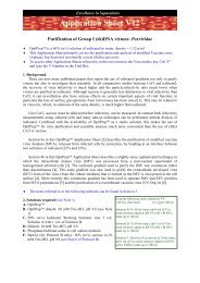
5. Either collect the whole gradient (Figure 1) in 1-2 ml fractions dense end first or use a syringe<br />
inserted at the 40%/54% interface to aspirate 4 ml of the 40% layer (see Section 4, Notes 5 and 6).<br />
3. Continuous gradient<br />
The method is adapted from Hermens et al [3] who used a preformed continuous gradient in a nearvertical<br />
rotor. After release of rAAV from cultured cells by freeze/thawing, the virus particles from the<br />
clarified fluid were first concentrated either by ammonium sulfate precipitation or by cellufine sulfate<br />
column chromatography [3].<br />
3a. Solutions required<br />
A. OptiPrep<br />
B. Phosphate-buffered saline<br />
Clarified virus fluid<br />
15% iodixanol in 1 M NaCl<br />
25% iodixanol<br />
40% iodixanol<br />
54% iodixanol<br />
3b. Ultracentrifuge rotor requirements<br />
Near-vertical rotor with approx 5 ml tubes, capable of approx 360,000g (see Section 4, Note 7)<br />
3c. Protocol<br />
1. After concentration by ammonium sulfate precipitation or chromatography, suspend rAAV in<br />
phosphate-buffered saline, pH 7.4.<br />
2. Transfer 2.7 ml of rAAV-containing fluid to a suitable tube and underlayer with OptiPrep to fill<br />
the tube.<br />
3. After sealing the tube, form the gradient in a Gradient Master by rotating at 20 rpm at 80° for 12<br />
min (see Section 4, Note 8)<br />
4. Centrifuge at 71,000 rpm (348,000 g av ) for 3 h at 16°C (see Section 4, Note 8).<br />
350,000g 1 h<br />
proteins<br />
adenovirus<br />
rAAV<br />
Figure 1 Banding of rAAV in a discontinuous iodixanol<br />
gradient [1]
3<br />
5. Collect the gradient from the bottom by tube puncture, the rAAV bands close to the bottom of the<br />
gradient (see Section 4, Note 9).<br />
4. Notes<br />
1. For smaller volume tubes scale down all volumes proportionally. It may be necessary to increase<br />
the centrifugation time proportionally if the rotor cannot achieve 350,000 g av .<br />
2. Phenol red (0.01 µg/ml) may be included in the alternate gradient layers to enhance visual<br />
identification of the layers. At 350,000 g, the iodixanol itself will sediment and may make the<br />
interfaces less obvious.<br />
3. Aggregation of rAAV with proteins in the cell lysate can pose a serious problem to its isolation as<br />
the aggregates are heterogeneous and consequently exhibit a broad range of densities. Inclusion of<br />
1 M NaCl in the 15% iodixanol prevents this aggregation and allows the rAAV to be isolated as a<br />
single band in the 40% iodixanol layer.<br />
4. Because of the large volumes used in this gradient, the use of a peristaltic pump to introduce the<br />
iodixanol solutions makes this task easier. For more information on preparing discontinuous<br />
gradients see <strong>Application</strong> <strong>Sheet</strong> V02.<br />
5. All of the contaminating proteins in the lysate band at the 25%/40% iodixanol interface, and more<br />
than 99% of the adenovirus contaminant bands at a lower density (