P and T wave analysis in ECG signals using Bayesian methods
P and T wave analysis in ECG signals using Bayesian methods
P and T wave analysis in ECG signals using Bayesian methods

Create successful ePaper yourself
Turn your PDF publications into a flip-book with our unique Google optimized e-Paper software.
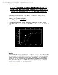
1.3 - Electrocardiography 17<br />
Figure 1.7: The P, Q, R, S, T, U <strong>wave</strong>s result<strong>in</strong>g from one depolarization / repolarization cycle.<br />
Image adapted from [Ala].<br />
<strong>ECG</strong> <strong>in</strong>tervals<br />
Intervals <strong>and</strong> segments of the <strong>ECG</strong> are important parameters for assess<strong>in</strong>g the normality or<br />
abnormality of the space between two electrical events, thus they are generally cl<strong>in</strong>ically relevant.<br />
Fig. 1.8 illustrates the normal cl<strong>in</strong>ical features of the electrocardiogram, which <strong>in</strong>clude<br />
<strong>wave</strong> amplitudes <strong>and</strong> <strong>in</strong>ter-<strong>wave</strong> tim<strong>in</strong>gs (<strong>ECG</strong> <strong>in</strong>tervals). Note that the <strong>in</strong>ter-beat tim<strong>in</strong>g (RR<br />
<strong>in</strong>terval) is not marked. The illustration uses the typical graph-paper presentation format,<br />
which stems from the early cl<strong>in</strong>ical years of electrocardiography, where <strong>analysis</strong> was done by<br />
h<strong>and</strong> measurements of hard copies. Each box is 1 mm 2 <strong>and</strong> the <strong>ECG</strong> paper is usually set to<br />
move at 25 mm/s. Therefore, each box represents 0.04 second <strong>in</strong> time. The amplitude scale is<br />
set to be 0.1 mV per square, although there is often a larger grid overlaid at every five squares<br />
(0.20 second/0.5 mV ) The values for the cl<strong>in</strong>ical features <strong>in</strong>dicated on the graph <strong>in</strong> Fig. 1.8<br />
are typical, although they can vary based upon gender, age, activity, <strong>and</strong> health.<br />
The cl<strong>in</strong>ical <strong>in</strong>terpretation of the <strong>ECG</strong> segments <strong>and</strong> <strong>in</strong>tervals can be summarized as follow<strong>in</strong>g:<br />
• PR (or PQ) <strong>in</strong>terval is measured from the beg<strong>in</strong>n<strong>in</strong>g of the P <strong>wave</strong> to the beg<strong>in</strong>n<strong>in</strong>g<br />
of the QRS complex. The PR <strong>in</strong>terval reflects the time the electrical impulse takes to<br />
travel from the s<strong>in</strong>oatrial node through the A-V node <strong>and</strong> enter<strong>in</strong>g the ventricles. The<br />
PR <strong>in</strong>terval is therefore a good estimate of A-V node function.<br />
• PR segment connects the P <strong>wave</strong> <strong>and</strong> the QRS complex. This co<strong>in</strong>cides with the<br />
electrical conduction from the A-V node to the His bundle <strong>and</strong> the bundle branches<br />
<strong>and</strong> then to the Purk<strong>in</strong>je Fibers. This electrical activity does not produce a contraction<br />
directly <strong>and</strong> is merely travel<strong>in</strong>g down towards the ventricles <strong>and</strong> this shows up flat on the