Animals. Health. Food Hygiene - Latvijas Lauksaimniecības ...
Animals. Health. Food Hygiene - Latvijas Lauksaimniecības ...
Animals. Health. Food Hygiene - Latvijas Lauksaimniecības ...

Create successful ePaper yourself
Turn your PDF publications into a flip-book with our unique Google optimized e-Paper software.
ISSN<br />
1407 – 1754<br />
LATVIJAS LAUKSAIMNIECĪBAS UNIVERSITĀTE<br />
VETERINĀRMEDICĪNAS FAKULTĀTE<br />
LATVIA UNIVERSITY OF AGRICULTURE<br />
FACULTY OF VETERINARY MEDICINE<br />
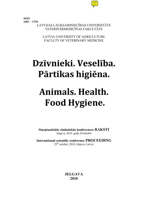
Dzīvnieki. Veselība.<br />
Pārtikas higiēna.<br />
<strong>Animals</strong>. <strong>Health</strong>.<br />
<strong>Food</strong> <strong>Hygiene</strong>.<br />
Starptautiskās zinātniskās konferences RAKSTI<br />
Jelgava, 2010. gada 29.oktobrī<br />
International scientific conference PROCEEDING<br />
29 th october, 2010, Jelgava, Latvia<br />
JELGAVA<br />
2010
Redkolēģija<br />
Editorial Board<br />
Henrikas Ţilinskas, Dr.habil.med.vet., profesors, Lietuvas Veterinārā<br />
akadēmija (Lietuva)<br />
Dr.habil.med.vet., professor, Lithuanian Veterinary Academy (Lithuania)<br />
Vidmantas Biţokas, Dr.habil.med.vet., profesors, Lietuvas Veterinārā<br />
akadēmija (Lietuva)<br />
Dr.habil.med.vet., professor, Lithuanian Veterinary Academy (Lithuania)<br />
Edīte Birģele, Dr.habil.biol., LZA korespondētājlocekle, <strong>Latvijas</strong><br />
Lauksaimniecības universitāte (Latvija)<br />
Dr.habil.biol., profesor, corresponding member of Latvian Academy of<br />
Sciences, Latvian University of Agriculture (Latvia)<br />
Vita Antāne, Dr.med.vet., asociētā profesore,<br />
<strong>Latvijas</strong> Lauksaimniecības universitāte (Latvija)<br />
Dr.med.vet., assoc.professor, Latvian University of Agriculture (Latvia)<br />
Aija Ilgaţa, Dr.med.vet., docente,<br />
<strong>Latvijas</strong> Lauksaimniecības universitāte (Latvija)<br />
Dr.med.vet., Latvian University of Agriculture (Latvia)<br />
Atbildīgais par izdevumu / Responsible for edition Edīte Birģele<br />
Izdevuma redaktore / Editor for edition Līga Kovaļčuka<br />
Maketētāja / Layout design Sandra Brande, Solvita Ţidkova<br />
Visi krājumā ievietotie raksti ir recenzēti.<br />
All articles are reviewed.<br />
2
SATURS<br />
TABLE OF CONTENTS<br />
PREPARĀTA MILDRONĀTA IEDARBĪBA UZ VAISLAS KUIĻU DZIMUMAKTIVITĀTI<br />
UN SPERMAS KVALITĀTI<br />
INFLUENCE OF PREPARATION MILDRONAT ON SEXUAL BEHAVIOR AND SEMEN<br />
QUALITY OF BOARS<br />
Vita Antāne 1 , Ilmārs Stonāns 2 , Jāzeps Rimeicāns 1 , Māra Mangale 1 ,<br />
Zigmunds Brūveris 1 , Alberts Auzāns 1 , Aleksandrs Mednis 1 , Ivars Lūsis 1 ................ 5<br />
MILDRONĀTA IETEKME UZ SAIMNIECISKO VAISLAS GATAVĪBU SASNIEGUŠU<br />
KUIĻU ASINS BIOĶĪMISKAJIEM RĀDĪTĀJIEM<br />
THE INFLUENCE OF MILDRONATE ON BIOCHEMICAL BLOOD INDICES OF BOARS<br />
AFTER REACHING THE BREEDING PREPAREDNESS<br />
Alberts Auzāns 1 , Zigmunds Brūveris 1 , Jāzeps Rimeicāns 1 , Vita Antāne 1 ,<br />
Māra Mangale 1 ,Aleksandrs Mednis 1 , Ivars Lūsis 1 , Ilmārs Stonāns 2 ...................... 12<br />
MILDRONĀTA IEDARBĪBA UZ KUIĻU SĒKLINIEKU MORFOLOĢIJU PĒC<br />
SAIMNIECISKĀS VAISLAS GATAVĪBAS SASNIEGŠANAS<br />
THE INFLUENCE OF MILDRONATE ON MORPHOLOGY OF BOARS TESTES AFTER<br />
REACHING THE BREEDING PREPAREDNESS<br />
Alberts Auzāns 1 , Zigmunds Brūveris 1 , Jāzeps Rimeicāns 1 , Vita Antāne 1 ,<br />
Māra Mangale 1 , Aleksandrs Mednis 1 , Ivars Lūsis 1 , Ilmārs Stonāns 2 ..................... 18<br />
MĀJAS SUĽU KUĽĢA GĻOTĀDAS VIRSMAS-BEDRĪŠU EPITĒLIJA MUKOĪDIE<br />
EPITELIOCĪTI UN HELIKOBAKTĒRIJU DAUDZUMS KUĽĢĪ<br />
MUCOID EPITHELIOCYTES IN THE SUPERFICIAL EPITHELIUM OF THE GASTRIC<br />
MUCOSA IN DOMESTIC DOGS AND AMOUNT OF HELICOBACTERIA IN THE<br />
STOMACH<br />
Dace Bērziņa, Edīte Birģele ...................................................................................... 24<br />
VAISLAS ĶĒVJU ASINS BIOĶĪMISKIE RĀDĪTĀJI DAŢĀDOS TURĒŠANAS<br />
APSTĀKĻOS<br />
BREEDING MARE BIOCHEMICAL PARAMETERS IN DIFFERENT HOUSING<br />
CONDITIONS<br />
Inta Caune, Aija Ilgaža ............................................................................................. 29<br />
INOVATĪVA SASTĀVA BROILERCĀĻU GAĻAS IEGUVE<br />
OBTAINING OF INNOVATIVE COMPOSITION BROILER CHICKEN MEAT<br />
PRODUCTION<br />
Sallija Ceriņa, Aleksandrs Jemeļjanovs, Vera Krastiņa, Īra Irēna Vītiņa ............. 36<br />
LATVIJAS BRŪNĀS ŠĶIRNES GOVJU PIENA MIKROBIĀLĀS PIESĀRĽOTĪBAS<br />
VĒRTĒJUMS PĒC TO ĢENĒTISKĀS IZCELSMES<br />
LB BOVINE MILK MICROBIAL CONTAMINATION EVALUATION BY RESEARCHING<br />
THEIR GENETIC ORIGIN<br />
Inese Dūjiņa, Aleksandrs Jemeļjanovs, Ināra Helēna Konošonoka ........................ 43<br />
1
STRAUSU (STRUTHIO CAMELUS VAR. DOMESTICUS) TIEVĀS UN RESNĀS ZARNAS<br />
MORFOMETRISKIE RĀDĪTĀJI PERINATĀLAJĀ PERIODĀ<br />
MORPHOMETRIC INDICES OF THE SMALL AND LARGE INTESTINE DURING<br />
PERINATAL PERIOD IN THE OSTRICH (STRUTHIO CAMELUS VAR. DOMESTICUS)<br />
Ilmārs Dūrītis, Arnis Mugurēvičs, Lauma Latkovska ............................................ 50<br />
THE EXPERIMENTAL IRON DISORDERS OF THE LIVER AND SMALL INTESTINE<br />
Gundega Knipše¹, Dmitrijs Babarikins², Jurijs Markovs¹, Nadežda Bērziņa³,<br />
Alise Grāmatniece¹ .................................................................................................... 56<br />
PĀRTIKAS TOKSIKOINFEKCIJAS IEROSINOŠĀS MIKROFLORAS SASTOPAMĪBA<br />
GOVS PIENA IEGUVES UN PIRMAPSTRĀDES PROCESĀ<br />
INCIDENCE OF FOODBORN TOXICOINFECTIONS SUGGESTIVE MICROFLORA IN<br />
MILKING AND FIRST STAGE TREATMENT PROCESSES<br />
Ināra Helēna Konošonoka, Aleksandrs Jemeļjanovs, Daina Ikauniece,<br />
Gundega Kleina ........................................................................................................ 62<br />
STRAUSA GAĻAS KVALITĀTE „SOUS VIDE” IEPAKOJUMĀ<br />
THE QUALITY OF PACKED OSTRICH MEAT IN „SOUS VIDE”<br />
Janīna Ķīvīte, Daina Kārkliņa, Irisa Mūrniece ....................................................... 68<br />
ENDOMETRIAL HYSTOLOGICAL CHANGES AND PREGNANCY RATES IN MARES<br />
IMPAIRED CERVICAL DRAINAGE<br />
Evija Liepina, Vita Antane ....................................................................................... 73<br />
STIRNU (CAPREOLUS CAPREOLUS) UN STALTBRIEŢU (CERVUS ELAPHUS) GAĻAS<br />
BAKTERIĀLĀ PIESĀRĽOJUMA NOTEIKŠANA PIELIETOJOT DAŢĀDAS<br />
MIKROBIOLOĢIJAS UN MOLEKULĀRĀS BIOLOĢIJAS METODES DETECTION OF<br />
BACTERIAL POLLUTION OF ROE DEER'S (CAPREOLUS CAPREOLUS) AND RED<br />
DEER'S (CERVUS ELAPHUS) MEAT USING CLASICAL MICROBIOLOGICAL AND<br />
MOLECULAR BIOLOGY METHODS<br />
Solveiga Liepiņa, Aleksandrs Jemeļjanovs, Ināra-Helēna Konošonoka ................. 78<br />
SARKOCISTOZE MĀJAS UN MEŢA CŪKĀM<br />
SARCOCYSTOSIS OF PIGS AND WILD BOAR<br />
Ruta Medne, Anna Krūklīte, Kristīne Šteina .......................................................... 82<br />
MĀKSLĪGI RADĪTĀ LAŠU SPURU NEKROZE<br />
ARTIFICAL INDUCED SALMON FIN NECROSIS<br />
Ruta Medne, Edgars Liepiņš .................................................................................... 88<br />
SIERU MIKROFLORAS ANALĪZE<br />
ANALYSIS OF CHEESE MICROFLORA<br />
Alla Miķelsone, Inga Ciproviča ................................................................................ 96<br />
2
RAPŠA RAUŠU IZMANTOŠANAS EKONOMISKIE UN EKOLOĢISKIE FAKTORI<br />
STALTBRIEŢU ĒDINĀŠANĀ<br />
RAPESEED OIL CAKE USING ECONOMICAL AND ECOLOGICAL FACTORS IN RED<br />
DEER FEEDING<br />
Līga Proškina, Aleksandrs Jemeļjanovs, Īra Irēna Vītiņa, Vera Krastiņa, Biruta<br />
Lujāne, Imants Jansons .......................................................................................... 103<br />
ŪDENS AKTIVITĀTE (A W ) UN PH KĀ LISTERIA MONOCYTOGENES RISKA RĀDĪTĀJI<br />
ŠĶĒLĒS SAGRIEZTIEM GAĻAS PRODUKTIEM<br />
WATER ACTIVITY (A W ) AND PH AS RISK SHOWING OF LISTERIA MONOCYTOGENES<br />
IN SLICED MEAT PRODUCTS<br />
Indulis Siliņš 1 , Edgars Liepiņš 2 ............................................................................... 110<br />
THE OVARY ACTIVITY IN COWS WITH AND WITHOUT RETAINED FETAL<br />
MEMBRANES<br />
Santa Skuja , Vita Antāne ....................................................................................... 117<br />
STUDY OF THE MILK PROTEIN GENETIC CHARACTERIZATION IN LATVIAN DAIRY<br />
CATTLE BREEDS POPULATIONS<br />
Dace Smiltiņa, Andris Bāliņš, Ziedonis Grīslis ...................................................... 125<br />
DAŢĀDA JERSĪNIJU SASTOPAMĪBA CŪKU LIEMEĽOS TRĪJĀS KAUTUVĒS LATVIJĀ<br />
DIFFERENT PREVALENCE OF YERSINIAE ON PIG CARCASSES IN THREE<br />
SLAUGHTERHOUSES IN LATVIA<br />
Margarita Terentjeva, Edgars Liepiņš, Aivars Bērziņš ......................................... 130<br />
PERSONALIZĒTA ANTIKOAGULANTU TERAPIJA KARDIOLOĢIJĀ<br />
INDIVIDUALIZED ANTICOAGULATION THERAPY IN CARDIOLOGY<br />
Inga Urtāne 1 , Jānis Ansabergs 2 , Aina Silvija Štokmane 1 , Dace Bandere 1 ............ 136<br />
BENZO(A)ANTRACĒNA, BENZO(A)PIRĒNA, BENZO(B)FLUORANTHĒNA UN<br />
KRIZĒNA SATURSKŪPINĀTĀ GAĻĀ UN ZIVĪS<br />
CONTENT OF BENZO(A)ANTHRACENE, BENZO(A)PYRENE,<br />
BENZO(B)FLUORANTHENE AND CHRYSENEIN SMOKED MEAT AND FISH<br />
Anda Valdovska 1 , Jānis Mičulis 2 , Ligita Plotiņa 2 .................................................. 142<br />
ANIMALS‟ BEHAVIORAL DATA ANALYSIS BASED ON BEHAVIOR PARAMETERS<br />
VISUALIZATION AND FRACTAL DIMENSION METHOD<br />
Viktor Veliks 1,2 , Alexander Grakovski 2 .................................................................. 148<br />
IMUNOGLOBULĪNU SEMIKVANTITATĪVO TESTU PIELIETOŠANAS NOZĪME GOVJU<br />
GANĀMPULKU VESELĪBAS KONTROLĒ<br />
THE IMPORTANCE OF APPLICATION OF IMMUNOGLOBULIN SEMI-QUANTITATIVE<br />
TESTS TO CONTROLTHE COW HERD HEALTH<br />
Māra Viduža 1 , Laima Liepa 2 .................................................................................. 154<br />
TESTING METHODS OF CANINE AND FELINE BLOOD GROUPS COMPATIBILITY IN<br />
CLINICAL CONDITIONS<br />
Jeļena Andrejeva, Aija Ilgaža ................................................................................. 160<br />
3
PRELIMINARY DATA ON TICK-BORNE DISEASES IN DOGS IN LATVIA<br />
Inese Berzina, Ilze Matise ....................................................................................... 161<br />
A RAPID EFFECT OF HANDLING ON COUNTS OF WHITE BLOOD CELLS IN A<br />
WINTERING PASSERINE BIRD: A MORE PRACTICAL MEASURE OF STRESS?<br />
Dina Cirule 1,2 , Tatjana Krama 2 , Jolanta Vrublevska 2 , Indrikis Krams 2 ............. 162<br />
MICROBIOLOGICAL SECURITY OF APPLES STORED AT MODIFIED<br />
ATMOSPHERE<br />
Karīna Juhnevica 1 , Gita Skudra 2 ........................................................................... 163<br />
OPPORTUNISTIC PARASITE: TOXOPLASMA GONDII<br />
Gunita Deksne ......................................................................................................... 164<br />
DYNAMICS OF CAMPYLOBACTER JEJUNI COUNTS IN RAW POULTRY MEET<br />
DEPENDING ON PACKAGING ATMOSPHERE<br />
Kaspars Kovaļenko 1 , Aija Ruzaiķe 2 , Mati Roasto 3 , Edgars Liepiņš 1 ................... 165<br />
EVALUATION OF THE TEAR PRODUCTION IN DOG FOLLOWING GENERAL<br />
ANESTHESIA<br />
Līga Kovaļčuka, Artis Bečs, Līga Strode ............................................................... 166<br />
CHANGES OF PHYSIOLOGICAL INDICES AFTER RENEWAL OF BALANCED<br />
FEEDING OF STARVATING COWS IN THE MAXIMUM PERIOD OF LACTATION<br />
Laima Liepa, Ilmārs Dūrītis, Daiga Rudevica ....................................................... 167<br />
SEASONAL OUTBREAKS OF TYZZER‟S DISEASE IN MALAGASY GIANT JUMPING<br />
RATS (HYPOGEOMYS ANTIMENA) IN THE RIGA ZOO<br />
Ilze Matise 1 , Tatjana Ivasenko 2 , Guna Vitola 2 , Arno Wunschmann 3 .................. 168<br />
RABIES ORAL VACCINATION IN LATVIA – PAST, PRESENT AND FUTURE<br />
Edvīns Oļševskis 1 , Edgars Liepiņš 2 ...................................................................... 169<br />
LYMPH NODES‟ HISTOLOGICAL STATUS OF PIGS WITH PORCINE CIRCOVIRUS-2<br />
Inga Piginka 1 , Edīte Birgele 2 .................................................................................. 170<br />
4
PREPARĀTA MILDRONĀTA IEDARBĪBA UZ VAISLAS KUIĻU<br />
DZIMUMAKTIVITĀTI UN SPERMAS KVALITĀTI<br />
INFLUENCE OF PREPARATION MILDRONAT ON SEXUAL<br />
BEHAVIOR AND SEMEN QUALITY OF BOARS<br />
Vita Antāne 1 , Ilmārs Stonāns 2 , Jāzeps Rimeicāns 1 , Māra Mangale 1 , Zigmunds<br />
Brūveris 1 , Alberts Auzāns 1 , Aleksandrs Mednis 1 , Ivars Lūsis 1<br />
1 LLU, Veterinārmedicīnas fakultāte, Latvija<br />
LUA, Faculty of Veterinary Medicine, Latvia<br />
2<br />
AS Grindeks<br />
ABSTRACT<br />
In 1984, the Institute of Organic Synthesis of Latvian Academy of Sciences developed a<br />
medical drug Mildronate that is widely used in medicine as anti-ishemic and stressprotective<br />
drug in treating various cardiovascular diseases, and in farm animals as<br />
growing and metabolic process stimulator. It is known, that Mildronate (Quaterin,<br />
Meldonium) interferes with carnitine biosynthesis, controlling carnitine and gammabutyrobetaine<br />
(GBB) concentration ratio. L-carnitine is implicate in reproductive<br />
biology and could be associated with the maturation process of spermatogenesis. In its<br />
turn, GBB normalizes and stimulates the sexual activity and potency in mammals.<br />
The aim of our study was to estimate the influence of preparation Mildronate on<br />
spermatogenesis, libido and semen quality of boars. Two groups (control and<br />
experimental) of boars were used to achieve the task of the research. Each group<br />
consisted of six animals, were kept and fed at the same circumstances. Experimental<br />
period started two months later when the boars had reached the breeding preparedness.<br />
The boars of experimental group got orally 2 g of Mildronate daily for 60 days. <strong>Animals</strong><br />
both control and experimental groups on a regular basis were tested for health status as<br />
well as the blood samples were taken. The semen was collected manually once every 10<br />
days. The semen quality parameters were assessed before the administration of<br />
Mildronate and further once in 39 days that corresponds to the length of<br />
spermatogenesis cycle in boars. The sexual behavior was detected for each boar. The<br />
results of the study indicate that the administration of Mildronate in boars improves<br />
their sexual activity and the semen quality: time from approaching the „dummy” to the<br />
start of ejaculation is significantly (p
ūtiski samazina L-karnitīna, bet paaugstina γ – butirobetaīna (GBB) koncentrāciju<br />
asins plazmā un sēkliniekos. GBB var pielietot seksuālās aktivitātes un potences<br />
stimulēšanai(4). Mūsu pētījuma mērķis bija pārbaudīt preparāta Mildronāta ietekmi uz<br />
kuiļu spermatoģenēzi un spermas kvalitāti, jo iepriekšējie pētījumi parādīja (1) ka<br />
izbarojot Mildronātu 60 dienas pa 2,0 g vaislas gatavību sasniegušiem kuiļiem<br />
palielinās spermatogēnā epitēlija slāľa biezums, testosterona koncentrācija asins<br />
serumā, un labāk attīstās sēklinieku interstīcijs un palielinās glandulocītu (Leidiga šūnu)<br />
skaits.<br />
MATERIĀLS UN METODIKA<br />
Pētījuma veikšanai no nosacīti analogiem kuilīšiem tika komplektēta<br />
eksperimentālā un kontroles grupa. Pētījums sastāvēja no diviem periodiem:<br />
sagatavošanas un eksperimentālā. Sagatavošanas periodā tika apmācīti 8-9 mēnešus veci<br />
kuiļi, kuri sasnieguši vaislas gatavību, atdot spermu uz fantoma. Eksperimentālais<br />
periods ilga trīs mēnešus. Šajā laikā kuiļi katru dienu individuāli kopā ar barību saľēma<br />
2 gramus Mildronātu atšķaidītu ar 2-3ml ūdens. Spermas noľemšana veikta manuāli ik<br />
pēc 10 dienām. Katram kuilim noteicām dzimumrefleksu (no kuiļa tuvošanās fantomam<br />
līdz ejakulācijas sākumam un ejakulācijas ilgumu) izteiktību laika periodā - sekundēs.<br />
Pirms Mildronāta saľemšanas un tālāk ik pēc 39 dienām, kas atbilst kuiļu viena<br />
spermatoģenēzes cikla garumam, noteicām kuiļiem spermas kvalitātes rādītājus<br />
(ejakulāta masu, spermiju koncentrāciju, spermatozoīdu aktivitāti un morfoloģiju).<br />
Spermas izmeklējumi tika veikti tūlīt pēc spermas noľemšanas uz vietas fermā.<br />
Spermatozoīdu morfoloģijas noteikšanai pagatavotās iztriepes nokrāsotas un izvērtētas<br />
Klīniskā institūta Dzīvnieku reprodukcijas laboratorijā.<br />
Iegūto datu apstrāde veikta ar matemātiskās statistikas metodēm, iegūtajiem<br />
rezultātiem aprēķināti vidējie aritmētiskie lielumi, dispersijas, standartnovirzes,<br />
izmantojot datoru ar Stata-9 un Microsoft Excel standartprogrammu paketi.<br />
REZULTĀTI UN DISKUSIJA<br />
Dzimumrefleksu, no kuiļa tuvošanās fantomam līdz ejakulācijai, izteiktība un<br />
ejakulācijas ilgums laika vienībā ik pēc 60 s grafiski attēloti izmantojot Kaplan-Meier<br />
(6) metodi (1 un 2 attēls). Aprēķini parādīja, ka pēc pirmajām 60 sekundēm 42% no<br />
eksperimenta dzīvniekiem (E), kuri saľēma Mildronātu, iestājās ejakulācija. Tajā pat<br />
laikā kontroles grupas kuiļiem (K), kuri preparātu nesaľēma, ejakulācija iestājusies tikai<br />
6% gadījumos. Pēc 120 s šie rādītāji ir attiecīgi 83% un 55%, pēc 180 s - 93% un 79%.<br />
Atšķirības starp grupām pēc Wilcoxon (Breslow) testa ir būtiskas - Pr>chi2=0,0001.<br />
Turpretīm ejakulācijas ilgums (2 zīm.) eksperimenta grupas kuiļiem būtiski neatšķīrās<br />
no kontroles grupas dzīvniekiem un tā vidējie rādītāji bija attiecīgi 276 ± 59 un 288 ± 52<br />
s. Pētījumos ar ţurkām (2) pierādījās, ka Mildronāts samazināja brīvā karnitīna<br />
koncentrāciju ţurku sēkliniekos un tas savukārt nepazemināja ţurku seksuālo aktivitāti<br />
un spermas kvalitāti.<br />
Kuiļu ejakulātu masas rādītāji attēloti 1. tabulā. Pirms Mildronāta pielietošanas<br />
eksperimenta un kontroles grupas kuiļu ejakulāta masas rādītāju atšķirības nav būtiskas<br />
(p>0.05). Pirmo 39 dienu laikā eksperimentālās grupas kuiļiem pēc Mildronāta<br />
pielietošanas palielinās ejakulāta masa no 120.8 ± 13.5 ml līdz 194.4 ± 16.1 (62%). Tai pat<br />
laikā kuiļiem, kuri preparātu nesaľēma (kontroles grupa) ejakulāta masas palielinājums<br />
nenozīmīgs un tas ir no 145.8 ± 24.9 uz 157.5 ± 18.2 (9%) Rādītāju atšķirības starp<br />
grupām ir ar varbūtību p
(80...99 diena) ejakulāta masas rādītāji starp E un K grupas kuiļiem ir līdzīgi.<br />
Mildronāts strukturāli ir radniecīgs karnitīnam (2) un tā L-karnitīna izbarošana kuiļiem<br />
pozitīvi ietekmēja spermas kvalitāti, jo par 11% palielinājās ejakulāta masa un kopējais<br />
spermiju daudzums tajā (3).<br />
time(sec)<br />
0 60 120 180 240 300 360<br />
0.80<br />
0.60<br />
1.00<br />
0.40<br />
0.20<br />
0.00<br />
experiment<br />
control<br />
1. attēls. Laiks (s) no kuiļa tuvošanās fantomam līdz ejakulācijai<br />
Figure 1. Time (sec) from approaching the “dummy” to the start of ejaculation<br />
time(sec)<br />
0 60 120 180 240 300 360<br />
0.80<br />
0.60<br />
1.00<br />
0.40<br />
0.20<br />
0.00<br />
experiment<br />
control<br />
2. attēls. Ejakulācijas ilgums (s)<br />
Figure 2. The length of ejaculation (sec)<br />
7
1. tabula/Table 1<br />
Ejakulāta masa pētījuma sākumā un vidēji pa 40 dienu periodiem eksperimenta<br />
(E) un kontroles (K) grupas kuiļiem, g<br />
The total wight of the ejaculate at the beginning of experiment and an average in<br />
40 days period in experimental (E) and control (K) group boars<br />
aritmētiskais vidējais/arithm. mean, standartkļūda/standart devision, t-tests<br />
p
mākslīgās apsēklošanas standartam un tāda paliek visu eksperimenta laiku. Tai pat laikā<br />
kontroles grupas kuiļu vidējā spermatozoīdu aktivitāte ejakulātā nesasniedz 7 balles un<br />
ir būtiski zemāka (p
10,0<br />
9,0<br />
8,0<br />
7,0<br />
Activity, points<br />
6,0<br />
5,0<br />
4,0<br />
K<br />
E<br />
Poly. (E)<br />
Poly. (K)<br />
3,0<br />
2,0<br />
1,0<br />
0,0<br />
0 10 20 30 40 50 60 70 80 90<br />
Time, days<br />
3. attēls. Spermatozoīdu vidējā aktivitāte kuiļiem, kuri saľēma Mildronātu un<br />
kontroles grupas kuiļiem, kuri Mildronātu nesaľēma<br />
Figure 3. The avarage spermatozoa motility in boars which were administrated<br />
Mildronate and control group<br />
Izvērtējot eksperimenta un kontroles grupas kuiļu spermiju aktivitātes vidējo<br />
rādītāju dinamiku pa atsevišķām spermas noľemšanas reizēm (3 attēls) redzam, ka<br />
spermatozoīdu aktivitātes svārstības ir vērojamas vienlaicīgi gan eksperimenta, gan<br />
kontroles grupas kuiļiem. Tomēr jāatzīmē, ka dzīvnieki, kuri saľēma Mildronātu<br />
spermatozoīdu aktivitātes dinamiskās izmaiľas pa spermas noľemšanas reizēm ir<br />
vienmērīgākas un izlīdzinātākas. Līdzšinējie pētījumi (7,9,10) liecina, ka Mildronāts<br />
(kvaterīns) pozitīvi ietekmē teļiem un sivēniem barības vielu uzsūkšanos zarnu traktā,<br />
uzlabo aminoskābju vielmaiľu un darbojas kā adaptogēns, mazinot sivēnu atšķiršanas<br />
stresu. Pētījumi ar cāļiem (8) parādīja, ka kvaterīns ir dzīvo šūnu augšanas stimulators<br />
un nedarbojas citotoksiski. Minēto pētījumu rezultātus var saistīt ar Mildronāta pozitīvo<br />
ietekmi uz kuiļu noturību pret stresu un aminoskābju vielu maiľu, kas ne tikai uzlaboja<br />
spermatozoīdu aktivitāti, bet mazināja tās svārstības starp atsevišķām spermas<br />
noľemšanas reizēm.<br />
SECINĀJUMI<br />
1. Preparāta Mildronāta pielietošana vaislas kuiļiem pozitīvi uzlabo dzimumrefleksu<br />
aktivitāti un spermas kvalitāti: būtiski samazinās laiks no kuiļu tuvošanās fantomam<br />
līdz ejakulācijas sākumam (p
2. Dzīvniekiem, kuri saľēma Mildronātu spermatozoīdu aktivitātes dinamiskās<br />
izmaiľas katrā spermas noľemšanas reizē ir vienmērīgākas un izlīdzinātākas.<br />
LITERATŪRA<br />
1. Auzāns ,A., Brūveris, Z., Rimeicāns, V. Antāne, M. Mangale, J., Mednis, I. Lūsis, I.<br />
Stonāns (2010) Mildronāta iedarbība uz kuiļu sēklinieku morfoloģiju pēc<br />
saimnieciskās vaislas gatavības sasniegšanas // Veterinārmedicīnas raksti. 2010.<br />
2. Dambrova, M., Cīrule, H., Svalbe, B., Zvejniece, L., Pugovichs, O., Zorenko, T.,<br />
Kalviľš, I., Liepiľš, E., Belozerova I. Effect of inhibiting carnitine biosynthesis on<br />
male rat sexual performance //Physiology & Behavior 2008; 95, 341-347.<br />
3. Jacino, E.et al Effect of L-Carnitine Supplementation on Boar Semen Quality//<br />
Acta.vet.Brno, .2007; 76:595-600.<br />
4. Kalvinsh, I., Veveris, M., Birmans, A. Pharmaceutial composition comprising<br />
gammabutirobetaine for stimulating the sexual activity and<br />
potency;,.WO/2003/022263[WO/2003/022263]. Ref. Type: Patent.<br />
5. Stira, A., Nudiens, J., Veide, Dz., Konošonoka, I.-H. Cūku reprodukcijas procesu<br />
pētījumi un to optimizācijas iespējas//Starptautiskā zinātniskā konference<br />
„Dzīvnieki.Veselība. Pārtikas higiēna.”, 2002; Jelgava: 266-271<br />
6. Veterinary epidemiologic research 2nd Edition Jan Dohoo, Wayne Martin, Henrich<br />
Stryhn /Published by VER Inc. Charlottetown · Prince Edward Island · Canada.<br />
Second printing January 2010; 475 – 476.<br />
7. Клинская, M. M., Романов, В. H. Влияние кватерина на пищеварение у телят //<br />
В кн.: Комбикорма, премиксы и добавки. Дубровники: 1985; 60-63.<br />
8. Kурашвили, Б. Е., Калвиньш, Я. И., Квинихадзе, С. Г., Кобахидзе, Л. Т.,<br />
Минкеладзе, Л. Г., Чумбуридзе, С. Р. Результаты пищеварительных испытаний<br />
на цыплят стимулятора «кватерина» // Известия академии наук СССР, серия<br />
«Биология», 1983;. 9. : 4:142-149.<br />
9. Романов, В. И. Процессы пищеварения и продуктивность телят при<br />
использовании кватерина в рационах // Авиорефeрат дисc…kанд. вет. наук,<br />
Дубровники: 1987; 1 – 23<br />
10. Тауритис, А. К., Андерсон, П. П., Бузлама, В. С., Калвиньш, И. Я. Применение<br />
кватерина для направленного изменения реакций поведения поросят при<br />
отьёмном стресе // Tезисы докладов научной конференции, Елгава: 1984; 22 –<br />
24<br />
11
MILDRONĀTA IETEKME UZ SAIMNIECISKO VAISLAS<br />
GATAVĪBU SASNIEGUŠU KUIĻU ASINS BIOĶĪMISKAJIEM<br />
RĀDĪTĀJIEM<br />
THE INFLUENCE OF MILDRONATE ON BIOCHEMICAL<br />
BLOOD INDICES OF BOARS AFTER REACHING THE<br />
BREEDING PREPAREDNESS<br />
Alberts Auzāns 1 , Zigmunds Brūveris 1 , Jāzeps Rimeicāns 1 , Vita Antāne 1 , Māra<br />
Mangale 1 ,Aleksandrs Mednis 1 , Ivars Lūsis 1 , Ilmārs Stonāns 2<br />
1 LLU Veterinārmedicīnas fakultāte, Latvija<br />
Faculty of Veterinary Medicine, LUA, Latvia<br />
2 AS Grindeks<br />
ABSTRACT<br />
The aim of the study was to find out the influence of preparation Mildronate (Quaterin,<br />
Meldonium) on a number of biochemical indices of boars blood after reaching the<br />
breeding preparedness. Mildronate was developed by the Institute of Organic Svnthesis<br />
of Latvian Academv of Sciences. Two groups (control and experimental) of boars were<br />
used to achieve the task of the research. Each group of 6 animals where kept and fed at<br />
the same circumstances. Experimental period started two month later when the boars<br />
had reached the breeding preparedness. The boars of experimental group daily got<br />
orally 2 g of Mildronate 60 days. <strong>Animals</strong> both control and experimental groups on a<br />
regular basis were tested for health status as well as the blood samples where taken. The<br />
results of our study indicate that levels of testosterone in the blood of boars of the<br />
Mildronate group increased statistifically significantly. Other blood biochemical<br />
characteristics did not differ between groups and correspondended to porcine<br />
physiological norm.<br />
KEY WORDS: mildronate, boars, biochemical indices of blood, testosterone.<br />
IEVADS<br />
Mūsu pētījuma mērķis bija noskaidrot, kā Mildronāts (Kvaterīns, Meldonijs)<br />
ietekmē vaislas gatavību sasniegušo kuiļu asins bioķīmiskos parametrus, jo iepriekšējais<br />
pētījums (2) parādīja, ka 2.0 g Mildronāta izbarošana vaislas gatavību sasniegušiem<br />
kuiļiem 60 dienas pozitīvi ietekmēja sēklinieku morfoloģiju: tika novērota tendence<br />
palielināties sēklinieku masai, tilpumam. Arī histoloģiskie pētījumi parādīja, ka<br />
Mildronāta ietekmē palielinās spermatogenā epitēlija slāľa biezums, labāk attīstās<br />
sēklinieku interstīcijs ar intersticiālajiem glandulocītiem (Leidiga šūnām) un asinsvadi.<br />
MATERIĀLS UN METODIKA<br />
Pētījuma veikšanai no nosacīti analogiem saimniecisko vaislas gatavību<br />
sasniegušiem kuiļiem, kas jau bija apmācīti atdot spermu uz fantoma, tika komplektēta<br />
e k s p e r i m e n t ā l ā un k o n t r o l e s g r u p a , katrā pa 6 dzīvniekiem. Sākot kuiļu<br />
apmācību spermas atdošanai, tie tika izvietoti katrs savā aizgaldā un baroti atbilstoši<br />
vecumam. Eksperimentālās grupas kuiļi, sākot ar 280 dienu vecumu, saľēma 2.0 g<br />
Mildronāta vienu reizi dienā, sajauktu kopā ar sauju sausās barības 60 dienas ilgi.<br />
Kontroles grupas kuiļi preparātu nesaľēma. Eksperimenta laikā abu grupu kuiļiem<br />
12
eksperimenta sākumā un beigās no v. saphena medialis laikā no 12 00 līdz 13 00 tika<br />
ľemti asinis paraugi, tie attiecīgi sagatavoti un nogādāti laboratorijās. Asins sērumā tika<br />
noteikta kreatinīna, kopējā holesterīna, kopējā bilirubīna, glikozes, testosterona<br />
koncentrācija un ASAT, ALAT aktivitāte.<br />
REZULTĀTI UN DISKUSIJA<br />
Asins bioķīmiskie rādītāji izmēģinājuma un kontroles grupas dzīvniekiem<br />
eksperimenta sākumā atspoguļoti 1. tabulā, bet eksperimenta beigās – 2. tabulā.<br />
1. tabula/Table 1<br />
Asins bioķīmiskie rādītāji eksperimentālās un kontroles grupas kuiļiem<br />
eksperimenta sākumā<br />
Biochemical indices of boars blood of experimental and control groups at the<br />
beginning of trial<br />
Kuiļu ident.Nr. /<br />
Boar<br />
identification<br />
Grupa E/K /<br />
Group E/K<br />
Kreatinīns/<br />
Creatinine,<br />
mknol/l<br />
Bilirubīns/<br />
Total<br />
bilirub.,<br />
mkmol/l<br />
Holesterīns/<br />
Cholesterol,<br />
mmol/l<br />
Glikoze /<br />
Glucose<br />
mmol/l<br />
ASAT,<br />
IU/l<br />
ALAT,<br />
IU/l<br />
4015 E 242 2.40 1.54 4.90 46 42<br />
4043 E 199 3.30 1.58 3.80 41 55<br />
4042 E 240 3.60 1.82 4.40 31 44<br />
5256 E 173 2.70 2.00 3.80 32 56<br />
5253 E 261 2.70 1.76 4.60 52 52<br />
4047 E 224 2.70 1.54 4.80 28 43<br />
Vid.aritm. / Mean arithm. 223.17 2.90 1.71 4.38 38.3 48.7<br />
Stand.nov. / Standard dev. 32.16 0.45 0.19 0.48 9.5 6.4<br />
Stand.kļūda / Standard error 13.13 0.18 0.08 0.20 3.9 2.6<br />
CI (95%)*<br />
189.4...<br />
256.9 2.43...3.37 1.51...1.90 3.88...4.89<br />
28.3...<br />
48.3<br />
42.0...5<br />
5.4<br />
4045 K 229 3.30 1.76 4.20 58 56<br />
5258 K 176 2.70 1.71 4.80 31 68<br />
4048 K 177 2.40 1.57 3.90 39 57<br />
4046 K 309 2.70 2.28 4.20 39 50<br />
5266 K 163 1.50 1.49 4.50 45 57<br />
5255 K 226 2.10 1.69 3.90 54 52<br />
Vid.aritm. / Mean arithm. 213.33 2.45 1.75 4.25 44.3 56.7<br />
Stand.nov. / Standard dev. 54.42 0.61 0.28 0.35 10.2 6.3<br />
Stand.kļūda / Standard error 22.22 0.25 0.11 0.14 4.1 2.6<br />
CI (95%)*<br />
156.2...<br />
270.4 1.81...3.09 1.46...2.04 3.88...4.62<br />
33.7...<br />
55.0<br />
50.1...<br />
63.2<br />
p-vērtība / p-value 0.36 0.09 0.38 0.30 0.16 0.03<br />
Nozīmīgums / Signifficance - - - - - +<br />
*Vidējā aritmētiskā 95% ticamības intervāls / 95% confidence interval of the arithmetric mean<br />
13
2. tabula/Table 2<br />
Asins bioķīmiskie rādītāji eksperimentālās un kontroles grupas kuiļiem<br />
eksperimenta beigās<br />
Biochemical indices of boars blood of experimental and control groups<br />
at the end of trial<br />
Kuiļu ident.Nr. /<br />
Boar<br />
identification<br />
Grupa E/K /<br />
Group E/K<br />
Kreatinīns /<br />
Creatinine,<br />
mknol/l<br />
Bilirubīns /<br />
Total<br />
bilirub.,<br />
mkmol/l<br />
Holesterīns /<br />
Cholesterol,<br />
mmol/l<br />
Glikoze /<br />
Glucose<br />
mmol/l<br />
ASAT,<br />
IU/l<br />
ALAT,<br />
IU/l<br />
4015 E 140 1.50 1.38 4.90 37 62<br />
4043 E 204 2.30 1.31 3.80 25 54<br />
4042 E 143 2.10 1.78 4.40 22 62<br />
5256 E 191 2.30 1.46 3.80 32 52<br />
5253 E 205 2.60 1.16 4.60 34 53<br />
4047 E 104 2.70 0.93 4.80 31 59<br />
Vid.aritm. / Mean arithm. 164.5 2.25 1.34 4.38 30.2 57.0<br />
Stand.nov. / Standard dev. 41.5 0.43 0.29 0.48 5.6 4.6<br />
Stand.kļūda / Standard error 17.0 0.17 0.12 0.20 2.3 1.9<br />
120.9...<br />
24.3... 52.2...<br />
CI (95%)* 208.1 1.80...2.70 1.04...1.64 3.88...4.89 36.1 61.8<br />
4045 K 183 1.20 1.41 5.20 34 66<br />
5258 K 155 1.50 1.25 4.50 38 73<br />
4048 K 133 1.50 1.31 4.60 36 60<br />
4046 K 214 2.30 0.92 4.50 42 52<br />
5266 K 163 2.30 1.66 4.00 34 56<br />
5255 K 140 1.50 1.77 4.40 22 74<br />
Vid.aritm. / Mean arithm. 164.7 1.72 1.39 4.53 34.3 63.5<br />
Stand.nov. / Standard dev. 29.9 0.47 0.30 0.39 6.7 9.0<br />
Stand.kļūda / Standard error 12.2 0.19 0.12 0.16 2.8 3.7<br />
133.3...<br />
27.3... 54.0...<br />
CI (95%)* 196.1 1.23...2.21 1.07...1.71 4.13...4.94 41.4 73.0<br />
p-vērtība / p-value 0.50 0.03 0.39 0.28 0.14 0.07<br />
Nozīmīgums / Signifficance - + - - - -<br />
*Vidējā aritmētiskā 95% ticamības intervāls / 95% confidence interval of the arithmetric mean<br />
Kā redzams 1. un 2. tabulā, abu grupu dzīvniekiem kreatinīna, kopējā bilirubīna,<br />
kopējā holesterīna, glikozes koncentrācija un ASAT, ALAT aktivitāte neuzrāda būtiskas<br />
atšķirības un atbilst šīs sugas dzīvnieku fizioloģiskajiem rādītājiem. Atšķirības<br />
vērojamas testosterona koncentrācijā (skat. 3. tabulu un 1. att.). Šīs tabulas mērījumu<br />
analīze rāda, ka eksperimentālās grupas kuiļiem testosterona koncentrācija asinīs<br />
pētījuma laikā ir būtiski palielinājusies, bet kontroles grupas dzīvniekiem tā nav<br />
mainījusies.<br />
14
3. tabula/Table 3<br />
Testosterona dinamika asins sērumā eksperimentālās (E)<br />
un kontroles (K) grupas kuiļiem<br />
Dynamics of the testosterone in boars blood in control (K) and<br />
experimental (E) groups, ng./ml<br />
Pētījjuma laiks<br />
Study time<br />
Mērījumu<br />
skaits<br />
Number of<br />
mesements<br />
Testosterona<br />
koncentrācija, ng/ml<br />
Conc. of testosterone<br />
ng/ml<br />
E K E K p-vērtība /<br />
p-value<br />
Atšķirību nozīmīgums<br />
The importance of<br />
distinction<br />
Būtiskums /<br />
Significance<br />
Eksperimenta sāk.<br />
At the beginn. of<br />
trial 4 6 2,0 ±0,6 1,5 ± 0,6 0.55 -<br />
Eksperimenta vidū<br />
Middle of trial 6 6 2,4 ±0,5 1,1±0,4 0.05 *<br />
Eksperimenta<br />
beigas<br />
At the end of trial 6 6 3,4 ± 0,7 1,2 ± 0,5 0.03 **<br />
p
Mūsu pētījums liecina, kā Mildronāts stimulē testosterona sintēzi kuiļu<br />
intersticiālajos glandulocītos (Leidiga šūnās). Saskaľā ar (3) novērojumiem,<br />
paaugstināts testosterona līmenis pozitīvi ietekmē kuiļu augšanu un to spermas<br />
produkciju, kaut arī daţi autori to neatzīst (14).<br />
Kā liecina literatūras dati, testosterona biosintēze ir ģenētiski kontrolēta (10), bet tā līmeni asinīs<br />
ietekmē arī rinda citu faktoru: dzīvnieka vecums (13,15), diennakts ritmika (5,7), ēdināšana (9),<br />
kā arī daţādas bioloģiski aktīvas vielas (1,4,8,9,12). Daţas no šīm vielām, piemēram,<br />
aromatozes inhibitors letrozols (1) un ārējais testosterons darbojas stimulējoši (4), bet 2-<br />
ethylhexyl ftalāts, estradiola benzoāts, tryfeniltins un tributylins nomāc testosterona biosintēzi<br />
(8,12). Visi minētie dati, ka arī mūsu pētījuma rezultāti liecina, ka testosterona līmenis asinīs ir<br />
ietekmējams.<br />
Al-Taras, Eeman Elsidding (1) aromatāzes inhibiciju ar letrozolu, kas paaugstina<br />
testoterona produkcijas līmeni kuiļu intersticiālajos glandulocītos (Leidiga šūnās),<br />
iesaka pielietot kuiļu reproduktīvo spēju uzlabošanai. Arī mūsu pētījums rāda, ka<br />
Mildronāts varētu būt iedarbīgs līdzeklis kuiļu testosterona deficīta gadījumos.<br />
Mildronāta stimulējošās iedarbības pamatā uz testosterona sintēzi kuiļu<br />
sēkliniekos varētu būt vairāki fizioloģiski mehānismi: 1) mildronāts, iedarbojoties uz<br />
sēklinieku mikrocirkulācijas sistēmu, uzlabo to audu trofiku, par ko liecina vairāki<br />
pētījumi par analoga preparāta kvaterīna iedarbību uz citiem orgāniem (16,17,18); 2)<br />
Mildronāts paaugstina gammabutirobetaīna koncentrāciju asinīs, bet šis savienojums<br />
pēc (6) datiem stimulē seksuālo aktivitāti; 3) iespējams, ka Mildronāts uzlabo aerobo<br />
vielmaiľu sēklinieku audos, kā to P. Mustafins (11) novērojis muskuļos.<br />
SECINĀJUMI<br />
1. Izbarojot saimniecisko vaislas gatavību sasniegušiem eksperimentālās grupas<br />
kuiļiem 60 dienas Mildronātu pa 2.0 g dienā kopā ar barību, eksperimentālās<br />
grupas kuiļiem testosterona koncentrācija asinīs palielinājās statistiski ticami,<br />
kamēr kontroles grupas dzīvniekiem tā nemainījās.<br />
2. Mildronāta izbarošana eksperimentālās grupas kuiļu asinīs neietekmēja kreatinīna,<br />
kopējā bilirubīna, kopējā holesterīna, glikozes koncentrāciju un ASAT, ALAT<br />
aktivitāti, salīdzinājumā ar kontroles grupas dzīvniekiem un šie rādītāji<br />
eksperimenta beigās atbilda sugas fizioloģiskajiem parametriem.<br />
LITERATŪRA<br />
1. Al-Taras, Eeman Elsidding. The role of endogenous estrogen in the regulation of<br />
reproductive function in the developing boar. Universitv of California, Davis, 2005:<br />
93 p.<br />
2. Auzāns, A., Brūveris, Z., Rimeicāns, J., Antāne, V. Mangale, M., Mednis, J., Lūsis,<br />
I., Stonāns, I. Mildronāta iedarbība uz kuiļu sēklinieku morfoloģiju pēc<br />
saimnieciskās vaislas gatavības sasniegšanas // Veterinārmedicīnas raksti. 2010.<br />
3. Bender, J. M., See, M. Т., Hanson, D. J., Lawrence, Т. E., Cassadv, J. P. Correlated<br />
response in growth, carcass, and meet quality traits to divergent selection for<br />
testosterone production in pigs // J. Anim. Sci. Savoy: Jun. 2006, v. 84 (6):1331-<br />
1337.<br />
4. Claus, R., Hausler, S., Lacorn, M. Rise of testosterone, nortestosterone, and 17 betaestradiol<br />
concentration in peripheral plasma of pigs after sublingual application in<br />
vivo // <strong>Food</strong> and Chemical Toxicology. Elsevier, Oxford, UK: 2007, 45: 225-228.<br />
5. Edqist, L. E. Diurnal variations in periphal plasma Ievels of testosterone,<br />
androsterons, and cortisol in boars // Acta vet. scandinavia, 1980, v. 21 (3): 451-453.<br />
16
6. Kalvinsh, I., Veveris, M., Birmans, A. Pharmaceutial composition comprising<br />
gammabutirobetaine for stimulating the sexual activity and potency;<br />
2003,.WO/2003/022263[WO/2003/022263].Ref.Type:Patent.<br />
7. Kattesh, H. G. Daily alterations in plasma testosterone in boars at diffent ages<br />
//Theriogenology, July 1982. v. 18 (1): 113-118.<br />
8. Ljungvall, K., Karlsson, Р., Hulten, F. Madej, A., Norrgren, L., Einarsson, S.<br />
Rodrigez-Martinez, H., Magnusson, U. Delayed effects on plasma concentration<br />
of testosterone and testicular mophology by intramuscular low-dose di (2-<br />
ethylhexyl) phtalate or estradiol bensoate in the prepubertal boar //<br />
Theriogenology. Elsevier, Nevv York, USA: 2005, 64 (5): 1170-1184.<br />
9. Louis, G. F. The effect of protein intake on boar libido, semen characteristics,<br />
and plasma hormone concentrations // J. Anim. Sci., Aug, 1994; 72 (8): 2038-<br />
2050.<br />
10. Lubritz, D. Genetic parametrs for testosterone production in boars // J. Anim.<br />
Sci. Aug. 1991; 69(8): 3220-3224.<br />
11. Mustafīns, P. Anaerobās slodzes sekas// Ţ. Doctus, jūnijs, 2010: 34-38.<br />
12. Ohno, S. Nakajima, Y. Nakajin, S. Trifenyltin and tributylin inhibit pig testicular<br />
17 beta-hydroxysteroid dehidrogenase activiry and supress testicular testosterone<br />
biosynthesis // Steroids. Elsevier, New York, USA: 2005; 70 (9): 645-651.<br />
13. Gray, R.C. Testosterone Ievels of boars at varieous ages // J. Anim. Sci., July<br />
1971; 33 (1):124-126.<br />
14. Walker, S. Effect of divergent selection for testosterone production on testicular<br />
morphology and daily sperm production in boars // J. Anim. Sci., Aug. 2004; 82<br />
( 8): 2259-2263.<br />
15. Zamaratskaia, G. Babol, J., Madej, A., Squires, A., Lundstrom, K. Age-related<br />
variation of plasma concentrations of scatole, androsterone, testosterone,<br />
oestradiol-17 beta oestrone sulphate, dehvdroepiandrosterone sulphate,<br />
trijodothvronine and IGF-1 in six entire male pigs // Reproduction in Domestic<br />
<strong>Animals</strong>. Blackvvell Wissenschafts-Verlag GmbH, Berlin, Germanv: 2004; 39<br />
(3): 168-172.<br />
16. Клинская, M. M., Романов, В. H. Влияние кватерина на пищеварение у<br />
телят // В кн.: Комбикорма, премиксы и добавки. Дубровники: 1985; 60-63.<br />
17. Курашвили, Б. Е., Калвинын, Я . И., Квинихадзе, С. Г., Кобахидзе, Л. Т.,<br />
Минкеладзе, Л. Г., Чумбуридзе. С. Р. Результаты пищеварительных<br />
испытаний на цыплят стимулятора «кватерина» // Известия академии наук<br />
СССР, 1983; серия «Биология», 9: 4:142-149.<br />
18. Романов, В. И. Процессы пищеварения и продуктивность телят<br />
прииспользовании кватерина в рационах II Автореф. дисс. канд. вет. наук,<br />
Дубровники: 1987; 1-23.<br />
17
MILDRONĀTA IEDARBĪBA UZ KUIĻU SĒKLINIEKU<br />
MORFOLOĢIJU PĒC SAIMNIECISKĀS VAISLAS GATAVĪBAS<br />
SASNIEGŠANAS<br />
THE INFLUENCE OF MILDRONATE ON MORPHOLOGY OF<br />
BOARS TESTES AFTER REACHING THE BREEDING<br />
PREPAREDNESS<br />
Alberts Auzāns 1 , Zigmunds Brūveris 1 , Jāzeps Rimeicāns 1 , Vita Antāne 1 , Māra<br />
Mangale 1 , Aleksandrs Mednis 1 , Ivars Lūsis 1 , Ilmārs Stonāns 2<br />
1 LLU Veterinārmedicīnas fakultāte, Latvija<br />
Faculty of Veterinary Medicine, LUA, Latvia<br />
2 AS Grindeks<br />
ABSTRACT<br />
The aim of the study was to estimate the influence of preparation Mildronate (Quaterin,<br />
Meldonium) on morphology of boars , testes after reaching the breeding preparedness.<br />
Mildronate was developed by the Institute of Organic Synthesis of Latvian Academy of<br />
Sciences. Two groups (control and experimental) of boars were used to achieve the task<br />
of the research. Each group of 6 animals where kept and fed at the same circumstances.<br />
Preparatory period lasted from weaning (21-28 day) until they reached the puberty and<br />
then the training of boars for collection of semen was started. Experimental period<br />
started two month later when the boars had reached the breeding preparedness. The<br />
boars of experimental group daily got orally 2,0 g of Mildronate 60 days. <strong>Animals</strong> both<br />
control and experimental groups on a regular basis were tested for health status as well<br />
as the blood and semen samples where taken. At the end of the experiment both groups<br />
of boars were euthanized following the study of testes (mass,volume) as well as<br />
histological research.<br />
Analysis of studies showed in boar‟s testes of the experimental group there was<br />
tendency of increase of testes mass and volume. Histological studies showed an increase<br />
the thickness of spermatogeny epithelium and considerably more interstitial<br />
glandulocytes (Leydig cells) in comparison with the animals of the control group.<br />
KEY WORDS: mildronate, boars, testes, morphometry, histology.<br />
IEVADS<br />
Postnatālajā ontoģenēzē turpinās sēklinieku augšanas un nobriešanas procesi. Šajā<br />
laikā ievērojami palielinās sēklinieku masa, kā arī notiek stromas un parenhīmas tālāki<br />
diferenciācijas procesi. Ľemot vērā, ka <strong>Latvijas</strong> Zinātľu akadēmijas Organiskās sintēzes<br />
institūtā sintezētais preparāts Mildronāts (Kvaterīns, Meldonijs) darbojas kā šūnu<br />
augšanas stimulators un pozitīvi ietekmē kā aminoskābju, tā arī enerģētisko vielmaiľu<br />
(4,7,8,9), izvirzījām uzdevumu noskaidrot, kā šīs preparāts ietekmē sēklinieku<br />
morfofunkcionālo stāvokli kuiļu postnatālās attīstības periodā pēc saimnieciskās vaislas<br />
gatavības sasniegšanas. Vienlaicīgi tika kontrolēti arī kuiļu asins bioķīmiskie rādītāji,<br />
seksuālā funkcija un spermas kvalitāte. *<br />
* Asins bioķīmiskās izmaiľas, kuiļu seksuālā aktivitāte un spermas kvalitāte tiks apskatīta atsevišķās<br />
publikācijās.<br />
18
MATERIALS UN METODIKA<br />
Pētījuma veikšanai no nosacīti analogiem kuilīšiem tika komplektēta<br />
eksperimentālā un kontroles grupa, ar aprēķinu, lai, uzsākot preparāta izbarošanu, katrā<br />
no tām būtu ne mazāk kā 6 dzīvnieki. Pētījums sastāvēja no diviem periodiem –<br />
sagatavošanas un<br />
eksperimentālā. S a g a t a v o š a n a s p e r i o d s ilga no sivēnu atšķiršanas (21-28 dienu<br />
vec.) līdz fizioloģiskās vaislas gatavības iestāšanās laikam un kuilīšu pieradināšanai<br />
atdot spermu uz fantoma. Sākot kuiļu apmācības periodu, tie tika izvietoti atsevišķos<br />
aizgaldos. E k s p e r i m e n t ā l a i s p e r i o d s sākās pēc tam, kad kuiļi bija apmācīti<br />
atdot spermu uz fantoma un tiem bija iestājusies saimnieciskā vaislas gatavība (248-279<br />
dienu vecumā). Eksperimentālās grupas kuiļi, sākot ar 280 dienu vecumu saľēma 2,0 g<br />
Mildronātu vienu reizi dienā, sajauktu kopā ar sauju sausās barības 60 dienas ilgi.<br />
Preparāta izbarošana tika veikta individuāli katram dzīvniekam un stingri sekots, lai tie<br />
saľemtu paredzēto devu. Kontroles grupas kuiļi preparātu nesaľēma. Eksperimentālā<br />
perioda laikā reizi mēnesī abu grupu kuiļi tika svērti, un noteikti hematoloģiskie un<br />
bioķīmiskie rādītāji. Eksperimentālā perioda beigās kuiļi eitanazēti. Izľemtiem<br />
sēkliniekiem noteikta masa un tilpums, kā arī veikta histoloģiskā izmeklēšana.<br />
Histoloģiskie izmeklējumi veiktizdarīti Rīgas P. Stradiľa Medicīnas akadēmijas<br />
Patoloģijas institūtā.<br />
PĒTĪJUMU REZULTĀTI UN DISKUSIJA<br />
SĒKLINIEKU MORFOMETRIJA<br />
Labā un kreisā sēklinieka svēršanas un tilpuma noteikšanas rezultāti atspoguļoti 1.<br />
tabulā. To analīze rāda, ka eksperimentālās grupas dzīvnieku kā labo tā kreiso<br />
sēklinieku vidējā masa ir lielāka, salīdzinot ar kontroles grupas kuiļu sēkliniekiem<br />
(eksperimentālai grupai attiecīgi 432.6 g un 440.8 g, bet kontroles grupai – 406.0 un<br />
422.0 g). Abās grupās kreisais sēklinieks svēra vairāk. Arī sēklinieku vidējie tilpumi<br />
eksperimentālās grupas kuiļiem ir lielāki. Eksperimentālās grupas kuiļu labā sēklinieka<br />
vidējais tilpums sastāda 414 ml un kreisais 420 ml, bet kontroles grupā – labais<br />
sēklinieks ir tikai 378.0 ml, bet kreisais – 384 ml liels. Atšķirības nav statistiski ticamas,<br />
tomēr kā sēklinieku masas, tā tilpuma palielināšanās tendence vērojama.<br />
Kā redzams 1. att., eksperimentālās grupas kuiļu sēklinieku masas un tilpuma<br />
rādītāji ir izlīdzinātāki, jo uzrāda mazākas individuālās svārstības.<br />
Sēklinieku izmēru – masas un tilpuma palielināšanās, ľemot vērā literatūrā satopamos<br />
datus (2.5), ļauj prognozēt, ka arī spermas apjoms eksperimentālās grupas kuiļiem būs<br />
augstāka, jo P. L. Senger (6) skaidri norāda, ka pastāv cieša pozitīva korelācija starp<br />
sēklinieku lielumu un to spēju producēt spermu.<br />
19
Sēklinieku morfometrija pēc kuiļu eitanāzijas<br />
Morphometry of testes after boars euthanasia<br />
(E - eksperimentālā grupa / experimental group; K - kontroles grupa / control group)<br />
1.tabula/Table 1<br />
Kuiļa id.nr.<br />
/Boar id.nr.<br />
Grupa / Group Sēklin.masa bez piedēkļa (g), /<br />
Weight of testis without<br />
epididymis (g),<br />
Sēklinieka tilpums (ml), /<br />
Volume of testis (ml),<br />
Sēklinieka piedēkļa masa (g), /<br />
Weight of epididymis (g),<br />
labais / right kreisais / left labais / right kreisais / left labais / right kreisais / left<br />
4042 E 394.0 422.0 390.0 400.0 130.0 129.0<br />
4043 E 435.0 424.0 440.0 420.0 114.0 114.0<br />
5256 E 399.0 387.0 360.0 380.0 130.0 132.0<br />
5253 E 436.0 451.0 400.0 400.0 131.0 131.0<br />
4047 E 499.0 520.0 480.0 500.0 144.0 131.0<br />
Vid.aritm. / Arithm. mean 432.6 440.8 414.0 420.0 129.8 127.4<br />
Stand.nov. / Standard dev. 41.97 49.77 46.69 46.90 10.64 7.57<br />
Stand.kļūda / Standard error 18.77 22.26 20.88 20.98 4.76 3.39<br />
CI (95%)* 380.5...484.7 379.0...502.6 356.0...472.0 361.8...478.2 116.6...143.0 118.0...136.8<br />
5254 K 334.0 384.0 310.0 320.0 107.0 119.0<br />
5258 K 523.0 514.0 480.0 480.0 153.0 154.0<br />
4048 K 331.0 353.0 300.0 320.0 132.0 133.0<br />
4046 K 441.0 428.0 420.0 400.0 98.0 101.0<br />
5255 K 405.0 431.0 380.0 400.0 125.0 129.0<br />
Vid.aritm. / Arithm. mean 406.8 422.0 378.0 384.0 123.0 127.2<br />
Stand.nov. / Standard dev. 80.19 60.80 75.63 66.93 21.60 19.42<br />
Stand.kļūda / Standard error 35.86 27.19 33.82 29.93 9.66 8.69<br />
CI (95%)* 307.2...506.4 346.5...497.5 284.1...471.9 300.9...467.1 96.2...149.8 103.1...151.3<br />
p-vērtība / p-value 0.27 0.30 0.20 0.18 0.27 0.49<br />
Nozīmīgums / Signifficance - - - - - -<br />
*Vidējā aritmētiskā 95% ticamības intervāls / 95% confidence interval of the arithmetric mean<br />
20
Histoloģiskie izmeklējumi<br />
Kā eksperimentālās tā kontroles grupu kuiļiem mikroskopā redzama normāla, sugai<br />
atbilstoša histoloģiskā uzbūve (2. att., 3. att.). Sēklinieku izlocītos kanālīšus izklāj<br />
perpendikulāri bazālajai membrānai novietotas balsta šūnas (Sertolī šūnas) un daţādās<br />
attīstības pakāpēs esošie spermatozoīdi. Spraugas starp līkumotajiem sēklinieka<br />
kanālīšiem aizľem labi attīstīti intersticiālie audi, kas ļoti bagāti ar intersticiālajiem<br />
glandulocītiem (Leidiga šūnām). Eksperimentālās grupas kuiļiem intersticiālo<br />
glandulocītu ir vairāk (2. att.) abu grupu dzīvniekiem intersticiālajos audos ir daudz<br />
nelielu asinsvadu, asins un limfas kapilāru.<br />
2. attēls. Līkumotie sēklinieku kanālīši un intersticiālie audi eksperimentālās<br />
grupas kuiļu sēkliniekos. Hematoksilīns-eozīns (x100)<br />
Figure 2. Seminiferous tubules and intertubular tissues of boars , testis<br />
(experimental group). Hematoxylin-eozin (x100)<br />
3. attēls. Līkumotie sēklinieku kanālīši un intersticiālie audi eksperimentālāsgrupas<br />
kuiļiem. Hematoksilīns-eozīns (x100)<br />
Figure 3. Seminiferous tubules and intertubular tissues of boars testis (control<br />
group). Hematoxylin-eozin (x100)<br />
21
Lai noskaidrotu Mildronāta ietekmi uz spermatoģenēzes procesu, tika mērīts<br />
spermatogenā epitēlija biezums abu grupu dzīvniekiem. Šo mērījumu rezultāti redzami<br />
2. tabulā.<br />
2. tabula/Table 2<br />
Kuiļu spermatogenā epitēlija biezums (µ)<br />
Thickness of spermatogeny epithelium (µ)<br />
(E – eksperimantālā grupa / experimental group, K – kontroles grupa / control group)<br />
Kuiļa id.nr. Grupa/ Mērījumu vieta / Place of measurement Vidējais<br />
/Boar id.nr. Group 1 2 3 4 5 / Mean<br />
4673 E 103.73 95.13 93.43 88.22 83.32 92.77<br />
4677 E 98.1 91.53 87.1 93.9 80.63 90.25<br />
4683 E 88.23 76.41 82.15 79.9 96.0 84.54<br />
4685 E 88.5 89.6 78.8 87.2 92.2 87.26<br />
4691 E 93.3 104.2 96.7 94.6 98.3 97.42<br />
Vid. aritmētiskais / Arithm. mean 90.45<br />
Standartnovirze / Standard deviation 7.43<br />
Standartkļūda / Standard error 1.49<br />
95% ticamības intervāls / 95% confidence interval 87.38......93.51<br />
5155 K 77.10 79.74 66.47 87.73 82.28 78.66<br />
4046 K 81.75 84.34 82.13 81.68 86.21 83.22<br />
40,48 K 78.73 74.79 74.77 82.46 72.01 76.55<br />
5258 K 69.01 72.78 70.18 57.54 71.27 68.16<br />
5758 K 70.07 68.63 83.31 80.46 68.91 74.28<br />
Vid. aritmētiskais / Arithm. mean 76.16<br />
Standartnovirze / Standard deviation 7.35<br />
Standartkļūda / Standard error 1.47<br />
95% ticamības intervāls / 95% confidence interval 73.14......79.21<br />
120<br />
Biezums / Thickness, mkm<br />
100<br />
80<br />
60<br />
E<br />
4. attēls. Spermatogenā epitēlija biezums<br />
Figure 4. Thickness of spermatogeny epithelium<br />
E - eksperimentālā grupa / experimental group, K – kontroles grupa / control group<br />
Mērījumu rezultātu analīze liecina, ka eksperimentālās grupas kuiļiem Mildronāta<br />
ietekmē spermatogenā epitēlija biezums statistiski ticami (p
kontroles grupas dzīvniekiem. Spermatoģenēzes procesu visticamāk ietekmē gamma<br />
butirobetaīna koncentrācijas palielināšanās audos, kas notiek mildronāta ietekmē (3),<br />
neskatoties uz faktu, ka notiek L-karnitīna biosintēzes inhibīcija (1).<br />
KOPSAVILKUMS<br />
Mildronāta piedeva vaislas kuiļu barībā pozitīvi ietekmē sēklinieku morfoloģiju:<br />
1. Novērojama neliela tendence palielināties to masai un tilpumam.<br />
2. Samazinās atsevišķu kuiļu sēklinieku masas atšķirības.<br />
3. Statistiski ticami palielinās spermatogenā epitēlija biezums.<br />
4. Sēklinieku stromā palielinās intersticiālo glandulocītu (Leidiga šūnu) skaits.<br />
LITERARŪRA<br />
1. Dambrova, M., H. Cīrule, B. Svalbe, L. Zvejniece, O. Pugovichs, T. Zorenko, I.<br />
Kalviľš, E. Liepiľš, I. Belozerova. Effect of inhibiting carnitine biosynthesis on<br />
male rat sexual performance //Physiology & Behavior XXX, 2008:7.<br />
2. . Hyang ,Y., Johnson, R. K. Effect of selection for size of testis in boars on semen<br />
and testis traits // J. Anim. Sci. (USA): 1996; 74 (4): 750-760.<br />
3. Kalvinsh, I., Veveris, M. Birmans, A.. Pharmaceutial composition comprising<br />
gammabutirobetaine for stimulating the sexual activity and potency;<br />
2003;.WO/2003/022263 [WO/2003/022263]. Ref. Type: Patent.<br />
4. Mustafīns, P. Anaerobās slodzes sekas// Ţ. Doctus, jūnijs, 2010; 34-38.<br />
5. Rathje, T., Johnson, R. K. Effect of selecting for increased testis size in boars on<br />
testis development and rates of sperm production // Nebraska, 1994; Swine Report:<br />
36 – 38.<br />
6. Senger, P. L. Pathways to Pregnancy and Parturition. 1 st revised Ed. Washington<br />
State University Res. And Technology Part., Pullman: 1999; 281.<br />
7. Крыжановский, С. А., Витинова, М. Б. Милдронат. В книге: Современные<br />
лекарственные средства. РИПОЛ Классик. 2007; 639.<br />
8. Kурашвили, Б. Е., Калвиньш, Я. И. Квинихадзе, С. Г. Кобахидзе, Л. Т.<br />
Минкеладзе, Л. Г. Чумбуридзе, С. Р. Результаты пищеварительных испытаний<br />
на цыплят стимулятора «кватерина» // Известия академии наук СССР, 1983;<br />
серия «Биология», 9: 4:142-149.<br />
9. Романов, В. И. Процессы пищеварения и продуктивность телят при<br />
использовании кватерина в рационах // Авторефират дисc. kанд. вет. наук,<br />
Дубровники: 1987; 1-23.<br />
23
MĀJAS SUĽU KUĽĢA GĻOTĀDAS VIRSMAS-BEDRĪŠU<br />
EPITĒLIJA MUKOĪDIE EPITELIOCĪTI UN HELIKOBAKTĒRIJU<br />
DAUDZUMS KUĽĢĪ<br />
MUCOID EPITHELIOCYTES IN THE SUPERFICIAL<br />
EPITHELIUM OF THE GASTRIC MUCOSA IN DOMESTIC DOGS<br />
AND AMOUNT OF HELICOBACTERIA IN THE STOMACH<br />
Dace Bērziľa, Edīte Birģele<br />
LLU, Veterinārmedicīnas fakultāte,Preklīniskais institūts, Latvija<br />
LUA, Faculty of Veterinary Medicine, Preclinical institute, Latvia<br />
dace.berzina@llu.lv, edite.birgele@llu.lv<br />
ABSTRACT<br />
This study was carried out to evaluate number of mucoid epiheliocytes of superficial<br />
epithelium of the gastric mucosa in domestic dogs related to the amount of<br />
helicobacteria. Mucosal samples were taken from several places of cardiac, fundic and<br />
pyloric gland region of the stomach to detect helicobacteria and superficial mucoid<br />
epitheliocytes of the gastric mucosa with the light histological examination. The amount<br />
of helicobacteria was relatively differentiated into four groups: as absente, mild,<br />
moderate and severe quantity of helicobacteria. The amount of mucoid epitheliocytes<br />
was evaluated in average numbers with standard derivation. There are no marked<br />
differences in the number of the mucoid epitheliocytes related to amount of<br />
helicobacteria in the cardiac and fundic gland region. Whereas in the pyloric gland<br />
region there is remarkable proliferation of superficial mucoid epitheliocytes and<br />
increased mucoid secretion related with higher amount of helicobacteria.<br />
KEY WORDS: helicobacteria, superficial mucoid epitheliocytes, dogs.<br />
IEVADS<br />
Pirmo reizi helikobaktērijas daţādu sugu dzīvniekiem konstatēja 19. gs. beigās –<br />
J. Rappin 1881. gadā un G. Bizzozero 1893. gadā, bet 1896. gadā H. Salomon tās<br />
aprakstīja kā spirāliski izliektas baktērijas suľu, kaķu un Norvēģijas ţurku kuľģos (Fox,<br />
Lee, 1997; Recordati et al., 2007). Tā kā helikobaktērijas ir grūti kultivēt barotnēs,<br />
sākotnēji tās nevarēja izolēt un identificēt (Owen, 1998). Tikai 1983. gadā pirmo reizi<br />
izolēja patogēno helikobaktēriju sugu Helicobacter pylori cilvēkiem, kas slimoja ar<br />
gastrītu (Marshall, Warren, 1983) un kuľģa čūlu (Marshall, Warren, 1984).<br />
Daudzas helikobaktēriju sugas tiek uzskatītas par patogēnām. Savukārt citas<br />
helikobaktērijas tiek uzskatītas kā normāla gremošanas trakta mikroflora (Fox, Lee<br />
1997; Simmons et al., 2000; Solnick, Schauter, 2001). Vairākos pētījumos pierādīts, ka<br />
suľiem un kaķiem kuľģa gļotādā patoloģiskās izmaiľas ir izteiktākas tieši dzīvniekiem,<br />
kuriem kuľģos atrod lielāku helikobaktēriju daudzumu (Hwang et al., 2002).<br />
Atzīmēts, ka dzīvniekiem helikobaktērijas kuľģī konstatētas gļotādas mukoīdā<br />
sekrētā pie gļotādas virsmas epitēlija, kuľģa bedrītēs, dziedzeru lūmenā un pat klājšūnu<br />
kanāliľos (Yamasaki et al., 1998; Hwang et al., 2002). Kuľģa gļotādas virsmas epitēlija<br />
mukoīdo epiteliocītu producētais sekrēts satur neitrālos mukopolisaharīdus un<br />
bikarbonātus, kas aizsargā gļotādu no mehāniskiem kairinājumiem skābās kuľģa sulas<br />
24
un fermentu iedarbības, tāpēc to skaita palielināšanās varētu liecināt par pastiprinātu<br />
kuľģa gļotādas aizsargreakciju (Owen , 2007).<br />
Šī darba mērķis bija izpētīt kuľģa gļotādas virsmas-bedrīšu mukoīdo epitēlijšūnu<br />
skaita dinamiku saistībā ar helikobaktēriju daudzumu tajā.<br />
Darba uzdevumi:<br />
1. Noteikt helikobaktēriju daudzumu suľu kuľģa kardiālo, fundālo un pilorisko<br />
dziedzeru zonas gļotādas virsmas-berīšu epitēlijā;<br />
2. Novērtēt mukoīdo epiteliocītu skaitu kuľģa gļotādas virsmas-bedrīšu epitēlijā<br />
kuľģa kardiālo, fundālo un pilorisko dziedzeru zonā saistībā ar helikobaktēriju<br />
daudzumu tajā.<br />
MATERIĀLS UN METODIKA<br />
Materiālu ieguvām LLU Veterinārmedicīnas fakultātes Mazo dzīvnieku klīnikā no 30<br />
daţāda vecuma, dzimuma un šķirľu mājas suľiem. Kuľģa gļotādas paraugus paľēmām<br />
no četrām vietām kuľģa kardiālo dziedzeru zonā, astoľām vietām fundālo dziedzeru<br />
zonā un piecām vietām pilorisko dziedzeru zonā, tādējādi no katra dzīvnieka iegūstot 17<br />
paraugus, kopumā izanalizējot 510 gļotādas paraugus.<br />
Gļotādas paraugus fiksējām 10% buferētā neitrālā formalīna šķīdumā. Parafīna bloku<br />
sagatavošanai izmantoja audu auto-procesoru Tissue-Tek II (Nīderlande). Tālāk<br />
preparātus sagriezām mikrotomā SLEE Mainz Cut 4 µm biezos griezumos un krāsojām,<br />
izmantojot Diff-Quick metodi helikobaktēriju noteikšanai, kā arī hematoksilīna un<br />
eozīna metodi mukoīdo epiteliocītu skaita noteikšanai. Histoloģiskos preparātus<br />
izvērtējām gaismas mikroskopā Leica DM5000B eļļas imersijā 400 un 1000 reiţu lielā<br />
palielinājumā.<br />
Katra kuľģa gļotādas paraugā 10 mikroskopa redzes laukos noteicām helikobaktēriju<br />
lokalizāciju kardiālo, fundālo un pilorisko dziedzeru zonas virsmas epitēlijā un kuľģa<br />
bedrītēs (Vorobjova et al., 2005). Helikobaktēriju daudzumu daţādajās dziedzeru zonās<br />
nosacīti iedalījām četrās grupās (Neiger et al., 1998): helikobaktēriju vienā mikroskopa<br />
redzes laukā nav – negatīvs, 1-10 helikobaktērijas vienā redzes laukā – maz<br />
helikobaktēriju, 10-50 helikobaktērijas vienā redzes laukā – vidēji daudz helikobaktēriju<br />
un vairāk kā 50 helikobaktērijas vienā mikroskopa redzes laukā – ļoti daudz<br />
helikobaktēriju.<br />
Kuľģa kardiālo, fundālo un pilorisko dziedzeru zonas gļotādas virsmas-bedrīšu<br />
epitēlijā analizējām mukoīdo epitēlijšūnu daudzumu, nosakot vidējo šūnu skaitu ±<br />
standartnovirze vienā mikroskopa redzes laukā, kas sastādīja 215000 µm² jeb attiecīgi<br />
0.215 mm².<br />
Mukoīdo epiteliocītu skaita atšķirības gļotādā analizējām ar Stjudenta t testu divu<br />
paraugkopu salīdzināšanai (Paura, Arhipova, 2002).<br />
REZULTĀTI UN DISKUSIJA<br />
Izmeklējot suľu kuľģa gļotādas paraugus izrādījās, ka 29 no 30 izmeklētajiem<br />
dzīvniekiem tika konstatētas helikobaktērijas. Kardiālo, fundālo un pilorisko dziedzeru<br />
zonas gļotādas mukoīdos epiteliocītus noteicām vietās, kurās helikobaktērijas nebija, vai<br />
bija mazā, vidēji lielā un ļoti lielā daudzumā.<br />
Rezultātu izklāstu sāksim ar kuľģa kardiālo dziedzeru zonu, kuras 120 paraugos<br />
kopumā novērtējām 1200 mikroskopa redzes laukus. Vidējais mukoīdo epiteliocītu<br />
skaits gļotādas virsmas–bedrīšu epitēlijā vietās ar daţādu helikobaktēriju daudzumu<br />
atspoguļots 1. attēlā.<br />
25
Vidējais Vidējais mukoīdo šūnu epiteliocītu skaits skaits<br />
vienā mikroskopa redzes redzes laukā/ laukā/<br />
average average number number of of the cells mucoid<br />
epitheliocytes Vidējais mukocītu in microscopic skaits redzes visual laukā<br />
in one microscopic visual field field<br />
230<br />
220<br />
210<br />
200<br />
190<br />
180<br />
170<br />
194 197 198 199<br />
1 2 3 4<br />
nav helikobaktēriju/ absent maz helikoabkēriju/ mild<br />
vidēji daudz helikobaktēriju/ moderate ļoti daudz helikobaktēriju /severe<br />
1. attēls. Mukoīdo epiteliocītu skaits suľu kuľģa kardiālo dziedzeru zonas gļotādas<br />
virsmas–bedrīšu epitēlijā<br />
Figure. 1. Number of mucoid epitheliocytes in the superficial epithelium of the dogs<br />
stomach`s cardiac gland region<br />
Konstatējām, ka virsmas–bedrīšu epitēlijā vietās, kur helikobaktēriju nav, vienā<br />
mikroskopa redzes laukā vidēji bija 194 ± 25 mukoīdo epiteliocītu, kas izrādījās<br />
nedaudz mazāk nekā vietās ar mazu, vidēji lielu un pat ļoti lielu helikobaktēriju<br />
daudzumu, kurās mukoīdie epiteliocīti vienā redzes laukā attiecīgi bija 197 ± 23,<br />
198 ± 26 un 199 ± 27.<br />
Fundālo dziedzeru zonas gļotādas virsmas–bedrīšu epitēlija mukoīdo epiteliocītu<br />
skaits atspoguļots 2. attēlā. Jāatzīmē, ka fundālo dziedzeru zonā kopumā 240 paraugos<br />
izmeklējām 2400 mikroskopa redzes laukus.<br />
Vidējais<br />
Vidējais<br />
mukoīdo<br />
šūnu<br />
epiteliocītu<br />
skaits<br />
skaits<br />
vienā mikroskopa redzes laukā/<br />
mikroskopa redzes laukā/<br />
average number of the mucoid<br />
epitheliocytes Vidējais mukocītu in microscopic skaits redzes visual laukā<br />
field<br />
average number of cells<br />
in one microscopic visual field<br />
225<br />
220<br />
215<br />
210<br />
205<br />
200<br />
195<br />
190<br />
185<br />
180<br />
195 198 197 199<br />
1 2 3 4<br />
nav helikobaktēriju/ absent maz helikoabkēriju/ mild<br />
vidēji daudz helikobaktēriju/ moderate ļoti daudz helikobaktēriju /severe<br />
2. attēls. Mukoīdo epiteliocītu skaits suľu kuľģa fundālo dziedzeru zonas gļotādas<br />
virsmas–bedrīšu epitēlijā<br />
Figure. 2. Number of mucoid epitheliocytes in the superficial epithelium of the dogs<br />
stomach`s fundic gland region<br />
26
Redzam, ka, līdzīgi kā tas bija kardiālo dziedzeru zonā, arī fundālo dziedzeru zonā<br />
virsmas–bedrīšu epitēlijā vietās, kur helikobaktēriju nebija, mukoīdo epiteliocītu skaits<br />
bija nedaudz mazāks nekā vietās ar daţāda daudzuma helikobaktērijām. Fundālo<br />
dziedzeru zonas gļotādas virsmas–bedrīšu epitēlijā vienā redzes laukā vidējais mukoīdo<br />
epiteliocītu skaits vietās bez helikobaktērijām vidēji bija 195 ± 16, bet vietās ar mazu,<br />
vidēji lielu un ļoti lielu helikobaktēriju daudzumu attiecīgi bija 198 ± 19, 197 ± 21 un<br />
199 ± 20 mukoīdie epiteliocīti vienā redzes laukā. Turklāt, arī virsmas-bedrīšu<br />
epitēlijšūnu izdalītais mukoīdais sekrēta daudzums gļotādā vietās ar daţādu<br />
helikobaktēriju skaitu praktiski neatšķīrās.<br />
Tātad, suľu kardiālo un fundālo dziedzeru zonas gļotādā pat vidēji liels un ļoti liels<br />
helikobaktēriju daudzums virsmas–bedrīšu epitēlijā praktiski neietekmē mukoīdo<br />
epiteliocītu un tā producētā mukoīdā sekrēta daudzumu.<br />
Savādāku ainu novērojām suľu pilorisko dziedzeru zonas gļotādā, kuras virsmas–<br />
bedrīšu epitēlija mukoīdo epiteliocītu skaits atspoguļots 3. attēlā. Jāatzīmē, ka pilorisko<br />
dziedzeru zonā kopumā 150 paraugos izmeklējām 1500 mikroskopa redzes laukus.<br />
Vidējais Vidējais mukoīdo šūnu epiteliocītu skaits skaits<br />
vienā mikroskopa mikroskopa redzes redzes laukā/ laukā/<br />
average average number number of the of cells mucoid<br />
Vidējais epitheliocytes in one mukocītu microscopic in microscopic skaits visual redzes field visual laukā<br />
field<br />
250<br />
200<br />
150<br />
100<br />
50<br />
0<br />
190 193 209 212<br />
1 2 3 4<br />
nav helikobaktēriju/ absent maz helikoabkēriju/ mild<br />
vidēji daudz helikobaktēriju/ moderate ļoti daudz helikobaktēriju /severe<br />
3. attēls. Mukoīdo epiteliocītu skaits suľu kuľģa pilorisko dziedzeru zonas gļotādas<br />
virsmas–bedrīšu epitēlijā<br />
Figure. 3. Number of mucoid epitheliocytes in the superficial epithelium of the dogs<br />
stomach`s pyloric gland region<br />
Redzam, ka piloriskās dziedzeru zonas gļotādas vietās, kurās helikobaktērijas<br />
nekonstatēja, virsmas–bedrīšu epitēlijā mukoīdo epiteliocītu skaits vidēji ir 190 ± 21<br />
šūnas vienā redzes laukā, bet Helicobacter spp. pozitīvajos paraugos mukoīdo<br />
epiteliocītu skaits bija būtiski lielāks (p
liecina par pilorisko dziedzeru zonas gļotādas aizsargreakciju (Vorobjova et al., 2005;<br />
Haesebrouck et al., 2009).<br />
SECINĀJUMI<br />
1. Suľu kuľģa kardiālo un fundālo dziedzeru zonā mukoīdo epiteliocītu skaits<br />
paraugos ar daţādu helikobaktēriju daudzumu būtiski neatšķiras.<br />
2. Suľu kuľģa pilorisko dziedzeru zonas gļotāda reaģē uz vidēji un ļoti daudz<br />
helikobaktērijām ar virsmas–bedrīšu epitēlijā mukoīdo epiteliocītu skaita pieaugumu<br />
un pastiprinātu mukoīdā sekrēta producēšanu.<br />
LITERATŪRA<br />
1. Fox, J.G., Lee, A. The role of Helicobacter species in newly recognized<br />
gastrointestinal tract diseases of animals. Laboratory Animal Science. 1997; 47(3):<br />
222-277.<br />
2. Haesebrouck, F., Pasmans, F., Flahou, B., Chiers, K., Baele, M., Meyns, T.,<br />
Decostere A., Ducatelle R. Gastric helicobacters in domestic animals and nonhuman<br />
primates and their significance for human health. Clinical Microbiology. 2009; 22:<br />
202-223.<br />
3. Hwang, C.Y., Han, H.R., Youn, H.Y. Prevalence and clinical characterization of<br />
gastric Helicobacter species infection of dogs and cats in Korea. Journal of<br />
Veterinary Science. 2002; 3(2): 123-156.<br />
4. Marshall, B.J., Warren, J.R. Unidentified curved bacilli in the stomach of patients<br />
with gastritis and peptic ulceration. Lancet. 1984; June 16, 1(8390): 1311-1315.<br />
5. Marshall, B.J., Warren, J.R. Unidentified curved bacilli on gastric epithelium in<br />
active chronic gastritis. Lancet. 1983; June 4, 1(8336): 1273 – 1275.<br />
6. Neiger, R., Dieterich, C., Burnens, A., Waldvogel, A., Corthesy – Theulaz, I.,<br />
Halter, F., Lauterburg, B., Schmassmann, A. Detection and prevalence of<br />
Helicobacter infection in pet cats. Journal of Clinical Microbiology. 1998; 36: 634-<br />
637.<br />
7. Owen, D.A. Somach. In: Mills S.E., Histology for pathologists, 3rd. ed.<br />
Philadelphia, Lippincott Williams&Wilkins, 2007; 589-602.<br />
8. Owen, R.J. Helicobacter – species classification and identification. British Medical<br />
Bullet. 1998; 54 (no.1): 17-30.<br />
9. Paura, L., Arhipova, I. Neparametriskās metodes. SPSS datorprogramma. LKC,<br />
Jelgava. 2002; 148.<br />
10. Recordati, C., Gualdi, V., Tosi, S., Facchini, R.V., Pengo, G., Luini, M., Simpson,<br />
K.W. Detection of Helicobacter spp. DNA in the oral cavity of dogs. Veterinary<br />
Microbiology. 2007; 119: 346-351.<br />
11. Simmons, J.h., Riley, L.K., Besch-Williford, C., Franklin, C.L. Helicobacter<br />
mesocricetorum sp. nov., a novel helicobacter isolated from the feces of Syrian<br />
hamsters. Journal of Clinical Microbiology. 2000; 38: 1811-1817.<br />
12. Solnick, J.V., Schauer, D.B. Emergence of diverse Helicobacter species in the<br />
pathogenesis of gastric and enterohepatic diseases. Clinical Microbiology. 2001;<br />
14(1): 59-97.<br />
13. Vorobjova, T., Hurlimann, S., Zimmermann, A., Uibo R., Halter F. Helicobacter<br />
pylori gastritis: glandular proliferation and homeostasis differ between gastric<br />
antrum and corpus. Acta Medica Lithuanica. 2005; 12(3): 18-27.<br />
14. Yamasaki, K., Suematsu, H., Takahasi, T. Comparison of gastric lesions in dogs and<br />
cats with and without gastric spiral organisms. Journal of American Veterinary<br />
Medicine Association. 1998; 212: 529-533.<br />
28
VAISLAS ĶĒVJU ASINS BIOĶĪMISKIE RĀDĪTĀJI DAŢĀDOS<br />
TURĒŠANAS APSTĀKĻOS<br />
BREEDING MARE BIOCHEMICAL PARAMETERS IN<br />
DIFFERENT HOUSING CONDITIONS<br />
Inta Caune, Aija Ilgaţa<br />
LLU, Veterinārmedicīnas fakultāte, Preklīniskais institūts<br />
LUA, Faculty of Veterinarmedicine, Preclinical institute<br />
intacaune@inbox.lv<br />
ABSTRACT<br />
Blood biochemical analysis is the additional methods of investigation. Clinically<br />
healthy animals are usually no blood diagnostic tests, for economic reasons in Latvia.<br />
Equine veterinary practice in pregnant mares does not take blood tests, except when the<br />
mare enters the supervision of a veterinarian and a thorough investigation of the animal<br />
to maintain the pregnancy and the mare health. <strong>Food</strong> consumption is increasing in<br />
mares from the ninth month of pregnancy until calving period, since this happens during<br />
the most intense fetal growth and reaches approximately 10% of the weight of the mare.<br />
Work aimed at clarifying the winter / spring and summer seasonal housing conditions<br />
on the blood biochemical parameters of one herd of horses, non-pregnant and pregnant<br />
clinically healthy mares. Conclusions. Blood biochemical parameters are slightly altered<br />
in pregnant mares during pregnancy in the second half, as well as more visible changes<br />
in older mares. Changes in blood biochemical parameters are slightly altered from the<br />
housing and feeding conditions and the physiological status of horses.<br />
KEY WORDS: horse, mares, blood biochemical analysis.<br />
IEVADS<br />
Asins bioķīmiskās analīzes veterinārmedicīnas praksē pieskaita pie papildus<br />
izmeklēšanas metodēm. Latvijā klīniski veseliem dzīvniekiem ekonomisko apsvērumu<br />
dēļ asins izmeklējumus profilaktiskos un diagnostiskos nolūkos parasti neveic. Arī<br />
grūsnām ķēvēm asins analīzes netiek veiktas, izľemot gadījumus, kad, lai saglabātu<br />
grūsnību un ķēves veselību, ķēve nonāk veterinārārsta uzraudzībā un ir jāveic rūpīga<br />
dzīvnieka izmeklēšana.<br />
Jaunākie pētījumi pierāda, ka pirmos grūsnības mēnešus auglis būtiski neietekmē<br />
vaislas ķēves organisma funkcijas un prasības. Sākot ar sesto grūsnības mēnesi ķēvēm<br />
palielinās vajadzība pēc barības piedevām, it sevišķi pēc mikro- un makroelementiem.<br />
Turklāt, ja ķēve tiek nodarbināta sportā, pētījuma autori iesaka samazināt fizisko slodzi.<br />
Kopējais barības patēriľš ķēvēm pieaug sākot no devītā grūsnības mēneša līdz pat<br />
atnešanās brīdim, jo šajā laikā notiek visintensīvākā augļa augšana un tas sasniedz<br />
apmēram 10% no ķēves svara (30- 60 kg) [1., 4., 5.] Bet trūkst ziľu, vai un kā grūsnība<br />
ietekmē asins bioķīmiskos rādītājus. Trūkst pētījumu, vai un kā šos rādītājus ietekmē<br />
katrai sezonai raksturīgie turēšanas un ēdināšanas apstākļi un vai vienādos apstākļos<br />
turētiem zirgiem šīs izmaiľas ir raksturīgas. Tāpēc mūsu darba mērķis bija noskaidrot<br />
ziemas un vasaras sezonai atbilstošu turēšanas apstākļu ietekmi uz asins bioķīmiskajiem<br />
rādītājiem viena ganāmpulka zirgiem- negrūsnām un grūsnām klīniski veselām ķēvēm.<br />
29
Darba uzdevumi:<br />
1. Salīdzināt asins bioķīmisko analīţu rezultātus vienādos (ziemas/pavasara sezonai<br />
atbilstošos) apstākļos turētām negrūsnām vaislas ķēvēm un ķēvēm otrajā grūsnības<br />
pusē.<br />
2. Noteikt iespējamās asins bioķīmisko rādītāju izmaiľas zirgiem turētiem un<br />
ēdinātiem atšķirīgos, vasaras sezonai raksturīgos apstākļos.<br />
3. Noskaidrot ziemas un vasaras sezonai atbilstošu turēšanas apstākļu ietekmi uz viena<br />
ganāmpulka zirgu asins bioķīmiskajiem rādītājiem.<br />
MATERIĀLS UN METODIKA<br />
Pētījums tika veikts nelielā stallī Latvijā, kurā mīt pieauguši sporta tipa zirgi<br />
vecumā no sešiem līdz 17 gadiem. Ievācot anamnēzes datus un veicot vispārīgo klīnisko<br />
izmeklēšanu, pētījumā iekļāvām sešus zirgus. Visi zirgi pētījuma laikā bija klīniski<br />
veseli. Trīs ķēves bija grūsnas- aprīļa sākumā viena no ķēvēm bija astotajā un divas -<br />
devītajā grūsnības mēnesī. Šie dzīvnieki veidoja pirmo jeb grūsno dzīvnieku pētījumu<br />
grupu. Ganāmpulkā vēl bija trīs negrūsnas ķēves, kurām pēdējos 6 mēnešus nav bijusi<br />
grūsnība un laktācija. Šos trīs dzīvniekus nosacīti iedalījām otrajā jeb kontroles grupā.<br />
Pirmo reizi asins paraugus visiem pētījumā iekļautajiem dzīvniekiem ľēmām aprīlī,<br />
tātad ziemas perioda beigās, kad vēl nav iestājusies ganību sezona. Zirgus šajā laikā<br />
baroja ar katram dzīvniekam atbilstošu daudzumu auzām, „Dobeles dzirnavnieka” kliju<br />
granulām un sienu (neierobeţotā daudzumā), kā arī papildus tika doti vitamīni un<br />
mikroelementi. Grūsnās ķēves atbilstoši ieteikumiem kliju granulas saľēma 1.5 reizes<br />
vairāk nekā kontroles grupas dzīvnieki. Zirgiem visu laiku bija pieejams dzeramais<br />
ūdens un laizāmie sāls bloki. Tos regulāri diennakts gaišajā laikā laida aplokos, kā arī<br />
regulāri nodarbinājām nelielās fiziskās slodzēs.<br />
Otro reizi asins paraugi tika ľemti jūlijā – tātad pēc trijiem mēnešiem. Ievācot<br />
anamnēzes datus, noskaidrojās, ka šajā laika periodā pētījuma grupas ķēves jau bija<br />
atnesušās. Ganību sezonas sākumā (maijā) visi zirgi bez ganību zāles vakaros stallī tika<br />
piebaroti ar sienu un kliju granulām, kā arī ar vitamīniem un mikroelementiem. Sākot ar<br />
jūniju abu grupu dzīvnieku tika turēti un baroti atšķirīgi. Kontroles grupas zirgi vairs<br />
nesaľēma spēkbarību un ganījās visu diennakti, bet pētījuma grupas ķēves ganījās tikai<br />
diennakts gaišajā laikā, bet vakarā stallī saľēma spēkbarību un sienu. Sāls un ūdens bija<br />
pieejams visiem zirgiem neierobeţotā daudzumā.<br />
Asins paraugi tika ľemti no rīta īsi pirms dzīvnieku barošanas un nogādāti uz<br />
laboratoriju Veterinārmedicīnas fakultātes Morfofunkcionālajā laboratorijā, kur veicām<br />
to pārbaudi. Asins bioķīmiskos rādītājus noteicām ar BS - 120 Chemistry Analyzer,<br />
Mindray. Katra zirga asins paraugā noteicām sekojošus bioķīmiskos rādītājus: kopējo<br />
proteīnu (KP), sārmaino fosfotāzi (AP), alanīnaminotransferāzi (ALAT), glikozi,<br />
urīnvielu (UREA), kreatinīnu (CREA), albumīnus, magniju (Mg) un fosforu (P).<br />
Datu statistisko analīzi veicām, izmantojot Microsoft Excel, katram bioķīmiskajām<br />
rādītājam aprēķinot vidējo aritmētisko un standartnovirzi visiem kontroles un pētījumu<br />
grupas dzīvniekiem. Lai noskaidrotu cik būtiskas ir konstatētās atšķirības, datus<br />
pārbaudījām ar t-testu vidējo rādītāju salīdzināšanai.<br />
REZULTĀTI UN DISKUSIJA<br />
Pirmais mūsu darba uzdevums bija noskaidrot, vai atšķiras asins bioķīmiskie rādītāji<br />
vienādos ziemas sezonai atbilstošos apstākļos turētām negrūsnām vaislas ķēvēm un<br />
ķēvēm otrajā grūsnības pusē. Tā kā pētījumā iekļauto dzīvnieku skaits ir neliels, analīzi<br />
uzsāksim apskatot no katra dzīvnieka iegūtos asins bioķīmisko rādītāju rezultātus.<br />
Iegūtie rezultāti apkopoti 1. tabulā.<br />
30
1.tabula/Table 1<br />
Grūsnu un negrūsnu ķēvju asins bioķīmisko analīţu rezultāti<br />
Blood biochemical analysis results in pregnant and non-pregnat mares grops<br />
Zirgs/horse,<br />
vecums gados/<br />
years old<br />
KP/ TP<br />
(g/l)<br />
AP / ALKP<br />
(IU/l)<br />
ALAT / ALT<br />
(IU/l)<br />
Glikoze/Glycose<br />
(mmol/l)<br />
UREA / BUN<br />
(mmol/l)<br />
CREA / CREA<br />
(µmol/l)<br />
Albumīni/Albumin<br />
(g/dl)<br />
Mg / Mg<br />
(mmol/l)<br />
P / P<br />
(mmol/l)<br />
grūsna<br />
ķēve/<br />
pregnant<br />
mare, 17<br />
grūsna<br />
ķēve/<br />
pregnant<br />
mare, 6<br />
grūsna<br />
ķēve/<br />
pregnant<br />
mare, 13<br />
negrūsna<br />
ķēve/<br />
nonpregnant<br />
mare, 11<br />
negrūsna<br />
ķēve/<br />
nonpregnant<br />
mare, 12<br />
negrūsna<br />
ķēve/<br />
nonpregnant<br />
mare, 10<br />
65 292.1 20.7 4.2 4.2 140.18 35.25 0.9 0.7<br />
64 442.8 23.2 6.1 3.1 114.02 32.47 1.6 0.8<br />
65 329.7 23.6 5.7 4.4 118.14 36.62 0.9 1.1<br />
60 292.4 21.8 4.6 4.2 146.08 25.13 1.0 0.9<br />
62 263.9 22.1 4.8 5.3 111.59 36.57 0.9 1.0<br />
72 202.4 23.1 4.9 3.9 131.03 39.33 1.0 1.0<br />
Normas<br />
robežas [3.]<br />
66 -<br />
77<br />
95 -<br />
233<br />
4 -<br />
18<br />
3.5 -<br />
5.6<br />
3.7 -<br />
8.8<br />
77 -<br />
175<br />
25 -<br />
38<br />
0.7 -<br />
1.1<br />
0.7 -<br />
1.7<br />
Aprīlī ľemto asins paraugu bioķīmiskās analīzes parādīja, ka visām ķēvēm ziemas<br />
perioda beigās, izľemot vienu, asinīs ir samazināts kopējā proteīna (KP) daudzums. Tas<br />
norāda, ka, kaut arī dzīvnieki tika baroti atbilstoši spēkbarības raţotāju ieteikumiem,<br />
izēdinātajā barībā trūkst proteīna - it sevišķi grūsno dzīvnieku grupas dzīvniekiem.<br />
Savukārt glikozes līmenis grūsnajām ķēvēm bija normas robeţās vai nedaudz virs<br />
normas, kas, savukārt, norāda, ka dzīvnieki necieš no enerģijas trūkuma.<br />
Sārmainās fosfotāze (AP) grūsnajām ķēvēm bija 1.5- 2 reizes augstāka par augšējo<br />
pieļaujamo normas robeţu. Arī ALAT rezultāti bija nedaudz palielināti visiem zirgiem<br />
(skat. 1. tab.). Sārmainās fosfotāzes paaugstināšanās varētu būt saistīta ar ķēvju 8-9<br />
grūsnības mēnesi, kad notiek ļoti intensīva augļa augšana, jo literatūrā ir atzīmēts, ka<br />
31
sārmainā fosfotāze atrodas placentā un ka tās līmenis var būt paaugstināts intensīvi<br />
augošiem dzīvniekiem. Pēc literatūras datiem par iespējamu klīnisku saslimšanu liecina<br />
sārmainās fosfotāzes palielināšanās 2.5- 3 reizes [2., 3.]. Mūsu pētījumā tik liela AP<br />
paaugstināšanās netika konstatēta nevienam dzīvniekam.<br />
Lai noskaidrotu, vai konstatētās atšķirības starp pētījuma grupā iekļautajām<br />
grūsnajām ķēvēm un kontroles grupas negrūsnajiem dzīvniekiem ir būtiskas,<br />
bioķīmisko rādītājus vidējās vērtības salīdzinājām, izmantojot t-testu divu paraugkopu<br />
vidējo aritmētisko salīdzināšanai. Iegūtie rezultāti apkopoti 2. tabulā.<br />
2.tabula/Table 2<br />
Asins bioķīmisko rādītāju atšķirības grūsno un negrūsno ķēvju grupu dzīvniekiem<br />
Differences in blood biochemical parameters in pregnant and non-pregnant mares<br />
groups<br />
Pētījuma grupas/<br />
Research groups<br />
Asins bioķīmiskie rādītāji (vidējais aritmētiskais ± standartnovirze)/<br />
Blood biochemical parameters<br />
(arithmetic mean ± standard deviation)<br />
KP/TP<br />
(g/l)<br />
AP /ALKP<br />
(IU/l)<br />
ALAT/ALT<br />
(IU/l)<br />
Glikoze/Glycose<br />
(mmol/l)<br />
UREA / BUN<br />
(mmol/l)<br />
CREA / CREA<br />
(µmol/l)<br />
Albumīni/Albumin<br />
(g/dl)<br />
Mg / Mg<br />
(mmol/l)<br />
P / P<br />
(mmol/l)<br />
Grūsnas ķēves/<br />
pregnant mare<br />
Negrūsnas<br />
ķēves/<br />
nonpregnant<br />
mare<br />
T-testa rezultāti /<br />
t-test (p)<br />
64.7±<br />
0.6<br />
61.3±<br />
0.9<br />
354.9±<br />
78.5<br />
266.2±<br />
10.8<br />
22.5±<br />
1.6<br />
22.3±<br />
0.5<br />
5.3±<br />
1.0<br />
4.8±<br />
0.1<br />
3.9±<br />
0.7<br />
4.5±<br />
0.6<br />
124.1±<br />
14.1<br />
129.6±<br />
11.9<br />
34.8±<br />
2.1<br />
33.7±<br />
5.7<br />
1.1±<br />
0.4<br />
0.96±<br />
0.04<br />
0.9±<br />
0.21<br />
0.96±<br />
0.04<br />
0.006 0.045 0.4 0.2 0.2 0.3 0.4 0.3 0.2<br />
Mūsu aprēķini parādīja, ka negrūsno ķēvju grupas dzīvniekiem kopēja proteīna<br />
daudzums asinīs bija būtiski zemāks nekā grūsno dzīvnieku grupas zirgiem (P=99%).<br />
Vēlreiz gribam uzsvērt, ka abu grupu dzīvniekiem kopēja proteīna līmenis asinīs bija<br />
zem apakšējās pieļaujamās normas robeţas - vidējie rādītāji attiecīgi 64.7 ± 0.6 g/l<br />
grūsnajām un 61.3 ± 0.9 g/l negrūsnajām ķēvēm. To varētu izskaidrot ar to, ka grūsnās<br />
ķēves tomēr saľēma 1,5 reizes lielāku spēkbarības daudzumu, kas acīmredzot tomēr bija<br />
nepietiekama. Arī citi autori iesaka - grūsnām ķēvēm pēdējos grūsnības mēnešos ir<br />
būtiski jāpalielina izēdināmās barības daudzumus [4., 5., 6.].<br />
Attiecībā uz sārmainās fosfatāzes (AP) līmeni jāsaka, ka gan grūsno ķēvju, gan<br />
negrūsno ķēvju grupas dzīvnieku asinīs tā vidējie rādītāji nedaudz pārsniedz<br />
pieļaujamās normas robeţas. Pēc literatūrā atrodamajām ziľām vairākkārtīgu (2.5-3<br />
reizes) sārmainās fosfotāzes palielināšanos var izskaidrot ar holestāzi un aknu<br />
problēmām vēl pirms klīnisko pazīmju sākšanās [2.]. Mūsu gadījumā šī nelielā<br />
paaugstināšanās var norādīt vienīgi uz palielinātu slodzi aknām. Turklāt grūsnajām<br />
ķēvēm AP līmenis bija būtiski augstāks - vidējie rādītāji attiecīgi 354.9 ± 78.5 un 266.2<br />
32
± 10.8 IU/l (P=95%). Pārējās asins bioķīmisko rādītāju vidējās vērtības abu grupu<br />
ķēvēm būtiski neatšķīrās, turklāt atradās fizioloģiski pieļaujamo normu robeţās.<br />
Grūsnības laikā ķēvju organismā paaugstinās vielmaiľas procesi, taču homeostāzes<br />
mehānisms cenšas saglabāt līdzsvaru organismā un līdz ar to asins bioķīmiskajās<br />
analīzēs novēro konstantu līmeni. Daţu parametru izmaiľas gan var konstatēt veicot<br />
regulāras asins analīzes, piemēram, reizi mēnesī. [7.]<br />
Otrs šī pētījuma uzdevums bija noskaidrot iespējamās asins bioķīmisko rādītāju<br />
izmaiľas zirgiem turētiem un ēdinātiem atšķirīgos, vasaras sezonai raksturīgos<br />
apstākļos. Kā jau minējām, asins analīzes tika ľemtas jūlija beigās, kad abu pētījuma<br />
grupu zirgi jau divus mēnešus tika turēti un baroti atšķirīgi - kontroles grupas zirgi tikai<br />
ganībās (ganību grupa), bet pētījuma grupas ķēves ganībās un stallī, kur vakaros saľēma<br />
spēkbarību un sienu (ganību/staļļa grupa). Iegūtie rezultāti apkopoti 3. tabulā.<br />
3. tabula/Table 3<br />
Ganību un ganību/ staļļa grupas zirgu asins bioķīmiskie rādītāji<br />
Grazing and grazing/stable groups of horse blood biochemical parameters<br />
Pētījuma grupas/<br />
Research groups<br />
Asins bioķīmiskie rādītāji (vidējais aritmētiskais ± standartnovirze)/<br />
Blood biochemical parameters (arithmetic mean ± standard<br />
deviation)<br />
KP/TP<br />
(g/l)<br />
AP /ALKP<br />
(IU/l)<br />
ALAT/ALT<br />
(IU/l)<br />
Glikoze/Glycose<br />
(mmol/l)<br />
UREA / BUN<br />
(mmol/l)<br />
CREA / CREA<br />
(µmol/l)<br />
Albumīni/Albumin<br />
(g/dl)<br />
Mg / Mg<br />
(mmol/l)<br />
P / P<br />
(mmol/l)<br />
Ganību grupa/<br />
Grazing gr.<br />
Ganību/staļļa<br />
gr./<br />
Grazing/stable<br />
gr.<br />
T-testa rezultāti/<br />
t-test (p)<br />
71,7±<br />
5,03<br />
71±<br />
3,0<br />
213,2±<br />
28,8<br />
187±<br />
15,2<br />
18,3±<br />
0,8<br />
15±<br />
0,9<br />
4,7±<br />
0,6<br />
4,96±<br />
1,0<br />
6±<br />
0,6<br />
5,6±<br />
0,2<br />
119,0±<br />
6,31<br />
120±<br />
3,5<br />
34,7±<br />
0,66<br />
36,95±<br />
0,475<br />
0,9±<br />
0,0<br />
0,93±<br />
0,15<br />
0,9±<br />
0,1<br />
0,9±<br />
0,29<br />
0,85 0,693 0,061 0,747 0,350 0,804 0,009 0,725 0,86<br />
Abu pētījuma grupu ķēvju asins bioķīmiskajās analīzēs redzam, ka kopējā proteīna<br />
līmenis vasaras periodā ir paaugstinājies un gandrīz sasniedz normas augstākās robeţas.<br />
Tas skaidrojams ar neierobeţotu ganību zāles pieejamību visiem pētījumā iekļautajiem<br />
zirgiem. Arī literatūrā pieejamos datos ir atzīmēts, ka zirgiem vislabāk ir atrasties<br />
ganībās un ēst ganību zāli [4.] Albumīnu vidējo rādītāju līmenis visiem pētāmajiem<br />
dzīvniekiem bija normas robeţās - attiecīgi 34.7 ± 0.66 g/dl ganību grupas zirgiem un<br />
36.95 ± 0.475 g/dl ganību/ staļļa grupas zirgiem. Tomēr izrādījās, ka ganību grupas<br />
zirgiem albumīnu daudzums asins serumā bija būtiski zemāks nekā ganību/ staļļa grupas<br />
zirgiem (P=99%). To var izskaidrot ar papildus spēkbarības devu, ko zirgi saľēma,<br />
vakaros atgrieţoties staļļos (skat. 3. att.).<br />
Lai to noskaidrotu ziemas un vasaras sezonai atbilstošu turēšanas apstākļu ietekmi<br />
uz viena ganāmpulka zirgu asins bioķīmiskajiem rādītājiem, asins bioķīmiskos rādītājus<br />
33
noteicām kontroles grupas negrūsnajiem dzīvniekiem, barotiem un turētiem ziemas<br />
periodam raksturīgos apstākļos (kontroles grupa, asins paraugi, ľemti aprīlī) un šiem<br />
pašiem dzīvniekiem ganību sezonas laikā (ganību grupa, jūlijs). Iegūtie rezultāti<br />
apkopoti 4. tabulā.<br />
4. tabula/Table 4<br />
Ziemas un vasaras perioda zirgu asins bioķīmisko analīţu salīdzinājums<br />
Horse blood biochemical parameters comparison of summer and winter<br />
period<br />
Pētījuma grupas/<br />
Research groups<br />
Asins bioķīmiskie rādītāji (vidējais aritmētiskais ± standartnovirze)/<br />
Blood biochemical parameters (arithmetic mean ± standard deviation)<br />
KP/TP<br />
(g/l)<br />
AP /ALKP<br />
(IU/l)<br />
ALAT/ALT<br />
(IU/l)<br />
Glikoze/Glycos<br />
e<br />
(mmol/l)<br />
UREA / BUN<br />
(mmol/l)<br />
CREA / CREA<br />
(µmol/l)<br />
Albumīni/Albu<br />
min<br />
(g/dl)<br />
Mg / Mg<br />
(mmol/l)<br />
P / P<br />
(mmol/l)<br />
Ziemas gr./<br />
Winter gr.<br />
Vasaras gr./<br />
Summer gr.<br />
T-testa<br />
rezultāti/<br />
t-test (p)<br />
61.3±<br />
0.9<br />
71±<br />
3,0<br />
266,2±<br />
10,8<br />
213,2±<br />
28,8<br />
22,3±<br />
0,5<br />
18,3±<br />
0,8<br />
4.8±<br />
0.1<br />
4,96±<br />
1,0<br />
4.5±<br />
0.6<br />
5,6±<br />
0,2<br />
129.6±<br />
11.9<br />
120±<br />
3,5<br />
33.7±<br />
5.7<br />
36,95±<br />
0,475<br />
0.96±<br />
0.04<br />
0,93±<br />
0,15<br />
0.96±<br />
0.04<br />
0,9±<br />
0,29<br />
0,007 0.054 0,08 0,75 0,06 0,41 0,49 0,74 0,59<br />
Pēc datu apstrādes redzam, ka ziemas periodā negrūsno zirgu grupas dzīvniekiem<br />
kopējo olbaltumvielu līmenis asinīs bija būtiski zemāks nekā šiem pašiem dzīvniekiem<br />
vasaras periodā (P=99%). Vasaras periodā šiem dzīvniekiem kopējā proteīna līmenis<br />
bija tuvu augšējai normas robeţai - 71.7 ± 5.03 g/l, bet ziemas periodā tas bija<br />
pazemināts - 61.3 ± 0.9 g/l. Kopējā proteīna līmeľa paaugstināšanos vasaras periodā var<br />
izskaidrot ar pilnvērtīgāku barību neierobeţotā daudzumā - ganību zāli. Literatūrā<br />
atrodami ieteikumi, ka zirgiem vasaras periodā vislabāk ir ganīties, taču mums<br />
neizdevās atrast asins bioķīmisko rādītāju vērtējumu ziemas un vasaras apstākļos<br />
turētiem zirgiem [4.]<br />
Latvijā veterinārmedicīnā, atšķirībā no humānās medicīnas, asinis bioķīmiski un arī<br />
hematoloģiski netiek izmeklētas profilakses nolūkos, kas dotu iespēju laicīgi labot<br />
iespējamās ēdināšanas un turēšanas kļūdas, kā arī novērstu veselības traucējumus.<br />
Grūsniem dzīvniekiem (arī ķēvēm) bioķīmiskie izmeklējumi tiek veikti tikai tad, kad<br />
dzīvniekiem jau ir sākušies klīniski konstatējumi veselības traucējumi, kas nereti<br />
apdraud veiksmīgu grūsnības iznākumu. Šis ir tikai ieskata pētījums, kas parādīja, ka<br />
turpmākie pētījumi šajā virzienā noteikti jāturpina.<br />
34
SECINĀJUMI<br />
1. Pazemināts kopējā olbaltumvielu daudzums asinīs norāda, ka ziemas perioda<br />
noslēgumā īpaša vērība jāpievērš proteīna daudzumam izēdināmā barībā, it sevišķi<br />
grūsnām ķēvēm.<br />
2. Grūsnības otrajā semestrī ķēvju asinīs būtiski paaugstinās sārmainās fosfotāzes<br />
līmenis, pārsniedzot normas robeţas.<br />
3. Papildus spēkbarības un siena deva diennakts tumšajā laikā, turot zirgus stallī, bet<br />
dienā ganībās, asinīs būtiski paaugstina albumīna, kā arī kopējo olbaltumvielu<br />
līmeni.<br />
LITERATŪRA<br />
1. Afanasjevs, I., Veterinārā dzemdniecība un ginekoloģija.- Zvaigzne. 1983; 352-353.<br />
2. Jemeļjanovs, Ļ., Mancevičs, Z. ,Dūrītis, I. Dzīvnieku iekšķīgo slimību klīniskā<br />
diagnostika.- Jelgava. 2007. 118-216.<br />
3. Jemeļjanovs, Ļ., Dūrītis, I., Beinerts, J. Dzīvnieku iekšķīgo slimību laboratoriskā<br />
diagnostika.- Jelgava. 2008; 1-78.<br />
4. http://www.levertonco.co.uk/acatalog/Feeding_Pregnant_Mares.html<br />
5. http://www.animavet.com/FeedingPregMares.pdf<br />
6. http://digitalcommons.unl.edu/cgi/viewcontent.cgi?article=1231&context=extension<br />
hist<br />
7. http://www.ncbi.nlm.nih.gov/pubmed/16134073<br />
8. http://cat.inist.fr/?aModele=afficheN&cpsidt=22731396<br />
35
INOVATĪVA SASTĀVA BROILERCĀĻU GAĻAS IEGUVE<br />
OBTAINING OF INNOVATIVE COMPOSITION BROILER<br />
CHICKEN MEAT PRODUCTION<br />
Sallija Ceriľa, Aleksandrs Jemeļjanovs, Vera Krastiľa, Īra Irēna Vītiľa<br />
LLU, Biotehnoloģijas un veterinārmedicīnas zinātniskais institūts ,,Sigra”, Latvija<br />
LUA, Research Institute of Biotechnology and Veterinary Medicine ,,Sigra” Latvia<br />
sigra@lis.lv<br />
ABSTRACT<br />
Innovative composition of broiler chicken meat, in comparison with commercial mass<br />
production, contains higher levels of omega-6 and omega-3 fatty acids and carotenoids<br />
complex which positively influence human health and prevent risk factors causing<br />
different diseases. The research aim was to evaluate the possibility to obtain broiler<br />
chicken meat of innovative composition, and to evaluate the expenses of production in<br />
bioeconomic aspects by using feed that contains an increased amount of omega-6 and<br />
omega-3 fatty acids and additives of carotenoids. Feeding trial was carried with cross<br />
ROSS 308 broiler chicken in the age from 1 to 42 days (n=300). It was concluded that<br />
the best combination in broiler chicken feed for producing innovative composition meat<br />
was 1% flax seed oil, 1% rapeseed oil, and 2% soya bean oil and 0.1% additives of<br />
carotenoids complex. Using the mentioned oil and antioxidant composition the obtained<br />
broiler chicken meat contained the amount of omega-6 fatty acids of 27.4%, omega-3<br />
fatty acids- 8.3% and carotenoids - 0.86 mg kg -1 , it is by 3.9%, 3.2% and 0.24 mg kg -1<br />
and live veidht by 7.87% higher in comparison with commercial mass productions and<br />
productivity.<br />
In trial the expenses of feed consumption per 1000 broiler chicken breeding were by<br />
1.7% higher than commercial feed, while broiler chicken productivity was 7.9% higher,<br />
since the total income from sales of 1000 broiler chicken breeding amounted to LVL<br />
293.47 or it was 15% higher than selling commercial mass production. Innovative<br />
composition of broiler chicken meat production do not change environment.<br />
KEY WORDS: broiler chicken, vegetable oil, fatty acids, environment, expenses.<br />
IEVADS<br />
Putnu gaļas raţošanas mērķis ir palielināt kvalitatīvu inovatīvu putnkopības<br />
produktu raţošanu. Inovatīvie pārtikas produkti satur tādas bioloģiski aktīvas vielas, kas<br />
labvēlīgi ietekmē cilvēka organisma funkcijas, samazina daţādu slimību riska faktorus<br />
un veicina veselības saglabāšanu. (Zariľš, Neimane, 2002). Kvalitatīva un veselībai<br />
labvēlīga putnu gaļa raksturojas ar augstu polinepiesātināto taukskābju (omega-6 un<br />
omega-3), vitamīnu, antioksidantu (karotinoīdu) un minerālvielu saturu, bet zemu<br />
piesātināto taukskābju un holesterīna līmeni (Holub, 2002; Bodnieks, 2008). Jāatzīmē,<br />
ka nepareiza uztura dēļ pasaulē un Latvijā iedzīvotāji visvairāk slimo ar sirds un<br />
asinsvadu slimībām (WHO, 2003). Viens no šo slimību augsta riska faktoriem ir<br />
omegas grupas taukskābju (linolēnskābes un linolskābes) un antioksidantu (karotinoīdu)<br />
deficīts ikdienas pārtikas produktos. Uzľemot ar uzturproduktiem omega grupas<br />
taukskābes un antioksidantus, tiek labāk sekmēta holesterīna vielu maiľa cilvēka<br />
organismā un novērsti riska faktori, kas izraisa sirds un asinsvadu, audzēju, reimatismu<br />
u.c. slimības (Aro, 2000). Taukskābes cilvēku organismā tiek pakļautas lielākā vai<br />
36
mazākā mērā oksidācijas procesiem. Taukskābju oksidācijas procesā rodas brīvie<br />
ķīmiskie radikāļi, kurus neitralizē antioksidanti: karotinoīdi, selēns, E vitamīns u.c.<br />
Tādēļ, raţojot taukskābēm bagātus produktus, tajos jāpalielina arī antioksidantu – īpaši<br />
karotinoīdu saturs (Surai, 2002). Latvijā ir visas iespējas attīstīt inovatīva sastāva<br />
broileru gaļas raţošanu ar paaugstinātu omega grupas taukskābju un antioksidantu<br />
saturu, jo Latvijā tiek raţoti barības līdzekļi ar paaugstinātu omega-6 un omega-3<br />
saturu. Inovatīvu broilercāļu gaļas raţošanas pamatā ir putnu ēdināšanā ar<br />
polinepiesātinātām taukskābēm bagātinātus barības līdzekļu izmantojot sojas, rapša un<br />
linsēklas eļļas kombinācijas.<br />
Viena no putnkopības produktu raţošanas problēmām ir vides piesārľojums.<br />
Broilercāļu mēsli un to sastāvā esošie slāpekļvielu un fosforu saturošie komponenti kā<br />
arī gremošanas mikroflora ir galvenie dabas piesārľotāji (Coelho, 1999; Nahm, Wathes,<br />
1998) Daţādu vēl pilnībā neizpētītu faktoru (barības komponenti, antinutritīvie barības<br />
savienojumi, aminoskābju nesabalansētība, stress u.c.) ietekmē broileru zarnās<br />
samazinās barības vielu sagremojamība un no putnu organisma apkārtējā vidē<br />
pastiprināti izdalās nesagremotās barības vielas, proteīna, slāpekļvielu substances un<br />
nevēlamā gremošanas mikroflora (Baidoo, 2000, Blair et al., 1999.) Nesagremotai<br />
kopproteīna masai bakterioloģiski sadaloties rodas nepatīkama smaka no slāpekļa<br />
savienojumiem (amonjaks, nitrīti, u.c.), ogļskābās gāzes un sērūdeľraţa. Tādējādi<br />
broileru mītnes, mēsli un gaiss piesārľo apkārtējo vidi ar cilvēku un dzīvnieku<br />
organismam nevēlamu un kaitīgu ķīmisko un bakterioloģisko piesārľojumu.<br />
Pētījuma mērķis - izvērtēt inovatīva sastāva broilercāļu gaļas raţošanas galvenos<br />
aspektus: iegūtās produkcijas kvalitāti, izmaksas un ietekmi uz apkārtējo vidi.<br />
MATERIĀLS UN METODIKA<br />
Broileru ēdināšanas izmēģinājumu veica LLU Biotehnoloģijas un<br />
veterinārmedicīnas zinātniskā institūta ,,Sigra” vivārijā ar krosa Ross-308 broilercāļiem<br />
no 1 - 42 dienu vecumam. Pēc analoga principa broilercāļus sadalīja 2 grupās (n=200).<br />
Izmēģinājumu veica pēc 1. tabulā norādītās shēmas.<br />
1. tabula/Table 1<br />
Ēdināšanas izmēģinājuma shēma ar broilercāļiem<br />
Broiler chickens trial scheme<br />
Grupa<br />
Group<br />
1.Grupa – kontrole<br />
1 St group –control<br />
2.Izmēģinājuma<br />
grupa<br />
2nd trial group<br />
Ēdināšanas programma<br />
Feeding programme<br />
PB – pamatbarība + 4% sojas eļļa<br />
Basic feed content 4% soyabean oil<br />
PB – pamatbarība + 2% sojas eļļa + 1%rapšu eļļa +1% linsēklu<br />
eļļa + 0.1% karotinoīdus saturošais komplekss<br />
Basic feed content 2% soyabean oil and 1% rapeseed oil and 1%<br />
linseed oil and 0.1% complex carotenoids<br />
Pamatbarības sastāvs visu grupu broilercāļiem sabalansēts atbilstoši krosa ROSS<br />
308 normatīvu norādēm (Ross Broiler Manegment, 2002). Kontroles grupas (1. grupa)<br />
broileriem izēdināja pamatbarība ar 4% sojas eļļas saturu. Izmēģinājuma (2. grupa)<br />
broileru barībai pievienoja 2% sojas, 1% rapša un 1% linsēklu eļļas un 0.1%<br />
karotinoīdu, antioksidantus saturošu kompleksu ar nolūku paaugstināt omega-3, omega-<br />
6 taukskābju un antioksidantu karotinoīdu saturu barības devās (1.tab.).<br />
37
Izmēģinājuma periodā broilercāļu produktivitāti novērtēja, uzskaitot:<br />
broileru dzīvmasu, sverot katru putnu individuāli 7, 14, 21, 28, 35 un 42 dienu<br />
vecumā;<br />
barības patēriľu – sverot barību katru dienu pa grupām;<br />
saglabāšanos – uzskaitīja katru dienu kritušos putnus.<br />
Aprēķināja: broilercāļu dzīvmasu un dzīvmasas pieaugumu, barības patēriľu 1 kg<br />
dzīvmasas ieguvei, barības patēriľa izmaksas. 42 dienu vecumā broileru gaļā analizēja<br />
omega-3 un omega-6 taukskābju daudzumu (realizācijas vecums), barības un mēslu<br />
ķīmisko sastāvu, slāpekļa sagremojamības koeficentu. Analīzes veica LLU<br />
Biotehnoloģijas un veterinārmedicīnas zinātniskā institūta ”Sigra” akreditētā bioķīmijas<br />
un mikrobioloģijas zinātniskā laboratorijā.<br />
REZULTĀTI UN DISKUSIJA<br />
Broileru produktivitātes pamatrādītāji ir dzīvmasa, dzīvmasas pieaugums, barības<br />
patēriľš un izmaksas.<br />
2. tabula/Table 2<br />
Broilercāļu produktivitāte<br />
Productivity of broiler chicken<br />
Rādītāji<br />
Parameters<br />
Broilercāļu dzīvmasa 42<br />
dienu vecumā, g<br />
Broiler chicken live weight at<br />
the age 42 days , g<br />
% pret kontroli/% to control<br />
Dzīvmasas pieaugums<br />
diennaktī, g<br />
Live weight gain per day, g<br />
% pret kontroli/% to control<br />
Saglabāšanās, %/<br />
Survival, %<br />
1.Grupa –<br />
kontrole<br />
1 st group-control<br />
2.Izmēģinājuma<br />
grupa<br />
2 nd group-trial<br />
± pret<br />
kontroli<br />
± to control<br />
2822 ±302 3044± +222<br />
100 107.86 +7.86<br />
66.21 71.49 +5.28<br />
100 107.97 +7.97<br />
98 99 +1.0<br />
Izmēģinājuma periodā broileru produktivitāte ir augsta. Vidējais dzīvmasas lielums<br />
broileriem realizācijas vecumā bija robeţās no 2822-3044 g un dzīvmasas pieaugums<br />
diennaktī 66.21-71.49 g (2.tab.).<br />
Izēdinot broileriem barību, kas bagātināta ar omega-6, omega-3 taukskābēm un<br />
karotinoīdiem, broileru dzīvmasa un dzīvmasas diennakts pieaugums ir attiecīgi par<br />
7.87% un 7.98% lielāks salīdzinot ar 1. grupu (kontroles variants).<br />
38
3. tabula/Table 3<br />
Broileru barības patēriľš un tā izmaksas izmēģinājuma periodā<br />
Feed consumption and feed costs for trial broilers<br />
Rādītāji<br />
Parameters<br />
Barības patēriľš vienam<br />
broilerim izaudzēšanas<br />
periodā, kg<br />
Feed consumption per one<br />
broiler during the breeding<br />
period, kg<br />
Barības patēriľš 1 kg<br />
dzīvmasas ieguvei, kg<br />
Feed consumption for<br />
production of 1 kg live weight,<br />
kg<br />
% pret kontroli/% to control<br />
1 kg barības cena, LVL<br />
Feed price of 1 kg, LVL<br />
Kopējās barības izmaksas 1<br />
putna izaudzēšanai, LVL<br />
Total feed costs for breeding<br />
one bird, LVL<br />
% pret kontroli/% to control<br />
Barības izmaksas 1 kg<br />
dzīvmasas pieauguma ieguvei,<br />
LVL<br />
Feed costs for production of 1<br />
kg live weight gain, LVL<br />
% pret kontroli/% to control<br />
1.Grupa – kontrole<br />
1 st group-control<br />
2.Izmēģinājuma grupa 2 nd<br />
group-trial<br />
5.40 5.20<br />
1.91 1.71<br />
100 89.5<br />
0.207 0.220<br />
1.12 1.14<br />
100 101.78<br />
0.40 0.38<br />
100 95.0<br />
Vidēji katrs broilercālis izaudzēšanas periodā patērēja šādu barības daudzumu:<br />
1.grupā 5.40 kg, 2. grupā 5.20 kg. Barības patēriľš viena broilera izaudzēšanai 2.<br />
grupā bija par 0.20 kg mazāks nekā 1. grupai. Rezultātā arī barības patēriľš 1 kg<br />
dzīvmasas ieguvei par 10.47% mazāks nekā 1. grupai. Barības izmaksas viena<br />
kilograma dzīvmasas pieauguma (0.38 LVL/kg) ieguvei bija mazāks nekā 1. grupai<br />
(3.tab.).<br />
39
4. tabula/Table 4<br />
Inovatīvas broilercāļu gaļas kvalitātes rādītāji<br />
Quality indices of innovative composition of broiler chicken meat<br />
Rādītāji<br />
Parameters<br />
∑ omega-6 (ω-6) taukskābe,<br />
% no kopējo lipīdu daudzuma<br />
∑ omega-6 (ω-6) fatty acids,<br />
% of total lipids<br />
∑ omega-3 (ω-3) taukskābe,<br />
% no kopējo lipīdu daudzuma<br />
∑ omega-3 (ω-3) fatty acids,<br />
1.Grupa –<br />
kontrole<br />
1 st group-control<br />
2.Izmēģinājuma<br />
grupa<br />
2 nd group-trial<br />
± pret<br />
kontroli<br />
± to control<br />
23.5 27.4 +3.9<br />
5.4 8.3 +2.9<br />
% of total lipids<br />
∑ (ω-6) : ∑(ω-3) 4.3 : 1 3.3 : 1 -1.0 : 1<br />
∑ kopējie karotinoīdi,<br />
mg kg -1 (antioksidanti)<br />
∑ total carotenoids,<br />
mg kg -1 (antioxidant)<br />
0.62 0.86 +0.24<br />
Ēdinot broilerus ar tradicionāla sastāva barību (1. grupa) broileru gaļa satur 23.5%<br />
omega-6 un 5.4% omega-3 taukskābes, rēķinot % no kopējiem lipīdiem. Pievienojot<br />
broilercāļu barībai 2% sojas, 1% rapša eļļu, 1% linsēklu eļļu kombinācija un 0.1% lielu<br />
karotinoīdu saturošu kompleksu (2. grupa), omega-6 taukskābju līmenis paaugstinājās<br />
par 3.9% un omega-3 taukskābju līmenis par 2.9% un karotinoīdu saturs par 0.24 mg<br />
kg -1 salīdzinot ar kontroles grupu (4.tab.).<br />
Izēdinot broileriem taukskābēm bagātinātu barību, (2.grupa) ieguva par 195.92 kg<br />
inovatīva sastāva broilercāļu kautmasas (rēķinot uz 1000 broileriem) vairāk nekā no<br />
broileriem, kuri ēdināti ar tradicionāla sastāva barību (1. grupa). Neskatoties uz to, ka<br />
taukskābēm bagātinātās barības izmaksas 1000 broileru izaudzēšanai bija par 20 LVL<br />
augstākas nekā kontroles grupai, ieľēmumi par inovatīva sastāva gaļas realizāciju bija<br />
par 293.47 LVL lielāki (rēķinot uz 1000 broileriem) nekā no kontrolers grupas. Lielākus<br />
ieľēmumus nodrošināja augstāka putnu saglabāšanās un lielāka iegūtā kopējā izaudzēto<br />
broileru dzīvmasa. Jānorāda, ka izmēģinājuma grupas broileru gaļa ar kontroli<br />
raksturojās ar augstāku omega-3 taukskābju saturu un optimālāku ω-6 : ω-3 taukskābju<br />
attiecību, tas ir, izmēģinājuma grupas broileru gaļai bija visaugstākā kvalitāte un arī 42<br />
dienu vecumā augstākā broileru dzīvmasa. Tātad pat pie vienādas broileru realizācijas<br />
cenas ir iegūts augstvērtīgāks, cilvēku veselībai veselīgāks inovatīvs produkts, un<br />
kopējie ieľēmumi raţotājam varētu būt par 15% lielāki nekā standarta broilercāļu<br />
ēdināšanas gadījumā. Lai izvērtētu inovatīva sastāva broilercāļu gaļas raţošanas ietekmi<br />
uz apkārtējo vidi – analizēja kopproteīna un fosforu saturošo savienojumu daudzumu<br />
putnu mēslos (5.tab.).<br />
40
Broilercāļu mēslu ķīmiskais sastāvs<br />
Chemical composition of broiler chicken manure<br />
5. tabula/Table 5<br />
Rādītāji<br />
Parameters<br />
Nitrāti, mg/kg<br />
Nitrate, mg/kg<br />
Kokslāpekļa substance, %<br />
Nitrogen substance, %<br />
Kopproteīns, %<br />
Total protein, %<br />
Kopējais fosfors, %<br />
Total phosphorus, %<br />
Grupas<br />
± pret<br />
Group<br />
kontroli<br />
1.Grupa – kontrole 2.Izmēģinājuma grupa<br />
± to control<br />
1 st group -control 2 nd trial group<br />
3.96 3.53 -0.43<br />
0.35 0.34 -0.01<br />
2.21 1.98 0.23<br />
0.28 0.25 -0.03<br />
Aprēķinot broilercāļu mēslos izdalīto nesagremoto un neizmantoto slāpekļvielu,<br />
koproteīna un fosfora daudzumu, konstatējām, ka izēdinot barību ar paaugstinātu<br />
omega-6 un omega-3 polinepiesātināto taukskābju daudzumu nesagremoto un<br />
neizmantoto slāpekļvielu, kopproteīnu, nitrātu un fosfora daudzums broilercāļu mēslos<br />
bija līdzvērtīgs salīdzinot ar kontroles grupas broilercāļiem (5.tab.). Inovatīva sastāva<br />
broilercāļu gaļas raţošana praktiski neietekmēja apkārtējo vidi un neradīja<br />
nesagremojamības un neizmantojamības dēļ izēdinātās barības papildus ekonomiskos<br />
zaudējumus.<br />
SECINĀJUMI<br />
1. Pievienojot broilercāļu barības sastāvā daţādu augu eļļu īpatsvaru un to savstarpējās<br />
kombinācijas, secināts, ka palielinās polinepiesātināto taukskābju (omega-6 un<br />
omega-3) daudzums broilercāļu gaļā, augstāka broilercāļu produktivitāte, ja broileru<br />
pamatbarībai pievieno 2% sojas eļļu, 1% rapšu eļļu, 1% linsēklu eļļu kombinācija<br />
un 0.1% karotinoīdus saturošu kompleksu.<br />
2. Iegūtā inovatīvā sastāva broilercāļu gaļa ar paaugstinātu omega-6 (linolskābes 3.9%)<br />
un omega-3 (linolēnskābes 2.9%) taukskābju daudzumu, kas ir kvalitatīvāka,<br />
augstvērtīgāka un veselīgāka cilvēku uzturā.<br />
3. Realizējot inovatīva sastāva broilercāļu gaļu ieľēmumi par 293.5 LVL vairāk<br />
(rēķinot uz 1000 broilercāļiem) nekā kontroles grupai.<br />
4. Inovatīva sastāva broilercāļu gaļas raţošana negatīvi neietekmē apkārtējo vidi.<br />
LITERATŪRA<br />
1. Aro, A. Diet Associated Changes in Coronary Heart Disease Mortality. No:<br />
Lopkopības produktu nekaitīgums, kvalitāte un kontroles metodes. Starptautiskās<br />
zinātniskās konferences materiāli, Sigulda, 2000. gada 15. Septembrī: 31 – 33.<br />
2. Baidoo, S.K. Environmental impact of swine, poultry nutrition discussed.<br />
Feedstuffs 2000; (June 26): 12-15.<br />
3. Blair, R., Jacob, I.P., Ibraham, S., Wahg, P. Diets for nitrogen use to minimize<br />
nitrogen pollutions from poultry caste. Journal of Applied Poultry Research 8.<br />
1999; 25-47.<br />
4. Bodnieks, E. Funkcionālās pārtikas klīniskie aspekti. P.Stradiľa KUS,<br />
Gastroenteroloģijas centrs, Rīga, 23. aprīlis 2008. gads.<br />
41
5. Coelho, M.B. Zoological nutrition; a costly or smart more? Feedstuffs. 1999;<br />
(June 10) 66:13-15.<br />
6. Harris, C.Meat products are perfect as functional foods. In: Meat Processing.<br />
2000; Jan/Feb, p.19.<br />
7. Holub, B.J. Clinical nutrition: 4. Omega – 3fatty acids in cardiovascular carve. In:<br />
Can. Medical. Assoc. I. 2002; 608-615<br />
8. Nahm, K.H. Evolution of the nitrogen content in poultry manure. World‟s Poultry<br />
Science Journal 59: 77-88.<br />
9. Surai, P.F. Organic selenium and the eeg: Lessons From nature. In: Feed<br />
Compounder. 2000; 20: 16-18.<br />
10. Zariľš, Z., Neimane, L.No: Uztura mācība. Rīga. 2000. 21-29: 99-103.<br />
11. Wathes, C.M. Aerial emission from poultry production. World‟s Poultry Science<br />
Journal. 1998; 54: 1-11.<br />
12. WHO. Diet, nutrition and the prevention of chronic disease. In: WHO Technicl<br />
report series, 916. World <strong>Health</strong> Organization, Genova, 2003; 54-60.<br />
13. Ross Broiler Managment manual. In: Managment essentials. Ross<br />
BreedersbLimited, Newbridge, Scotland, UK 2002; 33-35<br />
42
LATVIJAS BRŪNĀS ŠĶIRNES GOVJU PIENA MIKROBIĀLĀS<br />
PIESĀRĽOTĪBAS VĒRTĒJUMS PĒC TO ĢENĒTISKĀS<br />
IZCELSMES<br />
LB BOVINE MILK MICROBIAL CONTAMINATION<br />
EVALUATION BY RESEARCHING THEIR GENETIC ORIGIN<br />
Inese Dūjiľa, Aleksandrs Jemeļjanovs, Ināra Helēna Konošonoka<br />
LLU, Biotehnoloģijas un veterinārmedicīnas zinātniskais institūts ,,Sigra”, Latvija<br />
LUA, Research Institute of Biotechnology and Veterinary Medicine ,,Sigra” Latvia<br />
sigra@lis.lv<br />
ABSTRACT<br />
The most important target in dairy husbandry in the world is to harvest high quality,<br />
unpolluted milk. The most important measure in cultivation of dairy cow herd is to<br />
choose the correct genotypic and phenotypic brood stock for crossing. The aim of the<br />
research work is to analyze the fresh milk of different bulls‟ daughters, to find cows the<br />
effects of cows‟ genetic predisposition on microbial contamination, and somatic cell<br />
count (SCC) in milk. We can find in literature describing increasing resistance to some<br />
diseases like leukemia, tuberculosis, brucellosis, mastitis, foot disorders. This indicates<br />
that the genetic resistance and the animal‟s genetic immunity to diseases is found.<br />
Experimental work was carried in breed <strong>Latvijas</strong> Brūnās (LB) dairy cows shed which<br />
belongs to SIA Palsa and is situated in civil parish Varini, Smiltene counrty in year<br />
2008/2009. It is compared seven different bulls daughters microbial contamination of<br />
fresh milk and SCC. Comparing SCC samples between the lines of bull daughters<br />
researching average value of the uniformity of dispersion analysis, observed that the<br />
SCC was statistically significantly different (F = 2.1, p> 0.05) in different lines of bull<br />
daughters. But at the same housing and feeding conditions in two breeding bulls<br />
daughter groups drastically changed the common bacteria and coagulase negative<br />
number of bacteria in raw milk, indicating the resistance of different cows, after<br />
assessing the genetic background.<br />
KEY WORDS: cow's milk, microbial contamination, predisposition.<br />
IEVADS<br />
<strong>Latvijas</strong> Republikā viena no visplašāk attīstītajām lauksaimniecības nozarēm ir<br />
piena lopkopība, kurā piena ieguvei 2009.gadā izmantoja 75% <strong>Latvijas</strong> Brūnās (LB)<br />
šķirnes govis . Tādēļ mūsu pētijumos izvērtētas LB šķirnes govis, kuros skaidrota<br />
ģenētiskās tesmens slimību rezistences pārmantojamība tieši pa buļļu līnijām. Piena<br />
ieguvē visā pasaulē ļoti aktuāls jautājums ir iegūtā svaigā piena kvalitāte. Piena<br />
iepirkšanas uzľēmumi regulāri nosaka piena kvalitātes rādītājus – baktēriju kopskaitu<br />
un SŠS, bet nediferencē mikrobiālo piesārľojumu. Izslauktājā pienā var atrasties<br />
cilvēkam kaitīgi mikroorganismi: Salmonella spp., Listērijas, koagulāzes negatīvie<br />
stafilokoki, koliformas un Enterococcus spp. Par stafilokoku izplatību Latvijā liecina<br />
pētījumi, ka no slimo govju piena visbieţāk izdalītā mikroflora ir Staphylococcus<br />
aureus 62.5 %, Streptococcus agalactiae 21.88%, Escherichia Coli 12.50%, kuri ir<br />
ciešā saistībā ar vaislas buļļu meitām, izvērtējot rezultātus pa līnijām (Jemeļjanovs<br />
43
u.c.,1998). Staphylococcus aureus ir izturīgs pret sanitārās higiēnas pasākumiem,<br />
izturīgs ārējā vidē, ilgstoši saglabājas slaukšanas iekārtās (Myllys, 1995).<br />
Darba mērķis: noteikt atsevišķu LB šķirnes vaislas buļļu līniju meitu predispozīcijas<br />
piena mikrobiālai kontaminācijai,<br />
Darba uzdevumi:<br />
noteikt atsevišķu vaislas buļļu meitu pienā:<br />
1. somatisko šūnu skaitu (SŠS),<br />
2. svaiga piena bakteriālo piesārľojumu,<br />
3. noteikt LB šķirnes vaislas buļļu līnijas meitu.predispozīciju. piena bakteriālai<br />
piesārľotībai,<br />
4. noteikt LB šķirnes vaislas buļļu līnijas, kuras ir predisponētas saslimšanai ar mastītu<br />
un paaugstinātu SŠS.<br />
MATERIĀLS UN METODIKA<br />
Svaiga piena paraugi tika iegūti vienā no Smiltenes novada LB šķirnes slaucamo<br />
govju novietnē 2008/2009.gadā no 52 govīm. Daţādu šķirľu buļļu meitas izvēlētas<br />
apzināti, lai varētu salīdzināt un noteikt rezultatīvākos vaisliniekus pēc daţādām<br />
genotipa un fenotipa pazīmēm, novērtējot katra buļļa meitu grupu un salīdzinot<br />
rezultātus. Paraugi iegūti rīta slaukšanas reizē, pēc slaukšanas aparāta noľemšanas un<br />
pupa gala dezinfekcijas. Paraugi nogādāti laboratorijā aukstuma somā 5°C temperatūrā.<br />
Svaiga piena paraugu bakterioloģiskā izmeklēšana veikta LLU Biotehnoloģijas un<br />
veteinārmedicīnas zinātniskā institūta „Sigra”, Bioķīmijas un mikrobioloģijas<br />
zinātniskajā, sertificētajā laboratorijā (LATAK reģistrācijas Nr. LATAK-T-038-07-99-<br />
A) saskaľā ar LVS EN ISO standartu metodikām. Svaigā pienā noteikti sekojoši<br />
rādītāji: kopējais baktēriju skaits, Salmonella spp., Listeria spp., Enterococcus spp.,<br />
izolēti Staphylococcus aureus, koagulāzes negatīvie stafilokoki, Escherichia coli un<br />
koliformas. Datu matemātisko apstrādi veicām, izmantojot matemātiskās statistikas<br />
metodes (vienfaktoru un daudzfaktoru dispersijas analīzi), izmantojot MS Excel<br />
datorprogrammu, nosakot iegūto datu būtiskumu. Slaucamo govju produktivitātes<br />
rādītāji, SŠS (noteikts pēc metodes ISO 13366 – 3:1997), buļļu asinības rādītāji iegūti<br />
no V/A „Lauksaimniecības datu centrs”(V/A LDC) datu bāzes. Darbā izmantoto buļļu<br />
asinība attēlota 1.tabulā.<br />
1.tabula/Table 1<br />
Eksperimentā izmantoto buļļu asinības rādītāji<br />
The bloodiness indicators of bulls, used in experiment<br />
Bullis/<br />
Bull<br />
Asinība<br />
%<br />
Blodiness<br />
%<br />
Alters<br />
Disaks<br />
DS<br />
68.75<br />
ŠV<br />
31.25<br />
Primats<br />
Punčs<br />
DS<br />
75.00<br />
ŠV<br />
25.00<br />
Hoibru<br />
Bits<br />
DS<br />
59.37<br />
ŠV 4.38<br />
HS 6.25<br />
Aks<br />
Moments<br />
HS 50.00<br />
DS 43.75<br />
ŠV 6.25<br />
Lanis<br />
Moments<br />
HS 50.00<br />
DS 25.00<br />
ŠV 25.00<br />
Ryttargard<br />
Kvarnakra<br />
ZS 87.50<br />
NS 12.50<br />
Orkels<br />
Rudi<br />
AN<br />
50.00<br />
HS<br />
50.00<br />
1.tabula - izmantoto 7 buļļu grupu šķirnes asinības rādītāji %. Šķirľu nosaukumi: DS – Dānijas sarkanā<br />
(Danish Red), ŠV–Švices(Schwyz), HS–Holšteinas sarkanraibā(Holstein Red), ZS–Zviedru<br />
sarkanraibā(Sweden Red and white), NS–Norvēģu sarkanā, AN–Angleras (Angler) šķirne.<br />
Dzīvnieks noteiktajai šķirnei pieskaitāms, ja 75% izcelšanās nāk no šīs šķirnes asinības (Brunovska M.,<br />
Līdaks M., 2009; V/A LDC).<br />
44
REZULTĀTI UN DISKUSIJA<br />
Viens no piena kvalitātes rādītājiem ir SŠS pienā. Pienā somatiskās šūnas atrodas<br />
vienmēr, bet to skaits ir tesmeľa veselības raksturotājs. SŠS pienā ietekmē veselības<br />
stāvoklis, ģenētiskā predispozīcija, slaukšanas veids un laiks, laktācijas periods,<br />
gadalaiks, dzīvnieku produktivitāte, dzīvnieku vecums. Pārbaudot statistisko hipotēzi<br />
par izlašu vidējo vērtību vienādību ar dispersiju analīzi noskaidrojām, ka daţādu buļļu<br />
meitu piena vidējais somatisko šūnu skaits statistiski būtiski neatšķiras (F=2.1; p>0.5).<br />
Eksperimenta govju grupu SŠS 1 ml piena 2008.gada vidējie rādītāji attēloti 1.attēlā.<br />
400000<br />
Middle average SCC<br />
300000<br />
200000<br />
100000<br />
258750<br />
244333<br />
312000<br />
125600<br />
140600<br />
106857<br />
0<br />
44533<br />
Aks<br />
Moments<br />
Alters<br />
Disaks<br />
Hojbru<br />
Bits<br />
Lanis<br />
Moments<br />
Orkels<br />
Rudi<br />
Primats<br />
Punčs<br />
Ryttargard<br />
Kvarnakrae<br />
1.attēls. Dotas SŠS vidējās vērtības norādīto buļļu meitu pienā 2008.gadā<br />
Figure 1. Bulls’ daughters’ average values of SCC in milk in year 2008<br />
Vaislas buļļa Orkels Rudi meitu zemākais vidējais grupas SŠS bija 44 533; 2.<br />
zemākais SŠS bija buļļa Ryttargard Kvarnakra meitām – 106 857 vienā ml piena. Labi<br />
SŠS rādītāji ir vaislas buļļa Aks Moments –125 600 un Lanis Moments –140 600 buļļu<br />
meitu grupām. Augstākie SŠS rādītāji konstatēti buļļiem ar DS šķirnes asins pārākumu,<br />
piemēram, Hojbru Bits meitu grupā vidējais SŠS bija 244 333; Alters Disaks meitu<br />
grupā vidējais SŠS bija 258 750; Primats Punčs meitu grupā vidējais SŠS bija 312 000<br />
vienā ml piena. Šie dati apstiprina pieľēmumu, ka ar vaislas buļļu selekcijas palīdzību ir<br />
iespējams izlasīt īpatľus, kuru meitas uzrāda labākus produktivitātes rezultātus, kā arī<br />
mazāk slimo ar mastītiem, (Strautmanis , 2003; Schukken, 1992; Sanore, 2000; Olesen,<br />
2000). Salīdzinot iepriekšējo autoru pētījumu rezultātus 2005.gadā darbā „‟Buļļu<br />
iedzimtības ietekme SŠS izmaiľām meitu pienā‟‟ ar augstāk minētajiem rādītajiem,<br />
vērojama sakritība, ka mazākais SŠS salīdzinot sarkano govju šķirnes, bija Zviedru<br />
sarkanraibā (ZS) buļļu meitu pienā. Lielākais piena tauku daudzuma indekss ir govju<br />
grupām Angleras(AN) x LB šķirľu asinību, tīršķirnes LB un ZS, Latvijā augstākais<br />
tauku saturs pienā starp sarkanajām šķirnēm ir AN šķirnes govīm. Viszemākais tauku<br />
saturs ir tīršķirnes DS govju grupai. (Strautmanis, 1997).<br />
45
Lai izvērtētu. daţādu līniju buļļus, noteiktu to ģenētisko pārmantojamību jeb<br />
rezistenci uz piena mikrobiālo piesārľotību, kopā ir izmeklēti 7 buļļu 52 meitu 174<br />
svaiga piena paraugi. Kopējais baktēriju skaits noteikts katrā izmeklētajā svaiga piena<br />
paraugā. Izmeklējuma rezultāts parāda, ka no visiem paraugiem Salmonella spp. nav<br />
konstatēta, Listeria spp. izolēta 1 paraugā, Staphylococcus aureus 3 svaiga piena<br />
paraugos (vienai govij 2x), Staphylococcus ģints jeb koagulāzes negatīvie stafilokoki<br />
izolēti 167 svaiga piena paraugos, koliformas 17 svaiga piena paraugos, Enterococcus<br />
spp. - 75 paraugos. Izteikti augstais Staphylococcus ģints jeb koagulāzes negatīvo<br />
stafilokoku izolēšanas skaits - 167 svaiga piena paraugos, lika pievērst uzmanību SŠS,<br />
izslaukuma un koagulāzes negatīvo stafilokoku sakarībai. Papildus 5 slaucamajām<br />
govīm ar paaugstinātu SŠS iegūtajos piena paraugos tika izolēti Staphylococcus spp.,<br />
Streptococcus spp. (hemolizējošais), vides stafilokoki, gramnegatīvā mikroflora (Joffe<br />
u.c. 2006). Bīstamāks piena piesārľojums ir patogēnie mikroorganismi: salmonellas,<br />
listērijas, mezofilās baktērijas, pienam raksturīgie mikrokoki, koliformas un enterokoki,<br />
mastītus izraisošie, daţādie stafilokoki, kā arī piena patērētājam bīstamie piena<br />
piesārľotāji: dizentērijas ierosinātājas šigellas un salmonelozes ierosinātāji.<br />
(Konošonoka, Jemeļjanovs, 2007; Konošonoka u.c. 2008). Dzīvnieku selekcijas<br />
sekmīgai īstenošanai praksē, nepieciešams izdalīt gan izturīgus, gan uzľēmīgus šķirľu<br />
pārstāvjus, diferencēt atsevišķus dzīvniekus vai radniecīgas dzīvnieku grupas, kuri<br />
saslimst vai nesaslimst ar mastītiem, leikozi, vai ir neauglīgi. Tomēr attiecīgās pazīmes<br />
determinējošo gēnu ir grūti noteikt., tāpat kā grūti noteikt krasu robeţu starp ģenētiski<br />
mantoto rezistenci vai slimības uzľēmību (Petuhovs u.c., 1985).<br />
Tabulas 2. rādītāju kopējais baktēriju skaits uzskatāmi parādīts attēlā 2.<br />
2. attēls. Kopējais baktēriju skaits kvv ml -1 buļļu meitu pienā 2008. un 2009. gadā<br />
Figure 2. The total number of bacteria in cfu ml -1 in milk of daughters of bulls in<br />
year 2008 and 2009<br />
2008./2009. gada rādītāji iegūti no 174 paraugiem, ar lielāko kopējo baktēriju skaitu<br />
- buļļa Laľa Momenta meitām aprīļa un jūnija mēnesī – 1 484 889 kvv ml -1 piena.<br />
Salīdzinot ar SŠS skaitu, varam secināt, ka tas bija lielākais SŠS, kuru maksimālā<br />
vērtība bija 378 000 1 ml piena. Šis kopējo baktēriju skaits Laľa Momenta meitām<br />
konstatēts jūnija mēnesī, ganību periodā. Laľa Momenta meitas raksturīgas ar izteikti<br />
46
lieliem, zemiem tesmeľiem, lieliem, resniem pupiem, slaukšanai mīkstas, plašu pupu<br />
atveri un lielu piena strūklas izdalīšanos slaukšanas laikā. Laľa Momenta meitu grupā ir<br />
10 dzīvnieki, kas bija viena no vecākajām izmēģinājuma govju grupām (Butkus un<br />
Butkus, 1985). 2009. gadā Laľa Momenta 4 meitas no izmēģinājumu grupas saslimušas<br />
ar mastītu un likvidētas, tas liek domāt par Laľa Momenta meitu zemo rezistenci pret<br />
slimību ierosinātājiem un mastīta ģenētisko predispozīciju, kas pārmantota pa tēva<br />
līniju. Pēdējos gados aktuāls ir jautājums par koagulāzes negatīvo stafilokoku<br />
piesārľojumu svaiga piena paraugos. Koagulāzes negatīvie stafilokoki visbieţāk izraisa<br />
subklīniskos tesmeľa iekaisumus bez redzamām klīniskām izmaiľām un tiem raksturīgo<br />
somatisko šūnu skaita palielināšanos un izslaukuma samazināšanos. Par koagulāzes<br />
negatīvo stafilokoku izraisīto mastītu izplatību literatūrā atrodami dati, ka Norvēģijā,<br />
Zviedrijā, Somijā koagulāzes negatīvie stafilokoki ierosina vairāk nekā 30% subklīnisko<br />
mastītu un līdz 20% klīnisko tesmens iekaisumu. Mūsu pētījumā koagulāzes negatīvos<br />
stafilokokus izdalījām no klīniski veselu govju svaiga piena paraugiem (attēls 2.).<br />
3. attēls. Koogulāzes negatīvo stafilokoku skaits kvv ml -1 buļļu meitu pienā 2008. un<br />
2009. gadā<br />
Figure 3.The total number of coagulate negative staphylococci cfu ml -1 in bulls’<br />
daughters milk in years 2008 and 2009<br />
Kā redzam attēlā 3., koagulāzes negatīvo stafilokoku skaits pienā palielinājies<br />
augusta un septembra mēnešos iegūtajos piena paraugos visām izmēģinājuma govju<br />
grupām. Novietnē nomainīti pakaiši – zāģu skaidu vietā govis kaisa ar iepriekšējā gada<br />
sliktas kvalitātes ziemāju salmiem. Barības devā slaukšanas laikā turpina piebarot<br />
iepriekšējā gada skābsienu, gaisa temperatūra pazeminājusies, naktis govis pavada nakts<br />
aplokā pie fermas. Rezultātā vaislas buļļa Primats Punčs meitām visvairāk palielinājies<br />
koagulāzes negatīvo stafilokoku skaits pienā - 6113 kvv ml -1 , septembra mēnesī Primats<br />
Punčs meitām strauji paaugstinājies SŠS, līdz 981 000. Orkels Rudi koagulāzes<br />
negatīvo stafilokoku skaits 5575 kvv ml -1 , Lanis Moments - 4967 kvv ml -1 . Vaislas<br />
buļļa Aks Moments koagulāzes negatīvo stafilokoku skaits, salīdzinot otrā ceturkšľa un<br />
trešā ceturkšľa rezultātus, palielinājies gandrīz divas reizes no 898 kvv ml -1 uz 1612<br />
kvv ml -1 . Vaislas buļļa Aks Moments SŠS meitu pienā 2008. gada ceturtajā ceturksnī<br />
47
(oktobra – decembra mēnesī) strauji palielinājās - 981 000 vienā ml piena. Pārējo buļļu<br />
meitu grupās, palielinoties koagulāzes negatīvo stafilokoku skaitam pienā, SŠS pienā<br />
nav būtiski mainījies. Atsevišķu buļļu līniju meitām SŠS neizmainījās. Lielāks<br />
koagulāzes negatīvo stafilokoku skaits izraisa govs imūnsistēmas krasāku atbildes<br />
reakciju, kas rezultējās SŠS pieaugumā (Konošonoka, 2005).<br />
SECINĀJUMI.<br />
1. Vidējais somatisko šūnu skaits pienā statistiski ticami neatšķiras atsevišķu LB<br />
šķirnes buļļu meitām. Zemākais SŠS konstatēts vaislas buļļa Orkels Rudi meitām<br />
un augstākais SŠS vaislas buļļa Primats Punčs meitām vienā ml piena.<br />
Pasliktinoties ēdināšanas, turēšanas un klimatiskajiem apstākļiem SŠS krasi<br />
palielinās divu – Alters Disaks un Primats Punčs līniju buļļu meitu pienā.<br />
2. Lielākais kopējo baktēriju skaits svaigā pienā ir vaislas buļļa Laľa Momenta<br />
meitām un vaislas buļļa Orkels Rudi meitām.<br />
3. Koagulāzes negatīvo stafilokoku piesārľojums septembra mēnesī ievērojami<br />
palielinās Lanis Moments(HS 50.00%, DS 25.00%, ŠV 25.00%), Orkels Rudi(AN<br />
50.00%, HS 50.00%) un Primats Punčs(DS 75.00%, ŠV 25.00 %) līniju buļļu meitu<br />
pienā, bet decembra mēnesī Ryttargard Kvarnakrae(ZS 87.50%, NS 12.50%) līnijas<br />
buļļu meitu pienā – tas liek domāt par samazinātu buļļu meitu rezistences ģenētisko<br />
potenciālu.<br />
LITERATŪRA<br />
1. Brunovska, M., Līdaks, M. <strong>Latvijas</strong> Brūnās šķirnes govju ģenētisko resursu<br />
saglabāšanas programma (Latvian Brown breed cows genetic resources<br />
conservation program). Rīga, 2009;12.<br />
2. Jemeļjanovs, A., Mozgis, V., Blūzmanis, J., Jonins, V., Reine, A. Govju akūtais<br />
tesmeņa iekaisums un tā izraisītāji. (Cows‟ udder acute inflamation and it‟s<br />
reasons). <strong>Latvijas</strong> Veterinārārstu biedrības, LLU Veterinārmedicīnas fakultātes,<br />
<strong>Latvijas</strong> Republikas Valsts Veterinārā dienesta kopīgi veidots zinātniski praktiskais<br />
izdevums Veterinārmedicīnas raksti ’98. Jelgava, 1998; 67 – 71<br />
3. Joffe, R., Birģele, E., Baranovičs, E. The prevalence of enterotoxins producing<br />
staphylococcus aureus strains in milk products marketed in Latvia Starptautiskās<br />
zinātniskās konferences raksti. Jelgava, 2006; 112 – 119<br />
4. Konošonoka, I. H., Jemeļjanovs, A., Ikauniece, D. Svaiga govs piena mikrobiālā<br />
kvalitāte un to ietekmējošie faktori (Microbiological quality of cow‟s raw milk and<br />
it‟s affecting factors). In: Research Institute of Biotehnology and Veterinary<br />
Medicine „Sigra” of Latvia University of Agriculture Proceedings of International<br />
Scientific Conference. Sigulda, 2008; 123 – 127.<br />
5. Konošonoka, I. H. Govs piena mikrobiālā kontaminācija un izolētās<br />
mikroorganismu asociācijas. (Microbial contamination of cow's milk and isolated<br />
association of microorganisms) Sigulda, 2008; 56.<br />
6. Myllys, V. Effect of Abrasion of Teat Orifice Epithelium on Development of Bovine<br />
Staphylococcal Mastitis. Staphylococcal mastitis in heifers and dairy cows.<br />
Helsinki, 1995; 446-452.<br />
7. Olesen, M. Nye indekser for mastitis og vrige sygdomme. (Breeding values<br />
estimation of somatic cell count) RDMnyt 2000; 339.<br />
8. Sanore, A.B. Breeding values estimation of somatic cell count in the Italian<br />
Holstein. 51 st Animal Meeting of the EAAP, 2000; 51.<br />
48
9. Schukkan, Ontario bulk milk somatic cell count reduction programm. Journal of<br />
Dairy Sci, 1992; 75: 12: 3352 – 3358.<br />
10. Strautmanis, D. Latvian Brown dairy breed in current situation. Proceedings of the<br />
3rd Baltic Animal Breeding Conference, Riga, 1997; 46 – 47.<br />
11. Strautmanis, D. Daţādo govju šķirľu ietekme uz LB šķirnes buļļu māšu kandidāšu<br />
raţības ciltsvērtības indeksiem (Some dairy breeds influence on breeding valume<br />
indexes of Latvian Brown bull mothers candidates), Agronomijas Vēstis, Latvian<br />
Journal of Agronomy Nr. 5. Jelgava, LLU, 2003; 254 – 258.<br />
12. Valsts aģentūra „‟Lauksaimniecības datu centrs‟‟ datu bāze, (Database) Latvija,<br />
Available at: www.ldc.gov.lv.dzreg2.php 10 March 2009.<br />
13. Буткус, К., Буткус, П.. Влияние анормального молока на качество сыра (Effect<br />
of abnormal milk to cheese quality), Агропромиздат Москва, 1985; 78.<br />
14. Петухов, В.Л., Жигачев, А.И.., Назарова, Г.А.. Ветеринарная генетика с<br />
основами вариационной статистикu (Veterinary Genetics with the basics of<br />
variation statistics), Агропромиздат, Москва, 1985; 367.<br />
49
STRAUSU (STRUTHIO CAMELUS VAR. DOMESTICUS) TIEVĀS<br />
UN RESNĀS ZARNAS MORFOMETRISKIE RĀDĪTĀJI<br />
PERINATĀLAJĀ PERIODĀ<br />
MORPHOMETRIC INDICES OF THE SMALL AND LARGE<br />
INTESTINE DURING PERINATAL PERIOD IN THE OSTRICH<br />
(STRUTHIO CAMELUS VAR. DOMESTICUS)<br />
Ilmārs Dūrītis, Arnis Mugurēvičs, Lauma Latkovska<br />
LLU, Veterinārmedicīnas fakultāte, Preklīniskais institūts, Latvija<br />
LUA, Faculty of Veterinary Medicine, Preclinical Institute, Latvia<br />
ilmars.duritis@llu.lv<br />
ABSTRACT<br />
The aim of this research is to find out morphometric indices of the small and large<br />
intestine of the ostrich chicks from the 38 th day of embryonic development to the age of<br />
60 days. Forty-two individuals of African ostriches of both sexes bred on the ostrich<br />
farm Ozoliņi AB, Jekabpils region, Latvia, were used in the present research; 6 of them<br />
were embryos at the 38 th day of incubation, and 36 chicks – 1, 3, 7, 14, 30 and 60 days<br />
old, 6 individuals in each group.<br />
The absolute total weight, the absolute and relative weight of the small and large<br />
intestine separately were estimated, as well as the absolute and relative intestinal length<br />
was measured.<br />
The absolute total weight of intestines in chicks from the 38th day of embryonic<br />
development to the age of 60 days increases 67 times, i.e. from 8.81 ± 1.00 g to 593.64<br />
± 82.66 g. The relative intestinal weight increases linearly up to 30 days of age, while at<br />
the age of 60 days a significant decrease of the relative intestinal weight is observed.<br />
The total length of intestines in ostrich chicks from the 38th day of embryonic<br />
development to the age of 60 days increases 6.7 times, i.e. from 1188 ± 67 mm to<br />
7947.00 ± 773.67 mm. With the increase of the age of ostrich chicks, the relative length<br />
of the small intestine tends to decrease, the relative length of the large intestine, in its<br />
turn, tends to increase.<br />
KEY WORDS: ostrich, intestine, weight, length.<br />
IEVADS<br />
Zarnu uzbūve mājputniem detalizēti pētīta jau pagājušā gadsimta vidū, taču par<br />
strausa zarnu traktu, īpaši par zarnu attīstību ontoģenēzē, ir samērā maz pētījumu.<br />
Āfrikas strausam (Struthio camelus var. domesticus), zarnu trakta anatomiskajā uzbūvē<br />
vērojama virkne būtisku īpatnību, kas saistītas ar šīs sugas dabīgās izplatības areāla<br />
klimatiskajiem apstākļiem (tuksnesis) un samērā augsto kokšķiedras īpatsvaru barībā<br />
(Sales, 2006). Salīdzinoši ar citām putnu sugām strausveidīgajiem putniem raksturīga<br />
labi attīstīta resnā zarna, kas pieaugušam Āfrikas strausam veido apjomīgāko daļu no<br />
kuľģa-zarnu trakta (Bezuidenhout, 1993; Fowler, 1991; Порческу, 2007) un, kā<br />
zināms, īpaši resnajai zarnai ir svarīga loma kokšķiedras fermentācijas procesos.<br />
Šī pētījuma mērķis ir noskaidrot strausa cāļu tievās un resnās zarnas<br />
morfometriskos rādītājus no 38. embrionālās attīstības dienas līdz 60 dienu vecumam.<br />
50
MATERIĀLS UN METODIKA<br />
Pētījumā izmantoti 42 strausu fermā „Ozoliľi AB” (Jēkabpils rajons, Latvija)<br />
audzētu Āfrikas strausu abu dzimumu īpatľi no tiem seši embriji 38. inkubācijas dienā<br />
un 36 cāļi 1; 3; 7; 14; 30; 60 dienu veci, katrā grupā attiecīgi pa sešiem īpatľiem. Pirmās<br />
3 dienas pēc izšķilšanās cāļi atradās šķilšanās skapī. Sākot ar četru dienu vecumu putni<br />
uzturējās apsildāmā boksā ar smilšu grīdu un šajā vecumā uzsākta to ēdināšana ar<br />
komerciālo strausu cāļu barību Strus Premium - Strus 1. Barība un ūdens tika<br />
nodrošināti ad libidum.<br />
Pirms eitanāzijas attiecīgās vecuma grupas (7; 14; 30; 60d) putni tika 12 stundas<br />
badināti. Pēc tam tie tika narkotizēti izmantojot 0.5ml - 10% ketamīna un 0.5 ml - 2%<br />
ksilazīna šķīduma intramuskulāru injekciju un tad eitanizēti izmantojot 0.5 ml - 20%<br />
pentobarbitāla intrakardiālu injekciju. Pēc eitanāzijas katrs putna līķis tika nosvērts<br />
izmantojot elektroniskos svarus Kern 442-512N (± 1g) un secēts, tālākai izmeklēšanai.<br />
Noteicām zarnu kopējo absolūto masu, kā arī tievās un resnās zarnas absolūto masu<br />
izmantojot svarus Kern EW 420-3 NM (± 0,01g) un attiecīgi aprēķinot relatīvo zarnu<br />
masu (attiecība pret ķermeľa masu). Pirms svēršanas orgānu saturs netika evakuēts.<br />
Zarnu garuma noteikšanai izmantots lentmērs (±1mm). Zarnu relatīvais garums tika<br />
aprēķināts attiecinot konkrētās zarnas garumu pret kopējo zarnu trakta garumu.<br />
Pētījums veikts <strong>Latvijas</strong> Lauksaimniecības universitātes Veterinārmedicīnas<br />
fakultātes Preklīniskajā institūtā. Pētījumā iegūtie dati statistiski apstrādāti izmantojot<br />
SPSS 11.5 programmu. Katram rādītājam aprēķināta vidējā aritmētiskā vērtība un<br />
standartkļūda. Lai salīdzinātu vidējos rādītājus starp vecuma grupām izmantojām<br />
daudzfaktoru dispersijas analīzi ANOVA.<br />
REZULTĀTI UN DISKUSIJA<br />
Tievās un resnās zarnas absolūtās masas pieaugums pirmajās dienās pēc izšķilšanās<br />
samērā lēns, taču straujš masas kāpums konstatēts pēc 3 dienu vecuma sasniegšanas.<br />
Šajā vecumā tiek uzsākta cāļu ēdināšana. Kopējā zarnas masa pētītajā perinatālās<br />
ontoģenēzes periodā strausu cāļiem palielinās 67 reizes (tas ir no 8.81 ± 1.00 g –<br />
38.embr. attīstības dienā līdz 593.64 ± 82.66 g – 60. dienā), bet zarnu trakta garums<br />
pieaug 6.7 reizes (tas ir no 1188 ± 67mm – 38.embr. attīstības dienā līdz 7947.00 ±<br />
773.67mm - 60.dienā.). Būtiskākais masas pieaugums konstatēts starp 3 un 14 dienu<br />
(p
1.attēls. Strausa cāļu tievās (TZ) un resnās zarnas (RZ) absolūtā masa (g±SEM) no<br />
38.embrionālās attīstības dienas līdz 60 dienu vecumam<br />
Figure. 1.The absolute weight (g+SEM) of the small intestine (SI) and large<br />
intestine (LI) in ostrich chicks from the 38 th day of embryonic<br />
development to the age of 60 days<br />
Pēc vairāku autoru pētījumiem tievās zarnas relatīvais svars 3 dienu vecumā<br />
strausiem sastāda 2.1%, 72 dienu vecumā 4.2 % (Iji et al., 2003), bet pieaugušam<br />
putniem 1.06 % (Порческу, 2007) no kopējā ķermeľa svara. Savukārt mūsu pētījumā<br />
iegūtie rezultāti uzrāda tievās zarnas relatīvās masas lineāru pieaugumu līdz 30 dienu<br />
vecumam (skat. 2. att.), kad tā sasniegusi savu maksimumu un sastāda jau 4.91% ± 0.37<br />
% no ķermeľa masas, pie kam visstraujākais pieaugums vērojams tieši starp 7 un 14<br />
dienu dzīves dienu (p
Broileru cāļiem 35 dienu vecumā tievās zarnas relatīvais svars sastāda vairs tikai<br />
2.89 ± 0.141% no ķermeľa masas (Awad et al., 2009), bet savu maksimumu tas<br />
sasniedz jau 6 dienu vecumā (Uni et al., 1999).<br />
Analizējot tievās zarnas kopējo garumu konstatējām, ka 38. embrionālās attīstības<br />
dienā tas ir vidēji 509.2 ± 24.13mm, jeb 43.4 ± 2.04 % no kopējā zarnu garuma (skat. 3.<br />
un 4. att.). Strauja zarnas augšana garumā sākas pēc izšķilšanās, kad 7 dienu vecumā tās<br />
garums jau dubultojies un sastāda 1001.7 ± 65.52 mm, jeb 38.4 ± 1.10% no kopējā<br />
zarnas garuma.<br />
3.attēls. Strausa cāļu tievās un resnās zarnas garums (mm±SEM) no<br />
38.embrionālās attīstības dienas līdz 60 dienu vecumam<br />
Figure 3.The length (mm±SEM) of the small and large intestine in ostrich chicks<br />
from the 38 th day of embryonic development to the age of 60 days<br />
Tievās zarnas garuma īpatsvars (relatīvais garums) visiem izmeklētajiem cāļiem<br />
visās vecuma grupās svārstās no 33.8 ± 1.75% 14 dienu vecumā līdz 43.4 ± 2.04% 38.<br />
embrionālās attīstības dienā un tam novērojama tendence samazināties pieaugot cāļu<br />
vecumam (skat. 4. att.). Arī Fovlers (Fowler, 1991) konstatējis, ka tievās zarnas<br />
kopējais garums pieaugušam strausam ir 512 cm, kas sastāda 36% no kopējā zarnu<br />
garuma, bet 30 dienu vecumā attiecīgi 37%, kas norāda uz relatīvā zarnu garuma<br />
samazināšanos putnam pieaugot.<br />
4.attēls. Strausa cāļu tievās (TZ) un resnās zarnas (RZ) relatīvais garums<br />
(%±SEM) no 38.embrionālās attīstības dienas līdz 60 dienu vecumam<br />
Figure 4.The relative length (%±SEM) of the small (SI) and large intestine (LI) in<br />
ostrich chicks from the 38th day of embryonic development to the age of<br />
60 days<br />
53
Resnās zarnas absolūtā un relatīvā masa 38.embrionālās attīstības dienā sastāda<br />
attiecīgi 5.8 ± 0.62g, jeb 0.65 ± 0.07% no ķermeľa masas. Izšķilšanās dienā resnās<br />
zarnas absolūtā masa jau gandrīz trīskāršojusies (14.34 ± 1.55g; 1.73 ± 0.25%), bet<br />
sākot ar 3. dzīves dienu tā pieaug daudz straujāk un 14. dienu vecumā ir palielinājusies<br />
9 reizes sastādot vidēji 129.60 ± 16.94g; 12.17 ± 1.29% (skat.1. un 2. att.).<br />
30 dienu vecumā resnās zarnas relatīvā masa sasniedz maksimumu sastādot 13.63 ±<br />
0.71% no ķermeľa masas, taču otrā dzīves mēneša beigās (60 dienu vecumā) novērojām<br />
būtisku relatīvās masas kritumu (p
3. Cho, P., Brown, R., Anderson, M. Comparative gross anatomy of ratites. Zoo<br />
Biology.1984; 3: 133-144.<br />
4. Fowler, M.E. Comparative clinical anatomy of ratites. Journal of Zoo and Wildlife<br />
Medicine. 1991; 22 (2): 204-227.<br />
5. Iji, P.A., Van der Walt J.G., Brand T.S., Boomker E.A., Booyse D. Development of<br />
the digestive tract in the ostrich (Struthio camelus). Arch. Anim. Nutr.2003; 57(3):<br />
217-228.<br />
6. Sales, J. Digestive physiology and nutrition of ratites. Avian and poultry biology<br />
reviews. 2006; 17 (3): 41-55.<br />
7. Skadhauge, E., Warü, C.N., Kamau, J.M.Z., Maloiy, G.M.O. Function of the lower<br />
intestine and osmoregulation in the ostrich: preliminary anatomical and<br />
physiological observations. Quarter Jornal of Experimental physiology.1984; 69:<br />
809-818.<br />
8. Uni, Z., Noy, Y., Sklan, D. Posthatch development of small intestine function in the<br />
poult. Poultry Science.1999; 78: 215-222.<br />
9. Порческу, Г.С. Сравнительная морфология пищеварительного тракта<br />
Африканского черного страуса, курицы и индейки. Автореферат<br />
диссертации на соискание ученой степени доктора ветеринарных наук,<br />
Кишинев – 2007; 40 стр.<br />
55
THE EXPERIMENTAL IRON DISORDERS OF THE LIVER AND<br />
SMALL INTESTINE<br />
Gundega Knipše¹, Dmitrijs Babarikins², Jurijs Markovs¹, Nadeţda Bērziľa³,<br />
Alise Grāmatniece¹<br />
¹Faculty of Medicine, University of Latvia, Department of Anatomy and Histology<br />
²Institute of Innovative Biomedical Technologies<br />
³Agency of Latvian University “Institute of Biology of Latvian University”.<br />
gundegaknipse@lu.lv<br />
ABSTRACT<br />
Two feeding studies with rats and cockerels were conducted to evaluate the role of iron<br />
deficiency and overload in the morphofunctional changes in the liver and intestine.<br />
Administration of an iron – deficient diet to Wistar-Kyoto rats resulted within 20 days<br />
in significantly reduced serum iron concentrations, a hypochromic anemia, and<br />
increased intestinal villus height but no pronounced effects on cell proliferation was<br />
observed. The iron concentration in the liver of iron – deficient rats was found to<br />
increase. At the same time, the most common laboratory tests for the detection of liver<br />
injury – serum aminotransferases (ASAT and ALAT) were altered. We considered that<br />
moderate iron deficiency results in complex systemic disorders, including hypochromic<br />
anemia, duodenum mucosal hyperplasia and hepatocellular injury. Our observations<br />
emphasize also the importance of vacuolar compartmentalization in controlling iron<br />
fluxes in enterocyte and suggest that TRPV1- dependent iron uptake by subapical<br />
vesicles may play an important role in metal ion homeostasis of intestinal epithelium.<br />
KEY WORDS: iron deficiency, iron overload, TRPV1 expression.<br />
INTRODUCTION<br />
Iron is essential to life, because of its ability to donate and accept electrons. The<br />
most important group of iron-binding proteins contains the heme molecule, all of which<br />
contain iron at their centers. For example, animals use iron in the hemoglobin of<br />
erythrocytes. Iron is absorbed from food by enterocytes in the duodenum. It circulates in<br />
the bloodstream bound to transferrin and is delivered to sites of utilization and storage.<br />
In mammals the limited availability of soluble dietary iron is reflected in the lack of a<br />
true excretory system (iron is lost from the body through bleeding and sloughing of skin<br />
and mucosal cells) and the use of high affinity binding proteins for iron transport and<br />
storage. Iron deficiency and iron overload are among the most prevalent nutritional<br />
disorders worldwide. Severe iron deficiency results in complex systemic disorders<br />
including metabolism of energy and minerals. The role of iron deficiency as cause of<br />
liver and small intestine disfunction has never been studied in detail (8, 14). It should<br />
also be pointed out, that the data regarding villus height and crypt cell mitosis is such<br />
circumstances are still controversial (1, 10, 11). Other aspects of iron homeostasis, such<br />
as regulation of intracellular iron distribution, also remais largerly unexplored.<br />
Experimental approaches to analyze these mechanisms have been considerably<br />
advanced by the discovery of mammalian homologs of the Drosophila transient receptor<br />
potential cation channel, subfamily V, member 1 (TRPV1).TRPV1 is now known to be<br />
expressed not only in the peripheral sensory neurons, but also in the spinal cord, brain<br />
and wide-range of non-neural cells (e.g. enterocytes, urothelium, fibroblasts and<br />
hepatocytes) (2).<br />
56
Therefore, the objective of present experimental study conducted on laboratory<br />
animals was to evaluate the duodenal morphometry, the extent of hepatocellular injury<br />
and iron status indices of rats with iron – deficiency anemia under controlled<br />
environmental conditions. We wished to examine also the role of TRPV1 in the<br />
intracellular iron compartmentalization within the enterocyte.<br />
MATERIAL AND METHODS<br />
The effect of iron deficiency on liver and small intestine morphology have been<br />
studied in the first block of experiments. The experiment was carried out with 8 male<br />
rats Wistar-Kyoto, which were given a semi-synthetic ID-diet with suboptimal iron (3<br />
mg iron per kg diet) over a total of 20 days. The study also included healthy control<br />
group (n=8) rats fed with the same diet, but enriched by iron - 270 mg iron/kg diet<br />
(7).Blood erythrocytes and hemoglobin were detected on ILAB-300 analyzer<br />
(Instrumentation Laboratory), and blood serum ASAT, ALAT - on Colter Hmx<br />
(Beckman) ones. Fe determinations in liver dry ashed material (heated to 480ºC for 48h<br />
in silica crucibles), as well as in deproteinized blood serum were measured by flame<br />
atomic absorption spectroscopy (Perkin-Elmer, Analyst 700).<br />
One-to-thirty-day old Lohmann Brown cockerels have been used to investigate the<br />
intracellular distribution of TRPV1 and compartmentalization of iron in intestinal<br />
epithelial cells. One-day-old birds were received from Latvian poultry manufacture<br />
„BALTICOVO‟. Chickens were housed in cage units with free access to food and water.<br />
<strong>Animals</strong> were divided into two groups of seven heads in each. Birds of the first group<br />
were fed on a wheat-barely full-fed basal diet containing all the necessary nutrients<br />
(control). Chickens from the second group were provided with the same basal diet plus<br />
1000 mg Fe/kg. At the end of the experiment, the 30-day-old nonfasted cockerels were<br />
killed by decapitation in accordance with the Recommendations for thr Euthanasia of<br />
Experimental <strong>Animals</strong> of the Europe Convention. The abdomen was immediately<br />
opened, and the small intestine (duodenum) was dissected out. Each segment was<br />
washed out with an ice-cold saline solution (0, 9% NaCl, Merck, in nano-pure water), to<br />
remove food remnants. The experiments were approved by the local animal ethics<br />
committee.<br />
Routine hematoxylin – eosin sections were prepared from formalin – fixed, paraffin<br />
– embedded tissues. Immunohistochemical studies were performed by indirect staining<br />
methods using antibodies against CD68 (macrophages), CD 235 (glycophorin A), and<br />
TRPV1. Perl‟s stain is used to detect stainable tissue hemosiderin. The mitotic index<br />
representing the percentage of cells in mitosis in the crypt was calculated. The length of<br />
villi in the duodenum was also measured. The datasets were statistically compared using<br />
the Student‟s unpaired test.<br />
RESULTS AND DISCUSSION<br />
In the liver of control rats Perl‟s stain did not show the stainable tissue hemosiderin.<br />
Otherwise in experimental group Perl‟s stain showed the presence of hemosiderin in<br />
Kupffer cells and in some hepatocytes (Fig.1). Immunohistochemical analysis of liver<br />
specimens from iron – deficient rats revealed that CD235-positive erythrocytes are<br />
frequently aggregated around Kupffer cells. Some of these cells show<br />
erythrophagocytosis.These events was also accompanied with an accumulation of iron<br />
in the liver. Liver iron concentrations in the iron – deficient rats were 128% of control<br />
values (183.43 +/- 4.93 vs 236.8 +/- 6.31 micrograms/g dry tissue weight; p < 0.02).<br />
Rats fed an iron – deficient diet were anemic. These rats had significantly lower mean<br />
serum iron concentrations (1.15 +/- 0.35 vs 41.25 +/- 19.72 micrograms/ml serum; p <<br />
57
0.01), and mean hemoglobin concentrations (10.10 +/- 0.54 vs 17.71 +/- 0.92 g/dL; p <<br />
0.001). Statistically insignificant decrease in the erythrocyte count was also observed<br />
(6.11 +/- 0.55 vs 7.93 +/- 0.66 million/mm 3 ).<br />
Figure 1. Positive Perls’ staining is localized to some Kupffer cells and hepatocytes<br />
A slight, but statistically insignificant increase of ASAT (289.53 +/- 74.07 vs 278.28<br />
+/- 56.22) and ALAT (57.50 +/- 13.07 vs 52.00 +/- 6.08) was noted after 20 days of iron<br />
restriction. ASAT and ALAT were 114% and 111% of control values.<br />
The cell proliferation parameters (mitotic index) did not differ between groups (5.1<br />
+/- 0.1 vs 5.3 +/- 0.2 in the experimental group). In the experimental group, the mean<br />
length of the villi increased up to 623 +/- 41.2 μm in comparison to control group (437<br />
+/- 36.7 μm).<br />
Chickens fed a high iron diet had blue Perls‟-positive material within a subapical<br />
vesicular compartment of enterocytes, which may represent recycling endosomes. At<br />
the same time, enterocytes showed a preferred apical distribution of TRPV1, as<br />
demonstrated by immunohistochemistry (Fig.2).<br />
58
Figure 2. Positive apical vesicle TRPV1 immunohistochemical staining (arrows)<br />
In enterocytes, the polarizesd distribution of membrane enzymes, channels and<br />
transport proteins provide the basis for the vectorial transport functions of the<br />
epithelium. Intestinal iron absorption involves proteins located in the brush border<br />
membrane, cytoplasm, and basolateral membrane of duodenal enterocytes. The apical<br />
divalent metal transporter-1 transports ferrous iron from the lumen into the cells, while<br />
the basolateral transporter ferroportin extrudes iron from the enterocytes into the<br />
circulation. Once inside the enterocyte, there are two fates for iron: it may leave the cell<br />
or it can be bound to ferritin. We hypothesized, that the third homeostatic mechanism<br />
may include the uptake of iron into the apical vesicles. This conclusion is consistent<br />
with the some of our early investigations directed at clarifying the role of subapical<br />
vesicles in the regulation of zinc homeostasis (6). It appears reasonable to attribute the<br />
observed expression of TRPV1 on the apical vesicles to this phenomenon. Cellular iron<br />
homeostasis must be balanced to supply enough iron for metabolism and to avoid<br />
excessive, toxic levels. To perform intracellular compartmentalization, iron can cross a<br />
vesicle membrane with the aid of TRPV1. There would be a high probability that<br />
TRPV1 is not specific to calcium, and can transport other metal ions. Moreover, at<br />
higher iron concentrations other mechanisms (perhaps associated with the vesicular<br />
transport of iron) may come into play (4).<br />
As iron metabolism principally takes place in liver, intestines and blood, we studied<br />
the interference of iron deficient anemia in the morphofunctional changes of these<br />
organs and tissues. Intriguingly, iron deficiency results in accumulation of iron in the<br />
liver. We do not yet understand how this occurs, but we envision two possibilities. First<br />
of all, the induction of transferrin receptors might be one of the mechanisms of iron<br />
deposition in hepatocytes (9, 12). In the second place it may be that phagocytosis of<br />
damaged erytrocytes is closely related to the storage of iron derived from hemoglobin.<br />
Most of the iron in the organism is recycled when old erythrocytes are taken out of<br />
circulation and destroyed with their iron scavenged by macrophages, and returned to the<br />
storage pool for re-use. It is well established that peroxidative damage results in<br />
significant reduction in erythrocyte lifespan in iron deficiency (3). Present study showed<br />
59
that iron deposition in the liver occurs primarily in Kupffer cells and then “spills over”<br />
into hepatocytes to a lesser degree.<br />
Both inadequate and excessive iron cause significant mitochondrial malfunction<br />
(13). In our previous studies we have also demonstrated that mitochondria are target<br />
organelles for iron (5). Therefore, alterations of the most reliable markers of<br />
hepatocellulas injury (ALAT and ASAT) seemed to have been due more to iron induced<br />
damage of hepatocyte mitochondria and subsequent cell death in hepatocytes.<br />
After 20 days of iron deficiency a duodenum mucosal hyperplasia was observed.<br />
The increase of villus height in iron-deficient rats is most probably due to the decreased<br />
rate of cell death among mature enterocytes. We are of the opinion that these changes<br />
reflect an attempt to increase iron absorption to counteract iron deficiency. Serum iron<br />
concentration decreased approximately 35 times in iron – deficient rats compared to<br />
controls. It can be concluded that serum iron value is a sensitive indicator of possible<br />
iron deficiency anemia. A short – term moderate iron deficiency with ranging<br />
hemoglobin from 17.71 to 10.1, was accompanied by a decrease of erythrocyte count.<br />
The results obtained in this study indicate the occurrence of hypochromic anemia in<br />
experimental animals.<br />
We consider that our results offer a valuable tool for investigating nutritional<br />
disorders and for screening pharmaceutical compositions for improving iron absorbtion.<br />
Our observations also provide a basis for elucidation of the physiological role of TRPVI<br />
in iron homeostasis.<br />
REFERENCES<br />
1. Guha, D., Walia, B., Tandon, B., Deo, M., Ghai, O. Small bowel changes in irondeficiency<br />
anemia of childhood. Archives of Deceases in Childhood. 1968; 43: 239-<br />
244.<br />
2. Gunthorpe, M., Szallasi, A. Peripheral TRPV1 receptors as targets fo drug<br />
development: new molecules and mechanisms. Curr. Pharm. Des. 2008; 14: 32-41.<br />
3. Jain, S., Yip, R., Hoesh, R., Pramanok, A., Dallman, P., Shohet, S. Evidence of<br />
peroxidative damage to the erythrocyte membrane in iron deficiency. Am. J. Clin.<br />
Nutr. 1983; 37: 26-30.<br />
4. Linder, M., Moriya, M., Whon, A., Kassa, A., Gilley, C. Vesicular transport of Fe<br />
and interaction with other metal ions in polarized Caco2 cell monolayers. Biol. Res.<br />
2006; 39: 143-156.<br />
5. Markovs, J., Rozentale, B., Viksna, L. Histochemical and ultrastructural study of<br />
iron-loaded liver cells in rodents and men. <strong>Latvijas</strong> Universitātes raksti. 2004; 668:<br />
135-139.<br />
6. Markov, Y., Berzin, N., Valdman, A. Morphological analysis of zinc-accumulation<br />
capacity in organs of digestive system. Bull. Exp. Biol. Med. 1992; 3: 324-327.<br />
7. McCall, M., Newman, G., O‟Brien, J., Witts, L. Studies in iron metabolism. Brit. J<br />
Nutr. 1962; 16: 305 – 323.<br />
8. Morais, M., Lifschitz, C. Intestinal permeability to lactulose and mannitol in<br />
growing rats with iron-deficiency anemia. Biol. Trace. Elem. Res. 2004; 99: 233-<br />
240.<br />
9. Muller-Eberhard, U., Liem, H., Grasso, J., Giffhorn-Katz, DeFalco, M., Katz, N.<br />
Increase in surface expression of transferrin receptors on cultured hepatocytes of<br />
adult rats in response to iron deficiency. J. Biol. Chem. 1988; 263: 14753-14756.<br />
60
10. Naiman, J., Oski, F., Diamond, L., Vawter, G., Shwarchman, H. The gastrointestinal<br />
effects of iron deficiency anemia. Pediatrics. 1984; 33: 83-99.<br />
11. Shan, A. Iron deficiency anemia. Indian J. Med. Sci. 2004; 58: 79-81.<br />
12. Tong, X., Kawabata, H., Koeffler, H. Iron deficiency can upregulate expression of<br />
transferrin receptor at both the mRNA and protein level. Br. J. Hematol. 2002; 116:<br />
458-464.<br />
13. Walter, P., Knutson, M., Paler-Martinez, A., Lee, S., Viteri, F., Ames, B. Iron<br />
deficiency and iron excess damage mitochondria and mitochondrial DNA in rats.<br />
PNAS. 2002; 99: 2264-2269.<br />
14. Wayhs, M., Patricio, F., Amancio, O., Pedroso, M., Fagundes, U., Morais, M.<br />
Morphological and functional alterations of the intestine of rats with iron-deficiency<br />
anemia. Br. J. Med. Biol. 2004; 37: 1631-1635.<br />
61
PĀRTIKAS TOKSIKOINFEKCIJAS IEROSINOŠĀS<br />
MIKROFLORAS SASTOPAMĪBA GOVS PIENA IEGUVES UN<br />
PIRMAPSTRĀDES PROCESĀ<br />
INCIDENCE OF FOODBORN TOXICOINFECTIONS<br />
SUGGESTIVE MICROFLORA IN MILKING AND FIRST STAGE<br />
TREATMENT PROCESSES<br />
Ināra Helēna Konošonoka, Aleksandrs Jemeļjanovs, Daina Ikauniece, Gundega<br />
Kleina<br />
LLU, Biotehnoloģijas un veterinārmedicīnas zinātniskais institūts „Sigra”, Latvija<br />
LUA, Research Institute of Biotechnology and Veterinary Medicine „Sigra”, Latvia<br />
sigra@lis.lv<br />
ABSTRACT<br />
Bacterial food poisoning is defined as an illness caused by the consumption of food<br />
contaminated with bacteria or bacterial toxins. From dairy farm environment some food<br />
born pathogens gain entrance in raw milk. The presence of food born pathogens from<br />
Salmonella genus, spore forming Bacillus cereus and gram-negative Aeromonas<br />
hydrophila were investigated in the dairy farms environment – feed, manure and water.<br />
Salmonella spp. was detected only in manure samples. 24.6% of all feed samples were<br />
contaminated with vegetative cells, but 18.5% with spores of food born pathogen<br />
Bacillus cereus. Water used in dairy farms was contaminated with enterotoxins<br />
produced bacteria Proteus vulgaris (53.3%), Aeromonas hydrophila (33.3%) and<br />
Bacillus cereus (26.7%).<br />
KEY WORDS: foodborn toxicoinfections, pathogens, dairy farm environment.<br />
IEVADS<br />
Pārtikas izraisītās akūtās zarnu infekcijas t.sk. toksikoinfekcijas ir nopietna<br />
sabiedrības veselības problēma. Toksikoinfekcijas aizvien vēl ir bieţi sastopamas visā<br />
pasaulē, katru gadu no tām mirst 1.8 miljoni bērnu (<strong>Food</strong>born Disease Outbreaks).<br />
2009. gada 11 mēnešos (janvāris-novembris) <strong>Latvijas</strong> infektoloģijas centra stacionārā<br />
diagnosticēti 39 uztura toksikoinfekciju gadījumi. Salmoneloze konstatēta 86<br />
gadījumos. 367 gadījumos pacientiem bijusi caureja un gastroenterīts, kuru ierosinātājs<br />
nav identificēts, līdz ar to daudzos gadījumos slimības ierosinātājs varēja būt ar<br />
mikroorganismiem izdalīts toksīns (Stacionāra darba statistiskie rādītāji). Vairāki autori<br />
sniedz atšķirīgas toksikoinfekciju definīcijas. Tā, I.Mazjānis un E.Tirāns (2006)<br />
uzskata, ka toksikoinfekcijas ir slimību grupa, kas rodas pēc tādu uzturlīdzekļu<br />
lietošanas, kuros mikroorganismu darbības rezultātā ir uzkrājušies entero-, neiro- u.c.<br />
toksīni. Veselības enciklopēdijā (2009) toksikoinfekcijas definētas kā akūtas slimības,<br />
kas rodas, ja uzturā lieto pārtikas produktus, kuros savairojušies noteikti<br />
mikroorganismi un/ vai uzkrājušies to izdalītie toksīni. Kā visbieţāk toksikoinfekcijas<br />
izraisošie minēti nosacīti patogēni dabā plaši izplatīti mikroorganismi – Salmonella<br />
enteritidis u.c. Salmonella ģints sugas, Staphylococcus aureus (S. aureus), Clostridium<br />
perfringens, Clostridium botulinum, Campylobacter jejuni, Bacillus cereus (B. cereus),<br />
Shigella(S.) dysenteriae, S. flexneri, S. boydii, and S. sonnaei. A. Brila (2009) pārtikas<br />
62
toksikoinfekcijas un intoksikācijas iedala slimību grupā, kuras izraisa nosacīti patogēnie<br />
mikroorganismi ar pieaugošu patogenitāti, minot Escherichia, Klebsiella, Proteus,<br />
Aeromonas, Pseudomonas, Aerobacter, Citrobacter, Erlihia, Bacillus, Staphylococcus<br />
u.c. ģinšu pārstāvjus.<br />
Piena fermu vide – kūtsmēsli, pakaiši, barības līdzekļi, fermās lietojamais ūdens,<br />
govs ķermenis - apmatojums, tesmeľa un pupu āda, pupu slēdzējmuskuļa mikroflora,<br />
tesmeľa vai dzemdes iekaisuma sekrēts, slaukšanas iekārtu caurules, piena dzesēšanas<br />
un uzglabāšanas tvertnes ir iespējamie toksikoinfekcijas ierosinošo mikroorganismu<br />
avoti, no kuriem tie var kontaminēt izslaukto pienu un saglabāties piena produktos, tā<br />
apdraudot produktu patērētāju veselību.<br />
Darba mērķis bija noskaidrot toksikoinfekciju ierosinātāju Salmonella ģints<br />
baktēriju, B. cereus, Aeromonas hydrophila (A. hydrophila) u.c. nosacīti patogēnu<br />
toksīnus producējošu baktēriju sastopamību slaucamo govju fermu vidē.<br />
MATERIĀLS UN METODIKA<br />
Kopumā 2008. - 2009. gadā analizēti 175 dzīvnieku barības, fermās lietojamā ūdens<br />
un kūtsmēslu paraugi. Paraugi ľemti četrās saimniecībās: bioloģiskā saimniecībā<br />
„Grantskalni”, Pārgaujas novadā, konvencionālajās saimniecībās SIA „Lāčplēša piens”,<br />
Lielvārdes novadā, zemnieku saimniecībā „Robeţnieki” Salacgrīvas novadā un SIA<br />
„Palsa” Smiltenes novadā.<br />
Paraugu bakterioloģiskie izmeklējumi veikti Biotehnoloģijas un veterinārmedicīnas<br />
zinātniskā institūta „Sigra” <strong>Latvijas</strong> Nacionālā akreditācijas biroja akreditētajā<br />
Bioķīmijas un mikrobioloģijas zinātniskajā laboratorijā (LATAK-T-038-07-99-A).<br />
Salmonella ģints baktērijas izolētas saskaľā ar standarta LVS EN ISO 6579:2003<br />
metodiku; B. cereus identificēts saskaľā ar standarta LVS EN ISO 7932:2005 metodiku;<br />
S. aureus izolēts saskaľā ar standarta LVS EN ISO 6888-1:1999/A1:2003 metodiku;<br />
nosacīti patogēnās A. hydrophila un Enterobacteriaceae dzimtas baktērijas tika izolētas,<br />
izmantojot kompleksās un selektīvās barotnes: gaļas peptona agaru ar 5% asins piedevu,<br />
MacConkey agaru (Biolife, Spānija), RapidEcoli (Biorad, UK) u.c.<br />
Veikta iegūto datu statistiskā apstrāde, izmantojot SPSS 15.0 programmu.<br />
REZULTĀTI UN DISKUSIJA<br />
Toksikoinfekciju ierosinātājas baktērijas no Salmonella ģints netika konstatētas<br />
dzīvnieku barības paraugos (n = 130). Salmonella enteritidis konstatējām vienā no<br />
pieciem pārbaudītajiem mēslu paraugiem, līdz ar to mēsli uzskatāmi par vienu no<br />
salmonellu avotiem pienā.<br />
Lai gan pārtikas saindēšanos ierosina atsevišķi B. cereus celmi, bet citi šīs<br />
baktērijas celmi var būt probiotiķi dzīvniekiem, galvenokārt cāļiem, cūkām un trušiem<br />
(Ryan, Ray, 2004), tomēr jāľem vērā, ka B. cereus producē vemšanu izraisošu toksīnu<br />
cereulīdu, kā arī citus piecu veidu toksīnus, līdz ar to ir cilvēku pārtikas toksikoţu<br />
ierosinātājs. Pēdējos gados pasaulē bijuši 27 000 B. cereus ierosināti cilvēku<br />
saslimšanas gadījumi (Christiansson et al., 1998; Elnaga et al., 2002). Tā kā B. cereus<br />
veido sporas, kas iztur augstas temperatūras, tad nereti šo toksikoinfekciju ierosinātāju<br />
atklāj sausajā pienā (Snydman, Gorbach, 1991), kur tas nokļūst no fermu vides piena ieguves<br />
un pirmapstrādes procesa laikā.<br />
Daļa zinātnieku uzskata, ka dzīvnieku barība ir galvenais faktors B. cereus<br />
nokļūšanai pienā (Crielly et al., 1994), bet citi norāda, ka dzīvnieku barība ir nozīmīgs<br />
faktors piena kontaminēšanai ar B. cereus sporām no fekālijām tikai tad, ja sporu skaits<br />
ir virs 100 000 vienā gramā fekāliju (Magnusson et al., 2007). Mūsu pētījumā B. cereus<br />
veģetatīvās šūnas konstatētas 24.6 %, bet sporas – 18.5 % no visiem analizētajiem<br />
63
arības paraugiem (n = 130). Vērtējot B. cereus sastopamību katrā atsevišķā barības<br />
līdzekļu grupā (1.tabula), jāsecina, ka tikai 2 zāles paraugi (n = 14) saturēja šīs<br />
baktērijas veģetatīvās šūnas, bet sporas zālē netika konstatētas. Savukārt skābbarības (n<br />
= 48) un skābsiena (n = 16) paraugi 25.0% gadījumu bija kontaminēti ar B. cereus<br />
veģetatīvajām šūnām, bet 12.5% gadījumu ar sporām. B. cereus veģetatīvo šūnu un<br />
sporu klātbūtne skābbarībā liecina par kļūdainu skābbarības sagatavošanas procesu, t.i.,<br />
nepilnīgu skābbarības noblīvēšanu un skābbarības tvertnes nosegšanu, kā rezultātā gaiss<br />
piekļūst skābējamajai masai un ļauj attīstīties, veidot sporas un toksīnus aerobajām B.<br />
cereus baktērijām.<br />
Sienā (n = 10) B. cereus veģetatīvās šūnas konstatētas 20.0% gadījumu, bet sporas netika<br />
konstatētas nevienā paraugā. Savukārt visi salmu paraugi (n = 6) bija kontaminēti ar B. cereus<br />
sporām (1. tabula).<br />
1. tabula / Table 1<br />
B.cereus veģetatīvo šūnu un sporu izolēšanas bieţums daţādos barības līdzekļos<br />
Incidence of B.cereus vegetative cells and spores in different feedstuffs<br />
Barības līdzekļi<br />
Feedstuffs<br />
B. cereus veģetatīvās<br />
šūnas<br />
Vegetative cells n,<br />
(%)<br />
B. cereus sporas<br />
Spores<br />
n, (%)<br />
Zāle/ Green grass (n = 14) 2 (14.3) Nav<br />
Skābbarība/ Silage (n = 48) 12 (25.0) 6 (12.5)<br />
Skābsiens/ Haylage (n = 16) 4 (25.0) 2 (12.5)<br />
Siens/ Hay (n = 10) 2(20.0) Nav<br />
Spēkbarība/ Fodder (n = 32) 10 (31.3) 10 (31.3)<br />
Saimniecībā gatavots barības<br />
maisījums/ Self prepared feed<br />
concentrate (n = 4)<br />
2 (50.0) Nav<br />
Salmi/ Straw (n = 6) 2 (33.3) 6 (100.0)<br />
Tā kā salmi tika izmantoti kūtīs kā pakaiši, tad sporas kopā ar putekļiem kontaminē<br />
kūts gaisu, sienas, griestus, govju apmatojumu, radot iespēju sporām nokļūt izslauktajā<br />
pienā. Zinātnieku pētījumi apliecina, ka vispiesārľotākie ar B. cereus ir pakaiši, pie tam<br />
B. cereus sporu skaits pakaišos pozitīvi korelē (r = 0.72) ar sporu skaitu koppienā<br />
(Magnusson et.al., 2007). Spēkbarība - daţāda veida graudi, rapša rauši, saulespuķu<br />
spraukumi (n = 32) 31.3% gadījumu bija kontaminēti kā ar B. cereus veģetatīvajām<br />
šūnām, tā sporām. Konvencionālajā saimniecībā gatavotais barības maisījums divos<br />
paraugos no četriem bija kontaminēts ar B. cereus veģetatīvajām šūnām (1. tabula).<br />
Visbieţāk B.cereus veģetatīvās šūnas un sporas konstatējām ziemas periodā lietotajos<br />
barības paraugos. Konvencionālajās un bioloģiskajās saimniecībās izolēšanas bieţums bija līdzīgs<br />
(2. tabula).<br />
Pavasarī B. cereus izolēšanas bieţums no barības līdzekļiem samazinājās, salīdzinot<br />
ar ziemas periodu, abos saimniekošanas veidos. Vasaras periodā vairums analizēto<br />
paraugu bija svaiga zāle, tajā B. cereus baktērijas konstatējām reti. Iegūtie rezultāti nav<br />
pretrunā ar literatūras datiem (Vissers et al., 2007), kur norādīts, ka B. cereus sporu<br />
pieaugums koppienā vasaras periodā nav saistīts ar govju laišanu ganībās, bet gan ar<br />
skābbarības izbarošanu. Rudens periodā, kad dzīvnieku barība ir svaigi sagatavota,<br />
kontaminācija ar B. cereus bija neliela (2.tabula).<br />
64
2. tabula/Table 2<br />
B. cereus veģetatīvo šūnu un sporu izolēšanas bieţums barības līdzekļos<br />
atkarībā no saimniekošanas sistēmas un gadalaika<br />
Incidence of B. cereus vegetative cells and spores depending of management form and<br />
season<br />
Sezona/ paraugu<br />
skaits<br />
Season/number<br />
of samples<br />
Ziema/ Winter<br />
n=48<br />
Pavasaris/ Spring<br />
n=28<br />
Vasara/ Summer<br />
n=20<br />
Rudens/ Autumn<br />
n=34<br />
Bioloģiskā saimniekošana<br />
Organic farming<br />
B. cereus<br />
veģetatīvās<br />
šūnas/ vegetative<br />
cells, %<br />
B.cereus<br />
sporas/<br />
Spores, %<br />
Konvencionālā saimniekošana<br />
Conventional farming<br />
B. cereus<br />
veģetatīvās<br />
šūnas/ vegetative<br />
cells, %<br />
B.cereus<br />
sporas/<br />
Spores, %<br />
44.4 44.4 53.3 40.0<br />
27.1 14.3 33.4 15.4<br />
0.0 0.0 0.0 0.0<br />
9.5 14.0 17.6 5.9<br />
Piena fermās ūdens tiek izmantots gan govs tesmeľa apmazgāšanai, gan piena<br />
trauku, piena vadu, slaukšanas stobriľu un dzesēšanas tvertľu mazgāšanai. Tādējādi<br />
netīrs, ar mikroorganismiem kontaminēts ūdens viegli var piesārľot pienu. Pārbaudot 30<br />
piena fermu ūdensvadu un 10 piena sistēmas skalošanas ūdeľu paraugus, konstatēti<br />
vairāki toksikoinfekcijas ierosinoši mikroorganismi (3.tabula).<br />
3.tabula/ Table 3<br />
No ūdens paraugiem izolētā mikroflora un mikroorganismu izolēšanas bieţums<br />
Spectrum of microflora and incidence of microorganisms’ isolated from water<br />
samples<br />
Izolētais mikroorganisms<br />
Isolated microorganisms<br />
Piena fermas ūdensvada<br />
ūdens<br />
Water from water pipe,<br />
n = 30; n(%)<br />
65<br />
Paraugs<br />
Sample<br />
Piena dzesēšanas tvertnes<br />
skalojamais ūdens<br />
Milk cooling tank rinsing<br />
water, n = 10; n (%)<br />
Proteus vulgaris 16 (53.3) 10 (100.0)<br />
Enterococcus faecium;<br />
16 (53.3) 10 (100.0)<br />
Enterococcus faecalis<br />
Aeromonas hydrophila 10 (33.3) 6 (60.0)<br />
Pantoea agglomerans 10 (33.3) 2 (20.0)<br />
B.cereus 8 (26.7) 6 (60.0)<br />
Clostridium spp. 6 (20.0) 4 (40.0)<br />
Leuconostoc citreum 6 (20.0) 6 (60.0)<br />
E. coli 4 (13.4) 4 (40.0)<br />
Corynebacterium aquaticum 4 (13.3) 6 (60.0)<br />
Aeromonas cavia 2 (6.7) 0
Proteus vulgaris (P. vulgaris) satur daudzus virulences faktorus – flagellas, ārējos<br />
membrānu proteīnus, lipopolisaharīdus, kapsulu polisaharīdus u.c., t.sk. endotoksīnus<br />
(Rozalski et al., 1997). Tādējādi šis mikroorganisms pieskaitāms pie toksikoinfekciju<br />
ierosinātājiem. P. vulgaris konstatējām 53.3% ūdensvada un visos piena dzesēšanas<br />
tvertnes skalojamā ūdens paraugos. Enterococcus ģints pārstāvji ir indikatororganismi<br />
ūdens piesārľošanai ar fekālijām, līdz ar to fekālijās esošiem patogēniem t.sk.<br />
toksikoinfekciju ierosinātājiem no Salmonella ģints. Enterococcus spp. piesārľojumu<br />
uzrādīja 53.3% ūdensvada un 100.0% piena tvertľu skalojamā ūdens paraugi. Ūdens<br />
paraugos tika konstatētas divas Aeromonas ģints sugas Aeromonas cavia un A.<br />
hydrophila. No sešām Aeromonas ģintī esošajām sugām, A. hydrophila ir nosacīti<br />
patogēna, īpaši izturīga ārējā vidē, aug gan aerobos, gan anaerobos apstākļos, vairojas<br />
ledusskapja temperatūrās, ir rezistenta pret dezinfekcijas līdzekļiem. Pateicoties A.<br />
hydrophila struktūrai, tā ir toksiska dzīvajiem organismiem. Nokļūstot cilvēka vai<br />
dzīvnieka organismā, šīs baktērijas kopā ar asins straumi ceļo uz kādu no orgāniem,<br />
producējot aerolizīna citotoksisko enterotoksīnu (ACT), kurš bojā organisma audus<br />
(Bad Bug Book, 1991). 33.3% ūdensvada ūdens un 60.0% piena tvertľu skalojamā<br />
ūdens paraugu konstatētas šīs nevēlamās baktērijas, kas brīvi var kontaminēt izslaukto<br />
pienu. Sporas veidojošās baktērijas B. cereus saturēja 8 (26.7%) ūdensvada un 6<br />
(60.0%) skalošanas ūdeľu paraugi.<br />
Jāsecina, ka ūdens slaucamo govju fermās ir piesārľots ar mikroorganismiem, t.sk.<br />
toksikoinfekciju ierosinātājiem. Ľemot ūdens paraugus fermās, novērots, ka bieţi<br />
ūdensvada krānam pievienota gumijas caurule, kas izskaidro ievērojamo mikrobiālo<br />
piesārľojumu ūdenī. Gumijas caurules un krāna savienojuma vietās, kā arī caurules<br />
galā, kas nereti guļ uz grīdas, iespējama mikroorganismu uzkrāšanās un vairošanās,<br />
veidojot bioplēves uz šī organiskā materiāla. Šādi mikroorganismi kopā ar ūdeni nonāk<br />
slaukšanas sistēmas vados un piena tvertnē, tos mazgājot un skalojot.<br />
Pētījuma rezultāti apliecina, ka slaucamo govju fermu vide – dzīvnieku barība,<br />
mēsli un fermās lietojamais ūdens – ir potenciāli toksikoinfekciju ierosinātāju<br />
mikroorganismu avoti. Neievērojot sanitāri higiēniskos noteikumus piena ieguves un<br />
pirmapstrādes procesu laikā, nevēlamie mikroorganismi var piesārľot iegūto pienu un<br />
saglabāties tā pārstrādes produktos.<br />
SECINĀJUMI<br />
1. Salmonella ģints baktērijas netika konstatētas dzīvnieku barības paraugos.<br />
Salmonella enteritidis konstatētas vienā no pieciem kūtsmēslu paraugiem.<br />
Salmonella ģints baktērijas netika konstatētas ne bioloģiskās, ne konvencionālo<br />
saimniecību piena paraugos.<br />
2. Dzīvnieku barība ir riska faktors piena piesārľošanai ar toksikoinfekciju ierosinātāju<br />
B. cereus. B. cereus veģetatīvās šūnas konstatētas 24.6%, bet sporas – 18.5% visu<br />
barības paraugu. Skābbarības un skābsiena paraugi 25.0% gadījumos bija<br />
kontaminēta ar B. cereus veģetatīvajām šūnām. Vispiesārľotākie ar B. cereus<br />
sporām bija salmu paraugi.<br />
3. Piena fermu ūdensvada ūdens ir piesārľots ar toksikoinfekciju ierosinātājiem<br />
mikroorganismiem: P. vulgaris konstatēts 53.3%, A. hydrophila – 33.3% un B.<br />
cereus – 26.7% ūdens paraugu. Piena dzesēšanas tvertnes skalošanas ūdenī minēto<br />
mikroorganismu izolēšanas bieţums pieaug, atbilstoši, 100.0%, 60.0% un 60.0%.<br />
66
LITERATŪRA<br />
1. Bad Bug Book (1991) <strong>Food</strong>borne Pathogenic Microorganisms and Natural Toxins<br />
Handbook. "Aeromonas hydrophila." [Elektroniskais resurss]. – Tiešsaistes<br />
pakalpojums. – Pieejas veids: tīmeklis htpp: //www.cfsan.fda.gov/-mow/appl.-<br />
Resurss aprakstīts 2010.30.04.<br />
2. Brila, A. Infekcijas slimību epidemioloģija.- Rīga: Nacionālais apgāds, 2009; 198.<br />
ISBN 978-9984-26-409-7.<br />
3. Crielly, E.M., Logan, N.A., Anderton, A. Studies on the Bacillus flora of milk and<br />
milk products. - Journal of Applied Microbiology. 1994; 77: 256-263.<br />
4. Elnaga, H.I., Hegazi, F.Z., Elnaga, I.G. Spore-forming rods surviving boiling the<br />
raw milk and implicated in later spoilage of the product // Archiv fur<br />
Lebensmittelhygiene 2002; Vol.53, no 4: 86-89.<br />
5. <strong>Food</strong>born Disease Outbreaks: Guidelines for Investigations and Control. World<br />
<strong>Health</strong> Organization, 200; 146. ISBN 978-92-4-1547222.<br />
6. Konošonoka, I. H., Jemeļjanovs, A., Blūzmanis, J. Staphylococcus ģints baktēriju<br />
sastopamība veselu govju pienā un slimu govju tesmeľa sekrētā. Zinātniskā<br />
konferences “Dzīvnieki. Veselība. Pārtikas kvalitāte” rakstu krājums, Jelgava,<br />
Veterinārmedicīnas fakultāte. 2004; 150. – 155.<br />
7. Magnusson, M., Christiansson, A., Svensson, B. Bacillus cereus Spores During<br />
Housing of Dairy Cows: Factors Affecting Contamination of Raw Milk. – Journal of<br />
Dairy Science. 2007; 6: 2745-2754.<br />
8. Mazjānis, I., Tirāns, E. Infekcijas slimības // Mikrobu ierosinātās saindēšanās ar<br />
uzturlīdzekļiem –Rīga, 2006; 501-515. ISBN 9984-9236-5-7.<br />
9. Ryan, KJ; Ray, CG. Sherris Medical Microbiology (4th ed.). McGraw Hill. 2004;<br />
979.<br />
10. Rozalski,A., Sidorczyk, Z., Kotelko, K. Potential virulences factors of Proteus<br />
bacilli. – Microbial Mol Biol Rev.. 1997; 61 (1): 65-89.<br />
11. Snydman, D.R., Gorbach, S.L. Bacterial food poisoning // Bacterial infections of<br />
humans. Epidemiology and control. USA, 1991; 87-113.<br />
12. Stacionāra darba statistiskie rādītāji (pacientu skaits diagnoţu grupās). 2009.gada<br />
janvāris- novembris. <strong>Latvijas</strong> infektoloģijas centrs. [Elektroniskais resurss]. –<br />
Tiešsaistes pakalpojums. – Pieejas veids: tīmeklis<br />
http://www.lic.gov.lv/docs/268/2009-dok/Stac-atsk.pdf<br />
13. Veselības enciklopēdija. Uztura toksikoinfekcijas.- Rīga: Nacionālais apgāds, 2009;<br />
649-650.<br />
14. Vissers, M.M.M., Te Giffel, M.C., Driehuis, F., De Jong, P., Lankveld, J.M.G.<br />
Minimizing the Level of Bacillus cereus Spores in Farm Tank Milk. – Journal of<br />
Dairy Science. 2007; 90,7: 3286-3293.<br />
67
STRAUSA GAĻAS KVALITĀTE „SOUS VIDE” IEPAKOJUMĀ<br />
THE QUALITY OF PACKED OSTRICH MEAT IN „SOUS VIDE”<br />
Janīna Ķīvīte, Daina Kārkliľa, Irisa Mūrniece<br />
LLU, Pārtikas tehnoloģijas fakultāte, Latvija<br />
LUA, Faculty of <strong>Food</strong> Technology, Latvia<br />
daina.karklina@llu.lv<br />
ABSTRACT<br />
Fresh ostrich meat was marinated and prepared by „Sous vide” technology and stored<br />
28 days at +4±1 °C. Prepared meat samples were evaluated on changes in typical meat<br />
quality characteristics: pH, bacterial counts and vitamins B 1 , B 2 , B 12 . Obtained results<br />
showed that „Sous vide” technology was effective in protecting the ostrich meat from<br />
microbiological contamination. Minimal changes were observed in pH and in the<br />
content of vitamins B.<br />
KEY WORDS: ostrich meat, „Sous vide”, quality.<br />
IEVADS<br />
Problēmas, kas radās liellopu gaļas tirgū saistībā ar spongiformo encefalopātiju<br />
(BSE), bija viens no galvenajiem iemesliem strausa gaļas pieprasījuma kāpumam.<br />
Strausu audzēšana strauji ienāca mūsu lauksaimniecībā un ekonomikā un šodien ir likti<br />
pamati strausu gaļas putnu audzēšanai un šķirnes izkopšanai Latvijā.<br />
Strausa gaļa ir aprakstīta kā veselīga alternatīva citiem gaļas produktiem. Strausa<br />
gaļa ir veselīga, jo tai ir zems tauku un holesterīna līmenis, salīdzinoši augsts<br />
polinepiesātināto taukskābju līmenis attiecībā pret liellopu un vistas gaļu . Piesātināto<br />
taukskābju koeficients pret mono un polinepiesātinātajām taukskābēm strausa gaļā ir 1:<br />
1: 1, kas no diētiskā viedokļa ir ļoti labi. Vizuāli un strukturāli strausa gaļa ir ļoti līdzīga<br />
liellopa gaļai. Ar savām garšas un smarţas īpašībām strausa gaļa tiek vērtēta augstāk par<br />
liellopa gaļas fileju (Sales et al., 1996).<br />
Latvijā ir pieauguši strausa gaļas un pārstrādes produktu realizācijas apjomi.<br />
Interesi par strausa gaļu jau izrādījuši lielveikali „mc², „Rimi” un „Sky”, kuriem<br />
piedāvā no strausa gaļas gatavotu salami, auksti kūpinātu un vārīta strausa gaļas desu,<br />
karsti kūpināta strausa fileju un strausa gaļas pastētes konservus.<br />
Visdrošākā iespēja nogaršot strausa gaļu pagaidām ir saimniecībās, kas nodarbojas<br />
ar lauku tūrismu, taču strausa gaļu iecienījuši arī <strong>Latvijas</strong> restorāni. Steiks un fileja ir<br />
visplašāk izmantotie gaļas gabali. Filejas trijstūri, kas atrodas putna krūšu priekšdaļā var<br />
izmantot cepešu pagatavošanai. Kakls, aknas, sirds un mazie griezumi tiek izmantoti<br />
galvenokārt aukstās gaļas, pastēšu un maltās gaļas izstrādājumu gatavošanai.<br />
Lai palielinātu strausa gaļas izstrādājumus pieejamību tirdzniecības tīklos un<br />
sabiedriskās ēdināšanas sektorā, viena no nopietnākajām problēmām ir sagatavot gaļas<br />
izstrādājumus ar pagarinātu uzglabāšanas laiku. Strausu gaļas uzglabāšanas laiks ir<br />
atkarīgs no sākotnējā baktērijas daudzuma gaļā, uzglabāšanas temperatūras, kā arī no<br />
apkārtējā vidē esošā skābekļa daudzuma, kas veicina gaļā oksidācijas procesus. Tāpēc<br />
strausu gaļas sagatavošanai sabiedriskās ēdināšanas sektorā un tirdzniecības tīklos būtu<br />
jāizvēlas tāda tehnoloģija, kas varētu kontrolēt šos faktorus un kļūtu par atslēgu<br />
uzglabāšanas laika pagarināšanai. „Sous vide” ir tehnoloģija, kas piedāvā iepriekš<br />
sagatavotu produktu iepakot vakuumā, to termiski apstrādāt un tūlīt atdzesēt, tādejādi<br />
kavējot aerobo un fakultatīvi anaerobo mikroorganismu attīstību un tauku oksidāciju<br />
68
uzglabāšanas laikā un ļauj iegūt produktu ar uzlabotu sensoro kvalitāti un ilgāku<br />
uzglabāšanas laiku. „Sous vide” sagatavotais produkts pēc īsas siltumapstrādes ir<br />
lietojams tūlīt patēriľam (Insani, 2002). Tāpēc pētījuma mērķis bija noteikt Latvijā<br />
audzēto, ar „Sous vide” tehnoloģiju gatavoto strausu gaļas kvalitāti.<br />
MATERIĀLS UN METODIKA<br />
Pētījumu veikšanai izmantota strausa gaļa, kas iepirkta Latvijā strausu audzētāju<br />
fermā „Ozoliľi AB”. Svaigai strausa gaļai veica pirmapstrādi un gaļas paraugus sagrieza<br />
pa 250 g. Sensoro īpašību uzlabošanai gaļu marinēja izturot 12 stundas +6±1 ºC<br />
temperatūrā. Gaļas paraugus pēc marinēšanas nosusināja ar papīra dvieli, nosvēra un<br />
iepakoja vakuumā plēves maisiľos ar biezumu 60 m lietojot iepakošanas iekārtu<br />
„Multivac”. Iepakotos paraugus pasterizēja +90 ºC temperatūrā 40 min, karsēšanai<br />
izmantojot vannu „Clifton food range”. Paraugus pēc atdzesēšanas uzglabāja<br />
aukstumvitrīnā „Elcold” +4±1 ºC temperatūrā 28 dienas.<br />
Svaigai, marinētai un termiski apstrādātai strausa gaļai, pēc tās sagatavošanas un<br />
uzglabāšanas laikā (7.diena, 28.diena) noteica pH, mikroorganismu skaita un B grupas<br />
vitamīnu – tiamīna (turpmāk tekstā B 1 ), riboflavīna (B 2 ) un kobalamīna (B 12 ) satura<br />
izmaiľas.<br />
Datu apstrāde veikta ar matemātiskās statistikas metodēm, iegūtajiem rezultātiem<br />
aprēķināta vidējā aritmētiskā vērtība un standartnovirze.<br />
REZULTĀTI UN DISKUSIJAS<br />
pH skaitliskajai vērtībai gaļā ir būtiska nozīme, gaļu uzglabājot un novērtējot gan<br />
no veterināri sanitārā, gan arī tehnoloģiskā viedokļa. Jo zemāka pH skaitliskā vērtība<br />
gaļā, jo gaļa ir izturīgāka pret mikroorganismu iedarbību. Paaugstināts gaļas pH ir<br />
labvēlīga vide mikroorganismu attīstībai un veicina straujāku gaļas bojāšanos.<br />
Svaigiem, marinētiem, ar „Sous vide” tehnoloģiju termiski apstrādātiem un uzglabātiem<br />
strausa gaļas paraugiem tika noteiktas pH izmaiľas. Svaigas strausu gaļas pH parasti ir<br />
robeţās 5.00 – 6.00 un var svārstīties atkarībā no muskuļa grupas no 5.84 līdz 6.13. Kā<br />
rāda pētījuma rezultāti, analizētās svaigās strausa gaļas pH ir 6.23 ± 0.31. Pēc<br />
marinēšanas marinētās strausa gaļas pH ir zemāks un tā skaitliskā vērtība ir 5.48 ± 0.27.<br />
pH vērtības izmaiľas gaļā ir saistītas ar marinādē esošo etiķskābi, kas paaugstina pH<br />
vērtību. Termiski apstrādājot strausa gaļu ar „Sous vide” tehnoloģiju un pēc tam<br />
paraugus uzglabājot, nozīmīgas pH svārstības netika konstatētas (1. attēls).<br />
7<br />
6<br />
5<br />
4<br />
3<br />
2<br />
1<br />
0<br />
Svaiga / Fresh Marinēta /<br />
Marinade<br />
Sous vide, 1<br />
diena / 1 day<br />
69<br />
Sous vide, 7<br />
dienas / 7 days<br />
Sous vide, 28<br />
dienas / 28 days<br />
1. attēls. pH izmaiľas ar „Sous vide” tehnoloģiju sagatavotajos strausa gaļas<br />
paraugos<br />
Figure 1. Changes of pH in ostrich meat samples prepared by „Sous vide”
Gaļa ir ideāla vide mikroorganismu attīstībai, tādēļ to uzskata par ātri bojājošos<br />
produktu. Lai nenotiktu gaļas bojāšanās un pagarinātu tās uzglabāšanas laiku, ir<br />
nepieciešama piemērota vide tās uzglabāšanai. Gaļas uzglabāšanas laikā zemās<br />
temperatūrās bojāšanās mikroflorā dominē psihrotrofas, gramnegatīvas nūjiľveida<br />
baktērijas. Ja gaļa ir iepakota vakuumā un tā uzglabāta zemā temperatūrā, bojāšanās<br />
procesos aerobās baktērijas nomaina anaerobi mikroorganismi - Lactobacillus un<br />
Carnobactrium veidi.<br />
Sākotnējais mikroorganismu skaits ir viens no kritiskajiem faktoriem, kas ietekmē<br />
gaļas derīguma termiľu. Analizējot svaigas strausu gaļas mikrofloru, mezofīli aerobo un<br />
fakultatīvi anaerobo mikroorganismu skaits ir normas robeţās, kas strausa gaļas<br />
paraugos samazinās pēc marinēšanas un termiskās apstrādes. Arī uzglabāšanas laikā<br />
nozīmīga mikroorganismu skaita palielināšanās nav konstatēta (1. tabula). Mezofīli<br />
aerobo un fakultatīvi anaerobo mikroorganismu skaits „Sous vide” sagatavotajos strausa<br />
gaļas produktos atbilst mikrobioloģiski nekaitīgam gaļas produktam.<br />
1.tabula / Table 1<br />
Mikroorganismu skaits svaigos, marinētos un ar „Sous vide” tehnoloģiju<br />
sagatavotos un uzglabātos strausa gaļas paraugos<br />
Microbiological loads in fresh ostrich meat , marinated and stored ostrich meat<br />
samples prepared by „Sous vide”<br />
Mikroorganismu skaits, Svaiga /<br />
kvv.g -1 /<br />
Fresh<br />
Bacteria count, cfu./g -1<br />
MAFAm / Total aerobic<br />
and facultative anaerobic<br />
Enterobacteriaceae<br />
skaits / count<br />
Koliformas / Coliforms<br />
L. monocytogenes<br />
Pienskābes baktērijas /<br />
Lactic acid bacteria<br />
Sulfītreducējošās<br />
klostrīdijas / Sulphitereducing<br />
clostridia<br />
Marinēta /<br />
Marinated<br />
70<br />
„Sous<br />
vide”<br />
1 diena /<br />
1 day<br />
„Sous<br />
vide”<br />
7 dienas /<br />
7 days<br />
„Sous<br />
vide”<br />
28 dienas /<br />
28 days<br />
9.910 4 5.310³ < 110¹ 2.110²±78
Pēc eksperimenta datiem varam secināt, ka gaļā esošie B 1 un B 2 grupas vitamīnu<br />
saturs ir diezgan stabils. Tas saskan arī ar citu zinātnieku veiktajiem pētījumiem<br />
(Ginevra Lombardi-Boccia et al., 2005). B 12 satura izmaiľas strausa gaļas paraugos<br />
norādītas 3. attēlā.<br />
mg 100 g -1<br />
0.18<br />
0.16<br />
0.14<br />
0.12<br />
0.1<br />
0.08<br />
0.06<br />
0.04<br />
0.02<br />
0<br />
Svaiga / Fresh Marinēta /<br />
Marinade<br />
Sous vide, 1<br />
diena / 1 day<br />
Sous vide, 7<br />
dienas / 7 days<br />
Sous vide, 28<br />
dienas / 28<br />
days<br />
Tiamīns / Thiamin<br />
Riboflavīns / Riboflavin<br />
2. attēls. Tiamīna un riboflavīna (B 1 un B 2) ) satura izmaiľas ar „Sous vide”<br />
tehnoloģiju sagatavotajos strausa gaļas paraugos<br />
Figure 2. Changes of thiamin and riboflavin (B 1 un B 2) ) content in ostrich meat<br />
samples prepared by „Sous vide”<br />
25<br />
20<br />
mg 100 g -1<br />
15<br />
10<br />
5<br />
0<br />
Svaiga / Fresh Marinēta /<br />
Marinade<br />
Sous vide, 1<br />
diena / 1 day<br />
Sous vide, 7<br />
dienas / 7 days<br />
Sous vide, 28<br />
dienas / 28<br />
days<br />
Kobalamīns / Cobalamins<br />
3.attēls. Kobalamīna (B 12 ) satura izmaiľas ar „Sous vide” tehnoloģiju sagatavotajos<br />
strausa gaļas paraugos<br />
Figure 3. Changes of cobalamins (B 12 ) content in ostrich meat samples<br />
prepared by „Sous vide”<br />
Kobalamīna saturs ir samazinājies paraugos, kas sagatavoti „Sous vide” un<br />
zemākais tā saturs ir noteikts strausa gaļas paraugos, kas sagatavoti „Sous vide” un<br />
uzglabāti 28 stundas.<br />
71
SECINĀJUMI<br />
1. Termiski apstrādājot strausa gaļu ar „Sous vide” tehnoloģiju svaigas gaļas pH<br />
pazeminājās no 6.23 līdz 5.67. Paraugus uzglabājot, pH svārstības bija nelielas.<br />
2. MAFA, Enterobacteriaceae, un koliformu skaits izvēlētajā apstrādes reţīmā<br />
samazinājās salīdzinot ar svaigu gaļu. L. monocytogenes un sulfītreducējošo<br />
klostrīdiju klātbūtne netika konstatēta. „Sous vide” sagatavotajos strausa gaļas<br />
paraugos, uzglabājot tos aukstumvitrīnā “Elcold” +4±1 ºC temperatūrā, praktiski<br />
nedaudz paaugstinājās pienskābes baktēriju skaits.<br />
3. Strausa gaļas paraugos sagatavojot tos izvēlētajā „Sous vide” reţīmā un salīdzinot<br />
tos ar svaigas gaļas paraugiem, bioloģiski aktīvo vielu – B1 un B2 grupas vitamīnu<br />
zudumi ir nelieli. B12 vitamīna dinamikā vērojama lielāka diference starp svaigo<br />
gaļu un „Sous vide” gatavoto.<br />
LITERATŪRA<br />
1. Lombardi-Boccia, G., Lanzi, S., Aguzzi, A. Aspects of meat quality: trace elements<br />
and B vitamins in raw and cooked meats. Journal of <strong>Food</strong> Composition and<br />
Analysis, 2005; Vol 18, Issue 1: 39-46.<br />
2. Insani, E. M., Picallo, A. B., Gallinger, M. M., et al. „Sous vide” cooked beef<br />
muscles: effects of low temperature- long time ( LT-LT) treatments on their quality<br />
characteristics and storage stability. International Journal of <strong>Food</strong> Science and<br />
Technology, 2002; 37: 425- 441.<br />
3. Sales, J., Marals, D., Kruger, M. Fat content, Caloric Value, Cholesterol content,<br />
and Fatty acid Composition of Raw and Cooked Ostrich Meat. Journal of <strong>Food</strong><br />
Composition and Analysis, 1996;Vol 9, Issue 1: 85-89.<br />
72
ENDOMETRIAL HYSTOLOGICAL CHANGES AND<br />
PREGNANCY RATES IN MARES IMPAIRED CERVICAL<br />
DRAINAGE<br />
Evija Liepina, Vita Antane<br />
LUA, Faculty of Veterinary Medicine, Latvia<br />
evija_liepina@inbox.lv<br />
ABSTRACT<br />
Patency of the cervix of the mare is considered to be a prerequisite for efficient uterine<br />
drainage after mating. Endometriosis, also known as chronic degenerative endometritis,<br />
is described a periglandular and/ or stromal endometrial fibrosis, including glandular<br />
alteration in fibrotic foci. Endometrial fibrosis, a major case of reduced reproductive<br />
efficiency in mares, is a progressive and irreversible condition which tends to worsen<br />
with age. The aim of the study is to evaluate biopsy specimens from endometrium<br />
obtained during the first and fifth oestrus to determine initial microscopic status of<br />
endometrium and any changes in the endometrium that may have happened as a result<br />
of occluded cervix and analyze pregnancy rates after the experiment. A total of 24<br />
cycling light breed mares from MTT Agrifood Research and Equine College, Ypaja,<br />
Finland, where used in the study. The study was designed as a cross-over study where<br />
each mare received two treatments one of which was control treatment thus serving as<br />
internal control. The mares were observed during 5 oestrus periods and during two of<br />
these periods (second and fourth oestrus) the treatment was applied. According to the<br />
closing and opening of the cervix which was regulated using Bivona catheter, mares<br />
were distributed in 3 treatments. TRT A -- intrauterine catheter closed for 25 h; TRT B -<br />
- intrauterine catheter closed for 6h+19 h (catheter closed for 6h, opened and fluid<br />
collected; catheter for closed for another 19h); TRT C (control) - no intrauterine<br />
catheter. The results shows that occlusion of the cervix might influence the pregnancy<br />
rates since the uterus could not clear itself from inflammatory cells such as<br />
polymorphonuclear leukocytes. The impairment of uterine drainage increased<br />
periglandular fibrosis in endometrium and decreased pregnancy rates.<br />
KEY WORDS: endometriosis, mare, biopsy, inflammation, fibrosis.<br />
INTRODUCTION<br />
Patency of the cervix of the mare is considered to be a prerequisite for efficient<br />
uterine drainage after mating. If the cervix fails to open adequately, post-breeding<br />
endometritis will not resolve within 24 hours, as it should be in normal mares, but fluid,<br />
PMNs and cytokines will accumulate in the uterus. Reduced myometrial contractions,<br />
poor lymphatic drainage, large and overstretched uterus and cervical incompetence are<br />
predisposing factors for persistent mating-induced endometritis.<br />
Endometriosis, also known as chronic degenerative endometritis, is described a<br />
periglandular and/ or stromal endometrial fibrosis, including glandular alteration in<br />
fibrotic foci (Hofman et al., 2003). Endometrial fibrosis, a major case of reduced<br />
reproductive efficiency in mares, is a progressive and irreversible condition which tends<br />
to worsen with age. It is aggravated by frequent endometrial challenges such as<br />
breeding and infection (Ricketts and Alonso, 1991). The fibrosis reduces the efficacy of<br />
uterine defence mechanisms and the uterine capacity for foetal nutrition (Oddsdottir C.<br />
et al., 2008). Embrionic and fetal loss from endometria with widespread periglandular<br />
73
fibrosis has been observed and it has been considerd to be the major diagnosable case of<br />
this type of reproductive failure. Any injury that causes an inflammatory reaction may<br />
induce fibrosis.<br />
Endometrial biopsy is evolving as a useful technique for aiding in the diagnosis and<br />
prognosis of the endometrium's ability to carry a foal to term, and it plays an important<br />
role in fertility examination of the mare. It also helps the diagnosis of endometritis when<br />
clinical findings and culture results are not definite (Colahan et.al.,1999).<br />
The aim of the study is to evaluate biopsy specimens from endometrium obtained<br />
during the first and fifth oestrus to determine initial microscopic status of endometrium<br />
and any changes in the endometrium that may have happened as a result of occluded<br />
cervix and analyze pregnancy rates after the experiment.<br />
MATERIALS AND METHODS<br />
A total of 24 cycling light breed mares from MTT Agrifood Research and Equine<br />
College, Ypaja, Finland, where used in the study. Mares were 3–17 years old (mean 9.6<br />
years), had no history of reproductive failure and were clinical healthy. This study was<br />
carried out from the beginning of April to early September 2007.<br />
The study was designed as a cross-over study where each mare received two<br />
treatments one of which was control treatment thus serving as internal control. The<br />
mares were observed during 5 oestrus periods and during two of these periods (second<br />
and fourth oestrus) the treatment was applied. Treatments were as follows: Treatment<br />
A (TRT A) -- intrauterine catheter closed for 25 h, immediately after artificial<br />
insemination (AI) and occluded 65 –cm, 33fr diameter Bivona catheter (Equivet,<br />
Kruuse, Marslev, Denmark) was inserted into the uterus and the cuff was filled with a<br />
80-ml of air. The catheter was occluded at the end opposite to the cuff so that no fluid<br />
could escape from the catheter. After 25h the catheter was opened and the fluid drained<br />
into a sterile Falcon cylinder. Treatment B (TRT B) -- intrauterine catheter closed for<br />
6h+19 h, immediately after AI and a Bivona catheter was inserted as described for TRT<br />
A. The catheter was opened after 6h and fluid drained into a sterile Falcon cylinder.<br />
Then catheter was closed for another 19h after which it was opened and fluid collected<br />
as described above. Treatment C (TRT C) – control, no intrauterine catheter. A<br />
sterilized, regular-sized tampon (Tampax Inc., Palmer, MA, USA) with a long cotton<br />
string was placed within a 45-cm long plastic or metal tube which was inserted into the<br />
uterus by manual guidance through the cervix 25h after AI. A 53.5 cm long plastic AI<br />
catheter was used to push the tampon inside the uterus while the tube was withdrawn.<br />
After 20 minutes the tampon was removed through a sterilized speculum. Fluid<br />
absorbed into the tampon was squeezed out into a sterile plastic Falcon tube with a<br />
sterile garlic press.<br />
For mares of all treatment groups uterine lavage was performed using 500 ml<br />
Ringer's solution (Ringer-Acetat Viaflo, Baxter) after the last manipulation (collection<br />
of intrauterine fluid in TRT A, TRT B and and TRT C mares). All mares were given 10<br />
IU oxitocin i/v during or after lavage. Mares were randomly assigned to 1 of 4 groups;<br />
group 1 (TRT C/A)- mares at the second oestrus was control (C) but at the fourth<br />
oestrus the same group received treatment A, group 2 (TRT A/C)- mares at the second<br />
oestrus received treatment A but at the fourth oestrus the same group was control, group<br />
3 (TRT C/B)- mares at the second oestrus was control but at the fourth oestrus the same<br />
group received treatment B, group 4 (TRT B/C)- mares at the second oestrus received<br />
treatment B but at the fourth oestrus the same group was control.<br />
Oestrus was synchronised with 0.25 mg cloprostenol i/m (Estrumate vet., Schering-<br />
Plough A/S Farum, Denmark). The mares were examined by transrectal palpation and<br />
74
ultrasonography (Sono Site Vet 180 plus with a 5- MHz probe; Sono Site Inc., Bothell,<br />
WA, USA) on Monday, Wednesday and Friday. Relaxation of the cervix, the number<br />
and size of follicles, corpus luteum and the degree of endometrial oedema were<br />
assessed. It was presumed that the mare was in oestrus when a pre-ovulatory follicle of<br />
≥35mm was detected in association with uterine oedema and cervical softening. An<br />
approximate assessment of the amount of uterine fluid was made by measuring<br />
ultrasonographically the depth and lenght (height (mm) x width (mm)) of any pools of<br />
fluid in the uterus at the time of oestrus.<br />
During the second and fourth oestrus mares were inseminated with pooled semen<br />
from 2 stallions using a standard AI catheter. Semen was collected using an automated<br />
phantom (Equidame®) and diluted with skim milk extender to get an insemination dose<br />
of 500 x 10 6 progressively motile spermatozoa in a volume of 20 ml (250 x 10 6 from<br />
each stallion). All mares received an intravenous injection of 1500 IU of human<br />
chorionic gonodotrophin (hCG, Chorulon®, Intervet International B.V., Boxmeer, The<br />
Netherlands) right after AI to synchronize ovulation. Palpation and ultrasonographic<br />
examination continued daily until ovulation was detected.<br />
Pregnancy examination was done 15 – 17 days after AI by ultrasonography.<br />
Pregnant mares received 0.25 mg cloprostenol i.m (Estrumate vet., Schering- Plough<br />
A/S Farum, Denmark) to terminate the pregancy.<br />
Biopsy specimens from endometrium were obtained during the first and fifth<br />
oestrus to determine initial microscopic status of endometrium and changes in the<br />
endometrium such as inflammation and periglandular fibrosis that may have happened<br />
as a result of impaired uterine drainage.<br />
Endometrial biopsy specimens were obtained from the ventral wall at the base of<br />
either horn at first and fifth oestrus and processed as described by Kenney (1978). The<br />
histological slides were evaluated and categorized according to Kenney and Doig<br />
(1986).<br />
The biopsy slides were scored from 0 to 5 for the degree of inflammation according to<br />
intensity of polimorphonuclear leukocytes (PMNs) and mononuclear cells (MNS): score<br />
0- no cells, 1- small, inseqent infiltration or slight difuse under the epithelium, 2-<br />
smollish, relatively frequent infiltrations or a slight to moderete diffuse infiltration, 3-<br />
small to medium, frequent infiltrations or a moderate diffuse infiltration, 4- moderete or<br />
large, frequent infiltrations or a strong diffuse infiltrations, 5- the entire biopsy is<br />
covered by inflammatory cells. At the same time periglandular fibrosis was scored as<br />
follows: 0- none, 1- infrequent, slight 10<br />
fibrotic nests per field and 5- all glands are affected.<br />
All specimens from a mare were pooled for processing and evaluated without<br />
knowledge of their origin.<br />
RESULTS AND DISCUSSION<br />
Table 1 explains the changes of inflammation and periglandular fibrosis in<br />
endometrial histological samples obtained pre- and post- experiment (1 st and 5 th<br />
oestrus). The value represent the percentage of mares with increased, decreased and the<br />
same status of degree of inflammation and periglandular fibrosis.<br />
75
Table 1.<br />
Changes of inflammation and periglandular fibrosis in endometrial histological<br />
samples<br />
Inflammation score<br />
Periglandular fibrosis<br />
Group<br />
1(n=7)<br />
TRTC/A<br />
Group 2<br />
(n=6)<br />
TRTA/C<br />
Group 3<br />
(n=6)<br />
TRTC/B<br />
Group4<br />
(n=4)<br />
TRTB/C<br />
76<br />
Group 1<br />
(n=7)<br />
TRTC/A<br />
Group 2<br />
(n=6)<br />
TRTA/C<br />
Group 3<br />
(n=6)<br />
TRTC/B<br />
Group 4<br />
(n=4)<br />
TRTB/C<br />
Increase 14.3 0 16.7 0 57.2 33.3 50 50<br />
Decrease 42.9 0 50 50 0 16.7 16.7 0<br />
The<br />
same<br />
42.9 100 33.3 50 42.8 50 33.3 50<br />
The increase of inflammation in mares endometrium compared with pre- and postexperiment<br />
where found in groups TRTC/A (14.3%) and group TRT C/B (16.7%).<br />
There was no increase in inflammation score in groups TRT A/C and group TRT C/B<br />
(0%). Half of the mares in all groups, except TRT A/C, had 50% decrease or remained<br />
the same inflammation score of pre and post experiment. At the same time all treatment<br />
groups showed an increase or with no changes (same) in periglandular fibrosis, except<br />
TRT A/C and TRT C/B where one mare in each group showed an decrease of<br />
periglandular fibrosis pre- and post- experiment. In general impaired uterine drainage<br />
did not increase inflammation score in endometrium pre- and post- experiment but there<br />
was an increase of periglandular fibrosis.<br />
The pregnancy rates in mares with artificially occluded cervix are demonstrated in<br />
Table 2. As it was mentioned above each mare received two treatments one of which<br />
was control treatment thus serving as internal control.<br />
Table 2.<br />
Pregnancy rates (%) after AI in 2nd and 4th oestrus period<br />
Group Treatment Pregnancy rate<br />
% after<br />
2nd oestrus<br />
Treatment<br />
Pregnancy rate<br />
% after<br />
4th oestrus<br />
Group 1 TRT C 0 % TRT A 14.3 %<br />
Group 2 TRT A 66 % TRT C 33.3 %<br />
Group 3 TRT C 57 % TRT B 0 %<br />
Group 4 TRT B 50 % TRT C 25 %<br />
The results shows that occlusion of the cervix might influence the pregnancy rates<br />
since the uterus could not clear itself from inflammatory cells such as<br />
polymorphonuclear leukocytes. None of the mare became pregnant from TRT C after<br />
the 2 nd oestrus and TRT B after the 4 th oestrus. At the same time, the same group had<br />
increased periglandular fibrosis in endometrium. In group 2 and 4 pregnancy rates after<br />
the 2 nd oestrus were 66% and 50% and after the 4 th oestrus pregnancy rates were<br />
decreased for 50 % (33.3% and 25%). Group 2 and 4 like group 1 and 3 had increased<br />
periglandular fibrosis in endometrium. It seems likelihood that occlusion of the cervix<br />
and increased periglandular fibrosis in endometrium has influenced the pregnancy rates.<br />
The influx of polymorphonuclear neutrophils (PMN) starts approximately 30 min after<br />
AI (Katila, 1995) and the peak of endometrial inflammation is reached 6 to 24 h post<br />
breeding. If the uterine clearance fails, pregnancy rates are reduced (Adams et al.,
1987). Reduced myometrial contractions, poor lymphatic drainage, large and<br />
overstretched uterus and cervical incompetence are predisposing factors for persistent<br />
mating-induced endometritis and, as Troedsson (1997) suggests, inflammation,<br />
especially repeated inflammation may result in degenerative changes of the<br />
endometrium and early embryonic death.<br />
Pregnancy rates in all mares (Fig.1) where 2.5 times smaller after the 4th oestrus<br />
period (16.7%) than after the 2nd oestrus period (41.7%). These results also suggest that<br />
impaired uterine drainage affects the pregnancy rates.<br />
2nd oestrus<br />
4th oestrus<br />
16.70%<br />
41.70%<br />
Figure 1. Pregnancy rates (%) in all mares after the 2nd and 4th oestrus period<br />
CONCLUSION<br />
The impairment of uterine drainage increased periglandular fibrosis in endometrium<br />
and decreased pregnancy rates.<br />
REFFERENCES<br />
1. Colahan, P.T., Mayhew, A.M.M. and Moore, J.N.- Manual of Equine Medicine and<br />
Surger, Mosby, Inc.1999; 312-359.<br />
2. Hoffmann, C., Bazer, F.W., Klug, E., Allen, W.R., Aupperle, H., Ellenberger, C.,<br />
Schoon, H.A.- Morphofunctional studies regarding the pathogenesis of the equine<br />
endometrosis with special emphasis of uterine secretions-preliminary results.<br />
Pherdeheilkunde. 2003; 19 : 666-669.<br />
3. Katila, T. Onset and duration of uterine inflammatory response of mares after<br />
insemination with fresh semen. Biology of Reproduction. Monograph. 1995; 1: 515-<br />
517.<br />
4. Kenney, R.M. Cyclic and pathological changes of the mare endometrium as detected<br />
by biopsy, with a note on early embryonic death. Journal of the American<br />
Veterinary Medical Association. 1978; 172:241-262.<br />
5. Kenney, R.M., Doig, P.A. Equine endometrial biopsy. In: Morrow D.A. (Ed.)<br />
Current therapy in Theriogenology. W.B. Saunders.1986; 723-729.<br />
6. Oddsdottir, C., Bjornsdottir, S., Riley, S.C., Watson, E.D. Development of<br />
endometrial fibrosis in the mare. Reproduction in Domestic <strong>Animals</strong>, Willey-<br />
Blackwell 2008; 43:25.<br />
7. Ricketts, S. W., Alonso, S. Assessment of the breeding prognosis of mares using<br />
paired endometrial biopsy techniques. Equine Veterinary Journal 1991; 23:3:185–<br />
188.<br />
8. Squires, E.L., Barness, C.K., Rowley, H.S., McKinnon, A.O., Picket, B.W.,<br />
Shideler, R.K. Effect of uterine fluid and volume of extender on fertility. In:<br />
Proceedings of the 35 th Meeting of the American Association of Equine<br />
Practitioners, Elsevier. 1989; 25-30.<br />
9. Troedsson, M.H.T. Diseases of the uterus. In: Robinson, N.D. (Ed.), Current<br />
therapy on Equine Medicine. W.B Sounders. 1997; 517-523.<br />
77
STIRNU (CAPREOLUS CAPREOLUS) UN STALTBRIEŢU (CERVUS<br />
ELAPHUS) GAĻAS BAKTERIĀLĀ PIESĀRĽOJUMA<br />
NOTEIKŠANA PIELIETOJOT DAŢĀDAS MIKROBIOLOĢIJAS<br />
UN MOLEKULĀRĀS BIOLOĢIJAS METODES<br />
DETECTION OF BACTERIAL POLLUTION OF ROE DEER'S<br />
(CAPREOLUS CAPREOLUS) AND RED DEER'S (CERVUS<br />
ELAPHUS) MEAT USING CLASICAL MICROBIOLOGICAL AND<br />
MOLECULAR BIOLOGY METHODS<br />
Solveiga Liepiľa, Aleksandrs Jemeļjanovs, Ināra-Helēna Konošonoka<br />
LLU Biotehnoloģijas un veterinārmedicīnas zinātniskais institūts „Sigra”, Latvia<br />
LUA, Research Institute of Biotechnology and Veterinary Medicine ”Sigra”, Latvia<br />
sigra@lis.lv<br />
ABSTRACT<br />
To get high quality game meat, it is very important to observe strict deer farming and<br />
transporting conditions. Microbiological control of the meat is important factor<br />
providing harmlessness of the food. Clasical microbiology and molecular biology<br />
methods were applied to detect microorganisms. Investigations on bacterial pollution of<br />
roe deer's (Capreolus capreolus) and red deer's (Cervus elaphus) meat reveded amount<br />
number of microbes in venisions. Ratio of the numbers of Enterobacteriaceae and count<br />
of total colony count culture was constant in all storage period of meat of both animal<br />
species, and the numbers of Enterobacteriaceae composes 60% of total bacterial count.<br />
When storing fresh meat of captive deer (Cervus elaphus) and wild animals at different<br />
temperatures of microorganisms of Enterobacteriaceae species whose growth are not<br />
limited predominate. Increase in colony forming units culture count in roe meat during<br />
storage was slower than in deer meat however growth rate of Enterobacteriaceae was<br />
higher than in deer meat. Analysed venison samples were not contained verotoxinproducing<br />
strains of E.coli thus proving that this meat is safe for customers. It was<br />
proven by the sequestration reaction of E.coli and by comparison of the obtained results<br />
with the database of microorganism genome available on the internet.<br />
KEY WORDS: wild animals‟ meat, microbiological pollution.<br />
IEVADS<br />
Medījumu gaļa ir kvalitatīvs, cilvēka organisma fizioloģiskām prasībām atbilstošs<br />
uzturlīdzeklis, kas raksturojas ar diētiskumu, augstu proteīna līmeni, to aminoskābju<br />
saturu un to optimālām attiecībām (triptopāns: oksiprolīns - 2 : 1), raksturojas ar augstu<br />
33.7% nepiesātināto taukskābju saturu: linolskābe, linolēnskābe, arahidonskābe u.c.,<br />
zemu holesterīna līmeni, optimālu muskuļaudu un taukaudu attiecību, augstu<br />
minerālvielu saturu, (Cu, Fe, Zn). Lai gan medījumu gaļas patēriľš nav liels, salīdzinot<br />
ar mājdzīvnieku (cūku, liellopu, putnu) gaļu, tomēr pieprasījums pēc tās pieaug, un šīs<br />
gaļas ieguve kļūst par nozīmīgu pārtikas raţošanas sastāvdaļu.<br />
Vērtējot stirnu un brieţu gaļas mikrobiālo piesārľojumu, ir jāvērtē zarnu nūjiľu<br />
klātbūtne (E.coli) mezofīli aerobos, un fakultatīvi anaerobie mikroorganismus, kas plaši<br />
sastopami ārējā vidē, un līdz ar to viegli var kļūt par gaļas piesārľojuma cēloni. Ar<br />
pārtiku saistītās infekcijas slimības bieţi vien izraisa patogēnie mikroorganismi, kas<br />
78
nonāk cilvēka organismā ar dzīvnieku izcelsmes pārtiku. Patogēno baktēriju noteikšana<br />
ar tradicionālajām metodēm ir apgrūtināta, jo bieţi mikroorganismi ir nelielā skaitā un<br />
tiek nomākti ar organismā esošo vietējo mikrofloru, ienāk jaunas metodes, kas ir<br />
precīzas. Modernajām tehnoloģijām pilnveidojoties, lai atvieglotu un paātrinātu<br />
patogēno mikroorganismu noteikšanu un paaugstinātu identifikācijas jūtību, daţādu<br />
bakteriālo infekciju diagnostikā vienlaicīgi ar klasiskajām mikrobioloģijas metodēm<br />
aizvien vairāk izmanto molekulārās bioloģijas un ģenētisko izmeklējumu metodes<br />
(Olsen, 2000). Gaļas mikrobiālā kontrole ir svarīgs faktors pārtikas nekaitīguma<br />
nodrošināšanā. Šim nolūkam arvien vairāk tiek izmantotas tehnoloģijas, kas identificē<br />
un klasificē mikroorganismus DNS līmenī (Tutenel et al., 2003). Viena no bieţāk<br />
izmantotajām metodēm ir polimerāzes ķēdes reakcija - PĶR (Olsen, 2000).<br />
Mūsu pētījuma mērķis bija noskaidrot un izvērtēt savvaļas stirnu (Capreolus<br />
capreolus) un nebrīvē audzējamo staltbrieţu (Cervus elaphus) gaļas mikrobiālo<br />
piesārľojumu un tā pakāpi uzglabāšanas laikā daţādos temperatūras reţīmos, šim<br />
nolūkam izvirzot sekojošus uzdevumus:<br />
1) pētīt savvaļas dzīvnieku (Capreolus capreolus, Cervus elaphus) gaļas sākotnējo<br />
un turpmāk - glabāšanas laikā daţādās temperatūrās turētās gaļas mikrobiālo<br />
piesārľojumu, pielietojot klasiskās mikrobioloģijas metodes;<br />
2) veikt mikroorganismu identifikāciju savvaļas dzīvnieku gaļā un to<br />
mikroorganismu atbilstību sugai, pielietojot molekulārās bioloģijas metodi –<br />
polimerāzes ķēdes reakciju;<br />
3) salīdzināt ar mikrobioloģijas metodēm (klasisko) un PCR iegūtos rezultātus,<br />
vienlaicīgi nosakot, mikrobu celmos patogēnos verotoksīnus producējošus<br />
gēnus.<br />
MATERIĀLS UN METODES<br />
Medību sezonas laikā no 2009. gada oktobra līdz 2010. gada janvārim medībās,<br />
vairākos medību kolektīvos, tika iegūti 20 stirnu (Capreolus capreolus) gaļas paraugi,<br />
bet no nebrīvē audzētiem 20 dzīvniekiem (Cervus elaphus) - gaļas paraugi iegūti divās<br />
brieţaudzētāju saimniecībās Vidzemē.<br />
Saskaľā ar instrukciju, ievērojot standarta LVS ISO17604:2005 „Pārtikas un<br />
dzīvnieku barības mikrobioloģija, mikrobioloģiskai analīzei paredzētu paraugu ľemšana<br />
no liemeľa” atbilstošu metodiku, gaļas pārstrādes uzľēmumā no muskulatūras tika<br />
noľemti svaigas gaļas paraugi.<br />
Pētot iegūto 40 svaigas gaļas paraugu (no 20 savvaļas stirnām un no 20 nebrīvē<br />
audzētiem brieţiem) mikrobioloģiskā piesārľojuma pakāpi, mikrobioloģiskās analīzes<br />
un molekulārās bioloģijas metode – polimerāzes ķēdes reakcija (PĶR) tika veiktas LLU<br />
Biotehnoloģijas un veterinārmedicīnas zinātniskajā institūtā „Sigra”, nosakot sākotnējo<br />
gaļas paraugu mezofīlo aerobo un fakultatīvi anaerobo mikroorganismu (MAFAM)<br />
skaitu un Enterobacteriaceae dzimtas baktēriju skaitu vienlaicīgi. Ľemot vērā<br />
nepieciešamību noteikt optimālāko uzglabāšanas temperatūru, katras sugas dzīvnieku<br />
gaļas paraugi aseptiskos apstākļos tika sadalīti trīs daļās, turpmāk analizējamos gaļas<br />
paraugus ievietojot uz 168 stundām (7 diennaktīm) uzglabāšanas temperatūrās: +4°C,<br />
+8°C, +20 °C. Gaļas paraugu mikrobioloģiskie izmeklējumi tika veikti pēc 12, 24, 48,<br />
72, 96, 120, 144 un 168 stundām augstākminētās temperatūrās uzglabātiem gaļas<br />
paraugiem. Iegūtie rezultāti tika savstarpēji salīdzināti.<br />
Paraugu sagatavošana mikrobioloģiskai testēšanai tika veikta saskaľā ar standarta<br />
„Pārtikas un dzīvnieku barības mikrobioloģija. Testēšanas paraugu, sākotnējās<br />
suspensijas un decimālšķīdumu sagatavošana mikrobioloģiskām pārbaudēm. 2. daļa.<br />
Īpaši noteikumi kā sagatavot gaļu un gaļas izstrādājumus” (LVS EN ISO 6887, 2,<br />
79
2004). Atšķaidījumu pagatavošanai tika izmantots sāls-peptona šķīdums (Maximum<br />
recovery diluent, OXOID, CM0733). Visi mikrobioloģiskie izmeklējumi tika veikti<br />
saskaľā ar vispārpieľemtām metodikām.<br />
Lai apstiprinātu mikrobioloģiskās analīzēs konstatēto Escherichia coli (E.coli)<br />
klātbūtni gaļas paraugos un izolētās E.coli celmu tīrkultūras, pielietojot randomizācijas<br />
metodi, 10% izmeklējumiem papildus klasiskajām mikrobioloģiskajām metodēm - 8<br />
gaļas paraugu un 8 E.coli celmu tīrkultūras analīzēm, tika izmantota PĶR tehnika. PĶR<br />
reakciju arī veicām, lai noskaidrotu, vai kāds no celmiem nesatur patogēnos toksīnus<br />
producējošos gēnus VT1, VT2 un intimīna gēnu eaeA, kuri kalpo kā piesaistes vieta<br />
toksīniem.<br />
Ar PĶR izdalītās E.coli identifikācija tika pārbaudīta, iegūtos datus saskaľojot ar<br />
Nacionālā centra biotehnoloģiskās informācijas datu bāzi („National Center for<br />
Biotechnology Information”). Pārbaudot sekvencēšanas datu korektumu, izmantojām<br />
„Sekvences salīdzināšanas datu bāzi” („Sequence Similarity Search – Blast”).<br />
Mikroorganismu koloniju veidojošo vienību absolūtais skaits tika izteikts<br />
logaritmos pie bāzes 10. Datu apstrāde veikta ar matemātiskās statistikas metodēm.<br />
Metoţu novērtējumam un salīdzinājumam izmantota regresijas analīze, izmaiľu<br />
būtiskums pārbaudīts ar dispersiju salīdzinošiem testiem.<br />
REZULTĀTI UN DISKUSIJA<br />
Veiktās paraugu analīzes, pirms ievietošanas uzglabāšanai metodikā noteiktajās<br />
temperatūrās, parādīja ievērojamu gaļas virsmas mikrobioloģisko piesārľojumu:<br />
baktēriju kvv g -1 kopskaita lg bija 7.41 stirnu gaļai un kvv g -1 lg 6.84 brieţu gaļai, bet<br />
enterobaktēriju kopskaits - attiecīgi kvv g -1 lg 3.87 un 3.96. Iegūtie rezultāti parādīja, ka,<br />
stirnu un brieţu gaļas mikrobiālais piesārľojums pirms ievietošanas uzglabāšanai<br />
sasniedza sekojošas robeţvērtības 5 ×10 6 kvv g -1 (10 6 = lg 6).<br />
Mūsu rezultāti sakrīt ar citu pētnieku (H. Zaube, M.Šraiters) Заупе, Шрайтер<br />
(1985) un Lawrie (2006) iegūtiem datiem, ka pārtikas higiēnas prasībām atbilstoša gaļas<br />
kvalitāte saglabājas temperatūras robeţās no 0 līdz -10 °C. Vairumam baktēriju -10 °C<br />
temperatūras apstājas augšana un vairošanās, līdz ar to tiek pārtraukta arī taukus un<br />
proteīnus šķeļošo fermentu veidošanās procesi.<br />
Identificējot Enterobacteriaceae dzimtas baktērijas sugu līmenī, konstatējām, ka<br />
visbieţāk gaļu kontaminēja Escherichia coli, Escherichia freundii, Hafnia alvei,<br />
Kluyvera cryocrescens, Pantoea agglomerans, Klebsiella pneumoniae, Enterobacter<br />
cloacaea. Kā brieţu gaļas sporādisks infekcijas avots literatūras datos tiek minēts E.coli<br />
O157:H7 (Carter, 1990; Rabatsky-Ehr et al., 2002).<br />
Mūsu pētījums atklāj, ka MAFAM kvv g -1 kopskaita lg stirnas gaļai, uzglabājot<br />
168 stundas +4 °C temperatūrā, pieauga no 7.41 pirms uzglabāšanas uzsākšanas līdz<br />
9.55, bet staltbrieţu gaļai - kvv g -1 kopskaita lg atbilstoši no 6.84 līdz 9.40. Arī<br />
enterobaktēriju skaits kā stirnu gaļā, tā arī brieţu gaļā pieauga līdzvērtīgi.<br />
Uzglabājot gaļu +8 °C temperatūrā 168 stundas, MAFAM kopskaits stirnu gaļai<br />
pieauga no 7.20 pirms uzglabāšanas uzsākšanas līdz 9.73 lg kvv g -1 , bet staltbrieţu gaļai<br />
atbilstoši no 6.84 līdz 9.94 lg kvv g -1 . Tā kā gaļas paraugi jau pirms ievietošanas<br />
uzglabāšanai +8 °C temperatūrā bija stipri kontaminēti, tad uzglabāšanas laikā MAFAM<br />
skaits 1.5 reizes pārsniedza nekaitīguma kritērijus abām grupām.Kā stirnu gaļā, tā arī<br />
staltbrieţu gaļā palielinājās arī Enterobacteriaceae baktēriju skaits.<br />
Lai pārbaudītu klasisko mikrobioloģijas metoţu un PĶR reakcijas datu sakritību,<br />
pirmais tīrkultūras paraugs tika izvēlēts sekvenēšanas reakcijas veikšanai, kas arī ir bieţi<br />
izmantojama metode bakteriālo infekciju konstatēšanā. Pētījumi atklāja, ka neviens no 8<br />
80
izvēlētajiem E.coli celmu paraugiem nesaturēja ne verocitotoksīnus producējošos gēnus,<br />
ne intimīna gēnu eaeA, tādējādi pierādot, ka gaļa droši izmantojama pārtikā.<br />
Veicot E.coli DNS sekvenēšanas reakciju un mūsu iegūtos rezultātus salīdzinot ar<br />
internetā pieejamo mikroorganismu genomu datubāzi, varējām konstatēt, ka izolētās<br />
tīrkultūras atbilst E.coli 16S rDNS rajonam, tādejādi apstiprinot veikto tradicionālo<br />
mikrobioloģisko metoţu korektumu ( National Center for Biotechnology Information,<br />
„Sequence Similarity Search”).<br />
SECINĀJUMI<br />
1. Pieaugot stirnu (Capreolus, capreolus) un nebrīvē audzējamo staltbrieţu (Cervus<br />
elaphus) svaigas gaļas uzglabāšanas ilgumam, attiecīgi, pieaug arī MAFAM un<br />
Enterobacteriaceae skaits visos uzglabāšanas temperatūru reţīmos.<br />
2. Abu sugu dzīvnieku gaļas uzglabāšanas periodā (168 stundas) MAFAM un<br />
enterobaktēriju skaita attiecība bija konstanta un enterobaktēriju skaits sastādīja<br />
apmēram 60% no kopējo baktēriju skaita.<br />
3. Savvaļas dzīvnieku un nebrīvē audzējamo staltbrieţu (Cervus elaphus) gaļā<br />
uzglabājot temperatūrā +4°C +8°C +20°C, dominē Enterobacteriaceae dzimtas<br />
mikroorganismi, kuru augšana minētajās temperatūrās netiek kavēta.<br />
4. Gaļas uzglabāšanas laikā MAFAM skaita palielināšanās stirnu gaļā bija lēnāka nekā<br />
staltbrieţu gaļā, bet enterobaktēriju augšanas ātrums straujāks nekā brieţu gaļā,<br />
izľemot +20°C uzglabāšanas temperatūru, kad enterobaktēriju augšanas ātrums<br />
stirnu gaļā bija straujāks, salīdzinot ar staltbrieţu gaļu.<br />
5. Veicot E.coli DNS sekvenēšanas reakciju un iegūtos rezultātus salīdzinot ar<br />
internetā pieejamo mikroorganismu genomu datubāzi, varējām konstatēt, ka izolētās<br />
tīrkultūras atbilst E.coli 16S rDNS rajonam, parādot veikto mikrobioloģisko metoţu<br />
korektumu.<br />
LITERATŪRA<br />
1. Carter, M.E. Enterobacteria. In: Diagnostic procedures in veterinary bacteriology<br />
and mycology. Carter, M.E., Cole, J.R. (eds.). 5th ed. Academic Press. San. Diego.<br />
1990; 3 – 11.<br />
2. Lawrie, R.A. Meat Science. 7th edition, 2006; 456.<br />
3. National Center for Biotechnology Information.<br />
http://blast.ncbi.nlm.nih.gov/Blast.cgi<br />
4. Olsen, J.E. DNA-based methods for detection of food-borne bacterial pathogens.<br />
<strong>Food</strong> Research Internacional 33 2000; 257-266.<br />
5. Rabatsky-Ehr T, Dingman D, Marcus R, Howard R, Kinney A, Mshar P. Deer meat<br />
as the source for a sporadic case of Escherichia coli O157:H7 infection // Emerging<br />
Infectious Diseases, 2002 May; 8(5):525-7.<br />
6. Sequence Similarity Search – Blast. http: //blast.genome.jp/<br />
7. Tutenel, A.V., Pierard, D., Van Hoof, J., Cornelis, M., De Zutter, L. Isolation and<br />
molecular characterization of Escherichia coli O157 isolated from cattle, pigs and<br />
chickens at slaughter. International Journal of <strong>Food</strong> Microbiology, 84, 2003; 63 -<br />
69.<br />
8. Заупе Х., Шрайтер М. Микробиология продуктов животного происхождения.<br />
(Перевод с немецкого) Дрездэн, 1985; 34-63.<br />
81
SARKOCISTOZE MĀJAS UN MEŢA CŪKĀM<br />
SARCOCYSTOSIS OF PIGS AND WILD BOAR<br />
Ruta Medne, Anna Krūklīte, Kristīne Šteina<br />
LLU, Veterinārmedicīnas fakultāte, Latvija<br />
LUA, Faculty of Veterinary Medicine, Latvia<br />
Ruta.Medne@llu.lv<br />
ABSTRACT<br />
Sarcocystosis is zoonotic disease. In sarcocystosis, the endothelium and muscles and<br />
other soft tissues are invaded by protozoans of the genus Sarcocystis. Intermediate hosts<br />
of sarcocystosis are human, horses, cattle, sheep, goats, pigs, birds, rodents, camelids,<br />
wildlife, and reptiles. In our investigation we were clarifieing distribution of Sarcocystis<br />
sp. in pig and wild boar. Of 491 pig (364 wild boar, 127 pig) was examined that 9.8 %<br />
were found to contain Sarcocystis. Extensity of infection in wild boar was from 4.3 %<br />
(in year 2009) to 14.3% (year 2010). The number of cysts per case in wild boar varied<br />
from 0.18 (2008) up to 175.20 (2010). In home pig Sarcocystis was found in two case<br />
and in one of them was generalized sarcocystosis.<br />
KEY WORDS: sarcocystosis, pigs, wild boar, meat.<br />
IEVADS<br />
Cilvēki savā ikdienas uzturā pamatā lieto daţādus gaļas produktus – mājputnu,<br />
liellopu, mājas cūku, meţa cūku gaļu u.c. Lai patērētājs saľemtu veselībai drošu un<br />
kvalitatīvu pārtiku, gaļu pārbauda uz daţādām parazitozēm, cūkgaļai pamatā –<br />
cisticerkozi un trihinelozi. Taču gaļas kvalitāti nosaka ne tikai minētās, bet arī citas<br />
parazitozes, kuras ES nav noteiktas kā obligāti diagnosticējamās. Viena no šādām<br />
parazitozēm ir sarkocistoze. To ierosina vairākas sugas – Sarcocystis suihominis,<br />
S. miescheriana, S. porcifelis (Taylor et al., 2007; Fayer, 2004; Georgi, 1980).<br />
Sarkocistas ir vienšūnas organismi, ģints Sarcocystis, dzimta Sarcocystidae, kārta<br />
Eucoccidiida, klase Sporozoa. Dzīvnieka organismā sarkocistas sastop divās formās –<br />
cistas (Mišera maisiľi) un trofozoīti (Taylor et al., 2007; Georgi, 1980).<br />
Cistu lokalizējas muskuļšķiedrās un var būt cilindriskas, spirālveidīgas,<br />
elipsveidīgas vai neizteiksmīgas formas. Izmēri variē no daţiem mikroniem līdz 10 – 20<br />
mm. Cistas ir pildītas ar trofozoītiem (citomēriem, endodiocītiem), ko agrāk definēja kā<br />
sporas (Taylor et al., 2007; Georgi, 1980).<br />
Trofozoīti – bieţāk banānveida, retāk sirpjveida, spirālveida, ovālas, apaļas formas,<br />
garums 6 – 15 μm, platums 2 – 7 μm. Nokļūstot muskuļaudos, trofozoīti nogulsnējas<br />
vietās, kur ir vairāk sarkoplazmas, tur pārvēršas par ovālām (S.tenella) vai apaļām<br />
(S.miescheriana) šūnām un pēc tam dalās plazmatomijas ceļā(Taylor et al., 2007;<br />
Georgi, 1980).<br />
Sarkocistozes ierosinātāju attīstība norit ar divu saimnieku maiľu. Sarcocystis spp.<br />
patoģenitāte ir atkarīga no parazīta sugas un attīstības stadijas. Ar sarkocistām<br />
invadētiem dzīvniekiem, ne vienmēr ir izteiktas slimības klīniskās pazīmes un<br />
pataloganatomiskās izmaiľas muskuļaudos. Tas izskaidrojams ar to, ka cistu stadijā<br />
parazītu vielmaiľas procesi ir pazemināti un audu reakcija uz to klātbūtni ir zema<br />
(Taylor et al., 2007; Georgi, 1980).<br />
82
Atkarībā no sarkocistu sugas, definitīvie saimnieki ir suľi, kaķi vai cilvēki.<br />
Definitīvais saimnieks apēdot invadētu gaļu, saslimst ar sarkocistozes zarnu formu<br />
(Fayer, 2004).<br />
Cilvēks invadējas lietojot pārtikā invadētu gaļu. Inkubācijas periods nav noteikts.<br />
Slimojot ar enterālo sarkocistozi, novēro diareju, nelabumu, vājumu. Slimību izraisa<br />
S.hominis, S.suihominis un S.bovihominis. Inkubācijas periods 4 – 8 stundas,<br />
prepatentais periods 5 – 10 dienas, patentais periods 6 - 8 nedēļas. Sporocistu<br />
izdalīšanās ar fekālijām sākas 10 – 13 dienā pēc invadēšanās un var ilgt līdz 6<br />
mēnešiem. S.lindemanni ierosina muskuļu sarkocistozi cilvēkiem. Cilvēks invadējas<br />
norijot sporozoītus. Attīstība notiek kā starpsaimniekā asinsvados, limfvados, skeleta<br />
muskulatūrā, miokardā veidojot sarkocistas (Fayer, 2004).<br />
Cūkām nelielas vai vidējas invāzijas gadījumā slimība norit bez simptomiem.<br />
Intensīvas invāzijas gadījumā novēro diareju, īslaicīgas paralīzes, anoreksiju, drudzi,<br />
anēmiju, svara zudumu, apātiju un daţreiz izgulējumus, kā arī endokardītu un<br />
miokardītu. Sivēniem nomākums, temperatūras paaugstināšanās, anoreksija un izteiktas<br />
klīniskās saslimšanas pazīmes 10 – 13 dienā pēc invadēšanās (arī citām dzīvnieku<br />
sugām). Grūsnām sivēnmātēm sarkocistu invāzija var izraisīt abortus. Veidojas zarnu<br />
hemorāģisks iekaisums, gremošanas traucējumi, asins izlijumi galvas smadzenēs, kas<br />
var būt par iemeslu daţādu neiroţu izpausmēm, kuras novērojamas sarkocistozes<br />
gadījumā (Porter, Kaplan, 2005; Fayer, 2004).<br />
Sarkocistas cūkgaļā var iznīcināt, gaļu gatavojot 70°C 15 min, sasaldējot – 4°C 2<br />
dienas, vai sasaldējot - 20°C vienu dienu. Ja muskuļos atrod tikai sarkosporīdijas un<br />
tajos nav patoloģisku pārmaiľu, tad gaļu realizē brīvi, ja atrod Mišera maisiľus un<br />
muskuļu pārmaiľas, t.i., tie ir hidrēmiski, stipri gaiši vai arī tajos ir pārkaļķojumi, tad<br />
liemeni utilizē. Visu dzīvnieku iešu taukus un cūku speķi var realizēt brīvi. Stipri<br />
invadētus liemeľus un iekšējos orgānus brāķē un utilizē.<br />
Pēc Eiropas Savienības direktīvas 91/497/EEK par veselības problēmām, kas<br />
ietekmē Kopienas tirdzniecību ar svaigu gaļu, lai to attiecinātu uz svaigas gaļas<br />
raţošanu un tirdzniecību. Direktīva nosaka, ka valsts pilnvarots veterinārārsts atzīst par<br />
nederīgu lietošanai pārtikā, dzīvnieku gaļu, kurā ir konstatēta ģeneralizēta sarkocistoze.<br />
Mūsu darba mērķis bija noteikt Sarcocystis spp. invāzijas izplatību meţa un mājas<br />
cūkām.<br />
MATERIĀLS UN METODIKA<br />
Pētījums veikts <strong>Latvijas</strong> Lauksaimniecības universitātes, Veterinārmedicīnas<br />
fakultātes, Pārtikas un vides higiēnas institūta, Parazitoloģijas mācību laboratorijā.<br />
Laikā no 2007. gada septembra līdz 2010. gada aprīlim. Pētījumā izmeklētas 364 meţa,<br />
127 mājas cūkas. Paraugi iegūti kautuvēs un no daţādos medību kolektīvos nomedītām<br />
meţa cūkām.<br />
Izmeklēšanai izmantojām 10 g meţa un māju cūku šķērssvītroto muskuļaudu<br />
paraugus - diafragmas kājiľas, importētai cūkgaļai starpribu muskulatūru. Lai noteiktu<br />
sarkocistu klātbūtni izmeklējamos paraugos tika izmantotas sekojošas metodes:<br />
makroskopiskā novērtēšana, muskuļaudu kompresijas metode; sagremošana mākslīgā<br />
kuľģa sulā; histoloģiskā (diagnozes precizēšanai).<br />
Noteicām invadēšanās pakāpi, nosakot: invāzijas ekstensitāte (IE) un invāzijas<br />
intensitāte (II).<br />
Invāzijas ekstensitāti aprēķinājām, kā invadēto dzīvnieku procentuālo attiecību pret<br />
visu dzīvnieku skaitu izmeklējumu grupā.<br />
83
Invāzijas intensitāti aprēķinājām pēc formulas:<br />
II = X ± Sx (lim X min ... X max), kur<br />
X ±Sx – parazītu skaits vidēji uz vienu dzīvnieku<br />
X – aritmētiskais vidējais un Sx – standartkļūda<br />
(lim X min ... X max) – mazākais un lielākais parazītu skaits atsevišķiem dzīvniekiem (Keidāns<br />
u.c. 2008).<br />
REZULTĀTI UN DISKUSIJA<br />
Makroskopiskās izmeklēšanas laikā noteicām, ka sarkocistas ir daţāda lieluma un<br />
bieţi ir redzamas ar neapbruľotu aci- makrosarkocitas. Makrosarkocistas atradām tikai<br />
vienā gadījumā- mājas cūku gaļā, audos bija labi redzamas sarkocistu invāzijas radītās<br />
izmaiľas muskuļaudos – pārkaļķojumi prosas graudu lielumā, apkārtējie audi gaiši.<br />
Muskulatūras paraugus izmeklējām arī mikroskopiski, izmantojot kompresijas<br />
metodi un sagremošanu mākslīgā kuľģa sulā. Mikrosarkocistas ir labi saskatāmas ar<br />
kompresijas metodi, histoloģiskajos griezumos un atrodamas nesagremotajā masā,<br />
izmeklējot ar mākslīgās sagremošanas metodi.<br />
1.attēls. Meţa un mājas cūku sarkocistozes invāzijas ekstensitāte (%)<br />
Figure 1. Infection extensity (%) of sarcocystosis of wild boar and pigs<br />
Izmeklētajos meţa un mājas cūku paraugos 9.8% gadījumu atradām sarkocistu<br />
invāziju (1.att.): 8.2% gadījumu bija invadētas meţa cūkas, bet tikai 1.6% mājas cūkas.<br />
No izmeklētajiem 364 meţa cūku muskuļaudu paraugiem 30 paraugos diagnosticējām<br />
sarkocistas (IE 8.2%), mājas cūkām no 127 paraugiem sarkocistozi diagnosticējām<br />
divos paraugos (IE 1,6 %) Eiropā sarkocistoze reģistrēta 61% liellopu, Irānā liellopiem<br />
sarkocistozes IE ir 89% (Oryan et al., 2010). Vācijā invadēti 63% liellopu, 5% cūku Bet<br />
cūku invāzija ar sarkocistozi pasaulē ir zemāka no 3-36% (Fayer, 2004).<br />
84
2. attēls. Meţa un mājas cūku sarkocistozes invāzijas ekstensitāte (IE) izmaiľas pa<br />
gadiem<br />
Figure 2. Infection extensity (%) of sarcocystosis of wild boar and pigs<br />
Literatūrā neatradām datus par meţacūku sarkocistozi pasaulē. Mūsu pētījumā<br />
meţa cūkām sarkocistu IE daţādos gados atšķīrās, visaugstākā tā bija 2010. gadā –<br />
14.3%, bet viszemākā 2009. gadā – 4.3%. Mājas cūkām sarkocistozi diagnosticējām<br />
tikai 2010. gadā, pie kam invadēti bija tikai divi paraugi un viens no tiem ar<br />
ģeneralizētām muskuļaudu izmaiľām, kas norāda uz augstu invāzijas intensitāti.<br />
Cilvēka saslimšanu nosaka ne tikai parazītu klātbūtne gaļā, bet arī to daudzums.<br />
Tāpēc noteicām invāzijas intensitāti: t.i parazītu daudzumu. Parazītu skaits (II), vienā<br />
paraugā 10 g gaļas, variēja no 1 līdz 120. Invāzijas intensitāte pēdējā gadā ir lielāka<br />
175.2 ± 11.06 (1. tab.).<br />
1. tabula/Table 1<br />
Gads<br />
Sarkocistozes invāzijas intensitāte (II) meţa cūku gaļā<br />
Infection intensity (II) of sarcocystosis of wild boar meat<br />
Invāzijas intensitāte<br />
x±s<br />
2007 0,32 ± 0,42<br />
2008 0,18 ± 0,33<br />
2009 0,21 ± 0,35<br />
2010 175,20 ± 11,06<br />
Sarkocistozes laboratoriskajā diferenciāldiagnostikā jāľem vērā iespējamās<br />
biocenozes.<br />
Apskatot un novērtējot izmeklēšanai paredzētos cūku gaļas paraugus īpaši<br />
pievērsām uzmanību iespējamai makrosarkocistu invāzijas diferenciāldiagnostikai no<br />
Cistycercus celulosae invāzijas. Cistycercus celulosae invāzija netika konstatēta.<br />
85
3.1. attēls. Izmeklējumos uz sarkocistozi meţa cūkas gaļā vienlaicīgi diagnosticētie<br />
citu sugu parazīti<br />
Vienlaicīgi dzīvniekā var parazitēt vairākas parazītu sugas, tādēļ noteicām<br />
biocenozi. Sarcocystis sp. kopā ar Trichinella sp. ir atrastas 2 paraugos 2007. gadā un<br />
divos - 2009. gadā (kopā 0.8 % gadījumu). Bieţāk sastopama biocenoze: Sarcocystis sp<br />
un Alaria alata, kas 2009. gadā bija diagnosticēta 6 paraugos (3 % gadījumu).<br />
Savukārt, sarkocistu monoinvāzija tika diagnosticēta 8.2 %. Biocenozē parādījās,<br />
ka cūkām invadētām ar Sarkocistām ne vienmēr jābūt invadētām ar citiem muskuļaudu<br />
parazitoţu ierosinātājiem.<br />
SECINĀJUMI<br />
1. Meţa cūkām sarkocistu invāzijas ekstensitāte bija 14.3%, mājas cūkām IE 1.6%;<br />
2. Meţa cūkām augstāka sarkocistu invāzijas ekstensitāte salīdzinot ar mājas cūkām<br />
liek domāt par meţā dzīvojošo gaļēdāju (definitīvo saimnieku) lomu sarkocistu<br />
izplatībā;<br />
3. Muskuļaudos bez sarkocistām iespējama citu sugu parazītu klātbūtne (trihinelu<br />
kāpuri un alāriju mezocerkāriji).<br />
LITERATŪRA<br />
1. Keidāns, P., Krūklīte A., Keidāne D. Mājdzīvnieku parazitāro slimību diagnostika<br />
un profilakse, LLU, Jelgava, 2008; 137.<br />
2. Georgi, J.R., Parasitology for Veterinarians, Third Edition – USA, W.B. Saunders<br />
Company, 1980; 460.<br />
3. Taylor, M. A., Coop, R. L., Wall, R. L, Veterinary parasitology, Third Edition,<br />
Blackwell Publishing, 2007; 874.<br />
4. Porter, R., Kaplan J.L., The Merck Veterinary Manual Ninth Edition, Merck and<br />
Co, INC, Whitehouse Station, N. J., U.S.A. 2005; 2712.<br />
86
5. Oryan, A., Ahmadi N. and Mousavi S. M .M. Prevalence, biology, and distribution<br />
pattern of Sarcocystis infection in water buffalo (Bubalus bubalis) in Iran Tropical<br />
Animal <strong>Health</strong> and Production Vol. 42, N. 7: 2010.<br />
6. Fayer, R. Sarcocystis spp. in Human Infections Clin Microbiol Rev. 2004; 17(4):<br />
894–902.<br />
7. http://eurlex.europa.eu/LexUriServ/LexUriServ.do?uri=DD:03:12:31991L0497:LV:PDF<br />
resurss apskatīts 26.08.2010<br />
87
MĀKSLĪGI RADĪTĀ LAŠU SPURU NEKROZE<br />
ARTIFICAL INDUCED SALMON FIN NECROSIS<br />
Ruta Medne, Edgars Liepiľš<br />
LLU, Veterinārmedicīnas fakultāte, Latvija<br />
LUA, Faculty of Veterinary Medicine, Latvia<br />
ABSTRACT<br />
Fin necrosis is a progressive necrotic disease of many fish species. Several bacteria have<br />
been implicated in the etiology of the disease. To determine the bacteria associated with<br />
the disease in Baltic salmon, fish were sampled on hatcheries around the year. For<br />
determination of Aeromonas virulence, fish were artificially infected with A.<br />
salmonicidae and A. hydrophila.<br />
KEY WORDS: salmon, fin necrosis, Aeromonas spp.<br />
IEVADS<br />
Lielākā daļa zivju slimības izraisošo baktēriju ir ūdens vides parastie iemītnieki, un<br />
parastos apstākļos tie nerada nekādas problēmas. Tomēr zivīm, kuras atrodas viena vai<br />
vairāku negatīvu faktoru ietekmē (krasas temperatūras izmaiľas, slikta ūdens kvalitāte,<br />
transportēšana vai daţādas manipulācijas) var radīt stresu un novājināt imūnsistēmu.<br />
Tādas zivis kļūst daudz uzľēmīgākas pret bakteriālajām infekcijām. Turklāt stresa<br />
faktori, kas izjauc zivju imūnās sistēmas līdzsvaru, var palielināt zivju saslimšanas<br />
uzliesmojuma risku arī nākotnē (Stoskopf, 1993; Pipper et al., 1982). Slimības ātrāk<br />
attīstās siltā ūdenī- pavasarī, vasarā, turpretī ziemas laikā tā pāriet latentā formā- tas ir<br />
zivju mirstība samazinās, klīniskās pazīmes izzūd, taču ierosinātājs joprojām atrodas<br />
zivīs un ūdenī un gaida labvēlīgāku laiku (Stoskopf, 1993; Pipper et al., 1982).<br />
Zivīm patogēnas galvenokārt ir gramnegatīvās un daţas grampozitīvās baktērijas.<br />
Daţādu zivju bakterioloģisko slimību ierosinātāji (Aeromonas, Pseudomonas ģintis u.c.)<br />
plaši izplatīti ūdenstilpes sedimentā. Tā bakterioloģiskā analīzē visplašāk sastopamas<br />
Aeromonas hydrophila 24 - 33% gadījumu. Tā ir bieţāk no zivīm izolētā patogēnā<br />
Aeromonas ģints baktērija, kura izsauc karpu, zandartu, līdaku, lašu u.c. zivju, kā arī<br />
cilvēka saslimšanu (Hazen et al., 1978; Paniagua et al., 1990; Mateos et al., 1992;<br />
Kawula et al., 1996; Nielsen et al., 2001; Gonzalez et al., 2001; Sechi et al., 2002)<br />
Lašveidīgajām zivīm smagu saslimšanu miksobakteriozi izsauc Flavobacterium spp.<br />
(Pipper et al., 1982; Stoskopf, 1993).<br />
Baktērijas var izraisīt arī zivs spuru saslimšanu. Spuras ir zivs kustību orgāns, tām<br />
ir svarīga nozīme daţādu manevru veikšanā. Spuru patoloģiju pētījumi pasaulē notiek<br />
divu svarīgu iemeslu dēļ: pirmkārt, lai palielinātu zivju izdzīvošanas iespējas dabiskā<br />
vidē pēc zivju izlaišanas no audzētavām, otrkārt estētiskais faktors- makšķerniekiem un<br />
pircējiem labāk patīk zivis ar veselām spurām. Vairāk pētītas ir dzīves laikā iegūtās<br />
spuru patoloģijas: spuru nekroze/puve, deformācijas, defekti (Khan et al., 1981;<br />
Lakshmanaperumalsamy et al., 1984; Clayton et al., 1998; Lang et al., 1999; Van den<br />
Heuvel, et.al., 2000).<br />
Spuru nekroze un/vai spuru puve ir aprakstīta vairākām zivju sugām: zandartiem<br />
(Wiklund, Bylund, 1995; Van den Heuvel, et al., 2000), karpām (Kirubaharan et al.,<br />
1995), varavīksnes forelēm (Holm, 1988; Noble, Summerfelt, 1996; Moutou et al.,<br />
88
1998), plekstēm (Atallah et al., 1998; Lang et al., 1999; Bangaramma et al. 1999),<br />
skumbrijām (Khan et al., 1981; Lakshmanaperumalsamy et al., 1984;) u.c.<br />
Latvijā ir aktuāla problēma spuru nekroze zivju audzētavās Valsts zivju atraţošanas<br />
programmas ietvaros audzētajiem lašu mazuļiem.<br />
Darba mērķis pierādīt spuru nekrozes etioloģisko faktoru.<br />
MATERIĀLS UN METODIKA<br />
Darbu izstrādāja no 2000. līdz 2003. gadam <strong>Latvijas</strong> Lauksaimniecības<br />
universitātes Pārtikas un vides higiēnas institūtā, <strong>Latvijas</strong> zivsaimniecības pētniecības<br />
institūta Akvakultūras laboratorijā.<br />
Bakterioloģiskajai analīzei zivis ieguva valsts zivju audzētavās. Spuru bojājumus<br />
iedalīja trijās grupās: pirmā- sadzijusi brūce un vesela spura (B-1), otrā- akūta<br />
progresējoša nekroze (B-2), trešā- spuras vietā izveidojusies čūla (B-3).<br />
Lašu mākslīgo inficēšanu ar baktērijām A. salmonicidae un A hydrophila veica,<br />
LZPI Akvakultūras laboratorijas akvāriju telpā.<br />
Bioraudzei, lai pierādītu A. salmonicidae un A hydrophila ietekmi uz spuru<br />
nekrozes veidošanos, izmantoja Baltijas laša mazuļus, kas audzēti specializētās lašu<br />
audzētavas inkubatorā.<br />
Eksperimentu veica stikla akvārijos ar zivju izvietojamo platību 0.5 m 2 katram.<br />
Akvāriji bija aprīkoti ar ūdens temperatūras regulētājiem un aeratoriem. Ūdens<br />
temperatūru uzturēja 18 ± 1.3 ºC, skābekli - 5 g/L un kontrolēja 2 reizes dienā ar<br />
oksimetru DO - 300.<br />
Veica trīs pētījumu sērijas, izmantojot daţādus inficēšanas veidus. Izveidoja 9 zivju<br />
grupas, katrā 14 laši ar vidējo garumu 6.0 ± 0.3 cm. Zivis līdz eksperimentam inkubētas<br />
un audzētas specializētajā lašu audzētavā, izmantojot avota ūdeni. Visas zivis bija<br />
klīniski veselas, ar sugai raksturīgu uzvedību. Pirms inficēšanas manipulācijām lašus<br />
anestezēja ar 2-metilhinolīnu (hinaldīnu), izmantojot vannošanas metodi. Anestēzijas<br />
līdzekļa deva 10 mg/L ūdens.<br />
Pirmā pētījuma zivīm injicēja baktēriju suspensiju vēdera dobumā. Katrai<br />
eksperimentā iekļautajai zivij vēdera dobumā, netraumējot iekšējos orgānus, ar šļirci<br />
ievadīja baktēriju suspensiju 0.5 mL ar koncentrāciju 1.5 x 10 4 KVV. I-1 grupas zivīm<br />
injicēja A. hydrophila baktēriju suspensiju, I-2 grupas zivīm injicēja A. salmonicidae<br />
baktēriju suspensiju, bet I-K grupai injicējām 0.5 mL sterilu fizioloģisko šķīdumu.<br />
Otrā pētījuma sērijas zivis vannoja baktēriju suspensijā: 14 laši, kurus 24 h vannoja<br />
A. hydrophila baktēriju suspensijā 1.5 x 10 4 KVV /mL -1 (V-1) un 14 laši, kurus 24 h<br />
vannoja A.salmonicidae baktēriju suspensijā 1.5 x 10 4 KVV /mL -1 (V-2). Kontroles<br />
grupā iekļautos lašus izturēja tīrā ūdenī, radot eksperimenta grupām līdzīgus apstākļus<br />
(V-K).<br />
Trešā pētījuma zivīm pirms vannošanas baktēriju suspensijā, muguras spurā radīja<br />
brūci- veica spuras daļas ekstirpāciju. Radušos brūci neapstrādāja ar antiseptiskiem<br />
līdzekļiem, tādējādi imitējot dabiski radušos brūci, zivij dzīvojot baseinā. Pēc operācijas<br />
zivis vannoja baktēriju suspensijā: 14 laši, kurus 24 h vannoja A. hydrophila baktēriju<br />
suspensijā 1.5 x 10 4 KVV /mL -1 (VB-1) un 14 laši, kurus 24 h vannoja A.salmonicidae<br />
baktēriju suspensijā 1.5 x 10 4 KVV /mL -1 (VB-2). Kontroles grupā iekļautajiem lašiem<br />
veica iegriezumu muguras spurā, pēc tam izturēja tīrā ūdenī, radot eksperimenta grupām<br />
līdzīgus apstākļus (VB-K).<br />
89
REZULTĀTI UN DISKUSIJA<br />
No lašu spuru audiem izolēti ģinšu Aeromonas, Flavobacterium un Pseudomonas<br />
mikroorganismi. Vairumā gadījumu izolēta viena baktēriju sugu (1. att.), tomēr bija<br />
gadījumi, kad uz slimajām spurām bija divas patogēno baktēriju sugas. Vairāk par<br />
divām baktēriju sugām vienai zivij neatrada nevienā no izmeklētajiem gadījumiem. No<br />
veselām spurām visos gadījumos izolēta tikai viena patogēno baktēriju sugu.<br />
Maišinfekcija konstatēta 22 gadījumos. Daţādās kombinācijās atrasta ginšu<br />
Aeromonas un Flavobacterium baktērijas. Savukārt Pseudomonas ģints baktērija, visos<br />
diagnosticētajos gadījumos bija vienīgā patogēnā ģints pārstāve.<br />
1. attēls. Baktēriju kombinācija maišinfekcijas gadījumā<br />
Figure 1. Combination of bacteria in case of miscellaneous infection<br />
Analizējot literatūru, noskaidrojām, ka daţādās valstīs no spurām ir izolētas P.<br />
fluorescens P. anquilliseptica u.c. (El Altara et al., 1996; Khan et al., 1981), A.<br />
salmonicdae (Turnbull, 1993) Vibrio spp. (Khan et al., 1981). Mūsu pētījumā visbieţāk<br />
atrastas A.hydrophila un Flavobacterium spp.. A. hydrophila ir visbieţāk ūdens<br />
sedimentā atrastā baktērija (Mateos et al., 1992), bet Flavobacterium spp. <strong>Latvijas</strong><br />
apstākļos lašiem bieţāk sastopamā infekcijas izraisītāja. Tādēļ loģiska ir šo baktēriju<br />
kombinācija arī uz spurām. Kombināciju: A.hydrophila un A. salmonicidae<br />
diagnosticējām 7 gadījumos, bet neidentificētas Flavobacterium ģints baktērijas un<br />
A. salmonicidae 6 gadījumos.<br />
Lai pierādītu spuru nekrozes etioloģiju, eksperimentā izmantotos lašu mazuļus<br />
inficēja ar izolēto baktēriju tīrkultūru. Monoinfekcijai izmantoja Aeromonas hydrophila<br />
un A. salmonicidae. Pēc zivju inficēšanas katru dienu novēroja zivis, mirušajām zivīm<br />
veica bakterioloģisko analīzi.<br />
Otrā dienā pēc zivju inficēšanas vēdera dobumā, ap injekcijas vietu parādījās<br />
iekaisuma pazīmes. Inficējot vēdera dobumā ar A. salmonicidae pirmā zivs nomira jau<br />
nākamajā dienā pēc injekcijas, bet A. hydrophila gadījumā 4. dienā. Straujāka zivju<br />
mirstība bija inficējot A. salmonicidae, nedaudz lēnāk slimības attīstības process<br />
90
noritēja inficējot ar A. hydrophila un kumulatīvā mirstība sasniedza 100% attiecīgi 18.<br />
un 20. dienā pēc inficēšanas (2. att.).<br />
Lašiem, kuri tika inficēti vēdera dobumā spuru nekroze neattīstījās. Veicot<br />
ihtiopatoloģisko izmeklēšanu, noskaidrojām, ka visām mirušajām zivīm vēdera dobumā<br />
bija sarkans šķidrums, serozajās plēvēs skaidri redzams asinsvadu zīmējums. Pēc 10<br />
dienas mirušajiem lašiem palielināta liesa, ar noapaļotām malām, muskulatūra<br />
sarkanīga. Kontroles grupā līdz piektajai dienai bija mirusi viena zivs, kurai nekādas<br />
redzamas patoloģiskās izmaiľas nesaskatījām. Otra kontroles grupas zivs nomira 9.<br />
dienā un arī bez redzamām patoloģiskām izmaiľām.<br />
2.attēls. Kumulatīvā mirstība pēc baktēriju suspensijas injekcijas vēdera dobumā<br />
Figure 2. Cumulative mortality after intraapdominal injection of suspension of<br />
bacteria<br />
Vannojot lašus baktēriju suspensijā (2. pētījumu grupa) infekcijas ceļš ir<br />
gremošanas trakts un ţaunas. Mirstība zivju grupā, kuras vannotas A. salmonicidae<br />
suspensijā pieauga no 9. līdz 16. dienai (3.att). Šajā periodā nomira 50 % zivju,<br />
kumulatīvā mirstība 100% atzīmi sasniedza 34. eksperimenta dienā. Mirušajām zivīm<br />
novēroja asinsiplūdumus muskulatūrā un uz serozajām plēvēm, daţām zivīm bija<br />
redzami asinsizplūdumi ţaunās. Spuru nekrozes pazīmes šajā eksperimenta grupā<br />
nenovērojām.<br />
91
3.attēls. Kumulatīvā mirstība pēc zivju vannošanas baktēriju suspensijā<br />
Figure 3. Cumulative mortality after fish bathing in suspension of bacteria<br />
Trešās eksperimenta grupas lašiem, pirms ievietošanas baktēriju suspensijā, veica<br />
iegriezumu, (radīja brūci) muguras spurā. Brūci pēc šīs ķirurģiskās manipulācijas<br />
neapstrādāja ar dezinficējošu šķīdumu, lai imitētu brūču rašanos baseinā (4. att).<br />
C<br />
4.attēls. Kumulatīvā mirstība pēc mākslīgās brūces radīšanas un zivju<br />
vannošanas baktēriju suspensijā<br />
Figure 4. Cumulative mortality after fin incision and fish bathing in suspension of<br />
bacteria<br />
Spuru nekrozes klīniskās pazīmes novēroja tikai tajās eksperimenta grupās, kurām<br />
veicām iegriezumu spurā. Klīniskās spuru nekrozes pazīmes sāka veidoties no 6. dienā<br />
92
pēc vannošanas baktēriju emulsijā. Sākumā traumētajā vietā sāka veidoties iekaisums:<br />
apsārtums, bet 2-3 dienas vēlāk (8-9. dienā) jau labi saskatāms aplikums. Spuru nekroze<br />
attīstījās tikai VB-1 un VB-2 grupā.<br />
Pēc zivju inficēšanas jau pirmajā diennaktī nomira divas zivis, viena I-2 un viena I-<br />
K grupā. Kopumā visātrāk 100% kumulatīvo mirstību sasniedza I-2 grupa- 18. dienā,<br />
bet I-1 20. dienā pēc inficēšanas, turpretī, zivis vannojot baktēriju suspensijā, 100 %<br />
kumulatīvā mirstība V-2 un V-1 grupā iestājās attiecīgi tikai 33. un 34. dienā.<br />
Salīdzinot zivju kumulatīvo mirstību visās eksperimenta grupās, novērojām, ka<br />
zivis, kuras inficētas, injicējot vēdera dobumā baktēriju A. hydrophila suspensiju,<br />
nomira ātrāk nekā zivis, kuras vannotas suspensijā. Salīdzinot divus vannošanas veidus,<br />
noskaidrojām, ka vēlāk kumulatīvo mirstību sasniedza tā zivju grupa, kurai radījām<br />
brūci muguras spurā pirms vannošanas baktēriju A. hydrophila suspensijā.<br />
Nedaudz atšķirīga situācija veidojās eksperimenta grupās, kurās inficēšanai<br />
izmantota A. salmonicidae. Tāpat kā iepriekšējā gadījumā, visātrāk kumulatīvo mirstību<br />
100 % sasniedza eksperimentālā grupa, kurai vēdera dobumā injicēta baktēriju<br />
suspensija. Bet salīdzinot abas grupas, kurās izmantojām zivju vannošanu baktēriju<br />
suspensijā, pirmā kumulatīvo mirstību 100 % sasniedza zivju grupa, kurai radījām brūci<br />
muguras spurā pirms vannošanas baktēriju A salmonicidae suspensijā.<br />
Visas mirušās zivis atlasīja un veica bakterioloģisko izmeklēšanu. 1. un 2. pētījumā<br />
zivīm no spurām patogēnās baktērijas neizdalīja, taču I-1 un I-2 grupā no ţaunām un<br />
iekšējiem orgāniem izolējām attiecīgi A. hydrophila un A. salmonicidae. Kontroles<br />
grupas zivīm patogēnās baktērijas neizolēja.<br />
Toties trešajā pētījumu sērijā, kur zivis inficēja veicot iegriezumu spurās, no<br />
spurām izolējām patogēnās baktērijas attiecīgi A. hydrophila un A. salmonicidae. Visos<br />
gadījumos novēroja monoinfekciju.<br />
Inficējot mencas (Gadus morhua L.) ar atipisko A. salmonicidae Magnodottir et al<br />
(2002) pielietoja intramuskulārās un intraperitoneālās inficēšanas metodi. Mortalitāti un<br />
klīniskās pazīmes pētīja 28 dienas. Zivju mirstība LD 50 . zivīm pēc intramuskulārās<br />
injicēšanas visām zivīm parādījās klīniskās pazīmes un no visām mirušajām izolēja A.<br />
salmonicidae. Toties pēc intraperitoneālās injekcijas ne no visām zivīm izolēja A.<br />
salmonicidae, pat gadījumos, kad bija redzamas slimības pazīmes. Ar A. salmonicidae<br />
eksperimentāli inficētas mencas (Gadus morhua) pēc intraperitoneālās inficēšanas ar A<br />
salmonicidae 20 zivis no 49 mira pirmajās 15 dienās, pie kam viena zivs bija no<br />
kontroles grupas.<br />
SECINĀJUMI<br />
1. Pētījumā konstatēts, ka lašu mazuļu spuru nekrozes gadījumā no skartajiem audiem<br />
ir izolētas Flavobacterium., Aeromonas un Pseudomonas ginšu baktērijas.<br />
2. Uz lašu mazuļu spurām atrastas divas Aeromonas spp. sugas: A. hydrophila un<br />
A. salmonicidae.<br />
3. Eksperimentāli pierādīts, ka primārais spuru nekrozes cēlonis ir traumatisms un<br />
sekundāri pievienojusies mikroflora. Ir izdevies izsaukt spuru nekrozi inficējot zivis<br />
ar vannošanas metodi baktēriju suspensijā pēc mākslīgi radītās traumas.<br />
93
LITERATŪRA<br />
1. Atallah, O.A., Easa, M.E., Aly, A.A., Diab, A.S., Sakr, S.F. Pathological changes<br />
associated with the fin-rot-inducing bacterial diseases in fresh-water fish -<br />
Alexandria-Journal-of-Veterinary-Science (Egypt). 2008; 13(6): 629-644.<br />
2. Bangaramma, Y., Rao, L.M., Lakshmi, B.B. Frequency of occurrence of external<br />
diseases in mullets from polluted waters of Visakhapatnam harbor - Journal-of-<br />
Environmental-Biology. 20.1: 25-27<br />
3. Hazen, T.C., Fliermans C.B., Hirsch, R.P., Esch, G.W., Prevalence and Distribution<br />
of Aeromonas spp. In the United States. - Appl. Environ. Microbiol. 1978; 36: 731-<br />
739.<br />
4. Holm, J.C., Mono- and duo culture of juvenile Atlantic salmon (Salmo salar) and<br />
Arctic char (Salvenius alpinus) – Canadian Journal Fisheries aquatic science 1988;<br />
46 (4): 697-704.<br />
5. Kawula, T.H., Lelivelt. M.J., Orndorff, P.E., Using a new inbred fish model and<br />
cultured fish tissue cells to study Aeromonas hydrophila and Yersinia ruckeri<br />
pathogenesis. Microbiology and Pathology.1996; 20(2):119-125.<br />
6. Khan, R.A., Campbell. J., Lear. H., Mortality in captive Atlantic cod, Gadus<br />
morhua, associated with fin rot disease.- J Wildl Dis. 1981; 17(4):521-7<br />
7. Kirubaharan, J.J., Ravaneswaran, K., William, B.J., Balachandran, S., Studies on<br />
tail and fin - rot (Bacterioses pinnarum) disease in common carps (Cyprinus carpio).<br />
Cheiron. 1995; 24: 1: 31-34<br />
8. Lang, T., Mellergaard, S., Wosniok, W., Kadakas, V., Neumann, K., Spatial<br />
distribution of grossly visible diseases and parasites in flounder (Platichthys flesus)<br />
from the Baltic Sea: a synoptic survey. ICES Journal of Marine Science 1999; 56:<br />
138-147.<br />
9. Lakshmanaperumalsamy, P., Chandramohan, D., Natarajan, R., Fin rot and other<br />
diseases in marine fishes. Proceedings-of-the-Indian-Academy-of-Sciences,-Animal-<br />
Sciences. 1984; 93: 2: 107-114.<br />
10. Mateos, D., Anguita, J., Rivero, O., Naharro, G., Paniagua, C., Comparative study<br />
of virulence and virulence factors of Aeromonas hydrophila strains isolated from<br />
water and sediments of a river. Zentralbl Hyg Umweltmed 1992;193(2):114-122<br />
11. Moutou, K.A., McCarthy, I.D., Houlihan, D.F., The effect of ration level and social<br />
rank on the development of fin damage in juvenile rainbow trout. Journal of Fish<br />
Biology 1998; 52: 756-770.<br />
12. Nielsen, M.E., Hoi, L., Schmidt, A.S., Qian, D., Shimada, T., Shen, J.Y., Larsen,<br />
J.L., Is Aeromonas hydrophila the dominant motile Aeromonas species that causes<br />
disease outbreaks in aquaculture production in the Zhejiang Province of China? Dis<br />
Aquat Organ. 2001; 22; 46(1): 23-29.<br />
13. Noble, A.C., Summerfelt, S.T., Diseases encountered in rainbow trout cultured in<br />
recirculating systems. Annual-Review-of-Fish-Diseases. 1996. 6: 65-92<br />
14. Kirubaharan, J.J., Ravaneswaran, K., William, B.J., Balachandran, S., Studies on<br />
tail and fin - rot (Bacterioses pinnarum) disease in common carps (Cyprinus carpio).<br />
Cheiron. 1995. 24: 1, 31-34.<br />
15. Paniagua, C., Rivero, O., Anguita, J., Naharro, G., Pathogenicity factors and<br />
virulence for rainbow trout (Salmo gairdneri) of motile Aeromonas spp. isolated<br />
from a river. Journal of Clinic Microbiology. 1990; 28(2): 350-355.<br />
94
16. Piper, R. G., McElwain, I. B., Orme, L. E., McCraren, J. P., Fovler, L. G., Leonard,<br />
J. R. Fish Hatchery Management. In: Fish Hatchery Management. (ed.).<br />
Department of the Interior U S Fish and Wildfish service, Washington, 1982; 515.<br />
17. Sechi, L.A., Deriu, A., Falchi, M.P., Fadda, G., Zanetti, S., Distribution of virulence<br />
genes in Aeromonas spp. isolated from Sardinian waters and from patients with<br />
diarrhoea. J Appl Microbiol 2002; 92(2): 221-7.<br />
18. Stoskopf, M.K., Fish medicine. In: Fish medicine. Anderson, D. P., Gatzek, J. B<br />
(eds.). W B Saunders Company, London, 1993; 880.<br />
19. Turnbull, F., Inglis, V., Roberts, R.J., Bromage, N.R., Bacterial gill disease and fin<br />
rot. Bacterial-diseases-of-fish. 1993; 40-57.<br />
20. Van den Heuvel, M.R., Power, M., Richards, J., MacKinnon, M., Dixon., D.G.,<br />
Disease and gill lesions in yellow perch (Perca flavescens) exposed to oil sands<br />
mining-associated waters. Ecotoxicol Environ Saf. 2000; 46(3):334-341.<br />
21. Wiklund, T., Bylund, G. Fin abnormalities of pikeperch in coastal areas off the<br />
Finnish south coast. Journal of Fish Biology 1995; 48: 652-657.<br />
95
SIERU MIKROFLORAS ANALĪZE<br />
ANALYSIS OF CHEESE MICROFLORA<br />
Alla Miķelsone, Inga Ciproviča<br />
LLU, Pārtikas tehnoloģijas fakultāte, Latvija<br />
LUA, Faculty of <strong>Food</strong> Technology, Latvia<br />
Inga.Ciprovica@llu.lv<br />
ABSTRACT<br />
Cheese microflora consists of starter, secondary and advetious microorganisms.<br />
Adventious microflora mostly are heterofermentative lactobacilli that originate from<br />
raw milk and factory environment. This flora opposite to starter cultures increase during<br />
ripening reaching 10 8 cfu g -1 , and contribute to the formation of typical desirable or<br />
atypical flavour and texture of product. The aim of the paper was to establish diversity<br />
and changes in dynamic of Lactobacillus ssp. during ripening of Krievijas cheese. Three<br />
different Krievijas cheeses were chosen for further analysis. Strain identification and<br />
counting of colony forming units were made for unripened sample right away after<br />
salting, then after 15, 30, 45, and 60 days samples ripened at 6 o C and 12 o C. Serial<br />
dilutions of each cheese sample (1:10 000, 1:100 000, and 1 000 000) in saline were<br />
made. Lactobacillus was cultivated using MRS media. Strain identification was<br />
performed by the API 50 CHL system (BioMerieux, Marey l‟Etoile, France). Analysis<br />
were accomplished for unripened sample then after 15, 30, 45 and 60 days of ripening.<br />
Isolates were grouped by gram reaction by the 3% KOH method and catalase reaction<br />
by H 2 O 2 method. Obtained results shown that all isolated Lactobacillus were Gram<br />
positive, catalase negative. Diversity of Lactobacillus indicated prevalence of<br />
Lactobacillus curvatus in Krievijas cheese. Moreover Lactobacillus plantarum 1,<br />
L.delbrueckii ssp. lactis 1, L. acidophilus 3, Leuconostoc lactis were isolated from<br />
Krievijas cheese samples. Lactobacillus plantarum 1 was isolated only from samples<br />
ripened at 12 o C. Concentration of colony forming units after 60 days of ripening is<br />
higher in cheese ripened at 6 o C.<br />
KEY WORDS: cheese, API, Lactobacillus spp., ripening condition.<br />
IEVADS<br />
Sieru mikroflora ir ļoti daudzveidīga, tās sastāvā ietilpst pienskābes baktēriju<br />
ieraugi, sekundārā un netipiskā mikroflora. Ieraugu un sekundāro mikrofloru pievieno<br />
pienam, lai attiecīgajai siera šķirnei nodrošinātu garšas, smarţas un konsistences<br />
īpašības. Savukārt netipiskā mikroflora nonāk produktā ar izejvielu un tālāk jebkurā<br />
pārstrādes procesā, sieram saskaroties ar gaisu, tehnoloģiskajām iekārtām, sālījumu,<br />
u.c., būtiski ietekmējot tā kvalitāti. Siera mikrofloras raksturīgākie pārstāvji apkopoti<br />
1.tabulā.<br />
96
1.tabula/Table 1<br />
Siera mikrofloras raksturīgākie pārstāvji<br />
Typical representatives of cheese microflora (Stiles, Holzapfel, 1997)<br />
Ierauga mikroflora Sekundārā mikroflora Netipiskā mikroflora<br />
Lactococcus lactis<br />
Lactococcus cremoris<br />
Leuconostoc spp.<br />
Lactobacillus helveticus<br />
Lactobacillus delbrueckii<br />
Streptococcus thermophilus<br />
Propionibacterium<br />
shermanii<br />
Brevibacterium linens<br />
Penicillium camamberti<br />
Pencillium roqueforti<br />
Candida valida<br />
Geotrichum candidum<br />
Lactobacillus casei<br />
Lactobacillus paracasei<br />
Lactobacillus curvatus<br />
Lactobacillus plantarum<br />
Lactobacillus rhamnosus<br />
Lactobacillus brevis<br />
Lactobacillus reuteri<br />
Pediococcus spp.<br />
Mikrofloras daudzveidība un attīstības intensitāte sieros ir atkarīga no ūdens<br />
aktivitātes (a w ), sāls satura, pH, nogatavināšanas temperatūras, red-oks potenciāla un<br />
nitrātu/nitrītu satura. Ierauga mikrofloras dzīvotspēju sieros nosaka laktozes saturs, līdz<br />
ar tās pārraudzēšanu samazinās ierauga sastāvā esošo Lactococcus spp. un Leuconostoc<br />
spp., Lactobacillus spp., u.c. pienskābes baktēriju dzīvotspēja un paātrinās šūnu<br />
atmiršanas process. Sieru mikroflorā dominējošās kļūst netipiskās pienskābes baktērijas.<br />
Netipisko pienskābes baktēriju koncentrācija no 10 2 KVV g -1 nogatavināšanas sākumā<br />
sasniedz 10 8-9 KVV g -1 pāris nedēļu laikā (Fox et al., 1998).<br />
Lai gan netipiskās pienskābes baktērijas ir homofermentatīvas, fakultatīvi<br />
heterofermentatīvas un obligāti heterofermentatīvas, dominējošie netipisko pienskābes<br />
baktēriju pārstāvji sieros pieder pie fakultatīvi heterofermentatīvajiem. Tie laktozi<br />
pārraudzē glikolītiskā ceļā, bet glikonskābi utilizē fruktozes 6-fosfoketolāzes ceļā.<br />
Visbieţāk no sieriem izolē L.casei, L.paracasei, L.plantarum, L.rhamnosus un<br />
L.curvatus (Coppola et al., 1997, Fitzsimons et al., 1999). Sugu pārstāvniecība mainās<br />
nogatavināšanas laikā. Jaunos sieros dominē L.paracasei, L.plantarum un L.brevis, bet<br />
nogatavinātos L.paracasei, u.c.<br />
Analizējot mazumtirdzniecībā iegādājamos <strong>Latvijas</strong> uzľēmumos raţotos Krievijas<br />
sierus, tajos noteikts L.curvatus vai L.curvatus un L.plantarum, vai L.curvatus un<br />
L.acidophilus asociācijas (Miķelsone, Ciproviča, 2009). Katram sieram ir sava<br />
raksturīgā mikroflora, pat vienas šķirnes daţādu raţotāju sieriem mikrofloras sastāvs<br />
varbūt atšķirīgs.<br />
Zinātniskajā literatūrā diskusijas par mezofīlo pienskābes baktēriju, pie kurām pieder<br />
gan ierauga, gan netipiskās, augšanas dinamiku un sasaisti ar laktozes saturu sierā tiek<br />
nepārtraukti turpinātas. Kā minēts iepriekš, nogatavināšanas sākumā (līdz 15. dienai)<br />
laktozi pilnībā pārraudzē ierauga mikroflora (Turner, Thomas, 1980). Samazinoties<br />
laktozes saturam, ierauga mikroorganismi pamazām atmirst, tajā pat laikā mezofīlie<br />
netipisko pienskābes baktēriju pārstāvji turpina vairoties. Tas nozīmē, ka sierā ir citi šo<br />
mikroorganismu augšanai pieejamie substrāti. Netipisko pienskābes baktēriju enerģijas<br />
avoti ir no glikomakropeptīdiem atvasināti cukuri un tauku lodīšu apvalkos esošie<br />
glikoproteīni (Fox et al., 1998; Williams et al., 2000). Arī riboze, kas atbrīvojas ierauga<br />
šūnu atmiršanas rezultātā, ir jāuzskata par netipiskās mikrofloras enerģijas avotu<br />
(Thomas, 1987; Rapposch et al., 1999). Citi autori (Lane et al., 1997) uzskata, ka šūnu<br />
lizāts nav galvenais netipiskās mikrofloras barības avots un ir apstiprinājumi, ka tā<br />
nemaz nav spējīga utilizēt ribozi. Pie citiem sierā esošajiem netipisko pienskābes<br />
baktēriju augšanas substrātiem tiek minēti citrāti un arginīns. Vērojot daudzveidīgo<br />
97
netipisko pienskābes baktēriju pārstāvniecību sieros, to augšanai piemērotos apstākļus<br />
un barības vielu pieejamību, jautājums par to lomu sieru kvalitātes veidošanā aktuāls ir<br />
katram sieru raţotājam. Tāpēc vienīgais veids kā nodrošināt sieru kvalitāti, ir apzināt<br />
sieru mikrofloras raksturīgākos pārstāvjus, to mainību nogatavināšanas laikā un analizēt<br />
proteolīzes, lipolīzes un glikolīzes produktus, saistot ar sieru sensorajām īpašībām. Līdz<br />
ar to pētnieciskā darba mērķis bija noskaidrot Krievijas siera mikrofloras mainību<br />
nogatavināšanas laikā.<br />
MATERIĀLS UN METODE<br />
Pētījumiem izvēlēti Latvijā raţotie Krievijas siera paraugi ar 2.tabulā dotajiem<br />
kvalitātes rādītājiem.<br />
Analizēto sieru kvalitātes rādītāji<br />
Quality parameters of analysed cheese<br />
2.tabula/Table 2<br />
Siers<br />
Cheese<br />
Tauku saturs<br />
sausnā, %<br />
Fat in dry<br />
matter, %<br />
Ūdens saturs,<br />
%<br />
Moisture<br />
content, %<br />
98<br />
Sāls saturs, %<br />
Salt content,<br />
%<br />
E.coli, KVV g -<br />
1<br />
E.coli, CFU g -1<br />
*<br />
Krievijas siers I /<br />
Krievijas cheese I<br />
50+3 40-44 1.0-1.6<br />
Krievijas siers II /<br />
Krievijas cheese 50+2
Lactobacillus ģints sugu identifikācija veikta, lietojot API 50 CHL testu (Bio<br />
Merieux, Mareyl‟Etoile, Francija), pamatojoties uz fermentējamo substrātu krāsu<br />
reakcijām un to profilu analīzi. Sugas noteikšanai lietota API LAB plus version 4.0 (Bio<br />
Merieux) programma. Visas analīzes veiktas 3 atkārtojumos katrai atšķaidījumu sērijai.<br />
REZULTĀTI UN DISKUSIJA<br />
Krievijas siera raţošanā tiek lietots mezofīlo pienskābes baktēriju ieraugs<br />
(Lactococcus lactis, Lactococcus cremoris, Lactococcus diacetylactis, Leuconostoc<br />
cremoris). Pamatojoties uz siera raţošanas īpatnībām, pievienojot vairāk ierauga,<br />
intensificējot pienskābo rūgšanu siera graudu apstrādes laikā un paildzinot sieru<br />
izturēšanu starp presēšanu un sālīšanu, laktoze tiek pārraudzēta 2-3 nogatavināšanas<br />
dienu laikā. Nogatavināšanas sākumā strauji sarūk ierauga pienskābes baktēriju<br />
koncentrācija, atbrīvojot ceļu netipisko pienskābes baktēriju vairošanai. Netipisko<br />
pienskābes baktēriju kvantitāte, augšanas ātrums un sugu daudzveidība ir atkarīga gan<br />
no piena mikrofloras, tehnoloģiskā procesa reţīmiem un nogatavināšanas apstākļiem<br />
siera raţošanā. 3. un 4.tabulā ir apkopotas no Krievijas sieriem izdalītās pienskābes<br />
baktēriju sugas.<br />
3.tabula/Table 3<br />
Krievijas siera paraugu mikrofloras analīze, nogatavinot to 6oC temperatūrā<br />
Microflora analysis of Krievijas cheese ripened at 6oC<br />
Nogatavināšanas<br />
ilgums, dienas<br />
Ripening time,<br />
days<br />
Nenogatavināts /<br />
Unripened<br />
15 dienas /<br />
15 days<br />
30 dienas /<br />
30 days<br />
45 dienas /<br />
45 days<br />
60 dienas /<br />
60 days<br />
Krievijas siers I<br />
Krievijas cheese I<br />
Lactococcus lactis<br />
ssp. lactis 1<br />
Lactobacillus<br />
curvatus<br />
Lactococcus lactis<br />
ssp.lactis 2<br />
Lactobacillus<br />
curvatus<br />
Lactobacillus<br />
curvatus<br />
Krievijas siers II<br />
Krievijas cheese II<br />
Lactococcus lactis<br />
ssp. lactis 1<br />
Lactococcus lactis<br />
ssp. lactis 2<br />
Lactobacillus<br />
curvatus<br />
Lactobacillus<br />
curvatus<br />
Lactobacillus<br />
helveticus<br />
Lactobacillus<br />
curvatus<br />
Krievijas siers III<br />
Krievijas cheese III<br />
Leuconostoc lactis<br />
Lactobacillus<br />
acidophilus 3<br />
Lactobacillus<br />
delbrueckii ssp.<br />
delbrueckii<br />
Lactobacillus<br />
acidophilus 3<br />
Lactobacillus<br />
delbrueckii ssp. lactis 1<br />
Lactobacillus<br />
delbrueckii ssp.<br />
bulgaricus<br />
Lactobacillus<br />
delbrueckii ssp. lactis 1<br />
Lactobacillus<br />
acidophilus 3<br />
99
4.tabula/Table 4<br />
Krievijas siera paraugu mikrofloras analīze, nogatavinot to 12oC temperatūrā<br />
Microflora analysis of Krievijas cheese ripened at 12oC<br />
Nogatavināšanas<br />
ilgums, dienas<br />
Ripening time,<br />
days<br />
Nenogatavināts /<br />
Unripened<br />
15 dienas /<br />
15 days<br />
30 dienas /<br />
30 days<br />
45 dienas /<br />
45 days<br />
60 dienas /<br />
60 days<br />
Krievijas siers I<br />
Krievijas cheese I<br />
Lactococcus lactis<br />
ssp.lactis 1<br />
Lactobacillus<br />
curvatus<br />
Lactobacillus<br />
curvatus<br />
Krievijas siers II<br />
Krievijas cheese II<br />
Lactococcus lactis<br />
ssp. lactis 1<br />
Lactococcus lactis<br />
ssp. lactis 2<br />
Lactobacillus<br />
curvatus<br />
Lactobacillus<br />
plantarum 1<br />
Lactobacillus<br />
plantarum 1<br />
Lactobacillus<br />
curvatus,<br />
Lactobacillus<br />
plantarum 1<br />
Krievijas siers III<br />
Krievijas cheese III<br />
Leuconostoc lactis<br />
Lactobacillus<br />
acidophilus 3<br />
Leuconostoc lactis<br />
Lactobacillus brevis<br />
Leuconostoc lactis,<br />
Lactobacillus<br />
curvatus,<br />
Lactobacillus<br />
delbrueckii ssp.<br />
delbrueckii<br />
Lactobacillus<br />
curvatus,<br />
Leuconostoc lactis<br />
Analizētajos paraugos ir novērota pienskābes baktēriju sugu mainība, piemēram,<br />
Krievijas sieros I un II līdz 30 dienām, nogatavinot tos 6 o C temperatūrā, un līdz 15<br />
dienām, nogatavinot 12 o C temperatūrā. Pēc minētā nogatavināšanas laika sieros<br />
dominēja Lactobacillus curvatus un Lactobacillus plantarum 1. Tas sasaucas ar<br />
Fitzsimons un līdzautoru (2001) un Williams un līdzautoru (2002) pētījumu atziľām, ka<br />
nogatavināšanas laikā novērojama netipisko pienskābes baktēriju sugu mainība, bet<br />
sieros nogatavināšanas beigās ir pārstāvēta tikai viena Lactobacillus ģints suga.<br />
Atšķirīga situācija vērojama Krievijas siera III analīzē, nogatavinot paraugus 6 o C.<br />
Redzam, ka Leuconostoc lactis pamazām izkonkurē Lactobacillus acidophilus 3 un<br />
Lactobacillus delbrueckii ssp. lactis 1. Sugu daudzveidību šajā sierā ir jāskaidro ar<br />
izmainīto ieraugu sastāvu un īpašībām, jo šī produkta raţošanā lietotajam ieraugam<br />
sastāvs ir papildināts ar pienskābes baktēriju sugām, kurām ir lielāka proteolītiskā un<br />
lipolītiskā aktivitāte.<br />
Arī 12 o C temperatūrā nogatavinot Krievijas siera III paraugus, var novērot, ka<br />
Leuconostoc lactis saglabā savu dzīvotspēju visu sieru nogatavināšanas laiku, bet<br />
konkurencē par pastāvēšanu Lactobacillus acidophilus 3, Lactobacillus brevis un<br />
Lactobacillus delbrueckii ssp. delbrueckii zaudē pozīcijas, ļaujot nostiprināties<br />
Lactobacillus curvatus dominancei.<br />
Sugu mainību (3.4. tabula) un mikrofloras kvantitāti raksturo pienskābes baktēriju<br />
koloniju veidojošo vienību skaita dati, kas apkopoti 1. un 2.attēlā.<br />
100
1.attēls. Pienskābes baktēriju koloniju veidojošo baktēriju skaita izmaiľu<br />
dinamika, nogatavinot sierus 6 o C temperatūrā<br />
Figure 1. The dynamic of colony forming units changes in cheese ripened at 6 o C<br />
2.attēls. Pienskābes baktēriju koloniju veidojošo baktēriju skaita izmaiľu<br />
dinamika, nogatavinot sierus 12 o C temperatūrā<br />
Figure 2. The dynamic of colony forming units changes in cheese ripened at 12 o C<br />
Nogatavinot Krievijas sieru 6°C temperatūrā, Lactobacillus ssp. maksimālo<br />
koloniju veidojošo vienību skaitu sasniedza ap 45.dienu, izľemot Krievijas sieru III,<br />
kurā pienskābes baktēriju maksimālā koncentrācija tiek sasniegta ap 30.dienu. 12°C<br />
nogatavinātajā Krievijas sierā I un II Lactobacillus ssp. augstāko skaitu sasniedz ap<br />
30.nogatavināšanas dienu, bet Krievijas sierā III pienskābes baktēriju intensīva augšana<br />
turpinājās līdz 45.dienai. Pienskābes baktēriju koloniju veidojošo vienību skaita<br />
izmaiľas paraugos parāda, ka to pieaugums ir atkarīgs no nogatavināšanas temperatūras<br />
(Folkertsma et al., 1996), ierauga sastāva un tā īpašībām.<br />
101
SECINĀJUMI<br />
1. Analizētajos Krievijas sieru I, II un III paraugos nogatavināšanas beigās ir<br />
novērojama tendence dominēt vienai no Lactobacillus ģints sugām.<br />
2. Sieru mikrofloras sastāvs ir atkarīgs no nogatavināšanas temperatūras.<br />
3. Ierauga sastāvs un pilnveidotās tā funkcijas sieru mikrofloras kvalitatīvo sastāvu<br />
būtiski (p
RAPŠA RAUŠU IZMANTOŠANAS EKONOMISKIE UN<br />
EKOLOĢISKIE FAKTORI STALTBRIEŢU ĒDINĀŠANĀ<br />
RAPESEED OIL CAKE USING ECONOMICAL AND<br />
ECOLOGICAL FACTORS IN RED DEER FEEDING<br />
Līga Proškina, Aleksandrs Jemeļjanovs, Īra Irēna Vītiľa, Vera Krastiľa, Biruta<br />
Lujāne, Imants Jansons<br />
LLU, Biotehnoloģijas un veterinārmedicīnas zinātniskais institūts „Sigra”, Latvia<br />
LUA, Research Institute of Biotechnology and Veterinary Medicine “Sigra”, Latvia<br />
sigra@lis.lv<br />
ABSTRACT<br />
The rapeseed oil cake economical and environmental factors were evaluated by its using<br />
in red deer (Cervus elaphus) feeding, replacing the equivalent feed value of feed stock<br />
used in traditional rolled grain ration used during the winter period.<br />
We concluded that 0.2 kg dose in ratio of rapeseed oil cake per day in average for one<br />
red deer increased lean meat percentage of carcass by 4.43%, reduced the amount of<br />
consumed feed and expenses correspondingly by 3.2% and 8.13% increased feed crude<br />
protein utilization in animals organism by 6.18% in comparison with the control group.<br />
Environmental pollution with undigested protein in excreted manure decreased by<br />
15.02% and expenses decreased by 20.30% in comparison with the control group.<br />
KEY WORDS: red deer, rapeseed oilcake, economic, ecology.<br />
IEVADS<br />
Ilgtspējīgas brieţkopības nozares attīstību nosaka agro-ekoloģisko, sociālo un<br />
ekonomisko faktoru kopums (Fraser et al., 2007; Paulesich, et al.,2007). Nozares<br />
pamatuzdevums mūsu valstī ir raţot maksimālā daudzumā augstvērtīgu brieţu gaļu, ko<br />
nodrošina pilnvērtīga, ar proteīnu bagātinātas barības izēdināšana staltbrieţiem.<br />
Ziemas periodā staltbrieţi tiek pakļauti klimata ietekmei, samazinās vielmaiľa.<br />
Pieaugušie dzīvnieki ziemā zaudē līdz 20% no ķermeľa svara (Fletcher, 1989;<br />
Tuckwell, 2003; Paeglītis et.al, 2006). Lai saglabātu ķermeľa svaru ziemas periodā<br />
nepieciešama pilnvērtīga staltbrieţu ēdināšana, nodrošinot, lai 150 – 200 kg smags<br />
dzīvnieks atbilstoši normatīviem vidēji dienā saľemtu ar barības līdzekļiem 2.6 – 2.7 kg<br />
lielu sausnas, 320.0-330.0 g lielu proteīna un 25.0-39.0 MJ lielu maiľas enerģijas<br />
daudzumu (Adam, 1994).<br />
Latvijā staltbrieţiem šādu barības vērtību nodrošina izēdinot dienā vienam<br />
dzīvniekam vidēji ap 7 kg skābsienu un 1 kg placinātus graudus (auzas u.c.) (Paeglītis<br />
et. al, 2006). Literatūrā pieejamā informācija liecina, ka ārvalstu brieţaudzētāji ziemas<br />
barību bagātina ar proteīnu saturošiem zivju miltiem (Adam, 1994; Tuckwel, 2003) Arī<br />
Latvijā pastāv iespēja ziemas periodā bagātināt staltbrieţu barības devu ar proteīnu<br />
saturošiem barības līdzekļiem, t.i. var izmantot rapša raušus. Rapša rauši raksturojas ar<br />
augstu barotājvērtību, to sastāvā vidēji dabīgā paraugā ir 35.0 – 45.0% kopproteīna,<br />
14.0-15.0% koptauku, 7.27 MJ/kg maiľas enerģijas daudzums u.c. barības vielas.<br />
Rapša rauši staltbrieţu barības devā var sekmīgi aizvietot attiecīgu graudu<br />
daudzumu un nodrošināt dzīvniekiem nepieciešamo kopproteīna daudzumu. Līdz šim<br />
nav veikti pētījumi un vispusīgi izvērtēta rapša raušu izmantošanas ekonomiskā un<br />
103
ekoloģiskā efektivitāte staltbrieţu ēdināšanā <strong>Latvijas</strong> apstākļos. Īpaši svarīgi ir izpētīt<br />
rapša raušos esošā kopproteīna sagremojamību un izmantojamību staltbrieţu organismā.<br />
Izēdinātā barībā esošā kopproteīna sagremojamības un izmantojamības līmeni dzīvnieku<br />
gremošanas sistēmā būtiski raksturo arī nesagremotā un neizmantotā kopproteīna<br />
daudzums izdalītajos mēslos (Osītis, 2004). Izdalītais nesagremotā kopproteīna<br />
daudzums ķīmiski un bakterioloģiski noārdās un piesārľo apkārtējo vidi, vienlaicīgi<br />
rada ekoloģiskus un ekonomiskus zaudējumus sakarā ar barības nepilnvērtīgu<br />
izmantošanu. No ekonomiskā viedokļa nepieciešams izvērtēt rapša raušu izmantošanas<br />
izmaksas staltbrieţu ēdināšanā salīdzinot ar tradicionāla sastāva barības līdzekļu<br />
izmantošanu. No ekoloģiskā viedokļa ir nepieciešams izvērtēt vai rapša raušu<br />
izēdināšana staltbrieţiem negatīvi neietekmē apkārtējo vidi, t.i. nepalielina nesagremoto<br />
barības vielu izdali apkārtējā vidē. Tādēļ mūsu pētījuma mērķis bija izvērtēt rapša<br />
raušu izmantošanas ekonomisko un ekoloģisko efektivitāti staltbrieţu ēdināšanā ziemas<br />
periodā.<br />
MATERIĀLS UN METODES<br />
Rapša raušu ēdināšanas izmēģinājumu veica ar nebrīvē, ieţogotās teritorijās<br />
audzētiem staltbrieţiem (Cervus elaphus) 2010. gada ziemas periodā 3 mēnešus ilgi.<br />
Sniega segas biezums aplokos bija vidēji 53 cm, vidējā gaisa temperatūra - 5.6ºC, kas<br />
faktiski bija 0.7 grādus zemāka par normu (Meteoroloģijas centrs, 2010). Dabas apstākļi<br />
nodrošināja, ka staltbrieţi varēja uzľemt tikai tiem izēdināto barību. Ēdināšanas<br />
izmēģinājuma shēma norādīta 1. tabulā. Izmēģinājumā bija divas analoga vecuma<br />
staltbrieţu grupas: 1. grupa – kontrole, 2. grupa – izmēģinājuma.<br />
1. tabula/Table 1<br />
Staltbrieţu ēdināšanas izmēģinājuma shēma<br />
The scheme of the deer feeding trial<br />
Rādītāji<br />
Parameters<br />
Dzīvnieku skaits grupā /<br />
Number of animals<br />
Barības līdzekļi /<br />
Feed stuffs:<br />
skābsiens, kg /<br />
haylage, kg<br />
placināti graudi, kg /<br />
rolled grains, kg<br />
rapša rauši, kg /<br />
rapeseed oil cake, kg<br />
Barības devas sastāvs 1 staltbriedim dienā<br />
Ration composition of one red deer per day<br />
1. grupa – kontroles 2. grupa – izmēģinājuma<br />
Group 1 - control<br />
Group 2 - trial<br />
10 10<br />
7 7<br />
1 0.550<br />
- 0.200<br />
Kontroles un izmēģinājuma grupas staltbrieţus ēdināja ar vienādas vērtības barību.<br />
Barības devas sastāvs bija sabalansēts atbilstoši gadalaikam un staltbrieţu organisma<br />
fizioloģisko prasību normatīviem, lai viens dzīvnieks dienā varētu uzľemt 2.6 kg<br />
sausnu, kas saturēja 320.7g kopproteīna. (Fletcher, 1989; Adam, 1994). Izmēģinājuma<br />
grupas viena staltbrieţa dienas barības devā iekļāva 0.2 kg rapša raušus. Lai kontroles<br />
un izmēģinājuma grupas staltbrieţu barības deva saturētu vienādu sausnas, kopproteīna<br />
un citu barības vielu daudzumu, izmēģinājuma grupas staltbrieţu barības devā attiecīgi<br />
par 0.450 kg samazināja placināto graudu daudzumu.<br />
104
Izmēģinājuma periodā rapša raušu izmantošanas efektivitāti staltbrieţu ēdināšanā<br />
izvērtēja ekoloģiskā un ekonomiskā aspektā. Rapša raušu saturošās barības devas<br />
ekonomiskās efektivitātes novērtēšanai izmēģinājuma perioda beigās noteica<br />
muskuļaudu, kaulaudu, taukaudu īpatsvaru kontroles un izmēģinājuma grupas<br />
staltbrieţu liemenī, noteic barības patēriľu vienam dzīvniekam dienā, aprēķināja<br />
patērētās barības izmaksas un ieľēmumus par staltbrieţu gaļas realizāciju.<br />
Lai noteiktu ar rapša raušiem ēdinātu staltbrieţu iespējamo ietekmi uz apkārtējo<br />
vidi, analizēja kopproteīna saturu staltbrieţu mēslos un aprēķināja nesagremotā<br />
kopproteīna izdalīto daudzumu un tā izmaksas diennaktī ar mēsliem.<br />
Barības un mēslu bioķīmiskās analīzes veica LLU ZI Sigra akreditētā bioķīmijas<br />
zinātniskā laboratorijā (LATAK reģ. Nr. LATAK-T-038-06-99-A) pēc standarta LVS<br />
EN ISOIIEC 17025-2005, visas analīzes veic pēc atbilstošiem akreditētiem ISO<br />
standartiem. Iegūtos datus apstrādāja ar Microsoft Excel datu matemātiskās apstrādes<br />
metodēm.<br />
REZULTĀTI UN DISKUSIJA<br />
Rapša raušu izēdināšanas ietekmi uz staltbrieţu produktivitāti izvērtēja pēc<br />
muskuļaudu, taukaudu un kaulaudu īpatsvara liemenī. Šo daļu īpatsvars bija atšķirīgs<br />
starp grupām (2.tabula).<br />
2. tabula/Table 2<br />
Stalbrieţu liemeľa daļu īpatsvars un attiecība<br />
An average components percentage and ratio of deer carcasses<br />
Grupa<br />
Group<br />
1. grupa – kontole/<br />
Group 1 - control<br />
2. grupa –<br />
izmēģinājuma /<br />
Group 2 - trial<br />
+/- pret kontroli /<br />
+/- to control<br />
Muskuļaudi,%<br />
Muscle tissue,<br />
%<br />
Taukaudi,<br />
%<br />
Adipose<br />
tissue, %<br />
Kaulaudi,%<br />
Bone<br />
tissue, %<br />
Muskuļaudu un<br />
kaulaudu attiecība<br />
Ratio mass of muscle<br />
and bone tissues<br />
62.80 3.34 33.86 1.85<br />
67.23 5.66 27.11 2.48<br />
+4.43 +2.32 -6.75 +0.63<br />
Izēdinot rapša raušus saturošu barību staltbrieţu liemenī bija par 4.43% un par<br />
2.32% attiecīgi lielāks muskuļaudu un taukaudu īpatsvars, bet par 6.75% mazāks<br />
kaulaudu īpatsvars salīdzinot ar kontroles grupu. Pēc datu matemātiskās apstrādes,<br />
izmēģinājuma grupas staltbrieţiem muskuļaudu īpatsvars bija būtiski lielāks<br />
(p
3. tabula/Table 3<br />
Barības un kopproteīna patēriľš un izmaksas vienam staltbriedim diennaktī<br />
Feed and protein consumption and costs per red deer per day<br />
Rādītāji<br />
Parameters<br />
Barības sastāvā /<br />
In feed ration:<br />
skābsiens, kg/<br />
haylage, kg<br />
placinātie graudi, kg /<br />
rolled grain, kg<br />
rapša rauši, kg /<br />
rapeseed oil cake, kg<br />
1. grupa – kontole/<br />
Group 1 - control<br />
daudzums LVL*<br />
amount<br />
2. grupa – izmēģinājuma/<br />
Group 2 - trial<br />
daudzums LVL*<br />
amount<br />
7.00 0.126 7.00 0.126<br />
1.00 0.120 0.550 0.066<br />
- - 0.200 0.034<br />
Kopā / Total 8.00 0.246 7.750 0.226<br />
% pret kontroli /<br />
100 100 96.87 91.87<br />
% to control<br />
Kopproteīna saturs barības devā, g/<br />
Protein content to fed out feed<br />
ration, g<br />
320.7 0.148 320.7 0.136<br />
% pret kontroli /<br />
100 100 100 91.89<br />
% to control<br />
*Barības līdzekļu cenas ir aprēķinātas pēc LR CSP un LLKC cenu apkopojuma par 2009. gadu.<br />
Barības patēriľš vienam izmēģinājuma grupas staltbriedim dienā bija 7.750 kg, t.i.<br />
par 0.250 kg vai par 3.2% mazāks nekā barības patēriľš kontroles grupas staltbriedim<br />
diennaktī.<br />
Patērētās barības izmaksas izmēģinājuma periodā vienam izmēģinājumā grupas<br />
staltbriedim izēdinot rapša raušus bija par 8.13% zemākas nekā barības patēriľa<br />
izmaksas kontroles grupā. Patērētās barības izmaksu starpību noteica kopproteīna<br />
daudzuma un izmaksu atšķirība placinātos graudos un rapša raušos.<br />
Izēdinātā barībā esošā kopproteīna sagremojamības un izmantojamības līmeni<br />
dzīvnieku gremošanas sistēmā būtiski raksturo arī nesagremotā un neizmantotā<br />
kopproteīna daudzums izdalītajos mēslos (Osītis, 2004).<br />
Ar mēsliem izdalītais nesagremotais kopproteīns, kopslāpeklis un fosfors ir<br />
galvenie lauksaimniecības dzīvnieku radītie apkārtējās vides ķīmiskie piesārľotāji<br />
(Nahm, 2004, Patterson,1998). Atgremotājdzīvnieki, tajā skaitā staltbrieţi, fosforu<br />
galvenokārt izdala no organisma ar urīnu (Aron A. Bondi, 1992).<br />
Izēdinot rapša raušus, izmēģinājuma grupas staltbrieţu organismā ziemas periodā<br />
tika izmantots 75.23% no barībā esošā kopproteīna daudzuma, t.i. par 4.38% vairāk<br />
nekā izmantotais kopproteīna daudzums no kontroles grupas barības sastāva (4.tabula).<br />
106
4. tabula/Table 4<br />
Kopproteīna daudzums staltbrieţu barībā un mēslos un tā izmaksas<br />
(vienam briedim diennaktī)<br />
Crude protein amount and expenses in red deer feed and manure<br />
(for one deer per day)<br />
Rādītāji / Parameters<br />
Kopproteīns uzľemts ar barības sausnu, g /<br />
Crude protein taken up by dry matter, g<br />
Izdalīto mēslu daudzums diennaktī, sausna<br />
g / Excreten amount of manure per day,<br />
dry matter g<br />
Kopproteīna saturs izdalīto mēslu<br />
daudzumā, g / Crude protein content in the<br />
excreted manure, g<br />
% pret kontroles grupu / % to control<br />
group<br />
Organismā izmantotais kopproteīna<br />
daudzums, g / Utilized crude protein, g<br />
% pret kontroles grupu / % to control<br />
group<br />
Izmantotā kopproteīna daudzums<br />
organismā no uzľemtā, % / Utilized crude<br />
protein from taken up in organism, %<br />
Uzľemtā kopproteīna izmaksas, LVL /<br />
Expenses of taken up crude protein, LVL<br />
Izdalītā kopproteīna izmaksas, LVL /<br />
Expenses of excreted crude protein, LVL<br />
Izdalītā kopproteīna izmaksas % pret<br />
kontroli / Expenses of excreted crude<br />
protein to control, %<br />
1. grupa –<br />
kontole<br />
Group 1 -<br />
control<br />
2. grupa –<br />
izmēģinājuma<br />
Group 2 - trial<br />
320.7 320.7 -<br />
650.0 650.0 -<br />
+/- pret kontroli<br />
+/- to control<br />
93.47 79.43 - 14.04<br />
100 84.98 - 15.02<br />
227.23 241.27 + 14.04<br />
100 106.18 + 6.18<br />
70.85 75.23 + 4.38<br />
0.148 0.136 - 0.012<br />
0.043 0.034 - 0.009<br />
100 79.07 - 20.30<br />
Šie dati norāda, ka, izmantojot staltbrieţu ēdināšanā rapša raušus, kopproteīna un<br />
kopslāpekļa sagremojamība un izmantojamība bija salīdzinoši augstākā līmenī nekā<br />
kontroles grupā. Rezultātā samazinājās vides piesārľojums ar staltbrieţu ekskretēto<br />
kopproteīna un kopslāpekļa daudzumu. Izmēģinājuma grupas staltbrieţi apkārtējā vidē<br />
ar mēsliem izdalīja par 15.02% mazāk nesagremoto kopproteīnu nekā kontroles grupas<br />
staltbrieţi, t.i. samazinājās apkārtējās vides piesārľojums.<br />
Nesagremotais un no organisma ar mēsliem izdalītais kopproteīns ķīmiski un<br />
bakterioloģiski noārdoties no vienas puses ir apkārtējās vides piesārľotājs, bet no otras<br />
puses tas rada ekonomiskus zaudējumus, nepilnvērtīgas barības izmantojamības dēļ.<br />
Vērtējot naudas izteiksmē, izēdinot staltbrieţiem rapša raušus, nesagremotā un izdalītā<br />
kopproteīna izmaksas samazinājās par 20.30% salīdzinot ar kontroles grupu (4.tabula).<br />
107
5. tabula/Table 5<br />
Vides piesārľotāja – staltbrieţu nesagremotā kopproteīna izmaksas<br />
(vidēji uz 100 brieţiem diennaktī)<br />
Environmet pollution – deer undigested crude protein expenses<br />
(in average for 100 deer per day)<br />
Rādītāji<br />
Parameters<br />
Patērētās barības izmaksas, LVL / The<br />
utilised feed costs, LVL<br />
Nesagremotā kopproteīna izmaksas,<br />
LVL / The undigested crude protein<br />
costs, LVL<br />
Nesagremotā kopproteīna izmaksu<br />
īpatsvars no kopējām patērētās barības<br />
izmaksām, % / The undigested crude<br />
protein expenses from total utilized<br />
feed expenses,%<br />
1. grupa –<br />
kontole<br />
Group 1 - control<br />
2. grupa –<br />
izmēģinājuma<br />
Group 2 - trial<br />
+/- pret<br />
kontroli<br />
+/- to control<br />
24.60 22.60 -2.00<br />
4.30 3.40 - 0.90<br />
17.48 15.04 - 2.44<br />
Izmantojot staltbrieţu ēdināšanā rapša raušus, samazinājās kopējās barības patēriľa<br />
izmaksas par 8.13% (3.tabula) un ekonomiski efektīvāk tika izmantots barībā, tajā<br />
skaitā rapša raušos, esošais kopproteīns, nesagremotā un ar mēsliem izdalītā<br />
kopproteīna izmaksas izmēģinājuma grupā bija par 0.90LVL zemākas nekā kontroles<br />
grupā, t.i. barībā esošā kopproteīna zudumi samazinājās par 2.44% rēķinot no kopējām<br />
patērētās barības izmaksām un vienlaicīgi samazinājās apkārtējās vides piesārľojums<br />
(5.tabula).<br />
SECINĀJUMI<br />
1. Izmēģinājuma apstākļos izvērtēja rapša raušu izmantošanas ekonomisko un<br />
ekoloģisko efektivitāti staltbrieţu (Cervus elaphus) ēdināšanā ziemas periodā,<br />
iekļaujot 0.2 kg lielu rapša raušu devu vienam staltbriedim dienā. Salīdzinot ar<br />
kontroles grupu:<br />
palielinājās muskuļaudu īpatsvars staltbrieţu liemenī par 4.43%;<br />
samazinājās patērētās barības daudzums un izmaksas attiecīgi par 3.2% par<br />
8.13%;<br />
palielinājās organismā izmantotais kopproteīna daudzums par 6.18%;<br />
samazinājās apkārtējās vides piesārľojums ar ekskretētajos mēslos esošā<br />
nesagremotā kopproteīna daudzumu diennaktī par 15.02%;<br />
samazinājās izdalītā nesagremotā kopproteīna izmaksas par 20.30%.<br />
2. Rapša raušu izēdināšana staltbrieţiem palielina dzīvnieku produktivitāti – pārtikai<br />
derīgo daļu – muskuļaudu un taukaudu daudzumu liemenī un samazina apkārtējā<br />
vidē izdalītā nesagremotā kopproteīna daudzumu.<br />
LITERATŪRA<br />
108
1. Adam, C.L. Husbandry. In: Alexander T.L., Buxton D. (eds) Management and<br />
Diseases of Deer. A Handbook for Veterinary Surgeon (Second Edition).<br />
AVeterinary Deer Society Publication, London, 1994; 44-74.<br />
2. Bondi, A.A. Animal Nutrition. John Wiley&Sons. Chichester, New York, Brisbane,<br />
Toronto, Singapore, 1992; 540.<br />
3. Fletcher, J. Deer Farming in Europe. In: Hudson R.J., Drew K.R. and Baskin L.M.<br />
(eds.) Wildlife Production Systems. Cambridge University Press, Cambridge, UK,<br />
1989; 323-334.<br />
4. Fraser, D.G.E., Termansen, M. et al. Using Past Climate Variability to Understand<br />
how <strong>Food</strong> Systems are Resilient to Future climate change. In: Zollitsch W.,<br />
Winckler C., Waiblinger S. and Haselberger A.(eds) Sustainable <strong>Food</strong> Production<br />
and Ethics. Wageningen Academic Publishers, Vienna, Austria. 2007; 139-144.<br />
5. Meteoroloģijas centrs. Laika apstākļu raksturojums. (2010)<br />
Pieejams:http://www.meteo.lv/public/30669.html, Skatīts 01.06.2010.<br />
6. Nahm, K.H., Carlson, C.W. The Possible Minimum Chiken Nutrient Requirements<br />
for Protecting the Environment and Improving Cost Efficieny. In: Nahm K.H. and<br />
Nahm B.A. (eds) Poultry Production and Waste Management. Yu Han Publishing<br />
Co. 2004; 41-64.<br />
7. Osītis, U. Dzīvnieku ēdināšana kompleksā skatījumā. LLKC, Ozolnieki, 2004; 182.-<br />
210 ; 364.<br />
8. Paeglītis, D., Dusalijeva, I., Flečers, Dţ., Skriba, G. Staltbriežu audzēšana un<br />
selekcija (Breeding and Selection of Red Deer). Rīga: SDAA, 2006; 10.-30.<br />
9. Patterson P.H. Minimizing nutrient waste. Feed Management. 1998; 48 (12):21-22.<br />
10. Paulesich, R., Bohlander, K., Halsberger, A.G. Integration of Resilience into<br />
sustainability model for analaysis of Adaptive Capatities of Regions to Climate<br />
change: the EASY model. In: Zollitsch W., Winckler C., Waiblinger S. and<br />
Haselberger A.(eds) Sustainable <strong>Food</strong> Production and Ethics. Wageningen<br />
Academic Publishers, Vienna, Austria. 2007; 145-150.<br />
11. Tuckwell, C. The Deer Faming Handbook. Australian government, Rural Industries<br />
Research and Development Corporation, RIRDC Publication No. 03/029., Canbera,<br />
2003; 97.<br />
109
ŪDENS AKTIVITĀTE (A W ) UN PH KĀ LISTERIA<br />
MONOCYTOGENES RISKA RĀDĪTĀJI ŠĶĒLĒS SAGRIEZTIEM<br />
GAĻAS PRODUKTIEM<br />
WATER ACTIVITY (A W ) AND PH AS RISK SHOWING OF<br />
LISTERIA MONOCYTOGENES IN SLICED MEAT PRODUCTS<br />
Indulis Siliľš 1 , Edgars Liepiľš 2<br />
1 Pārtikas un veterinārais dienests, Dienvidzemgales pārvalde,<br />
<strong>Food</strong> and Veterinary Service, Dienvidzemgale branch, Latvia,<br />
2 LLU, Veterinārmedicīnas fakultāte, Latvija<br />
LUA, Faculty of Veterinary Medicine, Latvia<br />
forest.con@apollo.lv, Edgars.Liepins@llu.lv<br />
ABSTRACT<br />
The widespread non-spore forming gram-positive bacteria Listeria monocytogenes<br />
(LM) is a food pathogen bacteria and the causative agent of listeriosis. Outbreaks of<br />
listeriosis have been associated with milk, cheese, vegetables and meat products. The<br />
aim of study was to determine water activity (a w ) and pH values in meat products –<br />
sausages, smoked meat and cold smoked sausages, as a risk factor of L. monocytogenes<br />
growing or survival. Completely were examined 110 samples, acquired in Latvia shops.<br />
a w and pH was measured instrumentally at the <strong>Food</strong> and Environmental <strong>Hygiene</strong><br />
institute (Faculty of Veterinary medicine) of the Latvia University of Agriculture.<br />
Except cold smoked sausages, all meat products had a w >0.92 and 5.75
desmitu epidemioloģiskie pētījumi rāda, ka galvenais inficēšanās ceļš cilvēkiem ir<br />
alimentārais (Thevenot et al., 2005) - lietojot uzturā tehnoloģiski nepareizi sagatavotus<br />
vai nepareizi uzglabātus pārtikas produktus. Eiropā tā ir samērā reta slimība, starp 1<br />
miljons iedzīvotāju reģistrē vidēji 2 - 7 saslimšanas gadījumus. Listeriozei starp citām<br />
pārtikas infekcijām ir samērā augsta mirstība, vidēji 30 - 40%. Viens no apjomīgākiem<br />
saslimšanas gadījumiem pēc gaļas produktu lietošanas reģistrēts 2008. gada augustā<br />
Kanādā, kad no 57 saslimušajiem nomira 22 (38.6%) (Gilmour et al., 2010). Viena no<br />
pirmajām pasaulē Amerikas Savienotās Valstis ir noteikušas 0 kontaminācijas līmeni ar<br />
L. monocytogenes lietošanai gataviem gaļas produktiem (Shank et al., 1996). Latvijā<br />
reģistrētie listeriozes gadījumi zināmi pēc valsts aģentūras <strong>Latvijas</strong> infektoloģijas centrs<br />
(Latvia Infectology Centre, LIC) publicētajiem datiem un apkopoti 1. attēlā.<br />
n<br />
20<br />
15<br />
10<br />
5<br />
0<br />
16<br />
8<br />
0<br />
6<br />
2<br />
5 5 4 3<br />
2002 2003 2004 2005 2006 2007 2008 2009 2010<br />
(I...V)<br />
Gads / Year<br />
1.attēls. Latvijā reģistrētie humānās listeriozes gadījumi (n) 2002. – 2010. gg<br />
Figure 1. The approved cases (n) of human listeriosis in Latvia, yy 2002 – 2010<br />
(Data source: LIC http://www.lic.gov.lv/index.php?p=1327&pp=10756&lang=258)<br />
Kontaminācijas līmenis 0 nenozīmē, ka produktā nav dzīvotspējīgu listēriju.<br />
Nosakot L. monocytogenes skaitu 1 gramā produkta ar koloniju skaitīšanas metodi,<br />
iegūst rādītāju – kolonijas veidojošās vienības 1 gramā produkta (kvv g -1 ), kuram ir<br />
vērtība, ja pārbaudāmā produkta gramā reāli ir vairāk par 100 uz barotnes plates augt<br />
spējīgām šūnām (Augustinm, Carlier, 2000).<br />
Šodien pārtikas mikrobioloģiskā riska pētījumi sāk padziļināt izpratni par baktēriju<br />
un t.sk. L. monocytogenes augšanas īpatnībām daţādos produktos. Modificējot vides<br />
apstākļus, pētījumi notiek individuālu šūnu līmenī (Augustin, Guillier, 2006), kas tiek<br />
salīdzināti ar populācijas augšanas parametriem – ar iespējamo variāciju līdz 60%. J.C.<br />
Augustin un L. Guillier (2006) izdala 18 daţādus augšanas parametrus, kuri ar klasteru<br />
analīzi sagrupēti galvenajos 4: temperatūra, a w , pH un produkta sastāvs un struktūra.<br />
Jēdzienu ūdens aktivitāte (a w , angļu val. - water activity) pirmo reizi sāka lietot<br />
austrāliešu zinātnieks W.J. Skots (Scott, 1953), lai raksturotu mikroorganismiem<br />
izmantošanai pieejamā ūdens stāvokli pārtikas produktos un pierādītu mikroorganismu<br />
augšanas atkarību no šī stāvokļa. Pēc fizikālās būtības ūdens aktivitāte ir attiecība starp<br />
produktā esošā ūdens brīvās, parciālās daļas spiedienu p un teorētiski iespējamo<br />
piesātinātā tvaika spiedienu p o .<br />
a w skaitliski ir robeţās no 0.00 līdz 1.00 un ir noteikts kā mikrobioloģisko<br />
bīstamību raksturojošs parametrs daudziem pārtikas produktiem un, kā augšanu<br />
limitējošais rādītājs daudzām mikroorganismu grupām.<br />
L. monocytogenes augšanai nepieciešamā a w vērtība pārtikas produktā ir ≥0.92<br />
(Augustin, Carlier, 2000). Augstāk pieminētā formula ir tuvināts aprēķins, kas pilnībā<br />
111
apmierina prakses vajadzības. Dziļāka jēdziena teorētiskā izpratne balstās uz<br />
termodinamisko un fizikāli – ķīmisko sistēmas analīzi (Van den Berg and Bruin, 1981).<br />
Tiek uzskatīts, ka L. monocytogenes augšana optimāla sastāva barotnē nenotiek, ja<br />
pH vērtība ir ≤ 4.4 (George, Levett, 1990; Degenhardt, Sant‟Anna, 2007).<br />
Lai gan svarīgāko mikroorganismu augšanas faktoru – ūdens aktivitātes un pH<br />
mērījumi pārtikas produktiem ar šodienas ierīcēm un augstu precizitāti izdarāmi daţu<br />
minūšu laikā, to izmantošana raksturīga, galvenokārt, pētniecisko mērķu sasniegšanai.<br />
Lai ieinteresētu <strong>Latvijas</strong> pārtikas uzraudzības iestādes un raţotājus ātrā un praktiski<br />
viegli izpildāmā pārtikas produktu mikrobioloģiskā riska novērtēšanas metodē, par<br />
aprakstītā pētījuma mērķi kļuva a w un pH mērījumi šķēlēs sagrieztiem gaļas produktiem<br />
un to L. monocytogenes riska novērtējums.<br />
MATERIĀLS UN METODIKA<br />
Pētījums veikts LLU VMF Pārtikas un vides higiēnas institūtā 2010. gada maija –<br />
jūlija mēnešos. Pārtikas tirdzniecības vietās iegādātiem 110, sagrieztiem šķēlēs<br />
(gabalos) un iesaiľotiem gaļas produktiem instrumentāli izmērīja a w un pH. Ūdens<br />
aktivitāti noteica ar elektronisku mērīšanas ierīci „PawKit” (DECAGON, ASV), ko<br />
pārbaudīja un kalibrēja, izmantojot koncentrētu (6.0 mol) NaCl šķīdumu. Ierīces<br />
garantētā mērījuma precizitāte bija ± 0.02 a w , mērījuma ilgums 5 minūtes. pH<br />
mērījumus veica ar Testo 205 pH-metru, kas aprīkots ar automātisku temperatūras<br />
kompensācijas funkciju (raţotājs TESTO AG, Vācija). Ierīces kalibrāciju un<br />
verifikāciju veica pēc 2-punktu kalibrēšanas metodes izmantojot pH 4.01 un 7.00<br />
standarta šķīdumus. Mērījumu precizitāte ± 0.01 pH, indikācijas stabilizācija ~60<br />
sekundes. Riska novērtēšanai izmantota jautājumu – punktu matrica, kuras<br />
modifikācijas tiek pielietotas produktu raţošanas kvalitātes vadības standartu shēmās<br />
(LVS EN ISO 22000:2005, 2006), kas redzama 1. tabulā.<br />
112
1.tabula/Table 1<br />
L. monocytogenes riska novērtējuma kritēriji, to līmeľi un novērtējuma punkti<br />
The kvalitative and kvantitative assessment of L. monocytogenes risk parameters<br />
Kritēriji<br />
Parameters<br />
a) varbūtība, ka produktā ir LM<br />
possibility of LM contamination<br />
b) augšanas atkarība no temperatūras<br />
growth depending of temperature<br />
c) aw ietekme uz LM augšanu<br />
effect of aw<br />
d) pH ietekme uz LM augšanu<br />
effect of pH<br />
e) produkta sastāva ietekme<br />
effect of structure<br />
f) fona mikrofloras ietekme<br />
effect of common bacteria<br />
g) relatīvais uzglabāšanas laiks<br />
shelf storing time<br />
Kritēriju kvantitatīvais (punkti) un kvalitatīvais<br />
vērtējums<br />
Kvantitative (points) and kvalitative values of LM<br />
Risk parameters<br />
(1p) (2p) (3p)<br />
zema vidēja augsta<br />
low middle high<br />
neuztur uztur optimāla<br />
not support support optimal<br />
neuztur uztur optimāla<br />
not support support optimal<br />
neaug iespējams optimāla<br />
no growth growth/no growth optimal<br />
nomāc neietekmē veicina<br />
suppressed neitral support<br />
nomāc neietekmē veicina<br />
suppressed neitral support<br />
īss<br />
vidējs pārsniedz lag<br />
short medium exceed lag<br />
Atkarībā no kritēriju (a – g) kvalitatīvā novērtējuma, matricā tiek ierakstīta punktu<br />
vērtība. Konkrētajā modelī minimālā iespējamā punktu summa ir 7, bet maksimālā – 21.<br />
Ja novērtējums ir 14 punkti un vairāk, vērtējamais produkts tiek iekļauts paaugstināta<br />
riska produktu grupā kā potenciāls L. monocytogenes kontaminants. Ja kaut viena<br />
kritērija novērtējums ir 1 punkts, neatkarīgi no punktu kopsummas, produkts tiek<br />
iekļauts zema riska grupā – L. monocytogenes drošs produkts. Riska matricā ietver to<br />
kritēriju skaitu, kas ir nozīmīgi analizētajam riska uzturētājam – šajā gadījumā būtiskus<br />
parametrus, kas nepieciešami L. monocytogenes izdzīvošanai un augšanai lietošanai<br />
gatavos gaļas produktos.<br />
REZULTĀTI UN DISKUSIJA<br />
Šķēlēs vai gabalos sagrieztajām vārītajām desām un karstā kūpinājuma gaļas<br />
produktiem sekundārais piesārľojums ar L. monocytogenes no griešanas iekārtu<br />
virsmām un instrumentiem ir salīdzinoši bīstamāks nekā sākotnējais izejvielās.<br />
Sagrieztā iesaiľotā produkta uzglabāšana temperatūrā < 6<br />
o C dod augšanas<br />
priekšrocības tieši L. monocytogenes (Lunde´n et al., 2003). Gadījumos, kad produkta<br />
a w >0.92 un pH 5.0 – 6.5 intervālā, bet uzglabāšanas temperatūra bremzē saprofīto un<br />
pienskābo baktēriju augšanu, L. monocytogenes iespējamais augšanas temps gaļas<br />
produktos, ar plašu metodi nosakāmo skaitu palielinot par 1 lg, ir aptuveni 500 h<br />
(Augustin et al., 2005). Iepakoto, termiski apstrādāto un šķēlēs sagriezto gaļas produktu,<br />
kā arī aukstā kūpinājuma produktu LM riska novērtējuma rezultāti redzami 2. tabulā.<br />
113
2.tabula/Table 2<br />
Listeria monocytogenes riska vērtības šķēlēs sagrieztiem, iesaiľotiem gaļas<br />
produktiem<br />
The values of Listeria monocytogenes risk in sliced and packed meat products<br />
Riska vērtējuma<br />
parametri<br />
Risk assessment<br />
parameters<br />
Varbūtība<br />
Possibility<br />
Vārītas desas<br />
Boiled sausages<br />
Pusţāvētas desas<br />
Smoked sausages<br />
p** p p p p p<br />
a 1 3 1 3 1 3 1 3 1 3 1 3<br />
Temperatūra, o C<br />
Temperature<br />
b 6.0 2 6.0 2 6.0 2 6.0 2 6.0 2 6.0 2<br />
a w c 0.95 3 0.95 3 0.94 2 1 3 0.82 1 0.92 2<br />
pH d 6.12 3 5.85 3 6.05 3 6.3 3 4.72 1 5.75 2<br />
Sastāvs<br />
Structure<br />
e ± 2 ± 2 ± 2 ± 2 - 1 - 1<br />
Probiotiķi<br />
Probiotics<br />
f nav 2 nav 2 nav 2 nav 2 ir 1 ir 2<br />
Dienas<br />
Storing days<br />
g < 30 2 < 20 2 < 30 2 < 20 1 > 30 1 > 30 1<br />
Punkti<br />
Summary points<br />
17 17 16 16 10 13<br />
* – mean value; ** p – points<br />
114<br />
Kūpināta gaļa<br />
Smoked meat<br />
Vistas gaļas<br />
produkti<br />
Poultry products<br />
Fermentētas desas<br />
Cold smoked<br />
sausages<br />
Fermentēta gaļa<br />
Cold smoked meat<br />
Varbūtība, ka produkts sagriešanas laikā tiks kontaminēts ar L. monocytogenes,<br />
pieľemta kā 1.0 (neizbēgama) visām produktu grupām, jo bīstamība var realizēties tikai<br />
tad, ja tās ierosinātājs piedalās. Arī uzglabāšanas temperatūra 6 o C bija vienāda visām<br />
produktu grupām, līdz ar to a kritērija nozīme dod 3 punktus, bet b kritērija nozīme LM<br />
riska uzturēšanā novērtēta ar 2 punktiem.<br />
Ūdens aktivitāte (a w ) un pH (c un d kritēriji) būtiski neatšķīrās starp termiski<br />
apstrādāto produktu grupām un to vērtības ir optimālas L. monocytogenes augšanai,<br />
saľemot 3 punktus riska novērtējumā. Fermentēto desu un fermentētās gaļas produktu<br />
a w un pH mērījumi būtiski atšķīrās gan savstarpēji (p
nevar izslēgt L. monocytogenes skaita palielināšanos līdz kritiskajam līmenim – 2 lg kvv<br />
g -1 , iespēju raksturojot ar lag fāzes vērtību, kas, piemērojot J.C. Augustin et al. (2005)<br />
pētījumus, varētu būt ~20 dienas. Līdz ar to vārīto desu, pusţāvēto desu un karstā<br />
kūpinājuma gaļas produktu grupām uzglabāšanas ilguma risks novērtēts ar 2 punktiem,<br />
bet vistas gaļas produktiem (uzglabāšanas termiľš mazāks par 20 dienām) un aukstā<br />
kūpinājuma produktiem ar 1 punktu.<br />
Riska punktu summa 14 ir kritiskā robeţa, lai produktu iekļautu riska grupā un<br />
rūpīgi izstrādātu priekšnosacījumu pasākumus visā produkta tapšanas procesā.<br />
Neatkarīgi no punktu summas, ja kaut viens kritērijs novērtēts kā L. monocytogenes<br />
augšanu kavējošs, produkts nebūtu iekļaujams LM riska grupā.<br />
SECINĀJUMI<br />
Termiski apstrādāto šķēlēs sagriezto, iesaiľoto gaļas produktu ūdens aktivitātes un<br />
pH vērtības ir optimālas L. monocytogenes augšanai un skaita palielināšanai iespējamās<br />
kontaminācijas gadījumā, kas uztur vienas no pārtikas infekcijas slimībām – listeriozes<br />
risku.<br />
Aukstā kūpinājuma (fermentēto) desu un gaļas produktu ūdens aktivitātes un pH<br />
vērtības neuztur L. monocytogenes augšanu, kas atļauj šo gaļas produkta veidu iekļaut<br />
listeriozes zema riska grupā.<br />
Jautājumu – punktu matrica varētu tikt izmantota ne tikai listeriozes ierosinātāja<br />
riska novērtēšanai šķēlēs sagrieztiem, lietošanai gataviem gaļas produktiem, bet arī citu<br />
pārtikas infekciju izraisītāju riska apzināšanai un produktu raţošanas kvalitātes vadībā.<br />
LITERATŪRA<br />
3. Augustin, J.C. and Carlier V. Modelling the growth rate of Listeria monocytogenes<br />
with a multiplicative type model including interactions between environmental<br />
factors. International Journal of <strong>Food</strong> Microbiology, 2000; 56: 53–70.<br />
4. Augustin, J.C., Zuliani V., Cornu M. and Guillier L. Growth rate and growth<br />
probability of Listeria monocytogenes in dairy, meat and seafood products in<br />
suboptimal conditions. Journal of Applied Microbiology, 2005; 99: 1019-1042.<br />
5. Augustin, J.C. and Guillier L. Modelling the individual cell lag time distributions of<br />
Listeria monocytogenes as a function of the physiological state and the growth<br />
conditions. Journal of Applied Microbiology, 2006; 111: 241-251.<br />
6. Brooks, G.F., Butel J.S. and Morse S.A. Non-Spore forming gram-positive bacilli.<br />
In: E. Jawetz, J.L. Melnick and E.A. Adelberg‟s (eds) Medical Microbiology, 21 st<br />
ed. Appleton and Lange, Connecticut, 1998; 194-195.<br />
7. Degenhardt, R. and Sant‟Anna E.S. Survival of Listeria monocytogenes in low acid<br />
Italian sausage produced under Brazilian conditions. Brazilian Journal of<br />
Microbiology, 2007; 38: 309-314.<br />
8. George, A.E. and Levett P.N. Effect of temperature and pH on survival of Listeria<br />
monocytogenes in coleslaw. International Journal of <strong>Food</strong> Microbiology, 1990; 11:<br />
345-349.<br />
9. Gilmour, M.W., et al. High-throughput genome sequencing of two Listeria<br />
monocytogenes clinical isolates during a large foodborne outbreak. BMC Genomics,<br />
2010; 11: 120-124. http://www.biomedcentral.com/1471-2164/11/120 (Skatīts<br />
15.07.2010.)<br />
10. Hudson, J.A., Mott S.J. and Penney N. Growth of Listeria monocytogenes,<br />
Aeromonas hydrophila, and Yersinia enterocolitica on vacuum and saturated carbon<br />
dioxide controlled atmospherepackaged sliced roast beef. Journal of <strong>Food</strong><br />
Protection, 1994; 57: 204-208.<br />
115
11. Lunde´n J., Autio T. and Korkeala H. Persistent and nonpersistent Listeria<br />
monocytogenes contamination in meat and poultry processing plants. Journal of <strong>Food</strong><br />
Protection, 2003; 66: 2062–2069.<br />
12. LVS EN ISO 22000:2005 Pārtikas drošības pārvaldības sistēmas. Prasības jebkurai<br />
pārtikas aprites ķēdes organizācijai. LVS, 2006; 62.<br />
13. Scott, W.J. Water relations of Staphylococcus aureus at 30 o C. Australian Journal of<br />
Biological Science, 1953; 6: 549-564.<br />
14. Shank, F.R., Elliot E.L, Wachsmuth I.K. and Losikoff M.E. US position on Listeria<br />
monocytogenes in foods. <strong>Food</strong> Control, 1996; 7: 229–234.<br />
15. Thevenot, D., Delignette-Muller M.L., Christieans S. and Ver-nozy-Rozand C. Fate<br />
of Listeria monocytogenes in experimentally contaminated French sausages.<br />
International Journal of <strong>Food</strong> Microbioogy, 2005; 101: 189-200.<br />
16. Van den Berg, C. and Bruin S. Water activity and its estimation in food systems:<br />
Theoretical aspects. In: L. Rockland and G. F. Stewart (eds) Water activity:<br />
Influences on food quality, New York, Academic Press, 1981; 1–61.<br />
17. Vazquez-Boland, J.A., Kuhn M., Berche P., Chakraborty T., Dominguez-Bernal G.,<br />
Goebel W., Gonzalez-Zorn B., Wehland J. and Kreft J. Listeria pathogenesis and<br />
molecular virulence determinants. Clinical Microbiology Review, 2001; 14: 584-<br />
640.<br />
116
THE OVARY ACTIVITY IN COWS WITH AND WITHOUT<br />
RETAINED FETAL MEMBRANES<br />
Santa Skuja , Vita Antāne<br />
LLU Veterinārmedicīnas fakultāte, Latvija<br />
LUA, Faculty of Veterinary Medicine, Latvia<br />
santa.skuja@inbox.lv , vita.antane@llu,lv<br />
ABSTRACT<br />
The aim of the study was to find out the ovary activity in cattle with and without<br />
retained fetal membranes. Sixty cows of different age of Latvian black-and-white breed<br />
from two Joint Stock Companies were used in the present research. The animals were<br />
divided into two groups: control group (cows without retained fetal membranes -<br />
NRFM) of 15 cows and 45 cows with retained fetal membranes (RFM). Research was<br />
performed from September 2007 to January 2009. The level of progesterone in blood<br />
serum and rectal ultrasonography were used to establish the ovary activity in cows. The<br />
serum progesterone concentration was analyzed every two days for six weeks. The<br />
mean serum progesterone concentration during the first 14 days postpartum (PP) in<br />
cows with and without RFM was under 1ng/ml. Subsequently (day 18 to 32),<br />
progesterone concentration in cows with RFM increased from 1.24 ± 0.14ng/ml to 6.43<br />
± 0,76ng/ml but in NRFM (day 18 to30) the progesterone increased from 0.74 ±<br />
0.11ng/ml to 3.40 ± 1.15ng/ml. On day 42 PP, the progesterone level was decreased in<br />
both groups of cows: in RFM group to 3.34 ± 0.32 ng/ml, in the NRFM group to 1.97 ±<br />
0.68 ng/ml (P
on the 27 th day postpartum and the serum progesterone level 3.34 ng/ml, whereas in the<br />
control group cows the increase of progesterone level (4.66 ng/ml) and the corpus<br />
luteum are found on day 21 st postpartum.<br />
In animals with a delayed activity of ovaries, periparturient disorders such as<br />
dystocia, retained fetal membranes, metabolic disorders and metritis are observed<br />
(Thatcher et al., 2006). Könyves et al. (2009) report that retained fetal membranes result<br />
in a delayed renewal of the ovary activity and an increased interval between the<br />
parturition and the first ovulation. In cows with retained fetal membranes, bacterial<br />
number is increased in the uterus that reduces follicular activity in the ipsilateral ovary.<br />
Sheldon et al. (2002) in their research proved that the uterus bacterial contamination<br />
postpartum at the same time affected folliculogenesis but did not affect the ovarian<br />
follicle wave emergence.<br />
Early emergence of the ovary activity postpartum indicates an optimal calvingconception<br />
interval. Opsomer et al. (2000), Youngquist and Bierschwal (1985) report<br />
their research results that if the progesterone level increases before the 50 th day<br />
postpartum, then can observe a regular estrus. On the contrary, the absence of estrus<br />
until the 50 th , 60 th day postpartum may prolong the calving-conception period,<br />
consequently causing great economic losses (Zduńczyk et al., 2002; Opsomer et al.,<br />
2000).<br />
Domecq et al. (1997) report that the length of the dry period and body condition of<br />
cows are very important and later can affect the postpartum process (milk fever,<br />
retention of fetal membranes, metritis, abomasal displacement, lameness). The risk of<br />
retained placenta is greater for cows that are underconditioned at drying off (Markusfeld<br />
et al., 1997). On average, the dry period is 60 days. If this period is longer than 77 days,<br />
then the risk of development of delayed ovarian activity is 2.9 times greater (Opsomer<br />
et al., 2000).<br />
A negative energy balance influences the formation of hypothalamic gonadotropin<br />
releasing hormone which, in its turn, influences folliculogenesis and ovulation<br />
(Vanholder et al., 2006; Wathes et al., 2007). The negative energy balance is connected<br />
with irregular oestrus that increases the interval to first service and reduces conception<br />
rates. If the cow is underconditioned, the follicular development and ovulation is<br />
reduced (Wathes et al., 2007; Staples et al., 1990). Feeding cows additionally with<br />
dietary fat in early postpartum period, the ovary activity and folliculogenesis is usually<br />
stimulated (Butler 2010).<br />
The aim of the present research was to establish the ovary activity in cows with and<br />
without retained fetal membranes applying ultrasonography and progesterone level in<br />
blood serum.<br />
MATERIAL AND METHODS<br />
Sixty cows of different age of Latvian black-and-white breed from a Joint Stock<br />
Company “Agrofirma Tērvete” herd with 650 milking cows in Dobele region, and from<br />
“Daile Agro”, Ltd. herd with 280 milking cows in Jelgava region were used in this<br />
research. Animal feed ratio was adequate to the standards of milking cows. On both<br />
farms the animal keeping, care and feeding were similar. In winter, on both farms<br />
animals were kept in the barn in tie-stalls and fed on corn, alfalfa, clover and grass crop<br />
silage, which was prepared in plastic rolls and mixed in the feed mixer before feeding<br />
and distributed by a feed mixer. Before silage feeding, concentrated mixed feed<br />
(barley+wheat) with microelements, rape seed oil-cake and salt was fed. The herd of<br />
“Agrofirma Tērvete” was also fed with beer distillery refuse together with the<br />
concentrated mixed feed. Several times a week, hay was fed on both farms. Molasses<br />
118
was poured on the silage and hay. In summertime, cows of both farms were grazing<br />
twenty-four hours, and at the time of milking they were fed on concentrated mixed feed<br />
with microelements, salt and rape seed oil-cakes. On the farm of “Agrofirma Tērvete”,<br />
animals were fed with beer distillery refuse also in summertime.<br />
Research was carried out from September 2007 to January 2009.<br />
Cows were divided into groups according to the process of the third stage of<br />
parturition (expulsion of fetal membranes) and applied treatment: control group where<br />
fetal membranes were expulled within 8-12 hours (n=15) and three groups of cows with<br />
retained fetal membranes:<br />
1 st group – cows whose fetal membranes were removed manually and cows treated<br />
(n=15);<br />
2 nd group – cows whose fetal membranes were not removed but were treated<br />
(n=15);<br />
3 rd group – cows whose fetal membranes were not removed and were not treated<br />
(n=15).<br />
Cows with RFM were administered antibacterial and prostaglandin F 2α parenteral<br />
and intrauterin therapy.<br />
Blood for the progesterone level estimation was sampled 48 hours after 4, 6, 14, 16,<br />
18, 22, 24, 26, 28, 30, 32 un 42 days postpartum. The samples were collected in 7 ml<br />
sterile disposable vacuumteiner tubes without anticoagulant from the tail vein and were<br />
examined in the accredited laboratory „Central Laboratory”, Ltd. complying with the<br />
standard analyses requirements by Alecsis 2010 (Roche Diagnostics). Ultrasonography<br />
of ovaries was performed 14, 18, 22, 28 un 42 days postpartum by Tringa Linear VET (<br />
Esaote piemedical) ultrasonograph.<br />
This diagnostic ultrasound scanner equipped with a linear-array, 7.5 MHz rectal<br />
transducer was used to monitor follicular size, ovulation and formation and regression<br />
corpus luteum. Ovarian follicles are fluid-filled structures surrounded by an inner layer<br />
of granulose cells and an outer layer of thecal cells. Fluids absorb rather than reflect<br />
ultrasound waves, it means that follicles appear as black circular structures surrounded<br />
by echogenic ovarian tissue. Corpus luteum appears as distinctly echogenic as within<br />
the ovarian stroma. Mostly corpus luteum appears as a solid tissue masses but may also<br />
contain fluid filled cavities.<br />
A comparative assessment of progesterone parameters in cows with and without<br />
retained fetal membranes was carried out applying Microsoft Excel, performing<br />
ANOVA: single factor dispersion analysis, as well as calculating mean arithmetic<br />
indices. Differences were considered significant at P
0.11ng/ml to 3.40 ± 1.15ng/ml. The obtained results show that in cows with RFM luteal<br />
phases are longer because the progesterone concentration in the blood serum remains<br />
high even after the 30 th day postpartum. Whereas in cows of NRFM group at this time a<br />
decrease of progesterone concentration is observed. Mateus et al. (2002) and Opsomer<br />
et al. (2000) in their investigations report that cows with some health problems have<br />
prolonged luteal phases.<br />
Next time progesterone was established on the 42 nd day postpartum, the<br />
progesterone level was decreased in both groups of cows: in RFM group to 3.34 ± 0.32<br />
ng/ml, in NRFM group to 1.97 ± 0.68 ng/ml (P
progesterone concentration decreased. The difference was statistically significant<br />
between cows with RFM and control group on day 22, 28 and 32 (P
35<br />
30<br />
25<br />
20<br />
%<br />
15<br />
10<br />
5<br />
0<br />
Cows with retained fetal<br />
membranes<br />
Cows without retained fetal<br />
membranes<br />
Firgure 3. Progesterone levels under 1ng/ml<br />
It should be pointed out that the average dry period of cow groups involved in the<br />
experiment did not exceed 77 days and the body condition 3.75 points (Table 1).<br />
Nevertheless, some cows of group with RFM (4% of cases) the dry period lasted from<br />
77 to108 days and the body condition was 4.0 points.<br />
Dry period and body condition in cows with and without retained<br />
fetal membranes<br />
Table 1<br />
Group of cows Average dry period, days Average body condition,<br />
points<br />
Control group cows 66±4.84 3.66±0.05<br />
Cows with retained fetal<br />
membranes<br />
59±3.74 3.68±0.02<br />
CONCLUSIONS<br />
1. In total, in cows with retained fetal membranes the progesterone level postpartum<br />
was higher and decreased later in comparison with control group cows, that could be<br />
the reason for a delayed folliculogenesis and ovulation.<br />
2. During six weeks postpartum in 32% of retained fetal membranes cows and 20% of<br />
control group cows had a low ovary functional activity (P 4 < 1ng/ml)<br />
3. In cows with retained fetal membranes in postpartum period a longer luteal phase is<br />
observed.<br />
4. The ovary activity estimated by the progesterone concentration in blood gives a<br />
conception that is obtained by establishing ultrasonography.<br />
122
REFERENCES<br />
1. Bosu, W.T.K., Liptrap R.M., Leslie K.E. Peripartal changes in plasma progesterone<br />
and 15-keto-13,14-dihydro-prostoglandin F 2α concentrations in Holstein cows with<br />
and without retained foetal membranes – Animal Reproduction Science. 1984; 7:6<br />
497-510.<br />
2. Butler, W.R. Dietary fatty acid supplementation and reproduction in dairy cows –<br />
Department of Animal Science, Cornell University. Materials from Summer School<br />
“Reproductive challenges facing today‟s dairy industry”. May 24-28, 2010. Tartu,<br />
Estonia.<br />
3. Chew, B.P., Keller H.F., Erb E.R., Malven P.V. Periparturient concentrations of<br />
prolactin, progesterone and the estrogens in blood plasma of cows retaining and not<br />
retaining fetal membranes – Journal of Animal Science. 1977; 44:6 1055-1060.<br />
4. Domecq, J.J., Skidmore A.L., Lloyd J.W., Kaneene J.B. Relationship between body<br />
condition scores and milk yield in a large dairy herd of high yield in a large dairy<br />
herd of high yielding Holstein cows – Journal of Dairy Science. 72:2 233-239.<br />
5. Holt, L.C., WWhittier W.D., Gwazdauskas F.C., Vinson E. Early postpartum<br />
reproductive profiles in Holstein cows with retained placenta and uterine discharges<br />
– Journal of Dairy Science. 1989; 72: 533-539.<br />
6. Kaczmarowski, M., Malinowski E., Markiewicz H. Some hormonal and<br />
biochemical blood indices in cows with retained placenta and puerperal metritis -<br />
Bulletin of The Veterinary Institute in Pulawy. 2006; 50:89-92.<br />
7. Könyves, L., Szenci O., Jurkovich V., Tegzes L., Tiriάn A., Solymosi N., Gyulay<br />
G., Brydl E. Risk assessment and consequences of retained placenta for uterine<br />
health, reproduction and milk yield in dairy cows – Acta Veterinaria Scandinavica.<br />
2009; 78: 163-172/<br />
8. Markusfeld, O., Galon N., Ezra E. Body condition score, health, yield and fertility in<br />
dairy cows – The Veterinary Record. 1997; 141:3 67-72.<br />
9. Mateus, L., Lopes da Costa L., Bemardo F. Influence of puerperal uterine infection<br />
on uterine involution and postpartum ovarian activity in dairy cows – Reproduction<br />
in Domestic <strong>Animals</strong>. 2002; 37:1 31-35.<br />
10. Opsomer, G., Coryn M., de Kruif A. Postpartum anoestrus in high yielding dairy<br />
cows – Vlaams Diergeneeskundig Tijdshrift. 2004; 73: 112-118.<br />
11. Opsomer, G., Gröhn Y.T, Hertl J., Coryn M., Deluyker H., de Kruif A. Risk factor<br />
for post partum ovarian dysfunction in high producing dairy cows on Belgium: A<br />
field study – Theriogenology . 2000; 53:4 841-857.<br />
12. Opsomer, G., Wensing Th., Leavens H., Coryn M., de Kruif A. Insulin resistance:<br />
the link between metabolic disorders and cystic ovarian disease in high yielding<br />
dairy cows? 1999; 56: 211-222.<br />
13. Sheldon, M.I., Noakes D.E., Rycroft A.N., Pfeiffer D.U., Dobson H. Influence of<br />
uterine bacterial contamination after parturition on follicle growth and function in<br />
cattle – Reproduction. 2002; 123: 837-845.<br />
14. Singer, P.L. Pathways to pregnancy and parturition. Second revised edition –<br />
Current Conceptions,Inc.USA. 2005 187-341.<br />
15. Staples, C.R., Thatcher W.W., Clark J.H. Relationship between ovarian activity and<br />
energy status during the early postpartum period of high production dairy cows -<br />
Journal of Dairy Science. 1990; 73: 938-947.<br />
16. Thacher, W.W., Bilby T.R., Bartolome J.A., Silvestre F., Staples C.R., Santos J.E.P.<br />
Strategies for improving fertility in the modern dairy cow – Theriogenology. 2006;<br />
65:30-44.<br />
123
17. Vanholder, T., Opsomer G., de Kruif A., Leroy J.L.M.R. Interactions between<br />
energy balance and ovarian activity in high yielding dairy cows early postpartum: a<br />
review - Vlaams Diergeneeskundig Tijdshrift. 2006; 75: 79-85.<br />
18. Wathes, D.C., Fenwick M., Cheng Z., Bourne N., Llewellyn S., Morris D.G., Kenny<br />
D., Murphy J., Fitzpatrick R. Influence of negative energy balance on cyclicity and<br />
fertility in the high producing dairy cow – Theriogenology. 2007; 68S: S232-S241.<br />
19. Youngquist R.S. and Bierschwal C.J. Clinical management of reproductive<br />
problems in dairy cows – Journal of Dairy Science. 1985; 68:2817-2826.<br />
20. Zduńczyk, S., Mwaanga E.S., Małecki-Tepiciit J., Barański W., Janowski T. Plasma<br />
progesterone levels and clinical findings in dairy cows with post-partum anoestrus -<br />
Bulletin of The Veterinary Institute in Pulawy. 2002; 46: 79-86.<br />
124
STUDY OF THE MILK PROTEIN GENETIC<br />
CHARACTERIZATION IN LATVIAN DAIRY CATTLE BREEDS<br />
POPULATIONS<br />
Dace Smiltiľa, Andris Bāliľš, Ziedonis Grīslis<br />
LUA, Laboratory of Molecular Genetic Research, Faculty of Agriculture, Latvia<br />
dace.smiltina@yahoo.co.uk<br />
ABSTRACT<br />
The aim of this study in Laboratory of Molecular Genetic Research of Faculty of<br />
Agriculture in LLU was to perform an initial characterization of milk quality at the<br />
protein level of β-casein (CSN2) alleles A1 (CSN2A1) and A2 (CSN2A2) and also at the<br />
protein level of κ-casein (CSN3) in two different Latvian cattle breeds (Latvian Brown<br />
and Latvian Blue). A wide variation in the CSN3 allele B frequency among Latvian<br />
Blue (LZ) and Latvian Brown (LB) breeds was found suggesting that the molecular<br />
selection for animals carrying the allele B could impact breeding programs for dairy<br />
production in Latvia. The genotyping of CSN2 alleles A1 and A2 is of the practical<br />
importance since the CSN2A1 associated with the liberation of β-casomorphin-7 and<br />
other bioactive peptides with the opioid nature and cause of human non-communicable<br />
diseases. The relatively high incidence of the CSN2 allele A2 is characterisation for the<br />
Latvian cows breed populations and this special allele distribution could be used to<br />
develop selection strategies to breed specialised lines of Latvian local breeds. The<br />
results obtained are as follows: the LB breed (n=96) allele‟s frequency of CSN2A1 is<br />
0.703, the LZ breed (n=95) allele‟s frequency of CSN2A1 is 0.811.<br />
KEY WORDS: cows, κ-casein, β-casein, gene polymorphism, Gene Assisted Selection<br />
(GAS).<br />
INTRODUCTION<br />
Caseins are milk proteins secreted by mammary gland cells. They constitute about<br />
78-82% of bovine milk proteins and subdivided into four main groups: αS1-casein, αS2-<br />
casein, β-casein (CSN2), and κ-casein (CSN3) (Eigel et al., 1984). These proteins and<br />
their genetic variants have been extensively studied and reported as an important factor<br />
associated with lactation performance, milk composition and cheese yield efficiency<br />
(Aleandri et al., 1990). Bovine casein is encoded by a 200 kb DNA fragment located at<br />
chromosome (Chr) 6 q31-33 (Ferretti et. al., 1990) arranged in the order of αS1, αS2, β<br />
and κ. κ-CN fragment spans the 13 kb DNA sequence divided into five exons and<br />
intervening sequences and constitutes about 25% of the casein fraction (Martin et al.,<br />
2002). The αS1-, β-, and αS2-casein is at least 70 kb away from them (Ferretti et al.,<br />
1990).<br />
Kappa-casein (CSN3) has been extensively studied for its role in stabilizing the<br />
casein micelles and its influence on the manufacturing properties of milk. For several<br />
breeds, the genetic variability in the CSN3 locus has been reported each with a different<br />
allelic frequency based on genetic diversity among breeds. Various allelic variants have<br />
been described for CSN3 gene in different cattle breeds, which include alleles A, B, C,<br />
E, F, G, H, I and AI (See review by Soria et al., 2003). Among these, variants of CSN3<br />
alleles A and B are most commonly found and variant of CSN3 allele B is<br />
predominantly concerned with processing properties of milk and has better<br />
lactodynographic properties (Lin et al., 1992). In variant B, due to a single base<br />
125
mutation in the CSN3 locus, isoleucine substitute threonine and aspartic acid is<br />
substituted by alanine (Pinder et al., 1991). Selection for the B allele of CSN3 gene is<br />
integrated into cattle breeding programs in many countries and it also have should be<br />
done in Latvia too.<br />
Controversial and still unresolved is the role of β-casein (CSN2) genetic variants<br />
A1 and A2 on dietetic value of milk and it impact up on human health. Studies of CSN2<br />
genetic polymorphism, which began in 1961 with works of R. Aschaffenburg (1) have a<br />
wealth of material on the CSN2 gene, more than 10 genetic variants and their<br />
occurrence in different breeds of cows and their possible correlation with productivity<br />
traits. Therefore unanswered questions remain on the role of CSN2 allele A1 and partly<br />
also of CSN2 allele A2 in liberation of β-casomorphin-7 and other bioactive peptides in<br />
the human digestive tract, and their further fate. Many studies point to opioid nature<br />
of β-casomorphin-7 and other peptides and the possible effects up on the central<br />
nervous system, a certain relationship with the child sudden death syndrome,<br />
atherosclerosis, cardiovascular disease and insulin-dependent diabetes mellitus (DM-1)<br />
(McLachlan, 2001, Elliot et al., 1999, Teschemacher, 2003). The trustful association<br />
between using of CSN2A1 milk and DM-1 had shown by epidemiological situation in<br />
16 countries of the World, and had calculated correlation of that factor which is very<br />
high ( r=0.75) (Laugesen un Elliott, 2003). The first, which championed the need to<br />
reduce the frequency of A1 variant in herds, use only sires with the A2A2 genotype,<br />
were dairy cattle breeding organizations in New Zealand (A2 Corporation, 2009). This<br />
company sell the special branded milk of CSN2A2 Premium Brand A2 to New Zealand,<br />
the USA, Australia and Asia. We reserve the doubt that it is appropriate to identify<br />
someone A1 allele incidence in terms of Latvian dairy cattle populations. Milk protein<br />
gene research is needed to obtain the information GAS program development in Latvia.<br />
MATERIALS AND METODS<br />
<strong>Animals</strong> were chosen at random from different dairy cattle heards in Latvia. The<br />
material involved 191 cows. The blood was taken from the jugular vein and collected in<br />
K3-EDTA coated sterile vaccutainers and stored at -20°C until used for DNA<br />
extraction. DNA for the research of k-casein (CSN3) was extracted using the Fermentas<br />
Genomic DNA Purification Kit # KO512. CSN3 alleles A and B were identified using<br />
the PCR-RFLP (Polymerase Chain Reaction and Restriction Fragment Length<br />
Polymorphism) method in accordance with methodology provided by Medrano and<br />
Aguilar-Cordova (10) and Sulimova (15). DNA was extracted using the QIAGENE<br />
DNeasy Blood@ Tissue kit (USA). The CSN2 alleles A1 and A2 were identified using<br />
the PCR-RFLP method in accordance with methodology provided by Medrano and<br />
Sharrow (11) and McLachlan (13) with some our modifications. DNA primers<br />
described by McLachlan (2006) were used to PCR amplification.<br />
RESULTS AND DISCUSSION<br />
Milk protein genetic polymorphism was studied in Latvia raised breeds – Latvian<br />
Brown (LB) and Latvian Blue (LZ). LB breed at the number of animals is the leading in<br />
Latvia (150,000) but the LZ breed is one hundred times smaller but it is maintained as a<br />
genetic resource population. The study found the extent in which the favoured alleles of<br />
β- and κ-casein are represented in both breeds.<br />
Beta-casein. The most common is CSN2 allele A2. Unfortunately the adverse<br />
CSN2A1 frequency of CSN2A in Latvian Brown breed population is a high level<br />
(Table1); in the preliminary estimation we found frequency of CSN2 allele A1 0.697,<br />
but in the extended material the result of estimation was a little bit greater - 0.703. but<br />
126
for Latvian Blue breed population the adverse allele‟s A1 frequency have a much<br />
higher level: in the preliminary estimation we found frequency of CSN2 allele A1 is<br />
0.779 (n=61), but in the extended material the result of estimation - 0.811 (n=81).<br />
Table 1<br />
Frequency of β-casein (CSN2) genotypes and alleles estimated in Latvian Brown<br />
(LB) dairy cow breed population<br />
Genotypes Preliminary results (Smiltina,2010) Extended material<br />
and alleles n Frequency n Frequency<br />
De facto HW +/- De facto HW +/-<br />
A1A1 13 0.394 0.486 -0.092 41 0,427 0,494 -0,067<br />
A1A2 20 0.606 0.422 +0.184 53 0,552 0,418 0,135<br />
A2A2 0 0.000 0.092 -0.092 2 0,021 0,088 -0,067<br />
Total 33 1.000 1.000 0 96 1,000 1,000 -<br />
A1 0.697 - - - 0,703 - -<br />
A2 0.303 - - - 0,297 - -<br />
HW – Hardy-Weinberg equilibrium<br />
Breeding opportunities for increasing the desired allele A2 frequency of A2A2 and<br />
A1A2 determine the genotype of animals available. Over the two varieties of material (n<br />
= 191) found only 4 animals (2%) with the desired genotype A2A2. Also, heterozygous<br />
animals appeared relatively small quantities. Over the two varieties were a total of 85<br />
(44.5%) female animals with the genotype A1A2. The animals of the CSN2 undesired<br />
genotype A1A1 directly off of the reproduction is not inconceivable, but if they will<br />
have mated with the A2A2 genotype CSN2 breeding, then in the F1 generation will be<br />
able to obtain heterozygous individuals. The CSN2A2 frequency increases it is<br />
necessary to detect A2A2 bulls and the maximum use of breeding work. For increasing<br />
of the frequency of CSN2A2 need to discover A2A2 bulls and the maximum use of<br />
breeding work.<br />
Kappa – casein. In our research of CSN3 the most common allele B have shown<br />
( Table 2 ) that frequency of allele B is 0.316 for LB breed and 0.317 – for LZ breed.<br />
Table 2<br />
The corresponding genotypes and allele frequency’s analysis of CSN3 alleles A and<br />
B in Latvian cow breeds<br />
Latvian Brown<br />
Latvian Blue<br />
Genotypes<br />
Our results (Paura, 2009) (Paura,2009) Our results<br />
and genes<br />
Cows, n=30 Bulls, n=19 Cows, n=30 Cows, n=71<br />
AA 0.467 0.632 0.733 0.465<br />
AB 0.433 0.368 0.200 0.437<br />
BB 0.100 0 0 0.099<br />
A 0.684 0.816 0.833 0.683<br />
B 0.316 0.184 0.167 0.317<br />
Planning of the 2 mutually independent gene sampling (CSN2A2 and CSN3B),<br />
selection will also determine the effectiveness of the desired gene coincidence readable<br />
animals. Our data sample of 108 cases, data have simultaneously on the two locus<br />
(3.tab.). These data failed to find not one case where the animals would be homozygous<br />
for the preferred alleles while both locus. In seven cases of the 108 possible (6.49%)<br />
127
was found in one of the preferred homozygous alleles and heterozygous - in the second<br />
one. Since the two varieties of the desired alleles‟ frequencies (CSN2A2 and CSN3B)<br />
are in the minority, then in the genotype composition (3.tab.) only 25% of animals have<br />
in the desired allele‟s homo- or heterozygous state, which have also characterized by a<br />
random potential of selected for breeding cows.<br />
Table 3<br />
The incidence of the desirable genotypes of CSN2 and CSN3 in the total sample of<br />
LB and LZ dairy cows breeds (N=108)<br />
The number of desired genotypes<br />
N=108 22BB 22AB 12BB 12AB Total x<br />
n 0 1 6 20 27 x<br />
% 0 0,93 5,56 18,52 25,00 100,00<br />
Explanations: 22BB – CSN2 A2A2, CSN3 BB;<br />
22AB –CSN2 A2A2, CSN3 AB;<br />
12BB – CSN2 A1A2, CSN3 BB;<br />
12AB – CSN2 A1A2, CSN3 AB.<br />
Favorite opportunities may be able to find the desired alleles homozygous bulls and<br />
they are widely used in further breeding work.<br />
CONCLUSIONS<br />
1. The frequency of an undesirable bovine milk β-casein allele A1 (CSN2A1) has very<br />
high in both populations of the Latvian dairy breeds: Latvian brown breed – 0.703<br />
(n=96), Latvian Blue breed – 0.811 (n=95).<br />
2. The frequency of an undesirable bovine milk β-casein allele A1 (CSN2A1) can<br />
reduce by the breeding activities and can create the herds and breeds populations of<br />
the CSN2 genotype A2A2 (homozygous for desirable alleles). Critically important<br />
is to detect A2A2 breeding bulls and use artificial insemination.<br />
If we want also to increase milk κ-casein B allele frequency at the same time, then<br />
the task becomes more complicated, because in the tested two varieties of the breeding<br />
material (n=108) we did not find noone animal homozygous for both the preferred<br />
alleles‟ locus (A2A2BB). Work should begin with the heterozygous animals.<br />
ACNOWLEDGMENTS<br />
The study and publication financed by the project „Support for doctoral studies in<br />
LUA” No. / 2009/0180/1DP/1.1.2.1.2/09/IPIA/VIAA/017/ agreement No. 04.4-<br />
08/EF2.D1.13‟.<br />
The study was supported also by financining of research Project No. 09.1461 of the<br />
Latvian Science Council.<br />
REFERENCES<br />
1. Aleandri, RL., Buttazoni, G., Schneider, JC., Caroli, A., et al.. The effects of milk<br />
protein polymorphisms on milk components and cheese-producing ability. Journal<br />
of Dairy Sciences, 1990;73: 241-255.<br />
2. Aschaffenburg, R. Inherited casein variants in cow‟s milk. Nature, 1961; 192<br />
(4801): 431-432.<br />
128
3. A2 Corporation Limited Minutes of Annual Meeting of Shareholders, 2009;<br />
http://www.a2corporation.com/AGM Minutes_15.10.09.pdf<br />
4. Eigel, W.N., Butler, J.E., Ernstrom, C.A. and Farrell, H.M.. Nomenclature of<br />
proteins of cow‟s milk: Fifth revision. Journal of Dairy Sciences, 1984; 67: 1599-<br />
1631.<br />
5. Elliott, R.B., Harris, D.P., Hill, J.P., Bibby, N.J., Wasmuth, H.E. Type I<br />
(insulindependent) diabetes mellitus and cow milk: casein variant consumption.<br />
Diabetologia, 1999; 42: 292-296.<br />
6. Ferretti, L., Leone, P., Sgaramella, V. Long range restriction analysis of the bovine<br />
casein genes. Nucleic Acids Research, 1990; 18: 6829-6833.<br />
7. Laugesen, M., Elliott, R. Ischaemic heart disease, type 1 diabetes, and cow milk<br />
A1 beta-casein. The New Zealand Medical Journal, 2003; 116: 1-19.<br />
8. Lin, C.Y., Sabour, M. P. and Lee, A. J. Direct typing of milk protein as an aid for<br />
genetic improvement of dairy bulls and cows: A review. Animal Breed.Abstract,<br />
1992; 60: 1-10.<br />
9. Martin, P., Szymanowska, M., Zwierzchowski, L., Leroux. Ch., The impact of<br />
genetic polymorphisms on the protein composition of ruminant milks.<br />
Reproductive Nutrition Development, 2002; 42: 433-459.<br />
10. Medrano, J.F. and Aguilar-Cordova, E. Genotyping of bovine kappa-casein loci<br />
following DNA sequence amplification. Bio/Technology, 1990; 8:144-146.<br />
11. Medrano, J.F. and Sharrow, L. Genotyping of bovine beta- casein loci by restriction<br />
site modification of polymerase chain reaction (PCR) amplified genomic DNA.<br />
Journal of Dairy Sciences, 1991; 74(1):282<br />
12. McLachlan, C.N. Beta-casein A1, ischaemic heart disease mortality, and other<br />
illnesses. Medical Hypotheses, 2001; 56: 262-272.<br />
13. McLachlan, C.N. Breeding and milking cows for milk free of β-casein A1, 2006;<br />
United States Patent 709494<br />
14. Smiltiľa, D., Grīslis, Z., Bāliľš, A. Incidence of β-casein alleles A1 and A2 in<br />
Latvian dairy cattle populations. Baltic Animal Breeding XV Conference, Rīga,<br />
31st May – 1st June, 2010; 17 – 20.<br />
15. Sulimova, G.E., Azari, M.A., Rostamzadeh, J., Mohammad Abadi, M.R., Lazebny,<br />
O.E. kappa-casein gene (CSN3) allelic polymorphism in Russian cattle breeds and<br />
its information value as a genetic marker. Genetika, 2007; 43(1): 88-95.<br />
16. Paura, L., Jonkus, D., Jemeljanova, V. Milk protein κ-casein gene and its<br />
association to milk productivity traits in Latvian Brown cattle. Journal of<br />
International Scientific Publications: Materials, Methods & Technologies, 2009; 3<br />
(1): 287-294.<br />
17. Pinder, S. J., B. N. Perry, C. J. Skidmore and D. Savva Analysis of polymorphism<br />
in the bovine casein gene by use of polymerase chain reaction. Animal Genetic,<br />
1991; 22: 11-22.<br />
18. Soria, L. A., Iglesias, G. M., Huguet, M. U. and Mirande, S. L. A PCR-RFLP test<br />
to detect allelic variants of the bovine kappa-casein gene. Animal Biotechnology,<br />
2003; 14: 1-5.<br />
19. Teschemacher, H. Opioid receptor ligands derived from food proteins. Current<br />
Pharmaceutical Design, 2003; 9: 1331-134.<br />
129
DAŢĀDA JERSĪNIJU SASTOPAMĪBA CŪKU LIEMEĽOS TRIJĀS<br />
KAUTUVĒS LATVIJĀ<br />
DIFFERENT PREVALENCE OF YERSINIAE ON PIG CARCASSES<br />
IN THREE SLAUGHTERHOUSES IN LATVIA<br />
Margarita Terentjeva, Edgars Liepiľš, Aivars Bērziľš<br />
LLU, Veterinārmedicīnas fakultāte, Pārtikas un vides higiēnas institūts, Latvija<br />
LUA, Faculty of Veterinary Medicine, Institute of <strong>Food</strong> and Environmental <strong>Hygiene</strong>,<br />
Latvia<br />
Margarita.Terentjeva@llu.lv<br />
ABSTRACT<br />
The aim of the present study was to detect yersiniae species and their prevalence on<br />
carcasses of slaughter pigs. A total amount of 90 surface swabs of pig carcass were<br />
collected in three slaughterhouses in Latvia. All the samples were tested with direct<br />
plating, selective enrichment and cold enrichment according to the ISO and NCFA<br />
methods. Colonies, which resembled yersiniae on CIN agar plates, were confirmed with<br />
API 20E kit. Y. enterocolitica strains were bio-serotyped. In total, non-pathogenic<br />
yersiniae – Y. enterocolitica biotype 1A, Y. kristesenii and Y. fredriksenii, and<br />
pathogenic yersinia – Y. enterocolitica bioserotype 4/O:3 species and/or bioserotypes<br />
were isolated from pig carcasses. The prevalence of non-pathogenic and pathogenic<br />
yersiniae on pig carcasses was 52 and 9 %, respectively. Non-pathogenic yersiniae were<br />
isolated in all three slaughterhouses, while pathogenic Y. enterocolitica 4/O:3 in two<br />
slaughterhouses. In total, the prevalence of yersiniae on carcasses was 56 %, 66 % and<br />
60 % in slaughterhouses A, B and C, respectively. The prevalence of non-pathogenic<br />
yersiniae in slaughterhouses B and C was significantly higher than in slaughterhouse A<br />
(p
slaughterhouse with slaughtered animals and spread to carcasses of pigs during<br />
slaughtering procedures, indicating the hygiene status of certain plant (Harmon et al.,<br />
1984). As pathogenic yersiniae – Y. enterocolitica 4/O:3 and Y. pseudotuberculosis<br />
were isolated from pigs in previous studies in Latvia with the prevalence 35 % and 3 %,<br />
respectively, (Terentjeva and Bērziľš, 2010), possible that they could spread from<br />
tonsils and to contaminate carcasses at the slaughter .<br />
The aim of the present study was (i) to detect yersiniae species and/or their<br />
bioserotypes, distributed on pig carcasses and (ii) to evaluate differences in the<br />
prevalence of non-pathogenic and pathogenic yersiniae depending on sampling site and<br />
slaughterhouse, were samples were collected.<br />
MATERIAL AND METHODS<br />
A total amount of 90 surface swabs of pig carcasses were collected in three<br />
slaughterhouses - A, B and C, located in Zemgale, Vidzeme and Latgale. Samples were<br />
collected during July 2007 to November 2009. A total amount of 30 surface samples<br />
from each slaughterhouse were obtained. All slaughterhouses were large scale plants<br />
with slaughter capacity up to 50 pigs per hour. Slaughtering process included the<br />
following operations – stunning, bleeding, scalding, flaming, evisceration, carcass<br />
splitting, rinsing and cooling.<br />
Carcass surface swabs were collected from pig carcasses after evisceration and<br />
splitting, but prior to post-mortem inspection. An area of approximately 20cm 2 of head<br />
and costal regions was swabbed with sterile gauze tampon (5 cm x 5 cm), moistured in<br />
0.9% of saline, placed in sterile sample transporting bags and delivered to the laboratory<br />
on ice within 2 h after collection. Samples were diluted with 90 ml PMB broth<br />
(peptone-mannitol-bile salt broth) immediately after arrival to the laboratory.<br />
Samples were tested using the direct plating, the selective enrichment and the cold<br />
enrichment according to the ISO and NMKL methods. Prior to testing, swabs in PMB<br />
and were left for one hour at 22°C for resuscitation. For the direct plating, 10μl of<br />
suspension were streaked on CIN Agar. For the selective enrichment, 0.1ml of<br />
suspension was transferred into ITC (Irgasan Ticarcillin Chlorate) enrichment broth<br />
(Fluka, Switzerland) and CIN agar (Cefsulodin-Irgasan-Novobiocin agar, Yersinia<br />
selective agar, OXOID, Basingstoke, Hampshire, UK) and incubated at 25°C for 48 h.<br />
For the cold enrichment, samples in PMB broth were plated out onto CIN agar after<br />
one, two and three weeks of incubation at 4°C with alkali treatment with 0.25% KOH in<br />
case no positive isolates were obtained during the first or second weeks of cold<br />
enrichment.<br />
A quantity of 10μl of suspension from ITC broth after incubation, and PMB fater<br />
cold enrichment was streaked onto CIN agar plates. CIN agar was incubated at 30°C for<br />
48 h. CIN plates were evaluated after incubation in order to detect bacterial colonies<br />
with yersiniae-like appearance. Presumptive colonies with a “bull eye” like appearance -<br />
red centre and transparent surrounded margins, from CIN agar were tested for oxidase<br />
reaction and urea hydrolysis. Differentiation of species was carried out with API 20E<br />
system (BioMérieux, Marcy l‟Etoile, France).<br />
Biotyping of Y.enterocolitica positive isolates was performed as follows: strains<br />
were tested for pyrazinamidase activity, salicin, xylose, trehalose fermentation and<br />
lipase hydrolysis as described by Wauters et al., (1987). Indole reaction was obtained<br />
from API 20E kit. Serotyping was carried out as described by the manufacturer with<br />
Yersinia enterocolitica O:3 antisera (Sifin, Berlin, Germany).<br />
The Chi-square tests were used to detect differences in the prevalence of nonpathogenic<br />
and pathogenic yersiniae on pig carcasses in different slaughterhouses.<br />
131
RESULTS AND DISCUSSION<br />
Both non-pathogenic and pathogenic yersiniae were isolated from slaughtered pig<br />
carcasses. From the isolated species or/and their bioserotypes Y. enterocolitica biotype<br />
1A, Y. kristensenii and Y. fredriksenii belonged to non-pathogenic, while Y.<br />
enterocolitica 4/O:3 to pathogenic yersiniae (Figure 1).<br />
13% 2%<br />
14%<br />
71%<br />
Y. enterocolitica 4/O:3 Y. enterocolitica 1A Y. kristensenii Y. frederiksenii<br />
1. attēls. Jersīniju sugu izplatība cūku liemeľos<br />
Figure 1. The prevalence of yersiniae on pig carcasses<br />
Y. enterocolitica 1A was the most frequently isolated from yersiniae species,<br />
whereas Y. frederiksenii was isolated less frequently. In total, the percentage of isolated<br />
non-pathogenic species were significantly higher than percentage of pathogenic Y.<br />
enterocolitica 4/O:3 on pig carcasses.<br />
Pig heads and costal region of the carcasses were found to be contaminated with<br />
Yersinia spp., there the highest prevalence of yersiniae were observed on pig head,<br />
while the lowest on costal region (Table 1).<br />
1.tabula/Table 1<br />
Nepatogēno un patogēno jersīniju sastopamība cūku liemeľos<br />
The prevalence of non-pathogenic and pathogenic yersiniae on pig carcasses<br />
Parauga<br />
noľemšanas vieta<br />
Sampling site<br />
Paraugu<br />
skaits<br />
No. of<br />
samples<br />
132<br />
Pozitīvo paraugu skaits (%)<br />
No. of positive samples (%)<br />
Nepatogēnās jersīnijas<br />
Non-pathogenic<br />
yersiniae<br />
Patogēnās jersīnijas<br />
Pathogenic yersiniae<br />
Galvas reģions<br />
45 25 (56) 5 (11)<br />
Head region<br />
Ribu reģions<br />
45 22 (46) 3 (7)<br />
Costal region<br />
Kopā/ Total 90 47 (52)* 8 (9)<br />
* the prevalence of non-pathogenic yersiniae was significantly higher than the prevalence of pathogenic<br />
yersiniae (p
pig head and costal regions (p
In total, the prevalence of non-pathogenic yersiniae in slaughterhouses B and C was<br />
significantly higher than in slaughterhouse A (p
7. Laukkanen, R., Martinez, P. O., Siekkinen, K. M., Ranta, J., Maijala, R., Korkeala,<br />
H. Contamination of Carcasses with Human Pathogenic Yersinia enterocolitica<br />
4/O:3 Originates from Pigs Infected on Farms. <strong>Food</strong>borne Pathogens and Disease.<br />
2009; 6(6): 681-688.<br />
8. Ostroff, S.M., Kapperud, G., Huteagner, L.C., Nesbakken, T., Bean, N.H. Sources<br />
of sporadic Yersinia enterocolitica infection in Norway: a prospective case-control<br />
study. - Epidemiology and Infection. 1994; 112: 133-141.<br />
9. Terentjeva, M., Bērziľš, A. Prevalence and antimicrobial resistance of Yersinia<br />
enterocolitica and Yersinia pseudotuberculosis in pigs in Latvia. - Journal of <strong>Food</strong><br />
Protection. 2010; 73: 1135-1138.<br />
10. Wauters, E., Kandolo, K., Janssens, M. Revised biogrouping scheme of Yersinia<br />
enterocolitica. - Contributions in Microbiology and Immunology. 1987; 9: 14-21.<br />
135
PERSONALIZĒTA ANTIKOAGULANTU TERAPIJA<br />
KARDIOLOĢIJĀ<br />
INDIVIDUALIZED ANTICOAGULATION THERAPY IN<br />
CARDIOLOGY<br />
Inga Urtāne 1 , Jānis Ansabergs 2 , Aina Silvija Štokmane 1 , Dace Bandere 1<br />
1 Rīgas Stradiľa universitāte,<br />
Riga Stradins University,<br />
2 Paula Stradiľa Klīniskā universitātes slimnīca,Latvija<br />
Paul Stradins Clinical University Hospital, Latvia<br />
inga_ut@inbox.lv , Aina.Stokmane@rsu.lv, Dace.Bandere@rsu.lv<br />
ABSTRACT<br />
No standard dosage of warfarin will work for all people or even for the same person in<br />
all situations. Dosing of this medication must be individualized.<br />
The purpose was to investigate mechanisms of warfarin – drug interactions, identify<br />
possible warfarin interactions in the hospital drug prescribing for cardiologic patients.<br />
In this study patients from cardiological department of the hospital were included who<br />
received warfarin therapy from May 2008 to September 2008.<br />
A total of 100 patients (60 male, 40 female) met the study inclusion criteria. The mean<br />
age was 69 years. Patients received warfarin treatment for the following indications:<br />
atrial fibrillation (43 patients), tachyarrytmia (29 patients), prosthetic valve (7 patients),<br />
dilated cardiomyopathy (21 patients).<br />
Concomitant other drugs were analysed known either to prolong the prothrombin time<br />
or INR or to interact with warfarin. It is recommended that for patients who received<br />
combinations of warfarin with other drugs such as amiodarone ( 25%), anti-inflamatory<br />
drugs (aspirin (4%)), statins (13%), proton pump inhibitor (omeprazole (4%)) the daily<br />
warfarin dose is reduced to avoid increased bleeding risk.<br />
CONCLUSIONS<br />
• Risk of warfarin – drug interactions were found for combined use with<br />
amiodarone, digoxin, statins, spironolactone, aspirin, omeprazole, thyroid<br />
hormones and antithyroid drugs.<br />
• Physician and clinical pharmacist collaboration is important to recognize,<br />
analyze and manage potential warfarin – drug interactions during<br />
hospitalizations and if necessary prescribe other medications or adjust dose of<br />
warfarin.<br />
KEY WORDS: warfarin, drug-drug interactions, anticoagulation, dose.<br />
INTRODUCTION<br />
The number of elderly patients who are eligible to receive warfarin is steadily<br />
expanding, in part because of the increasing prevalence of atrial fibrillation. Both the<br />
risk of stroke in patients with atrial fibrillation and the risk of mortality in patients who<br />
experience a stroke increase with age. Warfarin therapy reduces the risk of stroke in<br />
patients with atrial fibrillation by 68%. It is highly effective for the treatment and<br />
prevention of venous and arterial thrombosis [10]. Synthetic coumarin derivate exerts<br />
136
its anticoagulant effect by inhibiting the cyclic interconversion of vitamin K and thereby<br />
impeding the production of clotting factors in the liver [6]. Because of substantial<br />
interpatient differences in sensitivity to warfarin, numerous variables that can alter the<br />
response to therapy with time, and the potential risk for major hemorrhage, a systematic<br />
approach to therapeutic drug monitoring must be carried out for every patient who has<br />
anticoagulant therapy [1, 2]. Warfarin – drug interactions are often encountered in the<br />
care of elderly potentially causing hospital admission and death. These interactions can<br />
be divided into pharmacokinetic and pharmacodynamic effects. Pharmacokinetic<br />
mechanisms of drug interaction with warfarin are mainly enzyme induction, enzyme<br />
inhibition, and reduced plasma protein binding. Pharmacidynamic mechanisms for<br />
interactions with oral anticoagulant are synergism (impaired hemostasis, reduced<br />
clotting factor synthesis), competitive antagonism (vitamin K), and an altered<br />
physiologic control loop for vitamin K (hereditary resistance to oral anticoagulants).<br />
The most serious interactions with warfarin are those that increase the anticoagulant<br />
effect and the risk of bleeding [3, 4]. By using the lowest possible required dose of<br />
warfarin, the physician can minimize the risk of bleeding while providing the benefits<br />
of anticoagulation.<br />
Warfarin combine 3 unfavorable properties which make them prone to potentially<br />
life threating drug - drug intearctions: high plasma protein binding, cytochrome P450<br />
dependent metabolism and narrow therapeutic range [6, 5, 7, 11]. It is important to pay<br />
attention when combinative therapy is appropriate.<br />
THE AIM was to investigate mechanisms of warfarin – drug interactions, identify<br />
possible warfarin interactions in the hospital drug prescribing, define the effects of<br />
interactions on the warfarin maintenance dose for cardiologic elderly patients with an<br />
international normalized ratio target between 2.0 and 3.0.<br />
MATERIALS AND METHODS<br />
In this study patients (age >65) from cardiologic department of the hospital were<br />
included who received warfarin concomitant with other drugs at the time from May<br />
2008 to September 2008. In the questionnaire such facts from patient`s medical history<br />
as gender, age, indication for oral anticoagulant therapy, presence of other medical<br />
diagnoses, used drugs, values of International Normalized Ratio (INR) and dates of<br />
testing were summarized.<br />
The warfarin dose was adjusted as necessary to maintain a therapeutic INR, which<br />
was defined in the majority of patients as an INR of 2 to 3. In a small subset of patients<br />
the INR was maintained between the ranges of 2.5 to 3.5. and 1.5 to 2.5 for valvular<br />
replacement and dilated cardiomyopathy indications, respectively.<br />
In statistical analysis comparisons of mean maximum percentage reductions in the<br />
warfarin dose observed during therapy of drugs which potentiate the anticoagulant<br />
effect of warfarin were performed using the Student`s t test. The level of statistical<br />
significance was defined as a p value
eceived combinations of warfarin with other drugs such as amiodarone (25%), antiinflamatory<br />
drugs (aspirin (4%)), statins (13%), proton pump inhibitor (omeprazole<br />
(4%)) the daily warfarin dose is reduced to avoid increased bleeding risk (Figure 1). In<br />
this study these combinations were found to be associated with a higher prevalence of<br />
hemmorrhage compared with warfarin used alone.<br />
percentage of interactions<br />
40<br />
35<br />
30<br />
25<br />
20<br />
15<br />
10<br />
5<br />
0<br />
36<br />
23<br />
19<br />
15<br />
9 7<br />
2 2<br />
amiodarone<br />
statins<br />
spironolactone<br />
omeprazole<br />
aspirin<br />
digoxin<br />
levothyroxin<br />
thyamazol<br />
warfarin combinations with interacting drugs<br />
1.attēls. Mijiedarbībā esošo medikamentu raksturojums stacionārā<br />
Figure 1. Drug Interactions with Warfarin in the Hospital<br />
The major mechanisms of warfarin drug interactions are inhibition of metabolism,<br />
affection of bioavailability, protein binding (Table 1).<br />
1. tabula/Table 1<br />
Medikamentu mijiedarbības iespējamo mehānismu apkopojums [8, 9]<br />
Mechanism of Selected Warfarin – Drug Interactions [8, 9]<br />
Mechanism of interaction<br />
Inhibit the metabolism of warfarin<br />
Displace warfarin from protein – binding<br />
sites<br />
Inhibit platelet aggregation<br />
Decrease or increase catabolism of vitamin<br />
K dependent clotting factors<br />
Diminish the ability of warfarin to prevent<br />
blood clots because the platelet cells and<br />
proteins are more concentrated after<br />
removal of sodium and water from the<br />
body<br />
Examples<br />
amiodarone (CYP2C9, CYP3A4,<br />
CYP1A2)<br />
omeprazole (CYP2C19)<br />
statins (rosuvastatin, atorvastatin)<br />
(CYP2C9)<br />
digoxin<br />
salicylates<br />
salicylates<br />
thyroid hormones and antithyroid drugs<br />
spironolactone<br />
138
Patient`s age, gender, other diseases such as diabetes, hypertension, liver diseases,<br />
impairment of coagulation system, history of bleeding, thyroid disorders may effect<br />
warfarin sensitivity resulting in increased or decreased warfarin requirements.<br />
Safety of the anticoagulation in the elderly can be maximized through careful<br />
monitoring and maintenance of an INR between 2 and 3. Because warfarin dose<br />
requirements are known to decrease with age, the mean used starting dose of warfarin<br />
6.0 ± 1.2 mg/d (p3.5) for<br />
many elderly (n=31). 27 patients with INR value >3.5 ± 0.2 received concomitant<br />
amiodarone maintenance dose 200 mg/d. This interaction has been described in<br />
literature. Amiodarone strongly potentiates the anticoagulant effects of warfarin,<br />
resulting in prolongation of the INR. In this study we found inverse correlation between<br />
starting dose of warfarin and maintenance dose of amiodarone (r 2 =0.94, p
2. Warfarin plays a significant role in the prevention and treatment of thrombembolic<br />
disease among elderly. Elderly patients (age >65 years) are more sensitive to<br />
warfarin therapy.<br />
3. Drug interactions and adverse drug reactions increase with increasing numbers of<br />
medications prescribed and represent a complex interplay of age, underlying<br />
disease, and number and types of medications. The clinical caveats in the elderly<br />
include reduced starting doses, elimination of unnecessary medications and<br />
anticipating and monitoring for drug interactions, especially when prescribing<br />
warfarin and amiodarone.<br />
4. Risk of warfarin – drug interactions was found for combined use with amiodarone,<br />
digoxin, statins, spironolactone, aspirin, omeprazole, thyroid hormones and<br />
antithyroid drugs.<br />
5. Safety of the anticoagulation in the elderly can be maximized through careful<br />
monitoring and maintenance of an INR between 2 and 3.<br />
6. The importance of patient education cannot be overstated. The patient must have an<br />
appreciation of the potential for adverse consequences due to other medications or<br />
alternative therapies used with warfarin. The clinical pharmacist with other health<br />
providers can offer reminders of signs of bleeding, amphasize vigilance in<br />
disclosing the usage of nonprescription remedies, promote diligence in medication<br />
adherence, and encourage adherence to regular INR monitoring.<br />
7. Physician and clinical pharmacist collaboration is important to:<br />
– recognize, analyze and manage potential warfarin – drug interactions during<br />
hospitalizations<br />
– prescribe other medications or change doses of warfarin based on changes of<br />
INR.<br />
FOOTNOTES<br />
Abbreviations: INR = international normalized ratio<br />
REFERENCES<br />
1. Ansell, J., Hirsh, J., Poller, L. et al. The pharmacology and management of the<br />
vitamin K antagonists: The Seventh ACCP Conference of Antithrombotic and<br />
Thrombolytic Therapy. – Chest. 2004; 126: 2045- 2335.<br />
2. Bennet, P.N., Brown, M.J. Clinical Pharmacology. 9 th ed. – Philadelphia: Churcill<br />
Livingstone, 2003; 567- 573.<br />
3. Chakrabarti, R., Das, S.K. Advances in Antitrombotic Agents. - Cardiovascular &<br />
Hematological Agents in Medicinal Chemistry. 2007; 5: 175– 185.<br />
4. Dentali, F., Douketis, J.D., Lim, W. et al. Combined aspirin – oral anticoagulant<br />
therapy alone among patients at risk for cardiovascular disease: a meta – analysis of<br />
randomized trials. – Arch Intern Med. 2007; 167(2): 117– 124.<br />
5. Edwards, L.D., Fletcher, A.J., Fox, A.W., Stonier, P.D. Principles and Practise of<br />
Pharmaceutical Medicine. 2 nd ed. – England: John Wiley & Sons Ltd, 2007; 193-<br />
203.<br />
6. Hirsh, J., Dalen, J.E., Anderson, D.R. et al. Oral Anticoagulants: Mechanism of<br />
Action, Clinical Effectiveness, and Optimal Therapeutic Range. – Chest. 2001;<br />
119(1): 8– 21.<br />
7. Katzung, B.G. Basic and Clinical Pharmacology. 10 th ed. - United States of<br />
America, 2007; 549- 552.<br />
8. Kurnik, D., Loebstein, R., Farfel, Z. et al. Complex drug – drug – disease<br />
interactions between amiodarone, warfarin, and the thyroid gland. - Medicine<br />
140
(Baltimore). 2004; 83(2): 107- 113.<br />
9. Lu, Y., Won, K.A., Nelson, B.J. et al. Characteristics of the amiodarone – warfarin<br />
interaction during long – term follow – up. - Am J <strong>Health</strong> Syst Pharm. 2008; 65(10):<br />
947- 952.<br />
10. Mycek, M.J., Harvey, R.A., Champe, P.C. Pharmacology. 2 nd ed. – United States of<br />
America: Lippincott Williams & Wilkins, 2000; 199- 201.<br />
11. Zhang, K., Young, C., Berger, J. Administrative claims analysis of the relationship<br />
between warfarin use and risk of hemorrhage including drug – drug and drug –<br />
disease interactions. - J Manag Care Pharm. 2006; 12(8): 640– 648.<br />
141
BENZO(A)ANTRACĒNA, BENZO(A)PIRĒNA,<br />
BENZO(B)FLUORANTHĒNA UN KRIZĒNA SATURSKŪPINĀTĀ<br />
GAĻĀ UN ZIVĪS<br />
CONTENT OF BENZO(A)ANTHRACENE, BENZO(A)PYRENE,<br />
BENZO(B)FLUORANTHENE AND CHRYSENEIN SMOKED<br />
MEAT AND FISH<br />
Anda Valdovska 1 , Jānis Mičulis 2 , Ligita Plotiľa 2<br />
1<br />
LLU Veterinārmedicīnas fakultāte, Latvija<br />
LUA, Faculty of Veterinary Medicine, Latvia<br />
2<br />
Biotehnoloģijas un veterinārmedicīnas zinātniskais institūts „Sigra”, Latvija<br />
Research Institute of Biotechnology and Veterinary Medicine „Sigra”, Latvia<br />
Anda.Valdovska@llu.lv, sigra@lis.lv<br />
ABSTRACT<br />
Polycyclic aromatic hydrocarbons (PAH) are widespread contaminants representing an<br />
important group of mutagens and carcinogens that have been detected in various foods –<br />
cereals, oils, fats, smoked meat and fish products. The contents of the PAH4 were<br />
analyzed from traditionally smoked fish and meat products using method of gas<br />
chromatography-mass spectrometry (GC-MS). The highest mean content of PAH4 was<br />
detected in canned smoked sprats (50.43 µg/kg). The analyzed smoked meat products<br />
showed mean content of PAH4 – 2.6 µg/kg and smoked fish products without smoked<br />
sprats mean content of PAH4 – 5.4 µg/kg. The results of our research confirms that<br />
maximum permissible limit of PAH4 for canned smoked sprats shall be establish higher<br />
in comparison with other smoked fish products.<br />
KEY WORDS: polycyclic aromatic hydrocarbons, PAH, smoked fish, smoked meat.<br />
IEVADS<br />
Zivju un gaļas tradicionālā kūpināšana koksnes nepilnīgas sadegšanas jeb pirolīzes<br />
radītajos dūmos ir sens pārtikas sagatavošanas veids. Gaļas un zivju kūpināšana aizkavē<br />
mikroorganismu attīstību produktos, piešķir tiem specifiskas garšas, krāsas un smarţas<br />
īpašības. Kūpināšanas laikā zivju un gaļas produkti zaudē mitrumu un piesātinās ar<br />
dūmu sastāvdaļām – formaldehīdiem, spirtiem un etiķskābi, kam piemīt baktericīdas<br />
īpašības, kā rezultātā paildzinās produktu derīguma termiľš (Bauer, 2004). Koksnes<br />
pirolīzes radītajos dūmos konstatē ap 200 daţādu ķīmisko savienojumu grupu, t.sk.,<br />
policikliskos aromātiskos ogļūdeľraţus (PAO).<br />
PAO ir liela nepolāro organisko savienojumu grupa, kas satur divus vai vairākus<br />
aromātiskos gredzenus ar oglekļa un ūdeľraţa atomiem. Bez koksnes pirolīzes PAO<br />
veidojas arī rūpniecisko procesu laikā un ar izmešiem nonāk atmosfērā. Savukārt ar<br />
nokrišľiem PAO nonāk apkārtējā vidē (Kulkarni, Mehendale, 2005). PAO konstatē<br />
ūdenī, gaisā, augsnē, augos, zivīs un citos jūras produktos, kā arī daţādos pārtikas<br />
produktos – graudaugu produktos, eļļās, taukos, kūpinātos gaļas un zivju produktos,<br />
kūpinātā sierā, kā arī tējā un kafijā.<br />
Gaļas un zivju kūpināšanas laikā PAO saturošas dūmu cietās daļiľas adsorbējas uz<br />
produktu virsmas un caur to difundē produktā (Kulkarni, Mehendale, 2005). Saistītais<br />
PAO saturs kūpinātos gaļas un zivju produktos atkarīgs no daudziem kūpināšanas<br />
142
faktoriem – kurināmā sadegšanas temperatūras, kūpināšanas ilguma, kūpināšanas krāsns<br />
tipa un kurināmā tipa un mitruma (Ogbadu, 2000; Bauer, 2004). Salīdzinot daţādu<br />
kūpināšanas tehnoloģiju pielietojumu, noskaidrots, ka augstus PAO līmeľus iegūst,<br />
pārtiku kūpinot ar tiešās kūpināšanas paľēmienu, t.i., kūpināšanas kamerā sadedzinot<br />
kurināmo tieši zem pārtikas produktiem, kā rezultātā dūmgāzes un karstais gaiss tiešā<br />
veidā nonāk kontaktā ar pārtiku (Karl, Leinemann, 1996).<br />
Kopumā ir konstatēti aptuveni 660 PAO, tomēr Pārtikas Zinātniskā komiteja (SCF)<br />
un ANO Pārtikas un lauksaimniecības organizācijas (FAO) un Pasaules Veselības<br />
organizācijas (WHO) Ekspertu komiteja par pārtikas piedevām (JECFA) ir atzinusi 15<br />
PAO savienojumus par īpaši kancerogēniem, mutagēniem un genotoksiskiem<br />
dzīvniekiem un cilvēkam (JECFA, 2005). Savukārt no tiem par viskaitīgāko cilvēka<br />
veselībai ir atzīts benzo(a)pirēns (BaP), kas šobrīd tiek izmantots par PAO pārtikas<br />
piesārľojuma marķieri (Sanders, Wise, 1997). Tomēr Eiropas Pārtikas nekaitīguma<br />
iestādes (EFSA) eksperti uzskata, ka BaP nav atbilstošs PAO grupas marķieris un tādēļ<br />
rekomendē izmantot benzo(a)pirēna, krizēna, benzo(a)antracēna un benzo(b)fluorantēna<br />
summu jeb PAO4 kā PAO piesārľojuma raksturotāju (EFSA, 2008).<br />
Pētījumā veikta Latvijā raţotu tradicionāli kūpinātu gaļas un zivju produktu PAO4<br />
līmeľa testēšana un sniegts PAO4 datu salīdzinošais izvērtējums.<br />
MATERIĀLS UN METODIKA<br />
PAO līmenis tika noteikts 9 kūpinātas gaļas un 10 kūpinātu zvejas produktu<br />
paraugos. Pētījumam paraugus ieguvām pārtikas uzľēmumos, kas izmanto iespējami<br />
vienādu kūpināšanas tehnoloģiju, t.i., strādā ar tradicionālo kūpināšanas metodi, dūmu<br />
ieguvei izmantojot alkšľu malkas šķeldu.<br />
Paraugu ľemšanu veicām saskaľā ar Komisijas 2007. gada 28. marta Regulas (EK)<br />
Nr. 333/2007 prasībām, ar ko nosaka paraugu ľemšanas un analīzes metodes svina,<br />
kadmija, dzīvsudraba, neorganiskās alvas, 3-MHPD un benzo(a)pirēna koncentrācijas<br />
oficiālajai kontrolei pārtikas produktos.<br />
PAO4 testēšana veikta LLUZI „Sigra” bioķīmijas laboratorijā, un tika izmantota<br />
šķidruma hromotgrāfijas (AEŠH) metode. paraugu sagatavošana tika veikta sekojošos<br />
etapos:<br />
parauga homogezinēšana un pārziepošana ar kālija hidroksīda/metanola šķīdumu<br />
un ekstrakcija ar cikloheksānu<br />
cikloheksāna šķidruma- šķidruma ekstrakcija ar N, N-dimetilformamīda<br />
(DMF)/ūdens šķīdumu(9:1)<br />
kombinētā DMF slāna ekstrakcija ar cikloheksānu<br />
cikloheksāna fāzes mazgāšana ar ūdeni<br />
ekstrakta ietvaicēšana izmantojot rotācijas ietvaicētāju un atšķaidīšana<br />
cikloheksānā<br />
ekstrakta attīrīšana izmantojot silikagela cietās fāzes ekstrakcijas kolonnu<br />
ekstrakta ietvaicēšana un atšķaidīšana kustīgajā fāzē (acetonitrils/ūdens)<br />
ekstrakta analīze<br />
REZULTĀTI UN DISKUSIJA<br />
Pētījumā noteikts PAO4 daudzums kūpinātos zivju un gaļas produktos (skat.<br />
1.tab.), jo, kā zināms, zivīs un jūras bezmugurkaulniekos no apkārtējās vides salīdzinoši<br />
maz absorbējas PAH (Stolyhwo, Sikorski, 2005).<br />
143
1.tabula/Table 1<br />
Vidējais PAO4 saturs kūpinātos gaļas un zivju produktos<br />
Average content of PAH4 in smoked meat and fish products<br />
Paraugu veids<br />
Type of samples<br />
Kūpināti gaļas<br />
produkti<br />
Smoked meat<br />
products<br />
Kūpināti zivju<br />
produkti<br />
BaP, µg/kg / LV<br />
0.6<br />
9<br />
3.1<br />
4<br />
BaP, µg/kg / EFSA<br />
CHR, µg/kg / LV<br />
0.2<br />
0 0.5<br />
3<br />
0.2<br />
9<br />
8.1<br />
0<br />
Smoked fish products<br />
BaP - Benzo(a)pirēns, Benzo(a)pyrene<br />
CHR - Krizēns, Chrysene<br />
BaA - Benzo(a)antracēns, Benzo(a)anthracene<br />
BbF - Benzo(b)fluorantēns, Benzo(b)fluoranthene<br />
CHR, µg/kg / EFSA<br />
BaA, µg/kg / LV<br />
0.5<br />
0 0.6<br />
8<br />
0.7<br />
5<br />
9.7<br />
6<br />
BaA, µg/kg / EFSA<br />
BbF, µg/kg / LV<br />
0.4<br />
2 0.7<br />
0<br />
0.5<br />
6<br />
2.7<br />
0<br />
BbF, µg/kg / EFSA<br />
0.1<br />
0<br />
0.3<br />
0<br />
PAO4, µg/kg / LV<br />
PAH4, µg/kg<br />
2.60<br />
23.7<br />
0<br />
PAO4, µg/kg / EFSA<br />
PAH4, µg/kg<br />
1.22<br />
1.90<br />
Salīdzinot mūsu pētījuma rezultātus ar EFSA PAO4 datiem, redzam, ka kūpinātu<br />
gaļas produktu grupā EFSA vidējie dati ir 2.1 reizes zemāki, turpretim daudz būtiskāka<br />
atšķirība ir rādītājam kūpinātu zivju produktu grupā. Daţādu kūpinātu zivju produktu<br />
vidējie PAO4 rādītāji (µg/kg) redzami 1.attēlā.<br />
No 1.attēla redzams, ka kūpinātās šprotēs ir ievērojami augstāks vidējais PAO4<br />
līmenis- 50.4 µg/kg salīdzinot ar vidējo rādītāju pārējos kūpināto zivju produktos, t.i.,<br />
5.4 µg/kg. Eiropas Pārtikas nekaitīguma iestāde, apkopojot datus par 336 kūpinātu zivju<br />
un 562 kūpinātas gaļas paraugiem, arī ziľojusi par benzo(a)pirēna, krizēna,<br />
benzo(a)antracēna un benzo(b)fluorantēna līmeľiem (EFSA, 2008), taču vidējais PAO4<br />
saturs mūsu pētījumā kūpinātās šprotēs 26.5 reizes pārsniedz EFSA vidējos PAO4<br />
rādītājus šajā zivju produktu kategorijā. Atsevišķu PAO4 komponentu analīze kūpināto<br />
šprotu kategorijā parādīja, ka benzo(a)pirēna, kas šobrīd ir PAO piesārľojuma<br />
indikators ES normatīvajos aktos, vidējais piesārľojums ir 6.72 µg/kg jeb 23.2 reizes<br />
augstāks nekā EFSA datos, krizēna piesārľojums vidēji ir 17.3 µg/kg jeb 23 reizes<br />
augstāks, benzo(a)antracēns – 21.5 µg/kg jeb 38.4 reizes augstāks un<br />
benzo(b)fluorantēns – 5.63 µg/kg jeb 18.7 reizes augstāks nekā EFSA datos.<br />
144
1.attēls. PAO4 daudzums kūpinātu zivju produktos<br />
Figure 1. Content of PAH4 in smoked fish products<br />
Kūpinātu šprotu konservu raţošanā tradicionāli izmanto Baltijas jūras brētliľas ar<br />
ķermeľa garumu līdz 120 mm. Pētnieki (Casini et al., 2006) ziľo par būtiskām<br />
izmaiľām Baltijas jūras ekosistēmā pēdējo 15 - 20 gadu laikā, kā rezultātā ir<br />
pasliktinājusies zivju, t.sk., brētliľu barības bāze, kas negatīvi ietekmē zivju biomasas<br />
pieaugumu. EFSA pētījumi (2008) liecina, ka PAO difūzija kūpinātā produkta iekšienē<br />
notiek caur produkta virsmu, tādēļ augsto PAO līmeni kūpinātām šprotēm skaidrojam ar<br />
relatīvi lielu brētliľas ķermeľa virsmu un vienlaikus nelielo zivs svaru, turklāt brētliľām<br />
ir salīdzinoši plāna āda, kas mazāk aizkavē PAO nokļūšanu zivīs. Izvirzīto hipotēzi<br />
apstiprina mūsu iegūtie dati par PAO4 saturu kūpinātās salakās, kas izmēra ziľā ir tuva<br />
brētliľām. Vienlaikus mūsu dati par PAO4 saturu salīdzinoši liela izmēra kūpinātās<br />
zivīs (mencā, lasī, forelē un skumbrijā) neatšķiras no EFSA vidējiem datiem<br />
iepriekšminētajām zivīm.<br />
EK Regulas projekts, kas grozīs Regulu Nr.1881/2006, plāno iespējamo maksimāli<br />
pieļaujamo normu kūpinātiem zivju produktiem – benzo(a)pirēnam 5.0 µg/kg un PAO4<br />
summai 25.0 µg/kg. Mūsu pētījums parāda, ka tradicionāli kūpināti šprotu konservi<br />
neatbilst EK Regulas projektā piedāvātajām normām kūpinātu zivju produktiem. Tādēļ<br />
tradicionāli kūpinātām šprotēm nepieciešams noteikt augstāku maksimāli pieļaujamo<br />
piesārľojuma līmeni nekā pārējiem kūpinātu zivju produktiem.<br />
Izvērtējot PAO4 saturu kūpinātos gaļas produktos (2.att.), konstatējām, ka<br />
visaugstākais tas ir „kūpinātā brokastu šķiľķī” (9.6 µg/kg), bet viszemākais – „kūpinātā<br />
karbonādē” (0.6 µg/kg). Vācijas zinātnieki (Jira, 2010) turpretim kūpinātam šķiľķim<br />
konstatējuši viszemāko PAO4 līmeni (0.1 µg/kg), kaut gan vidējais PAO4 līmenis visos<br />
izmeklētajos kūpinātās gaļas produktos bija 0.6 µg/kg, bet augstākais – tikai 0.58 µg/kg<br />
(„Frankfurtes desiľām”). Tātad mūsu pētījumā vidējais PAO4 līmenis ir par 77 %<br />
augstāks nekā Vācijas dati.<br />
Salīdzinot mūsu pētījumā iegūtos PAO4 datus ar EFSA rezultātiem (2008), redzam,<br />
ka EFSA vidējie rādītāji ir 2.1 reizi zemāki nekā mūsu. PAO4 rezultātus varētu skaidrot<br />
ar atšķirībām daţādu valstu kūpināšanas tehnoloģijās, sevišķi kūpināšanas ilgumā un<br />
kurināmā gruzdēšanas temperatūrā, jo tie ir noteicošie faktori PAO piesārľojumam<br />
tradicionāli kūpinātos gaļas un zivju produktos (EFSA, 2008; Jira, 2010).<br />
145
2.attēls. PAO4 daudzum kūpinātos gaļas produktos<br />
Figure 2. Content of PAH4 in smoked meat products<br />
SECINĀJUMI<br />
1. Latvijā raţoto tradicionāli kūpināto gaļas un zivju produktu vidējais PAO4 līmenis<br />
veiktā pētījuma ietvaros ir augstāks par EFSA datiem, attiecīgi 2.1 un 12.5 reizes<br />
2. PAO4 līmenis <strong>Latvijas</strong> tradicionāli kūpinātos gaļas un zivju produktos (izľemot<br />
kūpinātu šprotu konservus) atbilst Eiropas Komisijas Regulas projektā plānotajām<br />
PAO4 maksimāli pieļaujamajām piesārľojuma normām.<br />
LITERATŪRA<br />
1. Bauer, F. Residues Associated with Meat Production. In: Encyclopedia of Meat<br />
Sciences (ed. Jensen et al.) - Elsevier, Oxford: UK, 2004; 1189 -1190.<br />
2. Casini, M., Cardinale, M., Hjelm, J. Inter-annual variation in herring, Clupea<br />
harengus, and sprat, Sprattus sprattus, condition in the central Baltic Sea. -<br />
Synthesising ecology. 2006; 112 (3): 481 – 715.<br />
3. JECFA. Summary and conclusion of the Joint FAO/WHO Ekspert Committee on<br />
<strong>Food</strong> Additives, 64th meeting, Rome, 8-17 February 2005.<br />
4. Jira, W. Polycyclic aromatic hydrocarbons in German smoked meat products. -<br />
Europe <strong>Food</strong> Research Technology. 2010; 230: 447 - 455.<br />
5. Karl, H., Leinemann, M. Determination of Polycyclic Aromatic Hydrocarbons in<br />
smoked fishery products from different smoking kilns. - Z.Lebensm. Unters.<br />
Forsch. 1996; 202: 458 - 464.<br />
6. Komisijas 2006. gada 19. decembra Regula (EK) Nr. 1881/2006, ar ko nosaka<br />
konkrētu piesārľotāju maksimāli pieļaujamo koncentrāciju pārtikas produktos.<br />
Eiropas Savienības Oficiālais Vēstnesis. L 364/5.<br />
7. Komisijas 2007. gada 28. marta Regula (EK) Nr. 333/2007, ar ko nosaka paraugu<br />
ľemšanas un analīzes metodes svina, kadmija, dzīvsudraba, neorganiskās alvas, 3-<br />
MHPD un benzopirēna koncentrācijas oficiālajai kontrolei pārtikas produktos.<br />
Eiropas Savienības Oficiālais Vēstnesis. L 88/29.<br />
8. Kulkarni, S. G., Mehendale, H. M. Benzo(a)pyrene. In: Encyclopedia of<br />
Toxicology, (ed. Wexler), Elsevier, Oxford: UK, 2005; 257 - 259.<br />
146
9. Ogbadu, L. J. Traditional Preservatives–Wood Smoke. In: Encyclopedia of <strong>Food</strong><br />
Microbiology (ed. Robinson et all.), Vol. 3, Academic Press: San Francisko, 2000;<br />
1737 - 1743.<br />
10. Polycyclic Aromatic Hydrocarbons in <strong>Food</strong> Scientific Opinion of the Panel on<br />
Contaminants in the <strong>Food</strong> Chain. - The EFSA Journal. 2008; 724: 1 - 114.<br />
11. Sanders, L., Wise, S. Polycyclic aromatic hydrocarbons index. NIST special<br />
publication 922, National Institute of Standards and Technology, Gaithersburg.<br />
1997.<br />
12. Stolyhwo, A., Sikorski, Z. E. Polycyclic aromatic hydrocarbons in smoked fish – a<br />
critical review. - <strong>Food</strong> Chemistry. 2005; 91: 303 - 311.<br />
147
ANIMALS’ BEHAVIORAL DATA ANALYSIS BASED ON<br />
BEHAVIOR PARAMETERS VISUALIZATION AND FRACTAL<br />
DIMENSION METHOD<br />
Viktor Veliks 1,2 , Alexander Grakovski 2<br />
1 University of Latvia, Faculty of Biology, Department of Human and animals<br />
physiology, Latvia,<br />
2 Transport and Telecommunication Institute, Department of Electronic and<br />
Telecommunication, Latvia<br />
viktor.veliks@inbox.lv<br />
ABSTRACT<br />
The aim of this study was to develop advanced method of animals‟ behavior data<br />
representation which combined different behavior parameters and time-depending<br />
variation of these parameters.<br />
Experimental data from animals‟ observation in round open field were used for the<br />
analysis. The following behavior parameters data were used for calculations – distance,<br />
presence in zones and different animal‟s positions. Data preparation for zones and<br />
positions was done using specific coding procedure. A 3-D array was created and<br />
thereafter used for manipulations.<br />
In the first step of our algorithm 3-D plot of observation data was calculated, this gave<br />
possibility to conduct visual data analysis. In the second step Euclidean points from 3-D<br />
array were calculated and obtained time series data. Finally, fractal dimension was<br />
calculated from time series. We observed that animals with low behavior activity had<br />
lower fractal dimension values.<br />
KEY WORDS: behaviour, rats, fractal dimension, calculation method.<br />
INTRODUCTION<br />
During statistical analysis of behavioral data it is important to look on behavior as a<br />
complex process during observation period. Usually analysis of aggregated observation<br />
data (typically by mean or sum values) is conducted using standard statistical<br />
procedures usually Student t-test and/or analysis of variance. Nonetheless in some cases<br />
variation of the behavior parameters during experiment is “masked by mean value”,<br />
another disadvantage of such way of analysis is that measured behavior parameters are<br />
looking “unrelated”. Combination of two factors – measured parameters changes in time<br />
and interaction between observed parameters seem allow to take different behavior<br />
picture. Using fractal dimension mathematical apparatus may solve this task (4).<br />
Different fractal dimension methods are widely used in biological research.<br />
The fractal dimension calculations were used in a research on the influence of the<br />
electromagnetic field on movements of a C.elegans nematode. After two-day exposition<br />
of the electromagnetic field (900 MHz 100V/m) the nematodes were video recorded for<br />
10 minutes. The obtained pictures with the nematode‟s movements in the Petri container<br />
were analyzed with 2.5-minute periodicity. It was observed that the fractal dimension of<br />
the electromagnetic field changes from 1.36 to 1.44, while for the group not under the<br />
influence it changes from 1.35 to 1.58, which means more movement activity (1).<br />
The work by Nams is very interesting, in which he develops methodology for<br />
detecting animal movement track dependence from external environment (field, field<br />
148
with a few trees or bushes, forest). If conditions change, also the movement tactic of an<br />
animal changes, which is represented in the line segment length used for the calculation<br />
of the fractal dimension (5). His methodology is turned to comparison of the obtained<br />
under different conditions distance FD values using dispersion analysis, thus getting<br />
much more precise characterization of the behaviour. For example, a life areal of a field<br />
mouse C.gapperi is approximately 50x50m2 and the FD value of covered 50 m in this<br />
area was 1.11 – almost straight. However after using the VFractal method it turned out<br />
that the animal movement is best characterized by calculation step from 10 to 50 cm,<br />
biological interpretation of these results are the following: although the animal moves<br />
between two points as straight as possible, it tries to go round all obstacles.<br />
Foss group (2) examinate time variability of fluctuations of spontaneous pain in<br />
patients suffering from chronic back pain and chronic post-herpetic neuropathy, and<br />
contrast properties of these ratings to normal subjects‟ ratings of either acute thermal<br />
painful stimuli or of imagined back pain. They observed that the fluctuations of<br />
spontaneous pain do not possess stable mean or variance, implying that these timeseries<br />
can be better characterized by fractal analysis, applying time and frequency<br />
domain techniques to characterize variability of pain ratings with a single parameter -<br />
fractal dimension. Observed correlation between fractal dimension for pain ratings and<br />
for brain activity, in chronic back pain patients using fMRI.<br />
The aim of this study was to develop advanced method of animals‟ behavior data<br />
representation which combined different behavior parameters and time-depending<br />
variation of these parameters.<br />
MATERIALS AND METHODS<br />
Experimental data from animals‟ observation in round open field were used for the<br />
analysis. <strong>Animals</strong>‟ behavior was recorded using SMART software (v 1.2, Barcelona,<br />
Spain). The following behavior parameters data were used for calculations – distance,<br />
presence in zones and different animal‟s positions. Values of these parameters were<br />
calculated for each second.<br />
Our procedure for data analysis contain following steps – behavior parameters<br />
encoding, 3-D array creation and data visualization, calculation of time series from 3-D<br />
array and calculation of fractal dimension value. The special application in Matlab was<br />
developed for calculation of fractal dimension using correlation dimension formula<br />
from Grassberger and Procaccia (3) paper see Equation 1.<br />
First step, encoding of the behavior parameters. Data preparation for zones and<br />
positions was done using specific coding procedure. Presence in a zone and directions<br />
of zone changing were coded by positive numbers.<br />
For activities in zones, a graph of possible animals‟ movements was used.<br />
Encoding, longer distance covered by the animal from one zone to another in a round<br />
open field is given higher value. In the graph the first number represents movement in<br />
the round open field zones, see Figure 1. For example, 131 means that animal stayed in<br />
zone 1 then moved in to the zone 3 (center of open field) and returned back in to zone 1.<br />
Second number shows value that encodes this movement. Animal‟s positions were<br />
coded with negative numbers for scratching and grooming parameters and with positive<br />
numbers for vertical activities, holes observations and walking.<br />
Behavior position was recoded using the following values: any vertical position 30<br />
points, holes investigation 15, and walking 0 points, scratching -15 and grooming -30<br />
points.<br />
149
121 /2<br />
100 /0<br />
131 /4<br />
201 /16<br />
102 /6<br />
103 /8<br />
301 /28<br />
212 /12<br />
200 /10<br />
232 /14<br />
203 /18<br />
302 /26<br />
323 /22<br />
300 /30<br />
313 /24<br />
Figure 1. Graph of animal movements in the open field zone; first number<br />
movement in the zones, second number coding value for this movement<br />
Second step, 3-D array was created and thereafter used for manipulations. We<br />
assume that parameters that describe behavior have the same weight. Values of<br />
movements in zones and behavior position were divided by mean value of walking<br />
distance, as a result all scales that represent animal behavior are equal and describe<br />
animal individually. The main reason for the 3-D array creation was only human<br />
perception limit; from mathematical point of view there is no limit for adding another<br />
dimension (behavior parameter) to the array.<br />
Also in this step 3-D plot (presence in zones, animal positions and distance<br />
covered) of observation data was calculated, this gave the possibility to conduct visual<br />
data analysis. In figures 2 and 3 there are 3-D plots showing two animals‟ summary of<br />
behavior activities comparing to their movement track recorded during the experiment.<br />
Each point in 3-D plot represents behavior activity by values of animal positions, zones<br />
and distance covered during interval of one second. Figure 2 displays an animal<br />
behavior data with a low movement activity. A figure (2.b.) of relatively simple animal<br />
behavior phase with one pike on the zone axes can be seen that characterizes the animal<br />
entrance in the second zone (2.a.). During the experiment this animal was also less<br />
active in parameters of position and covered distance.<br />
150
Animal behavior<br />
distance<br />
10<br />
5<br />
0<br />
2<br />
-5<br />
1<br />
b.<br />
a.<br />
-10<br />
0.5<br />
0<br />
zones<br />
-0.5<br />
-1<br />
-2<br />
-1<br />
0<br />
position<br />
Figure 2. Animal with low behavior activity: a. walking path in open field zones, b.<br />
3-D plot of animal behavior<br />
Figure 3 displays an animal behavior with a very high movement activity. Figure<br />
(3.b.) of the behavior phase is already considerably different, the animal actively<br />
investigates the territory and even crosses the center of the open field (3.a.) and its<br />
behavior is much more varied.<br />
distance<br />
20<br />
Animal behavior<br />
10<br />
0<br />
-20<br />
4<br />
-2<br />
zones<br />
position<br />
-4 -10<br />
a. b.<br />
Figure 3. Animal with high behavior activity: a. walking path in open field zones, b.<br />
3-D plot of animal behavior<br />
In the third step Euclidean points from the 3-D array were calculated. As a result of<br />
this manipulation we obtained integrative behavior time series line. These data were<br />
used for calculation of value of “correlation dimension”. Figure 4.a. displays examples<br />
of plots of the data from the 3-D array and Figure 4.b. represents the calculated time<br />
series line.<br />
-10<br />
2<br />
0<br />
0<br />
10<br />
20<br />
151
30<br />
activity - zone - distance<br />
25<br />
Behavior time series<br />
25<br />
20<br />
20<br />
15<br />
15<br />
10<br />
-5<br />
5<br />
0<br />
10<br />
5<br />
-10<br />
0 100 200 300 400 500 600<br />
0<br />
0 100 200 300 400 500 600<br />
a. b.<br />
Figure 4. Animal behavior parameters: a. plot of the 3-D array, b. plot of the<br />
behavior time series<br />
In the last step calculation of the fractal dimension value took place. We assumed<br />
that correlation dimension represents complexity of animal behavior. Mathematical<br />
equation for calculation of the correlation dimension – Dcor is the following:<br />
ln C(<br />
r)<br />
|| Xi - Xj|| r<br />
Dcor lim , where C(<br />
r)<br />
<br />
2<br />
(1)<br />
r0<br />
ln r<br />
n<br />
where, r – length of square side, |Xi-Xj||
From another side the same Dcor value is observed in the case when animals have<br />
different behavior patterns – animal with active movements and less changes in<br />
positions and vice versa.<br />
Finally our method allows overall evaluating of animal behavior. An animal‟s<br />
behavior is visualized using 3-D or time series plots and correlation dimension gives it a<br />
numerical value.<br />
REFERENCES<br />
1. Alasonati, E., Comino, E., Ianoz, M., Korovkin, N., Rachidi, F., Saidi, Y., Zryd,<br />
J.P., Zweiacker, P. Fractal dimension: A method for the analysis of the biological<br />
effects of electromagnetic field. Proceedings of the 5th International Symposium on<br />
Electromagnetic Compatibility and Electromagnetic Ecology, St-Petersburg, Russia.<br />
2003; 405-407<br />
2. Foss, J., Apkarian, V., & Chialvo, D. Dynamics of Pain: Fractal Dimension of<br />
Temporal Variability of Spontaneous Pain Differentiates Between Pain States J<br />
Neurophysiol. 2006; 95: 730-736.<br />
3. Grassberger, P., & Procaccia I. Characterization of Strange Attractors. Physical<br />
Review Letters. 1983; 50(5), 346-349.<br />
4. Mandelbrot, В. В. The Fractal Geometry of Nature, New – York: Freeman, 1982;<br />
5. Nams, V. The VFractal: a new estimator for fractal dimension of animal movement<br />
paths. Landscape Ecology, 11.5,M 1996; 289-297<br />
153
IMUNOGLOBULĪNU SEMIKVANTITATĪVO TESTU<br />
PIELIETOŠANAS NOZĪME GOVJU GANĀMPULKU VESELĪBAS<br />
KONTROLĒ<br />
THE IMPORTANCE OF APPLICATION OF IMMUNOGLOBULIN<br />
SEMI-QUANTITATIVE TESTS TO CONTROLTHE COW HERD<br />
HEALTH<br />
Māra Viduţa 1 , Laima Liepa 2<br />
1 <strong>Latvijas</strong> Veterinārārstu biedrība, Latvija,<br />
Latvian Assotiation of Veterinarians, Latvia,<br />
2 LLU Veterinārmedicīnas fakultāte, Latvija ,<br />
LUA, Facultaty of Veterinary Medicine, Latvia<br />
lvb@lvb.lv, Laima.Liepa@llu.lv<br />
ABSTRACT<br />
The aim of the research was to find out the concentration of immunoglobulin (Ig) in<br />
subclinical acidosis affected cows and supply of immune substances in the newborn<br />
calves after consumption of colostrum. The research was carried out in a loose housing<br />
system cow shed with 400 cows, 210 of them were milking cows. Thirty-two milking<br />
cows were used for the experiment; they had calved from 24 April to 23 June 2010. The<br />
cows are fed on a total mixed ration with low fiber content that causes subclinical<br />
acidosis in all cows. Calves are kept in individual cages for 3 months after birth. Due to<br />
management peculiarities on the farm, the first feeding time of colostrum is not<br />
controllable. Respiratory and gastrointestinal diseases in calves are observed. Monthly<br />
mortality rate of calves is 8 to 10%.<br />
The concentration of immunoglobulin in colostrum and in the serum of calves was<br />
estimated by using semi-quantitative methods measuring the colostrum density with<br />
Kruuse Colostrum Densimeter, Ig in calves‟ serum was estimated with Na2SO3<br />
precipitation reaction, GGT activity in calves‟ serum was estimated in the Central<br />
Laboratory, Ltd.<br />
The average amount of immunoglobulin was 1054.6 ± 1.8 mg/dl, Ig concentration in the<br />
serum of calves after colostrum feeding was from 1000 to 1500mg/dl, and the average<br />
GGT result was 1744.4 ± 437.8 IU/l.<br />
The Ig concentration in colostrum and GGT parameter in calves are not dependent on<br />
the subclinical acidosis background in the herd. The level of Ig in colostrum does not<br />
differ significantly by lactations. GGT parameter in calves from the first-calf heifers is<br />
lower than in calves obtained from cows of lactations. The use of semi-quantitative tests<br />
in control of the herd health improves the breeding management of young animals and<br />
decreases mortality rate in calves during the first weeks of life.<br />
KEY WORDS: colostrum, calves, GGT, immunoglobulin (Ig).<br />
IEVADS<br />
Strādājot slaucamo govju novietnē, veterinārārstam bieţi jārisina teļu veselības<br />
problēmas. Ja saimniecībā dzīvniekus izmanto pēc intensīvas tehnoloģijas metodēm, tad<br />
vidējais govs raţības un arī dzīves ilgums reti pārsniedz divas laktācijas. Tāpēc, lai<br />
atraţotu slaucamās govis no pašu iegūtajiem teļiem, ir svarīgs katrs dzimušais<br />
154
jaundzīvnieks un viľa veselība. Tās raksturojošais lielums ir imūnglobulīnu līmenis<br />
asinīs.<br />
Lielākas dzīvnieku koncentrācijas apstākļos, ja vienkopus ir vairāk par 100<br />
laktējošām govīm un liels teļu daudzums, novietnē ir sava mikroflora, kas pasaţējoties<br />
veicina mikrofloras dzīvotspēju, virulences celšanos un izplatīšanās iespējas [5]. Turklāt<br />
atsevišķi nosacīti patogēni mikroorganismi iegūst stipri patogēnas īpašības [7].<br />
Jaundzimušo dzīvnieku rezistenci galvenokārt nosaka antivielu daudzums un kvalitāte,<br />
kā arī organisma spēja tās sintezēt. Jaundzimušo teļu organisma rezistences veidošanās<br />
procesā sevišķi liela nozīme ir jaunpiena olbaltumvielām un it īpaši imunoglobulīniem<br />
(Ig) [9,10]. Normālos apstākļos tie nepārveidoti uzsūcas organismā no zarnu trakta<br />
pirmo 12-24 stundu laikā pēc piedzimšanas un tieši piedalās antivielu veidošanā [7,8].<br />
Govs jaunpiens satur 1.4 reizes vairāk tauku, 4.5 reizes vairāk olbaltumvielu un 1.7<br />
reizes vairāk minerālvielu nekā parasts piens. Jaunpiena blīvums 1.040...1.080 g/ml.<br />
Piena blīvums atkarīgs no tā sastāva. Tas paaugstinās, ja pienā paaugstinās beztauku<br />
sausnas daudzums. No jaunpiena olbaltumvielām pirmajā slaukumā ir 71%<br />
imūnglobulīnu un 20% beta laktoglobulīna. Paaugstinātā imūnglobulīnu koncentrācija<br />
jaunpienā saglabājas līdz 5.- 6. laktācijas dienai [2]. Vidējais blīvums parastam govs<br />
pienam 1.030 g/ml [2]. Piena blīvumu nosaka ar laktodensimetru 20 o C temperatūrā.<br />
Izmeklējumu ar colostrum densimetru, kas ir graduēts un speciāli paredzēts tikai<br />
jaunpienam, iesaka dāľu uzľēmums Kruuse. Šī metode dod iespēju kontrolēt jaunpiena<br />
Ig koncentrāciju semikvantitatīvi [9]. Vācu veterinārārsti izmanto vēl citu<br />
semikvantitatīvu testu – refraktometrisko metodi jaunpiena ēdināšanas kvalitātes<br />
kontrolei [5]. Līdz šim trūkst pētījumu par Ig līmeni jaunpienā ar subklīnisku acidozi<br />
slimu govju ganāmpulkā.<br />
Teļu imunoloģiskā statusa novērtēšanai lauka apstākļos var izmantot vairākus<br />
semikvantitatīvos testus: refraktometrisko [5,7] reakciju, precipitācijas reakciju ar cinka<br />
sulfātu vai nātrija sulfītu [7,10]. Seruma Ig līmeľa noteikšana pamatojas uz seruma<br />
olbaltumvielu nogulsnēšanos ar daţādiem sāļiem, tas atkarīgs no sāls koncentrācijas un<br />
olbaltumvielu molekulārā rakstura [7,10].<br />
Tā kā pirmajā dzīves dienā jaunpiena imūnvielas bez izmaiľām spēj uzsūkties no<br />
zarnu trakta, tad teļa asins paraugā esošais Ig daudzums raksturo jaundzimušā dzīvnieka<br />
imunoloģisko statusu [8,9].<br />
Teļiem agrajā postnatālajā periodā (pirmajās 6-48 dzīves stundās), atkarībā no<br />
jaunpiena uzľemšanas daudzuma, strauji pieaug gamma-glutamiltransferāzes (GGT)<br />
aktivitāte serumā, pat par 100 reizēm pārsniedzot pieaugušas govs fizioloģisko normu.<br />
Jaunpiens ir bagāts ar šo fermentu, un to izmanto kā jaunpiena savlaicīgas un<br />
pietiekamas izēdināšanas rādītāju. Ja GGT aktivitāte serumā otrajā dzīves dienā ir<br />
zemāka par 100 IU/l, dzīvniekam nav pietiekoši izēdināts jaunpiens [4,9]. Normālā<br />
GGT koncentrācija asins serumā govīm ir 4-30 IU/l [3,4,6,9].<br />
Mūsu pētījuma mērķis: noskaidrot imūnglobulīnu koncentrāciju jaunpienā ar<br />
subklīnisku acidozi slimām govīm un imūnvielu nodrošinājumu šo govju teļu serumā<br />
pēc jaunpiena uzľemšanas.<br />
MATERIĀLS UN METODIKA<br />
Pētījums veikts 400 govju vaļējā tipa nepiesietā turēšanas veida novietnē, kurā<br />
atrodas 210 slaucamās govis. Ēdināšanas veids: pilnīgi samaisīta barība, kurā ir zems<br />
šķiedrvielu saturs un kas visām govīm rada subklīnisku acidozi. Slaukšana notiek<br />
slaukšanās zālē, piena uzskaite un kontrole elektroniska. Slaucamas govis ataudzē no<br />
pašu ganāmpulkā iegūtajiem teļiem.<br />
155
Teļi pēc piedzimšanas 3 nedēļas tiek turēti individuālajos sprostos. Jaunpiena pirmā<br />
izēdināšanas reize saimniecības menedţementa īpatnību dēļ nav kontrolējama. Teļiem<br />
novēro respiratoras un gastroenterālas saslimšanas. Teļu nobeigšanās ir 8-10 % mēnesī.<br />
Eksperimentā izmantotas 32 slaucamas govis, kuras atnesušās 2010. gadā no 24.<br />
aprīļa līdz 23. jūnijam.<br />
Jaunpiens iegūts no govīm pirmajā slaukšanas reizē. Jaunpienā, īsi pirms<br />
izdzirdināšanas teļiem, Ig koncentrācija noteikta ar semikvantitatīvu lauka testu, mērot<br />
jaunpiena blīvumu 20 o C temperatūrā ar Colostrum densimetru (“Kruuse”). Densimetra<br />
skala ir 1025 līdz 1075 mg/dl robeţās. Rezultātu interpretāciju veic atbilstoši raţotāja<br />
norādītajai tabulai: rezultāts mazāks par 1035 mg/dl – sliktas kvalitātes; robeţās no<br />
1035 līdz 1045 mg/dl – vidēji labas kvalitātes; rādījums no 1045 līdz 1075 mg/dl – labas<br />
kvalitātes pirmpiens.<br />
Teļu serums iegūts no 15 dzīvniekiem, asiľu paraugu noľemot vakuuma stobriľā<br />
24 stundas pēc piedzimšanas. Imūnvielas serumā noteiktas ar semikvantitatīvu metodi –<br />
imūnglobulīnu (Ig) precipitācijas reakciju ar Na 2 SO 3 – 14, 16, 18% šķīdumos. Reakciju<br />
nolasa 3 stobriľos, kuros ir pa 9,0 ml 14, 16 un 18% Na 2 SO 3 šķīduma, kam klāt<br />
pievienots pa 100 μl seruma. Precipitācijas reakciju ( gredzena veidošanās un pārslu<br />
izkrišana) novēro 1 stundas laikā ( 1.tabula).<br />
1.tabula/Table 1<br />
Teļa seruma Ig līmeľa noteikšana izmantojot nātrija sulfīta precipitācijas<br />
reakciju [7,10]<br />
Interpretation of Ig concentration in calf’ serum using Na 2 SO 3<br />
precipitation reaction [7,10]<br />
Na 2 SO 3 šķīduma koncentrācija<br />
Solution of sodium sulfite<br />
Ig( mg/dl)<br />
in serum<br />
Līmenis<br />
Level<br />
14% 16% 18%<br />
+ 1500 3<br />
SIA “Centrālā laboratorija” noteikta GGT aktivitāte teļu serumā. Iegūtie dati<br />
analizēti, izmantojot MS Excel datu statiskās apstrādes metodes.<br />
REZULTĀTI UN DISKUSIJA<br />
Eksperimenta govīm jaunpienā vidējais imūnglobulīnu daudzums noteikts 1054.6 ±<br />
1.8 mg/dl (1. attēls), rādītāju amplitūda konstatēta 1025 līdz 1066 mg/dl robeţās. Visi<br />
mērījumi (izľemot vienu) iekļaujas normas robeţās 1040 līdz 1080 mg/dl un atbilst<br />
vidējam un augstam Ig līmenim jaunpienā [10]. Konstatējot vienā jaunpiena paraugā<br />
1025 mg/dl Ig, tika veiktas pārrunas ar slaucēju, kurās noskaidrojās, ka jaunpiens pirms<br />
izdzirdināšanas teļam ir atšķaidīts. Teļš nobeidzies pirmajā dzīves diennaktī. Tas<br />
nozīmē, ka govīm subklīniskas acidozes apstākļi neietekmē jaunpiena Ig koncentrāciju.<br />
156
Ig mg/dl<br />
1070,0<br />
1065,0<br />
1060,0<br />
1055,0<br />
1050,0<br />
1045,0<br />
1040,0<br />
1054,6<br />
1056,5<br />
1053,3<br />
1046,5<br />
vidēji eksperimentā 1.laktācija 2.laktācija 4.laktācija<br />
1. attēls. Vidējā Ig koncentrācija govju jaunpienā<br />
Figure 1. Mean results of Ig in colostrum of cows<br />
Govis<br />
Ig mg/l<br />
Visi pārbaudītie 32 jaunpiena paraugi tika grupēti pa govju laktācijām: 13<br />
pirmpienes, 16 – otrās laktācijas, 2 – ceturtās laktācijas un 1 – piektās laktācijas govs.<br />
Eksperimenta laikā pārsvarā atnesās pirmpienes un 2. laktācijas govis. Starp 1. un 2.<br />
laktācijas govju jaunpiena vidējiem Ig daudzumiem (2.tabula) nav konstatētas būtiskas<br />
atšķirības (p>0,05) (1.attēls). Taču Ig koncentrācijai ir tendence samazināties, pieaugot<br />
govju laktāciju skaitam.<br />
Veicot semikvantitatīvo lauka testu ar Na 2 SO 3 , 15 teļu asinīs pēc colostrum<br />
izdzirdināšanas konstatētā Ig koncentrācija 1000 līdz 1500 mg/dl robeţās, kas atbilst<br />
vidējam imunoloģiskajam statusam [10] (2. tabula). Tātad, izdzirdinot teļiem ar<br />
subklīnisku acidozi slimu govju jaunpienu, viľu serumā tiek nodrošināta vidēji augsta<br />
Ig koncentrācija.<br />
2.tabula/Table 2<br />
Imunitāti raksturojošie vidējie rezultāti eksperimenta govīm un teļiem<br />
The mean results atributable to immunity of the experimental cows and calves<br />
Govis<br />
Cows<br />
Skaits<br />
Number<br />
Ig in<br />
colostrum<br />
mg/dl<br />
GGT konc. teļa<br />
serumā<br />
GGT in serum<br />
of calf (IU/l)<br />
Ig līmenis teļa<br />
serumā*<br />
Ig level in serum<br />
of calf<br />
1.laktācija /lactation 8 1056.5 ± 22 1262.9 ± 315.0 2.6 ± 0.2<br />
2.laktācija/lactation 5 1053.3 ± 2.62161.4 ± 977.8 2.2 ± 0.4<br />
*līmenis (level): 1. 1500 mg/dl<br />
Eksperimenta teļu grupai vidējā seruma GGT aktivitāte ir pietiekami augsta 1744 ±<br />
437.8 IU/l, kas pārsniedz fizioloģiskās normas rādītājus vidēji 145 reizes [1, 4, 6, 9, 10].<br />
Tātad govju subklīniskas acidozes apstākļos teļiem GGT aktivitāte pēc savlaicīgas un<br />
kvalitatīva jaunpiena izdzirdināšanas atbilst vispārpieľemtajiem fizioloģiskās normas<br />
rādītājiem.<br />
157
7000<br />
6000<br />
5000<br />
5865<br />
GGT (IU/l)<br />
4000<br />
3000<br />
2000<br />
1000<br />
0<br />
1938<br />
1623<br />
1256 1062<br />
755<br />
443<br />
121<br />
2905<br />
1530<br />
105<br />
2014<br />
1293<br />
3157<br />
1 3 5 7 9 11 13 15<br />
2. attēls. GGT aktivitāte (IU/l) eksperimenta Teļi teļu serumā<br />
Figure 2. GGT activity (IU/l) in the experimental calves’serum<br />
2099<br />
GGT (U/l)<br />
Vid<br />
Eksperimentā iegūtie rādītāji ir ļoti plašā amplitūdā: no 105 līdz 5865 IU/l (2.<br />
attēls), tomēr lielākais rezultātu skaits ir robeţās no 1000 līdz 2100 IU/l. Zemākā GGT<br />
aktivitāte 105 IU/l liecina par zemu imunitāti [1, 4, 6, 9, 10] un to konstatēja teļam, kas<br />
saľēma atšķaidītu jaunpienu [4]. Arī Na 2 SO 3 precipitācijas reakcijā konstatētā seruma<br />
Ig koncentrācija šim teļam noteikta zem 500 mg/dl, kas atbilst zemam imunoloģiskajam<br />
statusam [10]. No pirmpienēm iegūtajiem teļiem 24 stundu vecumā seruma GGT vidējā<br />
aktivitāte 1262.9 ± 315.0 IU/l ir ievērojami zemāka nekā otrajā laktācijā 2161.4 ± 977.8<br />
IU/l, tomēr atšķirības nav būtiskas (p>0.05) (2. attēlā).<br />
Eksperimenta laikā uzsāktā jaunpiena Ig līmeľa noteikšana uzlabojusi situāciju teļu<br />
veselības kontrolē – teļu saslimšana ar caureju un nobeigšanās samazinājusies no 10%<br />
līdz 5% mēnesī. Semikvantitatīvo testu veikšana lauka apstākļos ir vienkārša. Tā ir<br />
metode, kas ļauj noteikt Ig koncentrāciju jaunpienā un zemas Ig koncentrācijas<br />
gadījumā teļa dzirdināšanai izmantot citas govs jaunpienu. Ja pārbaudītajā piena<br />
paraugā Ig koncentrācija ir 1045 līdz 1075 mg/dl, tad teļam, pirmajā dzīves stundā<br />
saľemot šādu jaunpienu, pēc vienas diennakts seruma Ig koncentrācijai jābūt vismaz<br />
1000 līdz 1500 mg/dl (pēc Na 2 SO 3 precipitācijas testa rezultātiem) vai GGT aktivitātei<br />
jābūt vidēji 1744 ± 437.8 IU/l. Ja teļu imunitāti raksturojošie rādītāji ir zemāki, tad ir<br />
jāmeklē kļūdas viľu dzirdināšanā ar pirmpienu.<br />
SECINĀJUMI<br />
1. Subklīniskā acidoze neietekmē govju imūnglobulīnu koncentrāciju jaunpienā.<br />
2. Izdzirdinot ar subklīnisku acidozi slimu govju jaunpienu, teļu serumā imūnglobulīnu<br />
koncentrācija ir vidēji augstā līmenī un gamma-glutamiltransferāzes aktivitāte<br />
vairāk kā 100 reizes pārsniedz pieaugušas govs fizioloģiskās normas rādītājus.<br />
3. Ar katru nākošo laktāciju govju jaunpienā vidējai imūnglobulīnu koncentrācijai ir<br />
tendence samazināties (p>0.05).<br />
4. No pirmpienēm iegūtajiem teļiem 24 stundu vecumā seruma GGT vidējā aktivitāte<br />
ir nebūtiski (p>0.05) zemāka kā vēlāku laktāciju govju pēcnācējiem.<br />
158
5. Jaunpiena un teļu seruma semikvantitatīvo testu izmantošana govju ganāmpulka<br />
veselības kontrolē uzlabo jaundzīvnieku dzirdināšanas noteikumu izpildi un teļu<br />
veselības rādītājus: samazina gastroenterālās saslimšanas un nobeigšanās skaitu.<br />
LITERATŪRA<br />
1. Bertoni, G., Ferrari, A., Gubbiotti, A., Trevisi, E. Blood indicēs calves: relationship<br />
with mother values and changes in the first days of life. – Ial.J.Anim.Sci.vol 8<br />
(suppl 2) 2009; 595-597.<br />
2. Daugerts, R., Garančs, A. Laktācijas fizioloģija. – Rīga: Zvaigzne,1985; 149.<br />
3. Duncan & Passe`s. Veterinary Laboratory Medicine Clinical Pathology. – Iowa :<br />
State Press, 2003; 450.<br />
4. Jemeļjanovs, Ļ., Manevičs, Z., Dūrītis, I. Dzīvnieku iekšķīgo slimību klīniskā<br />
diagnostika. – Jelgava: 2007; 242.<br />
5. Kaske, M., Kunz, H.-J.Uberprufung des Kolostrum-Managements mittels<br />
Refraktometer:Optionen und Implikationen.<br />
6. Liepa, L. Asiľu bioķīmisko rādītāju klīniskā interpretācija govīm. – Jelgava, 2000;<br />
43.<br />
7. Mozgis, V. Vesela govs – zemnieka krājkase. – Rīga: Zinātne, 1994; 201.<br />
8. Neilands, J. Slimību profilakse piena lopkopībā. – Rīga: Avots,1986; 157.<br />
9. Radostits, O.W. & all. Veterinary medicine.10th Edition. – 2007; 2155.<br />
10. Rebhun, W.C., Guard, Ch., Ricards, C.M. Diseases of Dairy Cattle. – Lippincott<br />
Williams &Wilkins, 1995; 530.<br />
159
TESTING METHODS OF CANINE AND FELINE BLOOD GROUPS<br />
COMPATIBILITY IN CLINICAL CONDITIONS<br />
Jeļena Andrejeva, Aija Ilgaţa<br />
LLU, Veterinārmedicīnas fakultāte, Preklīniskais institūts, Latvija<br />
LUA, Faculty of Veterinary Medicine, Latvia<br />
INTRODUCTION: To carry out the blood transfusion procedure for animals, first of<br />
all the blood compatibility should be done. That is important in order to prevent<br />
haemolytic reactions after incompatible blood transfusion and also from the same<br />
agglutination reaction in animal during the neonatal period if the mother and the puppy<br />
or kitten shares the same type. Beside the most popular testing methods described in<br />
literature as "glass slide" and "test tube" methods or Kumba reactions, in the veterinary<br />
clinics use as well the direct test (slide test with mixing animal blood drops), and the<br />
special blood-grouping express-tests. In publications and study materials it‟s started that<br />
the agglutination reaction after blood transfusion is caused by more than first simple<br />
immunological reactions between donor \ recipient antigen and antibody. One to this<br />
several key methods that are used to determine the compatibility of blood – which is<br />
done before blood transfusion. The aim of our study was to compeer the usability of<br />
tests of the donor and recipient blood compatibility and to evaluate the accuracy of the<br />
obtained results of these tests in the veterinary clinic conditions.<br />
MATERIAL AND METHODS: The group of dogs (n=10) and cats (n=10) was<br />
formed. Blood samples were collected from randomly included animals (pure bred and<br />
no breed animals) from v. brachiocephalica or v. jugularis externa. We tested the<br />
donors and recipients blood compatibility with two basic methods, direct method and<br />
Alvedia Quick Express-test. Taking into account that the first blood transfusion in dogs<br />
usually is quite safe, and to make our research more close to the real conditions, we<br />
performed the immunisation in few animals with other blood from the same species<br />
animal. The Kumb reaction was very complex and problematic to perform in the clinic.<br />
During the research the equipment, performance and conditions were close to real clinic<br />
conditions.<br />
RESULTS: By compeering all four methods of donor and recipient blood compatibility<br />
we found that the faster way to compeer the blood compatibility was the express-test<br />
(not more that 5 min), and also the mixing a blood drops on the slide. The „glass slide”<br />
and the „test tube” methods performance required 60 min, but the Kumba test was not<br />
possible to perform at all for many reasons. Precision tests of the donor and recipient<br />
blood compatibility were Alvedia Quick Test 100 %, direct method 26 %, “glass slide”<br />
method 14,3% and “test tube” method14,3 %.<br />
We can conclude that in the veterinary clinic only the Express-test (for example<br />
Alvedia Quick) method is faster and more accurate than others.<br />
160
PRELIMINARY DATA ON TICK-BORNE DISEASES<br />
IN DOGS IN LATVIA<br />
Inese Berzina, Ilze Matise<br />
LLU, Veterinārmedicīnas fakultāte, Latvija<br />
LUA, Faculty of Veterinary Medicine, Latvia<br />
INTRODUCTION: There have been isolated case reports of tick-borne diseases (TBD)<br />
in dogs in Latvia, however no studies have been conducted to determine prevalence of<br />
these diseases in dogs in Latvia.<br />
MATERIALS AND METHODS: Clinically healthy dogs (group 1) and dogs with<br />
clinical signs suggestive of TBD (group 2) were enrolled in the study. Blood was<br />
drawn from all dogs and analyzed hematologically (analyzer and microscopy) and with<br />
IDEXX SNAP 4Dx test which detects antibodies against Anaplasma phagocytophilum,<br />
Erlichia canis, Borrhelia burgdorferi and Dirofilaria immitis. Information regarding<br />
tick exposure and ectoparasite control habits was collected from the owners of the dogs<br />
enrolled in the study.<br />
RESULTS: In group 1 (n=60), mean age of the dogs was 4.3 years (range 2 months -13<br />
years); sex: 28 female, 32 male; various breeds were represented. In group 2 (n=15),<br />
mean age of the dogs was 6.3 years old (range 1.5-10 years), sex: 7 female, 7 male, 1<br />
unknown, various breeds were represented. Among clinically healthy dogs one was<br />
positive for A.phagocytophilum. Three dogs with clinical signs were positive for<br />
A.phagocytophilum and one for E.canis. The prevalence of anaplasmosis was calculated<br />
at 1.6 % in healthy dogs and 20% in dogs with TBD clinical signs.<br />
CONCLUSIONS: Preliminary data show that TBD in dogs in Latvia are encountered<br />
with similar frequency as in Poland and Sweeden.<br />
161
A RAPID EFFECT OF HANDLING ON COUNTS OF WHITE<br />
BLOOD CELLS IN A WINTERING PASSERINE BIRD: A MORE<br />
PRACTICAL MEASURE OF STRESS?<br />
Dina Cirule 1,2 , Tatjana Krama 2 , Jolanta Vrublevska 2 , Indrikis Krams 2<br />
1 Institute of <strong>Food</strong> Safety, Animal <strong>Health</strong> and Environment BIOR, Riga, Latvia<br />
2 Institute of Systematic Biology, University of Daugavpils, Daugavpils, Latvia<br />
dina.cirule@bior.gov.lv<br />
ABSTRACT<br />
Measuring circulating glucocorticoids is a widely used method to assess stress in<br />
animals. However, hormones must be sampled within the first few minutes of capture,<br />
which makes it difficult to discriminate between hormone baseline levels and the levels<br />
caused by environmental stress. The use of white blood cell counts made from blood<br />
smears represents an alternate method for measuring physiological stress. Since the<br />
increase in glucocorticoid hormones causes characteristic long-lasting changes in the<br />
leukocyte numbers, we tested whether stress related handling of male great tits (Parus<br />
major) may cause rapid changes in their leukocyte profile. We found that handling<br />
stress significantly increased heterophil counts already between 30 and 60 min after<br />
capture, while lymphocyte and eosinophil counts significantly declined between 60 and<br />
120 min after capture. The increase in heterophil counts and reduction in lymphocyte<br />
counts caused an increase of heterophil and lymphocyte ratio (H/L) between 60 and 120<br />
min after capture. Overall these results indicate that leukocyte profiles in wintering male<br />
great tits may change more rapidly than previously thought, reflecting the condition of<br />
acute stress of individual birds.<br />
162
MICROBIOLOGICAL SECURITY OF APPLES STORED AT<br />
MODIFIED ATMOSPHERE<br />
Karīna Juhnevica 1 , Gita Skudra 2<br />
1 Latvia State Institute of Fruit-Growing,<br />
2 Latvia University of Agriculture<br />
karina.juhnevica@lvai.lv<br />
INTRODUCTION: It is substantial to maintain the quality of fruits during storage,<br />
including their microbiological security. Fruit microbiological security depends on<br />
several factors: cleanness of premises, extent of air pollution of storage chambers which<br />
could be one of the reasons of pollution and the following quality decrease. Many<br />
microorganism species are aerobic and their growth is inhibited in controlled<br />
atmosphere with low oxygen concentration. Therefore it is possible to reduce the<br />
development of some microorganism species.<br />
MATERIALS AND METHODS: The investigation was carried out to find<br />
relationships among gas compositions and development of microorganism species<br />
during storage. Air quality of storage chambers and microbiological cleanness of apple<br />
cultivars „Gita‟, „Saltanat‟, „Auksis‟, „Korichnoe Novoe‟, „Orlik‟ fruit surface were<br />
studied. As a result different microscopic fungi genus as Botrytis, Alternaria,<br />
Aspergilus, Penicillium were identified on the apple surface before storage not<br />
depending on cultivar. Penicillium italicum was the dominant species and from genus<br />
Cladosporium - Cladosporium macrocarpum; from bacteria species - Pseudomonas<br />
fluorescens, Bacillus cereus and from yeasts - Candida curvata, Candida fomata,<br />
Pichia etchellsii, Pichia carsonii.<br />
RESULTS: Microorganisms as Penicillium italicum, Bacilus cereus, Pseudomonas<br />
fluorescens, Candida curvata, Pichia etchellsii, Candida fomata were found after apple<br />
storage in the controlled modified atmosphere chamber ULO1 (Ultra Low Oxygen) with<br />
the content of:O 2 1%, CO 2 2% and N 2 97%. However, only Penicillium spp, Bacillus<br />
spp, Candida curvata, Candida fomata developed in the chamber ULO2 with O 2 1.5%,<br />
CO 2 2.5% and N 2 96%, while several microorganisms: Botrytis, Penicillium,<br />
Penicillium italicum, Pseudomonas fluorescens, Bacilus cereus, Candida curvata,<br />
Candida fomata, Pichia etchellsii, Pichia carsonii developed in the common cooling<br />
chamber with O 2 20.96%, CO 2 0.03% and N 2 78.06%.<br />
CONCLUSIONS: The investigation proved that fewer microorganisms were found in<br />
the chamber ULO2, testifying that this content of gases inhibits the growth of<br />
microorganisms. Apples stored with this composition of modified atmosphere can be<br />
used for food safely, even if they are not washed well. More microorganisms developed<br />
on apples stored in the common cooling chamber. It is important to record that bacteria<br />
Bacillus cereus which developed after storage exudes toxins and cause bacterial<br />
toxicopathy in humans.<br />
163
OPPORTUNISTIC PARASITE: TOXOPLASMA GONDII<br />
Gunita Deksne<br />
Pārtikas drošības, dzīvnieku veselības un vides zinātniskais institūts „BIOR”<br />
Institute of <strong>Food</strong> Safety, Animal <strong>Health</strong> and Environment „BIOR”<br />
gunita.deksne@bior.gov.lv<br />
INTRODUCTION: Toxoplasmosis is a common and cosmopolitan infection in<br />
animals and humans. It is caused by an obligate intracellular protozoan parasite,<br />
Toxoplasma gondii. Antibodies against T. gondii have been detected at different species<br />
of animals – insectivorous, bats, birds, rodents, predators etc. The parasite, however,<br />
only matures in cats and felids which are the definitive hosts.<br />
Case-control studies conducted in several European countries have shown that<br />
primary infection in strongly associated with eating raw or undercooked meat and more<br />
weakly associated with soil or with vegetables contaminated with soil. Direct contact<br />
with cats has not been identified as a risk factor.<br />
In humans, the majority of infections is asymptomatic or cause mild flu-like<br />
symptoms. However, toxoplasmosis can be life threatening especially for<br />
immunocompromised individuals. If acquired during pregnancy, toxoplasmosis can<br />
cause abortion or congenital malformation affecting the brain, eyes or other organs of<br />
the foetus.<br />
In animals, T. gondii is an important cause of abortion in sheep and goats. Among<br />
meat-producing animals, sheep and pigs are the most frequent carriers. Also the parasite<br />
is frequently reported in cats, dogs, wild carnivores, wild boars etc.<br />
The association of T. gondii with food-born and waterborn transmission has<br />
resulted in its classification as a B priority agent. Also, toxoplasmosis is on zoonoses<br />
that should me monitored according to the epidemiological situation (Directive<br />
2003/99/EC). Considering that there are only fragmentary studies according T. gondii<br />
distribution in human and animals which doesn‟t display real epidemiological situation.<br />
Correspondingly there is no regular monitoring in Latvia.<br />
164
DYNAMICS OF CAMPYLOBACTER JEJUNI COUNTS IN RAW<br />
POULTRY MEET DEPENDING ON PACKAGING ATMOSPHERE.<br />
Kaspars Kovaļenko 1 , Aija Ruzaiķe 2 , Mati Roasto 3 , Edgars Liepiľš 1<br />
1<br />
LLU, Veterinārmedicīnas fakultāte, Latvija<br />
LUA, Faculty of Veterinary Medicine, Latvia<br />
2 LLU, Pārtikas tehnoloģijas fakultātre, Latvija<br />
LUA, Faulty of food technology, Latvia<br />
3 EMÜ, Veterinaarmeditsiini ja loomakasvatuse instituut, Eesti<br />
EULS, Institute of Veterinary Medicine and Animal Sciences, Estonia<br />
kkovalenko@inbox.lv<br />
INTRODUCTION: Campylobacter spp. is the most common food borne enteritis<br />
cause in European Union. Campylobacter are mainly transmitted to human by<br />
consuming raw or undercooked poultry meet. Campylobacter jejuni is the main cause of<br />
human campylobacteriosis and is often found in raw poultry meat. Previous research<br />
shows that different atmospheric conditions contribute to different levels of<br />
Campylobacter jejuni isolated from raw chicken carcasses.<br />
MATERIALS AND METHODS: Fresh chicken leg samples (n=26) were artificially<br />
contaminated with Campylobacter jejuni in log 3 level (ml) of physiological saline<br />
solution. Two samples were examined with ISO 10272-2: 2006 for initial colony count<br />
per gram of product. After artificial contamination chicken meat samples were packed<br />
into modified atmosphere (MAP – CO 2 = 43,8%, O 2 = 0,2%, N 2 =57%), into vacuum<br />
and into air atmosphere separate packages. The number of samples for each different<br />
packaging was eight (n=8). All samples were stored in refrigerator temperatures from<br />
+2 o C till +4 o C. For each sample after every 48 hours of storage the Campylobacter<br />
colony counts were registrated in accordance of ISO 10272-2: 2006 and ISO 10272-<br />
1:2006 methods. The duration of storage experiment was 192 hours (eight days).<br />
RESULTS: The data obtained from present research showed that the most rapid<br />
decrease in Campylobacter counts was during the first 48 hours of storage in air packed<br />
samples from initial 23,1*10 4 CFU/g till 2,6*10 4 CFU/g, in vacuum till 9,2*10 4 CFU/g<br />
and in MAP till 16,8*10 4 CFU/g. In eighth day Campylobacter numbers in air<br />
atmosphere decreased till 0,7*10 4 CFU/g in vacuum till 5*10 4 CFU/g, in MAP till<br />
6,5*10 4 CFU/g.<br />
CONCLUSIONS: The best packaging atmosphere for fresh chicken meat is air<br />
atmosphere packaging and the most inappropriate is MAP packaging. There is need for<br />
additional studies to determine the best atmosphere modifications for reducing<br />
Campylobacter in raw chicken meat.<br />
165
EVALUATION OF THE TEAR PRODUCTION IN DOG<br />
FOLLOWING GENERAL ANESTHESIA<br />
Līga Kovaļčuka, Artis Bečs, Līga Strode<br />
LLU, VMF Preklīniskais institūts, Latvija<br />
LUA, VMF Preclinical institute, Latvia<br />
INTRODUCTION: The objective of this study was to evaluate the effect of<br />
premedication and general anesthesia on tear production in clinically normal dogs.<br />
MATERIAL AND METHODS: Twenty dogs of various ages, breeds and genders<br />
were examined clinically and ophthalmologicaly. The dogs have no history or clinical<br />
signs of ophthalmic disease. All dogs received atropine sulfate (0.04 mg/kg) and<br />
acepromazine maleate (0.1 mg/kg) intramusculary as a premedication. Ten minutes after<br />
premedication intravenous injection of ketamine hydrochloride (5.5 mg/kg) and<br />
diazepam (0.25 mg/kg) was added as a general anesthesia. During the surgery isoflurane<br />
was used as an inhalation anesthesia. Schirmer tear test 1 (STT 1) was measured<br />
bilaterally before and after premedication, one minute after intravenous anesthesia,<br />
during anesthesia and five minutes after the surgery.<br />
RESULTS: There was a significant decrease in tear production ten minutes after the<br />
premedication from the mean base-line value of 16.6 ± 2.6 mm/min to 13.9 ± 2.2<br />
mm/min. One minute after intravenous anesthesia tear production continued to decrease<br />
for an average 2.6 ± 2.8 mm/min. During the anesthesia STT 1 decreased for a more 4.3<br />
± 5.6 mm/min (P
CHANGES OF PHYSIOLOGICAL INDICES AFTER RENEWAL<br />
OF BALANCED FEEDING OF STARVATING COWS IN THE<br />
MAXIMUM PERIOD OF LACTATION<br />
Laima Liepa, Ilmārs Dūrītis, Daiga Rudevica<br />
LLU, Veterinārmedicīnas fakultāte, Latvija<br />
LUA, Faculty of Veterinary Medicine, Latvia.<br />
Laima.Liepa@llu.lv<br />
INTRODUCTION: The aim of our study was to investigate changes of biochemical<br />
indices of milk and serum of dairy cows in the middle period of lactation in association<br />
with extreme nutritional changes: in the second month (I) of lactation- at the 6 week<br />
underfeeding (UF) only with low quality alfa-alfa silage; and in the fourth month (II) –<br />
30 days after renewal of sufficient and balanced feeding (RF).<br />
MATERIALS AND METHODS:The experiment was carried out in one 200-cows<br />
herd with unified nutrition. The UF did not allow animals to reach the maximum milk<br />
yield. In I two groups were selected: 10 high productive (25.4 to 32.3 kg/day) and 10<br />
low productive (11.2-18.8 kg/day) cows. Milk and blood samples were collected 2 times<br />
(in I and II) at the monthly milk quality control. Biochemical indices were statistically<br />
analyzed by SPSS 11.5.<br />
RESULTS: In I cows reacted with lower body condition, coprophagia, reduction of<br />
milk protein (MP) and urea (MU), serum albumin (Alb) and urea (SU) concentration<br />
below physiological reference level (RL). There were higher serum triglyceride<br />
concentration and AST activity, but total bilirubin (TB) and glucosis concentration were<br />
in low RL. The direct bilirubin concentration (53% of TB) and high serum protein (TP)<br />
level is associated with dehydration and hyponatremia.<br />
In II cows had significant (p0.05) than in I. TP and Alb are significantly<br />
(p0.05).<br />
CONCLUSIONS: UF of cows in I reacted with lipolysis and subclinical extensive<br />
hepatocellular damage, without reduction of liver functions. There is high correlation<br />
(r=0.83) between MU and SU concentrations and both are objective indices for<br />
detection of deficiency of rumen digestable protein in UF and RF conditions. TP is not<br />
an objective parameter for control of feeding quality at nutritional salt deficiency. In II<br />
there is a significant reduction of milk production (more for high productive cows) and<br />
increase in MP, MU and SU concentrations (p
SEASONAL OUTBREAKS OF TYZZER’S DISEASE IN<br />
MALAGASY GIANT JUMPING RATS (HYPOGEOMYS<br />
ANTIMENA) IN THE RIGA ZOO<br />
Ilze Matise 1 , Tatjana Ivasenko 2 , Guna Vitola 2 , Arno Wunschmann 3<br />
1 LUA, Faculty of Veterinary Medicine, Latvia<br />
2 Riga Zoo<br />
3 College of Veterinary Medicine, University of Minnesota<br />
INTRODUCTION: Two outbreaks of sudden mortality in Malagasy giant jumping rats<br />
have occured in Riga‟s Zoo. During the first outbreak in fall of 2004 7/8 rats died. In the<br />
second outbreak (fall of 2009) 2/2 rats died. <strong>Animals</strong> of all ages were affected.<br />
MATERIALS AND METHODS: Necropsies, histopathology, special staining, and<br />
PCR were used to determine the cause of outbreaks.<br />
RESULTS: Acute hepatic necrosis was detected in 3/4 rats examined from 2004<br />
outbreak and in both rats from 2009 outbreak. Large, slender bacilli, typical of<br />
Clostridium piliforme were seen in Warthin-Starry silver stained sections. In 2009 cases<br />
C. piliforme DNA was found in formalin-fixed paraffin-embedded liver tissues by PCR.<br />
Sequencing of conserved region of C. piliforme showed that the strain was 99%<br />
homologous to mouse isolate. These findings support Tyzzer‟s disease as the cause for<br />
both outbreaks.<br />
CONCLUSIONS: Occurance of both Tyzzer‟s disease outbreaks in the fall indicates<br />
that wild rodents which start to move indoors during cooler nights may be carriers of C.<br />
piliforme. While the susceptibility of Malagasy giant jumping rats to C. piliforme is not<br />
known, these two outbreaks suggest that this rodent species may have increased<br />
sensitivity to Tyzzer‟s disease.<br />
168
RABIES ORAL VACCINATION IN LATVIA – PAST, PRESENT<br />
AND FUTURE<br />
Edvīns Oļševskis 1 , Edgars Liepiľš 2<br />
1<br />
Pārtikas un veterinārais dienests, Latvija<br />
<strong>Food</strong> and Veterinary Service, Latvia<br />
2<br />
LLU, Veterinārmedicīnas fakultāte, Latvija<br />
LUA, Faculty of Veterinary Medicine, Latvia<br />
Edvins.Olsevskis@pvd.gov.lv<br />
INTRODUCTION: Rabies is an enzootic disease in Latvia for more than 40 years. The<br />
main reservoirs of rabies virus are red fox (Vulpes vulpes) and raccoon dogs<br />
(Nyctereutes procinoides). There were 5881 rabies cases in a various animal species and<br />
three human cases registered from 1991 to 2009.<br />
MATERIALS AND METHODS: Rabies eradication using oral vaccination of wild<br />
animals has been started in 1991 by State Veterinary Service (<strong>Food</strong> and Veterinary<br />
Service since 2001). More than 12.6 million vaccine baits were distributed in a territory<br />
of Latvia from 1991 to 2009. In order to evaluate efficiency of rabies oral vaccination<br />
(ROV) since 1991 three separate periods were established:<br />
Period 1 – oral vaccine (without biomarker tetracycline) were distributed manually by<br />
hunters in small quantity irregularly in some regions (1991-1997). During this period<br />
27 943 vaccine baits were distributed (0.22% from total amount of 12.6 million);<br />
Period 2 – oral vaccine (containing biomarker tetracycline) were distributed manually<br />
by hunters in a limited amount and territory (1998-2004). During this period 1 106 100<br />
vaccine baits were distributed (8.73% from total amount of 12.6 million);<br />
Period 3 - aerial distribution of oral vaccine (containing biomarker tetracycline)<br />
covering all territory of Latvia (2005-2009). During this period 11 538 500 vaccine<br />
baits were distributed (91.05% from total amount of 12.6 million).<br />
Analysis of changes in rabies incidence in different periods was carried out.<br />
RESULTS: Results of the analysis showed that ROV implemented incompletely with<br />
limited resources were ineffective during Periods 1-2, moreover rabies incidence<br />
reached peak in 2003 (963 cases). After the change of ROV strategy in 2005 (Period 3)<br />
rabies incidence were decreased to 69 cases in 2009.<br />
CONCLUSIONS: There is need to continue ROV of rabies virus reservoirs in order to<br />
eradicate rabies in Latvia and move forward to officially rabies free country status.<br />
Detailed analysis of Period 3 is necessary for further possible improvements of rabies<br />
eradication programme in a future. Close collaboration with neighbouring countries is<br />
essential to ensure successful rabies eradication in Baltic countries. One of the further<br />
steps to protect Latvia from introduction of new rabies cases from Russia and Belarus is<br />
development of buffer zone where ROV is carried out on a regular basis.<br />
169
LYMPH NODES’ HISTOLOGICAL STATUS OF PIGS WITH<br />
PORCINE CIRCOVIRUS-2<br />
Inga Piginka 1 , Edīte Birgele 2<br />
1<br />
Institute of <strong>Food</strong> Safety, Animal <strong>Health</strong> and Environment "BIOR"<br />
2 LUA, Faculty of Veterinary Medicine<br />
INTRODUCTION: The effect of porcine circovirus-2 (PCV2) on the pig immune<br />
system are not yet fully know, but it has been reported that the main target cells for<br />
PCV2 replication are the monocyte/macrophage lineage cells. Aim of study is to<br />
investigate lymph nodes‟ histological sight of naturally infected pigs with PCV2. Study<br />
includes previous results about this investigation.<br />
MATERIALS AND METHODS: Investigation included three death pigs from one herd.<br />
The main clinical signs for animal selection criteria were lose of weight, respiratory<br />
diseases and diarrhea. Blood serum and lymph nodes samples (lnn. inguinales<br />
superficiales sinister at dexter, traheobronchales and jejunales) have been collected from<br />
each pig. Samples from pigs have been taken no longer those 12 hours after animal<br />
death.<br />
Blood samples were serologically tested for circovirus-2 antibody by enzyme-linked<br />
immunosorbent assay (Synbiotics, Serelisa PCV2 Ab Mono Blocking kit). The lymph<br />
nodes‟ tissues were fixed in 10% formalin approximately 24 hours, processed embedded in<br />
paraffin, sectioned at 4 m thickness, stained with Haematoxylin&Eosin. Lymph nodes<br />
have been investigated for PCV2 by immunohistochemistry with PCV2 monoclonal<br />
antibody (Ingenasa 36A9). Lymphoid tissues were evaluated of lymphoid depletion<br />
ranging, replacement of follicles ranging (Opriessnig et al., 2004) and PCV2 antigen<br />
presences from 0 (normal) to 3 (severe).<br />
RESULTS: All pigs were seropositive to the PCV2. All lymph nodes of one pig<br />
showed similar sight: follicles not visible, hard lymphoid depletion, multinuclear<br />
gigantic cells near the sinuses and PCV2 antigen presences near the sinuses with<br />
evaluation score 1.<br />
Second pig‟s lymph nodes had different histological sight. Follicles of lnn.<br />
inguinales superficiales did not visible, follicles of lnn. traheobronchales and lnn.<br />
jejunales did poor visible. Multinuclear gigantic cells have been presence only in lnn.<br />
inguinales superficiales and lnn. jejunales. Large histiocytic cells proliferation was found<br />
in lnn. traheobronchales. However evaluation of PCV2 antigen is score 3 and similar for<br />
all lymph nodes.<br />
Third pig‟s evaluation for PCV2 antigen and histological sight is different between<br />
lymph nodes. Follicles of lnn. inguinales superficiales did not visible (score 3), follicles<br />
of lnn. traheobronchales and lnn. jejunales did visible. Multinuclear gigantic cells have<br />
not been presence in third pig‟s lymph nodes.<br />
CONCLUSION: Pigs‟ lymph nodes have different histological sight between animals<br />
and lymph nodes and have specific changes (lymphoid depletion, multinuclear gigantic<br />
cells etc.), which have been reported in other authors study. On the histological status<br />
could to impact the amount of PCV2 and its distribution in lymph nodes. This<br />
investigation is in process.<br />
170
VETERINĀRMEDICĪNAS RAKSTI 2010<br />
LLU Veterinārmedicīnas fakultāte<br />
K. Helmaľa iela 8, Jelgavā. LV-3004<br />
Tālrunis 63024662<br />
Iespiests SIA tipogrāfija “Universums A”<br />
Pils ielā 11a, Tukumā, LV -3100<br />
Tālrunis 63181084<br />
171