The N-Methyl-D-Aspartate Receptor Co-agonist ... - Rene Hurlemann
The N-Methyl-D-Aspartate Receptor Co-agonist ... - Rene Hurlemann
The N-Methyl-D-Aspartate Receptor Co-agonist ... - Rene Hurlemann

Create successful ePaper yourself
Turn your PDF publications into a flip-book with our unique Google optimized e-Paper software.
<strong>The</strong> N-<strong>Methyl</strong>-D-<strong>Aspartate</strong> <strong>Receptor</strong> <strong>Co</strong>-<strong>agonist</strong><br />
D-Cycloserine Facilitates Declarative Learning and<br />
Hippocampal Activity in Humans<br />
Oezguer A. Onur, Thomas E. Schlaepfer, Juraj Kukolja, Andreas Bauer, Haang Jeung, Alexandra Patin,<br />
David-Marian Otte, N. Jon Shah, Wolfgang Maier, Keith M. Kendrick, Gereon R. Fink, and<br />
René <strong>Hurlemann</strong><br />
Background: <strong>The</strong> N-methyl-D-aspartate receptor (NMDAR) is critical for learning-related synaptic plasticity in amygdala and hippocampus.<br />
As a consequence, there is considerable interest in drugs targeting this receptor to help enhance amygdala- and hippocampus-dependent<br />
learning. A promising candidate in this respect is the NMDAR glycine-binding site partial <strong>agonist</strong> D-cycloserine (DCS). Accumulating clinical<br />
evidence indicates the efficacy of DCS in the facilitation of amygdala-dependent fear extinction learning in patients with phobic, social<br />
anxiety, panic, and obsessive-compulsive disorder. An important unresolved question though is whether the use of DCS can also facilitate<br />
hippocampus-dependent declarative learning in healthy people as opposed to being restricted to the fear memory domain.<br />
Methods: In the present study, we investigated whether or not DCS can facilitate hippocampus-dependent declarative learning. We have<br />
therefore combined functional magnetic resonance imaging with two different declarative learning tasks and cytoarchitectonic probabilistic<br />
mapping of the hippocampus and its major subdivisions in 40 healthy volunteers administered either a 250 mg single oral dose of DCS<br />
or a placebo.<br />
Results: We found that DCS facilitates declarative learning as well as blood-oxygen level dependent activity levels in the probabilistically<br />
defined cornu ammonis region of the hippocampus. <strong>The</strong> absence of activity changes in visual control areas underscores the specific action<br />
of DCS in the hippocampal cornu ammonis region.<br />
<strong>Co</strong>nclusions: Our findings highlight NMDAR glycine-binding site partial agonism as a promising pharmacological mechanism for facilitating<br />
declarative learning in healthy people.<br />
Key Words: <strong>Co</strong>gnitive enhancement, D-cycloserine, declarative<br />
learning, fMRI, hippocampus, memory, NMDA receptor<br />
Since the discovery of neuroenhancement by low-dose<br />
strychnine (1), ample evidence has accrued to show that<br />
drugs can augment learning. A crucial target of current<br />
neuroenhancement strategies is the N-methyl-D-aspartate receptor<br />
(NMDAR), which is the predominant molecular device for<br />
triggering learning-related synaptic plasticity in amygdala and<br />
hippocampus (2,3). Among the regulatory binding sites on the<br />
NMDAR is the glycine-binding site, which is distinct from the<br />
glutamate/aspartate-binding site and must be co-activated for<br />
NMDAR-mediated signaling (4,5). Whereas direct pharmacological<br />
stimulation via the glutamate/aspartate-binding site bears the<br />
risk of NMDAR overactivity and excitotoxicity (6), indirect stimulation<br />
via the co-<strong>agonist</strong> glycine-binding site offers a relatively<br />
safe and feasible pharmacological mechanism for facilitating<br />
From the Department of Psychiatry (OAO, TES, JK, HJ, AP, WM, RH) and<br />
Institute of Molecular Psychiatry (D-MO), University of Bonn, Bonn, Germany;<br />
Institute of Neurosciences and Medicine (OAO, JK, AB, NJS, GRF,<br />
RH), Research Center Jülich, Jülich, Germany; <strong>Co</strong>gnitive and Systems<br />
Neuroscience (KMK), <strong>The</strong> Babraham Institute, Cambridge, United Kingdom;<br />
Department of Psychiatry and Behavioral Sciences (TES), <strong>The</strong> Johns<br />
Hopkins University, Baltimore, Maryland; Department of Neurology (AB),<br />
University of Düsseldorf, Düsseldorf, Germany; Institute of Physics (NJS),<br />
University of Dortmund, Dortmund, Germany; and Department of Neurology<br />
(OAO, JK, GRF), University of <strong>Co</strong>logne, <strong>Co</strong>logne, Germany.<br />
Address correspondence to René <strong>Hurlemann</strong>, Ph.D., Department of Psychiatry,<br />
University of Bonn, Sigmund-Freud-Str. 25, 53105 Bonn, Germany.<br />
E-mail: renehurlemann@me.com.<br />
Received Nov 10, 2009; revised Jan 12, 2010; accepted Jan 22, 2010.<br />
0006-3223/$36.00<br />
doi:10.1016/j.biopsych.2010.01.022<br />
NMDAR function (7). One important candidate agent in this<br />
respect is the cyclic glycine analogue and high-affinity glycinebinding<br />
site partial <strong>agonist</strong> D-cycloserine (DCS). Studies in<br />
rodents indicate that DCS augments both amygdala- and hippocampus-dependent<br />
learning (8–10), and accumulating evidence<br />
from preclinical and clinical studies in humans suggests<br />
that DCS promotes both the consolidation (11) and extinction<br />
(12) of conditioned fear. Specifically, augmentation with DCS<br />
enhances responses to exposure-based cognitive-behavioral<br />
therapy (CBT) in patients with phobic (13), social anxiety<br />
(14,15), panic (16), and obsessive-compulsive disorder (17,18),<br />
most likely by potentiating amygdalar NMDAR activity related to<br />
fear extinction learning (19). While these human studies implicate<br />
the efficacy of DCS as a cognitive enhancer in the nondeclarative<br />
domain of fear memory, no such evidence has yet<br />
emerged for declarative (episodic and semantic) learning (20),<br />
despite its critical dependence on NMDAR activity in the hippocampus,<br />
and in particular, the cornu ammonis (CA) region<br />
([21]; see also [22,23]). Against this background, we devised a<br />
randomized controlled trial including 40 adult healthy volunteers,<br />
which combined functional magnetic resonance imaging<br />
(fMRI) with cytoarchitectonic probabilistic mapping of the hippocampus<br />
and its major subdivisions (24,25) to explore both the<br />
behavioral correlates and intrahippocampal location of putative<br />
DCS effects on declarative learning. Given evidence in rodents<br />
that DCS increased the rate of gradual learning in a hippocampus-dependent<br />
task (10), we used an fMRI paradigm that required<br />
gradual learning of item-category associations from visual<br />
trial-by-trial feedback (see also [26,27]), thereby enabling us to<br />
assess a DCS-induced modulation of task-related hippocampal<br />
responses on both the behavioral and neural level. <strong>The</strong> item-<br />
BIOL PSYCHIATRY 2010;67:1205–1211<br />
© 2010 Society of Biological Psychiatry
1206 BIOL PSYCHIATRY 2010;67:1205–1211 O.A. Onur et al.<br />
category association task was complemented by an objectlocation<br />
association task specifically addressing the spatial-contextual<br />
component of declarative learning (28–30), which has<br />
also been shown to be enhanced by DCS in rodents (22,23).<br />
Thus, the priority for the choice of these particular declarative<br />
learning tasks was their potential to evoke robust hippocampal<br />
responses and their susceptibility to the facilitative influence of<br />
DCS, as suggested by analogous experiments in rodents. In<br />
addition, all subjects were scanned on a checkerboard visual<br />
stimulation task, with the aim to control for nonspecific DCS<br />
effects possibly resulting from a global potentiation of NMDAR<br />
activity or homogeneous changes in cerebral hemodynamics.<br />
Methods and Materials<br />
Subjects<br />
Forty healthy volunteers (20 female volunteers, 20 male<br />
volunteers; mean age, 24.7 years; age range, 18.9–34.6 years)<br />
were recruited by advertisement and provided written informed<br />
consent before the study, which was approved by the University<br />
of Bonn Institutional Research Ethics Board (Identifier: 113/08)<br />
and the German Federal Institute of Drugs and Medical Devices<br />
(Identifier: 4033608). <strong>The</strong> study period commenced in June 2008<br />
and was completed by March 2009. <strong>The</strong> study was registered as<br />
a randomized controlled trial in the European Clinical Trials<br />
database (Identifier: 2007-005215-26) as well as in the Clinical<br />
Trials.gov database (Identifier: NCT00980408) provided by the<br />
US National Institutes of Health. All subjects were determined to<br />
be free of current or past physical (including daltonism) or<br />
psychiatric illness by medical history and diagnoses according to<br />
the Structured Clinical Interview for DSM-IV axis I disorders<br />
(SCID-I) and axis II disorders (SCID-II). Moreover, subjects were<br />
assessed with a comprehensive neuropsychological test battery<br />
(Table S1 in Supplement 1). Furthermore, subjects were briefed<br />
on magnetic resonance imaging (MRI) safety and instructed to<br />
maintain their regular bed and wake times and to abstain from<br />
caffeine and alcohol intake on the day before the fMRI scan.<br />
Experimental Protocol<br />
<strong>The</strong> rationale of this randomized, double-blind, placebo<br />
(PLC)-controlled, parallel-group study was to prove whether a<br />
250 mg single oral dose of DCS facilitates declarative learning in<br />
healthy subjects. According to the product information (King<br />
Pharmaceuticals, Ltd., Ballybofey, <strong>Co</strong>. Donegal, Ireland), DCS<br />
(D-4-amino-3-isoxazolidone) is an antibiotic effective against<br />
Mycobacterium tuberculosis. Following capsule ingestion,<br />
plasma concentrations are detectable within 1 hour, whereas<br />
peak plasma levels of approximately 10 mg/L are achieved 3 to<br />
4 hours after dosage administration. Data from the antibiotic use<br />
of DCS at doses of 1 g daily indicate that the drug has excellent<br />
central bioavailability (31), with peak cerebrospinal fluid levels<br />
corresponding to 80% to 100% of peak plasma concentrations<br />
([32]; see also [7]). <strong>The</strong> elimination half-life of DCS is in the range<br />
of 8 to 12 hours. In view of this pharmacokinetic profile, subjects<br />
received a single capsule containing either verum or a lactose<br />
PLC 4 hours before the fMRI scan. Drug allocation was genderbalanced.<br />
A 250-mg dose of DCS was administered, as cognitiveenhancing<br />
effects of the agent have been documented for a dose<br />
range of 50 to 500 mg daily (8,12). According to the scan<br />
protocol, we scanned 4 subjects per day, starting at 2:00 PM and<br />
finishing at 6:00 PM; until they were scanned, subjects were<br />
placed in a quiet room with reading materials. Before the fMRI<br />
scan, subjects performed training versions of the experimental<br />
tasks. Inside the scan room, a mirror system was used for<br />
stimulus presentation (viewing distance, 254 cm). Stimuli subtended<br />
a visual angle of 8.2° horizontally and 6.5° vertically.<br />
Stimulus delivery and response recording in the experimental<br />
tasks were carried out with Presentation12 (Neurobehavioral<br />
Systems, Inc, Albany, California).<br />
Imaging Paradigms<br />
Item-Category Association Task. This fMRI paradigm required<br />
subjects to make push-button responses to judge the<br />
category membership A or B of three-digit numerical items<br />
presented repeatedly on screen. Subjects were informed that<br />
there was no underlying rule defining which item belonged to<br />
category A or B and that category membership of each item was<br />
based on an arbitrary and randomized algorithm before the start<br />
of the task. Once assigned, category membership remained<br />
constant over six presentations (cycles). For the first cycle,<br />
subjects had no knowledge of the correct category membership<br />
and thus responded by guessing. Visual feedback immediately<br />
followed each category judgment, in which a gray circle changed<br />
to green for correct responses or to red for incorrect responses.<br />
<strong>The</strong> feedback informing subjects about the correct item-category<br />
association thereby enabled them to gradually improve response<br />
accuracy greater than chance over subsequent cycles. To avoid<br />
simple visuomotor learning, the response buttons for A and B<br />
changed depending on the random lateralization of A and B on<br />
screen. In the control condition of the task, subjects were<br />
instructed to dichotomically categorize numerical items smaller<br />
than 500 as A and items larger than 500 as B. In total, subjects<br />
completed three runs of the learning condition and one run of<br />
the control condition, with eight trials (four items in each<br />
category) presented over six cycles during each of these runs.<br />
Within each cycle, trials were presented in a random order.<br />
Hence, the number of trials per run was 48, leading to 192 trials<br />
over the entire paradigm. <strong>The</strong> trial duration was 3500 msec<br />
(stimulus-response duration 2500 msec; feedback duration 1000<br />
msec) and the jittered intertrial interval 2250 msec (1500–3000<br />
msec) (Figure 1A[i]). In contrast to previous studies (26,27),<br />
numerical items instead of symbols, objects, or scenes were<br />
presented to increment task difficulty and counteract near ceiling<br />
behavioral performance, which would render the paradigm<br />
insensitive to further DCS-induced improvement in performance.<br />
Object-Location Association Task. This fMRI paradigm was<br />
composed of an encoding phase separated from a retrieval phase<br />
(28–30). <strong>Co</strong>lored photographs of natural and artificial objects<br />
served as stimuli. <strong>The</strong> baseline display consisted of a green cross,<br />
which divided the screen into four quadrants. For encoding, 64<br />
stimuli were randomly selected from a pool of 96 stimuli. <strong>The</strong><br />
selected stimuli randomly occurred with a duration of 2000 msec<br />
in one of the four screen quadrants and were each followed by<br />
an interstimulus interval of 1450 msec. Subjects were instructed<br />
to memorize each item and its on-screen location. To ensure<br />
sufficient attentive processing, subjects engaged in a dichotomous<br />
push-button artificial-versus-natural judgment task. <strong>The</strong>re<br />
was a 5-min break between the encoding (duration 6.1 min) and<br />
retrieval (duration 10.4 min) phases, during which subjects<br />
maintained their position in the MRI scanner. During the retrieval<br />
phase, the complete set of 96 stimuli was presented in a random<br />
order. Stimuli were presented for 1500 msec followed by an<br />
interstimulus interval of 2650 msec. Subjects performed a pushbutton<br />
old-versus-new recognition judgment, combined with an<br />
object-location judgment for objects classified as old. Subjects<br />
www.sobp.org/journal
O.A. Onur et al. BIOL PSYCHIATRY 2010;67:1205–1211 1207<br />
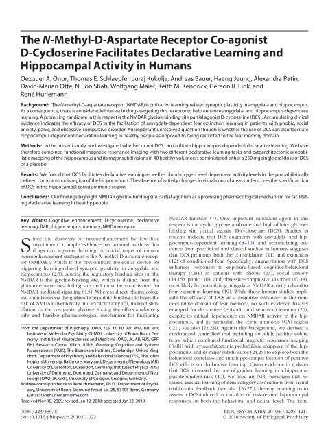
Figure 1. (A) Design of the feedback-guided item-category association task. (i) Subjects made push-button responses to judge the arbitrary category<br />
membership A or B of eight different three-digit numerical items presented repeatedly on-screen. In the first of six cycles, subjects had no knowledge of the<br />
correct category membership of each item and responded by guessing. Visual feedback (a gray circle changing to green for correct responses or to red for<br />
incorrect responses) immediately following each judgment informed subjects about the correct item-category association, thus enabling gradual increases<br />
in performance over subsequent cycles. (ii) A 250-mg single oral dose of D-cycloserine (DCS) decreased the number of cycles required to attain significantly<br />
improved performance compared with initial performance by two cycles (50%), thus resulting in a significant acceleration of learning although ultimate peak<br />
performance levels achieved were similar to the placebo (PLC) group. (B) Imaging data acquired with the item-category association task. (i) <strong>The</strong> probabilistic<br />
region-of-interest analysis revealed a differential effect of DCS over PLC treatment in the cornu ammonis (CA) region of the right hippocampus. (ii) Signal<br />
change courses demonstrated a trial repetition-related increase in CA responses under DCS treatment. (C) Imaging data acquired with the object-location<br />
association task. (i) Interaction contrasts revealed a differential effect of DCS over PLC treatment that probabilistically mapped to the CA region of the left<br />
hippocampus. (ii) <strong>The</strong> relative signal change profiles confirmed a DCS-induced enhancement of CA responses while encoding correct, but not incorrect,<br />
object-location associations. EC, encoding correct; FWE, family-wise error; SUB, subiculum.<br />
were instructed to make a guess if they were uncertain about<br />
object locations.<br />
Visual Stimulation Paradigm. We used a 10 10 checkerboard<br />
with a frequency of green-to-red switches of 8 per second<br />
over a block of 16-sec duration. Six blocks of checkerboard<br />
stimulation alternated with six blocks of rest, where a blank<br />
screen was presented.<br />
Acquisition of Imaging Data<br />
A TIM Trio MRI system (Siemens, Erlangen, Germany) operating<br />
at 3T was used to obtain T2*-weighted echo planar imaging<br />
(EPI) images with blood oxygenation level-dependent (BOLD)<br />
contrast. <strong>The</strong> following imaging parameters were applied for<br />
paradigm 1: repetition time, 2.24 sec; echo time, 30 msec; matrix<br />
size, 100 100; pixel size, 2 2mm 2 ; slice thickness, 2.0 mm;<br />
www.sobp.org/journal
1208 BIOL PSYCHIATRY 2010;67:1205–1211 O.A. Onur et al.<br />
distance factor, 10%; field of view, 200 mm; flip angle, 90°; 36<br />
axial slices (oriented centrally to the hippocampus); parallel<br />
acquisition technique (generalized autocalibrating partially parallel<br />
acquisitions); acquired volumes, 408. <strong>The</strong> following imaging<br />
parameters were applied for paradigms 2 and 3: repetition time,<br />
2.20 sec; echo time, 30 msec; matrix size, 64 64; pixel size,<br />
3.1 3.1 mm 2 ; slice thickness, 3.0 mm; distance factor, 10%; field<br />
of view, 200 mm; flip angle, 90°; 36 axial slices (oriented centrally<br />
to the hippocampus); acquired volumes, 464 (encoding, 172<br />
volumes; retrieval, 292 volumes) (paradigm 2) and 100 (paradigm<br />
3), respectively. <strong>The</strong> first four volumes were discarded to<br />
allow for T1 equilibration effects. In addition, we acquired<br />
high-resolution anatomical magnetic resonance images (T1-<br />
weighted three-dimensional magnetization-prepared rapid acquisition<br />
with gradient echo).<br />
Analysis of Imaging Data<br />
Image preprocessing was performed using Matlab7 (<strong>The</strong><br />
MathWorks, Inc., Natick, Massachusetts) and Statistical Parametric<br />
Mapping 5 (Wellcome Trust Centre for Neuroimaging, London,<br />
United Kingdom; http://www.fil.ion.ucl.ac.uk/spm). <strong>The</strong><br />
EPI images were corrected for head movements between scans<br />
by an affine registration (33). For realignment, a two-pass<br />
procedure was used by which images were initially realigned to<br />
the first image of the time series and subsequently realigned to<br />
the mean of all images. After completing the realignment, the<br />
mean EPI image for each subject was computed and spatially<br />
normalized to the Montreal Neurological Institute (MNI) template<br />
(34–36) using the unified segmentation function in Statistical<br />
Parametric Mapping 5. This algorithm is based on a probabilistic<br />
framework that enables the combination of image registration,<br />
tissue classification, and bias correction within the same generative<br />
model. <strong>The</strong> resulting parameters of a discrete cosine<br />
transform, which define the deformation field necessary to move<br />
the subjects’ data into the space of the MNI tissue probability<br />
maps, were then combined with the deformation field transforming<br />
between the latter and the MNI single subject template. <strong>The</strong><br />
ensuing deformation was subsequently applied to the individual<br />
EPI volumes. All images were hereby transformed into standard<br />
stereotaxic space and resampled at 2 2 2mm 3 voxel size.<br />
<strong>The</strong> normalized images were spatially smoothed using an 8-mm<br />
full-width at half maximum Gaussian kernel.<br />
Item-Category Association Task. An onset regressor was<br />
defined, indicating the onset times of all trials in which a correct<br />
behavioral response was recorded. An additional regressor indexing<br />
the number of repetitions of each stimulus as the<br />
parameter was included. <strong>The</strong> hemodynamic response to this<br />
event type was modeled using a canonical hemodynamic response<br />
function (HRF) and its first derivative, including the six<br />
head movement parameters as confounds. First-level linear<br />
baseline contrast was calculated comparing the regressors with<br />
the implicit baseline. This contrast was then taken to the second<br />
level, where it was subjected to an analysis of variance (ANOVA)<br />
with treatment (DCS vs. PLC treatment) as the between-subject<br />
factor. <strong>The</strong> t test analyses were used to constrain the direction of<br />
the observed effects. Unequal variances were compensated for<br />
by nonsphericity correction. In analogy to previous fMRI studies<br />
of gradual item-category learning that documented a linear<br />
adaptation of hippocampal responses as a function of trial<br />
repetitions (26,27), parametric modulation regressors were set to<br />
1 to test for voxels with a repetition-dependent incline in BOLD<br />
signal amplitude and set to 1 to test for voxels with a<br />
repetition-dependent decline in BOLD signal amplitude. Bidirectional<br />
contrasts (i.e., DCS PLC and PLC DCS) were calculated.<br />
To report parameter estimates separately for each of the six<br />
cycles, a new model was estimated based on separate onset<br />
regressors for each cycle, again including only those trials in<br />
which a correct behavioral response was recorded. For a hypothesis-driven<br />
analysis, the left and right hippocampi were defined<br />
as ROIs (regions-of-interest) based on cytoarchitectonic probability<br />
maps derived from the histological analysis of 10 human<br />
postmortem brains (24,25). We applied corrections for multiple<br />
comparisons based on family-wise error (FWE; significance<br />
threshold p .05). <strong>The</strong> feasibility of this probabilistic ROI<br />
approach has been confirmed by our previous work (37–41).<br />
Object-Location Association Task. We defined four onset<br />
regressors specifying the onset times of encoding- and retrievalrelated<br />
trials in which either correct or incorrect behavioral<br />
responses were recorded. Analogous to previous fMRI studies of<br />
object-location learning (28–30), our analysis focused on stimuli<br />
that were correctly recognized as “old.” Depending on whether<br />
the object-location judgment for these stimuli succeeded or<br />
failed, encoding (E) and retrieval (R) trials were classified as<br />
either correct (EC, RC) or false (EF, RF). <strong>The</strong> hemodynamic<br />
response to each of these four different event types (subsequently<br />
referred to as accuracy) was modeled using a canonical<br />
HRF and its first derivative, including the six head movement<br />
parameters as confounds. First-level linear baseline contrasts<br />
were calculated comparing each onset regressor with the implicit<br />
baseline. <strong>The</strong>se contrasts were then taken to the second level,<br />
where they were subjected to an ANOVA with accuracy as the<br />
within-subject factor and treatment as the between-subject factor.<br />
<strong>The</strong> t test analyses were used to constrain the direction of the<br />
observed effects. Unequal variances were compensated for by<br />
nonsphericity correction. Interactive contrasts were calculated<br />
separately for the encoding and retrieval phase, followed by an<br />
FWE-corrected probabilistic ROI analysis.<br />
Visual Stimulation Paradigm. <strong>The</strong> visual stimulation was<br />
modeled by a boxcar function convolved with a canonical HRF.<br />
A design matrix comprising contrasts of alternating intervals of<br />
visual stimulation and rest, the time derivative, and the six head<br />
movement parameters as confounds was created. A first-level<br />
linear baseline contrast was calculated by comparing the boxcar<br />
function with the implicit baseline. This contrast was then taken<br />
to the second level, where it was subjected to an ANOVA with<br />
treatment as the between-subject factor. Unequal variances were<br />
compensated for by nonsphericity correction. Again, bidirectional<br />
contrasts were calculated using t test analyses.<br />
Results<br />
Behavior<br />
Item-Category Association Task. In postscan interviews, 11<br />
subjects reported that scanner noise had made the task too<br />
challenging for them. This was confirmed by near-floor behavioral<br />
performance, i.e., response accuracy was not greater than<br />
chance in all cases. <strong>Co</strong>nsequently, the behavioral and fMRI data<br />
acquired from these subjects had to be discarded from subsequent<br />
analyses of this task. For the remaining 29 subjects (n 15<br />
DCS; n 14 PLC), a two-way repeated measures ANOVA with<br />
treatment group (PLC vs. DCS) as between-subject factor and<br />
cycle as within-subject factor revealed a main effect of group<br />
[F(1,27) 5.454; p .027] and a main effect of cycle [F(5,135) <br />
8.696; p .0001] on performance but no group cycle<br />
interaction effect [F(5,135) .233; p .05]. This indicates that<br />
treatment with a 250-mg single oral dose of DCS induced a<br />
www.sobp.org/journal
O.A. Onur et al. BIOL PSYCHIATRY 2010;67:1205–1211 1209<br />
general improvement of performance. In addition, one-way repeated<br />
measures ANOVAs showed that both the PLC [F(5,70) <br />
4.413; p .001] and DCS treatment groups [F(5,70) 5.412; p <br />
.001] significantly improved performance across cycles. Post hoc<br />
multiple comparisons between performance on cycle 1 and<br />
subsequent cycles using the Bonferroni paired t test revealed that<br />
for the DCS-treatment group performance on cycles 4 [t (14) <br />
3.812; p .004], 5 [t (14) 3.442; p .015], and 6 [t (14) 3.607;<br />
p .009] was significantly improved but that there were no<br />
significant differences between performance on these last three<br />
cycles (all p values .05). By contrast, in the PLC group, only<br />
performance on cycle 6 was significantly improved compared<br />
with cycle 1 [t (13) 3.386; p .018], although cycle 5 nearly<br />
achieved this [t (13) 2.944; p .067]. <strong>The</strong>se results imply that<br />
learning speed was considerably faster in the DCS-treated group.<br />
Results are shown in Figure 1A(ii). Neither reaction times nor<br />
response misses differed between the PLC and DCS treatment<br />
groups (Figure S1 in Supplement 1). Furthermore, treatment had<br />
no differential effect on performance in the control condition,<br />
with all 29 subjects achieving a response accuracy of nearly 100%<br />
(all p values .05).<br />
Object-Location Association Task. In contrast to the itemcategory<br />
association task, no subjects reported any difficulty in<br />
performing the object-location association task in postscan interviews<br />
and this was confirmed by their performance scores.<br />
Two-sample t tests confirmed that both groups (n 20 DCS; n <br />
20 PLC) performed almost identically on the retrieval of objectlocation<br />
associations [PLC group, 48.1 13.8%; DCS group,<br />
46.3 15.1%; t (38) .48; p .05]. Further, recognition of new<br />
objects did not differ between treatment groups [PLC group,<br />
82.7 14.7%; DCS group, 81.9 13.7%; t (38) 1.11; p .05].<br />
Neither reaction times nor response misses differed between<br />
treatment groups (all p values .05).<br />
Imaging<br />
Item-Category Association Task. <strong>The</strong> probabilistic ROI analysis<br />
demonstrated a parametric effect of DCS over PLC treatment<br />
in the CA region of the right hippocampus (MNI coordinates<br />
xyz 20, 11, 20; p .05, FWE-corrected) (Figure 1B[i]). In<br />
the DCS group, but not in the PLC group, this CA response<br />
increased across cycles (Figure 1B[ii]). Thus, the faster learning<br />
seen in the item-category association task following DCS administration<br />
was paralleled by increased CA activity.<br />
Object-Location Association Task. We found an interaction<br />
effect with the within-subject factor accuracy (EC vs. EF) and<br />
the between-subject factor treatment, which was restricted to the<br />
encoding phase of the task and probabilistically mapped to the<br />
left hippocampal CA region (MNI coordinates xyz 36, 10,<br />
21; p .05, FWE-corrected) (Figure 1C[i–ii]). An across-group<br />
main effect of accuracy (RC vs. RF) was restricted to the retrieval<br />
phase of the task, evident in robust bilateral hippocampal<br />
activation (Table S2 in Supplement 1). No further suprathreshold<br />
effects occurred in this analysis.<br />
Visual Stimulation Paradigm. Within-group analyses showed<br />
robust neural responses to checkerboard stimulation in bilateral<br />
visual cortex. However, between-group comparisons revealed<br />
no differential activations (Figure S2 in Supplement 1). This<br />
argues against nonspecific DCS effects resulting from a global<br />
potentiation of NMDAR activity or homogeneous changes in<br />
cerebral hemodynamics.<br />
Discussion<br />
In the present study, we combined conventional fMRI with<br />
cytoarchitectonic probabilistic mapping to make an initial attempt<br />
at a subdivision-level investigation of the effects of a<br />
250-mg single oral dose of DCS on hippocampal function probed<br />
with two declarative learning tasks. Parametric analysis of fMRI<br />
data acquired with the item-category association task revealed<br />
that DCS enhanced hippocampal activity, an effect that probabilistically<br />
mapped to the CA region. Specifically, the corresponding<br />
signal change courses illustrate that DCS elevated CA activity<br />
across trial repetitions, whereas CA activity remained at baseline<br />
in the PLC group. This profile supports the hypothesis that DCS<br />
may increase the efficiency of learning by indirectly upregulating<br />
glutamate signaling via NMDAR to above-threshold levels,<br />
thereby recruiting previously silent CA synapses for the benefit of<br />
faster learning (12,42). <strong>The</strong> latter is evident at the behavioral<br />
level, where DCS reduced the number of trial repetitions required<br />
to attain significant improvement compared with initial<br />
performance by two cycles (50%), hence resulting in an overall<br />
significant enhancement of declarative learning, although ultimate<br />
peak performance levels achieved were similar to the<br />
control group and reaction times were unaffected. Our results are<br />
thus compatible with a report of a 50% reduction of trial<br />
repetitions in DCS-treated rabbits tested on a hippocampusdependent<br />
gradual associative learning task (10).<br />
Our findings raise the crucial question as to whether DCS<br />
modulates item-category associative learning by facilitating encoding-<br />
and/or retrieval-related operations in the hippocampal<br />
CA region. To determine whether one or both of these operations<br />
are susceptible to DCS action, subjects also completed an<br />
object-location association task, which tested encoding and<br />
retrieval of object-location associations separately from each<br />
other (28–30). In rodents, object-location learning engages the<br />
hippocampal area CA1 and is enhanced by DCS treatment (23).<br />
However, consistent with the observed lack of a DCS behavioral<br />
effect on peak performance in item-category associative learning,<br />
no overall beneficial effect was found in the object-location<br />
association task either. Nevertheless, interaction contrasts revealed<br />
a differential effect of DCS over PLC that again projected<br />
to the hippocampal CA region and was exclusively restricted to<br />
the encoding phase of the task. From these results, it appears that<br />
DCS enhanced CA responses during encoding of object-location<br />
associations but did not affect retrieval of these associations.<br />
One important consideration in this context is the differentiation<br />
between a specific facilitation of learning-related NMDAR<br />
activity in the hippocampal CA region as opposed to nonspecific<br />
DCS effects resulting from a global potentiation of NMDAR<br />
activity or homogeneous changes in cerebral hemodynamics. To<br />
control for potential nonspecific DCS effects, all subjects were<br />
scanned on a checkerboard visual stimulation paradigm. Although<br />
the visual cortex is clearly an important site of NMDARmediated<br />
synaptic plasticity (43), between-group comparisons<br />
failed to find a differential effect of DCS on visual cortical<br />
responses to checkerboard stimulation. This absence of an effect<br />
supports the notion of a specific facilitative influence of DCS on<br />
learning-related CA activity. Thus, the present pharmacological<br />
fMRI study is the first to suggest that a 250-mg single oral dose of<br />
DCS improves declarative learning in healthy people through<br />
selective enhancement of hippocampal CA activity.<br />
Despite substantial evidence from behavioral studies in rodents<br />
that DCS facilitates performance in hippocampus-dependent<br />
learning tasks (10,22,23), behavioral studies in humans have<br />
www.sobp.org/journal
1210 BIOL PSYCHIATRY 2010;67:1205–1211 O.A. Onur et al.<br />
failed so far to demonstrate beneficial effects of DCS in tests of<br />
immediate and delayed recall of verbal and nonverbal items<br />
(7,44), suggesting that the facilitative influence of DCS on<br />
hippocampal function is limited (20). However, we note that<br />
these negative studies used a substantially lower dose of DCS (50<br />
mg) than the one we administered in our study (250 mg).<br />
Support for a dose-dependent variation in the efficacy of DCS<br />
comes from two fear extinction studies: Ledgerwood et al. (45)<br />
found effect sizes of .54, .80, and 1.43 for DCS versus PLC in<br />
rodents treated with 2.5, 5, and 10 mg, respectively, whereas<br />
Ressler et al. (13) found effect sizes of .36 and .86 for DCS versus<br />
PLC in phobic patients treated with 50 mg and 500 mg, respectively.<br />
Given this empirical background, dosing, frequency, and<br />
chronicity of DCS treatment appear to be critical as to whether<br />
the use of DCS can be expanded to promote hippocampusdependent<br />
declarative learning, aside from its experimental<br />
application in exposure-based CBT. Current hypotheses regarding<br />
the facilitation of declarative learning with DCS emphasize its<br />
potential to alleviate age-associated cognitive decline and to<br />
augment clinical response to CBT in patients, where a greater<br />
emphasis is placed on cognitive restructuring techniques, informational<br />
strategies, and skill acquisition interventions (44). However,<br />
if DCS indeed facilitates declarative learning by activating<br />
previously silent CA synapses, as is suggested by our results, it<br />
may be ineffective in conditions characterized by progressive<br />
hippocampal degeneration and synaptic loss (46,47).<br />
RH was supported by a German Research Foundation Grant<br />
(HU1302/2-2) and by a Starting Independent Researcher Grant<br />
jointly provided by the Ministry of Innovation, Science, Research<br />
and Technology of the State of North Rhine-Westphalia and the<br />
University of Bonn. KMK was supported by the Biotechnology<br />
and Biological Sciences Research <strong>Co</strong>uncil.<br />
We thank M.X. <strong>Co</strong>hen for helpful comments on a previous<br />
version of the manuscript and K. Linkert and A. Kirsch for<br />
research assistance.<br />
<strong>The</strong> authors report no biomedical financial interests or potential<br />
conflicts of interest.<br />
ClinicalTrials.gov: <strong>The</strong> influence of glutamate on memory in<br />
humans; http://clinicaltrials.gov; NCT00980408.<br />
Supplementary material cited in this article is available<br />
online.<br />
1. Lashley KS (1917): <strong>The</strong> effects of strychnine and caffeine upon the rate of<br />
learning. Psychobiology 1:141–170.<br />
2. Lee YS, Silva AJ (2009): <strong>The</strong> molecular and cellular biology of enhanced<br />
cognition. Nat Rev Neurosci 10:126–140.<br />
3. Li F, Tsien JZ (2009): Memory and the NMDA receptors. N Engl J Med<br />
361:302–303.<br />
4. Johnson JW, Ascher P (1987): Glycine potentiates the NMDA response in<br />
cultured mouse brain neurons. Nature 325:529–531.<br />
5. Kleckner NW, Dingledine R (1988): Requirement for glycine in activation<br />
of NMDA-receptors expressed in Xenopus oocytes. Science 241:835–<br />
837.<br />
6. Hardingham GE (2009): <strong>Co</strong>upling of the NMDA receptor to neuroprotective<br />
and neurodestructive events. Biochem Soc Trans 37:1147–1160.<br />
7. D’Souza DC, Gil R, Cassello K, Morrissey K, Abi-Saab D, White J, et al.<br />
(2000): IV glycine and oral D-cycloserine effects on plasma and CSF<br />
amino acids in healthy humans. Biol Psychiatry 47:450–462.<br />
8. Monahan JB, Handelmann GE, Hood WF, <strong>Co</strong>rdi AA (1989): D-cycloserine,<br />
a positive modulator of the N-methyl-D-aspartate receptor, enhances<br />
performance of learning tasks in rats. Pharmacol Biochem Behav 34:649–<br />
653.<br />
9. Flood J, Morley J, Lanthorn T (1992): Effect on memory processing by<br />
D-cycloserine, an <strong>agonist</strong> of the NMDA/glycine receptor. Eur J Pharmacol<br />
221:249–254.<br />
10. Thompson LT, Moskal JR, Disterhoft JF (1992): Hippocampus-dependent<br />
learning facilitated by a monoclonal antibody or D-cycloserine.<br />
Nature 359:638–641.<br />
11. Kalisch R, Holt B, Petrovic P, De Martino B, Kloeppel S, Buechel C, Dolan<br />
RJ (2009): <strong>The</strong> NMDA <strong>agonist</strong> D-cycloserine facilitates fear memory consolidation<br />
in humans. Cereb <strong>Co</strong>rtex 19:187–196.<br />
12. Norberg MM, Krystal JH, Tolin DF (2008): A meta-analysis of D-cycloserine<br />
and the facilitation of fear extinction and exposure therapy.<br />
Biol Psychiatry 63:1118–1126.<br />
13. Ressler KJ, Rothbaum BO, Anderson P, Zimand E, Tannenbaum L,<br />
Hodges L, Davis M (2004): D-cycloserine, a putative cognitive enhancer,<br />
accelerates extinction of fear in humans. Arch Gen Psychiatry 61:1136–<br />
1144.<br />
14. Hofmann SG, Meuret AE, Smits JA, Simon NM, Pollack MH, Eisenmenger<br />
K, et al. (2006): Augmentation of exposure therapy with D-cycloserine<br />
for social anxiety disorder. Arch Gen Psychiatry 63:298–304.<br />
15. Guastella AJ, Richardson R, Lovibond PF, Rapee RM, Gaston JE, Mitchell<br />
P, Dadds MR (2008): A randomized controlled trial of D-cycloserine<br />
enhancement of exposure therapy for social anxiety disorder. Biol Psychiatry<br />
63:544–549.<br />
16. Otto MW, Tolin DF, Simon NM, Pearlson GD, Basden S, Meunier SA, et al.<br />
(2010): Efficacy of D-cycloserine for enhancing response to cognitivebehavior<br />
therapy for panic disorder. Biol Psychiatry 67:365–370.<br />
17. Kushner MG, Kim SW, Donahue C, Thuras P, Adson D, Kotlyar M, et al.<br />
(2007): D-cycloserine augmented exposure therapy for obsessive-compulsive<br />
disorder. Biol Psychiatry 62:835–838.<br />
18. Wilhelm S, Buhlmann U, Tolin DF, Meunier SA, Pearlson GD, Reese HE, et<br />
al. (2008): Augmentation of behavior therapy with D-cycloserine for<br />
obsessive-compulsive disorder. Am J Psychiatry 165:335–341.<br />
19. Davis M, Ressler K, Rothbaum BO, Richardson R (2006): Effects of D-<br />
cycloserine on extinction: Translation from preclinical to clinical work.<br />
Biol Psychiatry 60:369–375.<br />
20. Grillon C (2009): D-cycloserine facilitation of fear extinction and exposure-based<br />
therapy might rely on lower-level, automatic mechanisms.<br />
Biol Psychiatry 66:636–641.<br />
21. Grunwald T, Beck H, Lehnertz K, Blümcke I, Pezer N, Kurthen M, et al.<br />
(1999): Evidence relating human verbal memory to hippocampal N-<br />
methyl-D-aspartate receptors. Proc Natl Acad SciUSA96:12085–12089.<br />
22. Quartermain D, Mower J, Rafferty M, Hering R, Lanthorn T (1994): Acute<br />
but not chronic activation of the NMDA-coupled glycine receptor with<br />
D-cycloserine facilitates learning and retention. Eur J Pharmacol 257:<br />
7–12.<br />
23. Assini FL, Duzzioni M, Takahashi RN (2009): Object location memory in<br />
mice: Pharmacological validation and further evidence of hippocampal<br />
CA1 participation. Behav Brain Res 204:206–211.<br />
24. Amunts K, Kedo O, Kindler M, Pieperhoff P, Mohlberg H, Shah NJ, et al.<br />
(2005): Cytoarchitectonic mapping of the human amygdala, hippocampal<br />
region and entorhinal cortex: Intersubject variability and probability<br />
maps. Anat Embryol (Berl) 210:343–352.<br />
25. Eickhoff SB, Stephan KE, Mohlberg H, Grefkes C, Fink GR, Amunts K, Zilles<br />
K (2005): A new SPM toolbox for combining probabilistic cytoarchitectonic<br />
maps and functional imaging data. Neuroimage 25:1325–1335.<br />
26. Strange BA, Fletcher PC, Henson RN, Friston KJ, Dolan RJ (1999): Segregating<br />
the functions of human hippocampus. Proc Natl Acad Sci USA<br />
96:4034–4039.<br />
27. Strange BA, <strong>Hurlemann</strong> R, Duggins A, Heinze HJ, Dolan RJ (2005): Dissociating<br />
intentional learning from relative novelty responses in the medial<br />
temporal lobe. Neuroimage 25:51–62.<br />
28. Cansino S, Maquet P, Dolan RJ, Rugg MD (2002): Brain activity underlying<br />
encoding and retrieval of source memory. Cereb <strong>Co</strong>rtex 12:1048–<br />
1056.<br />
29. Kukolja J, Thiel CM, Wilms M, Mirzazade S, Fink GR (2009): Ageingrelated<br />
changes of neural activity associated with spatial contextual<br />
memory. Neurobiol Aging 30:630–645.<br />
30. Kukolja J, Thiel CM, Fink GR (2009): Cholinergic stimulation enhances<br />
neural activity associated with encoding but reduces neural activity<br />
associated with retrieval in humans. J Neurosci 29:8119–8128.<br />
31. Nair KGS, Epstein IG, Baron H, Mulinos MG (1956): Absorption, distribution<br />
and excretion of cycloserine in man. Antibiot Annu 3:136–140.<br />
www.sobp.org/journal
O.A. Onur et al. BIOL PSYCHIATRY 2010;67:1205–1211 1211<br />
32. Holdiness MR (1985): Cerebrospinal fluid pharmacokinetics of the antituberculosis<br />
drugs. Clin Pharmacokinet 10:532–534.<br />
33. Ashburner J, Friston KJ (2003): Rigid body registration. In: Frackowiak RS,<br />
Friston KJ, Frith CD, Dolan RJ, Price CJ, Ashburner J, et al., editors. Human<br />
Brain Function, 2nd ed. London, UK: Academic Press, 635–655.<br />
34. Evans AC, Marrett S, Neelin P, <strong>Co</strong>llins L, Worsley K, Dai W, et al. (1992):<br />
Anatomical mapping of functional activation in stereotactic coordinate<br />
space. Neuroimage 1:43–53.<br />
35. <strong>Co</strong>llins DL, Neelin P, Peters TM, Evans AC (1994): Automatic 3D intersubject<br />
registration of MR volumetric data in standardized Talairach space.<br />
J <strong>Co</strong>mput Assist Tomogr 18:192–205.<br />
36. Holmes CJ, Hoge R, <strong>Co</strong>llins L, Woods R, Toga AW, Evans AC (1998):<br />
Enhancement of MR images using registration for signal averaging.<br />
J <strong>Co</strong>mput Assist Tomogr 22:324–333.<br />
37. <strong>Hurlemann</strong> R, Rehme AK, Diessel M, Kukolja J, Maier W, Walter H, <strong>Co</strong>hen<br />
MX (2008): Segregating intra-amygdalar responses to dynamic facial<br />
emotion with cytoarchitectonic maximum probability maps. J Neurosci<br />
Methods 172:13–20.<br />
38. Kukolja J, Schlaepfer TE, Keysers C, Klingmüller D, Maier W, Fink GR,<br />
<strong>Hurlemann</strong> R (2008): Modeling a negative response bias in the human<br />
amygdala by noradrenergic-glucocorticoid interactions. J Neurosci 28:<br />
12868–12876.<br />
39. Goossens L, Kukolja J, Onur OA, Fink GR, Maier W, Griez E, et al. (2009):<br />
Selective processing of social stimuli in the superficial amygdala. Hum<br />
Brain Mapp 30:3332–3338.<br />
40. Onur OA, Walter H, Schlaepfer TE, Rehme AK, Schmidt C, Keysers C, et al.<br />
(2009): Noradrenergic enhancement of amygdala responses to fear. Soc<br />
<strong>Co</strong>gn Affect Neurosci 4:119–126.<br />
41. <strong>Hurlemann</strong> R, Walter H, Rehme AK, Kukolja J, Santoro SC, Schnell K, et al.<br />
(2010): Human amygdala activity is diminished by the beta-noradrenergic<br />
ant<strong>agonist</strong> propranolol [published online ahead of print January 27].<br />
Psychol Med.<br />
42. Gomperts SN, Rao A, Craig AM, Malenka RC, Nicoll RA (1998): Postsynaptically<br />
silent synapses in single neuron cultures. Neuron 21:1443–<br />
1451.<br />
43. Artola A, Singer W (1987): Long-term potentiation and NMDA receptors<br />
in rat visual cortex. Nature 330:49–52.<br />
44. Otto MW, Basden SL, McHugh RK, Kantak KM, Deckersbach T, Cather C,<br />
et al. (2009): Effects of D-cycloserine administration on weekly nonemotional<br />
memory tasks in healthy participants. Psychother Psychosom 78:<br />
49–54.<br />
45. Ledgerwood L, Richardson R, Cranney J (2003): Effects of D-cycloserine<br />
on extinction of conditioned freezing. Behav Neurosci 117:<br />
341–349.<br />
46. Laake K, Oeksengaard AR (2002): D-cycloserine for Alzheimer’s disease.<br />
<strong>Co</strong>chrane Database Syst Rev 2:CD003153.<br />
47. Rosenzweig ES, Barnes CA (2003): Impact of aging on hippocampal<br />
function: Plasticity, network dynamics, and cognition. Prog Neurobiol<br />
69:143–179.<br />
www.sobp.org/journal