Journal of Medical Society, Sept 2008
Journal of Medical Society, Sept 2008
Journal of Medical Society, Sept 2008

Create successful ePaper yourself
Turn your PDF publications into a flip-book with our unique Google optimized e-Paper software.
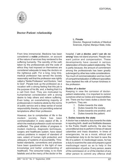
EDITORIAL<br />
Doctor-Patient relationship<br />
L. Fimate<br />
Director, Regional Institute <strong>of</strong> <strong>Medical</strong><br />
Sciences, Imphal, Manipur State, India.<br />
From time immemorial, Medicine has been<br />
considered a noble pr<strong>of</strong>ession, on account<br />
<strong>of</strong> the nature <strong>of</strong> services they rendered to the<br />
suffering humanity. The sanctity <strong>of</strong> the oath<br />
taken by the pr<strong>of</strong>essionals and the code <strong>of</strong><br />
ethics they had imposed on themselves are<br />
considered adequate to keep the doctors on<br />
the righteous path. For a long, long time,<br />
medical pr<strong>of</strong>ession has served the society<br />
reasonably well that the pr<strong>of</strong>ession was rightly<br />
called a “Noble Pr<strong>of</strong>ession” and Doctors, “next<br />
to God”. People took up the pr<strong>of</strong>ession as a<br />
vocation with a strong feeling that this job is<br />
the purpose <strong>of</strong> his life, and, a feeling that it is<br />
a call from God. They are motivated by<br />
humanitarian consideration with a strong<br />
desire to help others and relieve suffering.<br />
Even today, an overwhelming majority <strong>of</strong><br />
pr<strong>of</strong>essionals in medicine abide by the norms<br />
<strong>of</strong> public service and a deep sense <strong>of</strong> social<br />
responsibility thereby not permitting external<br />
temptations affect their pr<strong>of</strong>ession.<br />
However, due to complexities <strong>of</strong> life in the<br />
modem society, there has been<br />
commercialization in every aspect <strong>of</strong> life, in<br />
which the medical pr<strong>of</strong>ession could not remain<br />
unaffected. The great strides <strong>of</strong> progress in<br />
modem medicine, diagnostic techniques,<br />
surgery and healthcare system, have raised<br />
various problems in respect <strong>of</strong> standard <strong>of</strong><br />
care, extent <strong>of</strong> human right protection and<br />
adequacy <strong>of</strong> systems <strong>of</strong> accountability. Timetested<br />
standards, conventions and practices<br />
have been questioned in the light <strong>of</strong> new<br />
knowledge and better understanding <strong>of</strong><br />
healthcare. The consumer today, is no more<br />
fatalistic and at the mercy <strong>of</strong> the doctor or the<br />
JMS * Vol 22 * No. 3 * <strong>Sept</strong>ember, <strong>2008</strong><br />
hospital. I am a doctor, and I can do no<br />
wrong’ does not stand anymore. People now<br />
want justice and compensation. These<br />
developments have caused in serious<br />
deterioration <strong>of</strong> Doctor-patient relationship. This<br />
is partly because, the amount <strong>of</strong> commitment<br />
among the pr<strong>of</strong>essionals has been greatly<br />
submerged by other less noble considerations.<br />
Too much <strong>of</strong> commercialization and too much<br />
<strong>of</strong> compartmentalization <strong>of</strong> different specialties<br />
have depleted the milk <strong>of</strong> human kindness in<br />
the pr<strong>of</strong>ession.<br />
Duties <strong>of</strong> a doctor<br />
Keeping in view the corrosion <strong>of</strong> doctorpatient<br />
relationship, it is important to remind<br />
ourselves what our duties and responsibilities<br />
are. There are four basic duties a doctor has<br />
to perform. They are:<br />
1. Duties towards the state<br />
2. Duties towards the society,<br />
3. Duties towards one another, and<br />
4. Duties towards the patient.<br />
1. Duties towards the state<br />
A doctor has a statutory duty towards the state<br />
in reporting birth, death and Notifiable diseases<br />
to the Public Health authority. He also has<br />
unconditional duty to perform in times <strong>of</strong> natural<br />
calamities and mass disasters. In times <strong>of</strong><br />
emergencies, he as a moral, ethical and<br />
humanitarian duty to do his best to save the<br />
life <strong>of</strong> a patient. In medicolegal cases, he has<br />
a duty to examine the case and prepare a good<br />
medicolegal report so as to help in the<br />
administration <strong>of</strong> justice. Every person, aware<br />
<strong>of</strong> the commission <strong>of</strong>, or <strong>of</strong> the intention <strong>of</strong> any<br />
other person to commit any <strong>of</strong>fence shall<br />
107
EDITORIAL<br />
forthwith give information to the nearest<br />
Magistrate or Police <strong>of</strong>ficer <strong>of</strong> such<br />
commission or intention.<br />
2. Duties towards the society<br />
A doctor has a special duty to teach and<br />
demonstrate a healthful living in a society.<br />
3. Duty towards one another<br />
Doctors have to cooperate with one another.<br />
Never to criticize or condemn a colleague but<br />
should extend to him the same honour, respect<br />
good behavior and cordiality as he would<br />
expect from them.<br />
4. Duties towards the patient<br />
As soon as a doctor undertakes to treat a<br />
patient, he becomes duty bound to exercise a<br />
reasonable degree <strong>of</strong> skill, care and<br />
competence in his treatment. There has been<br />
an implied form <strong>of</strong> contract that he will do his<br />
best for the welfare <strong>of</strong> his patient. He need not<br />
guarantee a cure, nor will he be liable for an<br />
error <strong>of</strong> judgment. He should always be<br />
courteous, sympathetic, friendly and helpful.<br />
He should use his utmost skill, care and<br />
competence to cure his patients. Even when<br />
he fails to cure, the patient should not have<br />
any doubt about the sincerity <strong>of</strong> the doctor. He<br />
should always be a source <strong>of</strong> confidence and<br />
comfort to the sick. When a patient fails to<br />
see these conducts in a doctor, suspicion<br />
arose, faith falters. Doubt, challenges,<br />
litigations, and, even assaults occurred. In<br />
recent times, we have seen several<br />
embarrassing newspaper reports: “A doctor<br />
charged with homicide”, “Let Doctors<br />
pay”, “Hospital blunder” “Doctor in the<br />
Dock” “Neurosurgeon asked to pay Rs.5<br />
lakhs” “Doctor assaulted after patient’s<br />
death”, “Forceps left in the patient’s<br />
stomach after operation” etc.<br />
Remedy<br />
The best way to avoid all these problems is to<br />
observe the CODE OF MEDICAL ETHICS<br />
very scrupulously. We should practice our<br />
pr<strong>of</strong>ession with conscience and dignity,<br />
maintaining utmost respect to human life. Let<br />
us try to maintain the honour and noble tradition<br />
<strong>of</strong> our pr<strong>of</strong>ession by giving the health <strong>of</strong> our<br />
patient our first consideration. Let reward and<br />
financial gain be our secondary consideration.<br />
An ideal physician should be upright, pure in<br />
108<br />
character, diligent and dedicated in caring for<br />
the sick. He should be sober, patient, prompt<br />
in discharging duty without anxiety, conducting<br />
himself with propriety, humble, friendly and<br />
sympathetic. Let patient be considered the<br />
most important visitor <strong>of</strong> the day and, be<br />
hospitable to him. Holistic approach to the<br />
patient is the need <strong>of</strong> the hour in this era <strong>of</strong><br />
litigation where every patient becomes a<br />
potential going-to-court case. One should<br />
realize that “healing is not merely curing<br />
certain disease”. It is not like repairing and<br />
removing the defect <strong>of</strong> a machine: it deals with<br />
a person. So, let us be human, not mechanical<br />
in our approach. William Boyd has said, “The<br />
patient with a heart disease is not just an<br />
internal combustion engine with a leaking<br />
valve, but a sensitive human being with a<br />
disease heart”. Therefore, disease in man is<br />
not exactly the same as disease in<br />
experimental animal, for in man the emotion<br />
comes into play.<br />
Mushrooming <strong>of</strong> specialties and superspecialties<br />
in the modern medical science has<br />
complicated patient management. It has created<br />
too much <strong>of</strong> compartmentalization that, a time<br />
may come when a doctor is called a specialist<br />
in right kidney, and not for the left. Such<br />
multiple super-specialties have diluted the<br />
emotion and tend to be mechanical. The milk <strong>of</strong><br />
human kindness in a doctor has been<br />
submerged in a robotic approach <strong>of</strong> a doctor,<br />
thus shattering the bond <strong>of</strong> love, friendship and<br />
trust between a doctor and the patient.<br />
To bring back the past glory <strong>of</strong> the pr<strong>of</strong>ession,<br />
and restore the love and trust between doctor<br />
and the patient, let us administer a<br />
Reasonable degree <strong>of</strong> care to the patient –<br />
spend sometime with the patient everyday,<br />
explain his problems, be friendly, give comfort,<br />
let him not feel neglected, try to understand<br />
his problems. Give to your patient what you<br />
want a doctor to give when you are a patient.<br />
A sense <strong>of</strong> commitment and a sense <strong>of</strong><br />
stewardship are essential. To keep ourselves<br />
free from the slavery <strong>of</strong> covetousness,<br />
Let us accept our work,<br />
Not only as an occupation or even a pr<strong>of</strong>ession,<br />
But also as a CALLING-A VOCATION<br />
JMS * Vol 22 * No. 3 * <strong>Sept</strong>ember, <strong>2008</strong>
EDITORIAL<br />
Guest Editor<br />
Do we need re-examining undergraduate <strong>Medical</strong> Education<br />
Pr<strong>of</strong>. M. Amuba Singh<br />
Co-ordinator, <strong>Medical</strong> Education<br />
Unit, RIMS, Imphal<br />
Let us analyse the post-independence<br />
scenario <strong>of</strong> medical education in our country.<br />
There seems to be an overwhelming<br />
emphasis in promoting the production <strong>of</strong><br />
medical personnel. This has been well<br />
evidenced by the increasing number <strong>of</strong><br />
medical colleges throughout the country. At<br />
present, the annual output <strong>of</strong> MBBS doctors<br />
exceeds twenty five thousand. Are we not<br />
overproducing doctors in the country? The<br />
matter needs to be examined carefully. On the<br />
other hand, the quality <strong>of</strong> medical training<br />
programme has not been given due attention<br />
as it should be. The training programme for<br />
the medical students should be well planned<br />
so that it is relevant, useful and responsive to<br />
the needs <strong>of</strong> the common people/ society. If<br />
we examine teaching - training programmes/<br />
schedules for the MBBS students, there lies<br />
a number <strong>of</strong> points to be clarified.<br />
The curriculum needs to be planned properly.<br />
If we happen to examine the present curricula,<br />
it needs analysing common disease spectrum<br />
seen at outpatient and inpatient setups at<br />
primary health centres, district hospitals,<br />
general practitioners and medical college<br />
hospital. The emphasis in our training should<br />
match fairly with the common and important<br />
health problems <strong>of</strong> the society. It will be<br />
desirable to expose medical students to health<br />
problems throughout the period <strong>of</strong><br />
undergraduate training. 1<br />
Each department concerned should<br />
identify its own goals, objectives and core<br />
abilities. It should also be clearly<br />
mentioned as well as documented and be<br />
JMS * Vol 22 * No. 3 * <strong>Sept</strong>ember, <strong>2008</strong><br />
available to the medical students.<br />
It has been seen that the present system <strong>of</strong><br />
teaching is not active. This needs to be<br />
modified so that there is active learning where<br />
the participation <strong>of</strong> students or the learner is<br />
<strong>of</strong> utmost importance. Simply attending or<br />
listening to didactic lecture classes will not be<br />
appropriate.<br />
There should be provision for integrated<br />
learning along with other health pr<strong>of</strong>essionals<br />
from within the medical institute as well as from<br />
outside. This is needed to impart composite<br />
knowledge on health instead <strong>of</strong> fragmented<br />
and piece meal approach.<br />
The undergraduate medical education should<br />
be oriented towards training <strong>of</strong> medical<br />
students to become a physician <strong>of</strong> first hand<br />
contact who is capable <strong>of</strong> looking after the<br />
preventive, promotive, curative and<br />
rehabilitative aspects <strong>of</strong> medicine. 2 The<br />
graduate doctor should possess the skills to<br />
plan and manage community health<br />
programmes. Also he/she should have clear<br />
understanding <strong>of</strong> his / her role in different<br />
National Health Programmes.<br />
The basic doctor should invariably possess<br />
the requisite knowledge and skills to manage<br />
efficiently the clinical emergencies and<br />
medicolegal cases.<br />
The medical personnel/ doctor should also<br />
require the leadership and managerial skills<br />
to carry out his works with the health team <strong>of</strong><br />
primary health care centre or any health care<br />
set up. The National Health Policy (NHP)<br />
109
EDITORIAL<br />
decries the wrong priorities that crept into<br />
health care service <strong>of</strong> the country and clearly<br />
points to prepare doctors who will serve rural<br />
and urban settings and who will not give<br />
emphasis on curative aspects <strong>of</strong> medicine<br />
alone. 3<br />
The training <strong>of</strong> the undergraduates should be<br />
such that once graduated, he should develop<br />
humanistic attitudes in practising medicine.<br />
The curriculum contents for teaching the<br />
medical students should contain proper<br />
guideline for evaluation <strong>of</strong> students through a<br />
valid and reliable system which can assess<br />
the minimum skills/ care abilities <strong>of</strong> the<br />
learners.<br />
These are only a few facets <strong>of</strong> ongoing<br />
undergraduate medical teaching programme.<br />
Besides these, internship programme,<br />
methods <strong>of</strong> teaching, and use <strong>of</strong> newer and<br />
effective teaching aids etc. may be looked into<br />
properly.<br />
Some <strong>of</strong> the suggestions for a change or<br />
modification in the curriculum <strong>of</strong> the undergraduate<br />
medical education may be mentioned.<br />
- Faculty sensitisation and development<br />
- Discussion with student representatives<br />
- Identification <strong>of</strong> tasks <strong>of</strong> graduate doctor<br />
- Review and Revision <strong>of</strong> question papers<br />
with due approval from the university with<br />
a view to setting question papers properly<br />
balanced in terms <strong>of</strong> course content and<br />
its relevancy<br />
- Designing and Restructuring <strong>of</strong> evaluation<br />
tools/ Technique<br />
- Implementation <strong>of</strong> teaching innovations.<br />
- Continuous monitoring <strong>of</strong> changes by<br />
Feedback from students and faculty and<br />
modify if needed.<br />
<strong>Medical</strong> Education unit / cell <strong>of</strong> the Institute/<br />
College may be given the responsibility for coordinating<br />
and helping departments in<br />
successful implementation and achieving<br />
useful changes as suggested.<br />
References<br />
1. <strong>Medical</strong> Council <strong>of</strong> India, Regulations on<br />
undergraduate <strong>Medical</strong> Education, New Delhi:<br />
MCI; 1979.<br />
2. <strong>Medical</strong> Council <strong>of</strong> India, Regulations on<br />
Undergraduate <strong>Medical</strong> Education, New Delhi:<br />
MCI; 1997.<br />
3. National Health Policy. Ministry <strong>of</strong> Health and<br />
Family Welfare, New Delhi: Govt. <strong>of</strong> India; 1983.<br />
110<br />
JMS * Vol 22 * No. 3 * <strong>Sept</strong>ember, <strong>2008</strong>
ORIGINAL ARTICLE<br />
Prophylactic antiemetic therapy with dexamethasone in patients<br />
undergoing laparoscopic cholecystectomy<br />
1<br />
L. Deban Singh, 2 Hubert W. Majaw, 3 T. Hemjit Singh, 3 T. Rupendra Singh, 3 N. Anita Devi, 4 GS Moirangthem<br />
Abstract<br />
Objective : To evaluate the effectiveness <strong>of</strong><br />
intravenously administered dexamethasone<br />
for the prevention <strong>of</strong> postoperative nausea and<br />
vomiting in patients undergoing elective<br />
laparoscopic cholecystectomy. Methods :<br />
Fifty adult patients were randomly allocated<br />
to one <strong>of</strong> the two groups, to receive either 10mg<br />
(3ml) <strong>of</strong> dexamethasone i.v. (Group A), or 3ml<br />
normal saline i.v. (Group B). Results : The<br />
results showed that 8% <strong>of</strong> the patients in the<br />
dexamethasone group compared with 40% in<br />
the saline group reported vomiting (P
ORIGINAL ARTICLE<br />
<strong>Medical</strong> Sciences(RIMS), Imphal, during the<br />
period July 2003 to June 2005. After obtaining<br />
institutional ethics committee approval and<br />
informed written consent, fifty adult patients<br />
belonging to ASA grade I and II, aged 18-60<br />
years scheduled for elective laparoscopic<br />
cholecystectomy under general anaesthesia<br />
were selected for the study.<br />
Patients with any gastrointestinal, liver or renal<br />
diseases, psychological illness, a positive<br />
history <strong>of</strong> alcoholism or opioids addiction,<br />
those who had received steroids or antiemetic<br />
medication within 24h before surgery or<br />
complained <strong>of</strong> preoperative nausea or vomiting<br />
including motion sickness or headache,<br />
hypersensitivity to steroids and those who<br />
were pregnant, lactating, menstruating or<br />
weighing more than 90kg or suffered from a<br />
difficult intubation during the induction <strong>of</strong><br />
anaesthesia were excluded from the study.<br />
Patients were randomly allocated to two<br />
groups <strong>of</strong> twenty five (n=25) in each group.<br />
Group A received 10mg (3ml) dexamethasone<br />
intravenously while Group B (Placebo group)<br />
received 3ml normal saline.<br />
Pre-anaesthetic evaluation was done for all<br />
the patients scheduled for elective<br />
laparoscopic cholecystectomy. Detailed<br />
history, physical examination and basic<br />
investigations were done in these patients.<br />
Visual analogue scale (VAS) consisting <strong>of</strong> 10<br />
cms with 0 = no pain and 10 = most severe<br />
pain were explained to all the patients in the<br />
pre-anaesthesic visit. All patients were<br />
premedicated with oral diazepam 0.2mg/kg<br />
and ranitidine 150 mg orally the night before<br />
surgery.<br />
On arrival in the operation theatre, routine<br />
monitoring devices were placed and used,<br />
including non-invasive arterial pressure, heart<br />
rate and pulse oximetry. A suitable peripheral<br />
vein was cannulated for administration <strong>of</strong><br />
anaesthetic agents and intravenous fluids. All<br />
patients were premedicated with 50mg i.v.<br />
ranitidine and glycopyrrolate 0.2mg i.m. 1h<br />
before surgery. Then, study medications (3ml)<br />
were prepared. One minute before induction<br />
<strong>of</strong> anaesthesia, patients in Group A received<br />
112<br />
dexamethasone 10mg (3ml) i.v. and those in<br />
Group B, normal saline (3ml) i.v.<br />
The anaesthetic regimen and surgical<br />
procedure were standardized for all patients.<br />
Anaesthesia was induced with prop<strong>of</strong>ol 2mg/<br />
kg body weight and butorphanol 20µg/kg body<br />
weight i.v. Tracheal intubation was facilitated<br />
with succinylcholine 2mg/kg body weight i.v.<br />
and anaesthesia maintained with N 2<br />
O and<br />
halothane in oxygen and atracurium (0.5mg/<br />
kg body weight i.v. and repeated as required).<br />
Ventilation was controlled manually and<br />
adjusted to maintain the end-tidal partial<br />
pressure <strong>of</strong> CO 2<br />
(PETCO 2<br />
) between 4.7 and<br />
5.3kPa (35-40 mmHg). Laparoscopic<br />
cholecystectomy was performed under video<br />
guidance and involved four punctures <strong>of</strong> the<br />
abdomen. During surgery, patients were<br />
placed in the reversed Trendelenburg position<br />
with the right side <strong>of</strong> the bed elevated and<br />
abdomen insufflated with carbon dioxide<br />
through a veress needle to a maximum <strong>of</strong> 15<br />
mmHg. All patients received dicl<strong>of</strong>enac sodium<br />
75mg i.m. and tramadol 2mg/kg i.v., twenty<br />
minutes before the end <strong>of</strong> surgery. At the<br />
cessation <strong>of</strong> surgery, glycopyrrolate 0.5mg<br />
and neostigmine 2.5mg were administered i.v.<br />
to reverse the neuromuscular block, and<br />
tracheal tube removed. Intra-operative<br />
monitoring included continuous ECG, noninvasive<br />
blood pressure, capnography and<br />
pulse oximetry.<br />
After surgery, data were collected up to 24h<br />
postoperatively. The follow-up <strong>of</strong> the patients<br />
during the first 2h was undertaken in the postanaesthetic<br />
care unit (PACU) and thereafter<br />
(2-24h) in the ward. During the first 2h after<br />
anaesthesia (PACU), vital signs such as noninvasive<br />
blood pressure, heart rate, respiratory<br />
rate and haemoglobin oxygen saturation<br />
(SpO 2<br />
) were monitored in all patients. Nausea,<br />
emetic episodes, rescue antiemetic<br />
medication, pain treatment and adverse events<br />
were assessed on three occasions during the<br />
study period as follows: 1h, 2h and 24h after<br />
the end <strong>of</strong> the operation. Nausea and vomiting<br />
were evaluated on a 3 point ordinal scale<br />
(0=none; 1=nausea; 2=vomiting). The number<br />
<strong>of</strong> all emetic episodes were recorded. Rescue<br />
JMS * Vol 22 * No. 3 * <strong>Sept</strong>ember, <strong>2008</strong>
ORIGINAL ARTICLE<br />
antiemetic administration <strong>of</strong> ondansetron 4-8mg<br />
i.v. was given on patient request or when<br />
vomiting occurred. The number <strong>of</strong> antiemetic<br />
and analgesic doses were recorded during the<br />
study period (0-1h, 1-2h, 2-24h). Rescue<br />
analgesic was provided by a further dose <strong>of</strong><br />
tramadol 2mg/kg i.v. and dicl<strong>of</strong>enac sodium<br />
75mg i.m., repeated if necessary. Pain intensity<br />
was assessed using a 10-cm visual analogue<br />
scale (VAS; 0 = no pain, 10 = most severe pain)<br />
during the study period (1h, 2h, 24h).<br />
Statistical analysis <strong>of</strong> the data between the two<br />
groups was performed by student ‘t’ test. A<br />
‘P’ value <strong>of</strong> < 0.02 was considered to be highly<br />
significant.<br />
Results<br />
Over the study period, a total <strong>of</strong> 50 patients<br />
with 25 in each group were studied. There was<br />
no significant differences between the two<br />
groups with respect to patients<br />
characteristics, duration <strong>of</strong> CO 2<br />
insufflation,<br />
duration <strong>of</strong> surgery and duration <strong>of</strong> anaesthesia<br />
(table 1).<br />
Table 1. Patient characteristics, duration <strong>of</strong> CO 2<br />
insufflation, surgery and anaesthesia in Group A and<br />
Group B.<br />
Variables Group A Group B ‘P’<br />
(Dexame- (Saline)<br />
thasone) (n=25)<br />
(n=25)<br />
Age(yr) 35.1±10.2 39.5±14.1 >0.05<br />
Weight(kg) 56.7±10.8 58.1±8.6 >0.05<br />
Height (cm) 163.5±4.32 164.5±7.01 >0.05<br />
Sex (M:F) 5.20 8:17 >0.05<br />
Duration <strong>of</strong> CO 2<br />
insufflation(min) 44.3±13.2 46.9±14.07 >0.05<br />
Duration <strong>of</strong><br />
surgery(min) 53.7±15.15 53.9±14.9 >0.05<br />
Duration <strong>of</strong><br />
anaesthesia(min) 63.5±15.0 63.4±15.7 >0.05<br />
Table 2 showed total incidence <strong>of</strong> nausea and<br />
vomiting during 0-24h postoperatively. Only 8%<br />
<strong>of</strong> the patients in Group A compared to 36%<br />
<strong>of</strong> the patients in Group B experienced nausea<br />
with chi-square value <strong>of</strong> (x 2 ) 4.19 which is<br />
significant, i.e. p
ORIGINAL ARTICLE<br />
In Group A, 84% <strong>of</strong> the patients reported<br />
complete response compared to only 24% <strong>of</strong><br />
the patients in Group B during the 0-24h interval<br />
after surgery. Statistically, a chi-square value<br />
<strong>of</strong> (x 2 ) 15.78 was found which is highly<br />
significant, i.e. P 0.05. In the 1-2h interval, no patient in both<br />
the groups required any analgesic injection.<br />
However during the 2-24h period, 16 patients<br />
in Group A compared to 10 patients in Group<br />
B required 1 dose <strong>of</strong> analgesic supplement.<br />
Statistically, chi–square value <strong>of</strong> (x 2 ) 2.003<br />
was found which is not significant i.e. P> 0.05.<br />
Eight (8) patients in Group A required two<br />
doses <strong>of</strong> analgesic injection compared to 14<br />
patients in Group B during this interval with<br />
chi-square value <strong>of</strong> (x 2 ) 2.02 i.e. not significant<br />
P> 0.05 and 1 patient in both the groups<br />
required 3-6 doses with chi-square value <strong>of</strong><br />
(x 2 ) 0.52 which is not significant i.e. P> 0.05.<br />
Table 5 showed the incidence <strong>of</strong> adverse<br />
events in both groups. Drowsiness and<br />
sedation was reported as the maximum<br />
Table 4. The frequency <strong>of</strong> analgesia administration<br />
with injection dicl<strong>of</strong>enac sodium and injection<br />
tramadol with the number <strong>of</strong> doses administered<br />
during the three time intervals after operation.<br />
Group A(Dexame- Group B(Saline)<br />
Analgesic thasone)(n=25) (n=25)<br />
medication 0-1h 1-2h2-24h 0-1h 1-2h 2-4h.<br />
(yes/no) 1/24 0/25 25/0 0/25 0/25 25/0<br />
No.<strong>of</strong> doses<br />
1 1 0 16 0 0 10<br />
2 0 0 8 0 0 14<br />
3-6 0 0 1 0 0 1<br />
114<br />
adverse effect with 21 patients in Group A and<br />
23 patients in Group B. Sore throat was<br />
reported in 12 patients in Group A compared<br />
to 9 patients in Group B. Eleven (11) patients<br />
in Group A and 13 patients in Group B reported<br />
urinary retention. Four (4) patients in Group A<br />
complained <strong>of</strong> itching other than perineal<br />
compared to none in Group B. Headache was<br />
found in 3 patients in Group A compared to<br />
only 1 patient in Group B. Perineal itching was<br />
reported in 2 patients in Group A immediately<br />
after injection <strong>of</strong> dexamethasone whereas no<br />
patient in Group B reported perineal itching.<br />
No other side effects are observed.<br />
Table 5. Incidence <strong>of</strong> adverse effects. Values are<br />
numbers<br />
Group A<br />
(Dexamethasone)<br />
(n=25)<br />
Group B<br />
(Saline)<br />
(n=25)<br />
Drowsiness 21 23<br />
Sore throat 12 9<br />
Urinary retention 11 13<br />
Itching (other<br />
4 0<br />
than perineal)<br />
Headache 3 1<br />
Perineal itching 2 0<br />
Discussion<br />
Postoperative nausea and vomiting (PONV)<br />
are the most common complications after<br />
anaesthesia and surgery, with a relatively<br />
higher incidence after laparoscopic<br />
cholecystectomy. Several studies found high<br />
incidences <strong>of</strong> PONV (53-72%) in patients<br />
undergoing laparoscopic cholecystectomy.<br />
2,10,11,14-17<br />
The present study demonstrated the beneficial<br />
effects <strong>of</strong> intravenous dexamethasone in the<br />
prevention <strong>of</strong> PONV in patients undergoing<br />
laparoscopic cholecystectomy. Dexamethasone<br />
10mg (3ml) i.v. was given one minute<br />
before induction <strong>of</strong> anaesthesia whereas other<br />
previous authors used 2 to 16 mg <strong>of</strong> i.v.<br />
dexamethasone for their studies. 3,14-17<br />
In the present series16% <strong>of</strong> the patients in<br />
dexamethasone group reported nausea and<br />
vomiting compared to 76% <strong>of</strong> the patients in<br />
saline group during 0-24h postoperatively. Our<br />
values were not in full agreement with those<br />
<strong>of</strong> the other authors 14-16 where the frequency<br />
JMS * Vol 22 * No. 3 * <strong>Sept</strong>ember, <strong>2008</strong>
ORIGINAL ARTICLE<br />
<strong>of</strong> nausea and vomiting ranges from 23 to<br />
32.5% in dexamethsone group and 52.5 to<br />
64% in saline respectively. This discrepancy<br />
could be due to lack <strong>of</strong> full knowledge about<br />
its aetiology, use <strong>of</strong> large dose<br />
dexamethasone, various risk factors (CO 2<br />
insufflation, gall-bladder surgery, use <strong>of</strong> volatile<br />
anaesthetic and opiods and female sex) and<br />
less number <strong>of</strong> patient recruitment. The result<br />
shows that dexamethasone 10mg was more<br />
effective than placebo for the prevention <strong>of</strong><br />
PONV in patients undergoing laparoscopic<br />
cholecystectomy.<br />
In our study, 92% <strong>of</strong> the patients in<br />
dexamethasone and 60% in saline group<br />
reported no episode <strong>of</strong> vomiting during 0-24h<br />
interval. These results were identical to the<br />
previous authors 14-16 where no episode <strong>of</strong><br />
vomiting during the same period ranges from<br />
89 to 95% in dexamethsone group and 66 to<br />
72% in saline respectively.<br />
Rescue antiemetic was given to 8% <strong>of</strong> the<br />
patients in dexamethasone group compared<br />
to 40 % <strong>of</strong> the patients in the saline group.<br />
Our findings were in full agreement with those<br />
<strong>of</strong> previous investigators. 3,5 Complete<br />
response was seen in 84% <strong>of</strong> patients and<br />
24% in saline group. The values were similar<br />
to those <strong>of</strong> previous authors. 14-16<br />
The antiemetic action <strong>of</strong> dexamethasone is<br />
believed to be exerted through glucocorticoid<br />
receptors in nucleus <strong>of</strong> solitary tract, raphe<br />
nucleus and area postrema which are involved<br />
in the regulation <strong>of</strong> the vomiting reflex. It also<br />
inhibits the production or secretion <strong>of</strong> serotonin<br />
in central nervous system and prostaglandin<br />
synthesis is also impaired . 1,18<br />
Dexamethasone has been used in the dose<br />
<strong>of</strong> 8-32 mg. for the prophylaxis <strong>of</strong> emesis<br />
related to chemotherapy and after paediatric<br />
and gynaecological surgery. 2,19 Though<br />
dexamethasone 8 mg was used most<br />
frequently, we chose 10 mg to see whether<br />
large dose increased antiemetic effect.<br />
During the 24h study,arterial pressure,heart<br />
rate and ventilatory frequency were stable and<br />
there was no significant difference between<br />
groups. No patient had Spo 2<br />
less than 92%.<br />
Visual analogue score (VAS) after 1h, 2h and<br />
24h is found to be similar between the two<br />
group. Previous authors reported lower<br />
values. 15,16 This discrepancy could be due to<br />
the technique <strong>of</strong> anaesthesia and analgesia,<br />
low pain threshold in some patients and also<br />
less sample size <strong>of</strong> the present study. However<br />
in the present series, all the patients (100%)<br />
in both groups required at least one dose <strong>of</strong><br />
analgesic injection during the 0-24h<br />
postoperative period. The percentage <strong>of</strong><br />
patients requiring rescue analgesic in present<br />
study was higher than that <strong>of</strong> the previous<br />
studies. 14,16<br />
Reported side effect was similar to those <strong>of</strong><br />
previous studies. 14,17,20 Though adverse<br />
effects related to a single dose <strong>of</strong><br />
dexamethasone were extremely rare,we have<br />
reported a few adverse effects <strong>of</strong><br />
dexamethasone. Perineal itching was the most<br />
unusual feature that patients complaint.<br />
Several previous authors considered that use<br />
<strong>of</strong> dexamethasone therapy for less than 24 h<br />
was safe and almost without adverse<br />
effects. 12,13,15,19-23<br />
Conclusion<br />
It can be concluded from the present study<br />
that prophylactic intravenous dexamethasone<br />
10 mg significantly reduced the incidence <strong>of</strong><br />
PONV in patients undergoing laparoscopic<br />
cholecystectomy without any major side<br />
effects. Therefore, dexamethasone may be a<br />
useful and effective drug for the prevention <strong>of</strong><br />
PONV after laparoscopic procedures and is<br />
recommended for routine use.<br />
References<br />
1. Aapro MS, Alberts DS. High dose dexamethasone<br />
for prevention <strong>of</strong> cisplatin induced vomiting, Cancer<br />
Chemother Pharmacol.1981; 7(1) : 11-14.<br />
JMS * Vol 22 * No. 3 * <strong>Sept</strong>ember, <strong>2008</strong><br />
2. Sekine I, Nishiwaki Y, Kakinuma R, Kubota K,<br />
Hojo T, Matsumoto T, et al. Phase II study <strong>of</strong><br />
high-dose dexamethasone-based association<br />
115
ORIGINAL ARTICLE<br />
in acute and delayed high-dose cisplatin-induced<br />
emesis – JCOG study 9413. Br. J. Cancer 1997 ;<br />
76(1) : 90-92.<br />
3. Coloma M, White PF, Markowitz SD, Whitter<br />
CW, Macaluso AR, Berrisford SB. Dexamethasone<br />
in combination with dolasetron for prophylaxis<br />
in ambulatory setting: effect on outcome<br />
after laparoscopic cholecystectomy.<br />
Anaesthesiology 2002 ; 96(6) : 1346-50.<br />
4. Huang JC, Shieh JP, Tang CS, Tzeng JI, Chu<br />
KS, Wang JJ. Low dose dexamethasone effectively<br />
prevents postoperative nausea and<br />
vomiting after ambulatory laparoscopic surgery.<br />
Can. J. Anesth. 2001 ; 48(10) : 973-7.<br />
5. Wang JJ, Ho ST, Lee SC, Liu YC, Ho CM. The<br />
use <strong>of</strong> dexamethasone for pre-venting postoperative<br />
nausea and vomiting in females undergoing<br />
thyroidectomy : a dose-ranging study. Anesthesia<br />
Analgesia 2000 ; 91(6) : 1404-7.<br />
6. Wang JJ, Ho ST, Lee SC, Liu YC, Liu YH, Liao<br />
YC. The prophylactic effect <strong>of</strong> dexamethasone<br />
on postoperative nausea and vomiting in women<br />
undergoing thyroidectomy: a comparison <strong>of</strong><br />
droperidol with saline. Anesthesia Analgesia<br />
1999 ; 89(1) : 200-3.<br />
7. Wang JJ, Ho ST, Liu HS, Ho CM. Prophylactic<br />
antiemetic effect <strong>of</strong> dexamethasone in women undergoing<br />
ambulatory laparoscopic surgery. British<br />
<strong>Journal</strong> <strong>of</strong> Anaesthesia 2000 ; 84(4) : 459-62.<br />
8. Wang JJ, Ho ST, Tzeng JI, Tang CS: The effect<br />
<strong>of</strong> timing <strong>of</strong> dexamethasone administration on<br />
its efficacy as a prophylactic antiemetic for postoperative<br />
nausea and vomiting. Anesthesia Analgesia<br />
2000 ; 91(1) : 136-9.<br />
9. Wang JJ, Wang PC, Liu YH, Chien CC. Lowdose<br />
dexamethasone reduces nausea and vomiting<br />
after tympanomastoid surgery: a comparison<br />
<strong>of</strong> tropisetron and saline. Am. J. Otolaryngol<br />
2002 ; 23(5) : 267-71.<br />
10. Naguib M, el Bakry AK, Khoshim MH, Channa<br />
AB, el Gammal M, el Gammal K, et al. Prophylactic<br />
antiemetic therapy with ondansetron,<br />
tropisetron, granisetron and metoclopramide in<br />
patients undergoing laparoscopic cholecystectomy:<br />
a randomized, double-blind comparison<br />
with placebo. Can. J. Anaesth. 1996 ; 43(3) :<br />
226-31.<br />
11. Thune A, Appelgren L, Haglind E. Prevention <strong>of</strong><br />
postoperative nausea and vomiting after<br />
laparoscopic cholecystectomy. A prospective<br />
randomized study <strong>of</strong> metoclopramide and<br />
transdermal hyoscine. Eur. J. Surg. 1995 ;<br />
161(4) : 265-8.<br />
12. Splinter WM, Rhine EJ. Low dose ondansetron<br />
with dexamethasone more effectively decreases<br />
vomiting after strabismus surgery in children<br />
than high-dose ondansetron, Anaesthesiology<br />
1998 ; 88(1) : 72-75.<br />
13. Splinter WM, Robert DJ. Dexamethasone decreases<br />
vomiting by children after tonsillectomy,<br />
Anesthesia Analgesia 1996 ; 83(5) : 913-6.<br />
14. Bisgaard Thue, Klarskov Birthe RN, Henrik K,<br />
Jacob R. Preoperative dexamethasone improves<br />
surgical outcome after laparoscopic cholecystectomy:<br />
a randomized double-blind placebo-controlled<br />
trial, Annals <strong>of</strong> Surgery 2003 ; 238(5) :<br />
651-60.<br />
15. Wang JJ, Ho ST, Liu YH, Lee SC, Liu YC, Liao<br />
YC, et al. Dexamethasone reduces nausea and<br />
vomiting after laparoscopic cholecystectomy,<br />
British <strong>Journal</strong> <strong>of</strong> Anaesthesia 1999 ; 83(5) :<br />
772-5.<br />
16. Wang JJ, Ho ST, Uen YH, Lin MT, Chen KT,<br />
Huang JC, et al. Small dose dexamethasone<br />
reduces nausea and vomiting after laparoscopic<br />
cholecystectomy:a comparison <strong>of</strong> tropisetron<br />
with saline, Anesthesia Analgesia 2002 ; 95(1)<br />
:229-32.<br />
17. Elhakim M, Nafie M, Mahmoud K, Atef A. Dexamethasone<br />
8 mg in combination with<br />
ondansetron 4 mg appears to be the optimal<br />
dose for the postoperative nausea and vomiting<br />
after laparoscopic cholecystectomy, Can. J.<br />
Anaesth. 2002 ; 49(9) : 922-6.<br />
18. Jones AL, Hill AS, Soukop M, Hutcheon AW,<br />
Cassidy J, Kaye SB, et al. Comparison <strong>of</strong><br />
dexametha-sone and ondansetron in the prophylaxis<br />
<strong>of</strong> emesis induced by moderately<br />
emetogenic chemotherapy, Lancet 1991; 338:<br />
483-7.<br />
19. Fujii Y, Tanaka H, Toyooka H. The effects <strong>of</strong><br />
dexamethasone on antiemetics in female patients<br />
undergoing gynecologic surgery, Anesthesia<br />
Analgesia 1997; 85 : 913-7.<br />
20. Lopez-Olaondo L, Carrascosa F, Pueyo FJ,<br />
Mondero P, Busto N, Saez A. Combination <strong>of</strong><br />
ondansetron and dexamethasone in prophylaxis<br />
<strong>of</strong> postoperative nausea and vomiting, British<br />
<strong>Journal</strong> <strong>of</strong> Anaesthesia 1996 ; 76(6) : 835-40.<br />
21. Liu K, Hsu CC, Chia YY. Effect <strong>of</strong> dexamethasone<br />
on Postoperative emesis and pain, British<br />
<strong>Journal</strong> <strong>of</strong> Anaesthesia 1998 ; 80: 85-86.<br />
22. McKenzie R, Tantisira B, Karambelkar DJ, Riley<br />
TJ, Abdelhady H. Comparison <strong>of</strong> ondansetron<br />
with ondansetron plus dexamethasone in the<br />
prevention <strong>of</strong> postoperative nausea and vomiting,<br />
Anesthesia Analgesia 1994 ; 79(5) : 961-4.<br />
23. Pappas ALS, Sukhani R, Hotaling AJ, Mikat-<br />
Stens M, Javorski JJ, Donzelli J, et al. The effect<br />
<strong>of</strong> preoperative dexamethasone on immediate<br />
and delayed postoperative morbidity in<br />
children undergoing adenotonsillectomy, Anesthesia<br />
Analgesia 1998 ; 87(1) : 57-61.<br />
116<br />
JMS * Vol 22 * No. 3 * <strong>Sept</strong>ember, <strong>2008</strong>
ORIGINAL ARTICLE<br />
Anti-inflammatory, analgesic and anti-pyretic effects <strong>of</strong> Eupatorium<br />
birmanicum DC leaves in albino rats<br />
1<br />
Kh. Sharatchandra, 2 Kevilhulie Meyase, 3 Th. Imoba Singh<br />
Abstract<br />
Objective: To study anti-inflammatory,<br />
analgesic and antipyretic effects <strong>of</strong> aqueous<br />
extract <strong>of</strong> Eupatorium birmanicum DC leaves<br />
in albino rats. Methods: Carrageenan induced<br />
paw oedema method was adopted to study<br />
the anti-inflammatory activity in albino rats.<br />
Analgesic activity was studied by the tail flick<br />
method using analgesiometer and antipyretic<br />
activity was evaluated on brewer’s yeast<br />
induced pyrexia in albino rats. Results:<br />
Aqueous extract <strong>of</strong> Eupatorium birmanicum<br />
leaves 200 mg/kg bw, i.p. produced significant<br />
inhibition (23.08%) <strong>of</strong> carrageenan induced<br />
paw oedema (P
ORIGINAL ARTICLE<br />
was extracted with distilled water using a<br />
soxhlet apparatus till the eluent was colorless.<br />
On evaporation <strong>of</strong> the water extract, a deep<br />
brown residue (7.2 gm) was obtained and<br />
stored in a clean porcelain jar at room<br />
temperature. Freshly prepared solution <strong>of</strong> AEB<br />
was used in each experiment.<br />
The RIMS Institutional Ethical Committee<br />
approved the experimental protocol.<br />
Experimental animals<br />
Colony bred Wistar albino rats (NIN strain) <strong>of</strong><br />
either sex (120-180 gm) procured from<br />
National Institute <strong>of</strong> Nutrition, Hyderabad were<br />
used in the study. The animals were kept in<br />
polypropylene cages with 12 hour light – dark<br />
cycle in ambient temperature. The animals<br />
were maintained on standard laboratory diet<br />
with free access to clean drinking water.<br />
Acute toxicity study: No adverse effect or<br />
mortality was detected in albino rats fed upto<br />
3 gm/kg p.o. <strong>of</strong> AEB during 24 hr observation<br />
period.<br />
Drugs: Carrageenan, yeast, acetylsalicylic<br />
acid, Tween-80, paracetamol, pethidine.<br />
Anti-inflammatory activity <strong>of</strong> AEB<br />
The method <strong>of</strong> Winter CA et al 7 was followed<br />
to study anti-inflammatory activity.<br />
Carrageenan 1% in normal saline solution (w/<br />
v) was injected in a volume <strong>of</strong> 0.05 ml into the<br />
sub-plantar tissue in the right hind paw <strong>of</strong> rats.<br />
Aqueous extract <strong>of</strong> Eupatorium birmanicum<br />
(200 mg/kg) and acetyl salicylic acid (ASA) in<br />
3% Tween-80 (100 mg/kg) were administered<br />
orally in equal volumes <strong>of</strong> 1ml to the test and<br />
standard groups respectively.<br />
The animals in the control group received<br />
normal saline (10 ml/kg) 1 ml p.o. The foot<br />
volume was measured plethysmometrically<br />
as described by Ghosh MN and Singh H 8 in<br />
unanaesthetized rats immediately before and<br />
again after carrageenan injection. The<br />
difference between the two recordings was<br />
recorded as the oedema volume. The<br />
percentage anti-inflammatory activity was<br />
calculated as described by Khalaj A et al 9 .<br />
Anti-inflammatory activity = (1- dt )/100<br />
dc<br />
118<br />
Where, dt is the difference in paw volume <strong>of</strong><br />
drug treated groups and<br />
dc is the difference in paw volume<br />
<strong>of</strong> the control group.<br />
Analgesic activity <strong>of</strong> AEB<br />
The method <strong>of</strong> D’Amour FE et al 10 , as modified<br />
by Davies OL et al 11 was followed to evaluate<br />
analgesic activity <strong>of</strong> AEB by Tail flick method<br />
using analgesiometer. Animals showing<br />
variation <strong>of</strong> more than 3 sec from the group<br />
mean were discarded and cut-<strong>of</strong>f time was<br />
fixed at 10 sec to avoid tissue injury. The time<br />
interval taken by an animal to flick its tail from<br />
the hot nichrome wire was noted and taken<br />
as the “reaction time”. AEB and the standard<br />
drug (pethidine) were injected intraperitoneally<br />
at 200 mg/kg and 24 mg/kg doses<br />
respectively. Distilled water (DW) 2 ml/kg i.p.<br />
was used as control. The reaction time <strong>of</strong> each<br />
rat from each group was recorded at intervals<br />
<strong>of</strong> 15 and 30 minutes after administration <strong>of</strong><br />
the drugs.<br />
Antipyretic activity <strong>of</strong> AEB<br />
Antipyretic activity <strong>of</strong> E. birmanicum was studied<br />
on brewer’s yeast-induced method <strong>of</strong> Brownlee<br />
as described by Burn JH et al 12 . Animals were<br />
screened and only those animals showing<br />
approximately constant rectal temperature<br />
were selected. The room temperature was<br />
maintained throughout between 18 0 C and 20 0 C.<br />
Animals were fasted for 18 hours and after<br />
measuring the basal rectal temperature, pyrexia<br />
was induced by subcutaneous administration<br />
<strong>of</strong> two 1 ml <strong>of</strong> 15% suspension <strong>of</strong> dried yeast<br />
(1cc. per 100 gm wt.) in 2% gum acacia<br />
suspended in normal saline. Paracetamol 500<br />
mg/kg p.o. was used as the standard drug for<br />
comparing the antipyretic activity <strong>of</strong> EB 400 mg/<br />
kg p.o. Yeast induced control group received<br />
normal saline 5 ml/kg p.o. The various drugs<br />
were administered by gavage in equal volumes<br />
<strong>of</strong> 1 ml.<br />
Statistical analysis<br />
All values were expressed as mean ± SEM.<br />
Data were subjected to one-way ANOVA<br />
followed by Dunnett’s ‘t’ test. Kruskal-Wallis<br />
One-way ANOVA on ranks was used for the<br />
analysis <strong>of</strong> non-normally distributed data using<br />
NCSS s<strong>of</strong>tware. P values less than 0.05 were<br />
considered significant.<br />
JMS * Vol 22 * No. 3 * <strong>Sept</strong>ember, <strong>2008</strong>
Results<br />
Anti-inflammatory effect: The effect <strong>of</strong> AEB<br />
200 mg/kg p.o. on carrageenan induced paw<br />
edema in albino rats is shown in table 1. The<br />
mean increase in paw volume <strong>of</strong> the control<br />
group was 0.39 ± 0.01 ml whereas, it was 0.30<br />
± 0.01 and 0.32 ± 0.02 ml for groups treated<br />
with AEB and ASA respectively (P
ORIGINAL ARTICLE<br />
injection. Acetylsalicylic acid (100 mg/kg p.o.)<br />
also significantly reduced the paw volume and<br />
its effect as anti-inflammatory agent is<br />
reconfirmed. The test drug showed no<br />
significant difference (P >0.5) in the mean<br />
increase in paw volumes when compared to<br />
the standard drug and appears to be as potent<br />
as acetyl salicylic acid.<br />
In the present study it has been observed that<br />
the aqueous extract <strong>of</strong> AEB 200mg/kg p.o.<br />
produced highly significant increase (P<br />
ORIGINAL ARTICLE<br />
Clinico-pathological study <strong>of</strong> benign breast disease<br />
1<br />
Th. Sudhir Chandra Singh, 1 M. Birkumar Sharma, 2 Bernard Amer, 2 K. Nikishe Sumi, 3 Chito Thockchom,<br />
3<br />
Kh. Sridhartha<br />
Abstract<br />
Objective: To study the clinical patterns <strong>of</strong><br />
Benign Breast Disease (BBD) in females and<br />
to correlate them with pathological findings.<br />
Methods: One hundred females who attended<br />
Regional Institute <strong>of</strong> <strong>Medical</strong> Scieces (RIMS),<br />
Imphal with various forms <strong>of</strong> BBD during the<br />
period from May 2005 to June 2007 were<br />
studied. Clinical diagnosis was compared with<br />
cytological or histological findings wherever<br />
possible and accuracy <strong>of</strong> clinical diagnosis<br />
was evaluated. Results : Main presentations<br />
<strong>of</strong> BBD were a lump in the breast, breast pain<br />
and nipple discharge. The mean age at<br />
presentation was 28.26 years. Breast lumps<br />
accounted for 88 (88%) cases <strong>of</strong> which 18<br />
(20.45%) had associated complaints like<br />
breast-pain and nipple-discharge. Of the 26<br />
(26%) cases <strong>of</strong> breast-pain, 18 (69.23%) had<br />
an associated breast lump or nipple discharge.<br />
Nipple discharge was present in 8 (8%) case<br />
with or without a lump or pain in the breast.<br />
Fibroadenoma was the commonest lump<br />
accounting for more than half <strong>of</strong> the lumps.<br />
Fibroadenosis and galactocele came next with<br />
22.73% and 11.36% <strong>of</strong> the breast-lumps<br />
respectively. Clinical diagnosis <strong>of</strong> breast lump,<br />
as confirmed by cytology or histology, was<br />
accurate in 76.14% <strong>of</strong> the lumps as a whole<br />
while the accuracy was about 90% in case <strong>of</strong><br />
1. Associate Pr<strong>of</strong>essor; 2. PG trainee; 3. Registrar,<br />
Department <strong>of</strong> Surgery, Regional Institute <strong>of</strong> <strong>Medical</strong><br />
Sciences(RIMS), Imphal<br />
Corresponding author :<br />
Dr. Th. Sudhir Chandra Singh, Dept. <strong>of</strong> Surgery, RIMS,<br />
Lamphelpat, Imphal , Manipur 795004.<br />
e-mail: th_sudhir@yahoo.com<br />
JMS * Vol 22 * No. 3 * <strong>Sept</strong>ember, <strong>2008</strong><br />
fibroadenoma and fibroadenosis alone.<br />
Conclusion: A lump in the breast is the<br />
commonest presentation <strong>of</strong> BBD. Breast pain<br />
and nipple discharge are other symptoms.<br />
More than one symptom may be present in<br />
the same patient. Commonest age-group is<br />
21-30 years. Among breast-lumps,<br />
fibroadenoma is the commonest followed by<br />
fibroadenosis and galactocele. Clinical<br />
diagnosis <strong>of</strong> benign breast lump is accurate<br />
in 70 to 90 percent cases. Certain lumps like<br />
fat necrosis, parasitic infestation are <strong>of</strong>ten<br />
difficult to diagnose clinically and can be<br />
confirmed only histo-cytologically.<br />
Key words: Breast lump, breast pain, nipple<br />
discharge, fibroadenoma, fibroadenosis,<br />
galactocele, Cysticercus cellulosae.<br />
Introduction<br />
Benign Breast Disease (BBD) is a term for<br />
the group <strong>of</strong> breast diseases which are not<br />
cancer. It is by far the most common cause<br />
<strong>of</strong> breast problems in women. In fact, it is at<br />
least ten times more common than breast<br />
cancer in the west. 1 Up to 30% <strong>of</strong> women will<br />
suffer from benign breast disorder requiring<br />
treatment at some time in their lives. 2<br />
The popular classification <strong>of</strong> BBD according<br />
to the aberration <strong>of</strong> normal development and<br />
involution (ANDI) causes confusion due to lack<br />
<strong>of</strong> clarity in distinguishing between normal<br />
physiological changes and pathologic ones.<br />
One <strong>of</strong> the more satisfying classifications<br />
would be the one devised by Love S et al 3 ,<br />
the so called Nashville classification.<br />
According to this, BBD is classified by two<br />
121
ORIGINAL ARTICLE<br />
systems: Pathologically, BBD is divided into –<br />
(a) nonproliferative lesions (b) proliferative<br />
lesions without atypia and (c) atypical<br />
proliferative lesions. Clinically, BBD is<br />
classified as (a) physiologic swelling and<br />
tenderness, (b) nodularity, (c) breast pain, (d)<br />
palpable lumps, (e) nipple discharge and (f)<br />
infections or inflammation. In this study, we<br />
have pr<strong>of</strong>iled the incidence <strong>of</strong> BBD, the relative<br />
frequency <strong>of</strong> different types and their clinical<br />
features. Secondly, we have attempted to<br />
correlate the clinical and pathological findings,<br />
wherever possible.<br />
Material and methods<br />
The study was undertaken in the Department<br />
<strong>of</strong> Surgery, Regional Institute <strong>of</strong> <strong>Medical</strong><br />
Sciences, Imphal, during the period from May,<br />
2005 to June, 2007. The first 100 (one hundred)<br />
women treated for benign breast disease were<br />
included in the study.<br />
Inclusion criteria: Female patients with any<br />
benign disorder/disease <strong>of</strong> the breast – for<br />
example, breast lump, breast pain, nipple<br />
discharge – were included. Women with<br />
inflammatory diseases and malignancy were<br />
excluded from the study<br />
A detailed history and thorough physical<br />
examination were the basis <strong>of</strong> the study. After<br />
making an appropriate clinical diagnosis, one<br />
or more <strong>of</strong> the special investigations – fine<br />
needle aspiration cytology (FNAC),<br />
mammography and ultrasonography <strong>of</strong> the<br />
breasts – were carried out for confirmation <strong>of</strong><br />
the diagnosis. Histopathological examination<br />
was routinely performed if any lump was<br />
excised. Clinical diagnosis, particularly <strong>of</strong> the<br />
benign breast lumps, was compared with<br />
cytological or histological findings and<br />
accuracy <strong>of</strong> clinical diagnosis was evaluated.<br />
Results<br />
The patients were broadly divided into three<br />
groups depending on their symptoms or<br />
presentations such as breast lump, breast pain<br />
and nipple discharge.<br />
The commonest presentation was breast<br />
lump comprising 88(88%) cases out <strong>of</strong> which<br />
18 (20.45%) had associated complaints like<br />
breast pain and nipple discharge. Among 26<br />
122<br />
patients with breast pain, only 8 (30.77%) had<br />
no other complaint. Half <strong>of</strong> these had pain in<br />
both breasts. In the remaining 18 (69.23%)<br />
patients, the pain was associated with either<br />
a breast lump or nipple discharge or both. The<br />
pain was cyclical in 12 (46.15%) and noncyclical<br />
in 14 (53.85%) cases. Among 8<br />
patients with nipple discharge, 3 (37.5%)<br />
presented with nipple discharge as the only<br />
complaint. The discharge was serous and<br />
bilateral in 2 and unilateral and greenish in the<br />
other. The cause was found to be duct ectasia<br />
in the latter but no definite pathology could be<br />
detected in the former two. The remaining 5<br />
(62.5%) patients had nipple discharge<br />
associated with either a breast lump or breast<br />
pain. The different types <strong>of</strong> presentation and<br />
their incidence are shown in table 1.<br />
Table 1. Presentations <strong>of</strong> benign breast disease<br />
Presentation No. <strong>of</strong> Percenpatients<br />
tage<br />
#<br />
Breast lump only 69+1* 70<br />
#<br />
Breast lump + § Breast pain 14 14<br />
#<br />
Breast lump + © Nipple discharge 1 1<br />
#<br />
Breast lump + § Breast pain +<br />
©<br />
Nipple discharge 3 3<br />
§<br />
Breast pain only 8 8<br />
©<br />
Nipple discharge only 3 3<br />
#<br />
Breast pain+ © Nipple discharge 1 1<br />
Total 100 100<br />
* One patient had another lump in the axillary tail <strong>of</strong> the<br />
same side.<br />
#<br />
No. <strong>of</strong> patients with breast lump: 88<br />
§<br />
No. <strong>of</strong> patients with breast pain: 26<br />
©<br />
No. <strong>of</strong> patients with nipple discharge: 8<br />
Age <strong>of</strong> the patients with BBD ranged from 7 to<br />
58 years. The mean age at presentation was<br />
28.26 years. Fifty-five (55%) patients were in<br />
the age-group 21-30 years. The youngest was<br />
a 7 year-old girl who presented with a small<br />
lump in the right breast and the oldest was a<br />
58 year-old woman who had a lump <strong>of</strong><br />
parasitic origin in the left breast. Forty-six<br />
(52.27%) <strong>of</strong> the 88 women with benign breast<br />
lump were between 21 to 30 years. The lump<br />
was 3 cm across in 32 cases, which happened<br />
to be the commonest size in the series. The<br />
biggest lump was a galactocele <strong>of</strong> 10cm<br />
diameter. The patient was a 47 year-old<br />
woman. The age-wise distribution <strong>of</strong> BBB has<br />
been shown in figure 1.<br />
JMS * Vol 22 * No. 3 * <strong>Sept</strong>ember, <strong>2008</strong>
ORIGINAL ARTICLE<br />
%<br />
60<br />
50<br />
40<br />
30<br />
20<br />
10<br />
0<br />
1<br />
12<br />
Fig 1. Age-wise distribution <strong>of</strong> BBD<br />
Out <strong>of</strong> 100 patients, right breast was affected<br />
in 53 while the left breast was affected in 41.<br />
In 6 patients, both breasts were affected (table<br />
2). They included 4 cases <strong>of</strong> bilateral breast<br />
pain and 2 cases <strong>of</strong> bilateral nipple discharge.<br />
Thirty-one (35.23%) <strong>of</strong> the lumps were<br />
present in the upper outer quadrants.<br />
Fibroadenoma accounted for 45 (51.14%) <strong>of</strong><br />
the patients with breast lump(table 3). Twenty<br />
(22.73%) cases had fibroadenosis while 10<br />
(11.36%) others had galactocele. There was<br />
one case (1.14%) <strong>of</strong> parasitic infestation<br />
(Cysticercus cellulosae) in a 58 year-old<br />
woman. In 2 cases (2.27%) the lumps were<br />
found to be due to normal physiological<br />
changes (nodularity).<br />
JMS * Vol 22 * No. 3 * <strong>Sept</strong>ember, <strong>2008</strong><br />
55<br />
0 -10 11 - 20 21 - 30 31 - 40 41 - 50 51 - 60<br />
Age in years<br />
Table 2. Side-wise distribution <strong>of</strong> BBD<br />
Side involved No. <strong>of</strong> cases Percentage<br />
Right breast 53 53<br />
Left breast 41 41<br />
Both breasts 6 6<br />
Total 100 100<br />
Table 3. Incidence <strong>of</strong> different types <strong>of</strong> benign breast<br />
lumps<br />
Diagnosis No. <strong>of</strong> patients Percentage<br />
Fibroadenoma 45 (44 +1*) 51.14<br />
Fibroadenosis 20 22.73<br />
Galactocele 10 11.36<br />
Cyst 2 2.27<br />
Fat necrosis 3 3.41<br />
Phylloides tumour 2 2.27<br />
Epidermal inclusion cyst 3 3.41<br />
Parasitic infestation 1 1.14<br />
Normal (Nodularity) # 2 2.27%<br />
Total 88 100<br />
*This patient had 2 lumps –one each in the breast<br />
and axillary tail <strong>of</strong> the same side.<br />
#<br />
Subsequent cytology reported normal physiological<br />
change.<br />
20<br />
10<br />
2<br />
Clinical and histo-cytological correlation<br />
Diagnosis <strong>of</strong> the lumps was confirmed either<br />
cytologically or histologically or by both.<br />
Accuracy <strong>of</strong> clinical diagnosis <strong>of</strong> fibroadenoma<br />
was 90% (45 out <strong>of</strong> 50). Clinical diagnosis <strong>of</strong><br />
fibroadenosis was made in 22 cases and 20<br />
(90.91%) <strong>of</strong> them were correct. Diagnosis<br />
was wrong in 3 <strong>of</strong> the 10 cases <strong>of</strong> galactocele<br />
(70% accuracy). Only in one out <strong>of</strong> the 3 cases<br />
fat necrosis could be diagnosed clinically.<br />
There was a case <strong>of</strong> Cysticercus cellulosae<br />
<strong>of</strong> the breast which could not be diagnosed<br />
until cytology proved it to be parasitic<br />
infestation. On the whole, the clinical diagnosis<br />
was correct in 67 <strong>of</strong> the 88 patients <strong>of</strong> benign<br />
breast lump (76.14% accuracy).<br />
Discussion<br />
Patients <strong>of</strong> benign breast disease generally<br />
presented with one or more <strong>of</strong> the three<br />
complaints – breast lump, breast pain and<br />
nipple discharge. Foxcr<strong>of</strong>t LM et al 4 found that<br />
87.4% <strong>of</strong> women who attended the Wesley<br />
Breast Clinic were due to a breast lump while<br />
in the series <strong>of</strong> Ratanachaikanont T 5 , a breast<br />
lump was the presenting symptom in 77.35%<br />
<strong>of</strong> 371 BBD patients. The corresponding figure<br />
for our study was 88%.Fibroadenoma<br />
accounted for 51.14% <strong>of</strong> benign breast lumps<br />
in our study. Our finding is in agreement with<br />
most <strong>of</strong> the available literature on benign breast<br />
lumps where the frequency <strong>of</strong> fibroadenoma<br />
ranges from 46.6% to 55.6%. 6-9<br />
Many authors like Adesunkanmi AR and<br />
Agbakwuru EA 6 and Ikhewaba FN 7 found the<br />
incidence <strong>of</strong> fibroadenosis ranging from 29.5<br />
to 42.2% <strong>of</strong> benign breast lumps. We had a<br />
slightly smaller figure <strong>of</strong> 22.73%. The incidence<br />
<strong>of</strong> breast pain in our series was 26% <strong>of</strong> BBD<br />
which is quite within the range <strong>of</strong> 12.8% to<br />
30.3% found in most other series. 5,6 Leis HP<br />
Jr 10 reported that the incidence <strong>of</strong> breast<br />
discharge was only 9% <strong>of</strong> all breast complaints<br />
in his study, which is almost equal to 8%<br />
incidence in our study.<br />
The mean age at presentation was 28.26<br />
years. This is almost similar to the observation<br />
made by Adesunkanmi AR and Agbakwuru EA 6<br />
where the mean age <strong>of</strong> BBD patients at<br />
presentation was 28.7 SD+10.6 years. Most <strong>of</strong><br />
123
ORIGINAL ARTICLE<br />
the fibroadenomas were seen in the age group<br />
<strong>of</strong> 21 to 30 years. Greenal MJ 11 observed that<br />
fibroadenomas were mostly seen in girls aged<br />
16 to 24 years. Commonest quadrant to be<br />
affected by benign breast lumps was the upper<br />
outer quadrant (35.23%). The reason behind<br />
this higher incidence is due to larger mass <strong>of</strong><br />
tissue in this quadrant. 8,9<br />
In a study <strong>of</strong> 100 breast lumps by Sharma<br />
MB 12 , the accuracy <strong>of</strong> clinical diagnosis for<br />
breast lumps was 89.4%. In another series<br />
by Ratanachaikanont T 5 , the overall accuracy<br />
<strong>of</strong> clinical breast examination for a palpable<br />
lump was 91.44%. Both the figures are much<br />
higher than our corresponding finding <strong>of</strong><br />
76.14%. But both the studies included<br />
malignant cases also, which might have made<br />
the accuracy rate higher.<br />
Conclusion<br />
Benign breast disease is a common problem<br />
in women. A lump in the breast is the<br />
commonest presentation. Breast pain and<br />
nipple discharge are other symptoms. More<br />
than one symptom may be present in the same<br />
patient. Commonest age-group is 21-30 years.<br />
Among breast-lumps, fibroadenoma is the<br />
commonest followed by fibroadenosis and<br />
galactocele. Other lumps are relatively<br />
uncommon. Breast pain may occur alone or<br />
in association with a lump or nipple discharge.<br />
Incidences <strong>of</strong> cyclical and noncyclical pain are<br />
nearly equal. Nipple discharge, particularly if<br />
serous or greenish is usually harmless.<br />
Clinical diagnosis <strong>of</strong> benign breast lump is<br />
accurate in 70 to 90 percent cases. Certain<br />
lumps like fat necrosis, parasitic infestation<br />
are <strong>of</strong>ten difficult to diagnose clinically and can<br />
be confirmed only histo-cytologically.<br />
References<br />
1. Mansel RE. Benign breast disease.<br />
Practitioner 1982 ; 236 : 830-7.<br />
2. Sainsbury RC. Breast. In: Russel RCG,<br />
Norman WS, Bulsttrode CJK, editors. Baily and<br />
Love’s Short Practice <strong>of</strong> Surgery. 24th ed.<br />
London : Edward Arnold Ltd ; 2004. p.828-35.<br />
3. Love SM, Gelman RS, Silen W. Fibrocystic<br />
disease <strong>of</strong> the breast – a nondisease? N Eng<br />
J Med 1982 ; 307:1010-4.<br />
4. Foxcr<strong>of</strong>t LM, Evans EB, Hirst C, Hicks BJ.<br />
Presentation and diagnosis <strong>of</strong> adolescent<br />
breast disease. Breast 2001 ; 10(5) : 399-404.<br />
5. Ratanachaikanont T. Clinical breast<br />
examination, palpable breast lesion. J Med<br />
Assoc Thai 2005 ; 88(4) : 505-7.<br />
6. Adesunkami AR, Agbakwuru EA. Benign<br />
breast disease at Wesley Guild Hospital,<br />
Ilesha, Nigeria. West Afr J Med 2001 ; 20<br />
(2):146-51.<br />
7. Ihekwaba FN. Benign breast disease in<br />
Nigerian women: a study <strong>of</strong> 657 patients. J R<br />
Col Surg Edin 1994 ; 39(5) : 280 - 3.<br />
8. Fiorica JV. Fibrocystic changes, O and G Clin<br />
N Am 1994 ; 21(3):445- 59.<br />
9. Greenberg R, Skornick Y, Kaplan O.<br />
Management <strong>of</strong> Breast Fibroadenomas. J Gen<br />
Inter Med 1998 ; 13 : 640-5.<br />
10. Leis HP Jr., Cammarata A, LaRaja RD. Nipple<br />
discharge significant and treatment. Breast<br />
1985;11 : 6.<br />
11. Greenal MJ. Breast, Oxford Textbook <strong>of</strong><br />
Surgery. Ed. Morris PJ and Malt RA, 1st Edn.<br />
Vol. 1, New York : Oxford University Press Inc.<br />
; 1994. p.792 - 808.<br />
12. Sharma MB. FNAC <strong>of</strong> breast lumps : A comparative<br />
study <strong>of</strong> clinical histopathological examination.<br />
A thesis for the degree <strong>of</strong> Master <strong>of</strong><br />
Surgery, Manipur University ;1995.<br />
124<br />
JMS * Vol 22 * No. 3 * <strong>Sept</strong>ember, <strong>2008</strong>
ORIGINAL ARTICLE<br />
Study <strong>of</strong> lipid pr<strong>of</strong>ile among ABO blood groups <strong>of</strong> ischaemic heart disease<br />
1<br />
L.Lollen, 1 Mridul Das, 1 Arup Kumar Pal, 2 Alice Abraham Ruram, 3 T. Rajen Singh, 4 Th. Ibetombi Devi<br />
Abstract<br />
Objective: To study the association between<br />
ABO blood groups and lipid pr<strong>of</strong>ile in<br />
Ischaemic Heart Disease (IHD). Methods:<br />
The study was conducted during the period<br />
from October, 2005 to <strong>Sept</strong>ember 2006 in<br />
Department <strong>of</strong> Biochemistry, RIMS, Imphal. A<br />
total <strong>of</strong> 50 cases with severe acute chest pain<br />
already diagnosed and admitted in ICCU <strong>of</strong><br />
Medicine Dept. were taken as study subjects.<br />
Thirty normal individual were taken as controls.<br />
Blood samples were taken from each study<br />
and control for ABO grouping and lipid pr<strong>of</strong>ile,<br />
then the results were analysed statistically.<br />
Results: Total cholesterol (TC), tri-glycerides<br />
(TG), LDL, VLDL were increased in all blood<br />
groups <strong>of</strong> IHD when compared with control.<br />
However, HDL found to decrease significantly<br />
in all ABO blood groups <strong>of</strong> IHD cases. Fifty six<br />
percent <strong>of</strong> IHD cases were <strong>of</strong> group A where<br />
as 53.34% <strong>of</strong> controls were <strong>of</strong> group O. Among<br />
the healthy controls, TC level significantly<br />
increased in a group individuals compared to<br />
other blood groups. When lipid pr<strong>of</strong>ile were<br />
compared among various blood groups <strong>of</strong> IHD<br />
cases, TC, TG, LDL, and VLDL were found to<br />
be significantly higher in blood group A <strong>of</strong> IHD<br />
cases. Maximum cases (14) <strong>of</strong> group A <strong>of</strong> IHD<br />
cases exhibited TC level between the range<br />
1.PGT ; 2. Demonstrator ; 3. Associate Pr<strong>of</strong>essor ; 4.<br />
Pr<strong>of</strong>essor, Department <strong>of</strong> Biochemistry Regional<br />
Institute <strong>of</strong> <strong>Medical</strong> Sciences (RIMS), Imphal, Manipur,<br />
India.<br />
Corresponding author :<br />
Pr<strong>of</strong>. Th. Ibetombi Devi, Dept. <strong>of</strong> Biochemistry, RIMS,<br />
Imphal, Manipur, India. PIN – 795004.<br />
e-mail: drmriduldas@gmail.com<br />
JMS * Vol 22 * No. 3 * <strong>Sept</strong>ember, <strong>2008</strong><br />
<strong>of</strong> 231-250 mg %. Conclusion: Serum lipid<br />
parameters are increased in IHD irrespective<br />
<strong>of</strong> blood groups. However, TC level increases<br />
significantly among group A <strong>of</strong> IHD cases<br />
compared to other blood groups, which may<br />
be the reason for susceptibility <strong>of</strong> blood group<br />
A to IHD. It can be concluded that there is a<br />
familial, probably genetic component<br />
(ABOgroup) and also environmental factors<br />
influencing incidence <strong>of</strong> the disease.<br />
Key words: Ischaemic Heart Disease (IHD),<br />
ABO blood groups, Lipid pr<strong>of</strong>ile.<br />
Introduction<br />
Ischaemic Heart Disease (IHD) is usually<br />
associated with hypercholesterolemia. A linear<br />
increase was been observed in coronary heart<br />
disease (CHD) with increase in total plasma<br />
level from 180mg/dl onwards 1 . The Lipid<br />
research clinics Coronary primary prevention<br />
trials has postulated that in human 1% fall inTC<br />
predicts a 2% reduction in CHD risk. Lowdensity<br />
lipoprotein (LDL) is the carrier <strong>of</strong> total<br />
cholesterol (TC) transporting cholesterol to<br />
tissues and thus is the most potent<br />
atherogenic agent. An inverse relation between<br />
CHD and high-density lipoprotein (HDL) was<br />
been established. Elevated very low-density<br />
lipoprotein (VLDL) and higher tri-glycerides<br />
(TG) indicates less effective intravascular<br />
lipolysis, which in turn is associated with higher<br />
atherogenic effects. Significant association<br />
between ABO blood groups and IHD was been<br />
reported by several authors 2 . Serum TC level<br />
was significantly higher in subjects <strong>of</strong> blood<br />
group A resulting in higher incidence <strong>of</strong> IHD<br />
125
than with other blood groups. The reports <strong>of</strong><br />
various studies stated higher TC levels(at 1%<br />
significant level) among blood group A patients<br />
<strong>of</strong> IHD than those <strong>of</strong> B group patients<br />
and 0.1% significant than those <strong>of</strong><br />
group O indicating more<br />
susceptibility <strong>of</strong> blood group A<br />
persons to IHD 3,4 . Akhund IA et al 5<br />
found that blood group A was<br />
commonest among patients with MI<br />
and angina pectoris while these<br />
disease were least in blood group O<br />
patients. Patients with blood group O are more<br />
likely to bleed from peptic ulcer than A group 6 .<br />
The studies <strong>of</strong> many authors 5,6 found no<br />
significance association between TC and<br />
ABO blood groups and no significant<br />
predominance <strong>of</strong> group A over group O among<br />
patients with IHD. It is with this conflicting<br />
reports that this study is carried out in Regional<br />
Institute <strong>of</strong> <strong>Medical</strong> Sciences (RIMS),Imphal.<br />
Material and methods<br />
The study was conducted in the department<br />
<strong>of</strong> Biochemistry in collaboration with<br />
department <strong>of</strong> Medicine, RIMS during period<br />
from Oct. 2005 to <strong>Sept</strong>. 2006. A total <strong>of</strong> 50<br />
cases with severe acute chest pain attending<br />
emergency department and subsequently<br />
admitted in ICCU were selected as study<br />
group. The preliminary provisional diagnosis<br />
<strong>of</strong> IHD was based on history, clinical findings<br />
and findings from ECG. Thirty individuals age<br />
and sex matched from normal population were<br />
taken as controls excluding cardiac, renal,<br />
hepatic, pulmonary, metabolic or<br />
atherosclerotic disorders by clinical<br />
examination,relevant investigations and ECG.<br />
Screening investigations included serum<br />
urea,creatinine and liver function test. Blood<br />
grouping was done both in controls and IHD<br />
cases using a reagent kit, suuplied by Tulip<br />
(India) employing the method <strong>of</strong><br />
Harsh M 7 . Serum TG was estimated<br />
by the method <strong>of</strong> Gowland E 8 and<br />
serum TC by Steele et al 9 LDL and<br />
VLDL were estimated indirectly<br />
using formula <strong>of</strong> Friedwald and<br />
Fredickson. 10<br />
Results<br />
The mean age ± SD was found to<br />
be 52.66 ± 11.45 yrs in the control<br />
126<br />
ORIGINAL ARTICLE<br />
group <strong>of</strong> 30 individuals and 59.10 ± 9.47 yrs in<br />
the study group <strong>of</strong> 50 individuals as shown in<br />
table no.1. IHD cases was found to be<br />
Table1. Mean age with standard deviation in both control and IHD<br />
cases.<br />
Control(n=30) IHD cases(n=50)<br />
Sex No.<strong>of</strong> Mean age Sex No. <strong>of</strong> Mean age<br />
cases ± SD (yrs) cases ± SD (yrs)<br />
Male 17 50.29 ± 10.37 Male 28 58.96 ± 10.82<br />
Female 13 55.76 ± 12.44 female 22 59.27 ± 8.74<br />
Total 30 52.66 ± 11.45 Total 50 59.10 ± 9.74<br />
prevalent more in males than females with<br />
male:female ratio <strong>of</strong> 1.27 : 0.78. as revealed<br />
from table 1.<br />
In the table 2, it can be observed that majority<br />
<strong>of</strong> IHD cases (n=16, 56%) belonged to blood<br />
group A in the study group whereas most <strong>of</strong><br />
the control cases (n=28, 53:34%) belong to<br />
blood group O.<br />
Table2. Distribution <strong>of</strong> ABO blood groups in control<br />
and study cases.<br />
Blood group Control n(%) Study group<br />
n (%)<br />
A<br />
B<br />
AB<br />
0<br />
09 (30)<br />
03 (10)<br />
02 (6.66)<br />
16 (53.34)<br />
28 (56)<br />
05 (10)<br />
14 (28)<br />
Total 30 50<br />
Serum TC, TG, LDL and VLDL were significantly<br />
higher in IHD cases than the corresponding<br />
means <strong>of</strong> control group (table 3). The TC was<br />
220.44 ± 23.05 mg% in study group whereas<br />
in controls it was 183.42 ± 15.62 mg %. Like wise<br />
the TG was 158.77 ± 29.90 mg% in study group<br />
and 121.70 ± 31.42 mg% in control group,<br />
LDL was 145.06 ± 23.52 mg% in study group<br />
and 106.35 ± 16.61 mg% in control group, VLDL<br />
found as 43.56 ± 5.87 mg% in study group<br />
and 25.85 ± 7.84 mg% in control group.<br />
Table 3. Lipid pr<strong>of</strong>ile in control and IHD cases (Mean ± SD)<br />
Lipid parameters<br />
Controls (n=30)<br />
(Mean ± SD)<br />
IHD cases (n=50)<br />
(Mean ± SD)<br />
Total cholesterol(mg%) 183.42 ± 15.62 220.44 ± 23.05*<br />
Triglycerides (mg/dl) 121.70 ± 31.42 158.77 ± 29.90*<br />
LDL (mg/dI) 106.35 ± 16.61 145.06 ± 23.52*<br />
VLDL (mg/dl) 25.85 ± 7.84 43.56 ± 5.87*<br />
HDL (mg/dl) 51.21 ± 7.45 31.82 ± 6.74*<br />
*p
ORIGINAL ARTICLE<br />
Comparison <strong>of</strong> TC and TG level among ABO<br />
blood groups <strong>of</strong> IHD cases revealed<br />
significantly higher value among group A<br />
compared to other groups (table 4).<br />
Table 4. Serum TC and TG level among ABO blood<br />
groups <strong>of</strong> IHD cases.<br />
Blood groups TC TG<br />
A 221.3 ± 19.8* 164.3 ± 26.7*<br />
B 190.4 ± 5.2 140.4 ± 9.4<br />
AB 188.9 ± 10.72 142 ± 31.4<br />
O 194.5 ± 11.7 140.5 ± 24<br />
the findings <strong>of</strong> various investigators 11 who has<br />
reported highest incidence in the age group <strong>of</strong><br />
51-60 years. Preponderance <strong>of</strong> IHD in males<br />
were reported by eastern and western<br />
workers 12 which is also reflected in our study.<br />
Prevalence <strong>of</strong> IHD among group A followed<br />
by group O in our study is in conformity with<br />
reports <strong>of</strong> various investigators 13 . However,<br />
reports on higher incidence <strong>of</strong> IHD among AB<br />
group are also available in literature. 14<br />
* p
ORIGINAL ARTICLE<br />
References<br />
1. Tan MH. HDL-Cholesterol the negative risk<br />
factors for coronary heart disease. Ann Acad<br />
Med Singapore 1980 ; 9 : 491- 5.<br />
2. Bronte SB, Botha MC, Kruth LH. ABO blood<br />
groups in relation to ischaemic heart disease.<br />
BMJ 1962 ; 1 : 1646-50.<br />
3. Chakraborti S, Banerjee PK. Serum cholesterol<br />
in relation to blood groups in ischaemic heart<br />
disease. JIMA 1974 ; 63 : 150- 2.<br />
4. Madan PS. Serum cholesterol level in relation<br />
to ABO blood group. IMJ 1991 ; 85: 214-15.<br />
5. Akhund IA, Alvi IA, Ansari AK, Mughal MA,<br />
Akhund AA. A study <strong>of</strong> relationship <strong>of</strong> ABO<br />
blood groups and myocardial infarction and<br />
angina pectoris. Ayub Med Coll Abbotabad<br />
2001;13 : 15-6.<br />
6. Erikssen J, Thanlon E, Stormorken H,<br />
Brendemoen O, Hellem A. ABO blood groups<br />
and coronary heart disease (CHD). A study in<br />
subjects with severe CHD. Thromb Haemost<br />
1980 ; 43 : 137-40.<br />
7. Harsh M. ABO Blood grouping and Rh system.<br />
In: Praveen M, Tanya M, Sugandha M, editors.<br />
Pathology practical books. 2 nd ed. New Delhi :<br />
Jaypee Brothers ; 2007.p. 202-5.<br />
8. Gowland E. Determination <strong>of</strong> Triglycerides. In:<br />
Alan HG. Editor. Varley’s Practical Clinical<br />
Biochemistry. 6 th ed. New Delhi : CBS<br />
Publishers and Distributors ; 2006. p. 464 – 6.<br />
9. Steele BW, Donald FK, Miguel MA, Thomas<br />
PB, Kanta K, Mary ED. Enzymatic<br />
determinations <strong>of</strong> cholesterol in high density<br />
lipoprotein fractions prepared by a precipitation<br />
technique. Clin Chem 1976 ; 22 : 98-101.<br />
10. Fredickson DS, Fried WT, Dey RI. Estimation<br />
<strong>of</strong> the concentration <strong>of</strong> low density cholesterol<br />
in plasma, without use <strong>of</strong> the preparatory<br />
ultrafuge. Clin Chem 1972 ; 18: 499-504.<br />
11. Jhatakia KV, Thakkar PB, Vasanda PJ.<br />
Ischemic heart disease in the Indian<br />
Subcontinents: A comparative study with the<br />
western countries. JAPI 1976 ; 15 : 109-22.<br />
12. Krishnaswami V, Ramakrishna T, John MB,<br />
Mathew A. Association between cigarette smoking<br />
and coronary patients in India; How quantitative it<br />
is?. Ind Med Assoc 1970 ; 55:152-9.<br />
13. Allan TM, Dawson AA. ABO blood groups and<br />
ischaemic heart disease in men. Br Heart J<br />
1968 ; 30 : 377-82.<br />
14. Meade TW, Ruddock V, Chakrabarti R, Miller<br />
GJ. Fibrinolytic activity, clotting factors, and<br />
long-term incidence <strong>of</strong> ischaemic heart disease<br />
in the Northwick Park Heart Study. Lancet<br />
1993; 342 :1076-9.<br />
15. Miller GJ, Miller NE. Plasma-high-densitylipoprotein<br />
concentration and development <strong>of</strong><br />
ischaemic heart disease. Lancet 1975 ; l :16-9.<br />
16. Gillum RF. Blood groups, serum cholesterol,<br />
serum uric acid, blood pressure, and obesity in<br />
adolescents. J NatI Med Assoc 1991; 83:682-8.<br />
17. Kingsbury KJ. Relation <strong>of</strong> ABO blood-groups<br />
to atherosclerosis. Lancet 1971 ; 1 : 199-203.<br />
128<br />
JMS * Vol 22 * No. 3 * <strong>Sept</strong>ember, <strong>2008</strong>
ORIGINAL ARTICLE<br />
Infection with Hepatitis A, B, C and E viruses in HIV infected patients in<br />
Manipur<br />
1<br />
K. Pewezo Khalo, 2 Taruni Ngangbam, 3 H. Rebachandra Singh, 4 Ng. Brajachand Singh<br />
Abstract<br />
Objective: To study the prevalence <strong>of</strong> hepatitis<br />
virus infection in Human Immunodeficiency<br />
Virus (HIV) infected jaundice patients and to<br />
compare its prevalence with those <strong>of</strong> HIV noninfected<br />
jaundice patients and to analyse the<br />
liver functions <strong>of</strong> HIV infected patients taking<br />
antiretroviral therapy (ART). Methods: 200<br />
HIV infected jaundice (study group) and 200<br />
HIV non-infected jaundice (control group)<br />
patients attending Regional Institute <strong>of</strong> <strong>Medical</strong><br />
Sciences (RIMS) Hospital, Imphal during<br />
August 2005 to July 2007 were studied. 5 ml<br />
<strong>of</strong> blood was collected aseptically from all the<br />
patients. The sera were separated by<br />
centrifugation at 2000 g for 20 minutes and<br />
tested for detection <strong>of</strong> IgM anti-HAV, IgM anti-<br />
HEV, HBsAg, and anti-HCV Ab. Results:<br />
186(93%) HIV infected patients had coinfections<br />
with hepatitis viruses i.e. HAV<br />
70(35%), HBV 31(15.5%), HCV 22(11%) and<br />
HEV 63(31.5%) whereas 106(53%) HIV noninfected<br />
jaundice patients had hepatitis virus<br />
infection viz; HAV 54(27%), HBV 10(5%), HCV<br />
6(3%) and HEV 36(18%). Hepatitis virus<br />
infection was found to be more common in<br />
HIV infected patients. Liver function test (LFT)<br />
showed higher serum bilirubin and liver<br />
1. Post Graduate Student ; 3. Assistant Pr<strong>of</strong>essor ;<br />
4. Pr<strong>of</strong>essor & Head, Department <strong>of</strong> Microbiology,<br />
2. Associate Pr<strong>of</strong>essor, Department <strong>of</strong> Medicine,<br />
Regional Institute <strong>of</strong> <strong>Medical</strong> Sciences(RIMS), Imphal,<br />
Manipur-7950 04.<br />
Corresponding author:<br />
Dr. K. Pewezo Khalo, Department <strong>of</strong> Microbiology,<br />
RIMS, Imphal, Manipur – 795004, India.<br />
e-mail: pewezokhalo@yahoo.co.in<br />
JMS * Vol 22 * No. 3 * <strong>Sept</strong>ember, <strong>2008</strong><br />
enzymes among HIV infected jaundiced<br />
patients than the HIV non-infected jaundice<br />
patients. Conclusion: HIV infected patients<br />
are more prone to hepatitis virus infection.<br />
Hepatitis A and E virus infection may spread<br />
through unusual routes <strong>of</strong> transmission<br />
among high risk patients viz. homosexuals and<br />
IVDUs. Hepatotoxicity was found to be more<br />
in HIV patients co-infected with hepatitis virus.<br />
Therefore, screening for all hepatitis viruses<br />
in HIV infected patients whether they are on<br />
ART or naïve ART is recommended besides<br />
regular monitoring <strong>of</strong> liver functions.<br />
Key words: Human Immunodeficiency Virus<br />
(HIV), Hepatitis A, B, C and E Viruses,<br />
Co-infection.<br />
Introduction<br />
Hepatitis virus infection in HIV patients is a<br />
growing common problem worldwide. It is<br />
increasingly recognized that HIV infection can<br />
modify the clinical course <strong>of</strong> hepatitis virus<br />
infections. 1 There are many important<br />
epidemiological and clinical interactions<br />
between HIV and the hepatotropic viruses. The<br />
pattern <strong>of</strong> hepatotropic virus infection varies<br />
according to risk group. 2 HIV infection<br />
associated with Viral Hepatitis increases<br />
morbidity, mortality and utilization <strong>of</strong> medical<br />
care resources. 3 Parenteral transmission <strong>of</strong><br />
hepatitis A complicating transfusion <strong>of</strong> blood<br />
and blood products has now been reported<br />
many times besides its potential spread<br />
among the injecting drug-using population and<br />
homosexuals. 4 There are reports <strong>of</strong> an<br />
increased seroprevalence <strong>of</strong> anti-hepatitis E<br />
129
ORIGINAL ARTICLE<br />
virus antibodies in Italian injecting drug users<br />
and homosexual men. 5 An increase in the<br />
incidence <strong>of</strong> hepatocellular carcinoma and<br />
hepatotoxic effects were associated with<br />
antiretroviral drugs in patients with HCV and<br />
HBV co-infection. 6 Liver disease due to chronic<br />
HBV and HCV infection is becoming a leading<br />
cause <strong>of</strong> death among persons with HIV<br />
infection worldwide, and the risk <strong>of</strong> death<br />
related to liver disease is inversely related to<br />
the CD 4 cell count. 7 HIV positive patients on<br />
highly active ART (HAART) regimen should<br />
be tested for hepatitis virus co-infection at<br />
baseline and monitor liver function before and/<br />
or during antiretroviral therapy. 8<br />
Globally, an estimated 33 million people are<br />
infected with HIV. 9 Manipur with hardly 0.2%<br />
<strong>of</strong> India’s population contributes over 2% <strong>of</strong><br />
country’s AIDS cases and nearly 8% <strong>of</strong> India’s<br />
total HIV positive cases. 10, 11 Hepatitis B virus<br />
(HBV) causes an estimated 370 million<br />
chronic infections and hepatitis C virus (HCV)<br />
affects 130 million people worldwide. An<br />
estimated 2-4 million HBV and 4-5 million HCV<br />
respectively have co-infections with HIV<br />
worldwide. HBV, HCV and HIV shared<br />
common routes <strong>of</strong> transmission but they differ<br />
in their prevalence by geographic region and<br />
the efficiency <strong>of</strong> certain types <strong>of</strong> exposures. 1<br />
Hence this study was carried out to assess<br />
the association <strong>of</strong> hepatitis virus infection with<br />
HIV infected patients from Manipur, one <strong>of</strong> the<br />
six hard hit states <strong>of</strong> HIV/AIDS in India. 10 The<br />
study aims to find out the prevalence <strong>of</strong><br />
hepatitis virus infection in HIV infected patients<br />
with jaundice and to compare its prevalence<br />
with those <strong>of</strong> HIV non-infected jaundice<br />
patients and to analyse the liver functions <strong>of</strong><br />
HIV infected patients taking antiretroviral<br />
therapy (ART).<br />
Material and methods<br />
This prospective study consisted <strong>of</strong> 200<br />
diagnosed cases <strong>of</strong> HIV infected jaundice<br />
(study group) and 200 HIV non-infected<br />
jaundice patients (control group) attending<br />
Regional Institute <strong>of</strong> <strong>Medical</strong> Sciences (RIMS)<br />
Hospital, Imphal. The study was conducted in<br />
the Departments <strong>of</strong> Microbiology and Medicine,<br />
RIMS, Imphal during August 2005 to July 2007.<br />
130<br />
The study group consisted 169 males and 31<br />
females, whereas 177 males and 23 females<br />
in the control group. Age <strong>of</strong> the patients was<br />
from 18 - 50 years. 43(21.5%) HIV infected<br />
patients were on HAART.<br />
Patient suffering from the following conditions<br />
viz; alcoholic cirrhosis, chronic liver disease,<br />
hepatotoxic drugs except HAART drugs,<br />
toxins, autoimmune chronic hepatitis and other<br />
conditions like Wilson’s disease, a – 1<br />
antitrypsin deficiency etc. were not included<br />
in the study.<br />
Diagnosis <strong>of</strong> patients was made based on<br />
careful history taking, clinical examination and<br />
liver function tests for both study and control<br />
groups. 5 ml <strong>of</strong> venous blood was collected<br />
aseptically from all the patients and serum<br />
separated by centrifugation at 2000 g for 20<br />
minutes at room temperature. When testing<br />
was delayed, the samples were stored at 2 -<br />
8 O C for 1 week and at -20 O C for longer<br />
storage. Highly lipemic, icteric or haemolysed<br />
samples were not used.<br />
The serum samples were tested as per<br />
manufacturer’s guidelines for detection <strong>of</strong> IgM<br />
anti-HAV by ELISA based on the principle <strong>of</strong><br />
Microplates Enzyme “Capture” Immunoassay<br />
for the qualitative determination <strong>of</strong> IgM Class<br />
antibodies to Hepatitis A Virus in human serum<br />
and plasma (Equipar srl Diagnostici, Italy)<br />
having sensitivity at the Cut-<strong>of</strong>f <strong>of</strong> =10 PEI U/<br />
ml and specificity =98%, IgM anti-HEV by<br />
ELISA based on the principle <strong>of</strong> Microplates<br />
Enzyme Immunoassay for the determination<br />
<strong>of</strong> IgM antibodies to Hepatitis E Virus (Equipar<br />
srl Diagnostici, Italy) having sensitivity =98%<br />
and specificity =98%, HBsAg Hepacard<br />
(Biomed Industries, India), one step rapid<br />
immunoassay visual test for the qualitative<br />
detection <strong>of</strong> HBsAg based on the principle <strong>of</strong><br />
antigen capture or “sandwich” having<br />
sensitivity <strong>of</strong> 100% and specificity <strong>of</strong> 100%,<br />
and HCV TRI-DOT (Biomed Industries, India)<br />
4 th Generation rapid visual test for the<br />
qualitative detection <strong>of</strong> antibodies to Hepatitis<br />
C Virus based on the principle <strong>of</strong><br />
immun<strong>of</strong>iltration membrane having sensitivity<br />
100% and specificity 99.8%. The results were<br />
interpreted and recorded as per<br />
JMS * Vol 22 * No. 3 * <strong>Sept</strong>ember, <strong>2008</strong>
manufacturer’s instructions.<br />
Results<br />
A total <strong>of</strong> 200 HIV infected jaundice patients<br />
were studied for infection with hepatitis A, B,<br />
C and E viruses. 186(93%) patients were<br />
infected with various hepatitis viruses i.e.<br />
70(35%) from hepatitis A, 31(15.5%) hepatitis<br />
Table 1. Prevalence <strong>of</strong> Hepatitis Viruses in HIV infected jaundice<br />
patients<br />
Age Total no. HAV HBV HCV HEV<br />
<strong>of</strong> patients<br />
11-20 8 2 (1%) 1 (0.5%) 0 3 (1.5%)<br />
21-30 67 23 (11.5%) 12 (6%) 7(3.5%) 23 (11.5%)<br />
31-40 79 28 (14%) 16 (8%) 11(5.5%) 29 (14.5%)<br />
41-50 46 17 (8.5%) 2 (1%) 4 (2%) 8 (4%)<br />
TOTAL 200 70 (35%) 31(15.5%) 22(11%) 63(31.5%)<br />
% in parenthesis indicates prevalence rate<br />
Table 2. Prevalence <strong>of</strong> Hepatitis Viruses in HIV noninfected<br />
jaundice patients<br />
Age Total no. HAV HBV HCV HEV<br />
<strong>of</strong> patients<br />
11-20 17 9 (4.5%) 0 0 5 (2.5%)<br />
21-30 53 16 (8%) 3 (1.5%) 2 (1%) 10 (5%)<br />
31-40 68 20 (10%) 5 (2.5%) 3 (1.5%) 13 (6.5%)<br />
41-50 62 9 (4.5%) 2 (1%) 1 (0.5%) 8 (4%)<br />
TOTAL 200 54 (27%) 10 (5%) 6 (3%) 36 (18%)<br />
% in parenthesis indicates prevalence rate<br />
Table 3. Co-infections <strong>of</strong> hepatitis viruses in HIV infected &<br />
non-infected patients.<br />
HIV Status<br />
HAV + HEV HBV + HCV HAV + HBV HEV + HBV<br />
+ HCV + HCV<br />
HIV infected 17 (8.5%) 5 (2.5%) 2 (1%) 1 (0.5%)<br />
(Study group)<br />
HIV noninfected 4 (2%) 1 (0.5%) 0 0<br />
(Control group)<br />
% in parenthesis indicates prevalence rate<br />
Table 4. Liver Function Test <strong>of</strong> HIV infected & HIV non-infected<br />
jaundiced patients<br />
Test parameters HIV infected with jaundice HIV non-infected<br />
(average values) (n = 200)<br />
with jaundice<br />
On ART Naive ART (n = 200)<br />
(n = 43) (n = 157)<br />
Total Bilirubin<br />
(mg/dl) 5.2 3.1 2.5<br />
ALT/SGPT (U/L) 546.9 380 332.6<br />
AST/SGOT (U/L) 458 382.6 371.4<br />
Alkaline<br />
Phosphatase 106.1 70.8 79.4<br />
(U/L)<br />
ORIGINAL ARTICLE<br />
B, 22(11%) hepatitis C and 63(31.5%) from<br />
hepatitis E virus (table 1).<br />
Of the 200 HIV non-infected jaundice patients<br />
studied, 106(53%) patients suffered from<br />
various hepatitis viral diseases i.e. 54(27%)<br />
from Hepatitis A, 10(5%) Hepatitis B, 6(3%)<br />
Hepatitis C and 36(18%) from Hepatitis E<br />
(table 2).<br />
A high prevalence <strong>of</strong> HAV and HEV<br />
infections were noted in both HIV<br />
infected and HIV non-infected jaundice<br />
patients; 70(35%) against 54(27%)<br />
(p
ORIGINAL ARTICLE<br />
patients, the following risk factors were found<br />
viz. 126(63%) had heterosexual exposure,<br />
21(10.5%) were IVDUs, 8(4%) were men<br />
having sex with men (MSM), 8(4%) were<br />
heterosexual with IVDUs, 2 (1%) were MSM<br />
with IVDUs and 29(93.5%) were spouse <strong>of</strong> HIV<br />
infected husband.<br />
Discussion<br />
This study shows that a higher prevalence <strong>of</strong><br />
hepatitis virus infection occurs among the HIV<br />
infected patients as compared to the HIV noninfected<br />
patients i.e. 186(93%) and 106(52%)<br />
respectively. Similar finding <strong>of</strong> higher<br />
prevalence <strong>of</strong> hepatitis virus infection among<br />
the HIV infected persons was also observed<br />
by other workers. 12 Among the HIV infected<br />
jaundiced patients, high prevalence <strong>of</strong><br />
enterically transmitted Hepatitis A Virus (HAV)<br />
70(35%) and Hepatitis E Virus (HEV) 54(27%)<br />
infection were found. HAV and HEV has been<br />
reported amongst the homosexual and IVDUs<br />
by other’s study. 13 Therefore, possibilities <strong>of</strong><br />
HAV and HEV transmission through<br />
homosexual and IVDUs is envisaged from this<br />
study since HIV infected patients showed<br />
higher prevalence rate (p
ORIGINAL ARTICLE<br />
compared to HIV non-infected patients were<br />
attributed to its sharing <strong>of</strong> the same route <strong>of</strong><br />
transmission as to that <strong>of</strong> HIV and also<br />
exposure to high risk factors. However,<br />
enterically transmitted hepatitis A and E virus<br />
prevalence is equally high among HIV infected<br />
patients. Co-infection <strong>of</strong> hepatitis viruses and<br />
its co-infection with HIV were found to be<br />
significant in this study.<br />
This study suggests that besides its usual<br />
routes <strong>of</strong> transmission <strong>of</strong> hepatitis viruses,<br />
unusual routes <strong>of</strong> transmission <strong>of</strong> HAV and<br />
HEV should also be considered in view <strong>of</strong><br />
exposure to high risk factors amongst the<br />
homosexuals and IVDUs. Vaccination against<br />
HAV and HBV is therefore recommended for<br />
high risk patients as well as those HIV infected<br />
patients who are not co-infected with HAV and<br />
HBV. Screening for all hepatitis viruses in HIV<br />
infected patients whether they are on ART or<br />
naïve ART is recommended besides regular<br />
monitoring <strong>of</strong> liver functions.<br />
Acknowledgement<br />
The authors thank Director and <strong>Medical</strong><br />
Superintendent, Regional Institute <strong>of</strong> <strong>Medical</strong><br />
Sciences, Imphal for allowing to carry out this study.<br />
References<br />
1. Alter MJ. Epidemiology <strong>of</strong> viral hepatitis and<br />
HIV co-infection. J Hepatol. 2006 ; 44(1-Suppl):<br />
S6-9.<br />
2. McCarron B, Main J. Hepatitis in HIV-infected<br />
persons. In: Zuckerman AJ and Thomas HC.<br />
Viral Hepatitis.2 nd edition. London : Churchill<br />
Livingstone ; 1998 : 605-12.<br />
3. Post J, Williams C, Post E, Hobohm D . The<br />
seroprevalence <strong>of</strong> Viral Hepatitis in HIV infected<br />
persons attending a clinic in the Southwestern<br />
United States. Int Conf AIDS. ; 2002 July 7-<br />
12.p.14.<br />
4. Anonymous. Hepatitis A among drug abusers<br />
and homosexual men –United States, Canada<br />
and Australia. Morb. Mortal. Wkly. Rep. 1992 ;<br />
41(155) : 161-4.<br />
5. Montella F, Razza G, Di Sorra F,et al.<br />
Association between hepatitis E virus and HIV<br />
infection in homosexual men. Lancet. 1994 ;<br />
344 :1433.<br />
6. Margaret JK , Marion GP. Viral Hepatitis in<br />
HIV Infection. New England <strong>Journal</strong> <strong>of</strong> Medicine.<br />
2007 ; 14 (356): 1445-54.<br />
7. Annual Surveillance Report. HIV/AIDS, Viral<br />
Hepatitis and Sexually Transmissible Infections<br />
in Australia. National Centre in HIV<br />
Epidemiology and Clinical Research, University<br />
<strong>of</strong> New South Wales; 2007.<br />
8. Meraviglia P . Lopinavir/ritonavir treatment in HIV<br />
antiretroviral experienced patients: evaluation<br />
<strong>of</strong> risk factors for liver enzyme elevation. HIV<br />
Med. 2004 ; 5 : 334-51.<br />
9. UNAIDS Epidemic Update 2007. Joint United<br />
Nations Programme on HIV/AIDS and the World<br />
Health Organisation ; 2007.<br />
10. NACO. Tamil Nadu HIV/AIDS in India, the hard<br />
hit states. NACO ; 2003.<br />
11. Manipur State AIDS Control <strong>Society</strong> (MACS).<br />
Annual report ; 2006-07.<br />
12. Meyer JM. Prevalence <strong>of</strong> hepatitis A, hepatitis<br />
B and HIV among hepatitis C-seropositive<br />
patients. J Clin Psychiatry. 2003 ; 64(5) :<br />
540-5.<br />
13. Weissman S, Feucht C, Moore BA. Hepatitis<br />
virus infection in HIV positive patients. J Viral<br />
Hepat. 2006; 13(2): 81-6.<br />
14. Gökengin D , Yamazhan T. Hepatic adverse<br />
events during highly active antiretroviral therapy<br />
containing nevirapine: a case report. Annals <strong>of</strong><br />
Clinical Microbiology and Antimicrobials. 2002;<br />
1(10) :1186-91.<br />
15. Pasquazzi A, Zechini C, De Bac B, Carlo.<br />
Hepatotoxicity Development during Antiretroviral<br />
Therapy Containing Protease Inhibitors in<br />
Patients with HIV:The Role <strong>of</strong> Hepatitis B and C<br />
Virus Infection. <strong>Journal</strong> <strong>of</strong> Acquired Immune<br />
Deficiency Syndromes. 2002 Jan 1;29(1):41-48.<br />
16. Gonzalez-Garcia JJ, Mahillo B, Hernandez S,<br />
et al. Prevalence <strong>of</strong> hepatitis virus coinfection<br />
in Spanish HIV-infected patients. Enferm Infecc<br />
Microbiol Clin. 2005; 23(6): 340-8.<br />
JMS * Vol 22 * No. 3 * <strong>Sept</strong>ember, <strong>2008</strong><br />
133
ORIGINAL ARTICLE<br />
Study <strong>of</strong> effect <strong>of</strong> alcohol on antioxidant status in rat liver<br />
1<br />
Rajen Singh, 2 T. Rajen Singh , 3 Alice A Ruram , 4 A. Giridhari Singh, 5 W. Gyaneshwar Singh<br />
Abstract<br />
Objective: To see the effect <strong>of</strong> consumption<br />
<strong>of</strong> locally distilled alcohol (country liquor)<br />
continuously for few months on hepatic<br />
antioxidant status in albino rats. Methods: The<br />
study was carried out on thirty six (36) albino<br />
rats aged 3 – 4 months. They were divided<br />
into six groups. The first group received only<br />
normal diet (Control No. 2). The second, third,<br />
fourth and fifth groups were given alcohol along<br />
with normal diet. The sixth group was given<br />
both alcohol and nutrient antioxidant<br />
supplements (vitamins A, C, E) along with<br />
normal diet. Additional group <strong>of</strong> six albino rats<br />
(3 -4 months old) were sacrificed at the very<br />
beginning <strong>of</strong> study (Control No. 1). After getting<br />
the antioxidant level in Control No. 1, the<br />
antioxidant level in the alcoholic group was<br />
determined by sacrificing second, third, fourth<br />
and fifth group after one week, one month,<br />
three and six months respectively. The first<br />
and sixth groups were sacrificed after six<br />
months only. Results: After six months <strong>of</strong><br />
careful rearing the alcoholic groups showed<br />
much higher body weight gain <strong>of</strong> around 75<br />
gm as compared to the first and sixth group<br />
<strong>of</strong> around 37 gm. With progress <strong>of</strong> alcohol<br />
loading, all the sub-cellular fractions showed<br />
1. Post Graduate Student ; 2. Associate Pr<strong>of</strong>essor ;<br />
3. Demonstrator ; 5. Pr<strong>of</strong>essor and Head <strong>of</strong> Department<br />
<strong>of</strong> Biochemistry, Regional Institute <strong>of</strong> <strong>Medical</strong> Sciences<br />
(RIMS), Imphal, 4.Pr<strong>of</strong>essor, Department <strong>of</strong><br />
Biochemistry, Tripura <strong>Medical</strong> College and B.R.<br />
Ambedkar Hospital.<br />
Corresponding author:<br />
Pr<strong>of</strong>essor W. Gyaneshwar Singh, Head <strong>of</strong> Biochemistry<br />
Department, RIMS, Imphal – 795004, Manipur.<br />
134<br />
decrease in Vitamin E and Vitamin A levels.<br />
With antioxidant supplementation in the sixth<br />
group, the levels <strong>of</strong> vitamin E and A ware<br />
increased in all sub-cellular fractions. There<br />
was gradual fall in vitamin C content with<br />
progress <strong>of</strong> alcohol loading and the fall was<br />
equal in both alcoholic and non alcoholic<br />
antioxidant groups. Vitamin C supplement<br />
group showed a very high level <strong>of</strong> these<br />
vitamins in 15000 X g, 1hr supernatant fraction.<br />
Conclusion: Recommending a well defined<br />
dietary antioxidant vitamin supplement to<br />
alcoholics, a through trial study in human<br />
alcoholic is still necessary.<br />
Key words: Alcoholic, Antioxidant, Free<br />
radical.<br />
Introduction<br />
The liver injury due to acute or chronic ethanol<br />
abuse has been proved to be dependent on<br />
its oxidative effect at the cytosolic, peroxisomal<br />
and microsomal levels. 1 But despite extensive<br />
investigations, the molecular mechanism<br />
leading to hepatic damage still needs to be<br />
clarified. Based on technologically advanced<br />
procedures, it has been demonstrated that a<br />
group <strong>of</strong> reactive species known as free<br />
radicals might be taking a major role in the<br />
pathogenesis <strong>of</strong> tissue changes during<br />
hepatic ethanol loading. A free radical has been<br />
defined as a chemical species, capable <strong>of</strong><br />
independent existence that contains unpaired<br />
electrons. They are energetically unstable,<br />
highly reactive and short lived. 2 Drugs<br />
including alcohol may exert toxic effect by<br />
promoting free radical formation during their<br />
JMS * Vol 22 * No. 3 * <strong>Sept</strong>ember, <strong>2008</strong>
ORIGINAL ARTICLE<br />
metabolism and a decline <strong>of</strong> some <strong>of</strong> the<br />
antioxidant defenses like reduced gluthathione,<br />
vitamin C, vitamin E, vitamin A etc. thereby<br />
increasing the ratio between pro-oxidant and<br />
antioxidant reaction resulting to a condition<br />
known as oxidative stress. 3 Polyunsaturated<br />
fatty acids within cell membranes and<br />
lipoproteins are particularly susceptible to<br />
oxidative attack <strong>of</strong>ten as a result <strong>of</strong> metal ion<br />
dependent hydroxyl radical formation. Long<br />
chains <strong>of</strong> lipid peroxides may be formed<br />
causing serious disruption <strong>of</strong> cell membrane<br />
function. 4,5 Proteins exposed to free radical<br />
attack may fragment, cross link or aggregate.<br />
The consequences include interference with<br />
ion channels, failure <strong>of</strong> cell receptor etc. Free<br />
radical damage to DNA may cause<br />
destruction <strong>of</strong> bases, deoxyribose sugar and<br />
single or double strand breaks 6 and is<br />
implicated in mutagenesis, carcinogenesis<br />
and even cell death 7 . Antioxidants delay and<br />
protect against oxidative damage produced by<br />
free radicals. Vitamin A, C and E are usually<br />
known as nutrient antioxidants. Vitamin E is a<br />
lipid soluble antioxidant present on cell<br />
membrane and it suppresses free radical<br />
induced lipid peroxidation. Vitamin C, water<br />
soluble antioxidant which acts as free radical<br />
scavengers could improve liver functions in<br />
alcoholic patients. 8,9 It has been reported that<br />
Vitamin E, though a strong antioxidant may<br />
behave as a pro-oxidant if aqueous phase<br />
antioxidant falls short. 10<br />
The present study was undertaken to see<br />
whether the nutritional antioxidants like Vitamin<br />
C, E, A etc have got any definite role in<br />
checking liver injury in alcoholics.<br />
Material and methods<br />
The study was carried out in Department <strong>of</strong><br />
Biochemistry, Regional Institute <strong>of</strong> <strong>Medical</strong><br />
Sciences, Imphal, Manipur during June, 1999<br />
to December, 1999. Albino rats (Wistar strain),<br />
3 – 4 months old procured from National<br />
Institute <strong>of</strong> Nutrition, Indian Council <strong>of</strong> <strong>Medical</strong><br />
Research (ICMR), Hyderabad reared in the<br />
Central Animal House, RIMS Imphal were the<br />
animals used for the study. Diet chart<br />
formulation was done according to the method<br />
given for preparation <strong>of</strong> pellet diets as<br />
published in LAIIS Centre, News (Nov,1984).<br />
Ethical clearance was obtained from the<br />
institutional ethical committee for conducting<br />
the animal experiment.<br />
Thirty six male albino rats (36) with average<br />
mean weight <strong>of</strong> 165 gm were selected and<br />
divided into six groups. The first group was<br />
given only normal diet and served as Control<br />
No. 2. The second, third, fourth and fifth groups<br />
were given alcohol over and above normal diet.<br />
The sixth group was given both alcohol and<br />
nutrient antioxidant along with normal diet. An<br />
additional group (Control-I) <strong>of</strong> six albino rats<br />
(3 -4 months old) with an average mean weight<br />
<strong>of</strong> 165 gm was sacrificed at the very beginning<br />
<strong>of</strong> the study for determining antioxidant levels<br />
in various sub-cellular fractions.<br />
Determination <strong>of</strong> antioxidant level in alcoholic<br />
group was done by sacrificing the second,<br />
third, fourth and fifth groups after one week,<br />
one month, three and six months respectively.<br />
The first (Control No.2, C 2<br />
) and the sixth groups<br />
were sacrificed after six months only to<br />
determine the hepatic antioxidant levels.<br />
All the chemicals and reagents used for the<br />
study were <strong>of</strong> analytical grade and alcohol<br />
used for feeding animals was collected from<br />
a local distiller. This sample contains 37.95%<br />
alcohol as per analysis method given by<br />
Department <strong>of</strong> Food and Technology and<br />
Biochemical Engineering, Jadavpur University,<br />
Calcutta. Four hundred I.U. <strong>of</strong> Vitamin A<br />
(Retinol procured from Eupharma Lab. Ltd.<br />
Mumbai), 0.66 I.U. <strong>of</strong> Vitamin E (alpha<br />
tocopherol procured from E. Merck, India Ltd.)<br />
and 4 mg <strong>of</strong> Vitamin C (Ascorbic acid from<br />
ABBOTT, Lab. India) were supplemented to<br />
the sixth group per day per animal.<br />
For collection <strong>of</strong> rat liver, the abdomen was<br />
cut and tissue was dissected and then a<br />
homogenate was prepared in 20% 0.25 M<br />
sucrose solution using Potter Elvehjem type<br />
homogenizer. Differential centrifugation <strong>of</strong> the<br />
homogenate was done in high speed<br />
refrigerated centrifuge machine (Beckman’s<br />
Avanti – 30) to separate the various subcellular<br />
fractions. All the sub-cellular fractions<br />
and 15000 X g, 1hr supernatant were used for<br />
study <strong>of</strong> antioxidant levels in them. Methods<br />
<strong>of</strong> Natelson S 11 were used to estimate the<br />
levels <strong>of</strong> vitamin E, vitamin A and vitamin C.<br />
JMS * Vol 22 * No. 3 * <strong>Sept</strong>ember, <strong>2008</strong> 135
ORIGINAL ARTICLE<br />
Results<br />
In table 1, the sixth group and the first group<br />
(C 2 ) show mean body weight <strong>of</strong> 201 and 204<br />
gm respectively showing a weight gain <strong>of</strong> 36<br />
gm and 39 gm within a span <strong>of</strong> six months.<br />
The alcoholic group, on the other hand showed<br />
a better rate <strong>of</strong> weight gain showing the<br />
increase <strong>of</strong> 15 gm, 30 gm, 39 gm and 75 gm<br />
when recorded after one weak, one month,<br />
three months and six months respectively.<br />
loading. The table also shows the change in<br />
Vitamin E level in homogenate (H) as well as<br />
in supernatant. The change <strong>of</strong> the Vitamin level<br />
in alchohol and antioxidant supplement group<br />
is insignificant. With the progress <strong>of</strong> alcohol<br />
loading (i.e one week, one month, three<br />
months and six months <strong>of</strong> alcohol load) all the<br />
fractions showed decrease in vitamin E levels<br />
with a greater fall in 15000 X g, 1hr supernatant,<br />
which is statistically highly significant.<br />
Table 1. Comparative body weight changes in different<br />
groups <strong>of</strong> animals.<br />
Animal<br />
groups/duration<br />
Initial<br />
weight in<br />
gms<br />
(mean)<br />
Final<br />
weight in<br />
gms<br />
(Mean)<br />
Weight<br />
changes<br />
(mean)<br />
in gms<br />
1 week ALC (2nd gr) 165 180 15<br />
1 month ALC (3rd gr) 165 195 30<br />
3 months ALC (4th gr) 165 204 39<br />
6 months ALC (5th gr) 165 240 75<br />
6 months ALC + AO (6th gr) 165 201 36<br />
6 months Control – C2 (1st gr) 165 204 39<br />
P-value by T test between mean values <strong>of</strong> initial weight &<br />
final weight < 0.01 (.006), significant.<br />
ALC = Alcoholic & AO = Antioxidant<br />
Table 2 shows the comparative values <strong>of</strong><br />
Vitamin E in various sub-cellular fractions <strong>of</strong><br />
rat liver under different dietary conditions.<br />
Among the sub-cellular fractions, light<br />
mitochondrial fraction accommodates the<br />
maximum quantity <strong>of</strong> Vitamin E followed by<br />
nuclear fraction. Effect <strong>of</strong> alcohol on the subcellular<br />
distribution <strong>of</strong> Vitamin E can be<br />
recorded only after one month <strong>of</strong> alcohol<br />
Table 3 shows vitamin A distribution in all the<br />
sub-cellular fractions though the nuclear<br />
fraction and light mitochondrial fraction show<br />
slightly higher level. Effect <strong>of</strong> alcohol loading<br />
in level <strong>of</strong> vitamin A can be seen only after one<br />
month. Though, the homogenate (H) shows a<br />
negligible fall in its Vitamin A content (P > 0.2)<br />
the fall in the sub-cellular fractions are all<br />
significant. Heavy mitochondrial fraction<br />
shows the greatest fall (P < 0.001). Rearing<br />
the animal for 6 months showed decrease in<br />
the level <strong>of</strong> Vitamin A in nuclear fraction, heavy<br />
and light mitochondrial fractions in the first<br />
(Control 2, C 2 ) and sixth groups (Alchohol and<br />
antioxidant).<br />
The quantity <strong>of</strong> vitamin C recovered as sum<br />
<strong>of</strong> all the fractions seems to be much higher<br />
than that <strong>of</strong> the whole homogenate. The<br />
recovery is higher in the soluble fraction than<br />
that <strong>of</strong> the whole homogenate. On alcohol<br />
loading, Vitamin C level decreases in the<br />
soluble fraction and on antioxidant<br />
supplementation in the sixth group, the level<br />
Table 2. Comparative study <strong>of</strong> Vitamin E conc. In various subcellular fractions <strong>of</strong> alcoholic rat liver.<br />
S.F<br />
Control – I<br />
Mean ± SD<br />
1 week<br />
Mean ±<br />
SD<br />
1 month<br />
Mean ±<br />
SD<br />
Alcohol<br />
3 months<br />
Mean ±<br />
SD<br />
H 17.10 ±0.58 16.67 ±0.66 16.10 ±0.64 14.51 ±<br />
0.67**<br />
N 4.83 ± 0.42 4.77 ± 0.38 4.62 ± 0.37 4.16 ±<br />
0.32**<br />
M 1 3.83 ± 0.42 3.82 ± 0.39 3.80 ± 0.31 3.47 ±<br />
0.29*<br />
M 2 7.73 ± 0.79 7.77 ± 0.71 7.52 ± 0.70 6.49 ±<br />
1.18*<br />
Sup 3.57 ± 0.35 3.49 ± 0.32 3.35 ± 0.33 2.95 ±<br />
0.40**<br />
6 months<br />
Mean ±<br />
SD<br />
14.45 ±<br />
0.64**<br />
4.10 ±<br />
0.33**<br />
3.34 ±<br />
0.26**<br />
6.45 ±<br />
1.17*<br />
2.88 ±<br />
0.43**<br />
Alcohol<br />
Antioxidant<br />
6 months<br />
Mean ± SD<br />
17.23 ±<br />
0.69<br />
4.93 ±<br />
0.40<br />
3.97 ±<br />
0.37<br />
8.60 ±<br />
0.71<br />
3.60±<br />
0.34<br />
Control<br />
C2<br />
Mean ± SD<br />
17.39± 0.77<br />
4.85 ± 0.40<br />
3.93 ± 0.42<br />
7.77 ± 0.82<br />
3.58 ± 0.40<br />
Values expressed as mg/G liver<br />
*P < 0.05 ** P < 0.001 S.F - Subcellular fraction, H-Homogenate, N - Nuclear fraction, M1 - Heavy mitochondrial,<br />
M2 - Light mitochondrial, Sup. - Supernatant.<br />
136<br />
JMS * Vol 22 * No. 3 * <strong>Sept</strong>ember, <strong>2008</strong>
Table 3. Comparative study <strong>of</strong> Vitamin A concentration In various subcellular fractions <strong>of</strong> alcoholic rat liver.<br />
S.F<br />
Control – I<br />
Mean ± SD<br />
1 week<br />
Mean ± SD<br />
1 month<br />
Mean ± SD<br />
Alcohol<br />
<strong>of</strong> Vitamin C increases significantly in 15000<br />
X g, 1hr. supernatant. Sacrificing the animals<br />
after 6 months shows decreases in level <strong>of</strong><br />
vitamin C in 15000 X g, 1hr supernatant and<br />
the decrease is similar in both alcoholic and<br />
non alcoholic groups, suggesting a negligible<br />
role <strong>of</strong> alcohol in changing the hepatic Vitamin<br />
C level (Table 4). The changes must be simply<br />
because <strong>of</strong> aging.<br />
Discussion<br />
In this study, we found that alcoholic animals<br />
had a better weight gain when compared to<br />
control and alcoholic groups supplemented<br />
with antioxidant vitamins. Alcohol when given<br />
in reduced dose, instead <strong>of</strong> causing any<br />
harmful hepatic changes, might have simply<br />
stimulated the whole organ system thereby<br />
increasing different vital functions leading to<br />
better appetite and food intake. This may be<br />
one <strong>of</strong> the possible reasons <strong>of</strong> getting higher<br />
weight gain in alcoholic group. 12 The rapid<br />
3 months<br />
Mean ± SD<br />
6 months<br />
Mean ± SD<br />
Alcohol<br />
Antioxidant<br />
6 months<br />
Mean ± SD<br />
76.62 ±<br />
2.98**<br />
22.40 ±<br />
2.60*<br />
20.69 ±<br />
2.59*<br />
17.05 ±<br />
1.02<br />
17.46±<br />
0.52**<br />
Control<br />
C2<br />
Mean ± SD<br />
H 68.80 ±5.00 71.35 ±<br />
0.74<br />
66.75 ±<br />
1.68<br />
60.2 ±<br />
2.62*<br />
52.27 ±<br />
2.99*<br />
68.67±<br />
0.79<br />
N 18.36 ±0.99 18.35 ± 14.45 ± 14.53 ± 13.83 ±<br />
17.00 ±<br />
0.63 1.73* 1.69* 1.47*<br />
1.81<br />
M 1 16.67 ±0.47 17.14 ± 12.76 ± 12.50 ± 12.16 ±<br />
14.18 ±<br />
0.38 1.39** 1.41** 1.39**<br />
1.51*<br />
M 2 18.28 ±0.34 18.11 ± 14.27 ± 14.26 ± 13.77 ±<br />
17.09 ±<br />
0.43 1.74** 1.74* 1.52**<br />
0.74*<br />
Sup 15.31 ±0.67 15.88 ± 13.67 ± 12.82 ± 12.73 ±<br />
15.17 ±<br />
1.03* 1.95 1.35* 1.29*<br />
0.85<br />
Values expressed as mg/G liver *P < 0.05 ** P < 0.001 S.F - Subcellular fraction, H-Homogenate, N - Nuclear<br />
fraction, M1- Heavy mitochondrial, M2- Light mitochondrial, Sup.- Supernatant.<br />
Table 4. Comparative study <strong>of</strong> Vitamin C concentration In various subcellular fractions <strong>of</strong> alcoholic rat liver.<br />
S.F Control – I<br />
Mean ± SD<br />
Alcohol<br />
Control<br />
H 201.17 ±<br />
6.65<br />
N 44.95 ±<br />
2.44<br />
M 1 43.47 ±<br />
1.00<br />
M 2 43.47 ±<br />
2.07<br />
Sup 220.50 ±<br />
10.73<br />
1 week<br />
Mean ± SD<br />
189.33 ±<br />
4.84*<br />
44.70 ±<br />
2.51<br />
43.47 ±<br />
1.00<br />
43.75 ±<br />
0.76<br />
206.33 ±<br />
4.97*<br />
1 month<br />
Mean ± SD<br />
189.33 ±<br />
4.84**<br />
43.75 ±<br />
0.76<br />
43.75 ±<br />
0.76<br />
44.70 ±<br />
2.51<br />
207.33 ±<br />
2.51<br />
3 months<br />
Mean ± SD<br />
186.50 ±<br />
3.45*<br />
43.75 ±<br />
0.76*<br />
43.75 ±<br />
0.76<br />
44.70 ±<br />
2.51<br />
267.33 ±<br />
13.54*<br />
6 months<br />
Mean ± SD<br />
172.83 ±<br />
2.56**<br />
43.86 ±<br />
0.71<br />
44.70 ±<br />
2.51<br />
40.37 ±<br />
5.62<br />
203.83 ±<br />
6.52*<br />
ORIGINAL ARTICLE<br />
Alcohol<br />
Antioxidant<br />
6 months<br />
Mean ± SD<br />
203.43.62 ±<br />
5.20**<br />
43.47 ±<br />
2.07<br />
44.70 ±<br />
2.51<br />
43.43 ±<br />
1.00<br />
244.83±<br />
19.68**<br />
C2<br />
Mean ± SD<br />
181.83±<br />
3.71*<br />
43.43 ±<br />
1.00<br />
44.70 ±<br />
2.51<br />
43.80 ±<br />
0.71<br />
202 ±<br />
5.22*<br />
Values expressed as mg/G liver *P < 0.05 ** P < 0.001 S.F – Subcellular fraction, H-Homogenate, N – Nuclear<br />
fraction, M1 – Heavy mitochondrial, M2 – Light mitochondrial, Sup. – Supernatant.<br />
increase in weight may not be a good sign <strong>of</strong><br />
healthiness because most <strong>of</strong> chronic<br />
alcoholics are always on higher side <strong>of</strong><br />
expected normal weight. The cause <strong>of</strong><br />
increased body weight may be due to<br />
increased deposits <strong>of</strong> hepatic lipids and also<br />
other adipose tissues. Alcohol has extra<br />
calories <strong>of</strong> its own and if taken regularly<br />
becomes an appetizer.<br />
In this study, among the sub-cellular fractions,<br />
light mitochondria accommodates the highest<br />
quantity <strong>of</strong> the vitamins. It may be due to the<br />
fact that alpha - tocopherol is a membrane<br />
bound antioxidant associated mainly with the<br />
lipid component <strong>of</strong> the membrane. The<br />
localization <strong>of</strong> the vitamin in the 15,000Xg, 1hr<br />
supernatant must be due to microsomes<br />
present in it. The effect <strong>of</strong> regular intake <strong>of</strong> this<br />
locally distilled alcohol on the hepatic vitamin<br />
E content is seen more distinctly with the<br />
microsomal fraction when compared to that<br />
JMS * Vol 22 * No. 3 * <strong>Sept</strong>ember, <strong>2008</strong> 137
ORIGINAL ARTICLE<br />
in mitochondria. This is due to the presence<br />
<strong>of</strong> all the initial components <strong>of</strong> lipid peroxidation<br />
in the microsomes. Here supplementation <strong>of</strong><br />
antioxidant vitamins along with alcohol feeding<br />
does not affect the hepatic vitamin E content.<br />
A marginal increase in vitamin E content can<br />
rather be seen in all the sub-cellular fractions.<br />
The increase in light mitochondrial fraction is<br />
highest. This finding shows the importance <strong>of</strong><br />
dietary alpha tocopherol in checking the loss<br />
<strong>of</strong> this hepatic antioxidant due to ethanol<br />
mediated free radical injury. Ethanol feeding,<br />
if not supplemented by vitamin E shows a<br />
significantly reduced hepatic alpha tocopherol<br />
content. 13 The study also reaffirms that rearing<br />
the animals upto 5 to 6 months with proper<br />
dietary condition maintains the vitamin E<br />
content in all the sub-cellular fractions <strong>of</strong> rat<br />
liver showing that the effect <strong>of</strong> aging has not<br />
yet been started upto the age <strong>of</strong> 6 months.<br />
Further, in case <strong>of</strong> vitamin A low recovery after<br />
cell fractionation in the alcoholic group, it is<br />
suggested that the presence <strong>of</strong> naturally<br />
occurring protective antioxidants can work<br />
only in association with all the fractions by<br />
losing its protective nature after cell<br />
fractionation under alcoholic conditions due to<br />
alcohol mediated free radical reaction. Similar<br />
findings are reported by various other studies<br />
including that <strong>of</strong> Maria JE, et al 14 . Among non<br />
References<br />
1. Lieber CS. Biochemical and molecular basis <strong>of</strong><br />
alcohol-induced injury to liver and other tissues.<br />
N Engl J Med.1988; 319: 1639-50.<br />
2. Halliwell B, Gutterrigde JMC. Oxygen toxicity,<br />
oxygen radicals, transition metals and disease.<br />
Biochem J. 1984; 219: 1-14.<br />
3. Bast A, Haenen GR, Doelman CJ. Oxidants<br />
and antioxidants, State <strong>of</strong> art. An J Med. 1991;<br />
91: 2 – 13.<br />
4. Aust SD, Morehous LA, Thomas CE. Role <strong>of</strong><br />
metal in oxygen radical reactions, Free Radic<br />
Biol Med .1985; 1: 3-6.<br />
5. Sevenian A, Hachstein P. Mechanisms and<br />
consequence <strong>of</strong> lipid peroxidation in biological<br />
systems. Annu Rev Nutr. 1985; 5: 365 – 75.<br />
6. Aruoma OL, Halliwell B, Dizdaroglu M. Iron ion<br />
dependent modification <strong>of</strong> bases in DNA by the<br />
superoxide radical generation system<br />
hypoxanthine/xanthine oxidase. J Biol Chem.<br />
1989; 264: 13024 – 28.<br />
7. Scholes RJ. Radiation effects on DNA. Br. J<br />
Radiol. 1983; 56: 221 – 32.<br />
8. Feher J, Lengyel G, Blazovics A. Antioxidant<br />
and toxic liver disease. In: Proceeding <strong>of</strong><br />
International Symposium <strong>of</strong> Free radicals in<br />
138<br />
alcoholics, the recovery is almost 100%.<br />
Unlike vitamin E, the first fall in level <strong>of</strong> vitamin<br />
A is seen with mitochondrial fraction. Various<br />
authors reported that structural changes <strong>of</strong><br />
mitochondria after administration <strong>of</strong> alcohol<br />
may be related to the early fall in its vitamin<br />
content. 14 Antioxidant vitamin supplementation<br />
increases the level <strong>of</strong> this vitamin in all the subcellular<br />
fractions except in the light<br />
mitochondria which may be due to its<br />
sensitivity to alcohol which distort its structural<br />
integrity. Aging also seems to affect the<br />
vitamins A status <strong>of</strong> two mitochondrial fractions<br />
sparing rest <strong>of</strong> the fractions. Vitamin C is<br />
mainly recovered from soluble fraction and its<br />
level is not affected by progress <strong>of</strong> alcohol<br />
loading. The aging process seems to be the<br />
major factor in changing vitamin levels in<br />
soluble fraction <strong>of</strong> cells. The study reaffirms<br />
that antioxidant supplementation seems to be<br />
useful in maintaining the vitamin levels affected<br />
by process <strong>of</strong> aging and alcohol.<br />
Conclusion<br />
From all the findings, it is suggested that<br />
antioxidant vitamin supplements will be<br />
beneficial to alcoholic population but for it to be<br />
recommended, a through trial study in human<br />
alcoholics with a well controlled dietary chart<br />
and proper assessment <strong>of</strong> health status at<br />
different stages <strong>of</strong> aging be very much needed.<br />
Medicine and Biology, Udaipur (RNT <strong>Medical</strong><br />
college), India; 1997.p.15 – 16.<br />
9. Stocker R, Frei B. Endogenous antioxidant<br />
defences in human blood plasma. In: Oxidative<br />
stress, Oxidants and antoxidants. Ed. Sics H.<br />
London: Academic Press; 1991.p.213-42.<br />
10.Hennekens CH, Gasiano JM, Manson JE, Buring<br />
JE. Antoxidant Vitamins – cardiovascular disease<br />
hypothesis is still promising but still unproven:<br />
the need for randomized trial. Am J Clin Nutr. 1985;<br />
62: 1377 – 80.<br />
11.Natelson S. Estimation <strong>of</strong> Vitamin A, Vitamin C<br />
and alpha-tocopherol. In: Charles C, Thomas WB,<br />
editors. Techniques <strong>of</strong> Clinical Chemistry. 3 rd<br />
Edn, USA: Illionois; 1971c.p.162-758.<br />
12.Bharthi PG. Nutrition and Food. In: Health and<br />
Nutrition. 9 th Edn. London: Churchil Livingstone;<br />
1998.p.88-94.<br />
13. Maria JE, Stamfer MJ, Willett WC. Effect <strong>of</strong><br />
alcohol on antioxidant status. N Eng J Med.<br />
1982; 319: 1639 – 50.<br />
14.Tateo K, Shinzo K, Charles SL. Lipid peroxidation<br />
and antioxidant defence systems in rat liver after<br />
chronic ethanol feeding. Hepatolog. New York:<br />
Academic Press; 1989.p.815-21.<br />
JMS * Vol 22 * No. 3 * <strong>Sept</strong>ember, <strong>2008</strong>
ORIGINAL ARTICLE<br />
An outbreak <strong>of</strong> scrub typhus in Bishenpur district, Manipur 2006<br />
1<br />
K.Ghanachandra Singh, 2 Th. Nandakishore Singh, 3 T. Ghambhir Singh, 4 Tomcha Khuman,<br />
5<br />
S. Ibongochaouba Singh, 6 Kokindro Singh<br />
Abstract<br />
Objective:To survey the outbreak <strong>of</strong> fever with<br />
rash an epidemiological and entomological<br />
investigation was carried out in Bishenpur<br />
District <strong>of</strong> Manipur in 2006. Methods:11057<br />
persons were surveyed in the Bishenpur<br />
District. The multidisciplinary team<br />
investigating the outbreak included<br />
Physicians, Dermatologist, Microbiologist,<br />
Technicians and Paramedical staffs. Results:<br />
Out <strong>of</strong> the 11057 persons surveyed 24 cases<br />
were detected which included 5 fatal cases.<br />
The survey also showed that age group 10-<br />
45 years and both sexes were affected. The<br />
common presenting features were fever (high<br />
grade), skin rash mainly over trunk ,eschar at<br />
the site <strong>of</strong> bite, headache, body ache,<br />
hepatomegaly, splenomegaly, axillary/inguinal<br />
lymphadenopathy and altered sensorium in<br />
fatal cases. Serological tests with Weil-Felix<br />
OXK antigen showed titer >1:80. The source<br />
<strong>of</strong> infection in this outbreak was the rodents<br />
which were abundant in most <strong>of</strong> the<br />
households and surroundings. Conclusion :<br />
The disease outbreak occurring in Bishenpur<br />
district <strong>of</strong> Manipur was confirmed as scrub<br />
typhus. Health education regarding proper<br />
hygiene and maintenance <strong>of</strong> clean<br />
1.Assistant pr<strong>of</strong>essor, Medicine Deptt.,<br />
2.Assistant Pr<strong>of</strong>essor, Dermatology Deptt., RIMS,<br />
3.Associate pr<strong>of</strong>essor, PSM Deptt., Regional Institute<br />
<strong>of</strong> <strong>Medical</strong> sciences (RIMS), Imphal, 4. Deputy Director;<br />
5.<strong>Medical</strong> Officer; 6.Microbiologist, Manipur Health<br />
Service.<br />
Corresponding author :<br />
Dr. K. Ghanachandra Singh, Assistant pr<strong>of</strong>essor,<br />
Medicine Deptt., RIMS, Imphal 795004, Manipur.<br />
JMS * Vol 22 * No. 3 * <strong>Sept</strong>ember, <strong>2008</strong><br />
environment and improvement <strong>of</strong> health<br />
service seeking behavior <strong>of</strong> the inhabitants is<br />
essential for prevention <strong>of</strong> future outbreaks.<br />
Key Words : Epidemiological investigation,<br />
Entomological investigation, Orientia<br />
tsutsugamushi, Weil-Felix test.<br />
Introduction<br />
Outbreak <strong>of</strong> scrub typhus, a rickettsial disease<br />
caused by Orientia tsutsugamushi has been<br />
reported from different parts <strong>of</strong> India in recent<br />
years . Recent outbreak has occurred in<br />
Southern India 1 during October 2001 to<br />
February 2003 and in Himachal Pradesh<br />
affecting 113 cases in <strong>Sept</strong>ember 2003 2 and<br />
also in Himalayan region affecting more than<br />
100 cases in 2003 3 . The present report is the<br />
first outbreak reported from Manipur (2006) and<br />
is one <strong>of</strong> the most recent out breaks in India.<br />
A mysterious disease presenting with fever<br />
and one or more skin lesions has been<br />
occurring in Bishenpur district <strong>of</strong> Manipur<br />
since 2001. The disease followed a seasonal<br />
pattern with cases occurring in May to October<br />
months. A cluster <strong>of</strong> disease was reported in<br />
2006. Hence an epidemiological team<br />
investigated the outbreak <strong>of</strong> the disease.<br />
Material and methods<br />
Study area: Few villages in Bishenpur district<br />
affected by the febrile outbreak covering a<br />
population <strong>of</strong> 11057.<br />
Most <strong>of</strong> the villagers are involved in agricultural<br />
works and most were vegetable vendors and<br />
firewood cutters. The nearest Health Centre<br />
139
ORIGINAL ARTICLE<br />
is district hospital, Bishenpur. The<br />
multidisciplinary team investigating the<br />
outbreak included physicians, dermatologist,<br />
microbiologist, technicians and paramedical<br />
staffs. Morbidity and mortality survey and<br />
house to house survey was carried out in the<br />
villages to collect information about<br />
occurrence <strong>of</strong> fever with rash during last few<br />
days. A population <strong>of</strong> 11057 from the different<br />
wards No.1, to ward No 9 was covered. Death<br />
during the outbreak was also investigated by<br />
employing verbal autopsy method to ascertain<br />
the cause (s) <strong>of</strong> the death. The following<br />
investigation method was employed. Case<br />
definition: Fever with skin lesions among<br />
residents <strong>of</strong> Bishenpur since May 2006. Case<br />
search: Interview <strong>of</strong> cases, reviewing <strong>of</strong> case<br />
records, verbal autopsy. Laboratory<br />
investigation: Weil-Felix test, demonstration <strong>of</strong><br />
antibodies. Entomological study was carried<br />
out in the affected villages to study the<br />
prevalence and density <strong>of</strong> the transmitting<br />
agent. Indoor and outdoor collection <strong>of</strong> rodents<br />
and collection <strong>of</strong> mites from the rodents was<br />
carried out.<br />
details <strong>of</strong> the investigated population is shown<br />
in table1.<br />
Age group affected ranged from 10 years to<br />
45 years, maximum occurring in 15- 44 years<br />
(n=16). Both sexes were also affected, males<br />
occurring slightly more than females. The<br />
cases were scattered almost throughout the<br />
village occurring in many wards <strong>of</strong> the district,<br />
maximum number <strong>of</strong> cases (n=8) occurring<br />
in ward no.1, as shown in table 2. Most <strong>of</strong> the<br />
villagers had the habit <strong>of</strong> open air defaection<br />
and urination which gave more possible<br />
exposures to the infected larval mites<br />
(chiggers) which are present on the scrub<br />
vegetation. All the patients presented with fever<br />
with maculopapular skin rashes with central<br />
necrosis in many lesions(Fig 1). Some <strong>of</strong> the<br />
patients were also found to have eshcar at<br />
the site <strong>of</strong> the bite (Fig 2). Lymphadenopathy<br />
Results<br />
A total <strong>of</strong> 11057 population was surveyed from<br />
May to <strong>Sept</strong>ember 2006. Twentyfour cases<br />
were detected with 5 fatal cases .Overall<br />
mortality rate was 20.83%. The demographic<br />
Table 1. Age and sex distribution <strong>of</strong> Scrub Typhus in Bishenpur district, Manipur 2006<br />
Age<br />
group<br />
0-4<br />
5-9<br />
10-14<br />
15-44<br />
>45<br />
Total<br />
Population Male Female Cases Deaths Attack<br />
rate<br />
1105<br />
1238<br />
1255<br />
5376<br />
2083<br />
11057<br />
515<br />
630<br />
645<br />
3376<br />
1150<br />
5736<br />
505<br />
608<br />
610<br />
2152<br />
933<br />
5321<br />
Ward No. <strong>of</strong> cases No. <strong>of</strong> deaths<br />
1 8 4<br />
2 2 0<br />
3 4 0<br />
4 3 1<br />
5 2 0<br />
6 2 0<br />
7-9 3 0<br />
Total 24 5<br />
0<br />
0<br />
4<br />
16<br />
4<br />
24<br />
Table 2. Cases <strong>of</strong> Scrub typhus in different wards <strong>of</strong><br />
Bishenpur municipality area.<br />
Fig 1. Skin rashes with<br />
central necrosis in scrub<br />
typhus.<br />
involving mostly the inguinal region and<br />
cervical lymphadenopathy when lesions were<br />
on the scalp were<br />
0<br />
0<br />
0<br />
3<br />
2<br />
5<br />
0.0<br />
0.0<br />
0.32<br />
0.30<br />
0.32<br />
Case<br />
fatality<br />
rate<br />
0.0<br />
0.0<br />
0.0<br />
18.7<br />
50<br />
Fig 1. Eshcar at the site <strong>of</strong><br />
the bite in scrub typhus.<br />
also found.<br />
Additional signs<br />
included delirium,<br />
pneumonitis,<br />
myocarditis and<br />
encephalitis in<br />
seriously ill and<br />
terminal cases.<br />
Skin biopsy taken from the site <strong>of</strong> lesions<br />
showed nonspecific inflammatory features.<br />
Serum samples were collected and sent to<br />
NICD, New Delhi for Weil-Felix test which<br />
were found to show positive antibodies against<br />
OXK antigen (titre > 1:80) thus confirming the<br />
diagnosis <strong>of</strong> scrub typhus.<br />
Discussion<br />
The clinico-epidemiological and entomological<br />
investigations undertaken in the<br />
140<br />
JMS * Vol 22 * No. 3 * <strong>Sept</strong>ember, <strong>2008</strong>
ORIGINAL ARTICLE<br />
Bishenpur district <strong>of</strong> Manipur in 2006 indicated<br />
that the outbreak <strong>of</strong> fever with rash was due<br />
to scrub typhus. In recent years scrub typhus<br />
outbreak had occurred in many part <strong>of</strong> Asia<br />
including India 1,2,3 . However, the outbreak in<br />
Manipur is probably one <strong>of</strong> the most recent<br />
outbreaks <strong>of</strong> scrub typhus in India. The source<br />
<strong>of</strong> infection in this outbreak was the rodents<br />
which were abundant in most <strong>of</strong> the<br />
households and surroundings. Since most <strong>of</strong><br />
the inhabitants had the habit <strong>of</strong> open air<br />
defeacation and urination in the surroundings<br />
with thick vegetation and decayed materials<br />
they had increased risk <strong>of</strong> exposures to the<br />
mites particularly in the covered parts. Clinical<br />
presentations <strong>of</strong> the patients in this study were<br />
mostly similar to previous reports from other<br />
areas. 1,2,3<br />
Most <strong>of</strong> the patients in our study did not attend<br />
health care centres and indulged in local<br />
treatment from quacks in the false belief that<br />
medication from the doctors would aggravate<br />
the disease. This may account for the high<br />
fatality rate. Although the severity <strong>of</strong> scrub<br />
typhus varies considerably, involvement <strong>of</strong> the<br />
central nervous system is seen in almost all<br />
patients and can result in meningoencephalitis.<br />
4 A case <strong>of</strong> scrub typhus<br />
associated with acute disseminated<br />
encephalitides (ADEM) presenting with altered<br />
sensorium (stupurous), nuchal rigidity, left<br />
hemiparesis, tonic-clonic seizure have also<br />
been reported. Serum and cerebrospinal fluid<br />
samples were positive for anti-Orientia<br />
tsutsugamushi antibidy. This was the first<br />
identifiable case <strong>of</strong> ADEM temporarily<br />
associated with scrub typhus alone 5 .<br />
Therefore meningoencephalitis may be the<br />
presenting clinical feature when cases are<br />
brought first to referral hospitals. Recently few<br />
cases <strong>of</strong> myocarditis with cardiac<br />
manifestations <strong>of</strong> scrub typhus have also been<br />
reported 6 . One <strong>of</strong> the cases admitted in RIMS<br />
Hospital presented as myocarditis with history<br />
suggestive <strong>of</strong> scrub typhus. There is also a<br />
report <strong>of</strong> acute myocardial infarction (AMI)<br />
associated with scrub typhus which was<br />
successfully treated by percutaneous<br />
interventions (PCI) 6 . Although Weil-Felix test<br />
is not a very sensitive test in the diagnosis <strong>of</strong><br />
scrub typhus, due to lack <strong>of</strong> availability <strong>of</strong><br />
definite tests in India, it can be a useful tool<br />
when used and interpreted in the appropriate<br />
clinical context. 7 Though polymerase chain<br />
reaction (PCR) involving the amplification <strong>of</strong><br />
O. tsutsugamushi 165 RNA gene is taken as<br />
the definitive test for the diagnosis <strong>of</strong> scrub<br />
typhus, it has low sensitivity for the rapid<br />
diagnoses <strong>of</strong> scrub typhus in the endemic<br />
setting. 8 Because <strong>of</strong> this the Weil-Felix test<br />
may be more useful diagnostic tool in endemic<br />
settings. The treatment <strong>of</strong> choice <strong>of</strong> scrub<br />
typhus is tetracycline 500mg qid or<br />
doxycycline100mg bd given for 7-10 days.<br />
Azithromycin has also been found to be<br />
effective. 9,10 Recent reports <strong>of</strong> scrub typhus<br />
outbreaks in endemic areas, and a decreased<br />
effectiveness <strong>of</strong> antibiotics further highlights<br />
the necessity <strong>of</strong> a suitable vaccine 11 for<br />
prevention <strong>of</strong> scrub typhus.<br />
The public health importance <strong>of</strong> this disease<br />
is underestimated because <strong>of</strong> difficulties with<br />
clinical diagnosis and lack <strong>of</strong> laboratory<br />
facilities in many geographical areas. Since<br />
scrub typhus is known to occur all over India<br />
physicians should be aware <strong>of</strong> this potentially<br />
serious but easily treatable disease. 12<br />
Conclusion<br />
The disease outbreak occurring in Bishenpur<br />
district <strong>of</strong> Manipur was confirmed as scrub<br />
typhus. Almost all cases received treatment<br />
from Maibas (local quacks) who advised<br />
patients not take treatment from health<br />
facilities. This might be the reason for high<br />
mortality associated with the disease. Health<br />
education regarding proper hygiene and<br />
maintenance <strong>of</strong> clean environment and<br />
improvement <strong>of</strong> health service seeking<br />
behavior <strong>of</strong> the inhabitants is essential for<br />
prevention <strong>of</strong> future outbreaks. Furthermore<br />
awareness <strong>of</strong> such easily treatable condition<br />
is imperative amongst the health care<br />
providers for early and proper management.<br />
References<br />
1. Mathai E, Rolain JM, Verghese GM, Abraham<br />
OC, Mathai D, Mathai M, et al. Outbreak <strong>of</strong> scrub<br />
JMS * Vol 22 * No. 3 * <strong>Sept</strong>ember, <strong>2008</strong><br />
typhus in sourthern India during the cooler months.<br />
Ann NY Acad sci. 2003 June ;990 : 359 - 64.<br />
141
ORIGINAL ARTICLE<br />
2. Kumar K, Saxena VK, Thomas TG, Lal S.<br />
Outbreak in nvestigation <strong>of</strong> scrub typhus in<br />
Himachal pradesh (India). J. Commun Dis 2004<br />
Dec; 36(4): 277 - 83.<br />
3. Sharma A, Mahajan S, Gupta ML, Kanga A,<br />
Sharma V. Investigation <strong>of</strong> an outbreak <strong>of</strong> Scrub<br />
typhus in the Himalayan region <strong>of</strong> India. Jpn J<br />
Infect Dis. 2005 Aug; 58 (4); 208 - 10.<br />
4. Jeong YJ, Kim S, Wook YD, Lee JW, Kim KI,<br />
Lee SH. Scrub typhus : clinical, pathological,<br />
and imaging findings. Radiographics, 2007 Jan<br />
- Feb; 27(1) : 161 - 72.<br />
5. Chen PH, Hung KH, Cheng SJ, Hsu KN. Scrub<br />
typhus associated acute disseminated<br />
encophalomyelitis. Acta Nuraol Taiwan, 2006;<br />
15(4): 25 - 4.<br />
6. Kim DG, Kim JW, Choi YS, Kim SH, Kim SM,<br />
Park CG et al. Acute myocardial Infarction<br />
following scrub typhus infection. Int J Cardiol.<br />
2007 Jan 2; 114(1): e18- 20, E pub. 2006 Oct 19.<br />
7. Mahajan SK, Kashyap R, Kanga A, Sharma V,<br />
Prasher BS, Pal LS. Relevance <strong>of</strong> Weil-Felix<br />
test in diagnosis <strong>of</strong> Scrub typhus in India. J Assoc<br />
Physicians India, 2006 Aug; 54 : 619 - 21.<br />
8. Sonthayanon P, Chierakul W, Wuthiekanun V,<br />
Blacksell SD, Pimda K, Suputtamongkol Y et<br />
al. Rapid diagnosis <strong>of</strong> scrub typhus in rural<br />
Thailand using polymerase chain reaction. Am<br />
J Trop Med Hyg. 2006 Dec; 75 (6) : 1099- 102.<br />
9. Phimda K, Hoontrakul S, Suttinont C,<br />
Chareonwat S, Losuwanaluk K,<br />
Chueasuwanchai S et al. Doxycycline versus<br />
azithromycin for treatment <strong>of</strong> leptospirosis and<br />
scrub typhus. : Antimicrob Agents Chemother.<br />
2007 Sep; 51(9):3259-63. Epub 2007 Jul 16.<br />
10. Kim YS, Lee HJ, Chang M, Son SK, Rhee YE,<br />
Shim SK. Scrub typhus during pregnancy and<br />
its treatment: a case series and review <strong>of</strong> the<br />
literature. Am J Trop Med Hyg. 2006 Nov;<br />
75(5):955-9.<br />
11. Chatopadhyay S, Richards AL. Scrub typhus<br />
vaccines; Past History and Recent<br />
Developments. Hum Vaccin, 2007 May 8; 3(3)<br />
[ Epub ahead <strong>of</strong> print].<br />
12. Mahajan SK. Scrub typhus. J Assoc Physician<br />
India; 2005 Nov, 53 : 954 - 8.<br />
142<br />
JMS * Vol 22 * No. 3 * <strong>Sept</strong>ember, <strong>2008</strong>
SHORT STUDY<br />
Maintenance <strong>of</strong> stability during balloon positioning in balloon dilatation <strong>of</strong><br />
valvular aortic stenosis in paediatric cardiac cathlab - a new anaesthetic<br />
technique<br />
1<br />
Sampa Dutta Gupta, 2 Tapas Ghose, 3 Anupam Goswami, 4 Samarendra Pal, 5 Achyut Sarker<br />
Abstarct<br />
Objective : To evaluate the use <strong>of</strong> rapid pacing<br />
along with transient apnea in maintaining<br />
stability <strong>of</strong> balloon placement in balloon aortic<br />
valvuloplasty. Methods : A prospective pilot<br />
study was carried out on six paediatric patients<br />
with pure aortic stenosis requiring balloon<br />
dilatation <strong>of</strong> aortic valve. Patients who required<br />
re-inflation due to balloon dislodgement from<br />
first attempt were reinflated using rapid<br />
ventricular pacing for balloon positioning.<br />
Patients who required third re-inflation for<br />
failure from second attempt, transient apnoea<br />
along with rapid pacing was done for the<br />
positioning and inflation <strong>of</strong> balloon. Blood gas<br />
analyses done immediately before and after<br />
apnoea from both aortic root and int. jugular<br />
vein. Results: Balloon slipped through aortic<br />
valve in four patients using rapid pacing alone<br />
and required third reinflation. Following<br />
dilatation using rapid pacing with transient<br />
apnoea, systolic gradients across the aortic<br />
valves were significantly reduced no aortic<br />
regurgitation in the aortograms, no sustained<br />
arrhythmias or neurological deficit revealed in<br />
any patients. Tissue oxygenation was not<br />
hampered in any situation during the<br />
procedure. The time taken from placement <strong>of</strong><br />
1. Assistant Pr<strong>of</strong>essor ; 2. RMO cum Clinical Tutor ;<br />
3. Pr<strong>of</strong>essor ; 4. Pr<strong>of</strong>essor and Head, Department <strong>of</strong><br />
Anaesthesiology, 5. Assistant Pr<strong>of</strong>essor, Department <strong>of</strong><br />
Cardiology, Institute <strong>of</strong> Postgraduate <strong>Medical</strong> Education<br />
and Research (IPGME&R) / SSKM Hospital, Kolkata.<br />
Corresponding author:<br />
Dr. Sampa Dutta Gupta, 42 Lake Place, 1 st Floor.<br />
Kolkata – 29, Ph:9830392948, 033-24636407. e.mail:<br />
sdg_1603 @ yahoo.co.in.<br />
JMS * Vol 22 * No. 3 * <strong>Sept</strong>ember, <strong>2008</strong><br />
balloon to its deflation and re-establishment<br />
<strong>of</strong> ventilation did not exceed 16 secs in any <strong>of</strong><br />
the patients. Conclusion: Rapid right<br />
ventricular pacing along with transient apnea<br />
is a safe and effective method to provide<br />
balloon stability during balloon dilatation <strong>of</strong><br />
aortic valve.<br />
Key words: Balloon aortic valvuloplasty.<br />
Rapid ventricular pacing. Transient apnoea.<br />
Introduction<br />
Paediatric interventional cardiology has gained<br />
serious momentum in the last few decades.<br />
Interventional cardiologists can perform<br />
complex diagnostic procedures today as well<br />
as complicated therapeutic interventions such<br />
as coil and device closures <strong>of</strong> intracardiac and<br />
extracardiac shunts, angioplasties <strong>of</strong> major<br />
vessels and balloon dilatation <strong>of</strong> cardiac<br />
valves. Improvements in cardiac anaesthesia<br />
and monitoring equipment have helped in<br />
better understanding and handling <strong>of</strong><br />
haemodynamic disturbances during these<br />
procedures.<br />
A number <strong>of</strong> issues are therefore pertinent to<br />
a safe anaesthetic procedure in these children.<br />
Enduring haemodynamic stability throughout<br />
the perioperative period, provision <strong>of</strong> real life<br />
circumstances, to ensure diagnostic<br />
accuracy, and an easy titratibility <strong>of</strong> anaesthetic<br />
agents in order to cope with rapidly changing<br />
cardiac loading conditions and intense<br />
haemdynamic disturbances are thus<br />
hallmarks <strong>of</strong> paediatric cathlab anaesthesia.<br />
Till date, there are no clear cut<br />
143
SHORT STUDY<br />
recommendations for the conduct <strong>of</strong><br />
anaesthesia in these patients. Most <strong>of</strong> the<br />
techniques and concepts adopted by cathlab<br />
anaesthetists are usually established<br />
principles in other subspecialties <strong>of</strong> cardiac<br />
and paediatric anaesthesiology.<br />
Balloon aortic valvuloplasty (BAV) is an<br />
established procedure worldwide in the<br />
management <strong>of</strong> congenital aortic stenosis. It<br />
is <strong>of</strong>ten a safe and effective alternative to aortic<br />
valve replacements and provides excellent<br />
results in terms <strong>of</strong> improvement <strong>of</strong> left<br />
ventricular function. Above all, the procedure<br />
is relatively safe, and can be performed under<br />
normal circulation without the use <strong>of</strong> a<br />
cardiopulmonary bypass. It involves the<br />
placement <strong>of</strong> an angioplasty balloon at the<br />
level <strong>of</strong> the aortic valve followed by balloon<br />
dilatation to relieve the stenosis. But, powerful<br />
cardiac contraction and pulsatile blood flow in<br />
the systemic circuit can cause movement <strong>of</strong><br />
devices and balloons from their positions,<br />
leading to failure <strong>of</strong> dilatation, inadequate<br />
dilatation, valvular damage and even death. 1<br />
Several methods for achieving balloon stability<br />
during balloon aortic valvuloplasty have been<br />
proposed, but there is no single method which<br />
can guarantee adequate balloon stability.<br />
Rapid right ventricular pacing decreasing the<br />
stroke volume at the time <strong>of</strong> balloon inflation<br />
is an accepted technique worldwide, but<br />
despite rapid pacing, balloon movement is still<br />
reported leading to complications. A study<br />
showed that rapid pacing appears to be an<br />
effective and safe procedure to stabilize the<br />
balloon during balloon aortic valvuloplasty and<br />
is thought to decrease the incidence <strong>of</strong> aortic<br />
insufficiency. 2<br />
Transient apnea to prevent left ventricular filling<br />
is an established concept in cardiac<br />
anaesthesia and is used in a number <strong>of</strong><br />
cardiac surgical procedure including closed<br />
mitral valvotomy without a cardiopulmonary<br />
bypass. It prevents over distension <strong>of</strong> the left<br />
ventricle due to decreased filling as less blood<br />
returns to the left atrium from the pulmonary<br />
circulation as the lung movement ceases, also<br />
reducing the stroke volume as well.<br />
Thus transient controlled respiratory arrest<br />
144<br />
along with ventricular pacing should decrease<br />
the stroke output further leading to better<br />
stability <strong>of</strong> the balloon positioned across the<br />
aortic valve during inflation <strong>of</strong> balloon in balloon<br />
aortic valvuloplasty.<br />
An endeavour has been made to study the<br />
effectiveness <strong>of</strong> this technique regarding<br />
balloon stability during balloon aortic<br />
valvuloplasty. The aims <strong>of</strong> this study was to<br />
find out the effectiveness <strong>of</strong> transient controlled<br />
respiratory arrest along with rapid ventricular<br />
pacing as a method <strong>of</strong> achieving balloon<br />
stability during aortic balloon valvuloplasty in<br />
patients with congenital aortic stenosis in:<br />
1. Achieving successful placement and<br />
dilatation using apnoea along with rapid<br />
ventricular pacing in the study group.<br />
2. Detecting changes in systolic gradient<br />
across the aortic valve before and after the<br />
procedure.<br />
3. Detecting any procedure related<br />
complication.<br />
Material and methods<br />
This was a prospective pilot study <strong>of</strong> six<br />
patients with congenital aortic stenosis<br />
undergoing elective aortic balloon dilatation,<br />
performed at our tertiary paediatric cardiac<br />
care centre, <strong>of</strong> IPGME&R, Kolkata, spanned<br />
between May 2006 and August 2007.<br />
Six paediatric patients <strong>of</strong> age between 3 and<br />
6 years, <strong>of</strong> ASA physical status III, suffering<br />
from congenital aortic stenosis, were included<br />
in the study. In this patients balloon dilatation<br />
had failed on first attempt due to failure to place<br />
the balloon at the appropriate site across the<br />
stenosed aortic valve. Balloon placement was<br />
attempted for the second time with rapid<br />
ventricular pacing, and when a proper<br />
placement could not be achieved a third<br />
attempt was made along with transient<br />
controlled apnoea. Children with<br />
decompensated heart failure, failure to thrive,<br />
other associated cardiac lesions, and a stable<br />
balloon positioning during first attempt <strong>of</strong><br />
balloon dilatation were not included in the study.<br />
Informed consent was obtained from parents<br />
and a detailed history regarding the symptoms<br />
<strong>of</strong> heart failure and aortic stenosis and other<br />
congenital anomalies was obtained from each<br />
JMS * Vol 22 * No. 3 * <strong>Sept</strong>ember, <strong>2008</strong>
SHORT STUDY<br />
patient. A detailed physical examination was<br />
performed in every child to assess the ASA<br />
physical status, airway assessment and a<br />
complete cardio respiratory evaluation.<br />
Preoperative investigations included complete<br />
haemogram, ASO titer and blood grouping.<br />
ECG, chest X-Ray, and echocardiography.<br />
All children were premedicated with oral<br />
tricl<strong>of</strong>os syrup 1 hour before the procedure.<br />
Infective endocarditis prophylaxis was given<br />
to all patients with inj. ampicillin 50 mg per kg<br />
body weight i.v. thirty minutes prior to the<br />
procedure or with inj. vancomycin 20mg per<br />
kg body weight i.v over two hours, being<br />
completed thirty minutes prior to procedure.<br />
Intravenous access was secured either using<br />
local application <strong>of</strong> EMLA cream prior to<br />
induction or only after inhalational induction.<br />
All patients were pre-oxygenated with 100 %<br />
oxygen before the procedure. Induction <strong>of</strong><br />
anaesthesia was done using a co-induction<br />
technique with inj. midazolam 0.08-0.1 mg<br />
per kg body weight i.v., inj. fentanyl 1.5 µg per<br />
kg body weight along with 0.5- 1 % halothane<br />
in O 2 . Anaesthesia was maintained with 0.5 -<br />
0.75 % halothane in O 2 . Intubation and<br />
controlled ventilation was achieved with inj.<br />
vecuronium bromide 0.1 mg kg body weight<br />
i.v. Femoral artery and venous cannulation<br />
was done at the groin after induction and<br />
intubation. Through the femoral vein, a 4F<br />
bipolar pacing catheter was introduced upto<br />
the right ventricle. A single chamber<br />
pacemaker capable <strong>of</strong> rapid stimulation was<br />
connected, the VVI mode was chosen, and<br />
effective sensing and stimulation was<br />
confirmed prior to the procedure. A<br />
defibrillator was kept charged at 3 joules per<br />
kg body weight before beginning rapid<br />
ventricular pacing. Patients where the second<br />
attempt due to balloon dislodgement using<br />
rapid pacing as a sole method <strong>of</strong> achieving<br />
balloon stability for positioning had failed ,<br />
were put on 0.5-1% halothane in O 2 3 minutes<br />
prior to the third attempt for re-inflation with<br />
rapid ventricular pacing and transient<br />
controlled apnoea.<br />
The balloon dilatation system was positioned<br />
JMS * Vol 22 * No. 3 * <strong>Sept</strong>ember, <strong>2008</strong><br />
at the level <strong>of</strong> the aortic valve under<br />
fluoroscopic guidance just before ventricular<br />
pacing at a rate to establish a 50 % drop in<br />
pre-pacing aortic root blood pressure, and<br />
ventilation was stopped, the balloon was<br />
inflated till the waist disappeared and then kept<br />
inflated for 10 secs and deflated thereafter.<br />
Pressure readings were taken from the left<br />
ventricle and aortic root. SpO 2 % was checked<br />
in all patients throughout the procedure.<br />
Ventilation was resumed right after balloon<br />
deflation. Total apnoea time was noted for each<br />
patient. Blood gas analyses repeated<br />
immediately before and after apnoea from both<br />
aortic root and internal. jugular vein.<br />
Perioperative monitoring included continuous<br />
ECG/heart rate, pulse oximetry, EtCO 2 ,<br />
temperature, invasive blood pressure through<br />
appropriate left heart catheter, and blood gas<br />
analysis.<br />
Success <strong>of</strong> the procedure was determined by<br />
decrease in left ventricular end diastolic<br />
pressure, reduction in peak systolic gradient<br />
across the aortic valve, improvement <strong>of</strong><br />
ejection fraction and the absence <strong>of</strong> aortic<br />
regurgitation. 3<br />
At the end <strong>of</strong> the procedure, recovery from<br />
neuromuscular block was achieved with inj.<br />
neostigmine 50 µg per kg body weight i.v. with<br />
inj. glycopyrrolate 10 µg per kg body weight<br />
i.v. All patients were shifted to the paediatric<br />
ICCU after the procedure for monitoring.<br />
Results<br />
Two out <strong>of</strong> six patients had a successful<br />
balloon dilatation on second attempt using<br />
rapid pacing alone. Four <strong>of</strong> these required a<br />
third attempt.<br />
In these four patients BAV was successfully<br />
performed on third attempt without any balloon<br />
movement during the procedure, by using rapid<br />
ventricular pacing along with transient<br />
controlled respiratory arrest with optimal<br />
improvement in LVEF and reduction <strong>of</strong><br />
pressure gradient across the aortic valve.<br />
Fourth reinflation attempt were not required in<br />
any patient. The time taken from placement<br />
<strong>of</strong> balloon to its deflation and re-establishment<br />
145
<strong>of</strong> ventilation did not exceed 16 secs in any <strong>of</strong><br />
the patients.<br />
Table 1. Parameters related to evaluation <strong>of</strong> procedure.<br />
Case no.<br />
Peak Systolic<br />
Gradient<br />
(mmHg)<br />
Aortic<br />
Regurgitation<br />
It was observed that, systolic gradient across<br />
the aortic valve after the dilatations were<br />
significantly reduced in all four patients with<br />
no post procedural aortic regurgitation in<br />
aortograms(table 1). No sustained arrhythmias<br />
after cessation <strong>of</strong> ventricular stimulation<br />
revealed in any patients. No patients developed<br />
any neurological symptom or sign at the time<br />
or after the procedure. No marked changes in<br />
arteriovenous difference in oxygen tension (D<br />
(Aortic – Int. Jugular vein) pO 2<br />
in mm <strong>of</strong> Hg)<br />
were seen before and after the balloon<br />
dilatation using rapid ventricular pacing with<br />
transient controlled respiratory arrest. There<br />
were no procedure related complications<br />
revealed in any patient. SpO 2<br />
% maintained at<br />
100% in all patients throughout the procedure.<br />
Discussion<br />
Balloon dilatation is an established method <strong>of</strong><br />
treating congenital aortic stenosis. 4 Most<br />
catheter interventions are performed on<br />
normal circulation. Therefore use <strong>of</strong><br />
cardioplegia, fibrillators, or clamps to stop<br />
blood flow in the working field is not required.<br />
During catheterization, contraction <strong>of</strong><br />
hypertrophied left ventricle in valvular aortic<br />
stenosis tends to push the balloon across the<br />
stenosed valve into the aorta making proper<br />
positioning <strong>of</strong> balloon during dilatation difficult<br />
leading to various dreaded complications even<br />
death. So, balloon stability during the procedure<br />
is crucial for a successful procedure and also<br />
to reduce potential complications.<br />
by maneuvers such as the use <strong>of</strong> extra stiff wires<br />
and double balloons, pharmacological agents,<br />
LVEDP<br />
(mm <strong>of</strong> Hg)<br />
D(Aortic–Int.<br />
jugular vein)<br />
pO 2.<br />
mm <strong>of</strong> Hg<br />
1.Before dilatation 70 30 278<br />
After dilatation 24 Not developed 19 276<br />
2 Before dilatation 66 26 290<br />
After dilatation 20 Not developed 20 285<br />
3 Before dilatation 70 28 256<br />
After dilatation 26 Not developed 19 254<br />
4 Before dilatation 69 30 305<br />
After dilatation 18 Not developed 22 294<br />
SHORT STUDY<br />
and rapid pacing with<br />
their own merits and<br />
demerits. Stiff guide<br />
wires are sufficient to<br />
prevent balloon<br />
movement in neonates<br />
and most infants, but<br />
balloon displacement<br />
is still common in older<br />
children and<br />
adolescents, as stroke<br />
volume has increased<br />
and heart rate is<br />
decreased. Other<br />
mechanical methods include use <strong>of</strong> compliant<br />
balloons in SVC and IVC or PA.<br />
Adenosine and esmolol are used for<br />
pharmacological maintenance <strong>of</strong> balloon<br />
stability. Adenosine is a powerful drug that<br />
creates arterial hypotension and leads to<br />
transient cardiac standstill after bolus injection.<br />
De Giovanni JV et al 5 described the use <strong>of</strong><br />
adenosine to create transient cardiac standstill<br />
during balloon dilatation <strong>of</strong> congenital aortic<br />
valve stenosis, pulmonary valve stenosis,<br />
coarctation, and conduit stenosis. However,<br />
the dose–response to adenosine varies widely<br />
and has to be tested individually. 6 A standard<br />
adenosine dose may induce a wide range <strong>of</strong><br />
onset and duration <strong>of</strong> asystole. 7 In addition,<br />
adenosine does not prevent ventricular<br />
extrasystoles, which may occur<br />
spontaneously or can be triggered by the<br />
intraventricular part <strong>of</strong> the guide wire or the<br />
balloon itself during inflation. Therefore,<br />
adenosine has not become a standard tool<br />
for transcatheter interventions.<br />
Kahn RA et al 8 described cardiac standstill by<br />
induction <strong>of</strong> ventricular fibrillation to facilitate<br />
endovascular stent graft deployment in 1998.<br />
The authors claim that this technique has the<br />
advantage over high dose adenosine <strong>of</strong> being<br />
able to predict cardiac arrest time. However,<br />
defibrillation was necessary to restore<br />
synchronized cardiac activity after the<br />
procedure.<br />
Movement <strong>of</strong> the balloon by cardiac<br />
contractions during inflation can be prevented<br />
146<br />
Rapid right ventricular pacing decreases<br />
stroke volume and blood pressure without<br />
JMS * Vol 22 * No. 3 * <strong>Sept</strong>ember, <strong>2008</strong>
SHORT STUDY<br />
causing cardiac standstill. In older children and<br />
adolescents it may be advisable to use rapid<br />
pacing without a previous attempt at balloon<br />
inflation, as displaced balloons can still damage<br />
the aortic valve. In the study <strong>of</strong> Daehnert I et al<br />
9<br />
rapid right ventricular pacing was performed<br />
in 14 patients only after the balloon was<br />
displaced initially without rapid ventricular<br />
pacing. Out <strong>of</strong> these 14 patients, the balloon<br />
remained in stable position in 11 patients. In<br />
three patients the balloon was displaced again.<br />
In two <strong>of</strong> them an increase <strong>of</strong> the pacing rate to<br />
240 beats / min provided balloon stability. In the<br />
remaining one patient stability was obtained at<br />
an unchanged pacing rate on reattempt. No<br />
sustained arrhythmias occurred. There was no<br />
other procedure related complications. Thus,<br />
the method <strong>of</strong> rapid ventricular pacing provided<br />
balloon stability facilitating success in<br />
procedures and possibly reducing the risk <strong>of</strong><br />
complications but this procedure did not provide<br />
100% success on first attempt.<br />
In this study six patients were posted for<br />
balloon aortic valvuloplasty using rapid<br />
ventricular pacing for balloon stability after first<br />
attempt. Four out <strong>of</strong> these six patients required<br />
re-inflation due to balloon displacement using<br />
rapid pacing as a sole method for balloon<br />
stabilization. In these four patients balloon<br />
aortic valvuloplasty was successfully<br />
performed by using rapid ventricular pacing<br />
along with transient controlled respiratory<br />
arrest followed by optimal improvement in left<br />
ventricular ejection fraction (LVEF) and<br />
reduction <strong>of</strong> pressure gradient across the<br />
aortic valve. Further attempt to reinflate was<br />
not required in any patients. Balloon stability<br />
at time <strong>of</strong> inflation was achieved in all four<br />
cases without any balloon movement during<br />
the procedure. The time taken from the<br />
placement <strong>of</strong> balloon to its deflation and reestablishment<br />
<strong>of</strong> ventilation did not exceed 16<br />
secs in any <strong>of</strong> the patients. Systolic gradients<br />
across the aortic valve before and after the<br />
dilatation were significantly reduced in all four<br />
patients. No aortic regurgitation in aortograms<br />
were revealed in any <strong>of</strong> the four patients before<br />
and after balloon dilatation. No sustained<br />
arrhythmias after the cessation <strong>of</strong> ventricular<br />
stimulation was seen in any patients. No<br />
patient developed any neurological symptom<br />
JMS * Vol 22 * No. 3 * <strong>Sept</strong>ember, <strong>2008</strong><br />
or sign at the time or after the procedure, no<br />
marked changes in arteriovenous oxygen<br />
gradient seen following pacing along with<br />
transient controlled respiratory arrest. There<br />
were no procedure related complications<br />
revealed in any patients. SpO 2<br />
maintained at<br />
100% in all patients throughout the procedure<br />
Rapid right ventricular pacing reduces stroke<br />
volume due to decreased ventricular filling for<br />
shortened diastole, loss <strong>of</strong> AV synchrony,<br />
dyskinetic ventricular contraction. Balloon<br />
inflation increases left ventricular afterload.<br />
Rapid ventricular pacing and transient<br />
controlled apnoea should decrease the left<br />
ventricular preload and thereby the afterload<br />
to compensate for that.<br />
Transient apnea to prevent left ventricular filling<br />
is an established concept in cardiac<br />
anaesthesia and is used in a number <strong>of</strong> cardiac<br />
surgical procedure including closed mitral<br />
valvotomy without a cardiopulmonary bypass.<br />
It prevents over distension <strong>of</strong> the left ventricle<br />
and as well as reduction <strong>of</strong> stroke volume. Thus<br />
rapid pacing with apnea may significantly<br />
reduce forward ventricular ejection and prevent<br />
balloon dislodgement. Loss <strong>of</strong> ventilation further<br />
reduces stroke volume by reducing the inflow<br />
<strong>of</strong> blood into the left heart from the lungs.<br />
During the combination <strong>of</strong> pacing and apnoea<br />
running simultaneously along with balloon<br />
inflation there is no marked increase in the<br />
duration <strong>of</strong> reduced cardiac output over and<br />
above that produced by balloon inflation alone.<br />
Moreover cessation <strong>of</strong> ventilation provides a<br />
standstill field <strong>of</strong> work in a paediatric patient<br />
for proper placement <strong>of</strong> balloon to provide<br />
balloon stability during dilatation <strong>of</strong> aortic valve.<br />
Inspite <strong>of</strong> our utmost effort we could not find<br />
any comparable study combining pacing and<br />
apnoea for balloon placement during aortic<br />
valvuloplasty, in the literature.<br />
Limitation <strong>of</strong> the study: Small sample size,<br />
methodology not standardized and the findings<br />
not tested in other settings where low cardiac<br />
output state is desirable.<br />
Conclusion<br />
Rapid right ventricular pacing along with<br />
transient apnoea is a safe and effective<br />
147
SHORT STUDY<br />
method to provide stability <strong>of</strong> balloon placement<br />
during balloon dilatation <strong>of</strong> valvular aortic<br />
stenosis; however it needs further studies for<br />
proper evaluation <strong>of</strong> such a technique.<br />
References<br />
1. Cribier A, Savin T, Berland J, Roch P,<br />
Mechmeche R , Saoudi N, et al. Percutaneous<br />
transluminal balloon valvuloplasty <strong>of</strong> adult aortic<br />
stenosis: report <strong>of</strong> 92 cases. J Am Coll Cardiol,<br />
1987; 9:381-86.<br />
2. David F, Sanchez A, Yanez L, Velasqueze E,<br />
Jimenez S, Martinez A , et al. Cardiac pacing<br />
in balloon aortic valvuloplasty. Int J Cardiol 2007;<br />
116(3) : 327-30.<br />
3. Moon RE , Camporesi EM. Miller’s Anesthesia.<br />
Sixth Edition. Ronald D. Miller. Respiratory<br />
Monitoring Elsevier : Churchil Livingstone;<br />
2005.p.1473-81.<br />
4. Baram S , McCrindle BW, Han RK, Lee BN,<br />
Robert FM, David NG, et al. Outcomes <strong>of</strong><br />
uncomplicated aortic valve stenosis presenting<br />
in infants. Am Heart J 2003;145 : 1063–70.<br />
5. De Giovanni JV, Edgar RA, Cranston A.<br />
Adenosine induced transient cardiac standstill<br />
in catheter interventional procedures for<br />
congenital heart disease. Heart 1998;80:330-3.<br />
6. Hashimoto T , Young WL, Aagaard BD, Joshi<br />
S, Ostapkovich ND, Pile-Spellman J, et al.<br />
Adenosine-induced ventricular asystole to<br />
induce transient pr<strong>of</strong>ound systemic hypotension<br />
in patients undergoing endovascular therapy:<br />
dose-response characteristics. Anesthesiology<br />
2000 ; 93 : 998–1001.<br />
7. Kahn RA, Moskowitz DM, Marin ML, Hollier LH,<br />
Parsons R, Teodorescu V, et al. Safety and<br />
efficacy <strong>of</strong> high-dose adenosine-induced<br />
asystole during endovascular AAA repair. J<br />
Endovasc Ther 2000 ; 7 : 292–6.<br />
8. Kahn RA, Marin ML, Hollier L, Richard P, Randal<br />
G. Induction <strong>of</strong> ventricular fibrillation to facilitate<br />
endovascular stent graft repair <strong>of</strong> thoracic aortic<br />
aneurysms. Anesthesiology 1998 ; 88 : 534–6.<br />
9. Daehnert I, Rotzsch C, Wiener M, Schineider<br />
P. Rapid right ventricular pacing is an alternative<br />
to adenosine in catheter interventional<br />
procedures for congenital heart disease. Heart.<br />
2004 ; 90(9) : 1047–50.<br />
148<br />
JMS * Vol 22 * No. 3 * <strong>Sept</strong>ember, <strong>2008</strong>
CASE REPORT<br />
Petrified cardiac myxoma (Gamna body <strong>of</strong> the heart) mimicking atrial<br />
thrombus and causing severe mitral regurgitation : a case report<br />
1<br />
Dhanaraj Singh Chongtham, 2 Shyamkumar Thingnam, 3 Ratana Usham, 4 Th. Bipin<br />
This 52 year old female came in cardiology<br />
OPD <strong>of</strong> Regional Institute <strong>of</strong> <strong>Medical</strong> Sciences<br />
(RIMS), Imphal with history <strong>of</strong> fatigability,<br />
increasing exertional dyspnoea and<br />
palpitation since eight years, which was<br />
aggravated since last 15days. She was having<br />
in NYHA class III symptoms on initial<br />
examination. Clinical evaluation revealed<br />
irregularly irregular pulse <strong>of</strong> 92/min, blood<br />
pressure <strong>of</strong> 130/60mmHg with pan systolic<br />
murmur at apex radiating to axilla. Her<br />
electrocardiogram was suggestive <strong>of</strong> atrial<br />
fibrillation with feature <strong>of</strong> volume loaded left<br />
ventricular enlargement. X-ray chest showed<br />
dilated left atrial appendage with features <strong>of</strong><br />
pulmonary venous hypertension.<br />
Echocardiogram revealed thickened mitral<br />
Fig.1. Apical 4C view <strong>of</strong> transthoracic<br />
echocardiogram<br />
showing calcified mass in<br />
left atrium.<br />
valves, severe<br />
mitral regurgitation<br />
and jet area <strong>of</strong> 21/<br />
83sq.cm on apical<br />
4C view. Ejection<br />
fraction was 50%.<br />
There was a large<br />
left atrial mass 36x<br />
30mm size (Fig.1),<br />
which seemed to be<br />
layered in appearance, calcified and was<br />
attached to interatrial septum(IAS).<br />
She was referred to the cardio thoracic<br />
surgeons at Postgraduate Institute <strong>of</strong> <strong>Medical</strong><br />
Education and Research (PGIMER),<br />
Chandigarh for mitral valve replacement and<br />
excision <strong>of</strong> LA mass. Mitral valve replacement<br />
was done with St. Jude’s (31mm) valve with<br />
excision <strong>of</strong> left atrial mass along with fossa<br />
ovali septum. Mitral annulus was found to be<br />
dilated with thickened leaflets.<br />
The excised mass was 30x28x25mm in size,<br />
oval in shape and was calcified. It was found<br />
to be arising from IAS and posterior wall <strong>of</strong> the<br />
left atrium. On slicing, there were areas <strong>of</strong><br />
haemorrhage and calcification.<br />
The histopathology (Fig.2 & 3) report <strong>of</strong> left<br />
atrial mass shows extensive hyalinization,<br />
calcification and a tangled mass <strong>of</strong> thick<br />
collagen and elastic fibres impregnated with<br />
blue black pigment. This pigment stains<br />
1. Consultant Cardiologist and Associate Pr<strong>of</strong>essor,<br />
Medicine Department, RIMS, Imphal, 2. Additional<br />
Pr<strong>of</strong>essor, Cardiothoracic and Vascular Surgery<br />
Department, PGIMER, Chandigarh, 3. <strong>Medical</strong> Officer,<br />
Manipur Health Services, Manipur, 4. Consultant<br />
Pathologist, BABINA Diagnostic Centre, Imphal.<br />
Corresponding author :<br />
Dr. Dhanaraj Singh Chongtham, Thangmeiband<br />
Lourung Purel Leikai, Khoyathong, Imphal – 795004.<br />
e. mail: dhanaraj_chongtham@yahoo.com<br />
JMS * Vol 22 * No. 3 * <strong>Sept</strong>ember, <strong>2008</strong><br />
Fig. 2. Photomicrograph<br />
showing extensive<br />
calcification & hyalinization<br />
(hematoxylin and<br />
eosin stain, magnification<br />
X 550).<br />
Fig 3. Photomicrograph<br />
showing hypocellular<br />
myxoid areas with few<br />
lepidic type <strong>of</strong> cardiac<br />
myxoma cells (hematoxylin<br />
and eosin stain, magnification<br />
X 280).<br />
149
CASE REPORT<br />
positively for Prussian blue (for iron) and Von<br />
kossa (for calcium) stains. An occasional<br />
focus shows hypocellular myxoid areas with<br />
lepidic type <strong>of</strong> cardiac myxoma cell. Overall<br />
features are suggestive <strong>of</strong> a highly petrified<br />
cardiac myxoma also called Gamna body <strong>of</strong><br />
the heart. The excised mitral valve showed<br />
no features <strong>of</strong> rheumatic affection.<br />
Discussion<br />
Echocardiographic differentiation between left<br />
atrial clot and myxoma may at times be difficult<br />
especially in presence <strong>of</strong> dilated left atrial<br />
appendage and atrial fibrillation with<br />
echocardiographic features <strong>of</strong> mitral valve<br />
disease. Identification <strong>of</strong> stalk and its point <strong>of</strong><br />
attachment to the IAS and left atrial wall are<br />
important in differentiating myxoma. The<br />
thrombus present in posterior portion <strong>of</strong> left<br />
atrium and left atrial appendage has a layered<br />
appearance; whereas presence <strong>of</strong> stalk and<br />
mobility favour atrial myxoma. 1 In 75% <strong>of</strong><br />
cases, myxoma arises from fossa ovalis <strong>of</strong><br />
IAS. 2 Calcification is found in 10-20% <strong>of</strong> cases<br />
and microscopic calcification is less common<br />
in left atrial myxoma than in right atrial<br />
myxoma. 3 Only 10% <strong>of</strong> myxomas are reported<br />
to contain fluoroscopically detectable<br />
calcification; while approximately 28% cases<br />
have pathologically evident calcification or<br />
ossification. 2 The present index case <strong>of</strong><br />
Petrified cardiac myxoma is an uncommon<br />
histologic variant <strong>of</strong> cardiac myxoma. 4 It has<br />
been previously described as a fibrosiderotic<br />
or sclerosiderotic nodule or Gamna bodies <strong>of</strong><br />
the heart. 5 Such a tumor has also been <strong>of</strong>ten<br />
mistaken for an organized mural thrombus. 6<br />
Petrified myxoma are composed <strong>of</strong> vascular<br />
granulation tissue with many small vessels.<br />
Macrophages, many pigment laden, and a<br />
tangled mass <strong>of</strong> thick collagen and elastic<br />
fibres impregnated with blue-black pigment are<br />
prominent features. The pigment exhibits auto<br />
fluorescence in ultraviolet light, indicating that<br />
it consists <strong>of</strong> iron, calcium and ceroid. 6<br />
Camouflaged in this glaring morphology, the<br />
true elements <strong>of</strong> myxoma, which are myxoid<br />
areas with typical lepidic type cardiac myxoma<br />
cells are seen. Gamna’s hypothesis that it<br />
occurs because <strong>of</strong> recurrent hemorrhages<br />
with subsequent deposition <strong>of</strong> iron and calcium<br />
still holds good.<br />
The present index case also has severe mitral<br />
regurgitation, which may be due to recurrent<br />
collisions between myxoma and mitral valve<br />
causing permanent valvular damage known<br />
as wrecking ball effect. 7<br />
Cardiac magnetic resonance imaging plays a<br />
significant role in the evaluation <strong>of</strong> cardiac<br />
masses and is <strong>of</strong> greatest value when<br />
echocardiographic findings are equivocal or<br />
sub optimal or when the lesion has an atypical<br />
location or appearance. However, use <strong>of</strong> MR<br />
imaging generally will not help to differentiate<br />
myxoma from thrombus, as both are<br />
heterogeneous and can demonstrate variable<br />
signal intensity. Gadolinium enhancement<br />
study may be useful in differentiating thrombus<br />
from myxoma, as the former will enhance. 8<br />
References<br />
1. Ha JW, Kang We, Chung N. Echocardiographic<br />
and morphologic characteristics <strong>of</strong> left atrial<br />
myxoma and their relation to systemic<br />
embolization. Am J Cardiol 1999; 83:1579-82<br />
2. Gebenc ML, Rosado-de-Christnson ML. Cardiac<br />
myxoma: imaging features in 83 patients.<br />
Radiographics 2002;22:673-89.<br />
3. Burke AP, Virmani R. Cardiac myxoma: A<br />
clinicopathlogic study. Am J Clin Pathol<br />
1993;100:671-80.<br />
4. Lie JT. Petrified cardiac myxoma masquerading<br />
as organized atrial mural thrombus. Archiv Path<br />
Lab Med 1989;113:742-5.<br />
150<br />
5. Lie JT, Brown AL Jr. Fibrosiderotic nodule (<br />
Gamna body) in the heart. Arch Path Lab Med<br />
1972;94:255-7.<br />
6. Nandita Kakkar, RK Vasishta, AK Banerjee, RP<br />
Sapru, K Sud. Petrified cardiac myxoma<br />
mimicking atrial thrombus with focal segmental<br />
glomerulosclerosis. Asian Cardiovasc Thorac<br />
Ann 1997;5:250-3.<br />
7. Zipes, Libby, Bonow, Braunwald. Braunwald’s<br />
Heart Disease, 7 th Edition, Philadelphia: Elsevier<br />
Saunders ; 2005.p.1745-50.<br />
8. Funari M, Fujuta N, Peck WW. Cardiac tumors:<br />
assessment with Gd-DTPA enhanced MR imaging.<br />
J Camput Assist Tomogr 1991;15:953-8.<br />
JMS * Vol 22 * No. 3 * <strong>Sept</strong>ember, <strong>2008</strong>
CASE REPORT<br />
Primary malignant lymphoma <strong>of</strong> the spleen<br />
1<br />
Theophilus V. Bhushan, 2 Santhosh B. Patil, 3 B. Srinivas, 4 R.D. Mastiholimath<br />
A 60 years old lady presented with mass per<br />
abdomen, generalized weakness, on and <strong>of</strong>f<br />
breathlessness <strong>of</strong> over 2 years duration. She<br />
was a known diabetic, hypertensive and also<br />
was suffering from ischemic heart disease for<br />
which she was on medication.<br />
Examination <strong>of</strong> the patient was significant for<br />
conjunctival pallor, absence <strong>of</strong> generalized<br />
lymphadenopathy, breathless at rest. There<br />
was hepatomegaly <strong>of</strong> 2 cm below the right<br />
costal margin and massive spleenomegaly <strong>of</strong><br />
4 cm below the left costal margin crossing the<br />
mid line. There was no ascites.<br />
Laboratory data included hemoglobin <strong>of</strong> 5.5<br />
g/dl, haematocrit <strong>of</strong> 25%, total leukocyte count<br />
<strong>of</strong> 4200/ml (differential: neutrophil- 60,<br />
lymphocytes- 40). A peripheral smear showed<br />
pancytopenia with dimorphic anemia, platelet<br />
count <strong>of</strong> 80,000/ ml with reticular count <strong>of</strong> 1.4%<br />
and other investigation such as LFT, Urine and<br />
Serum chemistry were within normal limits.<br />
Special tests such as bone marrow revealed<br />
hyperplastic megaloblastic picture with mild<br />
dyserythropoiesis, and meta-myelocytosis.<br />
Chest X-ray showed cardiomegaly, and ECG<br />
showed sinus tachycardia with ischemic<br />
changes. USG abdomen revealed,<br />
hepatomegaly <strong>of</strong> 15cm with normal<br />
1. Associate Pr<strong>of</strong>essor ; 2. Assistant Pr<strong>of</strong>essor <strong>of</strong><br />
Surgery, 3. Pr<strong>of</strong>essor <strong>of</strong> Medicine, 4. Pr<strong>of</strong>essor <strong>of</strong><br />
Pathology, J.N. <strong>Medical</strong> college, KLES Hospital & MRC,<br />
Belgaum – 590 010. Karnataka. India<br />
Corresponding author :<br />
Grace Villa 4824 C/6. Kolhapur Cross. Ayodhya Nagar.<br />
Belgaum – 590 016.Karnataka, India.<br />
E-mail : theobhushan@rediffmail.com<br />
JMS * Vol 22 * No. 3 * <strong>Sept</strong>ember, <strong>2008</strong><br />
architecture, and massively enlarged spleen<br />
<strong>of</strong> 25cm, homogenous structure with pre-and<br />
para-aortic lymph- adenopathy.<br />
The patient was taken up for splenectomy as<br />
the patient clinically showed the signs <strong>of</strong> pancytopenia<br />
not responding to medical<br />
management. The patient also had many comorbid<br />
conditions such as diabetes,<br />
hypertension and ischemic heart disease<br />
which were affecting the patient’s general<br />
condition. The patient underwent splenectomy<br />
through left sub costal incision under G.A,<br />
along with excision <strong>of</strong> lymph nodes at the<br />
hilum. The postoperative period was<br />
uneventful surgically except for<br />
hyperglycemia and hypertension which was<br />
controlled with aggressive medications.<br />
Grossly, the spleen weighed 2500gms,<br />
measured 28cmx13cmx7cm in<br />
dimensions(Fig 1). Cut section showed<br />
multiple small grayish white miliary nodules.<br />
Microscopy showed large nodular areas <strong>of</strong><br />
lymphoid aggregates both in red and white<br />
pulp(Fig 2). Nodules were varying in size and<br />
follicles were composed <strong>of</strong> monotonous<br />
population <strong>of</strong> cells. Tissue from the hilar lymph<br />
node showed similar cell pattern, adherent<br />
pancreatic tissue is free <strong>of</strong> malignancy.<br />
Fig 1. Splenectomy<br />
Specimen 28x13x7 cm<br />
weight 2500 gms.<br />
Fig 2. Microscopy 20x H &E.<br />
Small lymphocytic type,<br />
follicular arrangement.<br />
151
Histopathological report was malignant<br />
lymphoma <strong>of</strong> spleen.<br />
Immuno-histochemistry was consistent with<br />
splenic marginal zone lymphoma, CD 20+Ve.<br />
Liver biopsy showed normal hepatic<br />
architecture with no tumour infiltration. The<br />
patient is doing well at 9 months follow up.<br />
Discussion<br />
Primary malignant lymphoma <strong>of</strong> the spleen<br />
(PMLS) is an unusual malignancy. It<br />
comprises less than 1% <strong>of</strong> all Non Hodgkin’s<br />
lymphomas. Cytopenia and anemia<br />
secondary to primary lymphoma <strong>of</strong> the spleen<br />
is refractory to medical therapy and responds<br />
well to splenectomy. 1<br />
We report a case <strong>of</strong> hypersplenism needed<br />
splenectomy which turned out to be Primary<br />
Malignant Lymphoma <strong>of</strong> the Spleen. Most<br />
PMLS are <strong>of</strong> B cell origin, most common<br />
histological picture being low grade or<br />
intermittent grade lymphomas. 2<br />
Clinical picture <strong>of</strong> anemia and pan-cytopenia<br />
which are usually refractory to medical therapy<br />
may be due to PMLS.<br />
Spleen is commonly involved in disseminated<br />
lymphoma. To qualify for primary splenic<br />
lymphomas, the lesion should be confined to<br />
the spleen and /or splenic hilar lymph nodes. 3<br />
Non Hodgkin’s lymphomas <strong>of</strong> various histologic<br />
types are represented amongst the primary<br />
splenic lymphomas. Majority <strong>of</strong> cases are <strong>of</strong><br />
B-cell lineage and, diffuse large cell<br />
lymphomas is the commonest type.<br />
CASE REPORT<br />
Malignant lymphomas <strong>of</strong> the spleen may<br />
present as a case <strong>of</strong> asymptomatic spleenomegaly<br />
or result in a picture <strong>of</strong> hypersplenism.<br />
Gross patterns are 1) Homogenous (diffuse) 2)<br />
Miliary (micro nodular) 3) Multiple masses (macro<br />
nodular) 4) Solitary masses (Macro nodular).<br />
According to World Health Organization<br />
classification system, splenic marginal zone<br />
lymphoma is described as an indolent B-cell<br />
lymphomas which generally presents as<br />
spleenomegaly with involvement <strong>of</strong> the bone<br />
marrow and peripheral blood. Splenic marginal<br />
zone lymphoma (SMZL) is characterized by<br />
micronodular infiltration <strong>of</strong> the spleen with<br />
marginal zone differentiation. 4<br />
SMZL is characterized by micro-nodular<br />
infiltrate <strong>of</strong> the splenic white pulp, centered on<br />
pre-existing follicles with a peripheral rim <strong>of</strong><br />
marginal zone B-cells, always accompanied<br />
by a variable degree <strong>of</strong> red pulp infiltration.<br />
Immuno-histochemistry is helpful in<br />
differentiating various types <strong>of</strong> lymphomas. 5<br />
In our case we did not make a pre-operative<br />
diagnosis <strong>of</strong> malignancy, even in the literature<br />
most <strong>of</strong> the splenic malignancy is made postoperatively<br />
by histopathology, as reported in<br />
many series. 6<br />
The clinical features <strong>of</strong> PMLS are<br />
characterized by nonspecific systemic<br />
symptoms, most <strong>of</strong> them are <strong>of</strong> B-cell origin<br />
and the most common picture is that <strong>of</strong><br />
cytopenias. 7<br />
There are some reports implicating long<br />
standing hepatitis B infection with PMLS. 8<br />
Acknowledgement: The author wishes to thank<br />
Dr. V.D. Patil, Principal, J.N. medical College and<br />
Dr. M.V. Jali, MD & CE <strong>of</strong> KLES Hospital for<br />
encouragement and permission to publish the article.<br />
References<br />
1. Brox A, Shustik C. Non Hodgkin’s lymphoma<br />
<strong>of</strong> the Spleen Leuk lymphoma 1993;11:165 –<br />
71(Medline).<br />
2. Gobbi PG, Grignani GE, Pozzetti U, et al.<br />
Primary Splenic lymphoma does it exist?<br />
Haemotologica 1994;79:286 – 93 (Medline)<br />
3. Christoper D.M Fletcher, Diagnostic<br />
Histopathology <strong>of</strong> tumor Vol II, Churchill Living<br />
Stone ; 2000.p.1252<br />
4. Thie Blemont C, Felman P, Callet-Bauchu E,et<br />
al. Splenic margin zone lymphoma: a distinct<br />
Clinical and Pathological entity. Lancet Oncol.<br />
2003 Feb; 4(2) : 95-103<br />
5. Piris MA, Mollejo M, Campo E, at al. A margin<br />
zone pattern may be found in different varieties<br />
152<br />
<strong>of</strong> Non Hodgkin’s lymphoma the morphology<br />
and immuno histology <strong>of</strong> spleen involvement by<br />
B-cell lymphoma simulating splenic marginal<br />
zone lymphoma. Histology 1998 Sep ; 33(3) :<br />
230–9.<br />
6. Falk S, Stutte HJ. Primary malignant lymphoma<br />
<strong>of</strong> the Spleen. A morphological immuno<br />
histochemical analysis <strong>of</strong> 17 cases. Cancer<br />
1990;66: 2612-9.<br />
7. Narang S, Wolf BC, Neiman RS. Malignant<br />
lymphoma presenting with prominent<br />
splenomegaly. Cancer 1985;55:1948-57.<br />
8. Koji M, Noboru Y, Tomio U, et al. Case <strong>of</strong> primary<br />
splenic malignant lymphoma. Yamaguchi<br />
<strong>Medical</strong> <strong>Journal</strong>. 1999;48(5): 213-7.<br />
JMS * Vol 22 * No. 3 * <strong>Sept</strong>ember, <strong>2008</strong>
CASE REPORT<br />
Cotard’s syndrome: a case report<br />
1<br />
Senjam Gojendra Singh, 2 N.Heramani, 3 R.K.Lenin, 4 Th.Bihari, 5 N.Khesoh, 5 Sidhartha Debbarma<br />
A 34 years old tribal married female from<br />
Tamenglong district was referred to Dept. <strong>of</strong><br />
Psychiatry for evaluation and treatment <strong>of</strong> her<br />
psychiatric symptoms. Her presenting<br />
symptoms, which had started 3 months before<br />
hospital admission were sleep disturbances,<br />
sad feeling, palpitation, tingling and numbness<br />
sensation, crying spells, inability to enjoy, lack<br />
<strong>of</strong> interest in her surroundings and inability to<br />
perform her daily household work. She also<br />
gradually developed nihilistic delusions and<br />
refused to take food. She believed that she<br />
did not have any stomach, intestine, brain and<br />
nerves and so she didn’t need to take any food<br />
which resulted in a decrease in her weight from<br />
68 kg to 60 kg in 1 month. Subsequently she<br />
denied existence <strong>of</strong> her body and other body<br />
parts. She also refused to take any medicines<br />
on above grounds. Subsequently, patient<br />
reportedly told her husband to under go his<br />
medical checkup as she believed that he also<br />
lacked these organs. Her problems gradually<br />
became more severe and she started saying<br />
that her children also did not have any vital<br />
organs and blood in their body, so they should<br />
be killed. She also believed that God has<br />
cursed her for her previous evil deeds and<br />
she would not be normal anymore. She also<br />
gradually stopped going outside her room and<br />
almost confined herself to her bed. She<br />
repeatedly expressed that she should be killed<br />
1. Registrar ; 2. Pr<strong>of</strong>essor & Head; 3. Assoc. Pr<strong>of</strong>.;<br />
4. Asstt. Pr<strong>of</strong>.; 5. PGT, Dept. <strong>of</strong> Psychiatry, Regional<br />
Institute <strong>of</strong> <strong>Medical</strong> Sciences (RIMS), Lamphel, Imphal-<br />
795004, Manipur (India)<br />
Corresponding author:<br />
Dr. Senjam Gojendra Singh, Dept. <strong>of</strong> Psychiatry, RIMS,<br />
Lamphel, Imphal-795004, Manipur (India). e-mail:<br />
sgojendra@yahoo.co.in Mobile: 9862032931.<br />
JMS * Vol 22 * No. 3 * <strong>Sept</strong>ember, <strong>2008</strong><br />
immediately as she is already dead without<br />
the vital organs.<br />
Patient didn’t take any psychiatric consultation<br />
before. However, she used to take<br />
tranquilizers prescribed by general physicians.<br />
She had some family problems prior to the<br />
onset <strong>of</strong> the illness. Family history revealed,<br />
one <strong>of</strong> her paternal uncle had mental problems<br />
but didn’t receive any treatment. She also didn’t<br />
suffer from any kind <strong>of</strong> psychiatric illness in<br />
the past.<br />
General physical examination didn’t reveal<br />
any abnormality. The results <strong>of</strong> routine<br />
hematological and biochemical tests including<br />
thyroid hormone tests were all normal. She<br />
underwent a thorough psychiatric evaluation.<br />
Mental Status Examination (MSE) revealed<br />
that she was well dressed and properly<br />
groomed. Her speech<br />
was coherent and<br />
relevant. Her mood<br />
was depressed and<br />
she had partial insights<br />
into her symptoms.<br />
We diagnosed the<br />
Fig 1. Patient with<br />
case as “Depression<br />
Cotard’s syndrome<br />
with psychotic<br />
features” according to International<br />
Classification <strong>of</strong> Diseases (ICD-10) 1 Criteria.<br />
She was prescribed a daily dose <strong>of</strong> Tablet<br />
haloperidol 10 mg, Tablet olanzapine 10 mg,<br />
Tablet lorazepam 2mg and Tablet<br />
escitalopram 10 mg. She showed gradual<br />
improvement with the treatment but she left<br />
the hospital against medical advice after 2<br />
weeks <strong>of</strong> treatment for visiting a local faith<br />
healer and a practitioner <strong>of</strong> the traditional<br />
system <strong>of</strong> medicine.<br />
153
CASE REPORT<br />
Discussion<br />
Cotard’s syndrome was originally described<br />
in 1880 by the French psychiatrist Jules<br />
Cotard, who called it the délire des negations 2<br />
Cotards syndrome is characterized by the<br />
presence <strong>of</strong> nihilistic delusions that one is<br />
dead or the world no longer exists. Typically,<br />
patients believe they have lost organs, blood<br />
or body parts, or even that they are dead.<br />
Patients suffering from the syndrome may<br />
deny that they exist or that a part <strong>of</strong> their body<br />
exists. They may also complain <strong>of</strong> damnation,<br />
possession or other delusional ideas, such as<br />
feeling enormous and immortal or believing<br />
that nothing exists or that another person’s<br />
identity is false. 3 According to others, the<br />
syndrome can be traced in the sphere<br />
involutional melancholy. 4 Cotard’s syndrome<br />
generally occurs in patients suffering from<br />
major depression with psychotic features, but<br />
it can also occur in patients suffering from<br />
schizophrenia or organic mental conditions<br />
(e.g., general paralysis, epilepsy). 5 This<br />
relatively rare syndrome is usually considered<br />
a precursor to a schizophrenic or depressive<br />
episode. With the use <strong>of</strong> antipsychotic drugs,<br />
the syndrome is seen even less frequently<br />
today than in the past. 6<br />
Our patient presented with the symptoms<br />
suggestive <strong>of</strong> Depression with psychotic<br />
features. Her nihilistic delusions were<br />
suggestive <strong>of</strong> Cotard’s syndrome. The<br />
syndrome appeared as case report in more<br />
than 200 times over the last century according<br />
to international literature. However, there has<br />
been a dramatic decline in the appearance <strong>of</strong><br />
it recently, probably due to the psychopharmacological<br />
treatment approach or/and<br />
because <strong>of</strong> the decrease in the number <strong>of</strong><br />
institutionalized patients. 7 The syndrome is<br />
usually encountered in middle-aged or older<br />
people. But, researchers like Wani et al 8<br />
reported this syndrome in a young pregnant<br />
female patient for the first time. Silva et al 9 also<br />
reported the syndrome with destructive<br />
behaviors directed at the self and/or others<br />
and self starvation. Cotards syndrome has<br />
been reported in a middle aged male with<br />
similar presentations by other investigators 10 .<br />
This is one <strong>of</strong> few reported cases <strong>of</strong> Cotard’s<br />
syndrome in a young female patient which<br />
presented with an extreme form <strong>of</strong> nihilistic<br />
delusion <strong>of</strong> non-existence <strong>of</strong> her body parts<br />
and self starvation.<br />
This case is being reported because <strong>of</strong> its<br />
rarity and to draw attention <strong>of</strong> its existence in<br />
this north eastern part <strong>of</strong> India and its being<br />
documented for the first time from Manipur.<br />
The patient’s rejection <strong>of</strong> food was life<br />
threatening and our case also highlight the<br />
importance <strong>of</strong> an urgent therapeutic approach<br />
in patients with Cotard’s syndrome who<br />
present with somatic and nihilistic delusions<br />
associated with self-starvation. The case also<br />
focuses on the need <strong>of</strong> various mental health<br />
awareness programmes in the remote areas<br />
where majority <strong>of</strong> people are still rely on local<br />
primitive system <strong>of</strong> medicine rather than<br />
accessing to the modern pharmacological<br />
method <strong>of</strong> treatment.<br />
References<br />
1. World Health Organisation (WHO), Geneva.<br />
The ICD-10 Classification <strong>of</strong> Mental and<br />
Behavioural Disorders. Clinical Descriptions<br />
and diagnostic guidelines,. AITBS Indian<br />
edition. Delhi : A.I.T.B.S Publishers &<br />
Distributors ; 2007.p.123-4.<br />
2. Pearn J, Gardner-Thorpe C, Cotard J. His life<br />
and the unique syndrome which bears his<br />
name. Neurology 2002 ; 58(9):1400-3.<br />
3. Berrios GE, Luque R. Cotard’s delusion or<br />
syndrome? A conceptual history. Compr<br />
Psychiatry 1995 ; 36(3):218-23.<br />
4. Majeron M, Finavera L. La sindrome di Cotard:<br />
aspetto atipico della melancolia involutiva.<br />
Minerva Med 1975;66:4269-72.<br />
5. Berrios GE, Luque R. Cotard’s syndrome:<br />
analysis <strong>of</strong> 100 cases. Acta Psychiatr Scand<br />
154<br />
1995;91(3):185-8.<br />
6. Kaplan H I, B. Sadock BJ. Other Psychotic<br />
disorders. In: Synopsis <strong>of</strong> Psychiatry, 8 th Edn.<br />
Baltimore: Williams & Wilkins ; 1997.p.498<br />
7. Vaxevanis A, Vidalis A. Cotards syndrome. A three<br />
case report, HippokratiaI 2005 ; 9 (1) : 41-44.<br />
8. Wani Z A, Khan A W, Baba A A, Khan H A,<br />
Wani Q A, Taploo R. Cotard’s syndrome and<br />
delayed diagnosis in Kashmir, India.<br />
International <strong>Journal</strong> <strong>of</strong> Mental Health Systems<br />
<strong>2008</strong>; 10(2):1186/1752-4458-2-1.<br />
9. Silva JA, Leong GB, Weinstock R, Gonzales<br />
CL. A case <strong>of</strong> Cotards syndrome associated<br />
with self starvation 2000 ; 45 (1) : 188-90.<br />
10. Arora SK, Valdiya PS. <strong>Medical</strong> <strong>Journal</strong> Armed<br />
Forces India 1988; 44(3) : 189-90.<br />
JMS * Vol 22 * No. 3 * <strong>Sept</strong>ember, <strong>2008</strong>
CASE REPORT<br />
Cystosarcoma phyllodes : a case report<br />
1<br />
Ksh. Raju Singh, 2 T.Y. Babu Singh, 3 S. Ranita Devi, 4 Sedengulie Nagi, 4 Subu Sumpi,<br />
5<br />
P.Robin Singh<br />
A 50 year old female tribal presented with a<br />
huge swelling on the left breast. She noticed<br />
the swelling 3 years back when it was small<br />
but it increased to its present size. Clinical<br />
examination revealed a well nourished woman<br />
whose left breast was replaced by a large,<br />
smooth, lobulated mass(fig 1).There was no<br />
skin ulceration. The<br />
swelling was mobile on<br />
the chest wall and there<br />
was no axillary lymph<br />
Fig 1. Cystosarcoma<br />
phyllodes <strong>of</strong> breast.<br />
1.Registrar; 2. Pr<strong>of</strong>essor; 3. Assistant Pr<strong>of</strong>essor; 4.<br />
PG Students; 5. House <strong>of</strong>ficer, Deptt. <strong>of</strong> surgery, Regional<br />
Institute <strong>of</strong> <strong>Medical</strong> Sciences (RIMS), Imphal.<br />
Corresponding author:<br />
Dr. Ksh. Raju Singh, Department <strong>of</strong> Surgery, RIMS,<br />
Imphal - 795004.<br />
JMS * Vol 22 * No. 3 * <strong>Sept</strong>ember, <strong>2008</strong><br />
node involvement.<br />
Results <strong>of</strong> routine blood<br />
and urine analysis were<br />
normal except Hb%<br />
which was found to be<br />
7gm% for which one unit compatible blood was<br />
transfused pre-operatively. Fine needle<br />
aspiration cytology from the mass revealed<br />
the overall features <strong>of</strong> phyllodes tumour likely<br />
<strong>of</strong> malignant variant. The patient was clinically<br />
Fig 2. Resected specimen<br />
<strong>of</strong> Cystosarcoma Phyllodes<br />
Fig 3. Photomicrograph<br />
showing pleomorphic<br />
<strong>of</strong> breast. Size - 16cm x malignant spindloid<br />
11cm.<br />
cells and increase<br />
mitotic figures, H xE, x 40.<br />
diagnosed as cystosarcoma phyllodes <strong>of</strong> left<br />
breast. A simple mastectomy <strong>of</strong> the left breast<br />
was performed. The resected specimen<br />
weighed 3 kg and it measured 16cmx11cm<br />
(fig 2). Histopathological reports revealed it as<br />
stromal sarcoma (fig 3).<br />
Discussion<br />
Cystosarcoma phyllodes is an uncommon<br />
tumour <strong>of</strong> the breast. Cystosarcoma phyllodes<br />
tumours account only for 0.5% to 1% <strong>of</strong> breast<br />
cancers. 1 These tumours can occur in women<br />
<strong>of</strong> any age but most frequently is present<br />
between ages 35 and 55. Skin ulceration may<br />
occur secondary to pressure <strong>of</strong> the underlying<br />
mass. FNAC can not reliably diagnose these<br />
tumours. Histologically, stromal overgrowth is<br />
the essential characteristic differentiating<br />
phyllodes tumours from fibroadenomas. The<br />
biologic behaviour <strong>of</strong> the malignant type is<br />
similar to that <strong>of</strong> sarcomas. Treatment is wide<br />
local excision to tumour free margins or total<br />
mastectomy. Axillary dissection is not indicated<br />
unless nodes are clinically positive(which is<br />
rare). Currently, there is no role for adjuvant<br />
radiation; however tumours >5cm in diameter<br />
and with evidence <strong>of</strong> stromal overgrowth may<br />
benefit from adjuvant chemotherapy with<br />
doxorubicin and ifosfamide. Patients whose<br />
tumours were malignant should be followed<br />
with semiannual physical examinations and<br />
annual mammograms and chest<br />
radiographs. 1<br />
Based on the histopathologic criteria , several<br />
classifications <strong>of</strong> cystosarcoma phylloides<br />
have been proposed. 2,3 However, a Phyllodes<br />
155
CASE REPORT<br />
is considered malignant if the stromal<br />
component shows a clear pattern <strong>of</strong> sarcoma.<br />
Generally , 10-40% <strong>of</strong> these tumours take a<br />
malignant course, with a high tendency toward<br />
local recurrence and systemic dissemination.<br />
Patients usually present with a large<br />
encapsulated tumour without infiltration <strong>of</strong> the<br />
surrounding tissues. The skin may be<br />
stretched over the tumour and may have a<br />
shiny appearance, sometimes with prominent<br />
veins. The mass is usually non-tender, free<br />
from overlying skin, and mobile. Pain or<br />
ulceration indicates pressure necrosis<br />
secondary to a bulky tumour.<br />
Approximately 3-12% <strong>of</strong> malignant Phylloides<br />
tumours metastasize. 4,5 Metastasis may<br />
occur at the time <strong>of</strong> presentation or as late as<br />
10 years later 6 , spreading hematogenously to<br />
the lungs (66%), bones(28%),and brain(9%)<br />
and, in rare instances ,to the liver and heart.<br />
Regional lymph node enlargement is common,<br />
but the lymph nodes are rarely involved by<br />
tumour. 5,6<br />
Only a few cases <strong>of</strong> cystosarcoma phylloides<br />
with lymph node involvement have been<br />
reported in the literature. Treves N 4 , in his<br />
series <strong>of</strong> 33 cases, reported only 1 case that<br />
showed metastasis to the axillary lymph<br />
nodes. In Norris HJ and Taylor HB’s series <strong>of</strong><br />
94 patients, 16(17%) had enlarged lymph<br />
nodes, but only 1 patient had histology proven<br />
metastasis. 5<br />
Management <strong>of</strong> cystosarcoma has always<br />
been controversial and cannot be<br />
standardized for all patients. 7 Treatment is<br />
directed at completer removal <strong>of</strong> tumour with<br />
adequate margins. Wide total excision for<br />
small tumours and simple mastectomy for<br />
larger ones are usually satisfactory.<br />
Excision <strong>of</strong> the pectoralis major muscle may<br />
be necessary if the fascia or muscle is<br />
infiltrated. Most <strong>of</strong> the studies showed that<br />
axillary node clearance was not required<br />
because <strong>of</strong> a very low incidence <strong>of</strong> lymph node<br />
involvement. Norris HJ and Taylor HB 5 have<br />
suggested that mastectomy with low axillary<br />
node dissection should be performed if the<br />
axillary lymph nodes are enlarged, the tumour<br />
is 4 cm or larger and biopsy shows an<br />
aggressive tumour.<br />
The role <strong>of</strong> adjuvant radiotherapy and<br />
chemotherapy remains uncertain, but<br />
encouraging results using radiotherapy and<br />
chemotherapy for s<strong>of</strong>t tissue sarcomas<br />
suggest that consideration be given for their<br />
use in cases <strong>of</strong> malignant cystosarcoma<br />
phylloides. Chemotherapy, including<br />
anthracyclines, ifosfamide (Ifex), Cisplatin, and<br />
etopovide, has been mentioned in various<br />
studies, though rarely used. 7 The use <strong>of</strong><br />
hormonal therapy, such as tamoxifen, has not<br />
been fully investigated in cystosarcoma<br />
phylloides. However, because estrogen and<br />
progesterone receptors have been<br />
documented in these tumours, further studies<br />
with hormonal manipulation in cystosarcoma<br />
phyllodes might be warranted.<br />
References<br />
1. Amos KD, Dietz JR. Breast Surgery: The<br />
Washington Manual <strong>of</strong> Surgery, 4 th Ed n .<br />
Philadelphia(USA) : Lippincott Williams ;<br />
2005.p. 521 – 37.<br />
2. Treves N. Study <strong>of</strong> Cystosarcoma Phylloides.<br />
Ann N Y Acad Sci 1964 ; 114: 922 – 36.<br />
3. World Health Organisation. The World Health<br />
Organisation histological typing <strong>of</strong> breast<br />
tumours – Second edition. Am J Clin Pathol<br />
1982 ; 78 : 806 – 16.<br />
4. Azzopardi JG, Ahmed A, Millis RR. Problems in<br />
breast pathology. Major Probl 1979; 11, 1 – 466.<br />
5. Azzopardi JG, Ahmed A, Millis RR. Problems<br />
156<br />
in breast pathology. Major Probl 1979; 11: 1 –<br />
466.<br />
5. Norris HJ, Taylor HB. Relationship <strong>of</strong> Histologic<br />
features to behaviour <strong>of</strong> Cystosarcoma<br />
phylloides: analysis <strong>of</strong> ninety four cases. Cancer<br />
1967 ; 20 : 2090– 99.<br />
6. Kessinger A, Foley JF, Lemon HM. Metastatic<br />
Cystosarcoma Phylloides: a case report and review<br />
<strong>of</strong> the literature. J. Surg Oncol 1972; 4:131– 47.<br />
7. Agrawal PP, Mohanta PK, Singh K, Bahadur<br />
AK. Community Oncology, Cystosarcoma<br />
Phylloides with lymph node metastasis 2005 ;<br />
3 : 44 – 48.<br />
JMS * Vol 22 * No. 3 * <strong>Sept</strong>ember, <strong>2008</strong>
CASE REPORT<br />
Pumonary lymphangitis carcinomatosa, a medical enigma : report <strong>of</strong> an<br />
autopsy case<br />
1<br />
R.K. Banashree Devi, 1 O.Okendrajit Singh, 2 Ksh. Achouba Singh, 3 N. Tombi Singh, 4 P. Punyabati Devi,<br />
5<br />
Durlav Chandra Sharma<br />
A 30 years old male was admitted on the morning<br />
<strong>of</strong> 7 th December 2002 in the department <strong>of</strong><br />
Chest Medicine with complaints <strong>of</strong> cough for<br />
the last 2months and difficulty in breathing for<br />
a few days not associated with chest pain or<br />
fever. He had sleep disturbance. He had no<br />
significant past, personal or family history.<br />
General physical examination showed average<br />
build, no pallor, jaundice, oedema, palpable<br />
cervical lymph nodes or signs <strong>of</strong> superior vena<br />
cava syndrome. Blood pressure was recorded<br />
as 110/80mm Hg and pulse rate, 80/min.<br />
On systemic examination, chest was clear on<br />
both sides. Cardiovascular examination<br />
revealed normal first and second heart<br />
sounds, no murmurs or jugular venous<br />
distension. Per abdominal, central nervous<br />
system examinations showed no significant<br />
findings. Routine haematological examination<br />
showed haemoglobin 13.4gm%, total<br />
leucocyte count 9000/cu mm, differential<br />
count revealed polymorph 67%,<br />
lymphocyte30%, monocyte1% and eosinophil<br />
2% and ESR 05mm in the first hour. Chest X-<br />
ray (PA view) revealed features <strong>of</strong><br />
pneumonitis. Urine examination showed pus<br />
1. Demonstrator ; 4. Associate Pr<strong>of</strong>essor ;<br />
5. Pr<strong>of</strong>essor and Head, Department <strong>of</strong> Pathology,<br />
3. Pr<strong>of</strong>essor, Department <strong>of</strong> Chest Medicine,<br />
Regional Institute <strong>of</strong> <strong>Medical</strong> Sciences(RIMS),<br />
Imphal, 2. Physician, Department <strong>of</strong> Medicine, J.N.<br />
Hospital, Imphal.<br />
Corresponding author :<br />
Dr. R.K. Banashree Devi, Demonstrator, Department<br />
<strong>of</strong> Pathology, RIMS, Imphal.<br />
JMS * Vol 22 * No. 3 * <strong>Sept</strong>ember, <strong>2008</strong><br />
1-2/high power field, no sugar or albumin. Blood<br />
sugar(random) was 72mg%, serum bilirubin<br />
0.7mg%, serum protein 6.4gm%, serum<br />
albumin 3.4gm%, serum globulin 3gm%,<br />
serum creatinine 0.8mg%, serum urea<br />
38.6mg%, serum sodium 145meq/L and<br />
serum potassium 4.8meq/L.<br />
Electrocardiography findings were within<br />
normal limits. Patient was planned for contrast<br />
CT examination. He was put on antibiotics,<br />
cough suppressant, bronchodilators and<br />
intermittent oxygen. On 10 th December 2002<br />
at1.30am, patient developed persistent cough<br />
and haemoptysis. Blood pressure was 100/<br />
70mmHg and pulse was feeble. A few<br />
crepitations were detected on both sides with<br />
reduced breath sounds. Treatment continued.<br />
But at 2am patient had sudden loss <strong>of</strong><br />
consciousness, not responding to verbal<br />
commands and painful stimuli. Resuscitation<br />
was given. But patient expired despite all<br />
maneuvers. A medical autopsy was<br />
conducted. On opening the chest, there was<br />
no evidence <strong>of</strong> pleural effusion. Right lung<br />
weighed 625gms, measured (20x13x7) cms,<br />
grossly unremarkable and floated in water.<br />
The pleura was not adherent to the underlying<br />
lung tissue. On cut section no gross area <strong>of</strong><br />
opacity or firmness detected but frothy fluid<br />
came out. Sections were taken from upper,<br />
middle and lower lobes. 2 hilar lymph nodes<br />
were identified measuring 1cm and 0.5cm in<br />
diameter respectively. Cut surface showed<br />
homogenous brownish appearance. Left lung<br />
weighed 575gms, (18x13x6)cms, grossly<br />
unremarkable and floated in water. Cut surface<br />
showed normal appearance but frothy fluid<br />
157
CASE REPORT<br />
came out. Sections were taken from upper and<br />
lower lobes. On microscopic examination <strong>of</strong><br />
the lung sections, there were multiple foci <strong>of</strong><br />
poorly differentiated adenocarcinoma<br />
predominantly within the peribronchial<br />
lymphatics(fig.1) and many tumour emboli<br />
lodged in the vessels.<br />
The involvement was<br />
bilateral and diffuse,<br />
more in the<br />
peribronchial areas.<br />
The tumour cells were<br />
composed <strong>of</strong> poorly<br />
Fig1. Photomicrograph<br />
showing tumour cells in<br />
the peribronchial lymphatics.<br />
A part <strong>of</strong> the lining<br />
mucosa and numerous<br />
carbon laden macrophages<br />
are also noted.(HE stain X400)<br />
Fig 2. Photomicrograph<br />
<strong>of</strong> the lymph node<br />
section showing metastatic<br />
tumour cells (HE<br />
stainX400).<br />
differentiated<br />
adenocarcinoma cells<br />
with scanty to<br />
moderate amount <strong>of</strong><br />
cytoplasm, nuclear<br />
pleomorphism with<br />
prominent nucleoli,<br />
arranged in clusters<br />
and vague acinar<br />
pattern. There were<br />
features <strong>of</strong> pulmonary<br />
congestion, oedema,<br />
chronic inflammatory<br />
cell infiltration and<br />
increased fibrosis in<br />
the interstitium and<br />
sclerosis <strong>of</strong> pulmonary<br />
vessels. Many <strong>of</strong> the<br />
vessels were<br />
thrombosed. Similar tumour cells were found<br />
in the hilar lymph nodes(fig 2).<br />
Spleen weighed 150gms, measured<br />
(12x7x3)cms and grossly unremarkable.<br />
Heart weighed 350gms, all coronary arteries<br />
were patent. Right ventricular wall was 0.3cm<br />
thick and left ventricular wall, 1.5cms. No<br />
intramural thrombi identified. Brain weighed<br />
1200gms, measured (16x10x9.5) cms. All<br />
sulci, gyri and meninges appeared normal.<br />
Liver weighed 1650gms, surface smooth and<br />
cut surface unremarkable. Gall bladder<br />
measured (6x3) cm, filled with bile, no localized<br />
thickening <strong>of</strong> the wall or stone identified.<br />
Pancreas measured (17x3x2) cms grossly<br />
unremarkable. Stomach measured<br />
(23x14)cms. On opening, rugae appeared<br />
normal, and no growth identified. Each kidney<br />
weighed 140gms, capsule could be easily<br />
158<br />
stripped. On cut section, corticomedullary<br />
junction could be easily identified, no gross<br />
abnormal appearing area identified. Prostate<br />
measured (3x4) cms and weighed 8gms. Cut<br />
surface was unremarkable. Colon appeared<br />
grossly unremarkable. Microscopic findings<br />
<strong>of</strong> the other organs were noncontributory.<br />
Discussion<br />
Lung is a very common site for metastatic<br />
diseases. Most metastases are multiple,<br />
bilateral and sharply defined. Some cases may<br />
present as isolated nodule with central<br />
cavitation. Lymphangitis carcinomatosa is a<br />
rare form <strong>of</strong> pulmonary metastasis where the<br />
tumour cells spread along the lymphatics.<br />
Lymphangitis carcinomatosa, as in our case,<br />
is usually adenocarcinoma 1 and <strong>of</strong>ten<br />
bilateral 2 . The most striking feature is<br />
progressive dypsnoea which is <strong>of</strong>ten<br />
disproportionate to the findings seen on chest<br />
radiographs. 3 In most cases it is thought to<br />
occur as a result <strong>of</strong> tumour microembolization.<br />
It may also result from retrograde spread from<br />
the hilar lymph nodes, though less common.<br />
Tumour emboli in the vessels, compression<br />
<strong>of</strong> the vessels by the distended lymphatics<br />
and progressive plugging <strong>of</strong> lymphatics around<br />
alveoli may account for the pr<strong>of</strong>ound dyspnoea<br />
observed in these patients. Pulmonary<br />
lymphangitis carcinomatosa is difficult to<br />
diagnose because the symptoms, as in this<br />
case, are suggestive <strong>of</strong> pneumonia or heart<br />
failure. 4 This is made worst by the fact that in<br />
30-50% <strong>of</strong> patients with pulmonary<br />
lymphangitis carcinomatosa the X-ray findings<br />
may be normal. The radiographic<br />
abnormalities, when present, consist <strong>of</strong> Kerley<br />
B lines 5 , coarse linear, reticular and nodular<br />
basal shadowing Bronchoalveolar lavage,<br />
though helpful, may fail to yield the dignosis 3 .<br />
So, flexible bronchoscopy with transbronchial<br />
lung biopsy is diagnostic procedure <strong>of</strong> choice.<br />
Pulmonary lymphangitis carcinomatosa is<br />
reported in cases <strong>of</strong> stomach, prostate, breast<br />
and pancreatic malignancies. In many cases<br />
<strong>of</strong> pulmonary lymphangitis carcinomatosa, as<br />
in our case, the primary site <strong>of</strong> tumour may<br />
be unknown. 6 High resolution Computed<br />
Tomography is the technique <strong>of</strong> choice in the<br />
diagnosis <strong>of</strong> diffuse lung diseases including<br />
lymphangitis carcinomatosa, sarcoidosis and<br />
JMS * Vol 22 * No. 3 * <strong>Sept</strong>ember, <strong>2008</strong>
CASE REPORT<br />
Langerhans cell histiocytosis. 7 The prognosis<br />
<strong>of</strong> pulmonary lymphangitis carcinomatosa is<br />
poor, with most patients dying before<br />
6months 1 . Lymphangitis carcinomatosa may<br />
be encountered not only in the lungs but also<br />
in other sites like bile duct wall 8 and lesser<br />
omentum 9 .<br />
Conclusion<br />
Pulmonary lymphangitis carcinomatosa is a<br />
pattern <strong>of</strong> metastatic spread in the lungs where<br />
it tends to present as widespread neoplastic<br />
involvement <strong>of</strong> the pulmonary perivascular<br />
and peribronchial lymphatics and is also<br />
known as lymphangitis carcinomatosis.<br />
Metastases from carcinomas <strong>of</strong> the stomach,<br />
pancreas prostate and breast tend to<br />
produce this type <strong>of</strong> morphological pattern <strong>of</strong><br />
involvement in the lungs. In some<br />
instances, the primary tumour may remain<br />
occult and even the chest X-ray findings may<br />
be within normal limits, except the development<br />
<strong>of</strong> severe dyspnoea clinically, as in this case.<br />
Pulmonary lymphangitis carcinomatosa<br />
should, therefore, be considered in cases <strong>of</strong><br />
unexplained progressive respiratory distress<br />
with normal X-ray findings. The importance <strong>of</strong><br />
medical autopsy underlines post mortem<br />
diagnosis <strong>of</strong> such cases after death, as in this<br />
one.<br />
References<br />
1. Leslie KO, Muller NL, Colby TV. Diffuse lung<br />
diseases. In: Steven G. Silverberg. Silverberg , s<br />
Principle and Practice <strong>of</strong> Surgical Pathology<br />
and Cytopathology. 4 th Edn. U.K.: Churchill<br />
Livingstone ; 2006.p.927-8.<br />
2. Rubens MB, Padley SPG. Tumours <strong>of</strong> the lung.<br />
In: David Sutton, editor. Textbook <strong>of</strong> Radiology<br />
and Imaging. 7 th Edn. U.K.: Churchill<br />
Livingstone; 2003.p.117.<br />
3. Sood N, Bandarenko N, Paradowski LJ. Acute<br />
respiratory failure secondary to lymphangitis<br />
carcinomatosa. <strong>Journal</strong> <strong>of</strong> Clinical Oncology<br />
2000 Jan ; 229.<br />
4. Lindqvist C, Lepantalo M, Juggel P.<br />
Lymphangitis carcinomatosa <strong>of</strong> the lungs : an<br />
unusual complication <strong>of</strong> oral cancer. Br J Oral<br />
Maxill<strong>of</strong>ac Surg. 1988 Jun ; 26(3) : 228-31.<br />
5. Murfitt J. The normal chest: Methods <strong>of</strong><br />
investigation and differential diagnosis. In: David<br />
Sutton, editor. Textbook <strong>of</strong> Radiology and<br />
Imaging. 7 th Edn. U.K.: Churchill Livingstone ;<br />
2003.p.19.<br />
6. Vaideeswar P, Deshpande JR, Sivaraman A.<br />
Pulmonary carcinomatous arteriopathy,a rare<br />
cause <strong>of</strong> dyspnoea and pulmonary hypertention.<br />
Indian J Pathol Microbiol. 1997 Oct ; 40(4) :<br />
511-4.<br />
7. Padley SPG, Rubens MB. Diffuse lung disease.<br />
In: David Sutton, editor. Textbook <strong>of</strong> Radiology<br />
and Imaging. 7 th Edn. U.K.: Churchill<br />
Livingstone; 2003.p.187.<br />
8. Gabata T, Matsui O, Kadoya M, Yoshikawa J,<br />
Ueda K, Kawamori Y, et al. Obstructive jaundice<br />
caused by lymphangitis carcinomatosa <strong>of</strong> bile<br />
duct wall from gastric carcinoma. Abdom<br />
imaging 1998 March-April ; 23(2) :177-9.<br />
9. Wetscher G, Bodner E, Redmond E, Perdikis<br />
G, Gadenstatter M, Klingler P. Lymphangitis<br />
carcinomatosa <strong>of</strong> the lesser omentum:a<br />
prognostic factor in gastric cancer. Surg Today<br />
1994 ; 24(11) : 963-5.<br />
JMS * Vol 22 * No. 3 * <strong>Sept</strong>ember, <strong>2008</strong><br />
159
CASE REPORT<br />
Duplicated median cubital vein : a case report<br />
1<br />
Nongthombam Saratchandra Singh, 1 Chongtham Rajendra Singh, 2 Kalpana Thounaojam,<br />
2<br />
Ashihe Kaini Pfoze<br />
During dissection in the Department <strong>of</strong><br />
Anatomy, Regional Institute <strong>of</strong> <strong>Medical</strong><br />
Sciences (RIMS), doubling <strong>of</strong> median cubital<br />
vein was observed in the left upper limb <strong>of</strong> a<br />
male cadaver<br />
(fig.1) whose age<br />
seemed to be<br />
about 50 years .<br />
The two median<br />
cubital veins<br />
Fig 1. Duplicated median<br />
cubital vein. SM-Superior<br />
median cubital vein;<br />
IM-Inferior median cubital<br />
vein; BV-Basilic vein;<br />
CV-Cephalic vein; BB-Biceps<br />
brachii; BR-Brachioradialis.<br />
connected the<br />
cephalic vein to the<br />
basilic vein. Both <strong>of</strong><br />
them had oblique<br />
courses and the<br />
angle <strong>of</strong> obliquity was<br />
more in the inferior<br />
one. The inferior one had a larger diameter than<br />
the superior. The two median cubital veins were<br />
found anterior to the elbow joint and the inferior<br />
median cubital vein was seen about 2.5 cm below<br />
the superior one. Despite the presence <strong>of</strong> two<br />
median cubital veins, the cephalic vein did not<br />
show any change in its usual course or diameter.<br />
Discussion<br />
The median cubital vein is a superficial vein<br />
connecting the cephalic and basilic veins in<br />
the cubital fossa. It crosses the bicipital<br />
aponeurosis which separates it from the<br />
1. Associate Pr<strong>of</strong>essor ; 2. P.G. student, Department <strong>of</strong><br />
Anatomy, Regional Institute <strong>of</strong> <strong>Medical</strong> Sciences (RIMS),<br />
Imphal-795004.<br />
Corresponding author :<br />
Dr. N. Saratchandra Singh, Associate Pr<strong>of</strong>essor,<br />
Department <strong>of</strong> Anatomy, RIMS, Imphal.<br />
E.mail : saratchandra _n @hotmail.com<br />
160<br />
underlying brachial artery and median nerve.<br />
It will be beneficial to be conscious <strong>of</strong> its<br />
variations because <strong>of</strong> its clinical implications.<br />
It is useful for drawing blood in venepuncture<br />
and inserting a catheter for right cardiac<br />
catheterization.<br />
To the best <strong>of</strong> our knowledge, there are only<br />
two documentations on the patterns <strong>of</strong><br />
superficial veins in the cubital region in Indians<br />
and their inferences are contradictory. Tewari<br />
SP et al 1 noticed that the commonest pattern<br />
in Indian subjects was a median vein <strong>of</strong> the<br />
forearm dividing into two branches, one joining<br />
the cephalic vein and the other the basilic vein.<br />
On the other hand, the median cubital vein<br />
connecting the cephalic and basilic veins was<br />
found as the commonest pattern in a study<br />
on Indians by Halim A and Abdi SHM 2 . The<br />
median cubital vein connecting the cephalic<br />
and basilic vein in a H-shaped pattern is the<br />
most common pattern described in textbooks.<br />
Tewari SP et al 1 observed double median<br />
cubital vein in 4.5% <strong>of</strong> cases in their study on<br />
Indians. Presence <strong>of</strong> a double median cubital<br />
vein is reported in a Male Malay. 3<br />
Median cubital vein was found arising from<br />
cephalic vein below the elbow in 62% male<br />
and 49% female Nigerians. 4 If this is to be<br />
considered, then the superior median cubital<br />
vein in our report can be regarded as an extra<br />
vein.<br />
If the median cubital vein is very large, most<br />
<strong>of</strong> the blood from the cephalic vein enters the<br />
basilic vein and thus the superior part <strong>of</strong><br />
cephalic vein may be diminished in size or<br />
JMS * Vol 22 * No. 3 * <strong>Sept</strong>ember, <strong>2008</strong>
CASE REPORT<br />
absent. 5 Our finding does not comply with this<br />
statement.<br />
Duplication <strong>of</strong> median cubital vein has not been<br />
detected in the cadavers dissected from 1982<br />
onwards in Department <strong>of</strong> Anatomy, RIMS until<br />
now. Because <strong>of</strong> its clinical utility, knowledge<br />
<strong>of</strong> this variation will be worthwhile.<br />
References<br />
1. Tewari SP, Singh SP, Singh S. The arrangement<br />
<strong>of</strong> superficial veins in cubital fossa in Indian<br />
subjects. J. Anat. Soc. India 1971; 20(2) : 99-<br />
102.<br />
2. Halim A, Abdi SHM. Superficial venous patterns<br />
in the cubital region <strong>of</strong> Indians. The Anatomical<br />
Record 1974 ; 178(3) : 631-5.<br />
3. Dharap AS, Shaharuddin MY. Patterns <strong>of</strong><br />
superficial veins <strong>of</strong> the cubital fossa in Malays.<br />
Med J Malaysia 1994 ; 49(3) : 239-41.<br />
4. Singh JD. Patterns <strong>of</strong> Superficial veins <strong>of</strong> the<br />
cubital fossa in Nigerian subjects. Acta<br />
Anat(Basel) 1982 ; 112(3) : 217-9.<br />
5. Moore KL, Dalley AF. Clinically oriented<br />
Anatomy, 4 th edition, Baltimore: Lippincott<br />
Williams and Wilkins ;1999.p.684.<br />
JMS * Vol 22 * No. 3 * <strong>Sept</strong>ember, <strong>2008</strong><br />
161
CASE REPORT<br />
Bilateral ovarian tumour with obstructed labour: a case report<br />
1<br />
Sandip Datta Roy, 2 L. Sushila Devi, 3 L. Bimolchandra Singh, 3 Y. Ajitkumar Singh, 4 Kh. Ambala Devi,<br />
5<br />
L. Ranjit Singh<br />
Mrs. M.T.D., a 29 years old unbooked 2nd<br />
gravida at term pregnancy reported to the<br />
emergency OPD <strong>of</strong> the Obstetrics and<br />
Gynaecology Department on 27 th Oct. 2006<br />
with complain <strong>of</strong> pain in the lower abdomen<br />
and back.<br />
Patient did not have regular antenatal checkup<br />
and did not receive tetanus toxoid injection.<br />
At the time <strong>of</strong> admission, on general physical<br />
examination, her vital parameters were within<br />
normal limits. Pallor, Icterus, edema and<br />
lymphadeno-pathy were clinically absent. BP<br />
was 110/70 mmHg and pulse rate was 70 bpm<br />
and regular. Body weight was 58 kgs.<br />
Systemic examination revealed no<br />
abnormality. Abdominal examination revealed<br />
a term size uterus with a single live fetus in<br />
cephalic presentation with non-engaged head.<br />
There was mild, irrigular uterine contraction.<br />
Fetal heart rate was 144 bpm and regular. On<br />
per vaginal examina-tion, cervix was 60%<br />
effaced, os 1.5 cm dila-ted, bag <strong>of</strong> forewater<br />
present and station <strong>of</strong> the head was high-up.<br />
Clinically, the pelvis appeared to be adequate<br />
for the baby.<br />
A cystic mass with smooth surface was felt<br />
with difficulty in the posterior fornix. Gentle<br />
digital rectal examination confirmed the<br />
presence <strong>of</strong> the mass.<br />
1. Junior Resident ; 3. Registrar ; 5. Associate<br />
Pr<strong>of</strong>essor, Department <strong>of</strong> Obst. & Gynae., 2. Junior<br />
Resident; 4. Demonstrator, Department <strong>of</strong> Pathology,<br />
Regional Institute <strong>of</strong> <strong>Medical</strong> sciences(RIMS), Imphal.<br />
Corresponding author:<br />
Dr. L. Ranjit Singh<br />
Deptt. <strong>of</strong> Obst. & Gynae, RIMS, Imphal, Manipur.<br />
162<br />
Hb was 10 gm% and blood group was A Rhpositive.<br />
Routine examination <strong>of</strong> urine, blood<br />
sugar, complete haemogram and liver and<br />
kidney functions were within normal limits.<br />
Patient went into active labour next morning.<br />
On examination, she had good uterine<br />
contraction and normal fetal heart rate. On P/<br />
V examination, cervix was 75% effaced, os 4<br />
cm dilated, bag <strong>of</strong> forewater present and station<br />
<strong>of</strong> the head was high up.<br />
There was no progress <strong>of</strong> labour inspite <strong>of</strong><br />
good uterine contraction and the head was still<br />
high up. With the diagnosis <strong>of</strong> obstructed<br />
labour due to pelvic mass, emergency<br />
caesarean section was performed. A live male<br />
baby weighing 3.4 kg with Apgar score <strong>of</strong> 9/10<br />
was delivered. The uterus was repaired in two<br />
layers and complete haemostasis maintained.<br />
Careful examination <strong>of</strong> the pelvis revealed two<br />
moderate sized cystic masses arising from<br />
both the ovaries. Left ovarian cyst was found<br />
impacted in the pouch <strong>of</strong> Douglas and it was<br />
tense, oedematous and dark-coloured due to<br />
congestion.<br />
The right ovarian cyst was found just lateral to<br />
the left ovarian cyst. The right and the left sided<br />
cysts measured 8 x<br />
5 x 4 cms and 6 x 5 x<br />
4 cms respectively.<br />
Bilaterial ovarian<br />
cystectomy was<br />
done and only<br />
minimal ovarian<br />
tissue could be<br />
preserved on both<br />
Fig 1. Bilateral ovarian<br />
tumour at caesarean<br />
section.<br />
JMS * Vol 22 * No. 3 * <strong>Sept</strong>ember, <strong>2008</strong>
CASE REPORT<br />
Fig 2 & 3. Photomicrograph showing features <strong>of</strong><br />
dermoid cysts <strong>of</strong> ovary<br />
the sides. On cut section, the right ovarian<br />
cyst was unilocular and the left one<br />
multilocular. Both the cysts contained<br />
pultaceous material and hair.<br />
Her post-operative period was uneventful and<br />
she was discharged on 5 th post operative day.<br />
Histopathological examination revealed<br />
keratin filled cysts lined by stratified<br />
squamous epithelium. Many dermal<br />
appendages and hair shafts were seen in the<br />
wall suggesting bilateral dermoid cysts.<br />
Discussion<br />
After an extensive search in the PubMed and<br />
Medline, we are unable to find a similar case<br />
<strong>of</strong> bilateral ovarian cystic teratoma causing<br />
obstructed labour. Although, there are very few<br />
cases <strong>of</strong> unilateral ovarian tumour causing<br />
obstructed labour. This is the first such case<br />
among the 52394 deliveries in our institute over<br />
the last five years.<br />
Kifle G 1 presented a case in 1987 in which<br />
ovarian cystic teratoma was reported as a<br />
cause <strong>of</strong> obstructed labour. Gupta B et al 2<br />
reported a case <strong>of</strong> osteoma <strong>of</strong> ovary causing<br />
obstructed labour.<br />
Ovarian tumours, though rare during pregnancy<br />
pose challenge to the obstetrician. Many <strong>of</strong> these<br />
cases are usually asymptomatic but can give<br />
rise to serious complications like torsion and<br />
haemorrhage presenting as acute abdomen.<br />
The incidence <strong>of</strong> ovarian masses vary during<br />
pregnancy depending on the age group<br />
studied and the frequency with which prenatal<br />
ultrasonography is done. Adnexal masses<br />
complicate approximately 1 per 2000<br />
pregnancies. 3<br />
Benign cystic teratomas are the most<br />
common ovarian neoplasms found in<br />
pregnancy followed by mucinous<br />
cystadenomas. 4 Teratomas represent upto<br />
36% <strong>of</strong> adnexal masses during pregnancy. 4<br />
Mature cystic teratomas are most common<br />
in young women with a median age at<br />
presentation <strong>of</strong> 30 years. 5 Hence, they are<br />
commonly found to complicate a pregnancy.<br />
It is bilateral in 12% <strong>of</strong> cases.<br />
Benign cystic teratomas in pregnant women<br />
may be responsible for complications such<br />
as torsion, rupture and obstruction <strong>of</strong> labour.<br />
These events may necessitate emergency<br />
surgical intervention with increased risk <strong>of</strong><br />
adverse outcome for both mother and fetus. 6<br />
If an ovarian mass obstructs the birth canal<br />
during labour, exploratory laparotomy is<br />
indicated for both delivery <strong>of</strong> the baby and<br />
management <strong>of</strong> the ovarian neoplasm.<br />
Allowing labour to proceed while an ovarian<br />
tumour obstruct the birth canal can result in<br />
rupture <strong>of</strong> the ovarian mass or haemorrhage<br />
into the tumour followed by necrosis and<br />
suppuration. 7<br />
The possibilty <strong>of</strong> an undiagnosed ovarian<br />
tumour should be kept in mind amongst many<br />
other causes <strong>of</strong> obstructed labour to avoid<br />
serious complications.<br />
References<br />
1. Kifle G. Ovarian cystic teratoma as a cause <strong>of</strong><br />
obstructed labour. Ethiop Med J 1987 ; 25(4) :<br />
177-9.<br />
2. Gupta B, Sehgal A, Punia RP, Bakshi P,<br />
Malhotra S. Osteoma <strong>of</strong> ovary causing<br />
obstructed labour. Aust N Z J Obstet Gynaecol<br />
2001 ; 41(2) : 230-1.<br />
3. Katz VL, Watson WJ, Hansen WF, Washington<br />
JL. Massive ovarian tumour complicating<br />
pregnancy-A case report. J Reprod Med 1993<br />
; 38(11) : 907-10.<br />
4. Hasan A, Amr S, Issa A, Bata M. Ovarian<br />
tumours complicating pregnancy. Int J<br />
Gynaecol Obstet 1983 ; 21(4) : 279-82.<br />
JMS * Vol 22 * No. 3 * <strong>Sept</strong>ember, <strong>2008</strong><br />
5. Comerci JT, Licciardi F, Bergh PA, Gregori C,<br />
Breen JL. Mature cystic teratoma: A<br />
clinicopathological evaluation <strong>of</strong> 517 cases and<br />
review <strong>of</strong> the literature, Obstetrics and<br />
Gynaecology 1994 ; 84 : 22-28.<br />
6. Giuntoli RL 2 nd , Vang RS, Britow RE. Evaluation<br />
and management <strong>of</strong> adnexal masses during<br />
pregnancy. Clinical Obstet Gynaecol 2006 ;<br />
49(3) : 492-505.<br />
7. Michael LB, Philip JDS, Krishnansu ST. Pelvic<br />
malignancies, gestational trophoblastic<br />
neoplasia and non pelvic malignancies. Robert<br />
K.Creasy, editor: 5th Ed.USA : Elsevier Inc ;<br />
2004.p. 1213-42.<br />
163
UPDATE<br />
Recent advances in breast cancer management<br />
1<br />
Th. Tomcha Singh<br />
Introduction<br />
Breast cancer is by far the most common<br />
female cancer worldwide accounting for 21%<br />
<strong>of</strong> all cancers diagnosed in women. The<br />
incidence <strong>of</strong> the disease vary globally with high<br />
rates in North America and Western Europe,<br />
intermediate rates in South America and<br />
Eastern Europe and low rates in Asia. But the<br />
disease is fast picking up in developing<br />
countries while it is getting stabilized or slightly<br />
down in developed countries and thus the<br />
difference in incidence and mortality is getting<br />
narrower. The reason <strong>of</strong> increased incidence<br />
in developing countries is because <strong>of</strong><br />
urbanization while in the later it is because <strong>of</strong><br />
more adoption <strong>of</strong> screening programmes and<br />
early detection. The focus <strong>of</strong> this review is to<br />
highlight the latest developments in the overall<br />
management <strong>of</strong> the disease.<br />
Diagnosis<br />
1.Newer trends in breast cancer biopsy:<br />
Newer approaches for the biopsy <strong>of</strong> non<br />
palpable breast lesions are aimed at obtaining<br />
diagnostically adequate tissue samples while<br />
minimizing invasiveness and the risk <strong>of</strong><br />
complications.<br />
1.1. Advanced breast biopsy instrumentation<br />
system(ABBI)<br />
It was designed as an alternative to<br />
1. Pr<strong>of</strong> & Head <strong>of</strong> Radiotherapy Department & Regional<br />
Cancer Centre, Regional Institute <strong>of</strong> <strong>Medical</strong><br />
Sciences(RIMS), Imphal-795004.<br />
Corresponding author :<br />
Pr<strong>of</strong> & Head <strong>of</strong> Radiotherapy Department & Regional<br />
Cancer Centre, RIMS, Imphal-795004. e-mail :<br />
drtomth@yahoomail.com<br />
164<br />
conventional excisional biopsy for the<br />
diagnosis <strong>of</strong> non palpable breast cancers.<br />
With 3 dimensional stereotactic localization,<br />
an axial wire is guided to the centre <strong>of</strong> the<br />
tumour. The wire is used as a guide to insert<br />
a surgical canula that removes a tissue<br />
cylinder encompassing the tumour. Tissue<br />
cylinders <strong>of</strong> varying lengths and 5-20 mm<br />
diameters can be removed. This is significantly<br />
larger tissue sample than could be removed<br />
through core biopsy and does not require<br />
multiple passes <strong>of</strong> the needle. The procedure<br />
can be carried out under local anaesthesia and<br />
on an outdoor basis. 1,2 Several institutions are<br />
testing the ABBI system with promising results.<br />
1.2. Vacuum assisted core biopsy<br />
This device uses the same general principle<br />
as the core needle biopsy but is capable <strong>of</strong><br />
obtaining upto 10 times as much tissue as a<br />
core needle. Two systems currently in use are<br />
the Mammotome and the Minimally Invasive<br />
Breast Biopsy (MIBB). These devices are<br />
positioned in the breast with the aid <strong>of</strong><br />
stereotactic or ultrasound guidance and uses<br />
high speed rotating knives to remove tissue<br />
that is removed by vacuum. False negative<br />
results are less with ultrasound guided biopsy<br />
and are preferable to stereotactic biopsy. They<br />
have the advantage <strong>of</strong> being able to obtain<br />
additional samples within one area by being<br />
turned in a circular motion. The probe may<br />
also be used to insert radio-opaque markers<br />
at the biopsy site to mark the lesion for further<br />
management. Histologic diagnostic accuracy<br />
rates <strong>of</strong> ultrasound guided large-core needle<br />
biopsy is comparable to that <strong>of</strong> open surgical<br />
JMS * Vol 22 * No. 3 * <strong>Sept</strong>ember, <strong>2008</strong>
UPDATE<br />
biopsy but has the advantage <strong>of</strong> being less<br />
invasive, less expensive, and less time<br />
consuming. 3<br />
2. Sentinel lymph node biopsy (SLNB)<br />
It has received much attention as possible<br />
alternative to Axillary Lymph Node Dissection<br />
(ALND) for the detection <strong>of</strong> axillary metastasis.<br />
If the sentinel lymph is negative, other nodes<br />
are presumed to be negative; thus morbidity<br />
<strong>of</strong> ALND could be avoided by voiding the<br />
axillary dissection. Sentinel lymph node (SLN)<br />
is identified by injecting either a vital dye or a<br />
colloidal suspension <strong>of</strong> radioactivity tagged<br />
substance into the periphery <strong>of</strong> the primary<br />
tumour site or intradermal injections into the<br />
skin over the tumour or the periareolar<br />
lymphatic plexus. In case <strong>of</strong> vital dye, SLN is<br />
detected during surgery but for the radioactivity,<br />
localization can be done before surgery and<br />
hence is a preferable practice. By this<br />
technique, SLN can be identified in as high as<br />
90% <strong>of</strong> the cases.<br />
SLNB is becoming an alternative to level I and<br />
II lymph node dissection for staging <strong>of</strong> patients<br />
with operable breast cancer. 4,5<br />
3. Immunohistochemistry techniques<br />
Occult micrometastases were defined as<br />
tumour deposits not larger than 2mm in the<br />
axillary lymph nodes (AJCC). Patients who<br />
were declared node negative histologically are<br />
found to have occult nodal metastases in 10-<br />
29% <strong>of</strong> the instances when stained with<br />
immunohistochemistry techniques. 6<br />
4. Molecular techniques<br />
A very fine level <strong>of</strong> resolution in the detection<br />
<strong>of</strong> isolated tumour cells and micrometastases<br />
is potentially available with the use <strong>of</strong> reverse<br />
transcriptase-polymerase chain reaction<br />
technique (RT-PCR). RT-PCR is theoretically<br />
<strong>of</strong> identifying single cells.<br />
5. Markers for predicting and evaluating<br />
response to therapy<br />
Modern therapies have improved the response<br />
and cure from breast cancer but at a cost life<br />
threatening toxicities. Moreover, with all the<br />
attendant toxicities, some patients may not still<br />
respond to the treatment. Now-a-days, 3<br />
JMS * Vol 22 * No. 3 * <strong>Sept</strong>ember, <strong>2008</strong><br />
categories <strong>of</strong> tumour markers are available to<br />
predict response to a particular therapy,<br />
thereby enabling to avoid a toxic treatment if it<br />
is not going to help much. Tumour-tissuebased<br />
markers such as oestrogen receptor,<br />
human epidermal growth factor receptor<br />
(HER-2) are quite useful for selecting patients<br />
for anti-oestrogen and anti-HER-2 therapy.<br />
Circulating markers such as soluble protein<br />
markers, CA-15(3), CA-27.29, CEA and extracellular<br />
domain <strong>of</strong> HER-2 or circulating tumour<br />
cells may be valuable in predicting metastatic<br />
disease and response to treatment there<strong>of</strong>.<br />
Most recently, germline markers that provide<br />
insight into inherited differences in genes that<br />
encode for the drug distribution enzymes or<br />
drug targets also may permit individualization<br />
<strong>of</strong> therapy. 7<br />
Advances in breast imaging<br />
1. Digital breast Tomosynthesis : 3 Dimensional<br />
mammography. The implementation <strong>of</strong> screening<br />
mammography (MMG) had a dramatic impact in<br />
the early detection <strong>of</strong> breast cancer; however, it<br />
is well recognized that conventional MMG cannot<br />
detect all breast cancers. In fact, as many as 20%<br />
<strong>of</strong> early breast cancer can be missed by MMG.<br />
This limitation becomes more pronounced as<br />
tissue density <strong>of</strong> breast increases as in young<br />
age and big breast. The major factor contributing<br />
to the limitations <strong>of</strong> conventional MMG is the<br />
structure overlap that results on a 2 dimensional<br />
MMG. Structure overlap can not only obscure<br />
images but it can also mimic abnormalities, thus<br />
leading to reduction in both sensitivity and<br />
specificity <strong>of</strong> MMG.<br />
Digital breast tomosynthesis is a new 3<br />
dimensional MMG technique which can<br />
provide cross sectional imaging study <strong>of</strong> the<br />
breast that virtually eliminates confounding<br />
structure overlap, allowing more accurate<br />
diagnosis, reduces false positive results and<br />
permits precise lesion localization. One <strong>of</strong> the<br />
distinct advantages <strong>of</strong> tomosynthesis is that it<br />
extends the capabilities <strong>of</strong> conventional MMG,<br />
a modality that is affordable, easily available,<br />
well studied and well understood. 8<br />
2. Breast Magnetic Resonance Imaging(MRI)<br />
The potential <strong>of</strong> breast MRI was recognized<br />
early but it is only recently that its diagnostic<br />
165
UPDATE<br />
power has been exploited in a major way. MRI<br />
failed to demonstrate any improvement in the<br />
diagnosis <strong>of</strong> breast disease till the advent <strong>of</strong> a<br />
contrast medium known as godalineum in<br />
Europe. Investigators found that breast cancer<br />
enhances early after a bolus <strong>of</strong> the contrast.<br />
Breast cancer enhances because <strong>of</strong><br />
increased capillary permeability. It so happens<br />
that godalineum complex used as a contrast<br />
agent is just the right size to leak from the<br />
abnormal breast tumour capillaries to a<br />
greater extent than from normal tissue<br />
vessels. 9,10 This unique feature is the key to<br />
success <strong>of</strong> this contrast MRI and its availability<br />
to detect diseases that cannot be seen by any<br />
other method. Even in-situ carcinoma has<br />
abnormal capillary permeability. Studies have<br />
shown that the negative predictive value <strong>of</strong><br />
breast MRI approaches 100%. Indeed the real<br />
value <strong>of</strong> breast MRI is its ability to exclude other<br />
diseases. No other imaging test can presently<br />
match this performance. 11<br />
3. Positron Emission Tomography (PET)<br />
The metabolism <strong>of</strong> tumour cells take place<br />
predominatly through glycolysis. Human<br />
benign tumours display some increased<br />
glycolytic activity but much to a lesser extent<br />
as compared to that <strong>of</strong> malignant tumours.<br />
The differential is the basis <strong>of</strong> breast cancer<br />
imaging with PET. The PET tracer is a<br />
cyclotron-produced 18F-labelled glucose<br />
analog, 2-[18F]-fluro-2-deoxy-d-glucose<br />
(FDG). When FDG is taken up by the tumour<br />
cells and phosphorylated by hexokinase to<br />
FDG-6-phosphate, it is not metabolized further<br />
and remains trapped in the cells because<br />
tumour cells do not contain significant amount<br />
<strong>of</strong> glucose-6-phosphate to reverse the action.<br />
The amount <strong>of</strong> FDG uptake is inversely<br />
proportional to the degree <strong>of</strong> differentiation <strong>of</strong><br />
the tumour cells. The major limitation <strong>of</strong> the<br />
PET is poor anatomic detail compared to CT<br />
or MRI10 but this can be overcomed by hybrid<br />
PET-CT.<br />
Surgery<br />
The present trend is to encourage early<br />
detection and opt for minimally invasive<br />
ablation techniques or breast conservation<br />
surgery. When more aggressive surgery is<br />
indicated, immediate breast reconstruction is<br />
the order <strong>of</strong> the day.<br />
166<br />
1. Minimally invasive ablation technique<br />
Minimally invasive ablation <strong>of</strong> primary tumour<br />
is possible with a variety <strong>of</strong> approaches like<br />
cryotherapy, heating (e.g. radi<strong>of</strong>requency<br />
ablation, focused ultrasound, laser interstitial<br />
therapy, microwave etc.) or chemical ablation<br />
(e.g. ethanol ablation). Techniques that are<br />
currently attracting the most attention for the<br />
treatment <strong>of</strong> breast cancer are radi<strong>of</strong>requency<br />
ablation, laser interstitial therapy and focused<br />
ultrasound.<br />
1.1. Radi<strong>of</strong>requency ablation (RFA)<br />
RFA destroys solid tumours through the<br />
generation <strong>of</strong> frictional heat by intracellular ions<br />
moving in response to an alternating current.<br />
This current flows from an electrode inserted<br />
into the tumour to a grounding pad placed<br />
externally on the skin. RFA results in cell<br />
membrane fluidity, destruction <strong>of</strong> cytoskeletal<br />
proteins and disruption <strong>of</strong> nuclear structure and<br />
DNA replication. RFA can take care <strong>of</strong> T1-2<br />
lesions but most effectively for tumours
UPDATE<br />
using MRI guided focused ultrasound to treat<br />
early stage breast cancer with lesion size upto<br />
2 cm in diameter.<br />
2. Breast conservation therapy (BCT)<br />
BCT has largely replaced mastectomy as the<br />
surgical treatment <strong>of</strong> choice for early stage<br />
breast cancer. Large prospective trials have<br />
shown no survival difference between the two<br />
surgical approaches, though there is a<br />
difference in the pattern <strong>of</strong> recurrence.<br />
Although loco-regional recurrences following<br />
mastectomy operations have a grim<br />
prognosis, most recurrences following BCT<br />
occur at the primary site that can be easily<br />
salvaged by additional surgery. Moreover, the<br />
psychological and cosmetic advantages <strong>of</strong><br />
BCT are paramount especially in younger<br />
women. However, because the risk <strong>of</strong> local<br />
recurrence in conservatively treated breast is<br />
lifelong, it is important to reduce that risk<br />
through careful patient selection 13 and regular<br />
follow up.<br />
3. Immediate breast reconstruction<br />
In late stages when conservative surgery is<br />
not possible and also in few <strong>of</strong> the early stage<br />
patients in whom conservation is<br />
contraindicated, mastectomies needs to be<br />
done. These women can now receive an<br />
immediate breast reconstruction in the same<br />
surgery as the mastectomy. Current<br />
reconstruction techniques <strong>of</strong>fer excellent<br />
cosmetic results and also provide outcomes<br />
equivalent to those <strong>of</strong> standard mastectomy.<br />
Albeit reconstructions using silicone gel or<br />
saline were initially very popular, the current<br />
trend is towards using autologous tissue - the<br />
most successful <strong>of</strong> which is the transverse<br />
rectus abdominalis muscle (TRAM) flap. In an<br />
opinion poll conducted by Sautes AS et al <strong>of</strong><br />
patients who underwent TRAM flap, tissue<br />
expanders and implant reconstructions<br />
revealed that majority <strong>of</strong> the patients preferred<br />
TRAM flap reconstruction albeit scar is a<br />
minus point. 14<br />
JMS * Vol 22 * No. 3 * <strong>Sept</strong>ember, <strong>2008</strong><br />
Chemotherapy<br />
The understanding <strong>of</strong> the fact that breast<br />
cancer is a systemic disease with as high as<br />
60% <strong>of</strong> the overall disease and 80% in late<br />
stages by the time <strong>of</strong> diagnosis, has reemphasized<br />
the importance <strong>of</strong> systemic<br />
treatment that chemotherapy is used more<br />
<strong>of</strong>ten now than ever in the past. The earlier<br />
concept that breast cancer spreads by<br />
contiguity is obsolete. It has been proved<br />
beyond doubt by a number <strong>of</strong> multicentric<br />
randomized trials that regardless <strong>of</strong> the extent<br />
or radicality <strong>of</strong> the surgical resection, the<br />
probability <strong>of</strong> cure is inversely proportionate<br />
to the initial stage or the extent <strong>of</strong> the regional<br />
nodal involvement or the presence <strong>of</strong> distant<br />
metastasis.<br />
1. Primary systemic therapy for operable<br />
breast cancer<br />
It may be defined as the first systemic<br />
treatment a patient receives after cancer is<br />
diagnosed and the term indicates that<br />
subsequent therapies are intended. Primary<br />
systemic therapy was first introduced in<br />
patients with technically inoperable patients.<br />
When it was noted that the majority <strong>of</strong> these<br />
patients had a remarkable response, some<br />
even a complete response, enthusiasm rose<br />
for primary systemic therapy even in operable<br />
breast cancer. 15<br />
National comprehensive cancer<br />
network(NCCN) <strong>2008</strong> recommends primary<br />
systemic therapy (PST) for women with large<br />
clinical stage IIA, IIB and T3N1M0 tumours who<br />
meet the criteria for BCT except for the tumour<br />
size and wish to have BCT. In this context, it<br />
is desirable to have FNA confirmation and not<br />
open biopsy <strong>of</strong> primary as well as palpable<br />
axillary nodes, should confirm invasive<br />
disease, sentinel lymph node biopsy for axilla<br />
negative patients and also placement <strong>of</strong><br />
percutaneous radio-opaque clips around the<br />
tumour with mammography or<br />
ultrasonography guidance to help in future<br />
guidance for extent <strong>of</strong> surgical remova. 15<br />
Maur M et al 16 in a review <strong>of</strong> PST in operable<br />
breast cancer derived from the first generation<br />
clinical trials that the treatment increased<br />
possibilities <strong>of</strong> conservative surgery, proportion<br />
<strong>of</strong> patients achieving complete pathological<br />
response and that response served as a good<br />
predictor for long term outcome. The second<br />
generation trials were designed to compare<br />
PST to postoperative adjuvant chemotherapy:<br />
167
UPDATE<br />
here again, the rate <strong>of</strong> conservative surgery<br />
was significantly improved and the<br />
pathological response demonstrated its<br />
prognostic value, however, no progressionfree<br />
or survival improvement was obtained in<br />
comparison with postoperative treatment.<br />
The main disadvantage <strong>of</strong> this treatment are<br />
imprecise clinical and radiological staging and<br />
delay in effective local treatment.<br />
2. Adjuvant chemo-hormonal therapy in nodenegative<br />
breast cancer<br />
In 1998, Early Breast Cancer Trialists'<br />
Collaborative Group (EBCTCG) found that the<br />
use <strong>of</strong> adjuvant chemotherapy leads to an<br />
almost equal proportionate risk reduction <strong>of</strong><br />
relapse and death in node-negative and nodepositive<br />
breast cancer patients. 17 The last<br />
EBCTCG overview presented in <strong>Sept</strong>ember,<br />
2000 entirely confirmed the findings. These<br />
findings support the concept that nodenegative<br />
breast cancer is not genetically and<br />
clinically different from node-positive patients<br />
and that nodal status is a variable prognostic<br />
factor only because it defines a time related<br />
disease stage and not because it is<br />
intrinsically associated to biologically different<br />
disease subtypes. In near future, it will be more<br />
prudent to tailor chemotherapy treatment<br />
decisions according to the following 4 main<br />
variables: molecular determinants <strong>of</strong> response<br />
to cytotoxics, pathologic and molecular<br />
determinants <strong>of</strong> disease recurrence and<br />
patient characteristics. 18<br />
Small primary tumours <strong>of</strong> up to 0.5 mm<br />
diameter that do not involve lymph nodes do<br />
not need adjuvant systemic therapy. Tumours<br />
<strong>of</strong> 0.6-10 mm diameter with adverse factors<br />
like nuclear grade, S phase, lymphovascular<br />
invasion and possibly HER 2/neu<br />
overexpression do need chemotherapy<br />
treatment. It tumour size is 10-20 mm and if<br />
hormone receptor is negative, combination<br />
chemotherapy is indicated and if receptor is<br />
positive, hormonal therapy ± combination<br />
chemotherapy is indicated. Here some<br />
histologies may not require systemic<br />
treatment: tubular, colloid, medullary and<br />
adenoid cystic. All other patients <strong>of</strong> lesion >20<br />
mm diameter are managed with combination<br />
168<br />
chemotherapy ± hormonal therapy. 17 For<br />
patients with HER 2/neu overexpression,<br />
combination <strong>of</strong> trastuzumab and<br />
chemotherapy gives better response.<br />
3. Adjuvant chemo-hormonal therapy in nodepositive<br />
breast cancer<br />
Any patient <strong>of</strong> any primary size whose axillary<br />
node(s) are positive, do need chemohormonal<br />
therapy. Pre-menopausal patient <strong>of</strong><br />
any primary size with positive axilla and<br />
negative hormonal status indicates<br />
combination chemotherapy (Taxane<br />
containing preferable). If more than 10 nodes<br />
are positive, high dose chemotherapy protocol<br />
must be used. In the same patient, if hormonal<br />
status is positive, combination chemotherapy<br />
and hormonal therapy is indicated. Post<br />
menopausal patients with negative hormone<br />
receptor status <strong>of</strong> any size indicates<br />
combination chemotherapy ± hormonal<br />
therapy. If HER 2/neu gene is overexpressed,<br />
trastuzumab must be added in the systemic<br />
therapy. The effects <strong>of</strong> HER 2/neu on<br />
tamoxifen resistance is unclear. 19<br />
Radiotherapy<br />
NCCN guidelines in Oncology <strong>2008</strong> for breast<br />
cancer recommends consideration <strong>of</strong><br />
radiation to the chest wall and supraclavicular<br />
area after chemotherapy for women with 1-3<br />
involved axillary nodes with consideration for<br />
inclusion <strong>of</strong> the epsilateral internal mammary<br />
field, though the later had some controversy.<br />
Women with 1-3 involved axillary nodes and<br />
with primary tumour >5 cm diameter or with<br />
positive pathological margins must be given<br />
postmastectomy and postchemotherapy<br />
radiotherapy to chest wall and supraclavicular<br />
area with consideration <strong>of</strong> inclusion <strong>of</strong> the<br />
epsilateral internal mammary field. The later<br />
still had some disagreement. Women with 4<br />
or more positive axillary nodes are at<br />
substantially increased risk <strong>of</strong> local recurrence<br />
<strong>of</strong> the disease and hence radiotherapy must<br />
definitely be recommended to chest wall and<br />
lymph node regions. 20<br />
Even in axilla negative patients, chest wall<br />
irradiation is recommended if primary tumour<br />
is >5 cm or with positive pathological margins<br />
or close (Less than 1 mm) pathological<br />
JMS * Vol 22 * No. 3 * <strong>Sept</strong>ember, <strong>2008</strong>
UPDATE<br />
margins. Radiotherapy is not recommended<br />
for patients with tumour 5 cm or smaller,<br />
negative margins and no positive axillary<br />
nodes. 20<br />
Axillary nodal irradiation may be as effective<br />
as axillary lymph node dissection that has an<br />
attendant morbidity in 60% <strong>of</strong> the patients -<br />
lymphoedema, numbness, limitation <strong>of</strong> arm<br />
movement and strength. The EBCTCG<br />
performed a meta-analysis <strong>of</strong> 8 trials<br />
comparing axillary nodal dissection to axillary<br />
irradiation for the treatment <strong>of</strong> early breast<br />
cancer and found no difference in mortality. 21<br />
Another emerging arena in radiation treatment<br />
is the consideration <strong>of</strong> partial breast irradiation<br />
(PBI) PBI can be done.<br />
1. Partial breast irradiation (PBI)<br />
Another emerging arena in radiation treatment<br />
is the consideration <strong>of</strong> partial breast irradiation<br />
which is an amalgamation <strong>of</strong> conservation and<br />
speed. Patients who have undergone BCT<br />
shall traditionally have to receive radiation<br />
treatment to the whole breast and needs about<br />
6.5 weeks treatment. PBI is a recent innovation<br />
in last about 10 years that advocates<br />
treatment <strong>of</strong> a part <strong>of</strong> the breast in a short<br />
treatment time. Accelerated Partial Breast<br />
Irradiation (APBI) enables to conclude radiation<br />
treatment in a matter <strong>of</strong> 1-5 days.<br />
The rationale <strong>of</strong> this treatment is the belief that<br />
majority <strong>of</strong> the recurrences at the primary site<br />
occurs close to the primary tumour and not<br />
more than 2 cm. beyond the edge <strong>of</strong> the<br />
tumour mass. Recht A et al 22 in an analysis<br />
found that 70-90% <strong>of</strong> the recurrences are at<br />
or near the original tumour bed in the first 5-<br />
10 years.<br />
PBI can be done with external beam<br />
radiotherapy, delivering 34-38.5 Gy over 10<br />
exposures treated twice daily to a target<br />
volume encompassing the tumour bed with a<br />
margin <strong>of</strong> 2.0-2.5 cm. But it is more typically<br />
treated with interstitial brachytherapy using<br />
mammoSite ballon applicator, 34 Gy over 10<br />
exposures being treated twice daily using<br />
HDR 23 . With a median follow up <strong>of</strong> 21 months,<br />
JMS * Vol 22 * No. 3 * <strong>Sept</strong>ember, <strong>2008</strong><br />
there have been no local recurrence and 88%<br />
had good to excellent cosmetic results and<br />
9% fair results. Another approach is to give<br />
intraoperative PBI (IOPBI) by a single<br />
exposure. The largest experience so far has<br />
been at the European Institute if Oncology,<br />
Milan using mobile linear accelerator to deliver<br />
3-9 MeV electrons delivering 17-21 Gy. IOPBI<br />
can also been given by 50 KV x-rays.<br />
The advantages <strong>of</strong> PBI are short treatment<br />
time, part treatment <strong>of</strong> breast thereby reducing<br />
side effects, better economy and better<br />
integration <strong>of</strong> radiotherapy and early<br />
chemotherapy. The disadvantages being need<br />
for proper patient selection, less understanding<br />
<strong>of</strong> optimal technical parameters <strong>of</strong> PBI and<br />
limited experience. Though promising, as <strong>of</strong><br />
now, it is not a standard care yet. Only large<br />
prospective randomized studies approved by<br />
institutional research boards will answer its<br />
future.<br />
Conclusion<br />
Gomdalineum enhanced MRI imaging is the<br />
single, most efficient diagnostic imaging tool<br />
for breast cancer. In the surgical management,<br />
the order <strong>of</strong> the day is towards more and more<br />
conservation. When conservation is not<br />
possible, immediate surgical reconstruction<br />
is order <strong>of</strong> the day. Sentinel lymph node biopsy<br />
has shown extremely promising results in<br />
assessing lymph nodal status and thus<br />
preferable alternative to axillary lymph node<br />
dissection for sampling. In the medical<br />
management <strong>of</strong> the disease, the shift is<br />
towards the broad application <strong>of</strong> systemic<br />
therapy endorsing the belief that breast<br />
cancer is a systemic disease. This is true<br />
regardless <strong>of</strong> the nodal status. The morbidity<br />
<strong>of</strong> axillary nodal dissection can be avoided by<br />
substituting with systemic therapy and<br />
irradiation <strong>of</strong> axilla. While taxane containing<br />
regimes are preferable to anthracycline<br />
combinations to CMF, inclusion <strong>of</strong><br />
trastuzumab for HER 2/neu gene<br />
overexpression , further enhances survival. In<br />
the radiation management, there is emerging<br />
role <strong>of</strong> partial breast irradiation particularly with<br />
mammoSite application.<br />
169
UPDATE<br />
References<br />
1. Portinacasa G, Lucci E, Navarra GG, et al. Initial<br />
experience with breast biopsy utilizing the<br />
Advanced Breast Biopsy Instrumentation<br />
(ABBI). J Surg Oncol 2000 ; 74 : 201-3.<br />
2. Schwartzberg BS, Goates JJ, Keeler SA, Moore<br />
JA. Use <strong>of</strong> Advanced Breast Biopsy<br />
Instrumentation while performing stereotactic<br />
breast biopsies : review <strong>of</strong> 150 consecutive<br />
biopsies. J Am Coll Surg 2000 ; 191: 9-15.<br />
3. Schueller G, Schueller-Weidekamm C, Helbich<br />
TH. Accuracy <strong>of</strong> ultrasound guided, large-core<br />
needle breast biopsy. Eur Radiol. <strong>2008</strong> April 15<br />
(Epub ahead <strong>of</strong> print)<br />
4. Veronesi U, Paganelli G, Viale G, et al. A<br />
randomized comparison <strong>of</strong> sentinel-node biopsy<br />
with routine axillary dissection in breast cancer.<br />
N Eng J Med 2003 ; 349 ; 546-53.<br />
5. Eleftherios PM. New issues in breast cancer<br />
surgical management. In: Gianni B, Gabriel NH,<br />
Pinuccia V editors. Textbook <strong>of</strong> breast cancer.<br />
3rd ed. London & New York : Taylor & Francis ;<br />
2006 : 115-44.<br />
6. Cote RJ, Peterson HF, Chaiwun B et al. Role <strong>of</strong><br />
immunohistochemical detection <strong>of</strong> lymph node<br />
metastasis in management <strong>of</strong> breast cancer.<br />
Lancet 1999 ; 354 : 896-900.<br />
7. Daniel F, Hayes. Markers for predicting and<br />
evaluating response to therapy for breast<br />
cancer. Americal <strong>Society</strong> <strong>of</strong> Clinical Oncology<br />
Education Book ; <strong>2008</strong>.p.30-34.<br />
8. Elizabeth AR. Breast tomosynthesis : 3<br />
dimentional mammography. In: American<br />
<strong>Society</strong> <strong>of</strong> Clinical Oncology education book.<br />
40th Annual Meeting. New Orleans: Los Angelis<br />
; 2004.p.45-47.<br />
9. Kelz F, Furman-Haran E, Grobgeld D, et al.<br />
Clinical testing <strong>of</strong> high-spacial-resolution<br />
parametric contrast-enhanced MR imaging <strong>of</strong><br />
the breast. AJR 2002 ; 179 : 1485-92.<br />
10. Jacobs MA, Barker PB, Bluemke DA, et al.<br />
Benign and malignant breast lesions : diagnosis<br />
with multiparametric MR imaging. Radiology<br />
2003 ; 229 : 225-32.<br />
11. Steven EH, Sally AH. New imaging techniques.<br />
In: Gianni B, Gabriel NH, Pinuccia V editors.<br />
Textbook <strong>of</strong> breast cancer. 3rd ed. London &<br />
New York: Taylor & Francis ; 2006 : 115-44.<br />
12. Schmitz AC, Gianfelice D, Daniel BL, Mali WP,<br />
Bosch MA. Image-guided focused ultrasound<br />
<strong>of</strong> breast cancer: current status, challenges,<br />
and future directions. Eur Radiol. <strong>2008</strong> July ;<br />
18(7) : 1431-41.<br />
13. Singletary SE. In : IJCP Group edition. Oncology<br />
special edition. Issue I. India : IJCP ; 2003.p.30-<br />
38.<br />
14. Sautes AS, Mustoe TA, Fine NA. A<br />
retrospective analysis <strong>of</strong> patient satisfaction with<br />
immediate breast reconstruction: Comparison<br />
<strong>of</strong> three common procedures. Plat Reconstr<br />
Surg 2007 ; 119(6) : 1669-76.<br />
15. National Comprehensive Cancer Network. NCCN<br />
Clinical practice guidelines in oncology on breast<br />
cancer ; <strong>2008</strong> V.1 MS 9-10.<br />
16. Maur M, Gaurneri V, Frassoldati A, Conte PF.<br />
Primary systemic therapy in operable breast<br />
cancer: Clinical data and biological fall-out. Ann<br />
Oncol 2006 May ; 17 (Suppl 5V) :158-64.<br />
17. Early Breast Cancer Trialists' Collaborative<br />
Group. Polychemotherapy for early breast<br />
cancer : an overview <strong>of</strong> the randomized trials.<br />
Lancet 1998 ; 352 : 930-42.<br />
18. Angelo DL, Laura B, Marco RT, Luigi C. Use <strong>of</strong><br />
chemotherapy in patients with node negative<br />
breast cancer. In : ASCO 2004 education book.<br />
40th Annual Meeting. Los Angeles ; 2004 June<br />
5-8 .p. 36-42.<br />
19. Karin G, Richard T. Chemohormonal<br />
management <strong>of</strong> nonmetastatic breast cancer.<br />
Oncology special edition. Issue I. India : IJCP ;<br />
2003 .p. 28-47.<br />
20. National Comprehensive Cancer Network. NCCN<br />
Clinical practice guidelines in oncology on breast<br />
cancer ; <strong>2008</strong> V1 MS11-12.<br />
21. Early Breast Cancer Trialists' Collaborative<br />
Group. Effects <strong>of</strong> radiotherapy and surgery in<br />
early breast cancer. N Eng J Med 1995 ; 333 :<br />
1444-55.<br />
22. Rect A, Come SE, Troyan S, et al. Locoregional<br />
recurrence after mastectomy or breast<br />
conserving therapy. In: Harris JR, Lippman ME,<br />
Morrow M,Osborne CK editors. Diseases <strong>of</strong> the<br />
breast. 2nd Ed. Philadelphia(Pennsylvania) :<br />
Lippincort Williams & Wilins ; 2000.p.731-48.<br />
23. Keisch M, Vicini F, Kuske RR et al. Treatment<br />
volume and optimization <strong>of</strong> the mammoSite<br />
breast brachytherapy applicator. Int J Radist<br />
Oncol Biol Phys 2003 ; 57 (2 Suppl) : 289-93<br />
170<br />
JMS * Vol 22 * No. 3 * <strong>Sept</strong>ember, <strong>2008</strong>