Chapter 27: Prokaryotes and Archaea
Chapter 27: Prokaryotes and Archaea
Chapter 27: Prokaryotes and Archaea

Create successful ePaper yourself
Turn your PDF publications into a flip-book with our unique Google optimized e-Paper software.
<strong>Chapter</strong> <strong>27</strong>:<br />
<strong>Prokaryotes</strong> <strong>and</strong> <strong>Archaea</strong>
Overview: Masters of Adaptation<br />
Utah’s Great Salt Lake can reach a salt concentration<br />
of 32%<br />
Its pink color comes from living prokaryotes
<strong>Prokaryotes</strong> <strong>and</strong> <strong>Archaea</strong><br />
<strong>Prokaryotes</strong> thrive almost everywhere, including<br />
places too acidic, salty, cold, or hot for most other<br />
organisms<br />
Most prokaryotes are microscopic, but what they<br />
lack in size they make up for in numbers<br />
• There are more in a h<strong>and</strong>ful of fertile soil than the number<br />
of people who have ever lived<br />
<strong>Prokaryotes</strong> are divided into two domains: bacteria<br />
<strong>and</strong> archaea
Video: Tubeworms
Concept <strong>27</strong>.1: Structural <strong>and</strong> functional<br />
adaptations contribute to prokaryotic success<br />
Earth’s first organisms were likely prokaryotes<br />
Most prokaryotes are unicellular, although some<br />
species form colonies<br />
Most prokaryotic cells are 0.5–5 µm, much smaller<br />
than the 10–100 µm of many eukaryotic cells<br />
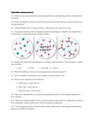
Prokaryotic cells have a variety of shapes<br />
• The three most common shapes are spheres (cocci), rods<br />
(bacilli), <strong>and</strong> spirals
Figure <strong>27</strong>.2<br />
1 m<br />
1 m<br />
3 m<br />
(a) Spherical (b) Rod-shaped (c) Spiral
Cell‐Surface Structures<br />
An important feature of nearly all prokaryotic cells is<br />
their cell wall, which maintains cell shape, protects the<br />
cell, <strong>and</strong> prevents it from bursting in a hypotonic<br />
environment<br />
• Bacterial cell walls contain peptidoglycan, a network of sugar<br />
polymers cross‐linked by polypeptides<br />
• <strong>Archaea</strong> contain polysaccharides <strong>and</strong> proteins but lack<br />
peptidoglycan<br />
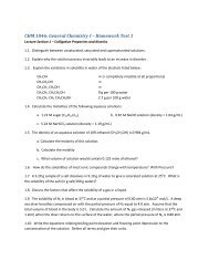
Scientists use the Gram stain to classify bacteria by cell<br />
wall composition<br />
• Gram‐positive bacteria have simpler walls with a large amount<br />
of peptidoglycan<br />
• Gram‐negative bacteria have less peptidoglycan <strong>and</strong> an outer<br />
membrane that can be toxic
Figure <strong>27</strong>.3<br />
(a) Gram-positive bacteria: peptidoglycan traps crystal violet.<br />
Gram-positive<br />
bacteria<br />
(b) Gram-negative bacteria: crystal violet is easily rinsed<br />
away, revealing red dye.<br />
Gram-negative<br />
bacteria<br />
Carbohydrate portion<br />
of lipopolysaccharide<br />
Cell<br />
wall<br />
Peptidoglycan<br />
layer<br />
Plasma<br />
membrane<br />
10 m<br />
Cell<br />
wall<br />
Outer<br />
membrane<br />
Peptidoglycan<br />
layer<br />
Plasma membrane
Cell‐Surface Structures<br />
Many antibiotics target peptidoglycan <strong>and</strong> damage<br />
bacterial cell walls<br />
Gram‐negative bacteria are more likely to be antibiotic<br />
resistant<br />
A polysaccharide or protein layer called a capsule covers<br />
many prokaryotes<br />
Some prokaryotes have fimbriae, which allow them to<br />
stick to their substrate or other individuals in a colony<br />
Pili (or sex pili) are longer than fimbriae <strong>and</strong> allow<br />
prokaryotes to exchange DNA
Figure <strong>27</strong>.4<br />
Bacterial<br />
cell wall<br />
Bacterial<br />
capsule<br />
Tonsil<br />
cell<br />
200 nm
Figure <strong>27</strong>.5<br />
Fimbriae<br />
1 m
Motility<br />
In a heterogeneous environment, many bacteria exhibit<br />
taxis, the ability to move toward or away from a<br />
stimulus<br />
• Chemotaxis is the movement toward or away from a chemical<br />
stimulus<br />
Most motile bacteria propel themselves by flagella<br />
scattered about the surface or concentrated at one or<br />
both ends<br />
Flagella of bacteria, archaea, <strong>and</strong> eukaryotes are<br />
composed of different proteins <strong>and</strong> likely evolved<br />
independently
© 2011 Pearson Education, Inc.<br />
Video: Oscillatoria
© 2011 Pearson Education, Inc.<br />
Video: Prokaryotic Flagella (Salmonella typhimurium)
Figure <strong>27</strong>.6<br />
Flagellum<br />
Filament<br />
20 nm<br />
Cell wall<br />
Hook<br />
Motor<br />
Plasma<br />
membrane<br />
Rod<br />
Peptidoglycan<br />
layer
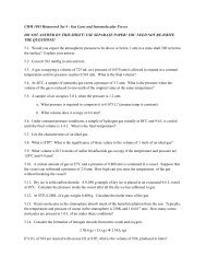
Internal Organization <strong>and</strong> DNA<br />
Prokaryotic cells usually lack complex compartmentalization<br />
Some prokaryotes do have specialized membranes that perform<br />
metabolic functions<br />
• These are usually infoldings of the plasma membrane<br />
The prokaryotic genome has less DNA than the eukaryotic genome<br />
• Most of the genome consists of a circular chromosome<br />
• The chromosome is not surrounded by a membrane; it is located in the<br />
nucleoid region<br />
• Some species of bacteria also have smaller rings of DNA called plasmids
Figure <strong>27</strong>.7<br />
0.2 m 1 m<br />
Respiratory<br />
membrane<br />
Thylakoid<br />
membranes<br />
(a) Aerobic prokaryote<br />
(b) Photosynthetic prokaryote
Figure <strong>27</strong>.8<br />
Chromosome<br />
Plasmids<br />
1 m
Reproduction <strong>and</strong> Adaptation<br />
<strong>Prokaryotes</strong> reproduce quickly by binary fission <strong>and</strong> can divide<br />
every 1–3 hours<br />
Key features of prokaryotic reproduction:<br />
• They are small<br />
• They reproduce by binary fission<br />
• They have short generation times<br />
Their short generation time allows prokaryotes to evolve quickly<br />
<strong>Prokaryotes</strong> are not “primitive” but are highly evolved<br />
Many prokaryotes form metabolically inactive endospores, which<br />
can remain viable in harsh conditions for centuries
Figure <strong>27</strong>.9<br />
Coat<br />
Endospore<br />
0.3 m
Concept <strong>27</strong>.2: Rapid reproduction, mutation,<br />
<strong>and</strong> genetic recombination promote genetic<br />
diversity in prokaryotes<br />
<strong>Prokaryotes</strong> have considerable genetic variation<br />
Three factors contribute to this genetic diversity:<br />
– Rapid reproduction<br />
– Mutation<br />
– Genetic recombination
Rapid Reproduction <strong>and</strong> Mutation<br />
<strong>Prokaryotes</strong> reproduce by binary fission, <strong>and</strong><br />
offspring cells are generally identical<br />
Mutation rates during binary fission are low, but<br />
because of rapid reproduction, mutations can<br />
accumulate rapidly in a population<br />
High diversity from mutations allows for rapid<br />
evolution
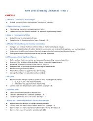
Genetic Recombination<br />
Genetic recombination, the combining of DNA from two<br />
sources, contributes to diversity<br />
Prokaryotic DNA from different individuals can be<br />
brought together by transformation, transduction, <strong>and</strong><br />
conjugation<br />
Movement of genes among individuals from different<br />
species is called horizontal gene transfer
Transformation <strong>and</strong> Transduction<br />
A prokaryotic cell can take up <strong>and</strong> incorporate foreign<br />
DNA from the surrounding environment in a process<br />
called transformation<br />
Transduction is the movement of genes between<br />
bacteria by bacteriophages (viruses that infect bacteria)
Figure <strong>27</strong>.11-4<br />
Phage<br />
A B <br />
Donor cell<br />
A <br />
B <br />
A <br />
Recombination<br />
A <br />
A <br />
B <br />
Recipient<br />
cell<br />
A <br />
B <br />
Recombinant cell
Conjugation <strong>and</strong> Plasmids<br />
Conjugation is the process where genetic material is<br />
transferred between prokaryotic cells<br />
Figure <strong>27</strong>.12<br />
Sex pilus<br />
1 m
Concept <strong>27</strong>.3: Diverse nutritional <strong>and</strong><br />
metabolic adaptations have evolved in<br />
prokaryotes<br />
<strong>Prokaryotes</strong> can be categorized by how they obtain<br />
energy <strong>and</strong> carbon<br />
• Phototrophs obtain energy from light<br />
• Chemotrophs obtain energy from chemicals<br />
• Autotrophs require CO 2 as a carbon source<br />
• Heterotrophs require an organic nutrient to make<br />
organic compounds
Table <strong>27</strong>.1
Nitrogen Metabolism<br />
Nitrogen is essential for the production of amino acids<br />
<strong>and</strong> nucleic acids<br />
In nitrogen fixation, some prokaryotes convert<br />
atmospheric nitrogen (N 2 ) to ammonia (NH 3 )
Metabolic Cooperation<br />
Cooperation between prokaryotes allows them to<br />
use environmental resources they could not use as<br />
individual cells<br />
In the cyanobacterium Anabaena, photosynthetic<br />
cells <strong>and</strong> nitrogen‐fixing cells called heterocysts<br />
exchange metabolic products
Figure <strong>27</strong>.14<br />
Photosynthetic<br />
cells<br />
Heterocyst<br />
20 m
Concept <strong>27</strong>.4: Molecular systematics is<br />
illuminating prokaryotic phylogeny<br />
Until the late 20th century, systematists based<br />
prokaryotic taxonomy on phenotypic criteria<br />
Applying molecular systematics to the investigation of<br />
prokaryotic phylogeny has produced dramatic results<br />
Molecular systematics led to the splitting of<br />
prokaryotes into bacteria <strong>and</strong> archaea
Figure <strong>27</strong>.15<br />
Eukaryotes<br />
Domain<br />
Eukarya<br />
UNIVERSAL<br />
ANCESTOR<br />
Korarchaeotes<br />
Euryarchaeotes<br />
Crenarchaeotes<br />
Nanoarchaeotes<br />
Proteobacteria<br />
Chlamydias<br />
Spirochetes<br />
Cyanobacteria<br />
Gram-positive<br />
bacteria<br />
Domain <strong>Archaea</strong><br />
Domain Bacteria
<strong>Archaea</strong><br />
<strong>Archaea</strong> share certain traits with bacteria <strong>and</strong> other<br />
traits with eukaryotes<br />
Eukarya<br />
<strong>Archaea</strong><br />
Bacteria<br />
Some archaea live in extreme environments <strong>and</strong> are<br />
called extremophiles<br />
Extreme halophiles live in highly saline environments<br />
Extreme thermophiles thrive in very hot environments
Table <strong>27</strong>.2
Figure <strong>27</strong>.16
<strong>Archaea</strong><br />
Methanogens live in swamps <strong>and</strong> marshes <strong>and</strong> produce<br />
methane as a waste product<br />
Methanogens are strict anaerobes <strong>and</strong> are poisoned by<br />
O 2
Bacteria<br />
Bacteria include the vast majority of prokaryotes of<br />
which most people are aware<br />
Diverse nutritional types are scattered among the<br />
major groups of bacteria<br />
Eukarya<br />
<strong>Archaea</strong><br />
Bacteria
Bacteria<br />
Proteobacteria<br />
These gram‐negative bacteria include<br />
photoautotrophs, chemoautotrophs, <strong>and</strong><br />
heterotrophs<br />
Some are anaerobic, <strong>and</strong> others aerobic<br />
Alpha<br />
Beta<br />
Gamma<br />
Delta<br />
Proteobacteria<br />
Epsilon
Figure <strong>27</strong>.17-a<br />
Subgroup: Alpha Proteobacteria<br />
Subgroup: Beta Proteobacteria<br />
Alpha<br />
Beta<br />
Gamma<br />
Delta<br />
Epsilon<br />
Proteobacteria<br />
2.5 m<br />
1 m<br />
Rhizobium (arrows) inside a root<br />
cell of a legume (TEM)<br />
Nitrosomonas (colorized TEM)<br />
Subgroup: Gamma Proteobacteria<br />
Subgroup: Delta Proteobacteria<br />
Subgroup: Epsilon Proteobacteria<br />
Thiomargarita namibiensis<br />
containing sulfur wastes (LM)<br />
200 m<br />
Fruiting bodies of Chondromyces<br />
crocatus, a myxobacterium (SEM)<br />
300 m<br />
Helicobacter pylori (colorized TEM)<br />
2 m
Subgroup: Alpha Proteobacteria<br />
Many species are closely associated with eukaryotic hosts<br />
• Example: Rhizobium,which forms root nodules in legumes <strong>and</strong> fixes<br />
atmospheric N 2<br />
• Example: Agrobacterium,which produces tumors in plants <strong>and</strong> is used in<br />
genetic engineering<br />
Subgroup: Alpha Proteobacteria<br />
2.5 m<br />
Rhizobium (arrows) inside a root<br />
cell of a legume (TEM)
Subgroup: Beta Proteobacteria<br />
Example: the soil bacterium Nitrosomonas,which<br />
converts NH 4+ to NO 2<br />
–<br />
Subgroup: Beta Proteobacteria<br />
1 m<br />
Nitrosomonas (colorized TEM)
Subgroup: Gamma Proteobacteria<br />
Examples include sulfur bacteria such as<br />
Chromatium <strong>and</strong> pathogens such as Legionella,<br />
Salmonella,<strong>and</strong> Vibrio cholerae<br />
Escherichia coli resides in the intestines of many<br />
mammals <strong>and</strong> is not normally pathogenic<br />
Subgroup: Gamma Proteobacteria<br />
Thiomargarita namibiensis<br />
containing sulfur wastes (LM)<br />
200 m
Subgroup: Delta Proteobacteria<br />
Example: the slime‐secreting myxobacteria<br />
Subgroup: Delta Proteobacteria<br />
Fruiting bodies of Chondromyces<br />
crocatus, a myxobacterium (SEM)<br />
300 m
Subgroup: Epsilon Proteobacteria<br />
This group contains many pathogens including<br />
Campylobacter,which causes blood poisoning, <strong>and</strong><br />
Helicobacter pylori,which causes stomach ulcers<br />
Subgroup: Epsilon Proteobacteria<br />
2 m<br />
Helicobacter pylori (colorized TEM)
Chlamydias<br />
These bacteria are parasites that live within animal<br />
cells<br />
Chlamydia trachomatis causes blindness <strong>and</strong><br />
nongonococcal urethritis by sexual transmission<br />
Chlamydias<br />
2.5 m<br />
Chlamydia (arrows) inside an<br />
animal cell (colorized TEM)
Spirochetes<br />
These bacteria are helical heterotrophs<br />
Some are parasites, including Treponema pallidum,<br />
which causes syphilis, <strong>and</strong> Borrelia burgdorferi,<br />
which causes Lyme disease<br />
Spirochetes<br />
5 m<br />
Leptospira, a spirochete<br />
(colorized TEM)
Cyanobacteria<br />
These are photoautotrophs that generate O 2<br />
Plant chloroplasts likely evolved from cyanobacteria<br />
by the process of endosymbiosis<br />
Cyanobacteria<br />
40 m<br />
Oscillatoria, a filamentous<br />
cyanobacterium
Gram-Positive Bacteria<br />
Gram‐positive bacteria<br />
include<br />
– Actinomycetes, which<br />
decompose soil<br />
– Bacillus anthracis, the<br />
cause of anthrax<br />
– Clostridium botulinum, the<br />
cause of botulism<br />
– Some Staphylococcus <strong>and</strong><br />
Streptococcus,which can<br />
be pathogenic<br />
– Mycoplasms, the smallest<br />
known cells<br />
5 m<br />
Streptomyces, the source of many<br />
antibiotics (SEM)<br />
2 m<br />
Hundreds of mycoplasmas covering<br />
a human fibroblast cell (colorized SEM)
Concept <strong>27</strong>.5: <strong>Prokaryotes</strong> play crucial roles<br />
in the biosphere<br />
<strong>Prokaryotes</strong> are so important that if they were to<br />
disappear the prospects for any other life surviving<br />
would be dim<br />
• <strong>Prokaryotes</strong> play a major role in the recycling of chemical<br />
elements between the living <strong>and</strong> nonliving components of<br />
ecosystems<br />
• Chemoheterotrophic prokaryotes function as<br />
decomposers, breaking down dead organisms <strong>and</strong> waste<br />
products<br />
• <strong>Prokaryotes</strong> can sometimes increase the availability of<br />
nitrogen, phosphorus, <strong>and</strong> potassium for plant growth
Concept <strong>27</strong>.6: <strong>Prokaryotes</strong> have both<br />
beneficial <strong>and</strong> harmful impacts on humans<br />
Some prokaryotes are human pathogens, but others<br />
have positive interactions with humans
Mutualistic Bacteria<br />
Human intestines are home to about 500–1,000<br />
species of bacteria<br />
Many of these are mutalists <strong>and</strong> break down food<br />
that is undigested by our intestines<br />
Pathogenic Bacteria<br />
<strong>Prokaryotes</strong> cause about half of all human<br />
diseases<br />
• For example, Lyme disease is caused by a<br />
bacterium <strong>and</strong> carried by ticks
Figure <strong>27</strong>.20<br />
5 m
<strong>Prokaryotes</strong> in Research <strong>and</strong> Technology<br />
Experiments using prokaryotes have led to important<br />
advances in DNA technology<br />
• For example, E. coli is used in gene cloning<br />
• For example, Agrobacterium tumefaciens is used to<br />
produce transgenic plants<br />
Bacteria can now be used to make natural plastics