Bacterial Colony Morphology
Bacterial Colony Morphology
Bacterial Colony Morphology

Create successful ePaper yourself
Turn your PDF publications into a flip-book with our unique Google optimized e-Paper software.
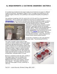
BACTERIAL COLONY MORPHOLOGY<br />
Bacteria grow on solid media as colonies. A<br />
colony is defined as a visible mass of<br />
microorganisms all originating from a single<br />
mother cell, therefore a colony constitutes a clone<br />
of bacteria all genetically alike.<br />
In the identification of bacteria and fungi much<br />
weight is placed on how the organism grows in or<br />
on media. This exercise will help you identify the<br />
cultural characteristics of a bacterium on an agar<br />
plate---called colony morphology. Although one<br />
might not necessarily see the importance of<br />
colonial morphology at first, it really can be<br />
important when identifying the bacterium.<br />
Features of the colonies may help to pinpoint the<br />
identity of the bacterium. Different species of<br />
bacteria can produce very different colonies.<br />
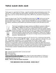
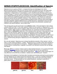
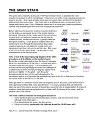
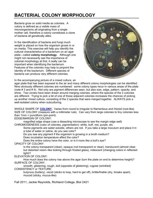
In the accompanying picture of a mixed culture, an<br />
agar plate that has been exposed to the air and many different colony morphologies can be identified.<br />
Nine obviously different colonies are numbered: some colony types recur in various areas of the plate<br />
(note # 3 and # 4). Not only are pigment differences seen, but also size, edge, pattern, opacity, and<br />
shine. Two circles have been drawn around merging colonies, where the species of the 2 colonies<br />
are different. Trying to pick a bit of one of those adjacent colonies increases the chances of picking<br />
up another mixed culture, consisting of the 2 species that were merged together. ALWAYS pick a<br />
well-isolated colony when subculturing.<br />
WHOLE SHAPE OF COLONY. Varies from round to irregular to filamentous and rhizoid (root-like)<br />
SIZE OF COLONY (measure with a millimeter rule). Can vary from large colonies to tiny colonies less<br />
than 1mm = punctiform (pin-point).<br />
EDGE/MARGIN OF COLONY:<br />
magnified edge shape (use a dissecting microscope to see the margin edge well)<br />
CHROMOGENESIS (color of colonies, pigmentation): white, buff, red, purple, etc.<br />
Some pigments are water-soluble, others are not. If you take a large inoculum and place it in<br />
a tube of water or saline, do you see color?<br />
Do you see any pigment if the organism is growing in a broth medium?<br />
Does incubation temperature affect the color?<br />
Does the entire colony have the color, or is it more like a bull’s eye?<br />
OPACITY OF COLONY:<br />
Is the colony transparent (clear), opaque (not transparent or clear), translucent (almost clear,<br />
but distorted vision–like looking through frosted glass), iridescent (changing colors in reflected<br />
light)?<br />
ELEVATION OF COLONY:<br />
How much does the colony rise above the agar (turn the plate on end to determine height)?<br />
SURFACE OF COLONY:<br />
smooth, glistening, rough, dull (opposite of glistening), rugose (wrinkled)<br />
CONSISTENCY or TEXTURE:<br />
butyrous (buttery), viscid (sticks to loop, hard to get off), brittle/friable (dry, breaks apart),<br />
mucoid (sticky, mucus-like)<br />
Fall 2011, Jackie Reynolds, Richland College, Biol 2421
OBJECTIVES:<br />
Describe features of colonies.<br />
See variations in colonial morphology among various species of bacteria.<br />
MATERIALS NEEDED:<br />
agar plates of various bacteria (examples = Pseudomonas, Chromobacterium, Micrococcus, Bacillus,<br />
Streptomyces, Streptococcus, and Neisseria)<br />
agar plates from sponge dilutions and cultures from last period<br />
THE PROCEDURES:<br />
1. Use a plate which has well-isolated colonies. Look at the largest colonies with the naked eye<br />
to determine general shape and chromogenesis.<br />
2. Use a dissecting/stereoscopic microscope for more detail. Place the plate RIGHTSIDE UP on<br />
the stage, leaving the petri dish cover ON (Otherwise, your culture will become contaminated.)<br />
There are 2 lenses on our scopes—10X and 20X: the black lens knob is on the right side of<br />
the head of the microscope. The magnification is especially helpful for the study of elevation,<br />
surface, opacity, size, and edge. There are 2 lights on these microscopes that you might find<br />
helpful, either using one at a time, or both, or even sometimes without them. Two small black<br />
rotating knobs on either side of the base control the 2 lights, one light from above and one light<br />
from below the stage.<br />
3. Or you may want to use the Quebec colony counter since it has a magnifying glass, and a light<br />
behind the plate stage. Make sure that the dish is right-side up.<br />
4. If you see water condensation on the lid cover, take a KimWipe and carefully remove the water<br />
from the cover, then quickly replacing the cover on the dish.<br />
5. In order to determine CONSISTENCY, you need to use your inoculating loop or needle to pick<br />
up the colony and determine the consistency of the inoculum material as the loop leaves the<br />
agar medium.<br />
2
Small Colonies<br />
Red Pigment<br />
Round, Entire<br />
Opaque<br />
Large Colonies<br />
Opaque<br />
Round, Entire<br />
Filamentous<br />
Irregular<br />
Opaque<br />
Punctiform<br />
Yellow<br />
Round<br />
Opaque<br />
Yellow<br />
Round, Entire<br />
Opaque<br />
Buff Pigment<br />
Irregular, Undulate<br />
Opaque<br />
Orange Pigment<br />
Round, Entire<br />
Opaque<br />
Brown Pigment<br />
Irregular<br />
Translucent<br />
GOOD RESOURCE: Science Buddies, Interpreting Plates<br />
http://www.sciencebuddies.org/mentoring/project_ideas/MicroBio_Interpreting_Plates.shtml<br />
3