BD Biosciences
BD Biosciences
BD Biosciences

Create successful ePaper yourself
Turn your PDF publications into a flip-book with our unique Google optimized e-Paper software.
<strong>BD</strong> PuraMatrix Peptide Hydrogel<br />
<strong>BD</strong> <strong>Biosciences</strong> Discovery Labware<br />
www.bdbiosciences.com<br />
<strong>BD</strong> <strong>Biosciences</strong><br />
Clontech<br />
Discovery Labware<br />
Immunocytometry Systems<br />
Pharmingen
<strong>BD</strong> <br />
PuraMatrix <br />
Peptide<br />
Hydrogel<br />
<strong>BD</strong> PuraMatrix Peptide<br />
Hydrogel, a novel synthetic<br />
matrix ideal for creating<br />
optimized 3D cell culture<br />
environments.<br />
<strong>BD</strong> PuraMatrix Peptide Hydrogel is a<br />
synthetic matrix that is used to create<br />
defined three-dimensional (3D) microenvironments<br />
for a variety of cell culture<br />
experiments. To achieve optimal cell growth<br />
and differentiation, it is necessary to<br />
determine the appropriate mixture of this<br />
material and bioactive molecules (e.g.,<br />
growth factors, extracellular matrix<br />
[ECM] proteins, and/or other molecules).<br />
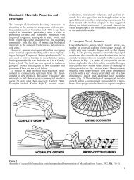
<strong>BD</strong> PuraMatrix Peptide Hydrogel consists<br />
of standard amino acids (1% w/v) and<br />
99% water. Under physiological conditions,<br />
the peptide component self-assembles into<br />
a 3D hydrogel that exhibits a nanometer<br />
scale fibrous structure (Figure 1). The<br />
hydrogel is readily formed in a culture dish,<br />
plate, or cell culture insert.<br />
The resulting hydrogel has been shown to<br />
promote the differentiation of hepatocyte<br />
progenitor cells 1 , rat pheochromocytoma<br />
cells (PC12) 2 , and hippocampal neurons. 3<br />
Studies have also demonstrated that<br />
<strong>BD</strong> PuraMatrix Peptide Hydrogel supports<br />
the attachment of a variety of primary (e.g.,<br />
neuronal, fibroblast, keratinocyte) and<br />
transformed (e.g., MG-63, SH-SY5Y,<br />
HEK293, NIH3T3) cell types. 4 Other<br />
potential applications include stem cell<br />
proliferation, tumor cell migration and<br />
invasion, angiogenesis assays, and in vivo<br />
analyses of tissue regeneration.<br />
<strong>BD</strong> PuraMatrix Peptide Hydrogel is<br />
biocompatible, resorbable, and devoid of<br />
animal-derived material and pathogens. For<br />
in vivo studies in animals, the soluble<br />
material can be injected and will subsequently<br />
form a 3D hydrogel upon contact<br />
with the physiological environment.<br />
Application Focus<br />
Differentiation of Hepatocyte Progenitor Cells 1<br />
Rat hepatocyte progenitor cells (Lig-8 5 ) were encapsulated in <strong>BD</strong> PuraMatrix Peptide<br />
Hydrogel and cultured overnight in defined medium at 37ºC. Samples were then used for<br />
bromodioxyuridine (BrdU) uptake and in situ immunofluorescence analyses. As shown in<br />
Figure 2, Lig-8 cells form spheroid colonies when cultured within the 3D hydrogel and<br />
express the hepatocyte markers CCAAT/enhancer binding protein α (C/EBPα) and cytochrome<br />
P450 1A1/1A2 (CYP1A1/1A2) in a manner that is independent of cellular mitotic<br />
activity. Therefore, while some cells are proliferating, the entire colony exhibits<br />
differentiation potential.<br />
Figure 1. Electron micrograph of <strong>BD</strong> PuraMatrix<br />
Peptide Hydrogel [bar, 100 nm].<br />
Figure 2. Lig-8 cells cultured in <strong>BD</strong> PuraMatrix<br />
Peptide Hydrogel. All cells in spheroid colonies,<br />
arrested or not, undergo differentiation.<br />
Spheroids were isolated, transferred to<br />
adherent cultures, and incubated with BrdU<br />
for 24 hours.<br />
(A) spheroid colony (phase contrast)<br />
(B) same optical layer as A immunostained for<br />
C/EBPα (red)<br />
References<br />
1. Semino, C.E., et al., Functional differentiation<br />
of hepatocyte-like spheroid structures from<br />
putative liver progenitor cells in threedimensional<br />
peptide scaffolds. Differentiation<br />
71:262 (2003).<br />
2. Holmes, T.C., et al., Extensive neurite outgrowth<br />
and active synapse formation on selfassembling<br />
peptide scaffolds. PNAS USA<br />
97:6728 (2000).<br />
3. Semino, C.E., et al., Entrapment of migrating<br />
hippocampal neural cells in 3D peptide nanofiber<br />
scaffold. Tissue Engineering 10:643 (2004).<br />
4. Zhang, S., et al., Self-complementary oligopeptide<br />
matrices support mammalian cell<br />
attachment. Biomaterials 16:1385 (1995).<br />
5. Lee, H-S., et al., Clonal expansion of adult rat<br />
hepatic stem cell lines by suppression of<br />
asymmetric cell kinetics (SACK). Biotechnology<br />
and Bioengineering 83:760 (2003).<br />
(C) same optical layer as A immunostained for<br />
BrdU (green)<br />
(D) spheroid colony (phase contrast)<br />
(E) same optical layer as D immunostained for<br />
CYP1A1/1A2 (red)<br />
(F) same optical layer as D immunostained for<br />
BrdU<br />
Data provided by 3D Matrix, Inc. and originally<br />
described in Semino, C.E., et al., Differentiation<br />
71:262 (2003).
Analysis of Neurite Outgrowth using PC12 cells 2<br />
PC12 cells were cultured on <strong>BD</strong> PuraMatrix Peptide Hydrogel according to a surface plating protocol. To examine the differentiated<br />
morphology of these cells, samples were incubated in the presence of nerve growth factor (NGF) and subsequently analyzed by scanning<br />
laser confocal microscopy. PC12 cells cultured under these conditions were found to differentiate and exhibit pronounced neurite outgrowth<br />
(Figure 3). Moreover, human SY5Y neuroblastoma cells and a variety of primary neurons were shown to exhibit comparable<br />
activity on this material (Table 1).<br />
Table 1. Neurite outgrowth from neuronal cells on <strong>BD</strong> PuraMatrix Peptide Hydrogel.<br />
Cell Type Neurite Length, µm Cell Source†<br />
NGF-treated Rat PC12 400-500 Cultured cell line<br />
NGF-preprimed PC12 400-500 Cultured cell line<br />
Human SY5Y neuroblastoma 400-500 Cultured cell line<br />
Mouse cerebellar granule neurons 200-300 Primary cells‡<br />
Mouse hippocampal neurons 100-200 Primary cells‡<br />
Rat hippocampal neurons 200-300 Primary cells§<br />
Figure 3. PC12 cell neurite outgrowth and<br />
differentiation on <strong>BD</strong> PuraMatrix Peptide<br />
Hydrogel. The image is a merged stack of<br />
multiple confocal optical sections. PC12 cells<br />
cultured in the presence of NGF attached to the<br />
hydrogel and projected extensive neurites. The<br />
black spots are holes in the hydrogel. The<br />
micrograph is representative of at least four<br />
independent experiments.<br />
† Cells were seeded onto <strong>BD</strong> PuraMatrix Peptide Hydrogel. The cell-bearing hydrogels were transferred<br />
to dishes with fresh medium. Maximum neurite length was estimated visually with scale bars 3 to 7<br />
days after cell attachment for primary cells and 10 to 14 days for the cultured cell lines.<br />
‡ Seven-day-old mouse<br />
§ One-day-old rat<br />
Data provided by 3D Matrix, Inc. and originally<br />
described in Holmes, T.C., et al., PNAS USA 97:<br />
6728 (2000).<br />
Differentiation of Neurons Derived from Hippocampal Slices 3<br />
Hippocampal tissue slices (~200 µm<br />
thickness) were isolated from 7.5-day-old<br />
postnatal rats and placed on top of a<br />
microporous membrane insert for up to<br />
two weeks. The inserts were either<br />
uncoated (control samples) or coated<br />
with <strong>BD</strong> PuraMatrix Peptide Hydrogel<br />
(~500 µm thick). When cultured on the<br />
hydrogel, the hippocampal slice exhibited<br />
tissue growth and cell migration into the<br />
material (Figure 4). While neuronal<br />
migration was not observed in the control<br />
samples (Figure 4g), neuronal and glial<br />
progenitor cells were found to migrate<br />
from the tissue slice into the hydrogel layer<br />
(Figure 4f and 4h). These results suggest<br />
that <strong>BD</strong> PuraMatrix Peptide Hydrogel<br />
may be useful for promoting neural cell<br />
differentiation and tissue growth in vivo.<br />
Figure 4. Hippocampal organotypic slice<br />
cultures on <strong>BD</strong> PuraMatrix Peptide Hydrogel<br />
develop extended tissue growth. Hippocampal<br />
slices were cultured on control<br />
membrane or <strong>BD</strong> PuraMatrix Peptide<br />
Hydrogel layers (~500 µm thick). Time lapse<br />
was carried out to follow tissue growth<br />
from the perimeter of the dentate gyrus<br />
region. (A) Time 0 (0 hour) of control slice<br />
culture; (B) Time 0 (0 hour) hydrogel<br />
(RAD16-I) slice culture; (C) 72 hours,<br />
control slice; (D) 72 hours, hydrogel slice;<br />
The red line indicates the original border<br />
of the tissue slice and the yellow line in d<br />
shows the extended tissue growth. The<br />
yellow arrow in d indicates the direction<br />
of tissue growth and extension. Black bars<br />
represent 100 µm. (E) 72 hours, control<br />
slice immunostained for GFAP (glial cell<br />
marker, green); (F) 72 hours, hydrogel<br />
slice immunostained for GFAP (green);<br />
(G) same optical layer as e immunostained<br />
for NeuN (neuron marker, red); (H) same<br />
optical layer as f immunostained for NeuN<br />
(red). Red lines in e and f and yellow in g<br />
and h indicate original perimeter of the<br />
tissue slice. The white line in e and g indicates<br />
the extended tissue scaffold in control<br />
cultures. The white line in f and h is<br />
used to compare the over extension<br />
obtained on hydrogel cultures. Yellow<br />
arrows in h indicate NeuN+ neurons (red)<br />
migrating into Area II in hydrogel slice cultures.<br />
White bar in f represents 100 µm.<br />
Data provided by 3D Matrix, Inc. and<br />
originally described in Semino, C.E., et al.,<br />
Tissue Engineering 10:643 (2004).
<strong>BD</strong> PuraMatrix Peptide Hydrogel<br />
Features and Benefits<br />
Purified Synthetic Peptide<br />
Composition (1% w/v)<br />
3D Hydrogel Structure<br />
Easy Handling<br />
Transparent Hydrogel<br />
Established Protocols<br />
Highly defined material that promotes cell attachment<br />
Assembles into fibrous structure with average pore size of 50-200 nm<br />
Easily mixed with cells and/or bioactive molecules (e.g., growth factors) prior to gelation;<br />
injectable for in vivo studies in animals<br />
Samples are readily visualized using standard staining methodologies and microscopy<br />
3D cell encapsulation cultures; Surface plating of adherent cells on microporous membrane<br />
inserts and microplates; cell recovery for sub-culturing or biochemical analyses; in vivo<br />
injection<br />
Characteristics<br />
• Peptide sequence promotes cell attachment, but does not activate RGD-dependent integrin signaling<br />
• In the presence of salt-containing solution, the peptide component of <strong>BD</strong> PuraMatrix Peptide Hydrogel<br />
self-assembles and forms a transparent 3D hydrogel<br />
• Exhibits nanometer scale fibrous structure<br />
• Biocompatible; devoid of animal-derived material and pathogens<br />
Technical Specifications<br />
• 1% solution (w/v) of purified synthetic peptide<br />
• Packaged material exhibits pH = 3.0<br />
• Quality Control:<br />
– Tested and found negative for bacteria, fungi, and Mycoplasma<br />
– Cell viability > 80% based on cytotoxicity analysis of NIH3T3 fibroblasts<br />
– Identity confirmed using Mass Spectrometry<br />
– Demonstration of fiber formation using a self-assembly assay<br />
Ordering Information<br />
Description Qty Cat. No.<br />
<strong>BD</strong> PuraMatrix Peptide Hydrogel 5 ml 354250<br />
To place an order in the U.S., contact Customer Service at:<br />
tel: 800.343.2035 or 978.901.7300; fax: 800.743.6200 or 978.901.7493<br />
For technical assistance, contact Technical Service at:<br />
tel: 800.343.2035 or 978.901.7300; fax: 800.743.6200 or 978.901.7493<br />
To place an order outside the U.S., contact your local distributor or nearest<br />
<strong>BD</strong> <strong>Biosciences</strong> office.<br />
For information about GMP-grade material, contact 3D Matrix, Inc. at:<br />
e-mail: sales@puramatrix.com; internet: www.puramatrix.com<br />
<strong>BD</strong> <strong>Biosciences</strong><br />
Two Oak Park<br />
Bedford, MA 01730 USA<br />
tel: 800.343.2035<br />
fax: 800.743.6200<br />
<strong>BD</strong><br />
2771 Bristol Circle<br />
Oakville, Ontario<br />
Canada L6H 6R5<br />
tel: 905.855.5550<br />
fax: 905.829.5405<br />
Nippon <strong>BD</strong><br />
Akasaka DS Bldg.<br />
5-26 Akasaka 8-chome<br />
Minato-ku, Tokyo 107 Japan<br />
tel: (81) 24 593 5405<br />
fax: (81) 24 593 5761<br />
<strong>BD</strong> <strong>Biosciences</strong><br />
Singapore Branch<br />
30 Tuas Avenue 2<br />
Singapore 639461<br />
tel: (65) 6861 0633<br />
fax: (65) 6860 1590<br />
<strong>BD</strong> <strong>Biosciences</strong> Europe<br />
Erembodegem-Dorp 86<br />
9320 Erembodegem, Belgium<br />
tel: (32) 53 720 211<br />
fax: (32) 53 720 450<br />
e-mail: contact_bdb@europe.bd.com<br />
<strong>BD</strong> <strong>Biosciences</strong><br />
4 Research Park Drive<br />
Macquarie University Research Park<br />
North Ryde NSW 2113 Australia<br />
tel: (612) 8875 5239<br />
fax: (612) 8875 7200<br />
For Research Use Only. Not for use in diagnostic or therapeutic procedures. Not for resale.<br />
PuraMatrix is a registered trademark of 3D Matrix, Inc.<br />
<strong>BD</strong>, <strong>BD</strong> Logo, and trademarks are the property of Becton, Dickinson and Company. ©2004 <strong>BD</strong><br />
B04B045<br />
<strong>BD</strong> <strong>Biosciences</strong>