BD Biosciences
BD Biosciences
BD Biosciences

Create successful ePaper yourself
Turn your PDF publications into a flip-book with our unique Google optimized e-Paper software.
Analysis of Neurite Outgrowth using PC12 cells 2<br />
PC12 cells were cultured on <strong>BD</strong> PuraMatrix Peptide Hydrogel according to a surface plating protocol. To examine the differentiated<br />
morphology of these cells, samples were incubated in the presence of nerve growth factor (NGF) and subsequently analyzed by scanning<br />
laser confocal microscopy. PC12 cells cultured under these conditions were found to differentiate and exhibit pronounced neurite outgrowth<br />
(Figure 3). Moreover, human SY5Y neuroblastoma cells and a variety of primary neurons were shown to exhibit comparable<br />
activity on this material (Table 1).<br />
Table 1. Neurite outgrowth from neuronal cells on <strong>BD</strong> PuraMatrix Peptide Hydrogel.<br />
Cell Type Neurite Length, µm Cell Source†<br />
NGF-treated Rat PC12 400-500 Cultured cell line<br />
NGF-preprimed PC12 400-500 Cultured cell line<br />
Human SY5Y neuroblastoma 400-500 Cultured cell line<br />
Mouse cerebellar granule neurons 200-300 Primary cells‡<br />
Mouse hippocampal neurons 100-200 Primary cells‡<br />
Rat hippocampal neurons 200-300 Primary cells§<br />
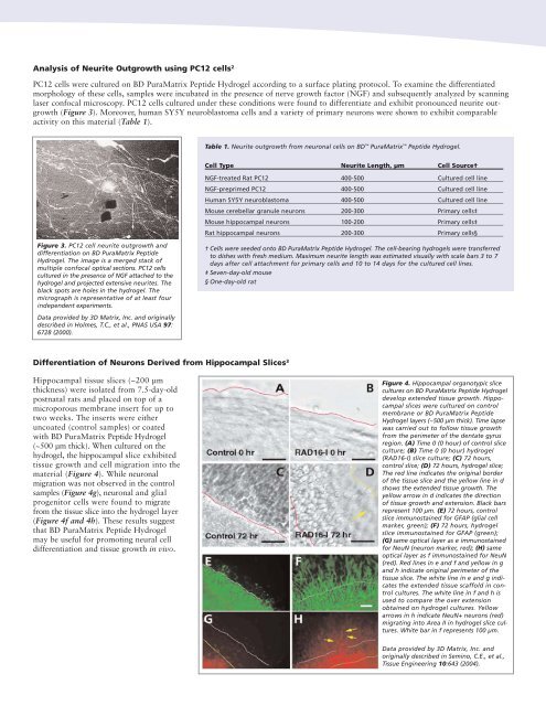
Figure 3. PC12 cell neurite outgrowth and<br />
differentiation on <strong>BD</strong> PuraMatrix Peptide<br />
Hydrogel. The image is a merged stack of<br />
multiple confocal optical sections. PC12 cells<br />
cultured in the presence of NGF attached to the<br />
hydrogel and projected extensive neurites. The<br />
black spots are holes in the hydrogel. The<br />
micrograph is representative of at least four<br />
independent experiments.<br />
† Cells were seeded onto <strong>BD</strong> PuraMatrix Peptide Hydrogel. The cell-bearing hydrogels were transferred<br />
to dishes with fresh medium. Maximum neurite length was estimated visually with scale bars 3 to 7<br />
days after cell attachment for primary cells and 10 to 14 days for the cultured cell lines.<br />
‡ Seven-day-old mouse<br />
§ One-day-old rat<br />
Data provided by 3D Matrix, Inc. and originally<br />
described in Holmes, T.C., et al., PNAS USA 97:<br />
6728 (2000).<br />
Differentiation of Neurons Derived from Hippocampal Slices 3<br />
Hippocampal tissue slices (~200 µm<br />
thickness) were isolated from 7.5-day-old<br />
postnatal rats and placed on top of a<br />
microporous membrane insert for up to<br />
two weeks. The inserts were either<br />
uncoated (control samples) or coated<br />
with <strong>BD</strong> PuraMatrix Peptide Hydrogel<br />
(~500 µm thick). When cultured on the<br />
hydrogel, the hippocampal slice exhibited<br />
tissue growth and cell migration into the<br />
material (Figure 4). While neuronal<br />
migration was not observed in the control<br />
samples (Figure 4g), neuronal and glial<br />
progenitor cells were found to migrate<br />
from the tissue slice into the hydrogel layer<br />
(Figure 4f and 4h). These results suggest<br />
that <strong>BD</strong> PuraMatrix Peptide Hydrogel<br />
may be useful for promoting neural cell<br />
differentiation and tissue growth in vivo.<br />
Figure 4. Hippocampal organotypic slice<br />
cultures on <strong>BD</strong> PuraMatrix Peptide Hydrogel<br />
develop extended tissue growth. Hippocampal<br />
slices were cultured on control<br />
membrane or <strong>BD</strong> PuraMatrix Peptide<br />
Hydrogel layers (~500 µm thick). Time lapse<br />
was carried out to follow tissue growth<br />
from the perimeter of the dentate gyrus<br />
region. (A) Time 0 (0 hour) of control slice<br />
culture; (B) Time 0 (0 hour) hydrogel<br />
(RAD16-I) slice culture; (C) 72 hours,<br />
control slice; (D) 72 hours, hydrogel slice;<br />
The red line indicates the original border<br />
of the tissue slice and the yellow line in d<br />
shows the extended tissue growth. The<br />
yellow arrow in d indicates the direction<br />
of tissue growth and extension. Black bars<br />
represent 100 µm. (E) 72 hours, control<br />
slice immunostained for GFAP (glial cell<br />
marker, green); (F) 72 hours, hydrogel<br />
slice immunostained for GFAP (green);<br />
(G) same optical layer as e immunostained<br />
for NeuN (neuron marker, red); (H) same<br />
optical layer as f immunostained for NeuN<br />
(red). Red lines in e and f and yellow in g<br />
and h indicate original perimeter of the<br />
tissue slice. The white line in e and g indicates<br />
the extended tissue scaffold in control<br />
cultures. The white line in f and h is<br />
used to compare the over extension<br />
obtained on hydrogel cultures. Yellow<br />
arrows in h indicate NeuN+ neurons (red)<br />
migrating into Area II in hydrogel slice cultures.<br />
White bar in f represents 100 µm.<br />
Data provided by 3D Matrix, Inc. and<br />
originally described in Semino, C.E., et al.,<br />
Tissue Engineering 10:643 (2004).