African Journal of Plant Science - Academic Journals
African Journal of Plant Science - Academic Journals
African Journal of Plant Science - Academic Journals
Create successful ePaper yourself
Turn your PDF publications into a flip-book with our unique Google optimized e-Paper software.
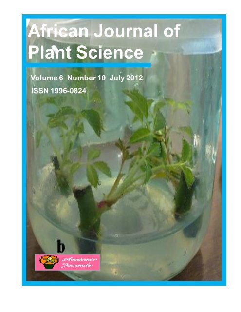
<strong>African</strong> <strong>Journal</strong> <strong>of</strong><br />
<strong>Plant</strong> <strong>Science</strong><br />
Volume 6 Number 10 July 2012<br />
ISSN 1996-0824
ABOUT AJPS<br />
The <strong>African</strong> <strong>Journal</strong> <strong>of</strong> <strong>Plant</strong> <strong>Science</strong> (AJPS) is published bi-monthly (one volume per year) by <strong>Academic</strong> <strong>Journal</strong>s.<br />
<strong>African</strong> <strong>Journal</strong> <strong>of</strong> <strong>Plant</strong> <strong>Science</strong> (AJPS) is an open access journal that provides rapid publication (bi-monthly) <strong>of</strong><br />
articles in all areas <strong>of</strong> <strong>Plant</strong> <strong>Science</strong> and Botany such as plant diseases and its treatment, paleoethnobotany,<br />
genetic modification <strong>of</strong> plants to achieve optimum productivity etc. The <strong>Journal</strong> welcomes the submission <strong>of</strong><br />
manuscripts that meet the general criteria <strong>of</strong> significance and scientific excellence. Papers will be published<br />
shortly after acceptance. All articles published in AJPS are peer-reviewed.<br />
Submission <strong>of</strong> Manuscript<br />
Submit manuscripts as e-mail attachment to the Editorial Office at: ajps@acadjournals.org. A manuscript number<br />
will be mailed to the corresponding author shortly after submission.<br />
The <strong>African</strong> <strong>Journal</strong> <strong>of</strong> <strong>Plant</strong> <strong>Science</strong> will only accept manuscripts submitted as e-mail attachments.<br />
Please read the Instructions for Authors before submitting your manuscript. The manuscript files should be given<br />
the last name <strong>of</strong> the first author.
Editors<br />
Pr<strong>of</strong>. Diaga Diouf<br />
Laboratoire de Biotechnologies végétales,<br />
Département de Biologie Végétale,<br />
Faculté des <strong>Science</strong>s et Techniques,<br />
Université Cheikh Anta Diop, BP 5005,<br />
Dakar, Sénégal.<br />
Pr<strong>of</strong>. Amarendra Narayan Misra<br />
Center for Life <strong>Science</strong>s, School <strong>of</strong> Natural <strong>Science</strong>s,<br />
Central University <strong>of</strong> Jharkhand,<br />
Ratu-Lohardaga Road, P.O. Brambe-835205,<br />
Ranchi, Jharkhand State,<br />
India.<br />
Associate Editors<br />
Dr. Ömür Baysal<br />
Mugla University<br />
ASMK Vocational School<br />
Technical Programs PB. 48300<br />
Fethiye -Mugla<br />
Turkey.<br />
Dr. R. Siva<br />
School <strong>of</strong> Bio <strong>Science</strong>s and Technology<br />
VIT University<br />
Vellore 632 014.<br />
Dr. Biswa Ranjan Acharya<br />
Pennsylvania State University<br />
Department <strong>of</strong> Biology<br />
208 Mueller Lab<br />
University Park, PA 16802.<br />
USA<br />
Dr. Ali Masoudi-Nejad<br />
Assistant Pr<strong>of</strong>essor <strong>of</strong> Bioinformatics<br />
Institute <strong>of</strong> Biochemistry and Biophysics (IBB)<br />
University <strong>of</strong> Tehran,Tehran,<br />
Iran.<br />
Pr<strong>of</strong>. H. Özkan Sivritepe<br />
Department <strong>of</strong> Horticulture Faculty <strong>of</strong><br />
Agriculture Uludag University Görükle<br />
Campus Bursa 16059<br />
Turkey.<br />
Pr<strong>of</strong>. Ahmad Kamel Hegazy<br />
Department <strong>of</strong> Botany, Faculty <strong>of</strong> <strong>Science</strong>,<br />
Cairo University, Giza 12613,<br />
Egypt.<br />
Dr. Annamalai Muthusamy<br />
Department <strong>of</strong> Biotechnology<br />
Manipal Life <strong>Science</strong> Centre,<br />
Manipal University,<br />
Manipal – 576 104<br />
Karnataka,<br />
India.<br />
Dr. Chandra Prakash Kala<br />
Indian Institute <strong>of</strong> Forest Management<br />
Nehru Nagar, P.B.No. 357<br />
Bhopal, Madhya Pradesh<br />
India – 462 003.
Editorial Board<br />
Pr<strong>of</strong>. Weimin Zhang<br />
Guangdong Institute <strong>of</strong> Microbiology<br />
100 Central Xian lie Road, Guangzhou, Guangdong 510070,<br />
China<br />
Specialization: Microbiology, biochemistry and molecular<br />
biology, natural<br />
product chemistry<br />
China<br />
Dr. Xu Suxia<br />
Fujian Institute <strong>of</strong> Subtropical Botany<br />
780-800, Jiahe Road, Xiamen, China361006<br />
Specialization: <strong>Plant</strong> physiology and biochemistry<br />
China<br />
Dr. Adaku Vivien Iwueke<br />
Federal University <strong>of</strong> Technology, Owerri<br />
Department <strong>of</strong> Biochemistry, Federal University <strong>of</strong><br />
Technology, Owerri<br />
Specialization: Pharmacological/Nutritional Biochemistry<br />
Nigeria<br />
Ass. Pr<strong>of</strong>. Turgay CELIK<br />
Gulhane Military Medical Academy,<br />
School <strong>of</strong> Medicine,<br />
Department <strong>of</strong> Cardiology<br />
Specialization: Interventional cardiology, clinical<br />
cardiology, intensive care<br />
Turkey<br />
Dr.Topik Hidayat<br />
Department <strong>of</strong> Biology Education<br />
Indonesia University <strong>of</strong> Education (UPI)<br />
Jalan Dr. Setiabudhi 229 Bandung 40154 Indonesia<br />
Specialization: Botany<br />
Indonesia<br />
Dr.Tariq Mahmood<br />
Quaid-i-Azam University,<br />
Department <strong>of</strong> <strong>Plant</strong> <strong>Science</strong>s, Quaid-i-Azam University,<br />
Islamabad, Pakistan<br />
Specialization: Biochemistry, Molecular Biology,<br />
Bioinformatics, Biotechnology<br />
Pakistan<br />
Dr.Neveen B. Talaat<br />
Department <strong>of</strong> <strong>Plant</strong> Physiology, Faculty <strong>of</strong> Agriculture,<br />
Cairo University, Egypt<br />
Specialization: <strong>Plant</strong> Physiology<br />
<strong>Plant</strong> Molecular Biology: <strong>Plant</strong> Transformation<br />
<strong>Plant</strong> nutrition<br />
Egypt<br />
Dr. Sudhamoy Mandal<br />
Central Horticultural Experiment Station (ICAR)<br />
Aiginia, Bhubaneswar, PIN-751019<br />
Specialization: Induced resistance (SAR, ISR) in plants,<br />
Oxidative burst, Biocontrol <strong>of</strong> plant diseases, Defense<br />
signaling in plants<br />
India<br />
Asso. Pr<strong>of</strong>. Chankova Stephka Georgieva<br />
Central Laboratory <strong>of</strong> General Ecology, Bulg Acad Sci<br />
1113 S<strong>of</strong>ia, 2 Gagarin str, Bulgaria<br />
Specialization: Genetics - environmental mutagenesis;<br />
molecular eco-toxicology, stress response<br />
Bulgaria<br />
Shijie Han<br />
Center <strong>of</strong> Forestry, Institute <strong>of</strong> Applied Ecology, Chinese<br />
Academy <strong>of</strong> <strong>Science</strong>s,<br />
72 Wenhua Road, Shenyang City, Liaoning Province 110016,<br />
PR China<br />
Specialization: Global change biology, <strong>Plant</strong> ecology, Soil<br />
ecology<br />
PR China<br />
Szu-Chuan Shen<br />
Department <strong>of</strong> Medical Nutrition,<br />
I-Shou University<br />
Yanchao Township,<br />
Kaohsiung County 824,Taiwan<br />
Specialization: Food Nutrition<br />
Taiwan<br />
Seddik Khennouf<br />
Dept <strong>of</strong> BIOLOGY, Faculty <strong>of</strong> <strong>Science</strong><br />
University Ferhat Abbas, SETIF, 19000, ALGERIA<br />
Specialization: Pharmacology <strong>of</strong> phenolic compounds,<br />
medicinal plants<br />
ALGERIA<br />
Saranyu Khammuang<br />
Mahasarakham University<br />
Department <strong>of</strong> Chemistry, Faculty <strong>of</strong> <strong>Science</strong><br />
Specialization: <strong>Plant</strong> biotechnology, Protein and peptide<br />
chemistry, Enzymology<br />
Thailand<br />
Samir Ranjan Sikdar<br />
Bose Institute<br />
P-1/12, C.I.T. Scheme VII M, Kolkata 700 054, India<br />
Specialization: Genetics, <strong>Plant</strong> Breeding, Tissue culture,<br />
protoplast culture and somatic hybridization with special<br />
reference to Brassica and edible mushrooms.<br />
India<br />
Dr. M. Abdul Salam<br />
Department <strong>of</strong> Agronomy, College <strong>of</strong> Agriculture<br />
Kerala Agricultural University, Vellayani 695 522,<br />
Trivandrum, Kerala<br />
Specialization: Agronomy <strong>of</strong> tropical crops, <strong>Plant</strong> nutrition,<br />
Cashew nut Production and Processing, Organic production<br />
<strong>of</strong> tropical vegetables and fruits, Irrigation water
Electronic submission <strong>of</strong> manuscripts is strongly<br />
encouraged, provided that the text, tables, and figures are<br />
included in a single Micros<strong>of</strong>t Word file (preferably in Arial<br />
font).<br />
The cover letter should include the corresponding author's<br />
full address and telephone/fax numbers and should be in<br />
an e-mail message sent to the Editor, with the file, whose<br />
name should begin with the first author's surname, as an<br />
attachment.<br />
Article Types<br />
Three types <strong>of</strong> manuscripts may be submitted:<br />
Regular articles: These should describe new and carefully<br />
confirmed findings, and experimental procedures should<br />
be given in sufficient detail for others to verify the work.<br />
The length <strong>of</strong> a full paper should be the minimum required<br />
to describe and interpret the work clearly.<br />
Short Communications: A Short Communication is suitable<br />
for recording the results <strong>of</strong> complete small investigations<br />
or giving details <strong>of</strong> new models or hypotheses, innovative<br />
methods, techniques or apparatus. The style <strong>of</strong> main<br />
sections need not conform to that <strong>of</strong> full-length papers.<br />
Short communications are 2 to 4 printed pages (about 6 to<br />
12 manuscript pages) in length.<br />
Reviews: Submissions <strong>of</strong> reviews and perspectives covering<br />
topics <strong>of</strong> current interest are welcome and encouraged.<br />
Reviews should be concise and no longer than 4-6 printed<br />
pages (about 12 to 18 manuscript pages). Reviews are also<br />
peer-reviewed.<br />
Review Process<br />
Instructions for Author<br />
All manuscripts are reviewed by an editor and members <strong>of</strong><br />
the Editorial Board or qualified outside reviewers. Authors<br />
cannot nominate reviewers. Only reviewers randomly<br />
selected from our database with specialization in the<br />
subject area will be contacted to evaluate the manuscripts.<br />
The process will be blind review.<br />
Decisions will be made as rapidly as possible, and the<br />
journal strives to return reviewers’ comments to authors as<br />
fast as possible. The editorial board will re-review<br />
manuscripts that are accepted pending revision. It is the<br />
goal <strong>of</strong> the AJPS to publish manuscripts within weeks after<br />
submission.<br />
Regular articles<br />
All portions <strong>of</strong> the manuscript must be typed doublespaced<br />
and all pages numbered starting from the title<br />
page.<br />
The Title should be a brief phrase describing the<br />
contents <strong>of</strong> the paper. The Title Page should include the<br />
authors' full names and affiliations, the name <strong>of</strong> the<br />
corresponding author along with phone, fax and E-mail<br />
information. Present addresses <strong>of</strong> authors should<br />
appear as a footnote.<br />
The Abstract should be informative and completely selfexplanatory,<br />
briefly present the topic, state the scope <strong>of</strong><br />
the experiments, indicate significant data, and point out<br />
major findings and conclusions. The Abstract should be<br />
100 to 200 words in length.. Complete sentences, active<br />
verbs, and the third person should be used, and the<br />
abstract should be written in the past tense. Standard<br />
nomenclature should be used and abbreviations should<br />
be avoided. No literature should be cited.<br />
Following the abstract, about 3 to 10 key words that will<br />
provide indexing references should be listed.<br />
A list <strong>of</strong> non-standard Abbreviations should be added.<br />
In general, non-standard abbreviations should be used<br />
only when the full term is very long and used <strong>of</strong>ten.<br />
Each abbreviation should be spelled out and introduced<br />
in parentheses the first time it is used in the text. Only<br />
recommended SI units should be used. Authors should<br />
use the solidus presentation (mg/ml). Standard<br />
abbreviations (such as ATP and DNA) need not be<br />
defined.<br />
The Introduction should provide a clear statement <strong>of</strong><br />
the problem, the relevant literature on the subject, and<br />
the proposed approach or solution. It should be<br />
understandable to colleagues from a broad range <strong>of</strong><br />
scientific disciplines.<br />
Materials and methods should be complete enough<br />
to allow experiments to be reproduced. However, only<br />
truly new procedures should be described in detail;<br />
previously published procedures should be cited, and<br />
important modifications <strong>of</strong> published procedures should<br />
be mentioned briefly. Capitalize trade names and<br />
include the manufacturer's name and address.<br />
Subheadings should be used. Methods in general use<br />
need not be described in detail.
Results should be presented with clarity and precision.<br />
The results should be written in the past tense when<br />
describing findings in the authors' experiments.<br />
Previously published findings should be written in the<br />
present tense. Results should be explained, but largely<br />
without referring to the literature. Discussion,<br />
speculation and detailed interpretation <strong>of</strong> data should<br />
not be included in the Results but should be put into the<br />
Discussion section.<br />
The Discussion should interpret the findings in view <strong>of</strong><br />
the results obtained in this and in past studies on this<br />
topic. State the conclusions in a few sentences at the end<br />
<strong>of</strong> the paper. The Results and Discussion sections can<br />
include subheadings, and when appropriate, both<br />
sections can be combined.<br />
The Acknowledgments <strong>of</strong> people, grants, funds, etc<br />
should be brief.<br />
Tables should be kept to a minimum and be designed to<br />
be as simple as possible. Tables are to be typed doublespaced<br />
throughout, including headings and footnotes.<br />
Each table should be on a separate page, numbered<br />
consecutively in Arabic numerals and supplied with a<br />
heading and a legend. Tables should be self-explanatory<br />
without reference to the text. The details <strong>of</strong> the methods<br />
used in the experiments should preferably be described<br />
in the legend instead <strong>of</strong> in the text. The same data should<br />
not be presented in both table and graph form or<br />
repeated in the text.<br />
Figure legends should be typed in numerical order on a<br />
separate sheet. Graphics should be prepared using<br />
applications capable <strong>of</strong> generating high resolution GIF,<br />
TIFF, JPEG or Powerpoint before pasting in the Micros<strong>of</strong>t<br />
Word manuscript file. Tables should be prepared in<br />
Micros<strong>of</strong>t Word. Use Arabic numerals to designate<br />
figures and upper case letters for their parts (Figure 1).<br />
Begin each legend with a title and include sufficient<br />
description so that the figure is understandable without<br />
reading the text <strong>of</strong> the manuscript. Information given in<br />
legends should not be repeated in the text.<br />
References: In the text, a reference identified by means<br />
<strong>of</strong> an author‘s name should be followed by the date <strong>of</strong><br />
the reference in parentheses. When there are more than<br />
two authors, only the first author‘s name should be<br />
mentioned, followed by ’et al‘. In the event that an<br />
author cited has had two or more works published during<br />
the same year, the reference, both in the text and in the<br />
reference list, should be identified by a lower case letter<br />
like ’a‘ and ’b‘ after the date to distinguish the works.<br />
Examples:<br />
Abayomi (2000), Agindotan et al. (2003), (Kelebeni,<br />
1983), (Usman and Smith, 1992), (Chege, 1998;<br />
1987a,b; Tijani, 1993,1995), (Kumasi et al., 2001)<br />
References should be listed at the end <strong>of</strong> the paper in<br />
alphabetical order. Articles in preparation or articles<br />
submitted for publication, unpublished observations,<br />
personal communications, etc. should not be included<br />
in the reference list but should only be mentioned in<br />
the article text (e.g., A. Kingori, University <strong>of</strong> Nairobi,<br />
Kenya, personal communication). <strong>Journal</strong> names are<br />
abbreviated according to Chemical Abstracts. Authors<br />
are fully responsible for the accuracy <strong>of</strong> the references.<br />
Examples:<br />
Chikere CB, Omoni VT and Chikere BO (2008).<br />
Distribution <strong>of</strong> potential nosocomial pathogens in a<br />
hospital environment. Afr. J. Biotechnol. 7: 3535-3539.<br />
Moran GJ, Amii RN, Abrahamian FM, Talan DA (2005).<br />
Methicillinresistant Staphylococcus aureus in<br />
community-acquired skin infections. Emerg. Infect. Dis.<br />
11: 928-930.<br />
Pitout JDD, Church DL, Gregson DB, Chow BL,<br />
McCracken M, Mulvey M, Laupland KB (2007).<br />
Molecular epidemiology <strong>of</strong> CTXM-producing<br />
Escherichia coli in the Calgary Health Region:<br />
emergence <strong>of</strong> CTX-M-15-producing isolates.<br />
Antimicrob. Agents Chemother. 51: 1281-1286.<br />
Pelczar JR, Harley JP, Klein DA (1993). Microbiology:<br />
Concepts and Applications. McGraw-Hill Inc., New York,<br />
pp. 591-603.<br />
Short Communications<br />
Short Communications are limited to a maximum <strong>of</strong><br />
two figures and one table. They should present a<br />
complete study that is more limited in scope than is<br />
found in full-length papers. The items <strong>of</strong> manuscript<br />
preparation listed above apply to Short<br />
Communications with the following differences: (1)<br />
Abstracts are limited to 100 words; (2) instead <strong>of</strong> a<br />
separate Materials and Methods section, experimental<br />
procedures may be incorporated into Figure Legends<br />
and Table footnotes; (3) Results and Discussion should<br />
be combined into a single section.<br />
Pro<strong>of</strong>s and Reprints: Electronic pro<strong>of</strong>s will be sent (email<br />
attachment) to the corresponding author as a PDF<br />
file. Page pro<strong>of</strong>s are considered to be the final version<br />
<strong>of</strong> the manuscript. With the exception <strong>of</strong> typographical<br />
or minor clerical errors, no changes will be made in the<br />
manuscript at the pro<strong>of</strong> stage.
Fees and Charges: Authors are required to pay a $550 handling fee. Publication <strong>of</strong> an article in the <strong>African</strong> <strong>Journal</strong> <strong>of</strong><br />
<strong>Plant</strong> <strong>Science</strong> is not contingent upon the author's ability to pay the charges. Neither is acceptance to pay the handling<br />
fee a guarantee that the paper will be accepted for publication. Authors may still request (in advance) that the<br />
editorial <strong>of</strong>fice waive some <strong>of</strong> the handling fee under special circumstances.<br />
Copyright: © 2012, <strong>Academic</strong> <strong>Journal</strong>s.<br />
All rights Reserved. In accessing this journal, you agree that you will access the contents for your own personal use<br />
but not for any commercial use. Any use and or copies <strong>of</strong> this <strong>Journal</strong> in whole or in part must include the customary<br />
bibliographic citation, including author attribution, date and article title.<br />
Submission <strong>of</strong> a manuscript implies: that the work described has not been published before (except in the form <strong>of</strong> an<br />
abstract or as part <strong>of</strong> a published lecture, or thesis) that it is not under consideration for publication elsewhere; that if<br />
and when the manuscript is accepted for publication, the authors agree to automatic transfer <strong>of</strong> the copyright to the<br />
publisher.<br />
Disclaimer <strong>of</strong> Warranties<br />
In no event shall <strong>Academic</strong> <strong>Journal</strong>s be liable for any special, incidental, indirect, or consequential damages <strong>of</strong> any<br />
kind arising out <strong>of</strong> or in connection with the use <strong>of</strong> the articles or other material derived from the AJPS, whether or<br />
not advised <strong>of</strong> the possibility <strong>of</strong> damage, and on any theory <strong>of</strong> liability.<br />
This publication is provided "as is" without warranty <strong>of</strong> any kind, either expressed or implied, including, but not<br />
limited to, the implied warranties <strong>of</strong> merchantability, fitness for a particular purpose, or non-infringement.<br />
Descriptions <strong>of</strong>, or references to, products or publications does not imply endorsement <strong>of</strong> that product or publication.<br />
While every effort is made by <strong>Academic</strong> <strong>Journal</strong>s to see that no inaccurate or misleading data, opinion or statements<br />
appear in this publication, they wish to make it clear that the data and opinions appearing in the articles and<br />
advertisements herein are the responsibility <strong>of</strong> the contributor or advertiser concerned. <strong>Academic</strong> <strong>Journal</strong>s makes no<br />
warranty <strong>of</strong> any kind, either express or implied, regarding the quality, accuracy, availability, or validity <strong>of</strong> the data or<br />
information in this publication or <strong>of</strong> any other publication to which it may be linked.
International <strong>Journal</strong> <strong>of</strong> Medicine and Medical <strong>Science</strong>s<br />
<strong>African</strong> <strong>Journal</strong> <strong>of</strong> <strong>Plant</strong> <strong>Science</strong><br />
Table <strong>of</strong> Contents: Volume 6 Number 10 July, 2012<br />
nces<br />
Review<br />
ARTICLES<br />
Allelopathy in aquatic macrophytes: Effects on growth and physiology <strong>of</strong><br />
phytoplanktons 270<br />
Yalew Addisie and Andrea Calderon Medellin<br />
Research Articles<br />
Propagation and growth from cultured single node explants <strong>of</strong> rosa (Rosa<br />
miniature) 277<br />
Farah Farahani and Soodeh Shaker<br />
Effect <strong>of</strong> time <strong>of</strong> harvest on physiological maturity and kenaf<br />
(Hibiscus canabinus) seed quality 282<br />
Olasoji J.O., A.O. Aluko, O.N. Adeniyan, S.O. Olanipekun ,<br />
A.A. Olosunde and Okoh J.O.
<strong>African</strong> <strong>Journal</strong> <strong>of</strong> <strong>Plant</strong> <strong>Science</strong> Vol. 6(10), pp. 270-276, July 2012<br />
Available online at http://www.academicjournals.org/AJPS<br />
DOI: 10.5897/AJPS12.008<br />
ISSN 1996-0824 ©2012 <strong>Academic</strong> <strong>Journal</strong>s<br />
Review<br />
Allelopathy in aquatic macrophytes: Effects on growth<br />
and physiology <strong>of</strong> phytoplanktons<br />
Yalew Addisie 1 * and Andrea Calderon Medellin 2<br />
1 Department <strong>of</strong> Biology, College <strong>of</strong> Natural and Computational <strong>Science</strong>s, Mekelle University, P. O. Box 231, Mekelle,<br />
Ethiopia.<br />
2 Universidad Militar Nueva Granada, Bogota, Bolivar, Colombia.<br />
Accepted 17 May, 2012<br />
The present review discussed allelophatic effects <strong>of</strong> aquatic macrophytes on phytoplankton with<br />
emphasis on growth and physiology; and also indicates implication for control <strong>of</strong> invasive aquatic<br />
macrophyte and phytoplankton species. In view <strong>of</strong> the fact, inhibition <strong>of</strong> phytoplankton by<br />
allelochemicals released by macrophytes is supposed to be one <strong>of</strong> the mechanisms that contribute to<br />
stabilisation <strong>of</strong> clear-water states in shallow lakes. This is achieved through the effects <strong>of</strong><br />
allelochemicals on growth, photosynthesis and enzymatic activities <strong>of</strong> the phytoplanktons. The<br />
reciprocal/counter act mechanisms <strong>of</strong> target phytoplankton to allelochemicals are still ill defined.<br />
Reciprocal allelopathic responses between macrophytes and other phytoplankton species would have<br />
implications for management <strong>of</strong> eutrophic waters. Therefore, understanding the molecular mechanisms<br />
and the genetic bases <strong>of</strong> such effect is important. In addition to these, the molecular and genetic bases<br />
<strong>of</strong> allelopathy <strong>of</strong> invasive species <strong>of</strong> aquatic macrophytes are not well studied and utilizing the outputs<br />
to develop potential control is done through engineering.<br />
Key words: Allelopathy, enzymatic activity, macrophytes, phytoplanktons.<br />
INTRODUCTION<br />
The phenomenon <strong>of</strong> chemical interactions among plants,<br />
fungi, bacteria, and animals has gained great attention by<br />
researchers from all over the world. Ever since, biological<br />
interactions have survival values to keep ecosystem<br />
integrations. Such chemically mediated interaction<br />
between plants or microorganisms is termed allelopathy<br />
(Rice, 1984). This mainly occurs in organisms releasing<br />
or secreting their own chemical substances named<br />
allelochemicals into a surrounding medium and eliciting<br />
either positive or deleterious response in target organism<br />
(Rice, 1984). Aquatic macrophytes (AM) including<br />
macroalgae from the long shore, floating as well as deep-<br />
water zone in the freshwater environment have<br />
*Corresponding author. E-mail: albertu2009@gmail.com.<br />
widespread allelopathy on phytoplankton (Gopal and<br />
Goel, 1993; Inderjit and Dakshini, 1994). Different<br />
investigations have shown the existence <strong>of</strong> diverse<br />
allelochemicals identified from AM and well documented<br />
their inhibition and stimulating effect (Gross, 2003; Hilt et<br />
al., 2006; Mulderij, 2006; Gross et al., 2007;<br />
Abdulrahman, 2010). Allelochemicals can interfere with<br />
many processes <strong>of</strong> target organisms (Einhellig, 2001).<br />
Allelopathically active compounds from AMs are <strong>of</strong>ten<br />
directed at numerous physiological processes such as<br />
photosynthesis and enzyme activities and growth and<br />
development <strong>of</strong> phytoplanktons. Therefore, the present<br />
review attempts to discuss allelophatic effects <strong>of</strong> some<br />
AMs on phytoplankton with emphasis on growth and<br />
physiology. Moreover, it tries to indicate implications for<br />
the control/management <strong>of</strong> invasive AM and<br />
phytoplankton species.
Figure 1. Schematic representation <strong>of</strong> allelopathic interactions between aquatic macrophytes and<br />
phytoplanktons.<br />
Allelopathy: An ecological overview<br />
Eutrophication and algal blooms are the most serious<br />
environmental problems. Search for allelopathic effects <strong>of</strong><br />
AMs on phytoplanktons’ growth and development<br />
has receiving world-wide attention in order to develop<br />
biological tools which are environmentally friendly to<br />
mitigate such challenges. This is due to the reason that<br />
inhibition <strong>of</strong> phytoplankton by allelochemicals released by<br />
submerged macrophytes is supposed to be one <strong>of</strong> the<br />
mechanisms that contribute to the stabilisation <strong>of</strong> clearwater<br />
states in shallow lakes. Since allelopathy <strong>of</strong> AMs is<br />
<strong>of</strong> significance on composition <strong>of</strong> communities in fresh<br />
water ecosystems and ecological rehabilitation <strong>of</strong><br />
eutrophic lakes (Korner and Nicklisch, 2002). This is<br />
achieved through allelopathic growth inhibition <strong>of</strong><br />
phytoplankton by AMs which might confer an advantage<br />
to macrophytes in the competition for light, carbon, and<br />
nutrients (Gross, 2003) and change the succession <strong>of</strong><br />
phytoplankton. However, understanding the factors which<br />
regulate or control the extent <strong>of</strong> allelopathic effects such<br />
as light and nutrient limitation <strong>of</strong> both the donor and the<br />
target organisms is vital. In view <strong>of</strong> the fact, production <strong>of</strong><br />
Addisie and Medellín 271<br />
secondary metabolites for plant defense related to light<br />
and nutrient availability (Cronin and Lodge, 2003),<br />
temperature (Albert et al., 2009) and biotic factors such<br />
as herbivory (Lemoine et al., 2009). Thus, allelopathy and<br />
other processes for plant defense cannot always be<br />
separated. Only few studies relate allelochemical<br />
production <strong>of</strong> AMs to abiotic and biotic influences.<br />
Although allelopathy also includes positive (stimulating)<br />
interactions, majority <strong>of</strong> the studies describe the inhibitory<br />
activity <strong>of</strong> allelopathically active compounds. Such<br />
inhibitory and simulative interactions are explained with<br />
tremendous growth, physiological and biochemical<br />
mechanisms which are discussed subsequently. Six<br />
prerequisites have to be met to show unequivocally the<br />
occurrence <strong>of</strong> allelopathy: (1) a pattern <strong>of</strong> inhibition <strong>of</strong><br />
target plant(s) or alga(e), (2) allelopathical compound(s)<br />
produced by donor plants, (3) release <strong>of</strong> these<br />
compounds by the producing plant, (4) transport and/or<br />
accumulation in the environment, (5) uptake by the target<br />
organism(s), and (6) that the inhibition cannot be<br />
explained solely by other physical or biotic factors,<br />
especially competition; and it is summarized<br />
schematically in Figure 1.
272 Afr. J. <strong>Plant</strong> Sci.<br />
Cell density (×10 3 cells ml -1 )<br />
Cell density (×10 6 cells ml -1 )<br />
Figure 2. Growth-inhibition and cell lysis effects <strong>of</strong> the extract <strong>of</strong> U. lactuca dry powder on harmful algal<br />
bloom species <strong>of</strong> (A) A. anophagefferens, (B) C. marina. Data points are means ± SD (n = 3) (Source:<br />
Tang and Gobler, 2011).<br />
Allelopathy and its effect on growth <strong>of</strong> phytoplankton<br />
and macrophytes<br />
Virtually various in situ and ex-situ experiments<br />
conducted to confirm allelopathy in AMs have shown its<br />
suppressing performance on phytoplankton growth. It is<br />
quite possible that allelochemicals may produce more<br />
than one effect on the cellular processes responsible to<br />
reduce phytoplankton growth. However, the details <strong>of</strong> the<br />
biochemical mechanism through which a particular<br />
allelochemical exerts a toxic effect on growth are not well<br />
known. The existing evidences suggest that the<br />
mechanism <strong>of</strong> growth inhibition <strong>of</strong> allelochemicals from<br />
macroalgae and macrophytes is dependent on the nature<br />
<strong>of</strong> the chemical elicited and genotype and diverse in type.<br />
For instance, the mechanism <strong>of</strong> growth inhibition <strong>of</strong> the<br />
green macro alga Ulva lactuca on harmful algal bloom<br />
species via allelopathy is achieved through cell lysing and<br />
decreasing cell density (Jin et al., 2005) (Figure 2).<br />
However, some macrophytes release allelochemicals<br />
which induce high rate <strong>of</strong> photosynthesis that might draw<br />
down CO2 and in turn increases pH level. Subsequently,<br />
this would have influence on processes involved in<br />
growth and developments <strong>of</strong> phytoplankton and other<br />
aquatic macrophytes (Lundholm et al., 2005). In this<br />
case, the presumption is that, high pH invoked<br />
allelopathic effects through interfering biochemical<br />
reactions and nutrient scavenging. Moreover, production<br />
<strong>of</strong> allelochemicals such as polyunsaturated fatty acids in<br />
Ulva (Alamsjah et al., 2005) and dithiolane and trithiane<br />
compounds identified from Chara sp. cause allelopathic<br />
effects on growth <strong>of</strong> epiphytic diatoms and other<br />
phytoplanktons (Wium-Anderson et al., 1982). Such<br />
influence <strong>of</strong> allelochemicals on growth <strong>of</strong> phytoplanktons<br />
would have negative consequences on the biomass<br />
production (Qiming et al., 2006).<br />
Effects on physiological activities<br />
Aquatic allelochemicals <strong>of</strong>ten target multiple physiological<br />
processes. Allelochemicals have significant effect on cell<br />
division, cell differentiation, respiration, photosynthesis,<br />
enzyme function, signal transduction as well as gene<br />
expression (Inderjit, 2003; Belz and Hurle, 2004) in both<br />
autotrophs <strong>of</strong> aquatic and terrestrial ecosystems. The
Figure 3. Inhibition <strong>of</strong> photosynthetic O2 evolution <strong>of</strong> Anabaena sp. PCC 7120 by addition <strong>of</strong><br />
different fractions <strong>of</strong> the solid phase extcraction(SPE) <strong>of</strong> M. spicatum extract (1, water, 2,<br />
25% (v/v) MeOH; 3, 50% (v/v) MeOH; 75 (v/v) MeOH; and 100% (v/v) MeOH). The amount <strong>of</strong><br />
exctract used in the experiment was equivalent to 19 µg TAE ml -1 . Error bars indicate SE; n =<br />
3 to 7 measurements per treatment. Significance differences between treatment and control<br />
are shown by asterisk (P
274 Afr. J. <strong>Plant</strong> Sci.<br />
Figure 4. The z-scheme <strong>of</strong> electron transport chain (red arrows) <strong>of</strong> the thylakoid membrane the (grey bar)<br />
in the chloroplasts with photosystem I and II (green ovals, PSI and PSII) and<br />
the target site <strong>of</strong> tellimagrandium II (blue arrow) between the quinine electron acceptors QA and QB. Abbrevi<br />
ations: Pheo: pheophytin; PQ: plastochinon; PQH2: reduced plastochinon; Fd: ferrodoxin and PC: phycocya<br />
nin. (Source: http://www.fsbio hannover.de/<strong>of</strong>theweek/120.htm).<br />
the secondary quinine electron acceptors, QA and QB in<br />
the PSII (Figure 4). These allelochemicals act on a<br />
different site <strong>of</strong> action. The decline in rate <strong>of</strong><br />
photosynthesis might be ascribed to decrease in amount<br />
<strong>of</strong> chlorophyll contents due to allelochemicals<br />
(Abdulrahman, 2010). For example, experiment<br />
conducted on the effect <strong>of</strong> allelochemicals from<br />
Myriophyllum aquaticum on photosynthetic pigments <strong>of</strong><br />
Microcystis aeruginosa showed that relative content <strong>of</strong><br />
chlorophyll a, phycocyanin and allophycocyanin<br />
decreased to 52.7, 15.3 and 7.6%, respectively (Wu et<br />
al., 2008) and the decline rate <strong>of</strong> assimilation is to that<br />
extent. The authors also reported that phycobiliprotein<br />
decreased more than Chlorophyll a.<br />
Inhibition <strong>of</strong> enzymes<br />
Many aquatic organisms produce extracellular enzymes<br />
that enable them to use complex substrates or are<br />
involved in the colonization <strong>of</strong> surfaces (Wetzel, 1991).<br />
However, several enzymatic processes are inhibited by<br />
allelochemicals from AMs. Interference with these<br />
enzymes can alter competitive interactions among<br />
organisms, change settling <strong>of</strong> organisms, and interfere<br />
with bi<strong>of</strong>ilm formation and/or epiphyte growth. For<br />
example, polyphenolic extracts from freshwater<br />
submersed angiosperms, M. spicatum and Stratiote<br />
aloides inhibit the enzyme alkaline phosphatase (Gross<br />
et al., 1996; Abdulrahman, 2010). But the inhibitory effect<br />
is genotype dependent (Figure 5). These findings<br />
suggested that inhibition <strong>of</strong> extracellular alkaline<br />
phosphatase could be one mode <strong>of</strong> action <strong>of</strong><br />
allelopathically active compounds. This might have<br />
growth inhibition effect as alkaline phosphatase activities<br />
are concomitant with growth <strong>of</strong> phytoplanktons (Hilt,<br />
2006). Enzyme inhibition can harm the whole organisms<br />
such as decreased dehydrogenase activity (for example,<br />
M. aeruginosa) and increased the level <strong>of</strong> reactive<br />
oxygen species (ROS) (Hong et al., 2009). The favoured<br />
production <strong>of</strong> ROS is as consequence <strong>of</strong> much lipid<br />
peroxidation by allelochemicals in algal cells directs to<br />
reduce algal cell proliferation (Wu et al., 2007). More to<br />
the point, a study conducted to test the inhibitory effect <strong>of</strong><br />
the allelochemical, gramine, on cyanobacterium (Hong et<br />
al., 2010) showed that gramine caused significant
(U mg -1 protein)<br />
APA activities (U mg -1 protein)<br />
(mg ml -1 Concentration <strong>of</strong> S. aloides extract (mg ml ) -1 )<br />
Figure 5. Effect <strong>of</strong> aqueous acetone extract <strong>of</strong> Stratiotes aloides on alkaline phosphatase activity<br />
<strong>of</strong> Scendesmus obliquus (After: Abdulrahman, 2010).<br />
increase <strong>of</strong> ROS. These also lowered activities <strong>of</strong><br />
antioxidant enzymes, such as superoxide dismutase and<br />
peroxidase, can lead to increased oxidative damage,<br />
metal ion leakage and changes in plasma integrity (Li and<br />
Hu, 2005). According to these authors also ethyl 2methylacetoacetate<br />
from Phragmites communis showed<br />
changes in plasma membrane integrity and leakage <strong>of</strong><br />
ions in the protoplast (Li and Hu, 2005). Furthermore,<br />
other modes <strong>of</strong> action <strong>of</strong> allelochemicals among<br />
phytoplankton are for example cellular paralysis reported<br />
<strong>of</strong> Anabaena flos-aquae affecting motile green algae<br />
(Kearns and Hunter, 2001) as well as the inhibition <strong>of</strong><br />
nucleic acid synthesis (Einhellig and Reigosa, 2002) or<br />
RNA polymerase by alkaloids (Doan et al., 2000).<br />
Future research areas and implication to invasive<br />
species control<br />
The research <strong>of</strong> the allelopathy <strong>of</strong> AMs on phytoplanktons<br />
is developing very fast. With many findings on new<br />
allelochemicals, it will become the most promising<br />
method to control algal bloom and invasive species.<br />
However, there still exist some important problems to be<br />
solved before it can be used in practice. The existing<br />
studies mainly focus on floating and submerged<br />
macrophytes, less studies on the emergent macrophytes.<br />
Addisie and Medellín 275<br />
Investigation on allelopathic activities <strong>of</strong> the AMs with<br />
abundant biomass is still extremely limited. Some<br />
allelochemicals have been identified from macrophytes,<br />
but the highly-effective allelochemicals are still very few.<br />
So, it still will be an important issue to search for highlyeffective<br />
allelochemicals from aquatic macrophytes. The<br />
studies on modes <strong>of</strong> action <strong>of</strong> allelochemicals will help to<br />
modify the structure <strong>of</strong> allelochemicals artificially and<br />
adjust their activities to be much more efficient. Most <strong>of</strong><br />
the existing researches are carried out in lab scale. The<br />
lab-scale system was too simple to reflect the actual<br />
circumstances <strong>of</strong> field. Since, under lab-conditions, it<br />
remains difficult to prove the influence <strong>of</strong> combination <strong>of</strong><br />
abiotic and biotic interactions’ on the allelochemicals in<br />
the same way as the in situ conditions. Therefore, future<br />
researches in analyzing the presence <strong>of</strong> allelopathic<br />
substances should focus on studies with intact<br />
macrophytes and experimental setups in which it is<br />
possible to make a clear distinction between allelopathy<br />
and other growth limiting factors.<br />
Currently, majority <strong>of</strong> allelopathic studies focus on<br />
negative effects exerted by one species on another,<br />
however, understanding the stimulation effect <strong>of</strong> some<br />
allelochemicals for the normal growth and development<br />
<strong>of</strong> some groups would have great contribution in<br />
developing mitigation mechanisms for harmful blooming<br />
algae and invasive macrophytes. Also, elucidating the
276 Afr. J. <strong>Plant</strong> Sci.<br />
reciprocal or counter act mechanisms <strong>of</strong> target<br />
phytoplankton to allelochemicals are still ill-defined and it<br />
could be possible future research areas. Reciprocal<br />
allelopathic responses between macrophytes and other<br />
phytoplankton species would have implications for the<br />
management <strong>of</strong> eutrophic waters. Majority <strong>of</strong> reports<br />
have indicated that allelochemicals negatively influence<br />
the growth and physiological processes such as enzyme<br />
activities and photosynthesis. Therefore, understanding<br />
the molecular mechanisms and the genetic bases <strong>of</strong> such<br />
effect is important. In addition to these, the molecular and<br />
genetics bases <strong>of</strong> allelopathy <strong>of</strong> invasive species <strong>of</strong> AMs<br />
are not well studied and utilizing the outputs to develop<br />
potential control is done through engineering.<br />
ACKNOWLEDGEMENT<br />
We would like to express our sincere appreciation and<br />
gratitude to Mr. Addisu Tegegne Jimma University,<br />
Ethiopia for his encouragement and comment on the<br />
work.<br />
REFERENCES<br />
Abdulrahman MA (2010). Differential Sensitivities <strong>of</strong> Different<br />
Scenedesmus obliquus Strains to the Allelopathic Activity <strong>of</strong> the<br />
Macrophytes Stratiotes aloides. J. Appl. Sci., 10: 1769-1774.<br />
Albert A, Sareedenchai V, Heller W, Seidlitz HK, Zidorn C (2009).<br />
Temperature is the key to altitudinal variation <strong>of</strong> phenolics in Arnica<br />
montana. Oecologia, 160:1-8.<br />
Alamsjah MA, Hirao S, Ishibashi F, Fujita Y (2005). Isolation and<br />
structure determination <strong>of</strong> algicidal compounds from Ulva fasciata.<br />
Biosci. Biotechnol. Biochem., 69: 2186–2192.<br />
Belz RG, Hurle K (2004). A novel laboratory screening bioassay for crop<br />
seedling allelopathy. J. Chem. Ecol., 3: 175-198.<br />
Cronin G, Lodge DM (2003). Effects <strong>of</strong> light and nutrient availability on<br />
the growth, allocation, carbon/nitrogen balance, phenolic chemistry,<br />
and resistance to herbivory <strong>of</strong> two freshwater macrophytes.<br />
Oecologia, 137: 32-41.<br />
Doan NT, Rickards RW, Rothschild JM, Smith GD (2000). Allelopathic<br />
actions <strong>of</strong> the alkaloid12-epi-hapalindole E isonitrile and calothrixin A<br />
from cyanobacteria <strong>of</strong> the genera Fischerella and Calothrix. J. Appl.<br />
Phycol., 12: 409-416.<br />
Gopal B,Goel U (1993). Competition and allelopathy in aquatic plant<br />
communities. Bot. Rev., 59: 155-210.<br />
Gross EM, Meyer H, Schilling G (1996). Release and ecological impact<br />
<strong>of</strong> algicidal hydrolysable polyphenols in Myriophyllum spectrum.<br />
Phytochemistry, 41:133-138.<br />
Gross EM (2003). Allelopathy <strong>of</strong> aquatic autotrophs. Crit. Rev. <strong>Plant</strong><br />
Sci., 22: 313–339.<br />
Gross EM, Hilt S, Lombardo P, Mulderij G (2007). Searching for<br />
allelopathy interaction–State <strong>of</strong> the art and open questions.<br />
Hydrobiologia., 584:77–88.<br />
Hilt S (2006). Allelopathic inhibition <strong>of</strong> epiphytes by submerged<br />
macrophytes. Aqua. Bot., 85: 252–256.<br />
Hong Y, Huang JJ, Hu HY (2009). Effects <strong>of</strong> a novel allelochemical<br />
Ethyl 2-Methyl Acetoacetate (EMA) on the ultrastructure and pigment<br />
composition <strong>of</strong> cyanobacterium Microcystis aeruginosa. Bull. Environ.<br />
Contam. Toxicol., 83: 502-508.<br />
Hong Y, Hu HY, Sakoda ASagehashi M (2010). Isolation and<br />
characterization <strong>of</strong> antialgal allelochemicals from Arundo donax.<br />
Allelopathy J.25: 357-368. http://www.fsbio hannover.de/<strong>of</strong> the<br />
week/120.htm.<br />
Inderjit S (2003). Ecophysiological aspects <strong>of</strong> allelopathy. <strong>Plant</strong>a, 217:<br />
529-539.<br />
Inderjit, S. and Dakshini, K. M. (1994). Algal allelopathy, Bot. Rev., 60:<br />
182-196.<br />
Jin Q, Dong SL, Wang CY (2005). Allelopathic growth inhibition <strong>of</strong><br />
Prorocentrum micans by Ulva pertusa and Ulva linza in laboratory<br />
cultures. Eur. J. Phycol., 40: 31–37.<br />
Kearns KD, Hunter MD (2001). Toxin-producing Anabaena flos-aquae<br />
induces settling <strong>of</strong> Chlamydomonas reinhardtii, a competing motile<br />
alga. Microb. Ecol., 42: 80-86.<br />
Korner S, Nicklisch A (2002). Allelopathic growth inhibition <strong>of</strong> selected<br />
phytoplankton species by submerged macrophytes. J. Phycol., 38:<br />
862–871.<br />
Lemoine DG, Barrat-Segretain MH, Roy A (2009). Morphological and<br />
chemical changes induced by herbivory in three common aquatic<br />
macrophytes. Int. Rev. Hydrobiol., 94: 282-289.<br />
Leu E, Krieger-Liszkay A, Goussias C,Gross EM (2002). Polyphenolic al<br />
lelochemicals from the aquatic angiosperm Myriophyllum spicatum<br />
inhibit photosystem II. <strong>Plant</strong> Physiol., 130:2011-2018.<br />
Li F M, Hu HY (2005). Isolation and characterization <strong>of</strong> a novel antialgal<br />
allelochemical from Phragmites communis. Appl. Environ. Microbiol.,<br />
11: 6545-6553.<br />
Lundholm N, Hansen PJ, Kotaki Y (2005). Lack <strong>of</strong> allelopathic effects <strong>of</strong><br />
the domoic acid-producing marine diatom Pseudo-nitzschia<br />
multiseries. Mar. Ecol. Prog. Ser., 288:21–33.<br />
Mulderij G (2006). Chemical warfare in freshwater. Allelopathic effects<br />
<strong>of</strong> macrophytes on phytoplankton. Ph.D. thesis, Netherlands Institute<br />
<strong>of</strong> Ecology, The Nether-lands.<br />
Qiming X, Haidong C, Huixian Z., Daqiang Y (2006). Allelopathic activity<br />
<strong>of</strong> volatile substance from submerged macrophytes on Microcystin<br />
aeruginosa. Acta Ecologica Sinica. 26: 3549−3554.<br />
Rice ELL (1984). Allelopathy. 2 nd Ed. <strong>Academic</strong>, New York.<br />
Smith GD, Doan NT (1999). Cyanobacterial metabolites with bioactivity<br />
against photosynthesis in cyanobacteria, algae and higher plants. J.<br />
Appl. Phycol., 11: 337-344.<br />
Srivastava A, Juttner F, Strasser RJ (1998). Action <strong>of</strong> the<br />
allelochemical, fischerellin A on photosystem II. Biochim. Biophys.<br />
Acta., 1364: 326-336.<br />
Tang Y, ZGobler JC (2011). The green macroalga, Ulva lactuca, inhibits<br />
the growth <strong>of</strong> seven common harmful algal bloom species via<br />
allelopathy. Harmful Algae. 10: 480–488.<br />
Wetzel RG, ChróstRJ (1991). Extracellular enzymatic interactions:<br />
storage, redistribution, and interspecific communication, Microbial<br />
Enzymes in Aquatic Environments, pp 6-28, Springer-Verlag, New<br />
York.<br />
Wium-Andersen S, Anthoni U, Christophersen CHouen G (1982).<br />
Allelopathic effects on phytoplankton by substances isolated from<br />
aquatic macrophytes (Charales). Oikos, 39: 187–190.<br />
Wu Z.B, Deng P, Luo WUX, Gao Y (2007). Allelopathic effects <strong>of</strong> the<br />
submerged macrophyte Potamogeton malaianus on Scenedesmus o<br />
bliquus. Hydrobiologia, 592: 465-474l.<br />
Wu C, Xuexiu C, Hongjuan D, Difu L Junyan L (2008). Allelopathic<br />
inhibitory effect <strong>of</strong> Myriophyllum aquaticum on Microcystis<br />
aeruginosa and its physiological mechanism. Acta Ecologica Sinica,<br />
28: 2595-2603.
<strong>African</strong> <strong>Journal</strong> <strong>of</strong> <strong>Plant</strong> <strong>Science</strong> Vol. 6(10), pp. 277-281, July 2012<br />
Available online at http://www.academicjournals.org/AJPS<br />
DOI: 10.5897/AJPS12.037<br />
ISSN 1996-0824 ©2012 <strong>Academic</strong> <strong>Journal</strong>s<br />
Full Length Research Paper<br />
Propagation and growth from cultured single node<br />
explants <strong>of</strong> rosa (Rosa miniature)<br />
Farah Farahani 1 * and Soodeh Shaker 2<br />
1 Department <strong>of</strong> Microbiology, Qom Branch, Islamic Azad University, Qom, Iran.<br />
2 <strong>Plant</strong> Biotechnology Department, Karaj Center, Payame Noor university, Karaj, Iran.<br />
Accepted 21 May, 2012<br />
Roses are one <strong>of</strong> the world's most important ornamentals for a long time and are most <strong>of</strong>ten used for<br />
ornamental, medicinal and aromatic purposes. A method for the proliferation and growth <strong>of</strong> rose (Rosa<br />
miniature) was developed. First to seventh nodal explants from young healthy shoots were excised and<br />
cultured on basal medium <strong>of</strong> Murashige and Skoog (1962, MS) containing several concentrations <strong>of</strong><br />
Benzyl amino purine (BAP) and indol butyric acid (IBA). Multiple shoot formation <strong>of</strong> up to 4 shoots from<br />
a single node was obtained on MS medium supplemented with 2 mgl -1 BAP and 0.1 mgl -1 IBA. The mean<br />
length <strong>of</strong> shoots were 38 mm. The mean number <strong>of</strong> leaves 13 and color <strong>of</strong> leaves red-green were<br />
observed. The regenerated shoot readily rooted on as the same MS medium with growth regulators,<br />
mean length <strong>of</strong> root were 33 mm. In vitro flowering will observe on rose plants cultured.<br />
Key word: Proliferation, Rosa miniature, ornamental, rooting.<br />
INTRODUCTION<br />
Roses have been one <strong>of</strong> the world’s most popular<br />
ornamental plants for a long time. They belong to the<br />
Rosaceae and are grown worldwide as cut flowers and<br />
potted plants and in home gardens. The flowers vary<br />
greatly in size, shape and color. There are more than<br />
20000 commercial cultivars <strong>of</strong> rose "the most important<br />
commercial crops" which collectively are based on only<br />
80 <strong>of</strong> the approximately 200 wild species in Rosa<br />
(Razavizadeh and Ehsanpour, 2008).<br />
Roses can be propagated by seeds, cutting, layering<br />
and grafting. Seed propagation <strong>of</strong>ten results in variation<br />
while other methods <strong>of</strong> rose propagation are low and time<br />
consuming. So, there is need to introduce efficient<br />
methods for faster propagation <strong>of</strong> roses (Shabbir et al.,<br />
2009). Tissue culture system in roses has been<br />
established (Hsia and Korban, 1996; Kintzios et al., 1999;<br />
Ibrahim and Debergh, 2001; Kim et al., 2003; Rout et al.,<br />
2006; Hameed et al., 2006; Drefahl et al., 2007; Previati<br />
*Corresponding author. E-mail: farahfarahni2000@yahoo.com.<br />
Tel: +989122778171. Fax: +982166551802.<br />
Abbreviation: BAP, Benzyl amino purine; IBA, indol butyric<br />
acid.<br />
et al., 2008). Recently, in vitro flower induction in roses<br />
was demonstrated (Wang et al., 2002; Vu et al., 2006).<br />
The flowering process is one <strong>of</strong> the critical events in the<br />
life <strong>of</strong> plant. This process involves the switch from<br />
vegetative stage to reproductive stage <strong>of</strong> growth and is<br />
believe to be regulated by both internal and external<br />
factors (Kanchanapoom et al., 2010). To establish an in<br />
vitro flowering research system, it is necessary to<br />
develop a reliable and rapid shoot organogenesis<br />
protocol. In this context, we describe an efficient tissue<br />
culture technique to yield large number <strong>of</strong> shoots from<br />
single node explants <strong>of</strong> rose. This study is part <strong>of</strong> a larger<br />
program designed to investigate in vitro flowering <strong>of</strong> Rosa<br />
species.<br />
MATERIALS AND METHODS<br />
<strong>Plant</strong> materials<br />
The single node explants containing lateral buds <strong>of</strong> actively fieldgrown<br />
‘Miniature’ rose were used for multiplication experiments.<br />
They were cut in 5-6 cm length segments and surface-disinfested<br />
using 70% ethanol for 20 s and then immersed in laundry bleach<br />
(5.25% Hypochorite Sodium) solution <strong>of</strong> commercial containing 2<br />
drops <strong>of</strong> Tween-20 emulsifier to aid wetting for 20 min (Nak-Udom<br />
et al., 2009). The sterilized explants were washed 2-3 times with
278 Afr. J. <strong>Plant</strong> Sci.<br />
Number <strong>of</strong> Leaves<br />
Lenght <strong>of</strong> Root (mm)<br />
Lenght <strong>of</strong> Shoot (mm)<br />
Number <strong>of</strong> Shoot<br />
50<br />
40<br />
30<br />
20<br />
10<br />
0<br />
6<br />
5<br />
4<br />
3<br />
2<br />
1<br />
0<br />
40<br />
30<br />
20<br />
10<br />
0<br />
25<br />
20<br />
15<br />
10<br />
5<br />
0<br />
a<br />
1 2 3 4 5 6 7 8 9 10 11 12 13 14 15<br />
Treatments<br />
1 2 3 4 5 6 7 8 9 10 11 12 13 14 15<br />
b<br />
Treatments<br />
c<br />
1 2 3 4 5 6 7 8 9 10 11 12 13 14 15<br />
Treatments<br />
d<br />
1 2 3 4 5 6 7 8 9 10 11 12 13 14 15<br />
Treatments<br />
Figure 1. In vitro culture <strong>of</strong> Rosa miniature (a) Mean length <strong>of</strong><br />
shoot; (b) mean <strong>of</strong> number <strong>of</strong> shoot; (c) length <strong>of</strong> root; (d) number <strong>of</strong><br />
leaves in different treatments.<br />
sterile distilled water to remove disinfecting solution. They were<br />
trimmed down to 1 cm long prior to transferring to shoot<br />
multiplication medium.<br />
Proliferation and growth<br />
The basal nutrient medium containing MS (Murashige and Skoog,<br />
1962) salts and vitamins was used with IBA and BAP. In the<br />
experiments, IBA at the concentrations <strong>of</strong> 0, 0.05 and 0.1 mgl -1 was<br />
combined with BAP at the concentrations <strong>of</strong> 0, 0.5, 1, 1.5 and 2 mgl -<br />
1 . Explants were subcultured to fresh medium every 6 weeks.<br />
In vitro conditions<br />
All media were supplemented with 3% sucrose and 7 gl -1 agar. The<br />
pH <strong>of</strong> all media was adjusted to 5.7 with 1 N NaOH or 1 N HCl prior<br />
to autoclaving at 1.05 kgcm -2 , 121°C for 20 min. Cultures were<br />
maintained at 25±1°C air temperatures in a culture room with a 16 h<br />
photoperiod under an illumination <strong>of</strong> 20 mmolm- 2 s- 1 photosynthetic<br />
photon flux density provided by cool-white fluorescent light. <strong>Plant</strong><br />
materials were stored in glass-capped culture jars (125 ml capacity)<br />
each containing 20 ml <strong>of</strong> medium.<br />
Statistical analysis<br />
Number <strong>of</strong> shoots and roots were evaluated after each culture<br />
period. A culture cycle was 6 weeks. Three explants were implanted<br />
per culture and 20 cultures were raised for each treatment unless<br />
otherwise stated. All <strong>of</strong> experiments were repeated for 3 times for<br />
each treatment used and morphological data were analyzed by<br />
analysis <strong>of</strong> variance test (ANOVA) followed by least significant<br />
difference test (LSD).<br />
RESULTS AND DISCUSSION<br />
After approximately one week, most explants turned<br />
green and the node began to expand. Visible elongation<br />
<strong>of</strong> the node on shoot induction medium was observed<br />
within 10 days. New budding gradually occurred and<br />
reached climax at 4 to 6 weeks (Figure 2a, b and c).<br />
After 6 weeks <strong>of</strong> initial culture, nodal explants<br />
containing lateral buds cultured on MS medium in the<br />
experiments developed multiple shoots at a high<br />
frequency <strong>of</strong> 100% with 4 ± 0.66 shoots on 2 mgl -1 BAP<br />
and 0.1 mgl -1 IBA (Figure 1a, b and c). There was no<br />
significant difference in shoot number per single node<br />
explants; however, regeneration frequency differed<br />
(Table 1). In the present study, multiplication occurred in<br />
all BAP containing media this may be attributed to the<br />
presence <strong>of</strong> IBA. Results revealed that highest level <strong>of</strong><br />
BAP (2 mgl -1 ) gave the highest number <strong>of</strong> 3.46 ± 0.6<br />
shoots (P
Farahani and Shaker 279<br />
Figure 2. In vitro culture <strong>of</strong> Rosa miniature. (a) Shoot initiation from single node explants (b) shoot multiplication<br />
(c) shoot elongation (d) rooting, with long roots (e) with large number <strong>of</strong> small roots.
280 Afr. J. <strong>Plant</strong> Sci.<br />
Table 1. Effect <strong>of</strong> different concentrations <strong>of</strong> BAP and IBA (Treatments) on adventitious shoot formation and rooting from single node<br />
culture <strong>of</strong> Rosa miniature (This is grouping according to Duncan's test).<br />
Treatment BAP (mg/l) IBA (mg/l)<br />
Length <strong>of</strong><br />
shoot (mm)<br />
Number <strong>of</strong><br />
branch<br />
Length <strong>of</strong><br />
root (mm)<br />
Number <strong>of</strong><br />
leaves<br />
Colour <strong>of</strong><br />
leaves<br />
T1 0 0 0 a 0 a 0 a 0 a _<br />
T2 0.5 0.5 1 a 3.44 bc 0 a 5.22 ab Green<br />
T3 1 1 22.22 b 3.66 c 0 a 8.55 b Green<br />
T4 1.5 1.5 12.33 b 3.66 c 0 a 10.55 bc Green<br />
T5 2 2 13.33 b 5.66 d 0 a 21.88 d Green<br />
T6 0 0 15 b 2.77 bc 0 a 11.11 bc Green<br />
T7 0.5 0.5 12.77 b 1.66 ab 0 a 6.66 ab Green<br />
T8 1 1 20.55 b 2.33 bc 0 a 6.11 ab Green<br />
T9 1.5 1.5 21.66 b 2.66 bc 0 a 10.22 bc Green<br />
T10 2 2 22.22 b 2.22 b 0 a 10 bc Green<br />
T11 0 0 20 b 2.33 bc 0 a 7.66 ab Green<br />
T12 0.5 0.5 21.66 b 2.77 bc 0 a 12.33 bc Green<br />
T13 1 1 20.55 b 1.77 bc 0 a 17.22 cd Green<br />
T14 1.5 1.5 34.33 c 3.26 bc 21.66 b 18 cd Red-Green<br />
T15 2 2 38.46 c 3.46 bc 33.07 c 13.38 bc Red-Green<br />
The mean differences are significant at the 0.05 level.<br />
stage. Previous research shows on other rose species<br />
regarding development <strong>of</strong> multiple shoot on regeneration<br />
media containing BA and IBA (Khosh-Khui and<br />
Jabbarzadeh, 2007; Kumar et al., 2001) or BA and NAA<br />
(Drefahl et al., 2007; Vu et al., 2006; Wang et al., 2002)<br />
with intervening callus phase. These in vitro regenerated<br />
shoots were further multiplied on this medium by<br />
successive subculture, after every six weeks.<br />
Telgen et al. (1992) studied effects <strong>of</strong> different growth<br />
regulators and inhibitors on sprouting and outgrowth <strong>of</strong><br />
isolated buds <strong>of</strong> different rose cultivars and reported bud<br />
growth stimulation by BAP. At all concentrations <strong>of</strong> BAP<br />
and IBA tested, multiple shoots developed without the<br />
intervening callus stage. BAP or NAA have been used for<br />
most experiments on shoot multiplication <strong>of</strong> a number <strong>of</strong><br />
rose species (Wang et al., 2002; Vu et al., 2006; Drefahl<br />
et al., 2007). These multiple shoots with green expanded<br />
leaves and single main stem continued to proliferate after<br />
several subcultures with an average <strong>of</strong> 4 shoots per<br />
cycle. The mean <strong>of</strong> number leaves is significant in T3 and<br />
T5 (Table 1). Clonal propagation <strong>of</strong> shoots was achieved<br />
by repeating subculture at 6 weeks intervals. Each nodal<br />
explant provided 16 shoots in 12 weeks, for a total<br />
production <strong>of</strong> 55 plantlets per six months.<br />
During investigation, it was also observed that 2 mgl -1<br />
BAP was best for shoot whereas lower (0.5 to 1.5 mgl -1 )<br />
and higher concentrations (> 2 mgl -1 ) inhibited it. This is<br />
in accordance with the study conducted by Kim et al.<br />
(2003) in which they reported that lower concentration <strong>of</strong><br />
BAP (0.5 to 1.5 mgl -1 ) stimulated the bud growth in six<br />
rose cultivars (Rosa hybrida L. cv. "Sequoia Ruby", "Play<br />
Boy") but higher concentration <strong>of</strong> BAP (2.5-4 mgl -1 )<br />
inhibited shoot proliferation (Hameed et al., 2006). Roots<br />
that developed on this medium were thick, long and<br />
fibrous (Figure 2d and e). In vitro-derived plants did not<br />
display any phenotypic variation during subsequent<br />
vegetative development. Up to 75% rooting was achieved<br />
on MS medium with growth regulators.<br />
It is interesting to note that in vitro flowering was<br />
observed after transferring regenerated shoot cultured on<br />
MS medium containing 0.1 mgl -1 IBA and 2 mgl -1 BAP to<br />
rooting medium BAP has been used for most<br />
experiments on in vitro flowering <strong>of</strong> a number <strong>of</strong> plants<br />
(Wang et al., 2002). In Rosa hybrida cultivar ‘Meirutral’<br />
and cultivar ‘Fairy’ could flower in propagation medium<br />
containing BAP (Dobres et al., 1998).<br />
Xing et al. (2010) reported rooting <strong>of</strong> multiplied rose<br />
cultivar rugosa shoots was achieved on medium with IBA<br />
alone, but Razavizadeh and Ehsanpour (2008)<br />
comprised, rooting <strong>of</strong> Rose hybrida with IBA and NAA<br />
and they resulted NAA (0.1 to 0.5 mgl -1 ) were better.<br />
In conclusion, a micropropagation system for Rosa<br />
(miniature cultivar) has been worked out utilizing single<br />
nodal explants. Micropropagated plants were rooted and<br />
established in vitro successfully. The preliminary result in<br />
this system enables in vitro flowering but requires further<br />
improvement.<br />
ACKNOWLEDGEMENT<br />
This research was financially supported in parts by the<br />
Zarrineh Rooz Tissue Culture Company, Tehran, Iran.<br />
The authors wish to thank the Islamic Azad University <strong>of</strong><br />
Qom Laboratory for the use <strong>of</strong> their equipments.
REFERENCES<br />
Dobres M, Williams L, Gail R (1998). Micropropagation <strong>of</strong> rose plants.<br />
United States patent, 5: 843: 782.<br />
Drefahl A, Quoirin MG, Cuquel FL (2007). Micropropagation <strong>of</strong> Rosa ×<br />
hybrida cv. Vegas via axillary buds. Acta Horticulturae, 751: 407-411.<br />
Hameed N, Shabbir A, Ali A, Bajwa R (2006). In vitro micropropagation<br />
<strong>of</strong> disease free rose (Rosa indica L.), Mycopath, 4: 35-38.<br />
Hsia CN, Korban SS (1996). Organogenesis and somatic<br />
embryogenesis in callus cultures <strong>of</strong> Rosa hybrida and Rosa chinensis<br />
minima. <strong>Plant</strong> Cell Tissue and Organ Culture, 44: 1-6.<br />
Ibrahim R, Debergh PC (2001). Factors controlling high efficiency <strong>of</strong><br />
adventitious bud formation and plant regeneration from in vitro leaf<br />
explants <strong>of</strong> roses (Rosa hybrida). Scientia Horticulturae, 88: 41-57.<br />
Kanchanapoom K, Sakpeth P, Kanchanapoom K (2010). In vitro<br />
flowering <strong>of</strong> shoots regenerated from cultured nodal explants <strong>of</strong> Rosa<br />
hybrida cv. 'Heirloom'. Sci. Asia, 36: 161-164.<br />
Khosh-Khui M, Jabbarzadeh Z (2007). Effects <strong>of</strong> several variables on in<br />
vitro culture <strong>of</strong> Damask Rose (Rosa damascena Mill.). Acta<br />
Horticulturae. 751: 389-393.<br />
Kim CK, Chung JD, Jee SO, Oh JY (2003). Somatic embryogenesis<br />
from in vitro grown leaf explants <strong>of</strong> Rosa hybrida L. Afri. J. <strong>Plant</strong><br />
Biotechnol., 5(3): 169-172.<br />
Kim SW, Oh SC, In DS, Liu JR (2003). <strong>Plant</strong> regeneration <strong>of</strong> rose (Rosa<br />
hybrida) from embryogenic cell-derived protoplasts. <strong>Plant</strong> Cell Tissue<br />
and Organ Culture, 73: 15-19.<br />
Kintzios S, Manos C, Makri, O (1999). Somatic embryogenesis from<br />
mature leaves <strong>of</strong> rose (Rosa sp.). <strong>Plant</strong> Cell. Reports, 18: 467-472.<br />
Kumar A, Sood A, Plani UT, Gupta AK, Plani LMS (2001).<br />
Micropropagation <strong>of</strong> Rosa damascene Mill. from mature bushes using<br />
thidiazuron, J. Horticult. Sci., 76: 30-34.<br />
Murashige T, Skoog F (1962). A revised medium for rapid growth and<br />
bioassays with tobacco tissue cultures. Physiologia <strong>Plant</strong>arum, 15:<br />
473-497.<br />
Farahani and Shaker 281<br />
Nak-Udom N, Kanchanapoom K, Kanchanapoom K (2009).<br />
Micropropagation from cultured nodal explants <strong>of</strong> rose (Rosa hybrida<br />
L. cv. 'Perfume Delight'). Songkalnakarin, J. Sci. Technol., 31(6):<br />
583-586.<br />
Previati A, Benelli C, Da Re F, Ozudogru A, Lambradi M (2008).<br />
Micropropagation and in vitro conservation <strong>of</strong> virus-free rose<br />
germplasm. Propagation <strong>of</strong> Ornamental <strong>Plant</strong>s, 8: 93-98.<br />
Razavizadeh R, Ehsanpour AA (2008). Optimization <strong>of</strong> in vitro<br />
propagation <strong>of</strong> Rosa hybrida L. Cultivar Black Red, American-<br />
Eurasian J. Agric. Environ. Sci., 3(1): 96-99.<br />
Rout GR, Mohapatra, A, Mohan Jain S (2006). Tissue culture <strong>of</strong><br />
ornamental pot plant: A critical review on present scenario and future<br />
prospect, Biotechnol. Adv., 24: 531-560.<br />
Shabbir A, Hameed N, Ali A, Bajwa R (2009). Effect <strong>of</strong> different<br />
cultural conditions on micropropagation <strong>of</strong> rose (Rosa indica L.), Pak.<br />
J. Bot., 41(6): 2877-2882.<br />
Telgen H, Elagoz V, Mil A, Paffen A, klerk G (1992). Role <strong>of</strong> plant<br />
hormones in lateral bud growth <strong>of</strong> rose and apple in vitro, Acta Hort.<br />
319: 137-142.<br />
Vu NH, Anh PH, Nhut DT (2006). The role <strong>of</strong> sucrose and different<br />
cytokinins in the in vitro floral morphogenesis <strong>of</strong> rose (hybrid tea) cv.<br />
‘First Prize’. <strong>Plant</strong> Cell Tissue and Organ Culture, 87: 315-320.<br />
Wang GY, Yuan MF, Hong Y (2002). In vitro flower induction in roses.<br />
In vitro Cellular and Developmental Biology-<strong>Plant</strong>, 38: 513-518.<br />
Xing W, Bao M, Qin H, Ning G (2010). Micropropagation <strong>of</strong> Rosa<br />
rugosa through axillary shoot proliferation. Acta Biologica<br />
Cracoviensia Series Botanica, 52(2): 69-75.
<strong>African</strong> <strong>Journal</strong> <strong>of</strong> <strong>Plant</strong> <strong>Science</strong> Vol. 6(10), pp. 282-289, July, 2012<br />
Available online at http://www.academicjournals.org/ajps<br />
DOI: 10.5897/AJPS12.083<br />
ISSN 1996-0824 ©2012 <strong>Academic</strong> <strong>Journal</strong>s<br />
Full Length Research Paper<br />
Effect <strong>of</strong> time <strong>of</strong> harvest on physiological maturity and<br />
kenaf (Hibiscus canabinus) seed quality<br />
Olasoji J. O. 1 *, A. O. Aluko 1 , O. N. Adeniyan 1 , S. O. Olanipekun 1 , A. A. Olosunde 2<br />
and Okoh J. O. 3<br />
1 Institute <strong>of</strong> Agricultural Research and Training, Obafemi Awolowo University, Moor <strong>Plant</strong>ation, Ibadan, Nigeria.<br />
2 National Centre for Genetic Resources and Biotechnology, Moor <strong>Plant</strong>ation, Ibadan, Nigeria.<br />
3 Department <strong>of</strong> <strong>Plant</strong> Breeding and Seed <strong>Science</strong>, University <strong>of</strong> Agriculture, PMB 2373, Markurdi, Nigeria.<br />
Accepted 15 May, 2012<br />
Field experiments were carried out between July and December 2010 and 2011 at research station <strong>of</strong><br />
the Institute <strong>of</strong> Agricultural Research and Training, Obafemi Awolowo University, Moor <strong>Plant</strong>ation,<br />
Ibadan, to determine the effects <strong>of</strong> time <strong>of</strong> harvesting on kenaf seed physiological maturity and quality.<br />
Kenaf seed <strong>of</strong> 10 genotypes were collected at seven harvesting periods. Seeds were harvested at 5 day<br />
interval from 15 to 45 days after flowering (DAF). Significant variation was observed in all the kenaf<br />
genotypes for all the parameters studied. Maximum seed quality as measured by seed viability,<br />
moisture content, seed mass and germination percentage were obtained at 35 DAF. However, seed<br />
quality <strong>of</strong> kenaf genotypes reduced thereafter with age and pre-harvest sprouting on mother plant<br />
occurred. Maximum seed weight (mass maturity) was achieved at 35DAF in both years when average<br />
seed moisture contents were 20.58 and 19.28%, respectively. For high seed quality, kenaf is better<br />
harvested at 35 DAF which could be regarded as the point <strong>of</strong> physiological maturity.<br />
Key words: Kenaf, harvesting period, seed quality, mass maturity, physiological maturity.<br />
INTRODUCTION<br />
Seed development is the period between fertilization and<br />
maximum fresh weight accumulation and seed<br />
maturation begins at the end <strong>of</strong> seed development and<br />
continues till harvest (Mehta et al., 1993). The seed<br />
reaches its maximum dry weight at physiological maturity.<br />
Studies on seed development and physiological maturity<br />
become important because seeds should be harvested at<br />
proper time to ensure their quality in terms <strong>of</strong> viability and<br />
vigor. Seed quality can be limited by environmental<br />
conditions both before and after physiological maturity,<br />
the stage <strong>of</strong> development at which the seed possesses<br />
its maximum dry mass (Indira and Dharmalingam, 1996).<br />
Seeds gradually attain viability and vigor during the<br />
developmental process as seed dry weight is<br />
accumulated. Maximum seed quality may be achieved at<br />
the end <strong>of</strong> seed filling period (Harington, 1972; Browne,<br />
1978; Tekrony and Hunter, 1995; Tekrony and Egli, 1997)<br />
*Corresponding author. E-mail: juliusolasoji@yahoo.com.<br />
or slightly after this phase (Pieta Filho and Ellis, 1991;<br />
Ellis et al., 1993; Zanakis et al., 1994; Sanhewe and Ellis,<br />
1996; Demir and Samit, 2001; Lehner et al., 2006;<br />
Ghassemi-Golezani and Mazloomi-Oskooyi, 2008;<br />
Ghassemi-Golezani and Hosseinzadeh-Mahootchy,<br />
2009). The end <strong>of</strong> seed filling phase is described as<br />
physiological maturity (Harington, 1972) or mass maturity<br />
(Ellis and Pieta Filho, 1992). Viability, the least<br />
discriminating measure <strong>of</strong> seed quality, is quickly gained<br />
during seed development and strongly maintained after<br />
maturity relative to germination ability.<br />
Stage <strong>of</strong> maturity at harvest is one <strong>of</strong> the most important<br />
factors that can influence the quality <strong>of</strong> seeds (Demir<br />
et al., 2008). Harvesting too early may result in low yield<br />
and quality, because <strong>of</strong> the partial development <strong>of</strong><br />
essential structures <strong>of</strong> seeds (Keller and Kollmann, 1999;<br />
Elias and Copeland, 2001; Ekpong and Sukprakarn,<br />
2008; Wang et al., 2008). Whereas, harvesting too late<br />
may increase the risk <strong>of</strong> shattering and decrease the<br />
quality <strong>of</strong> seeds due to ageing. Adverse environmental<br />
conditions such as rainfall or precipitation may also result
in sprouting <strong>of</strong> seeds on mother plants (Ellis and Pieta<br />
Filho, 1992; Elias and Copeland, 2001; Wang et al.,<br />
2008). Therefore, successful seed production depends<br />
on detection and prompt harvesting at this appropriate<br />
time. This time is a pre-requisite for the production <strong>of</strong><br />
maximum number <strong>of</strong> high quality seeds (Demir and<br />
Balkaya, 2005; Wang et al., 2008).<br />
Physiological maturity is a genotypic character which is<br />
influenced by environmental factors (Gupta and Kole,<br />
1982; Mahesha et al., 2001a). At this point <strong>of</strong> plant<br />
phenology, seeds attain maximum viability and vigor.<br />
Environmental conditions during seed development and<br />
maturity including temperature, water stress or excessive<br />
rain, nutrients shortage, diseases infection, and pest<br />
pressure influence seed quality (Delouche, 1980). One <strong>of</strong><br />
the major constraints facing kenaf productivity in Nigeria<br />
is lack <strong>of</strong> high quality seeds. Inadequate seeds supply to<br />
farmers as a result <strong>of</strong> rapid decline <strong>of</strong> kenaf seed causes<br />
poor viability. Seeds are stored on kenaf plants in the<br />
field after physiological maturity until the proper time <strong>of</strong><br />
harvest when suitable seed moisture content is attained.<br />
Any adverse conditions during this period such as heavy<br />
rainfall can affect seed viability. The need to determine<br />
the appropriate harvesting period for kenaf to enhance<br />
and ensure its quality and viability is imperative. Hence<br />
the study tried to determine the time <strong>of</strong> physiological<br />
maturity <strong>of</strong> kenaf seed and appropriate time <strong>of</strong> harvesting<br />
in order to produce qualitative kenaf seed.<br />
MATERIALS AND METHODS<br />
The experiments were carried out at research station <strong>of</strong> the Institute<br />
<strong>of</strong> Agricultural Research and Training, Obafemi Awolowo University,<br />
Moor <strong>Plant</strong>ation, Ibadan, in the late humid months <strong>of</strong> 2010 and<br />
2011 to determine the effects <strong>of</strong> harvesting stages on physiological<br />
maturity and kenaf seed quality. The trials set up in July both years<br />
in random block design with three replicates. Conventional<br />
cultivation practices were applied. The first harvest was performed<br />
15 days after flowering. Subsequent harvests took place at 5 days<br />
intervals till 45 days after flowering. A total <strong>of</strong> 7 harvests were<br />
performed. Harvested capsules were carefully shelled and the<br />
seeds extracted. Germination test, seed moisture content and seed<br />
mass determination were performed on the extracted seeds<br />
immediately after harvest. Seed moisture and seed mass were<br />
determined by drying the seed at 105°C for 24 h. Seed viability was<br />
determined using water soaked filter paper. Data were collected on<br />
seed mass and viability. Meteorological data were received from<br />
Institute’s meteorological station. Experimental data were<br />
statistically processed by the analysis <strong>of</strong> variance and also by<br />
regression analysis.<br />
RESULTS AND DISCUSSION<br />
Mean values for germination percentage at different<br />
harvesting period for both years are shown in Table 1. It<br />
can be seen that viability was lowest at 15 DAF in both<br />
years, but it increased with progressing seed<br />
development. This improvement continued until<br />
maximum seed viability was obtained at 35 days after<br />
Olasoji et al. 283<br />
flowering for all the genotypes. The freshly harvested<br />
kenaf seeds are capable <strong>of</strong> germinating during the period<br />
between 15 and 30 DAF but the germination capacity is<br />
low. Highest germination percentage was realized in 35<br />
DAF (days after flowering) across all the tested kenaf<br />
genotypes in both years. Mehta et al. (1993) reported that<br />
seeds harvested at 37 days after anthesis recorded<br />
higher germination percentage while seeds harvested at<br />
33DAA showed the lowest germination percentage. From<br />
the study, kenaf seeds reach highest viability values in<br />
both years (PM) at 35 days after flowering, a period when<br />
developing seed reaches its maximum dry weight and the<br />
quality declines thereafter. Seed maturation in kenaf is an<br />
important process during which morphological (seed size<br />
and colour), physiological (dry weight, moisture content<br />
and germination), chemical (oil, protein and<br />
carbohydrate) and functional (vigor and viability) change<br />
from the time <strong>of</strong> fertilization until the seed are ready for<br />
harvest (Demir and Balkaya, 2005).<br />
Maximum seed quality as measured by seed viability,<br />
moisture content, seed mass and germination percentage<br />
was obtained at the point <strong>of</strong> mass maturity. These results<br />
agreed with the suggestions that seed quality is at the<br />
maximum at the end <strong>of</strong> seed filling phase (Harington,<br />
1972; Browne, 1978; Tekrony and Hunter, 1995; Tekrony<br />
and Egli, 1997). Low seed quality at the early stages <strong>of</strong><br />
seed development was due to immaturity while the<br />
decline in quality parameter at later stages in both years<br />
caused by seed ageing (Ghassemi-Golezani and<br />
Mazloomi-Oskooyi, 2008) and pre-harvest sprouting<br />
(Figure 3) on mother plant (Ellis and Pieta Filho, 1992;<br />
Elias and Copeland, 2001; Wang et al., 2008). From the<br />
analysis <strong>of</strong> variance in Table 1, genotype, year and<br />
genotype x year interaction had significant (p < 0.05)<br />
effects on viability at some <strong>of</strong> the harvesting period.<br />
Throughout the course <strong>of</strong> development, kenaf seeds<br />
undergo changes in dry weight (Table 2). Between 15<br />
and 35 DAF, dry weight increase very rapidly. The<br />
increase in seed dry weight is associated with the greater<br />
accumulation <strong>of</strong> photosynthates in to the sink (capsule)<br />
up to 35 DAF after which the growth remained static<br />
which may be due to decrease in photosynthesis and<br />
accumulation <strong>of</strong> photosynthates (Indira and<br />
Dharmalingam, 1996). The seed dry weight increase to<br />
its maximum at 35 days after flowering and the moisture<br />
content <strong>of</strong> the seed decline from 70.39% in 2010 and<br />
75.18% in 2011 at 15 days after flowering to a stable<br />
values <strong>of</strong> about 20% at 35 days after flowering in both<br />
years. There was an overall reduction in seed moisture<br />
content from 15 to 35 DAF as reserve materials<br />
accumulated. Seed moisture content dropped from 70.39<br />
and 75.18% in both years at 15 DAF to 14.39 and<br />
13.78%, respectively at 45 DAF (Table 3). Developing<br />
seeds at early stages <strong>of</strong> formation have a relatively high<br />
water content compared with the matured seeds. Mehta et<br />
al. (1993) reported that seed harvested at 29 DAA<br />
showed the highest moisture percentage while seed<br />
harvested at 45 DAA showed the lowest moisture
284 Afr. J. <strong>Plant</strong> Sci.<br />
Table 1. kenaf genotypes seed viability (%) at different harvest dates in 2010/2011.<br />
Genotype Year<br />
G-45<br />
Ex-funtua<br />
Tainung<br />
Ex-Shika<br />
S-72-78-10<br />
A-60-282<br />
Ifeken-100<br />
A-60-284<br />
Ifeken-400<br />
Local line<br />
Average<br />
Days after flowering<br />
15 20 25 30 35 40 45<br />
2010 18.67 18.00 26.67 58.67 94.67 88.00 48.00<br />
2011 0.00 5.00 5.00 33.33 70.00 56.67 38.33<br />
2010 18.67 11.33 32.00 60.00 90.67 78.66 53.33<br />
2011 0.00 11.67 21.67 38.33 76.67 50.00 48.33<br />
2010 17.33 14.00 22.67 62.67 90.67 68.00 50.67<br />
2011 1.33 13.33 23.33 28.33 80.00 45.00 63.33<br />
2010 17.33 22.00 8.00 46.67 86.67 80.00 34.67<br />
2011 0.00 15.00 15.00 36.67 65.00 63.33 60.00<br />
2010 13.33 12.67 37.33 64.00 93.33 88.00 40.00<br />
2011 0.00 10.00 15.00 38.33 71.67 68.33 51.67<br />
2010 12.00 20.00 32.00 48.00 93.33 90.67 48.00<br />
2011 0.00 15.00 25.00 40.00 76.67 61.67 60.00<br />
2010 10.67 19.33 30.67 56.00 92.00 80.00 56.00<br />
2011 0.00 18.33 28.33 50.00 83.33 71.67 40.00<br />
2010 6.67 30.00 24.00 53.33 96.00 78.66 40.00<br />
2011 0.00 8.33 18.33 30.00 65.00 38.33 53.33<br />
2010 4.00 8.00 36.00 53.33 89.33 90.67 38.66<br />
2011 0.00 10.67 16.67 45.00 68.33 55.00 58.33<br />
2010 1.33 9.33 16.00 69.33 96.00 76.00 69.33<br />
2011 0.00 13.33 23.33 51.66 71.67 60.00 65.00<br />
2010 12.00 16.45 26.53 62.37 92.27 81.87 47.87<br />
2011 0.13 12.07 19.17 39.17 72.83 57.00 53.83<br />
F-test<br />
Genotype * * ns ** ns ** **<br />
Year *** ns ** *** *** *** *<br />
G x Y * * ns * ns ns **<br />
***, ** and * significant at 0.001, 0.01 and 0.5% level <strong>of</strong> probability, respectively. Ns- not significant.<br />
percentage. Moisture content was the highest in H1<br />
stage, that is, seed collected at 30 days after flowering<br />
(DAF) and the lowest moisture content in H3 stage, that<br />
is, at 40 DAF (Mahesha et al., 2001a). The obtained<br />
results (Table 3) indicated that marked reduction in<br />
moisture occurred in the seeds <strong>of</strong> all the kenaf genotypes<br />
tested after 25 days <strong>of</strong> observation and moisture dropped<br />
abruptly 35 days after flowering. A drastic reduction in<br />
seed moisture (about 60%) is observed between 30 and<br />
35 DAF. This is exactly the natural desiccation period for<br />
kenaf seed. The results showed that physiological<br />
maturity <strong>of</strong> field grown kenaf seeds was attained at 35<br />
days after flowering in both years. Local line recorded low<br />
percent reduction in viability after physiological maturity in<br />
both years as compared to other genotypes. At 40 DAF<br />
and thereafter, germination percentage declined in both<br />
years with the exception <strong>of</strong> Ifeken-400 that recorded an<br />
increase in germination percentage in 2010. Thus,<br />
variation in germination percentage in the kenaf seeds<br />
obtained in two different seasons seems mainly due to
Table 2. Seed mass (g/50 seeds) <strong>of</strong> kenaf genotypes in 2010 and 2011.<br />
Genotype Year<br />
G-45<br />
Ex-funtua<br />
Tainung<br />
Ex-Shika<br />
S-72-78-10<br />
A-60-282<br />
Ifeken-100<br />
A-60-28<br />
Ifeken-400<br />
Local line<br />
Average<br />
Days after flowering<br />
15 20 25 30 35 40 45<br />
2010 0.61 0.90 1.20 1.23 1.30 1.19 1.29<br />
2011 0.68 0.86 0.98 1.14 1.18 1.17 1.17<br />
2010 0.64 0.97 1.17 1.23 1.34 1.34 1.30<br />
2011 0.71 1.06 1.11 1.23 1.32 1.30 1.33<br />
2010 0.71 0.89 1.17 1.24 1.21 1.23 1.22<br />
2011 0.68 0.94 1.01 1.11 1.27 1.13 1.19<br />
2010 0.76 0.94 1.18 1.36 1.28 1.16 1.20<br />
2011 0.69 0.97 1.26 1.46 1.45 1.26 1.30<br />
2010 0.83 1.01 1.19 1.28 1.31 1.29 1.21<br />
2011 0.69 0.82 0.99 1.31 1.21 1.15 1.27<br />
2010 0.72 0.92 1.15 1.29 1.37 1.25 1.21<br />
2011 0.73 0.89 1.12 1.20 1.32 1.28 1.24<br />
2010 0.73 0.96 1.26 1.30 1.23 1.33 1.28<br />
2011 0.71 0.87 1.06 1.37 1.23 1.37 1.34<br />
2010 0.66 0.93 1.25 1.31 1.37 1.31 1.24<br />
2011 0.61 0.87 1.16 1.30 1.41 1.20 1.26<br />
2010 0.56 0.92 1.22 1.31 1.32 1.35 1.24<br />
2011 0.66 0.81 1.10 1.20 1.28 1.24 1.26<br />
2010 0.71 0.91 1.05 1.23 1.28 1.24 1.22<br />
2011 0.71 1.03 1.01 1.20 1.32 1.24 1.24<br />
2010 0.69 0.94 1.18 1.28 1.30 1.27 1.24<br />
2011 0.69 0.91 1.08 1.25 1.30 1.23 1.26<br />
F-test<br />
Genotype *** * ** *** ** *** *<br />
Year ns ns *** ns ns ns ns<br />
G × Y ** ** * * * ** ns<br />
***, ** and * significant at 0.001, 0.01 and 0.5 % level <strong>of</strong> probability, respectively. Ns - not significant.<br />
Table 3. Kenaf seed moisture (%) in 2010 and 2011.<br />
Genotype Year<br />
G-45<br />
Ex-funtua<br />
Tainung<br />
Days after flowering<br />
15 20 25 30 35 40 45<br />
2010 74.03 65.13 52.20 46.99 24.55 26.29 13.43<br />
2011 75.33 63.06 56.62 43.44 20.50 12.46 13.77<br />
2010 71.77 64.74 58.74 50.62 16.09 22.29 19.11<br />
2011 74.72 55.37 55.28 52.03 16.00 14.29 15.07<br />
2010 66.21 65.29 56.02 47.91 20.99 25.90 12.67<br />
2011 75.55 60.01 56.00 59.01 22.47 16.13 12.85<br />
Olasoji et al. 285
286 Afr. J. <strong>Plant</strong> Sci.<br />
Table 3. Contd.<br />
Ex-Shika<br />
S-72-78-10<br />
A-60-282<br />
Ifeken-100<br />
A-60-284<br />
Ifeken-400<br />
Local line<br />
2010 69.92 60.64 57.02 44.41 20.35 14.88 11.94<br />
2011 75.61 59.61 52.62 48.77 16.35 15.95 14.56<br />
2010 68.12 62.46 57.69 49.61 19.02 21.23 18.08<br />
2011 75.86 66.27 55.79 49.60 15.73 19.35 12.04<br />
2010 68.76 58.43 56.16 44.93 15.99 15.31 11.26<br />
2011 73.89 62.12 53.91 55.75 18.07 12.87 10.89<br />
2010 69.26 65.87 55.10 47.67 19.86 17.93 11.91<br />
2011 74.67 61.20 50.58 48.21 23.16 13.64 12.72<br />
2010 72.90 64.51 56.45 50.25 24.89 21.98 21.33<br />
2011 76.57 64.53 53.44 46.03 18.27 13.58 16.17<br />
2010 76.87 62.63 55.62 52.49 28.44 22.30 11.95<br />
2011 76.29 66.42 56.68 49.48 17.27 16.35 16.98<br />
2010 66.08 61.23 55.90 46.29 15.61 19.96 12.26<br />
2011 73.27 56.06 55.09 47.67 24.97 16.29 12.70<br />
Average<br />
F-test<br />
2010<br />
2011<br />
70.39<br />
75.18<br />
63.09<br />
61.47<br />
56.09<br />
54.60<br />
48.11<br />
50.00<br />
20.58<br />
19.28<br />
20.81<br />
15.09<br />
14.39<br />
13.74<br />
Genotype *** ** ns *** *** *** ***<br />
Year *** * ** ** ns *** **<br />
G × Y *** ** ns *** *** *** ns<br />
***, ** and * significant at 0.001, 0.01 and 0.5% level <strong>of</strong> probability, respectively. Ns - not significant.<br />
Figure 1. Rainfall data during planting.<br />
the variation in environment during seed development<br />
(Figures 1 and 2). Pre-harvest sprouting <strong>of</strong> seeds on the<br />
mother plants occurred after 35 DAF till the point <strong>of</strong><br />
harvest in year 2010 due to high rainfall before the
Temperature (°C)<br />
Figure 2. Maximum and minimum temperature during planting.<br />
Figure 3. Seed germination on kenaf plant.<br />
attainment <strong>of</strong> harvesting maturity (Figure 3).<br />
The regression analysis showed that the theoretical<br />
maximum viability in all the kenaf genotypes tested was<br />
achieved at seed mass and moisture content <strong>of</strong> about 1.3<br />
g/50 seeds and 20.00% in both years 2010 and 2011<br />
(Figures 4 and 5). Coefficient <strong>of</strong> determination (0.715)<br />
was highest between moisture content and seed viability<br />
in 2011.<br />
Conclusion<br />
2010 Min Temp (°C)<br />
2010 Max Temp (°C)<br />
2011 Min Temp (°C)<br />
2011 Max Temp (°C)<br />
Olasoji et al. 287<br />
Highest values <strong>of</strong> average seed viability were registered<br />
in the genotypes A-60 284 and Local line in 2010 and<br />
Ifeken 400 in 2011. Highest values <strong>of</strong> average seed<br />
viability were registered in the seed harvested at 35 days<br />
after flowering in both years. Average seed mass was at<br />
the maximum 35 DAF in both years and this could be
288 Afr. J. <strong>Plant</strong> Sci.<br />
Figure 4. Relationship between seed mass and moisture content on seed viability in 2010.<br />
Figure 5. Relationship between seed mass and moisture content at harvest on seed viability in 2011.<br />
regarded as the point <strong>of</strong> physiological maturity. For high<br />
seed quality, kenaf is better harvested 35 DAF. After<br />
physiological maturity, local line recorded lowest seed<br />
viability loss in both years. Therefore, this genotype can<br />
be used for future breeding program in kenaf.<br />
ACKNOWLEDGEMENT<br />
This research was supported by research grant from the<br />
Institute <strong>of</strong> Agricultural Research and Training, Obafemi<br />
Awolowo University, Moor <strong>Plant</strong>ation, Ibadan, Nigeria.<br />
REFERENCES<br />
Browne CL (1978). Identification <strong>of</strong> physiological maturity in sunflower<br />
(Helianthus annuus), Austr. J. Agric. Anim. Husbandry, 18:282-286.<br />
Delouche JC (1980). Evironmental effects on seed development and<br />
seed quality, Hort. Sci., 15: 775-780.<br />
Demir I, Ashirov AM, Mavi K (2008). Effect <strong>of</strong> seed production<br />
environment and time <strong>of</strong> harvest on Tomato (Lycopersicon<br />
esculentum) seedling growth. Res. J. Seed. Sci., 1: 1-10.<br />
Demir I, Balkaya A (2005). Seed development stages <strong>of</strong> kale<br />
(Brassica oleracea var. acephala L.) genotypes in Turkey. Hort.<br />
Sci., 32: 147-153.<br />
Demir I, Samit Y (2001). Seed quality in relation to fruit maturation<br />
and seed dry weight during development in tomato. Seed Sci.<br />
Tech., 29: 453-462.
Ekpong B, Sukprakarn S (2008). Seed physiological maturity in dill<br />
(Anethum graveolens L.). Kasetsart J., 42: 1-6.<br />
Elias SG, Copeland LO (2001). Physiological and harvest maturity <strong>of</strong><br />
canola in relation to seed quality. Agron. J., 93: 1054-1058.<br />
Ellis RH, Pieta-Filho C (1992). Seed development and cereal seed<br />
longevity, Seed. Sci. Res., 3: 247-257.<br />
Ellis RH, Hong TD, Jackson MT (1993). Seed production<br />
environment, time <strong>of</strong> harvest and potential longevity <strong>of</strong> seeds <strong>of</strong><br />
three cultivars <strong>of</strong> rice (Oryza sativa L.). Ann. Bot., 72: 583-590.<br />
Ghassemi-Golezani K, Hosseinzadeh-Mahootchy A (2009). Changes<br />
in seed vigour <strong>of</strong> faba bean (Vicia faba L.) cultivars during<br />
development and maturity. Seed Sci. Tech., 37: 713-720.<br />
Ghassemi-Golezani K, Mazloomi-Oskooyi R (2008). Effect <strong>of</strong> water<br />
supply on seed quality development in common bean (Phaseolus<br />
vulgaris var.). Int. J. <strong>Plant</strong> Prod., 2: 117-124.<br />
Gupta K, Kole S (1982). Maturity and seed quality in sunflower:<br />
physiobiochemical assessment through accelerated ageing. Proc.<br />
<strong>of</strong> the 10 th Inter. Sunflower Conf., pp. 112-115.<br />
Harington JF (1972). Seed storage and longevity, In: Kozlowski TT<br />
(Ed.). Seed Biology <strong>Academic</strong> Press, New York, pp. 145-245.<br />
Indira, K, Dharmalingam C (1996). Seed development and maturation<br />
in fenugreek, Madras. Agric. J., 83: 239-240.<br />
Keller M, Kollmann J (1999). Effects <strong>of</strong> seed provenance on<br />
germination <strong>of</strong> herb for agricultural Compensation, sited. Agric.<br />
Ecosystem. Environ., 72: 87-99.<br />
Lehner A, Bailly C, Flechel B, Poels P, Comea D, Corbineau F (2006).<br />
Changes in wheat seed germination ability, soluble carbohydrate<br />
and antioxidant enzyme activities in the embryo during the<br />
desiccation phase <strong>of</strong> maturation, J. Cereal. Sci., 43: 175-182.<br />
Mahesha CR, Channaveeraswami AS, Kurdikeri MB, Shekhargouda<br />
M, Merwade MN (200la). Seed maturation studies in sunflower<br />
genotypes. Seed Res., 29(1): 95-97.<br />
Olasoji et al. 289<br />
Mehta CJ, Kuhad MS, Sheoran IS, Nandwal AS (1993). Studies on<br />
seed development and germination in chickpea cultivars. Seed<br />
Res., 21(2): 89-91.<br />
Pieta Filho C, Ellis RH (1991). The development <strong>of</strong> seed quality in<br />
spring barley in four environments. I. Germination and longevity,<br />
Seed. Sci. Res., 1: 163-177.<br />
Sanhewe AJ, Ellis RH (1996). Seed development and maturation in<br />
Phaseolus vulgaris. II. Post-harvest longevity in air-dry storage, J.<br />
Exp. Bot., 47: 959-965.<br />
Tekrony DM, Egli DB (1997). Accumulation <strong>of</strong> seed vigour during<br />
development and maturation. In: Ellis RH, Black M, Murdoch AJ,<br />
Hong TD (Eds.). Basic and applied aspects <strong>of</strong> seed biology,<br />
Dordercht: Kluwer <strong>Academic</strong> Publishers, pp. 369-384.<br />
Tekrony DM, Hunter JL (1995). Effect <strong>of</strong> seed maturation and<br />
genotype on seed vigour in maize. Crop Sci., 35: 857-862.<br />
Wang Y, Mu C, Hou Y, Li X (2008). Optimum harvest time <strong>of</strong> Vicia<br />
cracca in relation to high seed quality during pod development,<br />
Crop Sci., 48: 709-715.<br />
Zanakis G, Ellis RH, Summerfield RJ (1994). Seed quality in relation<br />
to seed development and maturation in three genotypes <strong>of</strong> soybean<br />
(Glycine max). Exp. Agric., 30: 139-156.
UPCOMING CONFERENCES<br />
18th Australasian Weeds Conference, Melbourne,<br />
Australia, 8-11 Oct 2012<br />
10th International Congress on <strong>Plant</strong> Molecular Biology,<br />
Jeju, South Korea, 21 Oct 2012
Conferences and Advert<br />
August 2012<br />
International Congress on Crop <strong>Science</strong>, Bento Goncalves, Brazil, 10 Aug 2012<br />
October 2012<br />
18th Australasian Weeds Conference, Melbourne, Australia, 8 Oct 2012<br />
2013<br />
September 2013<br />
61st International Congress and Annual Meeting <strong>of</strong> the Society for Medicinal <strong>Plant</strong><br />
and Natural Product Research, Muenster, Germany, 1 Sep 2013
<strong>African</strong> <strong>Journal</strong> <strong>of</strong><br />
<strong>Plant</strong> <strong>Science</strong><br />
Related <strong>Journal</strong>s Published by <strong>Academic</strong> <strong>Journal</strong>s<br />
■ <strong>African</strong> <strong>Journal</strong> <strong>of</strong> Food <strong>Science</strong><br />
■ <strong>Journal</strong> <strong>of</strong> Bacteriology Research<br />
■ <strong>Journal</strong> <strong>of</strong> Entomology and Nematology<br />
■ <strong>African</strong> <strong>Journal</strong> <strong>of</strong> Biochemistry Research<br />
■ <strong>African</strong> <strong>Journal</strong> <strong>of</strong> Microbiology Research<br />
■ <strong>African</strong> <strong>Journal</strong> <strong>of</strong> Biotechnology


