Small-angle X-ray scattering - Hasylab - Desy
Small-angle X-ray scattering - Hasylab - Desy
Small-angle X-ray scattering - Hasylab - Desy

You also want an ePaper? Increase the reach of your titles
YUMPU automatically turns print PDFs into web optimized ePapers that Google loves.
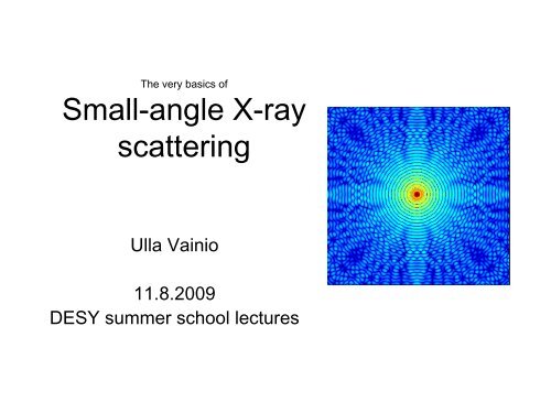
The very basics of<br />
<strong>Small</strong>-<strong>angle</strong> X-<strong>ray</strong><br />
<strong>scattering</strong><br />
Ulla Vainio<br />
11.8.2009<br />
DESY summer school lectures
History<br />
• Beginning of 20 th century x-<strong>ray</strong> diffraction studies of metals show some strange<br />
<strong>scattering</strong> in the middle of the <strong>scattering</strong> pattern. It is later named “small <strong>angle</strong><br />
<strong>scattering</strong>” and becomes a separate field.<br />
• A. Guinier, P. Debye, V. Luzatti, G. Porod and others (1940-1960) develop<br />
interpretation for the basic features in SAXS patterns<br />
• Early 1970s first synchrotron SAXS studies on muscle fibers at DESY, end of 1970s<br />
time resolved studies at DORIS<br />
• Mandelbrot invents the term ‘fractal’ in 1975 and writes a book about it in 1982,<br />
shortly after Bale and Schmidt apply fractal concepts to small <strong>angle</strong> <strong>scattering</strong><br />
(1984) (their paper is cited 498 times 15.7.2009)<br />
• Stuhrmann (1983) builds the first anomalous small-<strong>angle</strong> x-<strong>ray</strong> <strong>scattering</strong> (ASAXS)<br />
beamline in the world at DORIS synchrotron for low x-<strong>ray</strong> energies and studies<br />
biological systems. The idea was copied from anomalous diffraction.<br />
• Haubold et al. (1994) and Simon and Lyon (1994) give examples of ASAXS in book<br />
Resonant anomalous X-<strong>ray</strong> <strong>scattering</strong><br />
• Svergun and his team at DORIS synchrotron develop SAXS software for data<br />
analysis of SAXS from proteins (early 1990s until now). The software becomes<br />
popular also among other SAXS community, because it’s freely available from the<br />
webpage and is very automatized. Glatter develops simultaneously in Austria another<br />
approach to data analysis. In many laboratories people develop their own software.<br />
• SAXS boom: More than 1.2 million articles have been published in this field and 0.8<br />
million of them after year 1999!<br />
Photo source: A. Guinier (1969), Physics today 22, 25.
20<br />
40<br />
60<br />
80<br />
<strong>Small</strong> is big & big is small<br />
Real space<br />
100 0<br />
20 40 60 80 100<br />
(after you take a Fourier transform)<br />
1<br />
0.8<br />
0.6<br />
0.4<br />
0.2<br />
20<br />
40<br />
60<br />
80<br />
100<br />
Inverse space<br />
20 40 60 80 100<br />
10<br />
8<br />
6<br />
4<br />
2
20<br />
40<br />
60<br />
80<br />
<strong>Small</strong> is big & big is small<br />
Real space<br />
100 0<br />
0 50 100<br />
(after you take a Fourier transform)<br />
1<br />
0.8<br />
0.6<br />
0.4<br />
0.2<br />
FFT<br />
-0.3<br />
20<br />
40<br />
0<br />
60<br />
80<br />
Inverse space<br />
0.3<br />
-0.3 0 0.3<br />
Pixel q-pixel (1/pixel)<br />
100<br />
0 50 100<br />
Period d = 10 pixels 1 q-pixel is of length 2π/100 ≈ 0.06 1/pixel<br />
Period d = 2π/q = 2π/(10*0.06) = 10 pixels<br />
x 10 6<br />
8<br />
6<br />
4<br />
2<br />
0
Oriented samples (example)<br />
Organoclay players in polypropylene<br />
Packing material for your food in the grocery store<br />
<strong>Small</strong>-<strong>angle</strong> X-<strong>ray</strong> <strong>scattering</strong><br />
Transmission electron microscopy<br />
Ristolainen et al. (2005) J. Polymer Sci. B-Polymer Physics 43, 1892.<br />
Studied volume ratio SAXS:TEM about 1000 000 000:1
20<br />
40<br />
60<br />
80<br />
Real space<br />
100 0<br />
20 40 60 80 100<br />
Experiment<br />
1<br />
0.8<br />
0.6<br />
0.4<br />
0.2<br />
20<br />
40<br />
60<br />
80<br />
100<br />
Inverse space<br />
Center is covered by beamstop<br />
⇒Large structures are not seen<br />
because of the beamstop and<br />
because of limited detector resolution.<br />
Largest structure observed<br />
about 100 nm.<br />
q<br />
20 40 60 80 100<br />
10<br />
Largest q restricted<br />
by the detector size<br />
⇒<strong>Small</strong>est structure<br />
about 1 nm<br />
8<br />
6<br />
4<br />
2
Example of a SAXS beamline, B1<br />
SAXS detector<br />
100<br />
200<br />
300<br />
400<br />
500<br />
600<br />
0 100 200 300 400<br />
Reference<br />
samples<br />
WAXS<br />
detector<br />
Samples<br />
Monochromatized<br />
X-<strong>ray</strong> beam from a<br />
bending magnet on<br />
DORIS synchrotron
SAXS beamline 7T-MPW SAXS at BESSY<br />
Monochromatized<br />
X-<strong>ray</strong> beam from<br />
a wiggler at<br />
BESSY<br />
synchrotron
0<br />
200<br />
400<br />
600<br />
800<br />
Scattering from a sphere<br />
Real space<br />
1000<br />
200 400 600 800 1000<br />
0<br />
Linear scale<br />
R = 30 pixels<br />
1<br />
0.8<br />
0.6<br />
0.4<br />
0.2<br />
0<br />
200<br />
400<br />
600<br />
800<br />
1000<br />
Inverse space<br />
200 400 600 800 1000<br />
Logarithmic scale<br />
14<br />
12<br />
10<br />
8<br />
6<br />
4<br />
2
50<br />
100<br />
150<br />
200<br />
250<br />
300<br />
350<br />
Projected electron density of a sphere in 3D on 2D plane<br />
Real space<br />
400 0<br />
100 200 300 400<br />
50<br />
40<br />
30<br />
20<br />
10<br />
50<br />
100<br />
150<br />
200<br />
250<br />
300<br />
350<br />
400<br />
Inverse space<br />
ϕ<br />
100 200 300 400<br />
20<br />
15<br />
10<br />
5
Intensity (arbitrary units)<br />
10 10<br />
10 5<br />
10 0<br />
Intensity of a sphere<br />
10 -1<br />
q (1/nm)<br />
Sphere (projected)<br />
Circle in 2d<br />
q -4<br />
q -3<br />
Theoretical<br />
10 0<br />
de = 3<br />
de = 2<br />
Many SAXS curves<br />
follow a power law<br />
I(q) ∝ q -α<br />
α is the power law exponent<br />
d e = Euclidian dimension (3 in real life)
Mass fractal<br />
Menger sponge, mass fractal dimension D m = 2.72<br />
(Source: http://commons.wikimedia.org/wiki/File:Menger_sponge_(Level_1-4).jpg)
Surface fractal<br />
Koch snowflake, surface fractal dimension D s = 1.26 (in d e = 2)<br />
(http://commons.wikimedia.org/wiki/File:KochFlake.png)<br />
In nature the fractals are not mathematically self similar.<br />
Avnir et al. (1998) criticize the use of fractal theory to everything in<br />
their paper in Science “Is the geometry of nature fractal?”<br />
In their opinion the power law should extend more than one order of<br />
magnitude, which may correspond just to one step!
Intensity (arbitrary units)<br />
10 12<br />
10 10<br />
10 8<br />
10 6<br />
10 4<br />
10 -1<br />
Fractals, self similar objects<br />
q (1/nm)<br />
Sphere (projected)<br />
Circle in 2d<br />
q -4<br />
q -3<br />
d e = 2!<br />
D m = mass fractal dimension<br />
D s = surface fractal dimension<br />
d e = Euclidian dimension (3 in real life)<br />
10 0<br />
Many SAXS curves follow a power law<br />
I(q) ∝ q -α<br />
Mass fractal:<br />
α = Dm (0 − 3)<br />
Surface fractal:<br />
α = 2d e –D s (6 − D s , when d e = 3)<br />
Shape D m D s<br />
Line 1 0<br />
Platelet 2 1<br />
Sphere 3 2<br />
Porod law<br />
I(q) ∝ q -4
Form factor & structure factor of particles<br />
• Intensity = form factor x structure<br />
factor I(q) = P(q)S(q)<br />
– This equation works if you have<br />
spherical monodisperse particles,<br />
otherwise the factors may be<br />
more mixed<br />
• Structure factor S(q) is related to<br />
the ordering and distance between<br />
particles<br />
• Form factor tells about the shape<br />
and size of the particles<br />
– In a very dilute solution of<br />
particles I(q) = P(q) because S(q)<br />
= 1<br />
P(q)<br />
Form factor<br />
More on form factors of differently shaped particles:<br />
Jan Skov Pedersen: Analysis of small-<strong>angle</strong> <strong>scattering</strong> data from colloids and polymer solutions:<br />
modeling and least-squares fitting. Advances in Colloid and Interface Science 70 (1997) 171-210.<br />
q<br />
P(q)S(q)<br />
S(q)<br />
q<br />
Structure factor<br />
q<br />
Total <strong>scattering</strong>
Far from ideal<br />
Real systems have nasty effects which disturb the perfect SAXS measurement of particles<br />
• “Concentration effect”: The contribution of<br />
the structure factor increases when the particle<br />
concentration increases (order is increased)<br />
•Concentration series is usually needed<br />
and then one can extrapolate to 0concentration<br />
and determine the particle<br />
shape and size<br />
• “Polyelectrolyte effect”: Charged particles<br />
(negative or positive) in solution give always a<br />
structure factor<br />
•Add salt to suppress the interaction<br />
caused by charges (but this can make the<br />
particles willing to aggregate!)<br />
Problem 1: Why measuring very dilute<br />
solutions is not usually possible?<br />
Problem 2: Why you cannot measure forever<br />
long a sample with a synchrotron beam?<br />
Intensity / concentration<br />
Concentration effect<br />
Concentration<br />
increases<br />
Concentration<br />
increases<br />
q<br />
Polyelectrolyte effect
Scattering from particles<br />
Distance distribution function a.k.a. p(r) function<br />
Svergun & Koch, Rep. Prog. Phys. 66 (2003) 1735–1782<br />
Do you find a mistake here?
Scattering from particles<br />
Distance distribution function a.k.a. p(r) function<br />
Svergun & Koch, Rep. Prog. Phys. 66 (2003) 1735–1782
N(R) = number size distribution<br />
R = size of the particle<br />
Polydispersity*<br />
(*) lot’s of different sized particles<br />
Vainio et al. (2006) Latvian journal of physics and technical sciences 4, 14.
How to perform an experiment?
Example: Ontime chemistry at beamline A2<br />
by Vivian Rebbin<br />
• The formation of micelles is observed<br />
with SAXS. With ongoing<br />
condensation of silica, the ordering<br />
process starts.
Courtesy of Vivian Rebbin<br />
On-time chemistry with SAXS<br />
Sample containing the triblock copolymer Pluronic P123 as structuredirecting<br />
agent, 1,4-bis(triethoxysilyl)benzene as silica source, pH = 1,<br />
increasing temperature<br />
Structure: From diffuse <strong>scattering</strong> to 2d hexagonal
Example: Spinodal decomposition<br />
in amorphous metals<br />
3d atom probe of yttrium atoms<br />
N. Mattern et al. Acta Materialia 57 (2009) 903–908
Anomalous small-<strong>angle</strong> X-<strong>ray</strong> <strong>scattering</strong> (ASAXS)<br />
“What is the unit of intensity?!”
Two-phase approximation<br />
• Only mean electron density difference counts<br />
B<br />
A (vacuum)<br />
Absorption<br />
Anomalous<br />
<strong>scattering</strong> factors<br />
Intensity (log scale)<br />
Conclusion: f’’<br />
Pink atoms are distributed<br />
homogeneously into one phase<br />
(in small-<strong>angle</strong> <strong>scattering</strong>)!<br />
f’<br />
Energy<br />
Absorption edge of pink element<br />
I(E1)<br />
I(E2)<br />
I(E1) - I(E2)<br />
Scattering <strong>angle</strong> (log scale)<br />
Intensity ∝ |ρB (E) - ρA |<br />
Real space Fourier space<br />
2<br />
Atomic <strong>scattering</strong> factor<br />
Z + f’(E) + if’’(E)
Three or more phases<br />
• One more phase now ASAXS can help<br />
S bb<br />
S pp<br />
S bp<br />
B<br />
A (vacuum)<br />
Absorption<br />
Anomalous<br />
<strong>scattering</strong> factors<br />
Energy<br />
f’’<br />
f’<br />
Intensity (log scale)<br />
Absorption edge of pink element<br />
I(E1)<br />
I(E2)<br />
I(E1) - I(E2)<br />
Scattering <strong>angle</strong> (log scale)<br />
Intensity(E) = Spp (E) + Sbp (E) + Sbb Real space Fourier space<br />
Partial structure factors<br />
Assumption: Atoms in phase B have constant f (no absorption edge at this energy)!
Lignosulfonate, the brown stuff in a tree<br />
Trees<br />
Pulp & paper mills<br />
Paper products,<br />
cellulose<br />
Lignin (in the form of<br />
Kraft lignin or lignosulfonate)
Intensity (relative units)<br />
10 10<br />
10 8<br />
10 6<br />
10 4<br />
Simple model<br />
Fourier transform<br />
10 -2 10 2<br />
10 -1<br />
Lignosulfonate ASAXS<br />
Partial structure factors<br />
q (1/nm)<br />
10 0<br />
Rubidium (Rb) counterions<br />
Rb<br />
Rb-LS<br />
LS<br />
Total<br />
10 1<br />
Intensity (arb. units)<br />
10 2<br />
10 1<br />
10 0<br />
10 -1<br />
10 -2<br />
10 -3<br />
10 -4<br />
Comparison to experiment<br />
model<br />
experiment<br />
10 0<br />
q (1/nm)<br />
I(E1)-I(E2) (model)<br />
I(E1) (model)<br />
I(E1)-I(E2) (Rb-LS)<br />
I(E1) (Rb-LS)<br />
10 1
SAXS beamlines at DORIS III<br />
• SAXS at A2, B1<br />
• USAXS at BW4<br />
• ASAXS at B1<br />
• GISAXS at BW4<br />
• Rheology & SAXS BW1
Books about SAXS<br />
(in the order I like them most)<br />
• A Guinier (1956/1994) X-<strong>ray</strong> diffraction. In crystals, imperfect crystals, and<br />
amorphous bodies. Chapter 10 <strong>Small</strong>-<strong>angle</strong> x-<strong>ray</strong> <strong>scattering</strong>.<br />
• Glatter & Kratky (ed.) (1982) <strong>Small</strong> Angle X-<strong>ray</strong> Scattering.<br />
(http://physchem.kfunigraz.ac.at/sm/Software.htm)<br />
• Feigin & Svergun (1987) Structure analysis by small-<strong>angle</strong> X-<strong>ray</strong> and neutron<br />
<strong>scattering</strong>.<br />
(http://www.embl-hamburg.de/ExternalInfo/Research/Sax/reprints/feigin_svergun_1987.pdf)<br />
• Guinier and Fournet (1955) <strong>Small</strong> <strong>angle</strong> <strong>scattering</strong> of X-<strong>ray</strong>s.<br />
ASAXS review<br />
• D. Tatchev (2008) Structure analysis of multiphase systems by anomalous small<strong>angle</strong><br />
X-<strong>ray</strong> <strong>scattering</strong>. Philosophical Magazine 88, 1751 – 1772.
Programs to analyze SAXS data<br />
But be careful: you get always a result. It doesn’t mean it’s right.<br />
Examples of standalone programs that one can get from the web:<br />
• Dimitri Svergun’s ATSAS package at EMBL<br />
http://www.embl-hamburg.de/ExternalInfo/Research/Sax/software.html<br />
• Joachim Kohlbrecher’s SASfit at PSI http://kur.web.psi.ch/sans1/SANSSoft/sasfit.html<br />
• S. Förster’s Scatter at Uni Hamburg<br />
http://www.chemie.uni-hamburg.de/pc/sfoerster/software.html<br />
• Richard Heenan’s FISH at ISIS<br />
http://www.isis.rl.ac.uk/LargeScale/LOQ/FISH/FISH_intro.htm<br />
• G. Fritz’s Singlebody http://physchem.kfunigraz.ac.at/sm/Software.htm<br />
• More programs (standalone and those requiring Matlab or Igor) at<br />
http://www.small-<strong>angle</strong>.ac.uk/small-<strong>angle</strong>/Software.html