Spirulina Abstracts Draft 6 - Cyanotech
Spirulina Abstracts Draft 6 - Cyanotech
Spirulina Abstracts Draft 6 - Cyanotech

You also want an ePaper? Increase the reach of your titles
YUMPU automatically turns print PDFs into web optimized ePapers that Google loves.
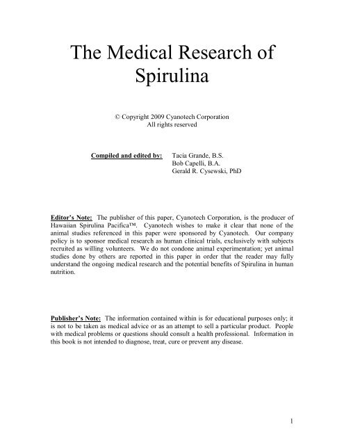
The Medical Research of<br />
<strong>Spirulina</strong><br />
© Copyright 2009 <strong>Cyanotech</strong> Corporation<br />
All rights reserved<br />
Compiled and edited by: Tacia Grande, B.S.<br />
Bob Capelli, B.A.<br />
Gerald R. Cysewski, PhD<br />
Editor’s Note: The publisher of this paper, <strong>Cyanotech</strong> Corporation, is the producer of<br />
Hawaiian <strong>Spirulina</strong> Pacifica. <strong>Cyanotech</strong> wishes to make it clear that none of the<br />
animal studies referenced in this paper were sponsored by <strong>Cyanotech</strong>. Our company<br />
policy is to sponsor medical research as human clinical trials, exclusively with subjects<br />
recruited as willing volunteers. We do not condone animal experimentation; yet animal<br />
studies done by others are reported in this paper in order that the reader may fully<br />
understand the ongoing medical research and the potential benefits of <strong>Spirulina</strong> in human<br />
nutrition.<br />
Publisher’s Note: The information contained within is for educational purposes only; it<br />
is not to be taken as medical advice or as an attempt to sell a particular product. People<br />
with medical problems or questions should consult a health professional. Information in<br />
this book is not intended to diagnose, treat, cure or prevent any disease.<br />
1
Table of Contents<br />
Section One: Medical Research on <strong>Spirulina</strong><br />
I. Introduction……………………………………………………………………4<br />
II. Antioxidant……………………………………………………………………5<br />
III. Anti-Inflammatory………………………………………………………….. 15<br />
IV. Immunity……………………………………………………………………..22<br />
V. Chemopreventative…………………………………………………………..43<br />
VI. Diabetes………………………………………………………………………68<br />
VII. Anti-Viral…………………………………………………………………….73<br />
VIII. Anemia and Blood Improvement………………………………………….…81<br />
IX. Cardioprotective (Heart)……………………………………………….…….87<br />
X. Hepatoprotective (Liver)……………………………………………………108<br />
XI. Renal Protective (Kidneys)…………………………………………………117<br />
XII. Neuroprotective (Brain)…………………………………………………….127<br />
XIII. Heavy Metal Removal……………………………………………………...132<br />
XIV. Allergies………………………………………………………….…………140<br />
XV. Additional Areas of <strong>Spirulina</strong> Research……………………………………147<br />
a. Energy/Endurance/Muscle Damage<br />
b. Pregnancy<br />
c. H.Pylori (Gastric Ulcers)<br />
d. Protein Absorption<br />
e. Eye (Cataract)<br />
f. Trace Minerals<br />
g. Malnutrition<br />
h. Anti-Depressant<br />
i. Muscle Protein Synthesis<br />
j. Arthritis<br />
k. General Health<br />
2
Section Two: Medical Research on the<br />
Key Nutrients in <strong>Spirulina</strong><br />
XVI. Introduction to Research on the Key Nutrients in <strong>Spirulina</strong>……….….……161<br />
XVII. Beta Carotene<br />
a. Beta Carotene and Skin Health…………………………………….…...162<br />
b. Beta Carotene and Immunity……………………………………….…..182<br />
c. Beta Carotene as a Chemoprentative…………………...........................197<br />
d. Beta Carotene and Eye Health…………………………………….……217<br />
XVIII. Zeaxanthin<br />
a. Zeaxanthin and Eye Health……………………………………………..234<br />
b. Zeaxanthin and Brain Health…………………………………………...253<br />
c. Zeaxanthin as a Hepatoprotective………………………………………256<br />
XIX. Iron<br />
a. Iron and Immunity……………………………………………………...261<br />
b. Iron and Cognitive Function …………………………………………...281<br />
c. Iron and Anemia………………………………………………………..301<br />
d. Iron and Blood Improvement…………………………………….……..322<br />
XX. Vitamin B-12<br />
a. Vitamin B-12 and Brain Health……………………………………...…339<br />
b. Vitamin B-12 and Psychiatric Disorders…………………………….....355<br />
c. Vitamin B-12 and Anemia…………………………………………...…367<br />
XXI. Vitamin K<br />
a. Viamin K and Bone Health………………………………………….….381<br />
b. Vitamin K and Blood Improvement……………………………………401<br />
c. Vitamin K and Alzheimer’s…………………………………………….418<br />
XXII. Superoxide Dismutase<br />
a. SOD Introduction ………………………………………………………421<br />
b. SOD and Arthritis………………………………………………………422<br />
c. SOD and Skin Health…………………………………………………...433<br />
3
Introduction<br />
There has been an extensive amount of research on the species Arthrospira Platensis,<br />
more commonly known as “<strong>Spirulina</strong>.” This research dates back decades and has<br />
been conducted at universities, at government sponsored research facilities, as well as<br />
by private researchers throughout the world.<br />
<strong>Cyanotech</strong> Corporation* feels that it is important to have a library of this research<br />
available for interested persons; hence we have created this document. Below the<br />
reader will find selected research abstracts on the health benefits of <strong>Spirulina</strong> and<br />
some of the major nutrients contained within Spiruilna. It was not practical to include<br />
full studies due to the massive amount of literature available; in addition, it was not<br />
practical to include all available abstracts. Particularly in the case of some of<br />
<strong>Spirulina</strong>’s key nutrients, there were hundreds of abstracts available, so editing was<br />
necessary in order to present a manageable document.<br />
The abstracts are presented according to health benefit as noted in the table of<br />
contents. In the case of studies that focused on more than one health benefit, the<br />
study is categorized according to the primary area of research within the abstract. All<br />
of the studies contained in this document are published and most can be found at<br />
www.pubmed.com<br />
Any questions may be directed to <strong>Cyanotech</strong> Corporation, Kailua-Kona, Hawaii,<br />
USA, by e-mail at info@cyanotech.com or by telephone at 808.326.1353.<br />
• <strong>Cyanotech</strong> Corporation is the producer of Hawaiian <strong>Spirulina</strong> Pacifica since<br />
1984. Hawaiian <strong>Spirulina</strong> Pacifica is widely regarded as the world’s highest<br />
quality <strong>Spirulina</strong>, with far superior levels of key nutrients than all other<br />
<strong>Spirulina</strong> products.<br />
4
Antioxidant Research<br />
Food Chem Toxicol. 2007 Dec;45(12):2412-9. Epub 2007 Jun 28.<br />
Antioxidant<br />
<strong>Spirulina</strong> fusiformis provides protection against mercuric chloride<br />
induced oxidative stress in Swiss albino mice.<br />
Sharma MK, Sharma A, Kumar A, Kumar M.<br />
Department of Zoology, RLS Government (PG) College, Kaladera, Jaipur<br />
303801, India. mkshrma@hotmail.com<br />
Oxidative stress induced by mercuric chloride (5 mg/kg body weight i.p.) in mice<br />
substantially increases the lipid peroxidation level along with corresponding<br />
decrease in the reduced glutathione and various antioxidant enzymes in liver and<br />
increase in serum transaminases activity. Supplementation of <strong>Spirulina</strong> (800<br />
mg/kg body weight orally, in olive oil, along with mercuric chloride) for 40 days<br />
resulted in decreased LPO level, serum glutamate oxaloacetate and serum<br />
glutamate pyruvate transaminase activity along with increase in liver GSH level.<br />
The activities of antioxidants enzymes superoxide dismutase, catalase and<br />
glutathione-S-transferase were also concomitantly restored to near normal level<br />
by <strong>Spirulina</strong> supplementation to mercuric chloride intoxicated mice. The results<br />
clearly demonstrate that <strong>Spirulina</strong> treatment augments the antioxidants defense<br />
mechanism in mercuric chloride induced toxicity and provides evidence that it<br />
may have a therapeutic role in free radical mediated diseases.<br />
Publication Types:<br />
• Research Support, Non-U.S. Gov't<br />
PMID: 17706852 [PubMed - indexed for MEDLINE]<br />
5
Biochem Biophys Res Commun. 2000 Aug 18;275(1):20-5.<br />
Antioxidant<br />
C-phycocyanin: a potent peroxyl radical scavenger in vivo and in<br />
vitro.<br />
Bhat VB, Madyastha KM.<br />
Department of Organic Chemistry, Indian Institute of Science, Bangalore, 560<br />
012, India.<br />
C-Phycocyanin (from <strong>Spirulina</strong> platensis) effectively inhibited CCl(4)-induced<br />
lipid peroxidation in rat liver in vivo. Both native and reduced phycocyanin<br />
significantly inhibited peroxyl radical-induced lipid peroxidation in rat liver<br />
microsomes and the inhibition was concentration dependent with an IC(50) of<br />
11.35 and 12.7 microM, respectively. The radical scavenging property of<br />
phycocyanin was established by studying its reactivity with peroxyl and hydroxyl<br />
radicals and also by competition kinetics of crocin bleaching. These studies have<br />
demonstrated that phycocyanin is a potent peroxyl radical scavenger with an<br />
IC(50) of 5.0 microM and the rate constant ratios obtained for phycocyanin and<br />
uric acid (a known peroxyl radical scavenger) were 1.54 and 3.5, respectively.<br />
These studies clearly suggest that the covalently linked chromophore,<br />
phycocyanobilin, is involved in the antioxidant and radical scavenging activity of<br />
phycocyanin. Copyright 2000 Academic Press.<br />
Publication Types:<br />
• Research Support, Non-U.S. Gov't<br />
PMID: 10944434 [PubMed - indexed for MEDLINE]<br />
6
J Sep Sci. 2005 Jun;28(9-10):1031-8.<br />
Antioxidant<br />
Characterization via liquid chromatography coupled to diode array<br />
detector and tandem mass spectrometry of supercritical fluid<br />
antioxidant extracts of <strong>Spirulina</strong> platensis microalga.<br />
Mendiola JA, Marín FR, Hernández SF, Arredondo BO, Señoráns FJ,<br />
Ibañez E, Reglero G.<br />
Sección de Ciencias de la Alimentación, Universidad Autónoma de Madrid,<br />
Ciudad Universitaria de Cantoblanco, 28049 Madrid, Spain.<br />
<strong>Spirulina</strong> platensis microalga has been extracted on a pilot scale plant using<br />
supercritical fluid extraction (SFE) under various extraction conditions. The<br />
extraction yield and the antioxidant activity of the extracts were evaluated in order<br />
to select those extracts with both the highest antioxidant capacity and a good<br />
extraction yield. These extracts were characterized using LC coupled to diode<br />
array detection (DAD) and LC coupled to mass spectrometry (MS) with two<br />
different interfaces, atmospheric pressure chemical ionization (APCI) and<br />
electrospray (ESI) which allowed us to perform tandem MS by using an ion trap<br />
analyzer. The best extraction conditions were as follows: CO2 with 10% of<br />
modifier (ethanol) as extraction solvent, 55 degrees C (extraction temperature)<br />
and 220 bar (extraction pressure). Fractionation was achieved by cascade<br />
depressurization providing two extracts with different activity and chemical<br />
composition. Several compounds have been identified in the extracts,<br />
corresponding to different carotenoids previously identified in <strong>Spirulina</strong> platensis<br />
microalga along with chlorophyll a and some degradation products. Also, the<br />
structure of some phenolic compounds could be tentatively identified. The<br />
antioxidant activity of the extracts could be attributed to some of the above<br />
mentioned compounds.<br />
Publication Types:<br />
• Research Support, Non-U.S. Gov't<br />
PMID: 16013830 [PubMed - indexed for MEDLINE]<br />
7
J Agric Food Chem. 2005 May 18;53(10):4207-12.<br />
Antioxidant<br />
Antioxidant and antiproliferative activities of <strong>Spirulina</strong> and<br />
Chlorella water extracts.<br />
Wu LC, Ho JA, Shieh MC, Lu IW.<br />
Department of Applied Chemistry, National Chi-Nan University, Puli, Nantou,<br />
Taiwan. lw25@ncnu.edu.tw<br />
Liver fibrosis is a chronic liver disease that will further develop to cirrhosis if<br />
severe damage continues to form. A potential treatment for liver fibrosis is to<br />
inhibit activated hepatic stellate cell (HSC) proliferation and, subsequently, to<br />
induce HSC apoptosis. It has been reported that antioxidants are able to inhibit the<br />
proliferation of HSCs. In this study, the aqueous extract of spirulina was chosen<br />
as the source of antioxidant to investigate the inhibitory effect on the proliferation<br />
of HSC. The growth inhibitory effects of aqueous spirulina and chlorella extract<br />
on human liver cancer cells, HepG2, were also studied and compared in pairs.<br />
Results indicated that the total phenol content of spirulina was almost five times<br />
greater than that of chlorella (6.86 +/- 0.58 vs 1.44 +/- 0.04 mg tannic acid<br />
equivalent/g of algae powder, respectively). The antioxidant activity of spirulina<br />
determined by the ABTS*+ method was higher than chlorella (EC50: 72.44 +/-<br />
0.24 micromol of trolox equivalent/g of spirulina extract vs 56.09 +/- 1.99<br />
micromol of trolox equivalent/g of chlorella extract). Results of DPPH* assay<br />
also showed a similar trend as the ABTS*+ assay (EC50: 19.39 +/- 0.65 micromol<br />
of ascorbic acid equivalent/g of spirulina extract vs 14.04 +/- 1.06 micromol of<br />
ascorbic acid equivalent/g of chlorella extract). The aqueous extracts of these two<br />
algae both showed antiproliferative effects on HSC and HepG2, but spirulina was<br />
a stronger inhibitor than chlorella. Annexin-V staining showed that aqueous<br />
extract of spirulina induced apoptosis of HSC after 12 h of treatment. In addition,<br />
the aqueous extract of spirulina triggered a cell cycle arrest of HSC at the G2/M<br />
phase.<br />
Publication Types:<br />
• Comparative Study<br />
• Research Support, Non-U.S. Gov't<br />
PMID: 15884862 [PubMed - indexed for MEDLINE]<br />
8
J Chromatogr A. 2004 Aug 27;1047(2):195-203.<br />
Antioxidant<br />
Pressurized liquid extracts from <strong>Spirulina</strong> platensis microalga.<br />
Determination of their antioxidant activity and preliminary analysis<br />
by micellar electrokinetic chromatography.<br />
Herrero M, Ibáñez E, Cifuentes A, Señoráns J.<br />
Instituto de Fermentaciones Industriales (CSIC), Juan de la Cierva 3, 28006<br />
Madrid, Spain.<br />
In this work, different extracts from the microalga <strong>Spirulina</strong> platensis are obtained<br />
using pressurized liquid extraction (PLE) and four different solvents (hexane,<br />
light petroleum, ethanol and water). Different extraction temperatures (115 and<br />
170 degrees C) were tested using extraction times ranging from 9 to 15 min. The<br />
antioxidant activity of the different extracts is determined by means of an in vitro<br />
assay using a free radical method. Moreover, a new and fast method is developed<br />
using micellar electrokinetic chromatography with diode array detection (MEKC-<br />
DAD) to provide a preliminary analysis on the composition of the extracts. This<br />
combined application (i.e., in vitro assays plus MEKC-DAD) allowed the fast<br />
characterization of the extracts based on their antioxidant activity and the UV-vis<br />
spectra of the different compounds found in the extracts. To our knowledge, this<br />
work shows for the first time the great possibilities of the combined use of PLE-in<br />
vitro assay-MEKC-DAD to investigate natural sources of antioxidants.<br />
Publication Types:<br />
• Research Support, Non-U.S. Gov't<br />
PMID: 15460249 [PubMed - indexed for MEDLINE]<br />
9
J Neural Transm. 2007 Sep;114(9):1217-25. Epub 2007 May 26.<br />
Antioxidant<br />
Effect of spirulina maxima on the haloperidol induced tardive<br />
dyskinesia and oxidative stress in rats.<br />
Thaakur SR, Jyothi B.<br />
Department of Pharmacology, School of Pharmaceutical Sciences, Sri Padmavathi<br />
Mahila Viswa Vidyalayam, Andhra Pradesh, India. drsanthrani@gmail.com<br />
Haloperidol is a widely used neuroleptic drug for the treatment of acute and<br />
chronic psychosis. The use of haloperidol is limited by extrapyramidal movement<br />
disorders such as Parkinsonism, akathesia, dystonia, and tardive dyskinesia (TD).<br />
Treatment with haloperidol increases oxyradicals which are implicated in TD.<br />
<strong>Spirulina</strong> is widely used as nutritional supplement rich in proteins and<br />
antioxidants. The present study is proposed to study the effect of spirulina on<br />
haloperidol induced TD and oxidative stress by studying TD, various enzymatic<br />
and nonenzymatic antioxidants and lipid peroxidation. Haloperidol 1 mg/kg/i.p<br />
was used to induce vacuous chewing movements in rats. <strong>Spirulina</strong> maxima<br />
suspended in 1% between 80 at a dose of 45, 90 and 180 mg/kg were<br />
administered by gavage along with haloperidol from 21st day to 49th day of<br />
treatment. <strong>Spirulina</strong> supplementation at a dose of 180 mg/kg significantly<br />
improved enzymatic and nonenzymatic antioxidants and decreased the tardive<br />
dyskinesia induced by haloperidol. In conclusion, the results of present<br />
investigation suggest that spirulina decreases haloperidol induced oxidative stress<br />
and TD by many mechanisms as it is cocktail of antioxidants. On chronic use it<br />
may inhibit haloperidol induced reduced expression of DNA thereby increases the<br />
expression of enzymatic and nonenzymatic antioxidants and protects against<br />
oxidative stress induced neurodegeneration and TD.<br />
PMID: 17530160 [PubMed - in process]<br />
10
Indian J Biochem Biophys. 2006 Feb;43(1):25-31.<br />
Antioxidant<br />
Antioxidant potential of C-phycocyanin isolated from cyanobacterial<br />
species Lyngbya, Phormidium and <strong>Spirulina</strong> spp.<br />
Patel A, Mishra S, Ghosh PK.<br />
Central Salt and Marine Chemicals Research Institute, GB Marg, Bhavnagar,<br />
India.<br />
The antioxidant activity of C-Phycocyanin (C-PC) isolated from three<br />
cyanobacterial species Lyngbya (marine), Phormidium (marine) and <strong>Spirulina</strong><br />
(fresh water) was studied in vitro. The results demonstrate that C-PCs from<br />
Lyngbya, Phormidium and <strong>Spirulina</strong> spp. are able to scavenge peroxyl radicals<br />
(determined by crocin bleaching assay) with relative rate constant ratio of 3.13,<br />
1.89 and 1.8, respectively. C-PCs also scavenge hydroxyl radicals (determined by<br />
deoxyribose degradation assay) with second order rate constant values of 7.87 x<br />
10(10), 9.58 x 10(10) and 6.42 x 10(10), respectively. Interestingly, Lyngbya C-<br />
PC is found to be an effective inhibitor of peroxyl radicals (IC50 6.63 microM),<br />
as compared to <strong>Spirulina</strong> (IC50 12.15 microM) and Phormidium C-PC (IC50<br />
12.74 microM) and is close to uric acid (IC50 2.15 microM). Further, the studies<br />
suggest that the covalently-linked tetrapyrrole chromophore phycocyanobilin is<br />
involved in the radical scavenging activity of C-PC. The electron spin resonance<br />
(ESR) spectra of C-PCs indicate the presence of free radical active sites, which<br />
may play an important role in its radical scavenging property. This is the first<br />
report on the ESR activity of native C-PCs without perturbations that can cause<br />
radical formation.<br />
Publication Types:<br />
• Research Support, Non-U.S. Gov't<br />
PMID: 16955748 [PubMed - indexed for MEDLINE]<br />
11
J Appl Microbiol. 2009 Apr;106(4):1093-100. Epub 2009 Feb 23.<br />
Antioxidant<br />
A potent anti-oxidant property: fluorescent recombinant alphaphycocyanin<br />
of <strong>Spirulina</strong>.<br />
Guan XY, Zhang WJ, Zhang XW, Li YX, Wang JF, Lin HZ, Tang XX, Qin S<br />
.<br />
Ocean University of China, Qingdao, China.<br />
AIMS: To express and product a fluorescent antioxidant holo-alpha-phycocyanin<br />
(PC) of <strong>Spirulina</strong> platensis (Sp) with His-tag (rHHPC; recombinant holo-alphaphycocyaninof<br />
<strong>Spirulina</strong> platensis with His-tag) in 5-l bench scale. METHODS<br />
AND RESULTS: A vector harbouring two cassettes was constructed: cpcA along<br />
with cpcE-cpcF in one cassette; ho1-pcyA in the other cassette. Lyases CpcE/F of<br />
Synechocystis sp. PCC6803 (S6) could catalyse the 82 site Cys in apo-alpha-PC<br />
of Sp linking with bilin chromophores, and rHHPC was biosynthesized in<br />
Escherichia coli BL21. The constant feeding mode was adopted, and transformant<br />
reached the biomass of rHHPC up to 0.55 g l(-1) broth in 5-litre bench scale.<br />
rHHPC was purified by Ni(2+) affinity column conveniently. The absorbance and<br />
the fluorescence emission spectra of rHHPC had lambda(max) at 621 and 650 nm,<br />
respectively. The IC(50) values of rHHPC were 277.5 +/- 25.8 microg ml(-1)<br />
against hydroxyl radicals and 20.8 +/- 2.2 microg ml(-1) against peroxyl radicals.<br />
CONCLUSIONS: Combinational biosynthesis of rHHPC was feasible, and the<br />
constant feeding mode was adopted to produce good yields of rHHPC.<br />
Fluorescent rHHPC with several unique qualitative and quantitative features was<br />
effective on scavenging hydroxyl and peroxyl radicals. SIGNIFICANCE AND<br />
IMPACT OF THE STUDY: A potent antioxidant rHHPC was co-expressed,<br />
produced and characterized for nutritional and pharmacological values, which<br />
would help to develop phycobiliproteins' applications in their fluorescent and<br />
biological activities.<br />
PMID: 19239531 [PubMed - in process]<br />
12
Phytother Res. 2008 May;22(5):627-33.<br />
Antioxidant<br />
Antioxidant potential of selected <strong>Spirulina</strong> platensis preparations.<br />
Dartsch PC.<br />
Dartsch Scientific GmbH, Institut für zellbiologische Testsysteme, Horb am<br />
Neckar, Germany. pc.dartsch@dartsch-scientific.com<br />
Recent studies suggest that <strong>Spirulina</strong>, a unicellular blue-green alga, may have a<br />
variety of health benefits and therapeutic properties and is also capable of acting<br />
as an antioxidant and antiinflammatory agent. In this study, a cell-free and a cellbased<br />
test assay were used to examine the antioxidant and antiinflammatory<br />
properties of four selected <strong>Spirulina</strong> platensis preparations: (1) Biospirulina, (2)<br />
SpiruComplex, a preparation with naturally bound selenium, chromium and zinc,<br />
(3) SpiruZink, a preparation with naturally bound zinc, (4) Zinkspirulina +<br />
Acerola, a preparation with naturally bound zinc and acerola powder. The cellfree<br />
test assay used potassium superoxide as a donor for superoxide radicals,<br />
whereas the cell-based test assay used the formation of intracellular superoxide<br />
radicals of functional neutrophils upon stimulation by phorbol-12-myristate-13acetate<br />
as a model to investigate the potential of <strong>Spirulina</strong> preparations to<br />
inactivate superoxide radicals. In accordance with the recommended daily dosage,<br />
test concentrations ranging from 50 to 1000 microg/mL were chosen. The results<br />
showed a dose-dependent inactivation of free superoxide radicals (antioxidant<br />
effect) as well as an antiinflammatory effect characterized by a dose-dependent<br />
reduction of the metabolic activity of functional neutrophils and a dose-dependent<br />
inactivation of superoxide radicals generated during an oxidative burst. The<br />
results demonstrate that the tested <strong>Spirulina</strong> preparations have a high antioxidant<br />
and antiinflammatory potential. Especially SpiruZink and Zinkspirulina + Acerola<br />
might be useful as a supportive therapeutic approach for reducing oxidative stress<br />
and/or the generation of oxygen radicals in the course of inflammatory processes.<br />
PMID: 18398928 [PubMed - indexed for MEDLINE]<br />
13
Acta Pol Pharm. 2007 Jul-Aug;64(4):335-44.<br />
Antioxidant<br />
In vitro evaluation of protective effects of ascorbic acid and water<br />
extract of <strong>Spirulina</strong> plantesis (blue green algae) on 5-fluorouracilinduced<br />
lipid peroxidation.<br />
Ray S, Roy K, Sengupta C.<br />
Division of Pharmaceutical Chemistry, Himalayan Pharmacy Institute, Majhitar,<br />
Rangpo, East Sikkim, 737 136, India. supratimray_in@yahoo.co.in<br />
Considering drug-induced lipid peroxidation as a possible mediator of druginduced<br />
toxicity and exploiting the free radical scavenging action of antioxidants,<br />
the present study was designed to evaluate the protective effects of ascorbic acid<br />
(AA) and water extract of <strong>Spirulina</strong> plantesis (SP) to minimize 5-fluorouracil (5-<br />
FU)-induced lipid peroxidation. The study has been performed in vitro using goat<br />
liver as an experimental model. This evaluation was done by measuring the<br />
malondialdehyde (MDA), reduced glutathione (GSH), 4-hydroxy-2-nonenal (4-<br />
HNE) and nitric oxide (NO) content of the tissue as markers of lipid peroxidation.<br />
The results suggest that ascorbic acid and water extract of <strong>Spirulina</strong> plantesis<br />
could suppress the 5-FU-induced lipid peroxidation to a significant extent.<br />
Publication Types:<br />
• In Vitro<br />
PMID: 18536159 [PubMed - indexed for MEDLINE]<br />
14
J Med Food. 2007 Dec;10(4):566-70.<br />
Anti-Inflammatory<br />
Clinical potential of <strong>Spirulina</strong> as a source of phycocyanobilin.<br />
McCarty MF.<br />
NutriGuard Research, Encinitas, CA 92024, USA. mccarty@pantox.com<br />
Recent research reveals that free bilirubin functions physiologically as a potent<br />
inhibitor of NADPH oxidase activity. The chromophore phycocyanobilin (PCB),<br />
found in blue-green algae and cyanobacteria such as <strong>Spirulina</strong>, also has been<br />
found to be a potent inhibitor of this enzyme complex, likely because in<br />
mammalian cells it is rapidly reduced to phycocyanorubin, a close homolog of<br />
bilirubin. In light of the protean roles of NADPH oxidase activation in pathology,<br />
it thus appears likely that PCB supplementation may have versatile potential in<br />
prevention and therapy -- particularly in light of rodent studies demonstrating that<br />
orally administered <strong>Spirulina</strong> or phycocyanin (the <strong>Spirulina</strong> holoprotein that<br />
contains PCB) can exert a wide range of anti-inflammatory effects. Until PCBenriched<br />
<strong>Spirulina</strong> extracts or synthetically produced PCB are commercially<br />
available, the most feasible and least expensive way to administer PCB is by<br />
ingestion of whole <strong>Spirulina</strong>. A heaping tablespoon (about 15 g) of <strong>Spirulina</strong> can<br />
be expected to provide about 100 mg of PCB. By extrapolating from rodent<br />
studies, it can be concluded that an intake of 2 heaping tablespoons daily would<br />
be likely to have important antioxidant activity in humans -- assuming that<br />
humans and rodents digest and absorb <strong>Spirulina</strong>-bound PCB in a comparable<br />
manner. An intake of this magnitude can be clinically feasible if <strong>Spirulina</strong> is<br />
incorporated into "smoothies" featuring such ingredients as soy milk, fruit juices,<br />
and whole fruits. Such a regimen should be evaluated in clinical syndromes<br />
characterized and in part mediated by NADPH oxidase overactivity in affected<br />
tissues.<br />
Publication Types:<br />
• Review<br />
PMID: 18158824 [PubMed - indexed for MEDLINE]<br />
Anti-Inflammatory<br />
15
Anesth Analg. 2009 Apr;108(4):1303-10.<br />
Antiinflammatory and antihyperalgesic activity of C-phycocyanin.<br />
Shih CM, Cheng SN, Wong CS, Kuo YL, Chou TC.<br />
Chia-Yi Christian Hospital, Taipei, Taiwan, Republic of China.<br />
BACKGROUND: C-phycocyanin (C-PC), a biliprotein found in blue green algae,<br />
such as <strong>Spirulina</strong> platensis, is often used as a dietary nutritional supplement due to<br />
its various therapeutic values. In addition, the antiinflammatory activity of C-PC<br />
partly through inhibition of proinflammatory cytokine formation, inducible nitric<br />
oxide synthase (iNOS) and cyclooxygeanase-2 (COX-2) expression has been<br />
demonstrated in many in vitro and in vivo studies. However, whether C-PC also<br />
has antihyperalgesic activity in inflammatory nociception has not been<br />
investigated. METHODS: Using a carrageenan-induced thermal hyperalgesia<br />
model, we evaluated the effect of C-PC on nociception by measuring paw<br />
withdrawal latency. To clarify the mechanisms involved, the expression of iNOS<br />
and COX-2 and the formation of nitrate and tumor necrosis factor-alpha (TNFalpha)<br />
in the rat paw were determined. RESULTS: Pre- or posttreatment with C-<br />
PC (30 or 50 mg/kg, IP) significantly attenuated carrageenan-induced<br />
inflammatory nociception and the induction of iNOS and COX-2 at the late phase,<br />
(4 h) accompanied by an inhibition of the formation of TNF-alpha, prostaglandin<br />
E(2), nitrate and myeloperoxidase activity. CONCLUSIONS: Based on these<br />
results, it is suggested that the inhibition of NO and prostaglandin E(2) overproduction<br />
through suppressing iNOS and COX-2 induction and attenuation of<br />
TNF-alpha formation and neutrophil infiltration into inflammatory sites by C-PC<br />
may contribute, at least in part, to its antihyperalgesic activity.<br />
Publication Types:<br />
• Research Support, Non-U.S. Gov't<br />
PMID: 19299804 [PubMed - indexed for MEDLINE]<br />
Anti-Inflammatory<br />
16
Biochem Biophys Res Commun. 2000 Nov 2;277(3):599-603.<br />
Selective inhibition of cyclooxygenase-2 by C-phycocyanin, a<br />
biliprotein from <strong>Spirulina</strong> platensis.<br />
Reddy CM, Bhat VB, Kiranmai G, Reddy MN, Reddanna P, Madyastha KM<br />
.<br />
Department of Organic Chemistry, Indian Institute of Science, Bangalore, 560<br />
012, India.<br />
We report data from two related assay systems (isolated enzyme assays and whole<br />
blood assays) that C-phycocyanin a biliprotein from <strong>Spirulina</strong> platensis is a<br />
selective inhibitor of cyclooxygenase-2 (COX-2) with a very low IC(50) COX-<br />
2/IC(50) COX-1 ratio (0.04). The extent of inhibition depends on the period of<br />
preincubation of phycocyanin with COX-2, but without any effect on the period<br />
of preincubation with COX-1. The IC(50) value obtained for the inhibition of<br />
COX-2 by phycocyanin is much lower (180 nM) as compared to those of<br />
celecoxib (255 nM) and rofecoxib (401 nM), the well-known selective COX-2<br />
inhibitors. In the human whole blood assay, phycocyanin very efficiently<br />
inhibited COX-2 with an IC(50) value of 80 nM. Reduced phycocyanin and<br />
phycocyanobilin, the chromophore of phycocyanin are poor inhibitors of COX-2<br />
without COX-2 selectivity. This suggests that apoprotein in phycocyanin plays a<br />
key role in the selective inhibition of COX-2. The present study points out that the<br />
hepatoprotective, anti-inflammatory, and anti-arthritic properties of phycocyanin<br />
reported in the literature may be due, in part, to its selective COX-2 inhibitory<br />
property, although its ability to efficiently scavenge free radicals and effectively<br />
inhibit lipid peroxidation may also be involved. Copyright 2000 Academic Press.<br />
Publication Types:<br />
• Comparative Study<br />
• Research Support, Non-U.S. Gov't<br />
PMID: 11062000 [PubMed - indexed for MEDLINE]<br />
Anti-Inflammatory<br />
17
Arzneimittelforschung. 2000 Dec;50(12):1106-9.<br />
Effects of phycocyanin extract on prostaglandin E2 levels in mouse<br />
ear inflammation test.<br />
Romay C, Ledón N, González R.<br />
Departamento de Farmacología, Centro Nacional de Investigaciones Científícas,<br />
CNIC, Habana, Cuba. Cheyla@quimica.cneuro.edu.cu<br />
Recently it was demonstrated that phycocyanin, a biliprotein isolated from<br />
microalgae <strong>Spirulina</strong>, exerts anti-inflammatory activity in several animal models<br />
of inflammation. In this report, the effects of phycocyamin on prostaglandin E2<br />
(PGE2) concentrations and phospholipase A2 (PLA2) activity were determined in<br />
arachidonic acid (AA) and 12-O-tetradecanoyl phorbol 13-acetate (TPA)-induced<br />
mouse ear oedema, respectively. Phycocyanin (50-200 mg/kg p.o.) inhibited in a<br />
dose-dependent manner PGE2 levels in mouse ear treated with AA. Also,<br />
phycocyanin (100-400 mg/kg p.o.) moderately reduced PLA2 activity in TPAinduced<br />
mouse ear inflammation test. In this model triamcinolone (10 mg/kg p.o.)<br />
used as reference drug exerted a remarkable inhibitory effect on PLA2 activity.<br />
These results provide the first evidence that the anti-inflammatory effects of<br />
phycocyanin may result, at least partially, from inhibition of PGE2 production<br />
and a moderate inhibition of PLA2 activity.<br />
PMID: 11190776 [PubMed - indexed for MEDLINE]<br />
Anti-Inflammatory<br />
18
Mediators Inflamm. 2002 Apr;11(2):81-5.<br />
Role of histamine in the inhibitory effects of phycocyanin in<br />
experimental models of allergic inflammatory response.<br />
Remirez D, Ledón N, González R.<br />
Ozone Research Center, National Center for Scientific Research, Havana, Cuba.<br />
It has recently been reported that phycocyanin, a biliprotein found in the bluegreen<br />
microalgae <strong>Spirulina</strong>, exerts anti-inflammatory effects in some animal<br />
models of inflammation. Taking into account these findings, we decided to<br />
elucidate whether phycocyanin might exert also inhibitory effects in the induced<br />
allergic inflammatory response and on histamine release from isolated rat mast<br />
cells. In in vivo experiments, phycocyanin (100, 200 and 300mg/kg post-orally<br />
(p.o.)) was administered 1 h before the challenge with 1 microg of ovalbumin<br />
(OA) in the ear of mice previously sensitized with OA. One hour later,<br />
myeloperoxidase activity and ear edema were assessed. Phycocyanin significantly<br />
reduced both parameters. In separate experiments, phycocyanin (100 and 200<br />
mg/kg p.o.) also reduced the blue spot area induced by intradermal injections of<br />
histamine, and the histamine releaser compound 48/80 in rat skin. In concordance<br />
with the former results, phycocyanin also significantly reduced histamine release<br />
induced by compound 48/80 from isolated peritoneal rat mast cells. The inhibitory<br />
effects of phycocyanin were dose dependent. Taken together, our results suggest<br />
that inhibition of allergic inflammatory response by phycocyanin is mediated, at<br />
least in part, by inhibition of histamine release from mast cells.<br />
PMID: 12061428 [PubMed - indexed for MEDLINE]<br />
PMCID: PMC1781653<br />
Anti-Inflammatory<br />
19
Mediators Inflamm. 2002 Apr;11(2):75-9.<br />
Inhibitory effects of <strong>Spirulina</strong> in zymosan-induced arthritis in mice.<br />
Remirez D, González R, Merino N, Rodriguez S, Ancheta O.<br />
Ozone International Center, Havana, Cuba. ozono@infomed.sld.cu<br />
The anti-inflammatory effect of microalgae <strong>Spirulina</strong> was studied in zymosaninduced<br />
arthritis in mice. Four days after the intra-articular injection of zymosan<br />
(15 mg/ml), <strong>Spirulina</strong> (100 and 400 mg/kg perorally) was administered to animals<br />
for 8 days. The mice were than killed and beta-glucuronidase was measured in the<br />
synovial fluid. Each knee joint was totally removed for histopathological studies.<br />
<strong>Spirulina</strong> significantly reduced the levels of beta-glucuronidase that had been<br />
increased by zymosan. Histopathological and ultrastructural studies showed<br />
inhibition of the inflammatory reaction, whereas no destruction of cartilage, wellpreserved<br />
chondrocytes, and normal rough endoplasmic reticulum and<br />
mitochondria were seen. The anti-arthritic effect exerted by <strong>Spirulina</strong> as shown in<br />
this model may be at least partly due to the previously reported antiinflammatory<br />
and antioxidative properties of its constituent, phycocyanin. To our knowledge,<br />
this is the first report on the anti-inflammatory effect of <strong>Spirulina</strong> in an<br />
experimental model of arthritis.<br />
PMID: 12061427 [PubMed - indexed for MEDLINE]<br />
PMCID: PMC1781650<br />
Anti-Inflammatory<br />
20
Biol Pharm Bull. 2006 Dec;29(12):2483-7.<br />
Anti-inflammatory effect of <strong>Spirulina</strong> fusiformis on adjuvantinduced<br />
arthritis in mice.<br />
Rasool M, Sabina EP, Lavanya B.<br />
School of Bio-engineering and Biosciences, Vellore Institute of Technology,<br />
Deemed University, Tamilnadu, India. mkr474@rediffmail.com<br />
The present study was carried out to evaluate the anti-inflammatory effect of<br />
<strong>Spirulina</strong> fusiformis on adjuvant-induced arthritis in mice. Arthritis was induced<br />
by intra dermal injection of complete freund's adjuvant (0.1 ml) into the right hind<br />
paw of Swiss albino mice. <strong>Spirulina</strong> fusiformis (800 mg/kg/b.wt) was orally<br />
administered for 8 d (from 11th to 18th day) to arthritic animals after adjuvant<br />
injection. The anti-inflammatory activity of <strong>Spirulina</strong> fusiformis was assessed by<br />
measuring paw volume, body weight, levels of lysosomal enzymes, tissue marker<br />
enzymes and glycoproteins in control and experimental animals. In adjuvantinduced<br />
arthritic animals, the levels of lysosomal enzymes, tissue marker<br />
enzymes, glycoproteins and the paw volume were increased significantly.<br />
However the body weight was found to be reduced when compared to control<br />
animals. Oral administration of <strong>Spirulina</strong> fusiformis (800 mg/kg/b.wt)<br />
significantly altered these above physical and biochemical changes observed in<br />
arthritic animals to near normal conditions. Hence results of this study clearly<br />
indicate that <strong>Spirulina</strong> fusiformis has promising anti-inflammatory activity against<br />
adjuvant-induced arthritic animals.<br />
PMID: 17142986 [PubMed - indexed for MEDLINE]<br />
Anti-Inflammatory<br />
21
Immunity<br />
Zh Mikrobiol Epidemiol Immunobiol. 2001 Mar-Apr;(2):114-8.<br />
Immunity<br />
[Biological activity of <strong>Spirulina</strong>]<br />
[Article in Russian]<br />
Blinkova LP, Gorobets OB, Baturo AP.<br />
Mechnikov Research Institute for Vaccines and Sera, Moscow, Russia.<br />
In this review information of <strong>Spirulina</strong> platensis (SP), a blue-green alga<br />
(photosynthesizing cyanobacterium) having diverse biological activity is<br />
presented. Due to high content of highly valuable proteins, indispensable amino<br />
acids, vitamins, beta-carotene and other pigments, mineral substances,<br />
indispensable fatty acids and polysaccharides, PS has been found suitable for use<br />
as bioactive additive. SP produces an immunostimulating effect by enhancing the<br />
resistance of humans, mammals, chickens and fish to infections, the capacity of<br />
influencing hemopoiesis, stimulating the production of antibodies and cytokines.<br />
Under the influence of SP macrophages, T and B cells are activated. SP<br />
sulfolipids have proved to be effective against HIV. Preparations obtained from<br />
SP biomass have also been found active against herpesvirus, cytomegalovirus,<br />
influenza virus, etc. SP extracts are capable in inhibiting cancerogenesis. SP<br />
preparations are regarded as functional products contributing to the preservation<br />
of the resident intestinal microflora, especially lactic acid bacilli and<br />
bifidobacteria, and to a decrease in the level of Candida albicans. The biological<br />
activity of SP with respect to microorganisms holds good promise for using these<br />
microalgae as components of culture media.<br />
Publication Types:<br />
• English Abstract<br />
• Review<br />
PMID: 11548244 [PubMed - indexed for MEDLINE]<br />
22
Int Immunopharmacol. 2002 Mar;2(4):423-34.<br />
Immunity<br />
Activation of the human innate immune system by <strong>Spirulina</strong>:<br />
augmentation of interferon production and NK cytotoxicity by oral<br />
administration of hot water extract of <strong>Spirulina</strong> platensis.<br />
Hirahashi T, Matsumoto M, Hazeki K, Saeki Y, Ui M, Seya T.<br />
Department of Immunology, Osaka Medical Center for Cancer and<br />
Cardiovascular Diseases, Japan.<br />
<strong>Spirulina</strong> platensis is a cyanobacterial species that is surmised to potentiate the<br />
immune system leading to suppression of cancer development and viral infection.<br />
Here, we identified the molecular mechanism of the human immune potentiating<br />
capacity of <strong>Spirulina</strong> by analyzing blood cells of volunteers with pre and post oral<br />
administration of hot water extract of <strong>Spirulina</strong>. NK functions represented by IFN<br />
gamma production and cytolysis were enhanced after administration of <strong>Spirulina</strong><br />
in >50% subjects. IFN gamma was produced in an IL-12/IL-18-dependent<br />
fashion. In vitro stimulation of blood cells with BCG cell wall skeleton (CWS)<br />
allowed more potent IL-12 p40 production in cells from volunteers given<br />
<strong>Spirulina</strong> than in cells without pre-exposure to <strong>Spirulina</strong>. As BCG-CWS serves as<br />
a ligand for Toll-like receptor (TLR) 2 and 4 to raise the maturation stage of<br />
monocytes/macrophages, <strong>Spirulina</strong> may be involved in the signaling responses<br />
through Toll in blood cells even when orally administered. These observations<br />
indicated that in humans <strong>Spirulina</strong> acts directly on myeloid lineages and either<br />
directly or indirectly on NK cells. The presence of co-operative IL-12 and IL-18<br />
is critically important for NK-mediated IFN gamma production.<br />
Publication Types:<br />
• Clinical Trial<br />
• Research Support, Non-U.S. Gov't<br />
PMID: 11962722 [PubMed - indexed for MEDLINE]<br />
23
J Med Food. 2008 Jun;11(2):313-22.<br />
Immunity<br />
Enhancement of human adaptive immune responses by<br />
administration of a high-molecular-weight polysaccharide extract<br />
from the cyanobacterium Arthrospira platensis.<br />
Løbner M, Walsted A, Larsen R, Bendtzen K, Nielsen CH.<br />
Department of Clinical Immunology and Blood Bank, Herlev Hospital, University<br />
of Copenhagen, Herlev, Denmark. mortenlobner@hotmail.com<br />
The effect of consumption of Immulina, a high-molecular-weight polysaccharide<br />
extract from the cyanobacterium Arthrospira platensis, on adaptive immune<br />
responses was investigated by evaluation of changes in leukocyte responsiveness<br />
to two foreign recall antigens, Candida albicans (CA) and tetanus toxoid (TT), in<br />
vitro. Consumption of Immulina by 11 healthy male volunteers caused an<br />
immediate, but temporary, increase of CA-induced CD4+ T-helper (Th) cell<br />
proliferation (P < .02). TT-induced Th cell proliferation was increased in<br />
individuals over 50 years of age (P < .05) and correlated with age (P < .02).<br />
Consumption for 8 days enhanced the CA-induced B cell proliferation (P < .02),<br />
but after 56 days a diminution was seen (P < .03). The CA-elicited production of<br />
the Th1 cytokines tumor necrosis factor (TNF)-alpha, interleukin (IL)-2, and<br />
interferon (IFN)-gamma was increased after Immunlina administration for 3 days<br />
(P < .001, < .03, and < .007, respectively), and increased IL-2 production<br />
persisted after 56 days (P < .004). The TNF-alpha, IFN-gamma, and IL-6<br />
responses to TT were enhanced after 8 and 14 days (P < .002-.05), while IL-5<br />
responses increased significantly within 3 days (P < .04) and fell below baseline<br />
levels after 14 days (P < .008). Conversely, consumption for 3 days inhibited the<br />
IL-4 responses to both CA and TT (P < .008 and P < .03, respectively). No effects<br />
on IL-10 responses were observed. Upon addition to normal mononuclear cells in<br />
vitro, Immulina elicited strong TNF-alpha, IL-1beta, and IL-6 responses,<br />
indicating that it acts by inducing a pro-inflammatory state. Taken together, the<br />
data suggest that Immulina causes an age-dependent, temporary enhancement of<br />
adaptive immune responses.<br />
Publication Types:<br />
• Research Support, Non-U.S. Gov't<br />
PMID: 18598175 [PubMed - indexed for MEDLINE]<br />
24
J Nutr Sci Vitaminol (Tokyo). 2004 Apr;50(2):129-36.<br />
Immunity<br />
Phycocyanin enhances secretary IgA antibody response and<br />
suppresses allergic IgE antibody response in mice immunized with<br />
antigen-entrapped biodegradable microparticles.<br />
Nemoto-Kawamura C, Hirahashi T, Nagai T, Yamada H, Katoh T, Hayashi<br />
O.<br />
Department of Health and Nutrition, Kagawa Nutrition University, Chiyoda,<br />
Sakado, Saitama 350-0288, Japan.<br />
In the present study, we have investigated the effects of phycocyanin, a biliprotein<br />
of <strong>Spirulina</strong> platensis, on mucosal and systemic immune responses and allergic<br />
inflammation in C3H/HeN and BALB/cA mice. To induce the antigen-specific<br />
antibodies in the peripheral lymphoid tissues such as Peyer's patches and<br />
mesenteric lymph nodes, biodegradable ovalbumin-entrapped poly (DL-lactideco-glycolide)<br />
particles were used as an antigen. Two weeks after the onset of<br />
phycocyanin ingestion, mice were immunized with an aqueous ovalbumin (OVA)<br />
solution. Starting at one week after the primary immunization, the mice were<br />
subjected to oral immunization with the biodegradable OVA microparticles twice<br />
a week. IgA, IgE and IgG1 antibodies were determined by ELISA. The OVA<br />
microparticles of 4-microm diameter successfully induced antigen-specific<br />
antibodies. In the mice that received phycocyanin treatment for 6 wk, a marked<br />
increase in the antigen-specific, as well as the total, IgA antibody level was<br />
observed in the Peyer's patches, mesenteric lymph nodes and intestinal mucosa as<br />
well as in the spleen cells. Both antigen-specific IgG1 and IgE antibody levels in<br />
the serum were suppressed by ingestion of phycocyanin for 8 wk. However,<br />
inflammation of the small intestine, monitored as vascular permeability by the<br />
Evans blue-leaking method was reduced by phycocyanin at 6 wk, which preceded<br />
the suppression of antigen-specific IgG1 and IgE antibody production by 2 wk.<br />
These results suggest that phycocyanin enhances biological defense activity<br />
against infectious diseases through sustaining functions of the mucosal immune<br />
system and reduces allergic inflammation by the suppression of antigen-specific<br />
IgE antibody.<br />
PMID: 15242017 [PubMed - indexed for MEDLINE]<br />
25
Arch Biochem Biophys. 2007 Mar 15;459(2):169-77. Epub 2007 Jan 29.<br />
Immunity<br />
C-Phycocyanin inhibits 2-acetylaminofluorene-induced expression<br />
of MDR1 in mouse macrophage cells: ROS mediated pathway<br />
determined via combination of experimental and In silico analysis.<br />
Roy KR, Arunasree KM, Dhoot A, Aparna R, Reddy GV, Vali S, Reddanna<br />
P.<br />
Department of Animal Sciences, School of Life Sciences, University of<br />
Hyderabad, Hyderabad 500046, India.<br />
We studied the effects of C-Phycocyanin (C-PC), a biliprotein from <strong>Spirulina</strong><br />
platensis on the 2-acetylaminofluorene (2-AAF)-induced expression of MDR1,<br />
encoded by the multidrug resistance (MDR1) gene, in mouse macrophage cell line<br />
(RAW 264.7). Our experimental and In silico studies revealed a significant<br />
inhibition of 2-AAF-induced expression of MDR1 protein in C-PC treated mouse<br />
macrophage cell line. MDR1 induction by 2-AAF was dependent on ROS<br />
(reactive oxygen species)-Akt (protein kinase B)-NF-kappaB (Nuclear factor<br />
kappa B) signaling pathway. Generation of ROS, phosphorylation of Akt and<br />
corresponding nuclear translocation of NF-kappaB, the events that play a major<br />
role in the induction of MDR1 expression, were decreased significantly in C-PC<br />
treated cells. NADPH oxidase inhibitor, DPI (Diphenyl iodide), and<br />
pharmacological inhibitor of Akt, Akt inhibitor IV, also showed a reduction in<br />
MDR1 expression, although not to the same extent as C-PC mediated inhibition<br />
of MDR1 expression. To further understand the mechanism, we created a<br />
computational model of the detailed ROS-Akt-NF-kappaB pathway. C-PC was<br />
modeled purely as a ROS scavenger and this representation matched the<br />
experimental trends accurately. Also the ROS levels determined through In silico<br />
investigation showed that C-PC was more effective in reduction of MDR1<br />
expression than inhibitors of NADPH oxidase and Akt. Our experimental and In<br />
silico studies collectively suggest that 2-AAF induces MDR1 by ROS dependent<br />
pathway and C-PC is a potential negative regulator of MDR1 expression. This<br />
down regulation of MDR1 expression, induced by xenobiotics such as 2-AAF,<br />
suggests C-PC's usefulness in overcoming the drug resistance in cellular systems.<br />
Publication Types:<br />
• Research Support, Non-U.S. Gov't<br />
PMID: 17303067 [PubMed - indexed for MEDLINE]<br />
26
Zh Mikrobiol Epidemiol Immunobiol. 1979 Dec;(12):75-9.<br />
Immunity<br />
[Immunostimulating activity of the lipopolysaccharides of bluegreen<br />
algae]<br />
[Article in Russian]<br />
Besednova NN, Smolina TP, Mikheĭskaia LV, Ovodova RG.<br />
The whole cells of blue-gree algae and lipopolysaccharides isolated from these<br />
cells were shown to stimulate the production of macro-(mainly) and<br />
microglobulin antibodies in rabbits. The macro- and microphage indices in rabbits<br />
increased significantly after the injection of LPS isolated from blue-green algae<br />
24--48 hours before infecting the animals with a virulent Y. pseudotuberculosis<br />
strain. Besides, the inhibiting action of this strain on the migration of phagocytes<br />
to the site of infection was abolished immediately after the injection. The use of<br />
the indirect hemagglutination test allowed to prove the absence of close antigenic<br />
interrelations between blue-green algae and the following organisms: <strong>Spirulina</strong><br />
platensis, Microcystis aeruginosa, Phormidium africanum and P. uncinatum.<br />
Publication Types:<br />
• English Abstract<br />
PMID: 117655 [PubMed - indexed for MEDLINE]<br />
27
Vopr Pitan. 2006;75(2):19-21.<br />
Immunity<br />
[Studies of immunomodulation caused by selenium-enriched<br />
phycocyanin]<br />
[Article in Russian]<br />
Egorova EA, Gmoshinskiĭ IV, Zorin SN, Mazo VK.<br />
An influense was studied in rats of selenium enriched phycocyanin (Se-PC) from<br />
food microalgae <strong>Spirulina</strong> on anaphylactic reaction severity and circulating<br />
antibody response against model allergen--hen's egg white ovalbumin. Se-PC was<br />
introduced into diet in form of protein isolate precipitated with ammonia sulphate.<br />
Se-PC dosage made up to 450 mcg per rat daily that corresponded to 5 mcg of<br />
selenium. There were no differences revealed between experimental and control<br />
group that received standard diet in severity of anaphylactic reaction.<br />
Nevertheless rats receiving Se-PC demonstrated significantly increased specific<br />
IgG response. The probable immunomodulating properties of Se-PC included into<br />
food are discussed.<br />
Publication Types:<br />
• English Abstract<br />
PMID: 16729754 [PubMed - indexed for MEDLINE]<br />
28
Immunity<br />
Immunopharmacol Immunotoxicol. 2001 May;23(2):281-9.<br />
Enhancement of chicken macrophage phagocytic function and<br />
nitrite production by dietary <strong>Spirulina</strong> platensis.<br />
Al-Batshan HA, Al-Mufarrej SI, Al-Homaidan AA, Qureshi MA.<br />
Department of Animal Production, College of Agriculture, King Saud University,<br />
Riyadh, Kingdom of Saudi Arabia.<br />
The effects of dietary <strong>Spirulina</strong> platensis on chicken macrophage phagocytic<br />
function and nitrite production were examined. Day old broiler (meat-type) chicks<br />
were randomly assigned to various pens of electrically heated wire batteries.<br />
Dietary treatment groups included a basal diet with no dietary <strong>Spirulina</strong> added,<br />
and three additional groups with 0.5, 1.0 and 2.0% dietary <strong>Spirulina</strong>. Feed and<br />
water were provided for ad libitum consumption from one day of age. Sephadexelicited<br />
macrophages were harvested at 14, 35 and 42 days of age. Phagocytosis<br />
assay was performed by co-incubating sheep red blood cells (SRBC) with the<br />
adherent macrophage monolayers. For nitrite quantification, macrophage cultures<br />
from various dietary treatment groups were stimulated in the presence or absence<br />
of 1 microg/mL of Escherichia coli lipopolysaccharide. These culture supernatant<br />
fractions were then tested for nitrite levels using the Greiss reagent technique. All<br />
<strong>Spirulina</strong> dietary group macrophages exhibited an enhanced phagocytic activity in<br />
terms of overall phagocytic percentage (range = 28 to 39% versus 24 to 25% in<br />
the basal group) and the average number of SRBC per phagocytic macrophage<br />
(range = 2.2 to 3.6 versus 1.8 to 2.5 in the basal group). This increase was linear<br />
with each incremental increase of dietary <strong>Spirulina</strong>. While LPS-induced nitrite<br />
levels in macrophages from basal diet group ranged from 60 to 278 microM over<br />
the three developmental ages, these levels in all <strong>Spirulina</strong> dietary groups were<br />
significantly higher (0.5% group range = 198 to 457 microM; 1.0% group range =<br />
161 to 359 microM and 2.0% group range = 204 to 420 microM. These data<br />
clearly show that <strong>Spirulina</strong> platensis feeding upregulates macrophage phagocytic<br />
as well as metabolic pathways leading to increased nitric oxide synthase activity.<br />
These findings therefore imply that <strong>Spirulina</strong> platensis may enhance the functions<br />
of mononuclear phagocytic system thereby increasing the disease resistance<br />
potential in chickens.<br />
Publication Types:<br />
• In Vitro<br />
• Research Support, Non-U.S. Gov't<br />
• Research Support, U.S. Gov't, Non-P.H.S.<br />
PMID: 11417854 [PubMed - indexed for MEDLINE]<br />
29
J Med Food. 2000 Fall;3(3):135-40.<br />
Immunity<br />
Effect of spirulina on the secretion of cytokines from peripheral<br />
blood mononuclear cells.<br />
Mao TK, VAN DE Water J, Gershwin ME.<br />
ABSTRACT The purpose of this study was to evaluate the immunomodulatory<br />
activity of <strong>Spirulina</strong>, a bluegreen alga used as a food supplement. The effects of<br />
<strong>Spirulina</strong> on the secretion of three cytokines from unstimulated and stimulated<br />
human peripheral blood mononuclear cells (PBMC) were examined. In resting<br />
PBMC, <strong>Spirulina</strong> stimulated secretion of interleukin (IL)-1beta, IL-4, and<br />
interferon (IFN)-gamma to nearly 2.0, 3.3, and 13.6 times basal levels,<br />
respectively. <strong>Spirulina</strong> induced levels of IFN-gamma (229 +/- 104 pg/ml) that<br />
were comparable to those seen after phytohemagglutinin (PHA) stimulation (476<br />
+/- 121 pg/ml). However, it was much less mitogenic than PHA (13.1 +/- 6.9<br />
pg/ml) with respect to the induction of IL-4 secretion (0.34 +/- 0.1 pg/ml). In<br />
PHA-stimulated cells, <strong>Spirulina</strong> enhanced secretion of IL-1beta, IL-4, and IFNbeta<br />
by 2.9, 4.0., and 1.6 times, respectively. Although <strong>Spirulina</strong> stimulates<br />
several cytokines, it is clearly more effective in the generation of a Thl-type<br />
response. This in vitro study offers additional data for consideration of the<br />
potential therapeutic benefits of <strong>Spirulina</strong>.<br />
PMID: 19281334 [PubMed - in process]<br />
30
Lik Sprava. 2003 Jul-Aug;(5-6):102-5.<br />
Immunity<br />
[Evaluation of the efficacy of a plant adaptogen (spirulina) in the<br />
pathognic therapy of primary tuberculosis in children]<br />
[Article in Ukrainian]<br />
Kostromina VP, Derkach OV, Symonenkova NV, Riechkina OO,<br />
Otroshchenko AO.<br />
The use of spirulina and its efficiency have been studied in a comparative aspect<br />
as a systemic biocorrector, in a combined treatment of tuberculosis in 26 children.<br />
It has been ascertained that application of spirulina as a pathogenetic means of<br />
remediation permits shortening the intoxication syndrome regression time,<br />
reducing the frequency of adverse reactions in administering antituberculous<br />
preparations.<br />
Publication Types:<br />
• Clinical Trial<br />
• Comparative Study<br />
• English Abstract<br />
PMID: 14618819 [PubMed - indexed for MEDLINE]<br />
31
Planta Med. 2001 Nov;67(8):737-42.<br />
Immunity<br />
Isolation of three high molecular weight polysaccharide<br />
preparations with potent immunostimulatory activity from<br />
<strong>Spirulina</strong> platensis, aphanizomenon flos-aquae and Chlorella<br />
pyrenoidosa.<br />
Pugh N, Ross SA, ElSohly HN, ElSohly MA, Pasco DS.<br />
Department of Pharmacognosy, School of Pharmacy, University of Mississippi,<br />
University, Mississippi 38677, USA.<br />
This research describes the identification of three new high molecular weight<br />
polysaccharide preparations isolated from food-grade microalgae that are potent<br />
activators of human monocytes/macrophages: "Immulina" from <strong>Spirulina</strong><br />
platensis, "Immunon" from Aphanizomenon flos-aquae, and "Immurella" from<br />
Chlorella pyrenoidosa. These polysaccharides are structurally complex and have<br />
estimated molecular weights above ten million daltons. All three polysaccharides<br />
are highly water soluble and comprise between 0.5 % and 2.0 % of microalgal dry<br />
weight.Immunostimulatory activity was measured using a transcription factorbased<br />
bioassay for nuclear factor kappa B (NF-kappa B) activation in THP-1<br />
human monocytes/macrophages. Using this system the EC(50) values for these<br />
microalgal polysaccharides are between 20 and 110 ng/ml (about 10pM). THP-1<br />
activation was confirmed by measuring immune cytokine mRNA induction using<br />
reverse transcriptase-polymerase chain reaction (RT-PCR). Each polysaccharide<br />
substantially increased mRNA levels of interleukin-1beta (IL-1beta) and tumor<br />
necrosis factor-alpha (TNF-alpha). These polysaccharides are between one<br />
hundred and one thousand times more active for in vitro monocyte activation than<br />
polysaccharide preparations that are currently used clinically for cancer<br />
immunotherapy.<br />
Publication Types:<br />
• Research Support, U.S. Gov't, Non-P.H.S.<br />
PMID: 11731916 [PubMed - indexed for MEDLINE]<br />
32
Biochim Biophys Acta. 1997 Mar 1;1355(3):241-7.<br />
Immunity<br />
Calcium spirulan as an inducer of tissue-type plasminogen activator<br />
in human fetal lung fibroblasts.<br />
Hayakawa Y, Hayashi T, Hayashi K, Ozawa T, Niiya K, Sakuragawa N.<br />
Department of Clinical Laboratory Medicine, Faculty of Medicine, Toyama,<br />
Japan. hayakawa@ms.toyama-mpu.ac.jp<br />
Calcium spirulan (Ca-SP), a novel sulfated polysaccharide isolated from the bluegreen<br />
alga <strong>Spirulina</strong> platensis, has been found to have antiviral and heparin<br />
cofactor II-dependent antithrombin activities. We have obtained evidence that Ca-<br />
SP is a potent inducer of tissue-type plasminogen activator (t-PA) production. The<br />
addition of Ca-SP to a culture of IMR-90 human fetal lung fibroblasts increased t-<br />
PA concentrations in the conditioned medium, in a dose- and time-dependent<br />
manner, but in the cell lysate, t-PA concentrations were unchanged, suggesting<br />
that t-PA induced by Ca-SP is easily secreted into the conditioned medium. The<br />
amount of newly synthesized t-PA in IMR-90 cells, as measured by labeling with<br />
[35S]methionine and subsequent immunoprecipitation of t-PA from conditioned<br />
medium, was significantly increased by Ca-SP-stimulation. However, Ca-SP did<br />
not increase the t-PA mRNA levels. As previously reported, thrombin stimulated<br />
t-PA gene transcription in IMR-90 cells, and the simultaneous treatment with Ca-<br />
SP and thrombin caused further enhancement of t-PA production, in a synergistic<br />
manner. It would thus appear that Ca-SP increases t-PA production through posttranscriptional<br />
processes. IMR-90 cells also produce plasminogen activator<br />
inhibitor type-1 (PAI-1), but Ca-SP showed little effect on the PAI-1 production.<br />
H-SP, which was obtained by removing the calcium from Ca-SP, had no effect on<br />
the t-PA production. Na-SP, which was prepared by replacement of the calcium<br />
with sodium, stimulated the t-PA production similarly to Ca-SP. Thus, Ca-SP<br />
specifically induces t-PA production, and the molecular conformation of Ca-SP<br />
maintained by Ca or Na may be essential for the stimulation of t-PA synthesis.<br />
PMID: 9060995 [PubMed - indexed for MEDLINE]<br />
33
Immunopharmacol Immunotoxicol. 1996 Aug;18(3):465-76.<br />
Immunity<br />
Dietary <strong>Spirulina</strong> platensis enhances humoral and cell-mediated<br />
immune functions in chickens.<br />
Qureshi MA, Garlich JD, Kidd MT.<br />
Department of Poultry Science, North Carolina State University, Raleigh 27695-<br />
7608, USA.<br />
Cornell K-strain White Leghorns and broiler chicks were raised to 7 wks and 3<br />
wks of age respectively, with diets containing various levels (0, 10, 100, 1,000<br />
and 10,000 ppm) of <strong>Spirulina</strong> platensis from day of hatch. Chicks in all treatment<br />
groups had comparable body weights. While bursal and splenic weights did not<br />
change, the K-strain chicks had larger thymuses (P < or = .05) over the controls (0<br />
ppm group). No differences were observed in anti-sheep red blood cells<br />
antibodies during primary response. However, during secondary response, Kstrain<br />
chicks in all <strong>Spirulina</strong>-dietary groups had higher total anti-SRBC titers with<br />
10,000 ppm group being the highest (6.8 Log2) versus the 0 ppm (5.5 Log2)<br />
group. In broiler chicks, a one Log increase in IgG (P < or = .05) was observed in<br />
10,000 ppm group over the controls. Similarly, chicks in 10,000 ppm <strong>Spirulina</strong><br />
group had a higher PHA-P-mediated lymphoproliferative response over the 0 ppm<br />
controls. Macrophages isolated from both K-strain (10,000 ppm group) and<br />
broilers from all <strong>Spirulina</strong> groups had higher phagocytic potential than the 0 ppm<br />
groups. <strong>Spirulina</strong> supplementation at 10,000 ppm level also increased NK-cell<br />
activity by two fold over the controls. These studies show that <strong>Spirulina</strong><br />
supplementation increases several immunological functions implying that a<br />
dietary inclusion of <strong>Spirulina</strong> at a level of 10,000 ppm may enhance disease<br />
resistance potential in chickens.<br />
PMID: 8872497 [PubMed - indexed for MEDLINE]<br />
34
Immunopharmacol Immunotoxicol. 1996 Aug;18(3):457-63.<br />
Immunity<br />
<strong>Spirulina</strong> platensis exposure enhances macrophage phagocytic<br />
function in cats.<br />
Qureshi MA, Ali RA.<br />
Department of Poultry Science, North Carolina State University, Raleigh 27695-<br />
7608, USA.<br />
Bronchoalveolar lavage macrophages isolated from cats were cultured on glass<br />
coverslips. Macrophages were exposed to a water-soluble extract of <strong>Spirulina</strong><br />
platensis in concentration range of 0 to 60 micrograms per mL for two hours.<br />
<strong>Spirulina</strong>-extract exposure did not cause significant macrophage cytotoxicity over<br />
untreated control cultures. Macrophage monolayers from treated and control<br />
cultures were incubated with sheep red blood cells (SRBC) as well as viable<br />
Escherichia coli. The percentages of phagocytic macrophages for both of these<br />
particulate antigens were higher (a two-fold increase in SRBC phagocytosis and<br />
over 10% increase in Escherichia coli uptake) in cultures treated with various<br />
concentrations of <strong>Spirulina</strong>-extract. However, the numbers of either types of<br />
particles internalized by phagocytic macrophage were not different between the<br />
control and treated cultures. These data which showed that <strong>Spirulina</strong> platensis<br />
extract enhances macrophage phagocytic function imply that dietary <strong>Spirulina</strong><br />
supplementation may improve the disease resistance potential in cats.<br />
PMID: 8872496 [PubMed - indexed for MEDLINE]<br />
35
J Nutr Sci Vitaminol (Tokyo). 1994 Oct;40(5):431-41.<br />
Immunity<br />
Enhancement of antibody production in mice by dietary <strong>Spirulina</strong><br />
platensis.<br />
Hayashi O, Katoh T, Okuwaki Y.<br />
Department of Health and Nutrition, Kagawa Nutrition University, Sakado, Japan.<br />
Mice fed a <strong>Spirulina</strong> platensis diet showed increased numbers of splenic antibodyproducing<br />
cells in the primary immune response to sheep red blood cells (SRBC).<br />
However, immunoglobulin G (IgG)-antibody production in the secondary<br />
immune response was hardly affected. The percentage of phagocytic cells in<br />
peritoneal macrophages from the mice fed S. platensis diet, as well as the<br />
proliferation of spleen cells by either concanavalin A (Con A) or<br />
phytohemagglutinin (PHA) was significantly increased. Addition of a hot-water<br />
extract of S. platensis (SHW) to an in vitro culture of spleen cells markedly<br />
increased proliferation of these cells, whereas culture of thymus cells was scarcely<br />
affected. The <strong>Spirulina</strong> extract also significantly enhanced interleukin-1 (IL-1)<br />
production from peritoneal macrophages. Addition to the in vitro spleen cell<br />
culture of SHW as well as the supernatant of macrophages stimulated with SHW<br />
resulted in enhancement of antibody production, that is, an increase of the number<br />
of PFC. These results suggest that <strong>Spirulina</strong> enhances the immune response,<br />
particularly the primary response, by stimulating macrophage functions,<br />
phagocytosis, and IL-1 production.<br />
PMID: 7891204 [PubMed - indexed for MEDLINE]<br />
36
Phytother Res. 2004 Sep;18(9):754-7.<br />
Immunity<br />
Antibacterial activity of volatile component and various extracts of<br />
<strong>Spirulina</strong> platensis.<br />
Ozdemir G, Karabay NU, Dalay MC, Pazarbasi B.<br />
Ege University, Faculty of Science, Department of Biology, 35100 Bornova,<br />
Izmir, Turkey. gozdemir@sci.ege.edu.tr<br />
The methanol, dichloromethane, petroleum ether, ethyl acetate extracts and<br />
volatile components of <strong>Spirulina</strong> platensis were tested in vitro for their<br />
antimicrobial activity (four Gram-positive, six Gram-negative bacteria and<br />
Candida albicans ATCC 10239). GC-MS analysis of the volatile components of<br />
S. platensis resulted in the identification of 15 compounds which constituted<br />
96.45% of the total compounds. The volatile components of S. platensis consisted<br />
of heptadecane (39.70%) and tetradecane (34.61%) as major components. The<br />
methanol extract showed more potent antimicrobial activity than<br />
dichloromethane, petroleum ether, ethyl acetate extracts and volatile components.<br />
Copyright (c) 2004 John Wiley & Sons, Ltd.<br />
Publication Types:<br />
• Research Support, Non-U.S. Gov't<br />
PMID: 15478198 [PubMed - indexed for MEDLINE]<br />
37
Vopr Pitan. 2007;76(2):21-5.<br />
Immunity<br />
[The influence of <strong>Spirulina</strong> and Selen-<strong>Spirulina</strong> on some indexes of<br />
rat's immune status]<br />
[Article in Russian]<br />
Trushina EN, Gladkikh O, Gadzhieva ZM, Mustafina OK, Pozdniakov AL.<br />
This paper reviews evidence for the immune-enhancing effect of <strong>Spirulina</strong> (Sp)<br />
and Selen-<strong>Spirulina</strong> (Se-Sp) in male Wistar rats. The rats of control group fed<br />
half-synthetic diet. Rats of experimental groups consumed the half-synthetic diets<br />
with Sp (10 g/kg diet) or Se-Sp (350 microg Se/kg diet) for 2 weeks. Using rats<br />
lymphocytes in vitro after phytohemagglutinin stimulation was demonstrated that<br />
lymphocytes from Sp and Se-Sp groups secreted of interleukin-2 and interferongamma<br />
more control group. Induction of interleukin-4 was comparable with once<br />
of control group. We believed that Sp and Se-Sp are more effective in stimulating<br />
a Th-1--type response and hence potentiates cell-mediated immunity. The<br />
immunostimulatory effect of Sp and Se-Sp was confirmed by morphologic and<br />
morphometric investigation of rats spleen, also with by NBT-test of peritoneal<br />
macrophages.<br />
Publication Types:<br />
• Comparative Study<br />
• English Abstract<br />
PMID: 17561650 [PubMed - indexed for MEDLINE]<br />
38
Int Immunopharmacol. 2006 Dec 5;6(12):1808-14. Epub 2006 Aug 28.<br />
Immunity<br />
Toll-like receptor 2-dependent activation of monocytes by <strong>Spirulina</strong><br />
polysaccharide and its immune enhancing action in mice.<br />
Balachandran P, Pugh ND, Ma G, Pasco DS.<br />
National Center for Natural Products Research, Research Institute of<br />
Pharmaceutical Sciences, School of Pharmacy, University of Mississippi,<br />
University, MS 38677, USA.<br />
We reported previously that a high molecular weight polysaccharide fraction<br />
(Immulina) from <strong>Spirulina</strong> was a potent activator of NF-kappa B and induced<br />
both IL-1 beta and TNF-alpha mRNAs in THP-1 human monocytes. In the<br />
present study, we show that NF-kappa B activation by Immulina is suppressed by<br />
antibodies to CD14 and TLR2 but not by antibodies to TLR4. Similarly, NFkappa<br />
B directed luciferase expression was enhanced by Immulina treatment<br />
when cells were co-transfected with vectors expressing proteins supporting<br />
TLR2- (CD14 and TLR2) but not TLR4-(CD14, TLR4, and MD-2) dependent<br />
activation. Mice that consumed a chemically defined chow mixed with an extract<br />
containing Immulina exhibited changes in several immune parameters. The ex<br />
vivo production of IgA and IL-6 from Peyer's patch cells was enhanced 2-fold and<br />
interferon-gamma production from spleen cells was increased 4-fold in Immulinatreated<br />
mice. The enhanced production of these factors was most notable with<br />
mice that had consumed this extract for 4 or 5 days. These studies shed light on<br />
how Immulina activates cells of the innate immune system and suggests that oral<br />
consumption of this polysaccharide can enhance components within both the<br />
mucosal and systemic immune systems.<br />
Publication Types:<br />
• Research Support, Non-U.S. Gov't<br />
• Research Support, U.S. Gov't, Non-P.H.S.<br />
PMID: 17052671 [PubMed - indexed for MEDLINE]<br />
39
J Altern Complement Med. 2006 Jun;12(5):429-35.<br />
Immunity<br />
Immolina, a high-molecular-weight polysaccharide fraction of<br />
<strong>Spirulina</strong>, enhances chemokine expression in human monocytic<br />
THP-1 cells.<br />
Grzanna R, Polotsky A, Phan PV, Pugh N, Pasco D, Frondoza CG.<br />
RMG Biosciences, Inc., Baltimore, MD, USA.<br />
INTRODUCTION: <strong>Spirulina</strong> (<strong>Spirulina</strong> platensis) is a dietary supplement valued<br />
for its immune-enhancing properties. We previously reported that the<br />
immunostimulatory effect of spirulina can be traced to a high-molecular- weight<br />
polysaccharide fraction. This fraction, labeled Immolina, activates nuclear factor<br />
kappa-B in human monocytic THP-1 cells and increases expression of<br />
proinflammatory cytokines. OBJECTIVE: To characterize further the<br />
immunostimulatory effects of Immolina on THP-1 cells, we evaluated its effect<br />
on genes encoding the chemokines interleukin (IL)-8, MCP-1, MIP-1alpha, MIP-<br />
1beta, IP-10, the cytokines tumor necrosis factor (TNF)-alpha, IL-1beta, and the<br />
enzyme cyclo-oxygenase-2 (COX-2). METHODS: THP-1 cells were exposed to<br />
concentrations of Immolina ranging from 1 ng/mL to 100 microg/mL and changes<br />
in gene expression were assessed by reverse transcriptase-polymerase chain<br />
reaction (RT-PCR). For comparison, THP-1 cells were activated with 1 ng/mL of<br />
TNF-alpha, 10 ng/mL of IL-1beta, or 10 ng/mL of lipopolysaccharide using the<br />
same assay conditions. To assess the response of THP-1 cells to Immolina at the<br />
protein level, we probed culture supernatants using a cytokine array immunoblot<br />
assay. RESULTS: RT-PCR analysis revealed that Immolina dose-dependently<br />
increased the expression of all 5 chemokines tested as well as the expression of<br />
TNF-alpha, IL-1beta, and COX-2. The cytokine array immunoblot assay revealed<br />
an increase in the chemokines IL-8 and MIP-1beta. Thymidine uptake<br />
experiments verified that Immolina did not affect the viability and growth rate of<br />
THP-1 cells. CONCLUSIONS: The results of the experiments demonstrate that<br />
Immolina activates THP-1 cells in a manner that is consistent with the recruitment<br />
of diverse populations of leukocytes in response to inflammatory and infectious<br />
signals.<br />
Publication Types:<br />
• Research Support, Non-U.S. Gov't<br />
• Research Support, U.S. Gov't, Non-P.H.S.<br />
PMID: 16813506 [PubMed - indexed for MEDLINE]<br />
40
Nat Med (Tokyo). 2009 Apr;63(2):169-75. Epub 2008 Dec 18.<br />
Immunity<br />
Appraisal of immunomodulatory potential of <strong>Spirulina</strong> fusiformis:<br />
an in vivo and in vitro study.<br />
Rasool M, Sabina EP.<br />
Faculty of Heath and Life Sciences, Management and Science University,<br />
Sekysen 13, Shah Alam, Selangor Darul Ehsan, Malaysia. mkr474@gmail.com<br />
In recent years, <strong>Spirulina</strong> has gained more and more attention from medical<br />
scientists as a nutraceutical and a source of potential pharmaceuticals. The present<br />
study was conducted to elucidate the immunomodulatory effect of <strong>Spirulina</strong><br />
fusiformis (a cyanobacterium of the family Oscillatoriaceae) in vivo and in vitro.<br />
The in vivo effect of S. fusiformis (400 or 800 mg/kg body wt.) on humoral<br />
immune response, cell-mediated immune response and tumour necrosis factor<br />
alpha was investigated in mice. We also evaluated the effect of S. fusiformis (50<br />
or 100 microg/ml) in vitro on mitogen (phytohaemagglutinin)-induced T<br />
lymphocyte proliferation in heparinized human peripheral blood. For comparison,<br />
dexamethasone was used as a standard. In mice, S. fusiformis (400 or 800 mg/kg<br />
body wt.) administration significantly inhibited the humoral immune response,<br />
cell-mediated immune response (delayed-type hypersensitivity reaction (DTH))<br />
and tumour necrosis factor alpha in a dose-dependent manner. In vitro, S.<br />
fusiformis (50 or 100 microg/ml) decreased the mitogen (phytohaemagglutinin)induced<br />
T lymphocyte proliferation in a concentration-dependent manner when<br />
compared with control cells. These observations clearly suggest that S. fusiformis<br />
has a remarkable immunosuppressive effect, which provides a scientific<br />
validation for the popular use of this drug, and helped us in further work on<br />
investigating its complete mechanism of action.<br />
Publication Types:<br />
• Comparative Study<br />
• Research Support, Non-U.S. Gov't<br />
PMID: 19093070 [PubMed - indexed for MEDLINE]<br />
41
Evid Based Complement Alternat Med. 2008 Sep 14. [Epub ahead of print]<br />
Immunity<br />
<strong>Spirulina</strong> in Clinical Practice: Evidence-Based Human Applications.<br />
Karkos PD, Leong SC, Karkos CD, Sivaji N, Assimakopoulos DA.<br />
Specialist Registrar in Otolaryngology, 36 Hopkinsons Court, Walls Avenue,<br />
Chester CH1 4LN, UK. E- pkarkos@aol.com.<br />
<strong>Spirulina</strong> or Arthrospira is a blue-green alga that became famous after it was<br />
successfully used by NASA as a dietary supplement for astronauts on space<br />
missions. It has the ability to modulate immune functions and exhibits antiinflammatory<br />
properties by inhibiting the release of histamine by mast cells.<br />
Multiple studies investigating the efficacy and the potential clinical applications<br />
of <strong>Spirulina</strong> in treating several diseases have been performed and a few<br />
randomized controlled trials and systematic reviews suggest that this alga may<br />
improve several symptoms and may even have an anticancer, antiviral and<br />
antiallergic effects. Current and potential clinical applications, issues of safety,<br />
indications, side-effects and levels of evidence are addressed in this review. Areas<br />
of ongoing and future research are also discussed.<br />
PMID: 18955364 [PubMed - as supplied by publisher]<br />
42
Chemopreventative<br />
Clin Exp Metastasis. 1998 Aug;16(6):541-50.<br />
Inhibition of tumor invasion and metastasis by calcium spirulan (Ca-SP), a<br />
novel sulfated polysaccharide derived from a blue-green alga, <strong>Spirulina</strong><br />
platensis.<br />
Mishima T, Murata J, Toyoshima M, Fujii H, Nakajima M, Hayashi T, Kato T,<br />
Saiki I.<br />
Research Institute for Wakan-Yaku, Toyama Medical and Pharmaceutical University,<br />
Japan.<br />
We have investigated the effect of calcium spirulan (Ca-SP) isolated from a blue-green<br />
alga, <strong>Spirulina</strong> platensis, which is a sulfated polysaccharide chelating calcium and mainly<br />
composed of rhamnose, on invasion of B16-BL6 melanoma, Colon 26 M3.1 carcinoma<br />
and HT-1080 fibrosarcoma cells through reconstituted basement membrane (Matrigel).<br />
Ca-SP significantly inhibited the invasion of these tumor cells through<br />
Matrigel/fibronectin-coated filters. Ca-SP also inhibited the haptotactic migration of<br />
tumor cells to laminin, but it had no effect on that to fibronectin. Ca-SP prevented the<br />
adhesion of B16-BL6 cells to Matrigel and laminin substrates but did not affect the<br />
adhesion to fibronectin. The pretreatment of tumor cells with Ca-SP inhibited the<br />
adhesion to laminin, while the pretreatment of laminin substrates did not. Ca-SP had no<br />
effect on the production and activation of type IV collagenase in gelatin zymography. In<br />
contrast, Ca-SP significantly inhibited degradation of heparan sulfate by purified<br />
heparanase. The experimental lung metastasis was significantly reduced by co-injection<br />
of B16-BL6 cells with Ca-SP. Seven intermittent i.v. injections of 100 microg of Ca-SP<br />
caused a marked decrease of lung tumor colonization of B16-BL6 cells in a spontaneous<br />
lung metastasis model. These results suggest that Ca-SP, a novel sulfated polysaccharide,<br />
could reduce the lung metastasis of B16-BL6 melanoma cells, by inhibiting the tumor<br />
invasion of basement membrane probably through the prevention of the adhesion and<br />
migration of tumor cells to laminin substrate and of the heparanase activity.<br />
Publication Types:<br />
• Research Support, Non-U.S. Gov't<br />
PMID: 9872601 [PubMed - indexed for MEDLINE]<br />
Chemopreventative<br />
43
Nutr Cancer. 1995;24(2):197-202.<br />
Evaluation of chemoprevention of oral cancer with <strong>Spirulina</strong><br />
fusiformis.<br />
Mathew B, Sankaranarayanan R, Nair PP, Varghese C, Somanathan T,<br />
Amma BP, Amma NS, Nair MK.<br />
Regional Cancer Centre, Medical College Campus, Kerala, India.<br />
The blue-green microalgae <strong>Spirulina</strong>, used in daily diets of natives in Africa and<br />
America, have been found to be a rich natural source of proteins, carotenoids, and<br />
other micronutrients. Experimental studies in animal models have demonstrated<br />
an inhibitory effect of <strong>Spirulina</strong> algae on oral carcinogenesis. Studies among<br />
preschool children in India have demonstrated <strong>Spirulina</strong> fusiformis (SF) to be an<br />
effective source of dietary vitamin A. We evaluated the chemopreventive activity<br />
of SF (1 g/day for 12 mos) in reversing oral leukoplakia in pan tobacco chewers<br />
in Kerala, India. Complete regression of lesions was observed in 20 of 44 (45%)<br />
evaluable subjects supplemented with SF, as opposed to 3 of 43 (7%) in the<br />
placebo arm (p < 0.0001). When stratified by type of leukoplakia, the response<br />
was more pronounced in homogeneous lesions: complete regression was seen in<br />
16 of 28 (57%) subjects with homogeneous leukoplakia, 2 of 8 with erythroplakia,<br />
2 of 4 with verrucous leukoplakia, and 0 of 4 with ulcerated and nodular lesions.<br />
Within one year of discontinuing supplements, 9 of 20 (45%) complete<br />
responders with SF developed recurrent lesions. Supplementation with SF did not<br />
result in increased serum concentration of retinol or beta-carotene, nor was it<br />
associated with toxicity. This is the first human study evaluating the<br />
chemopreventive potential of SF. More studies in different settings and different<br />
populations are needed for further evaluation.<br />
Publication Types:<br />
• Clinical Trial<br />
• Randomized Controlled Trial<br />
• Research Support, Non-U.S. Gov't<br />
PMID: 8584455 [PubMed - indexed for MEDLINE]<br />
Chemopreventative<br />
44
Nutr Cancer. 1988;11(2):127-34.<br />
Prevention of experimental oral cancer by extracts of <strong>Spirulina</strong>-<br />
Dunaliella algae.<br />
Schwartz J, Shklar G, Reid S, Trickler D.<br />
Department of Oral Medicine and Oral Pathology, Harvard School of Dental<br />
Medicine, Boston, MA 02115.<br />
An extract of <strong>Spirulina</strong>-Dunaliella algae was shown to prevent tumor<br />
development in hamster buccal pouch when a 0.1% solution of 7,12dimethylbenz[a]anthracene<br />
(DMBA) in mineral oil was applied topically three<br />
times weekly for 28 weeks. The algae extract was delivered by mouth in<br />
continued dosages of 140 micrograms in 0.4 ml mineral oil three times per week.<br />
After 28 weeks, the animals given vehicle and untreated controls all presented<br />
gross tumors of the right buccal pouch. Animals fed canthaxanthin presented a<br />
notably and statistically significant reduction in tumor number and size compared<br />
with controls. Animals fed beta-carotene demonstrated a smaller but statistically<br />
significant reduction in tumor number and size. The algae animals presented a<br />
complete absence of gross tumors. However, microscopic sections of the buccal<br />
pouch in the algae group showed localized areas of dysplasia and early<br />
carcinoma-in-situ undergoing destruction.<br />
Publication Types:<br />
• Research Support, Non-U.S. Gov't<br />
PMID: 3129701 [PubMed - indexed for MEDLINE]<br />
Chemopreventative<br />
45
Space Med Med Eng (Beijing). 2003;16 Suppl:514-8.<br />
[Protective effect of natural dietary antioxidants on space radiationinduced<br />
damages]<br />
[Article in Chinese]<br />
Chen B, Zhou XC.<br />
Institute of Space Medico-Engineering, Beijing, China.<br />
This paper described the radiation-induced damage on human body in space and<br />
summarized the studies of antioxidants such as Vit C, Vit E, Vit A, beta-carotene,<br />
flavonoids, polysaccharide, green-tea and <strong>Spirulina</strong> protection against radiationinduced<br />
damage. Application prospects of natural antioxidants in space food were<br />
also put forward in this article.<br />
Publication Types:<br />
• English Abstract<br />
PMID: 14989308 [PubMed - indexed for MEDLINE]<br />
Chemopreventative<br />
46
J Agric Food Chem. 2008 Jun 25;56(12):4352-8. Epub 2008 Jun 4.<br />
In vitro antioxidant and antiproliferative activities of seleniumcontaining<br />
phycocyanin from selenium-enriched <strong>Spirulina</strong> platensis.<br />
Chen T, Wong YS.<br />
Research Laboratory for Food Protein Production and Food and Nutritional<br />
Sciences Programme, Department of Biology, The Chinese University of Hong<br />
Kong, Hong Kong SAR, China.<br />
Both selenium and phycocyanin have been reported to show potent cancer<br />
chemopreventive activities. In this study, we investigated the in vitro antioxidant<br />
and antiproliferative activities of selenium-containing phycocyanin (Se-PC)<br />
purified from selenium-enriched <strong>Spirulina</strong> platensis. The antioxidant activity of<br />
Se-PC was evaluated by using four different free radical scavenging assays,<br />
namely, the 2,2'-azinobis-3-ethylbenzothiazolin-6-sulfonic acid (ABTS) assay,<br />
1,1-diphenyl-2-picryhydrazyl (DPPH) assay, superoxide anion scavenging assay,<br />
and erythrocyte hemolysis assay. The results indicated that Se-PC exhibited<br />
stronger antioxidant activity than phycocyanin by scavenging ABTS, DPPH,<br />
superoxide anion, and 2,2'-azobis-(2-amidinopropane)dihydrochloride free<br />
radicals. Se-PC also showed dose-dependent protective effects on erythrocytes<br />
against H 2O 2-induced oxidative DNA damage as evaluated by the Comet assay.<br />
Moreover, Se-PC was identified as a potent antiproliferative agent against human<br />
melanoma A375 cells and human breast adenocarcinoma MCF-7 cells. Induction<br />
of apoptosis in both A375 and MCF-7 cells by Se-PC was evidenced by<br />
accumulation of sub-G1 cell populations, DNA fragmentation, and nuclear<br />
condensation. Further investigation on intracellular mechanisms indicated that<br />
depletion of mitochondrial membrane potential (DeltaPsi m) was involved in Se-<br />
PC-induced cell apoptosis. Our findings suggest that Se-PC is a promising organic<br />
Se species with potential applications in cancer chemoprevention.<br />
PMID: 18522403 [PubMed - indexed for MEDLINE]<br />
Chemopreventative<br />
47
Radiats Biol Radioecol. 2000 May-Jun;40(3):310-4.<br />
[The postradiation use of vitamin-containing complexes and a<br />
phycocyanin extract in a radiation lesion in rats]<br />
[Article in Russian]<br />
Karpov LM, Brown II, Poltavtseva NV, Ershova ON, Karakis SG, Vasil'eva<br />
TV, Chaban IuL.<br />
Mechnikov Odessa State University, Ukraina.<br />
Wistar rats have been exposed to X-rays with a dose of 5 Gy. Significant decrease<br />
in dehydrogenase activity, energy-rich phosphate level and efficiency of<br />
antioxidant defence and significant increase in pyruvate amount were observed<br />
within 4 weeks. It was also found that the feeding of exposed rats with<br />
phycocyanin extract from blue-green algae <strong>Spirulina</strong> platensis lead to correcting<br />
effect. The same result was observed after injections of tocopherol or complex of<br />
six water-soluble vitamins. The combination of above mentioned compounds had<br />
more marked effect, especially at the presence unitiole and Na2Se.<br />
Publication Types:<br />
• Comparative Study<br />
• English Abstract<br />
PMID: 10907410 [PubMed - indexed for MEDLINE]<br />
Chemopreventative<br />
48
Biotechnol Appl Biochem. 2006 Mar;43(Pt 3):155-64.<br />
Molecular immune mechanism of C-phycocyanin from <strong>Spirulina</strong><br />
platensis induces apoptosis in HeLa cells in vitro.<br />
Li B, Gao MH, Zhang XC, Chu XM.<br />
College of Marine Life Sciences, Ocean University of China, Qingdao 266003,<br />
People's Republic of China.<br />
C-phycocyanin (C-PC), a water-soluble protein pigment, isolated from <strong>Spirulina</strong><br />
platensis, is of great importance because of its various medical and<br />
pharmacological properties. In the present study, we first investigated the effect of<br />
highly purified C-PC on growth and proliferation of HeLa cells in vitro. The<br />
results indicated that there was a significant decrease in the number of cells that<br />
survived for HeLa cells treated with C-PC compared with control cells untreated<br />
with C-PC. Further electron-microscopic studies revealed that C-PC could induce<br />
characteristic apoptotic features, including cell shrinkage, membrane blebbing,<br />
microvilli loss, chromatin margination and condensation into dense granules or<br />
blocks. Agarose electrophoresis of genomic DNA of HeLa cells treated with C-<br />
PC showed fragmentation pattern (DNA ladder of oligomers of 180-200 bp)<br />
typical for apoptotic cells. Flow-cytometric analysis of HeLa cells treated with<br />
different concentrations of C-PC demonstrated an increasing percentage of cells<br />
in sub-G0/G1 phase. In addition, we found that C-PC could promote the<br />
expression of Fas and ICAM-1 (intercellular cell-adhesion molecule 1) protein,<br />
while it held back the Bcl-2 (B-cell lymphocytic-leukaemia proto-oncogene 2)<br />
protein expression. This suggested that C-PC could induce the activation of proapoptotic<br />
gene and downregulation of anti-apoptotic gene expression and then<br />
facilitate the transduction of tumoural apoptosis signals that resulted in the<br />
apoptosis of HeLa cells in vitro. Caspases 2, 3, 4, 6, 8, 9, and 10 were activated in<br />
C-PC-treated HeLa cells, which suggested that C-PC-induced apoptosis was<br />
caspase-dependent. C-PC treatment of HeLa cells also resulted in release of<br />
cytochrome c from the mitochondria into the cytosol that was related to apoptosis<br />
of C-PC-treated HeLa cells.<br />
Publication Types:<br />
• Research Support, Non-U.S. Gov't<br />
PMID: 16316316 [PubMed - indexed for MEDLINE]<br />
Chemopreventative<br />
49
Biomed Pharmacother. 2005 Dec;59(10):551-60. Epub 2005 Oct 25.<br />
Effects of CD59 on antitumoral activities of phycocyanin from<br />
<strong>Spirulina</strong> platensis.<br />
Li B, Zhang X, Gao M, Chu X.<br />
Department of Marine Biology, Institution of Life Science and Technology,<br />
Ocean University of China, 5 Yushan Road, Qingdao 266003, China.<br />
The regulatory effect of phycocyanin (PC) from <strong>Spirulina</strong> platensis on cluster of<br />
differentiation 59 (CD59) gene expression of Hela cells and antitumoral<br />
mechanism of PC was investigated in this study. PC was purified by<br />
hydroxylapatite (HA) and sephacrylHR-200 gel-filtration columns<br />
chromatography. The molecular weight of PC was determined by SDS-PAGE<br />
electrophoresis. The CD59 cDNA was inserted into the eukaryotic expression<br />
plasmid pALTER-MAX, and the recombinant vector pALTER-MAX-CD59 was<br />
successfully constructed. By using cationic liposome (Lipfectamine-2000)mediated<br />
transfection method, the recombinant plasmid pALTER-MAX-CD59<br />
and the selective marker PcDNA were cotransfected into Hela cells and normal<br />
Chinese hamster ovary (CHO) cells. Stable positive cell clones were sorted out<br />
and disposed with different concentrates of PC. The expression of CD59 protein<br />
was determined by in situ hybridization, immunofluorescence and enzyme linked<br />
immunosorbent assay (ELISA). In addition, the effect of PC on the proliferation<br />
of Hela cells was determined by MTT method and the expression of Fas protein<br />
was by immunohistochemistry. Results showed that PC can promote the<br />
expression of CD59 protein in Hela cells, hold back it is reproductions of Hela<br />
cells, and moreover, a dosage effect was found between them. Namely, with the<br />
ascendance of PC concentration, the expression quantities of CD59 protein and<br />
apoptosis-inducing Fas protein increased and the multiplication activity of Hela<br />
cells declined, whereas PC was of no use to CD59 and Fas protein expression, and<br />
reproduction of normal CHO cells as well. Besides an imaginable antitumoral<br />
molecular immune mechanism of PC was brought forward and discussed.<br />
Publication Types:<br />
• Comparative Study<br />
• Research Support, Non-U.S. Gov't<br />
PMID: 16271846 [PubMed - indexed for MEDLINE]<br />
Chemopreventative<br />
50
Biochem Pharmacol. 2004 Aug 1;68(3):453-62.<br />
Molecular mechanisms in C-Phycocyanin induced apoptosis in<br />
human chronic myeloid leukemia cell line-K562.<br />
Subhashini J, Mahipal SV, Reddy MC, Mallikarjuna Reddy M, Rachamallu<br />
A, Reddanna P.<br />
Department of Animal Sciences, School of Life Sciences, University of<br />
Hyderabad, Hyderabad 500046, India.<br />
C-Phycocyanin (C-PC), the major light harvesting biliprotein from <strong>Spirulina</strong><br />
platensis is of greater importance because of its various biological and<br />
pharmacological properties. It is a water soluble, non-toxic fluorescent protein<br />
pigment with potent anti-oxidant, anti-inflammatory and anti-cancer properties. In<br />
the present study the effect of highly purified C-PC was tested on growth and<br />
multiplication of human chronic myeloid leukemia cell line (K562). The results<br />
indicate significant decrease (49%) in the proliferation of K562 cells treated with<br />
50 microM C-PC up to 48 h. Further studies involving fluorescence and electron<br />
microscope revealed characteristic apoptotic features like cell shrinkage,<br />
membrane blebbing and nuclear condensation. Agarose electrophoresis of<br />
genomic DNA of cells treated with C-PC showed fragmentation pattern typical<br />
for apoptotic cells. Flow cytometric analysis of cells treated with 25 and 50<br />
microM C-PC for 48 h showed 14.11 and 20.93% cells in sub-G0/G1 phase,<br />
respectively. C-PC treatment of K562 cells also resulted in release of cytochrome<br />
c into the cytosol and poly(ADP) ribose polymerase (PARP) cleavage. These<br />
studies also showed down regulation of anti-apoptotic Bcl-2 but without any<br />
changes in pro-apoptotic Bax and thereby tilting the Bcl-2/Bax ratio towards<br />
apoptosis. These effects of C-PC appear to be mediated through entry of C-PC<br />
into the cytosol by an unknown mechanism. The present study thus demonstrates<br />
that C-PC induces apoptosis in K562 cells by cytochrome c release from<br />
mitochondria into the cytosol, PARP cleavage and down regulation of Bcl-2.<br />
Publication Types:<br />
• Research Support, Non-U.S. Gov't<br />
PMID: 15242812 [PubMed - indexed for MEDLINE]<br />
Chemopreventative<br />
51
Biotechnol Appl Biochem. 2007 Jul;47(Pt 3):159-67.<br />
Alteration of mitochondrial membrane potential by <strong>Spirulina</strong><br />
platensis C-phycocyanin induces apoptosis in the<br />
doxorubicinresistant human hepatocellular-carcinoma cell line<br />
HepG2.<br />
Roy KR, Arunasree KM, Reddy NP, Dheeraj B, Reddy GV, Reddanna P.<br />
Department of Animal Sciences, School of Life Sciences, University of<br />
Hyderabad, Hyderabad, India.<br />
C-PC (C-phycocyanin) is a water-soluble biliprotein from the filamentous<br />
cyanobacterium <strong>Spirulina</strong> platensis with potent antioxidant, anti-inflammatory<br />
and anticancerous properties. In the present study, the effect of C-PC was tested<br />
on the proliferation of doxorubicin-sensitive (S-HepG2) and -resistant (R-HepG2)<br />
HCC (hepatocellular carcinoma) cell lines. These studies indicate a 50% decrease<br />
in the proliferation of S- and R-HepG2 cells treated with 40 and 50 microM C-PC<br />
for 24 h respectively. C-PC also enhanced the sensitivity of R-HepG2 cells to<br />
doxorubicin. R-HepG2 cells treated with C-PC showed typical apoptotic features<br />
such as membrane blebbing and DNA fragmentation. Flow-cytometric analysis of<br />
R-HepG2 cells treated with 10, 25 and 50 microM C-PC for 24 h showed 18.8,<br />
39.72 and 65.64% cells in sub-G(0)/G(1)-phase respectively. Cytochrome c<br />
release, decrease in membrane potential, caspase 3 activation and PARP<br />
[poly(ADP-ribose) polymerase] cleavage were observed in C-PC-treated R-<br />
HepG2 cells. These studies also showed down-regulation of the anti-apoptotic<br />
protein Bcl-2 and up-regulation of the pro-apoptotic Bax (Bcl2-associated Xprotein)<br />
protein in the R-HepG2 cells treated with C-PC. The present study thus<br />
demonstrates that C-PC induces apoptosis in R-HepG2 cells and its potential as an<br />
anti-HCC agent.<br />
Publication Types:<br />
• Research Support, Non-U.S. Gov't<br />
PMID: 17274761 [PubMed - indexed for MEDLINE]<br />
Chemopreventative<br />
52
Phytother Res. 1999 Mar;13(2):111-4.<br />
Modulatory potential of <strong>Spirulina</strong> fusiformis on carcinogen<br />
metabolizing enzymes in Swiss albino mice.<br />
Mittal A, Kumar PV, Banerjee S, Rao AR, Kumar A.<br />
Department of Zoology, University of Rajasthan, Jaipur, India.<br />
The modulatory potential of <strong>Spirulina</strong> fusiformis was observed on the hepatic and<br />
extrahepatic carcinogen metabolizing enzymes in Swiss albino mice at a dose of<br />
800 mg/kg b.w. given orally. A significant reduction in the hepatic cytochrome P-<br />
450 content was observed in the group treated with <strong>Spirulina</strong> in comparison with<br />
the control group. The hepatic glutathione S-transferase activity was induced<br />
significantly by <strong>Spirulina</strong> treatment. There was no change in the extrahepatic<br />
glutathione S-transferase activity after the animals were fed with <strong>Spirulina</strong>.<br />
Publication Types:<br />
• Research Support, Non-U.S. Gov't<br />
PMID: 10190182 [PubMed - indexed for MEDLINE]<br />
Chemopreventative<br />
53
Sheng Wu Yi Xue Gong Cheng Xue Za Zhi. 2002 Jan;19(1):1-3.<br />
[Inhibition activity of spirulina platensis proteins photoimmobilization<br />
biomaterial on proliferation of cancer cells]<br />
[Article in Chinese]<br />
Guan Y, Guo B.<br />
Biotechnology Research Institute, South China Normal University, Guangzhou<br />
510631.<br />
The bioactive protein-phycocyanin and all the proteins of <strong>Spirulina</strong> Platensis were<br />
isolated and purified. Photo-reactive proteins were synthesized by coupling the<br />
proteins with (N-(4-azidobenzoyloxy)succinimide) and were spread onto the 24well<br />
cell culture polystyrene plate. Then the coated surface was exposed to<br />
ultraviolet irradiation for chemical fixation of proteins via the conversion of the<br />
phenylazido group to the highly reactive phenyl-nitrene which spontaneously<br />
formed covalent bonds with neighboring hydrocarbons. On these proteinsimmobilized<br />
polystyrene plates, the liver cancer cells 7402 were cultured under<br />
the serum-free conditions, and the inhibition activity on proliferation of liver<br />
cancer cells was investigated and analyzed.<br />
Publication Types:<br />
• English Abstract<br />
• Research Support, Non-U.S. Gov't<br />
PMID: 11951491 [PubMed - indexed for MEDLINE]<br />
Chemopreventative<br />
54
Acta Pharmacol Sin. 2001 Dec;22(12):1121-4.<br />
Chemo- and radio-protective effects of polysaccharide of <strong>Spirulina</strong><br />
platensis on hemopoietic system of mice and dogs.<br />
Zhang HQ, Lin AP, Sun Y, Deng YM.<br />
The Medical and Pharmaceutical Academe of Yangzhou University, Yangzhou 225001,<br />
China.<br />
AIM: To observe polysaccharide of <strong>Spirulina</strong> platensis (PSp) on the hematopoietic<br />
system of mouse and dogs which were damaged by injection of cyclophosphamide<br />
(CTX) and 60Co-gamma irradiation. METHODS: CTX and 60Co gamma ray were used<br />
to induce bone marrow damage, and the experimental animals were ig with different dose<br />
of PSp in vivo, after 12-d and 21-d administration, the whole blood cells and nucleated<br />
cells in bone marrow were measured, and the DNA in bone marrow were inspected by<br />
UV-spectrophotometer. RESULTS: CTX and 60Co-gamma irradiation induced<br />
hemopoietic system damage in mice and dogs, respectively. PSp 30, 60 mg/kg increased<br />
the level of the white cells in blood and nucleated cells and DNA in bone marrow in mice<br />
but had no effects on red cells and hemoglobins. PSp 12 mg/kg increased the level of red<br />
cells, white cells, and hemoglobins in blood and nucleated cells in bone marrow in dogs<br />
(P < 0.01), and the effects of PSp 60 mg/kg were better than that of berbamine<br />
hydrochloride 60 mg/kg. CONCLUSION: PSp has chemo-protective and radio-protective<br />
capability, and may be a potential adjunct to cancer therapy.<br />
PMID: 11749812 [PubMed - indexed for MEDLINE]<br />
Chemopreventative<br />
55
Mol Cell Biochem. 2001 Oct;226(1-2):27-38.<br />
Chemomodulation of carcinogen metabolising enzymes, antioxidant<br />
profiles and skin and forestomach papillomagenesis by <strong>Spirulina</strong><br />
platensis.<br />
Dasgupta T, Banejee S, Yadav PK, Rao AR.<br />
Cancer Biology and Applied Molecular Biology Laboratories, School of Life<br />
Sciences, Jawaharlal Nehru University, New Delhi, India.<br />
Numerous reports have revealed an inverse association between consumption of<br />
some selective natural products and risk of developing cancer. In the present<br />
study the effect of 250 and 500 mg/kg body wt. of <strong>Spirulina</strong> was examined on<br />
drug metabolising phase I and phase II enzymes, antioxidant enzymes, glutathione<br />
content, lactate dehydrogenase and lipid peroxidation in the liver of 7-week-old<br />
Swiss albino mice. The implications of these biochemical alterations have been<br />
further evaluated adopting the protocol of benzo(a)pyrene induced forestomach<br />
and 7,12 dimethylbenz(a)anthracene (DMBA) initiated and croton oil promoted<br />
skin papillomagenesis. Our primary findings reveal the 'Monofunctional' nature of<br />
<strong>Spirulina</strong> as deduced from its potential to induce only the phase II enzyme<br />
activities associated mainly with carcinogen detoxification. The glutathione Stransferase<br />
and DT-diaphorase specific activities were induced in hepatic and all<br />
the extrahepatic organs examined (lung, kidney and forestomach) by <strong>Spirulina</strong><br />
pretreatment (significance level being from p < 0.05 to p < 0.005) except for the<br />
low dose treatment in forestomach. With reference to antioxidant enzymes viz.,<br />
superoxide dismutase, catalase, glutathione reductase, glutathione peroxidase and<br />
reduced glutathione were increased significantly by both the chosen doses of<br />
<strong>Spirulina</strong> from p < 0.01 to p < 0.005. Chemopreventive response was quantitated<br />
by the average number of papillomas per effective mouse (tumor burden) as well<br />
as percentage of tumor bearing animals. There was a significant inhibition of<br />
tumor burden as well as tumor incidence in both the tumor model systems studied.<br />
In the skin tumor studies tumor burden was reduced from 4.86 to 1.20 and 1.15 by<br />
the low and high dose treatment respectively. In stomach tumor studies tumor<br />
burden was 2.05 and 1.73 by the low and high doses of <strong>Spirulina</strong> treatment<br />
against 3.73 that of control.<br />
Publication Types:<br />
• Research Support, Non-U.S. Gov't<br />
PMID: 11768236 [PubMed - indexed for MEDLINE]<br />
Chemopreventative<br />
56
Toxicol Lett. 1989 Aug;48(2):165-9.<br />
Radioprotective effect of extract from <strong>Spirulina</strong> platensis in mouse<br />
bone marrow cells studied by using the micronucleus test.<br />
Qishen P, Guo BJ, Kolman A.<br />
Department of Biotechnology, Zhongkai Agriculture and Technology College,<br />
Guangzhou People's Republic of China.<br />
The radioprotective effect of an extract of <strong>Spirulina</strong> platensis has been studied<br />
using the micronucleus test in polychromatic erythrocytes of bone marrow of<br />
mice. In this system the extract caused a significant reduction of the micronucleus<br />
frequencies induced by gamma-radiation.<br />
PMID: 2505406 [PubMed - indexed for MEDLINE]<br />
Chemopreventative<br />
57
Eur J Cancer Clin Oncol. 1988 May;24(5):839-50.<br />
Tumor necrosis factor in experimental cancer regression with<br />
alphatocopherol, beta-carotene, canthaxanthin and algae extract.<br />
Shklar G, Schwartz J.<br />
Department of Oral Medicine and Oral Pathology, Harvard School of Dental<br />
Medicine, Boston, MA 02115.<br />
Regression of established hamster buccal pouch carcinoma has recently been<br />
demonstrated in association with an induction of tumor necrosis factor alpha in<br />
macrophages. Regression of hamster buccal pouch tumors has also been<br />
demonstrated following the local injection of alphatocopherol, canthaxanthin and<br />
an extract of <strong>Spirulina</strong>-Dunaliella algae. The current study demonstrates that<br />
cancer regression is also accompanied by a significant induction of tumor necrosis<br />
factor in macrophages in the tumor area, suggesting a possible mechanism of<br />
tumor destruction. One hundred and forty young, male adult hamsters were<br />
divided into seven equal groups of 20 animals. Epidermoid carcinomas were<br />
induced in right buccal pouches by 14 weeks of painting, three times per week, of<br />
a 0.5% solution of 7,12-dimethylbenz(a)anthracene. Groups 1 and 2 were<br />
untreated and sham injected controls. Groups 3-7 had injected twice weekly into<br />
the right buccal pouches 0.1 ml (1.9 mg/ml of 13-cis-retinoic acid, canthaxanthin,<br />
algae extract, beta-carotene and alphatocopherol. After 4 weeks the tumors in<br />
groups 3-7 demonstrated varying degrees of regression and the animals were<br />
sacrificed and the right buccal pouches excised. Tumor necrosis factor alpha<br />
(TNF-alpha) was demonstrated by immunohistochemical techniques. A very<br />
significant increase in TNF-alpha positive macrophages was found in the tumorbearing<br />
pouches of animals in groups 5-7. Smaller numbers of TNF-alphapositive<br />
macrophages were found in group 4 pouches and a very slight increase in<br />
group 3 pouches.<br />
Publication Types:<br />
• Research Support, Non-U.S. Gov't<br />
PMID: 3139418 [PubMed - indexed for MEDLINE]<br />
Chemopreventative<br />
58
J Oral Maxillofac Surg. 1987 Jun;45(6):510-5.<br />
Regression of experimental hamster cancer by beta carotene and<br />
algae extracts.<br />
Schwartz J, Shklar G.<br />
The effect of algae extract on tumor regression was studied. Phycotene (extract of<br />
<strong>Spirulina</strong> and Dunaliella algae) 250 micrograms in 0.1 ml MEM (minimum<br />
essential medium) was injected locally into DMBA (7, 12<br />
dimethylbenz(a)anthracene)-induced squamous cell carcinomas of hamster buccal<br />
pouch in 20 animals. DMBA-induced carcinomas in 20 hamsters were injected<br />
locally with beta carotene 250 micrograms in 0.1 ml MEM; DMBA-induced<br />
carcinomas in 20 animals were injected locally with canthaxanthin, 250<br />
micrograms in 0.1 ml MEM, and DMBA-induced carcinomas in 20 animals were<br />
injected locally with 13-cis-retinoic acid, 250 micrograms in 0.1 ml MEM.<br />
Twenty animals with DMBA-induced carcinomas were sham-injected controls<br />
using 0.1 ml MEM. The various agents were injected into the tumor bearing right<br />
buccal pouches twice-weekly for four weeks. Total tumor regression was found in<br />
30% of phycotene animals, 20% of beta carotene animals and 15% of<br />
canthaxanthin animals after four weeks. Partial tumor regression was found in the<br />
remaining 70% of phycotene animals, 80% of beta carotene animals and 85% of<br />
canthaxanthin animals. None of the 13-cis-retinoic acid animals had total tumor<br />
regression, but 70% showed partial regression. No tumor regression was found in<br />
the DMBA control group and the sham-injected group.<br />
Publication Types:<br />
• Research Support, Non-U.S. Gov't<br />
PMID: 3108474 [PubMed - indexed for MEDLINE]<br />
Chemopreventative<br />
59
Cancer Sci. 2009 May 6. [Epub ahead of print]<br />
Enhancement of antitumor natural killer cell activation by orally<br />
administered <strong>Spirulina</strong> extract in mice.<br />
Akao Y, Ebihara T, Masuda H, Saeki Y, Akazawa T, Hazeki K, Hazeki O,<br />
Matsumoto M, Seya T.<br />
Department of Microbiology and Immunology, Hokkaido University Graduate<br />
School of Medicine, Kita-15, Nishi-7, Kita-ku Sapporo 060-8638, Japan.<br />
Oral administration of hot-water extract of <strong>Spirulina</strong>, cyanobacterium <strong>Spirulina</strong><br />
platensis, leads to augmentation of NK cytotoxicity in humans. Here, we applied<br />
to syngeneic tumor-implant mice (C57BL/6 versus B16 melanoma) <strong>Spirulina</strong> to<br />
elucidate the mechanism of raising antitumor NK activation. A B16D8 subcell<br />
line barely expressed MHC class I but about 50% expressed Rae-1, a ligand for<br />
NK activation receptor NKG2D. The Rae-1-positive population of implant B16<br />
melanoma was effectively eliminated in the tumor mass progressed in mice. This<br />
antitumor activity was induced in parallel with IFN-gamma and abolished in mice<br />
by treatment with asialoGM-1 but not CD8beta Ab, suggesting the effector is NK<br />
cell. NK cell activation occurred in the spleen of wild-type mice medicated with<br />
<strong>Spirulina</strong>. This <strong>Spirulina</strong>-mediated enhanced NK activation was abrogated in<br />
MyD88 -/- mice but not in TICAM-1 -/- mice. The NK activating properties of<br />
<strong>Spirulina</strong> depending on MyD88 were confirmed with in vitro bone marrowderived<br />
dendritic cells expressing TLR2/4. In D16D8 tumor challenge studies, the<br />
antitumor effect of <strong>Spirulina</strong> was abolished in MyD88 -/- mice. Hence, orally<br />
administered <strong>Spirulina</strong> enhances tumoricidal NK activation through the MyD88<br />
pathway. <strong>Spirulina</strong> exerted a synergistic antitumor activity with BCG-cell wall<br />
skeleton, which is known to activate the MyD88 pathway via TLR2/4 with no NK<br />
enhancing activity. <strong>Spirulina</strong> and BCG-cell wall skeleton synergistically<br />
augmented IFN-gamma production and antitumor potential in the B16D8 versus<br />
C57BL/6 system. We infer from these results that NK activation by <strong>Spirulina</strong> has<br />
some advantage in combinational use with BCG-cell wall skeleton for developing<br />
adjuvant-based antitumor immunotherapy. (Cancer Sci 2009).<br />
PMID: 19432881 [PubMed - as supplied by publisher]<br />
Chemopreventative<br />
60
Med Oncol. 2009 Jan 21. [Epub ahead of print]<br />
Long-term effect of <strong>Spirulina</strong> platensis extract on DMBA-induced<br />
hamster buccal pouch carcinogenesis (immunohistochemical study).<br />
Grawish ME, Zaher AR, Gaafar AI, Nasif WA.<br />
Oral Biology Department, Faculty of Dentistry, Mansoura University, Mansoura,<br />
Egypt, grawish2005@yahoo.com.<br />
In cancer research, the use of complementary and alternative medicine has<br />
increased over the past decade. In this study, 80 male golden Syrian hamsters<br />
were divided into four equal groups; the right buccal pouches of the hamster rats<br />
in group 1 were painted with 0.5% solution of 7, 12-dimethylbenz[a]anthracene<br />
(DMBA), three times a week for 32 weeks. The same pouches of group 2 were<br />
subjected to the same DMBA painting; but at the same time, the animals received<br />
10 mg/daily <strong>Spirulina</strong> platensis extract for the same period. In group 3, the same<br />
regimen of DMBA painting was done but for 24 weeks only and the daily<br />
systemically S. platensis was received for the 32 weeks. In group 4, neither<br />
DMBA painting nor S. platensis administration was done but pouches were<br />
painted with saline and served as a control one. Five rats from each group were<br />
sacrificed at 12, 24, 28, and 32 weeks, respectively. The required pouches were<br />
excised, fixed, and embedded in paraffin to be immunostained with proliferating<br />
cell nuclear antigen (PCNA). The results showed that increased PCNA expression<br />
was directly related to the severity of pathological alterations from normal<br />
epithelium to dysplasia and from dysplasia to squamous cell carcinoma (SCC) in<br />
the study groups at the different extended periods of DMBA application and S.<br />
platensis extract administration. Analysis of variance and Duncan's multiple-range<br />
test for PCNA labeling index were proved a high significant difference (P < 0.01)<br />
between the different groups. From the pervious results, it can be concluded that<br />
S. platensis extract has a beneficial role in regression of cancer progression.<br />
PMID: 19156551 [PubMed - as supplied by publisher]<br />
Chemopreventative<br />
61
Food Chem Toxicol. 2008 Feb;46(2):567-74. Epub 2007 Sep 5.<br />
Chemoprotective effect of <strong>Spirulina</strong> (Arthrospira) against<br />
cyclophosphamide-induced mutagenicity in mice.<br />
Chamorro-Cevallos G, Garduño-Siciliano L, Barrón BL, Madrigal-Bujaidar<br />
E, Cruz-Vega DE, Pages N.<br />
Departamento de Farmacia, Escuela Nacional de Ciencias Biológicas, Instituto<br />
Politécnico Nacional, Prolongación Carpio y Plan de Ayala S/N, 11340 México<br />
D.F., México. gchamcev@yahoo.com.mx<br />
The aim of this study was to investigate the antimutagenic effects of <strong>Spirulina</strong><br />
(SP) on male and female mice by the dominant lethal test using<br />
cyclophosphamide (CP) as a mutagen. Animals of both sex were given SP orally<br />
at 0, 200, 400 or 800 mg/kg body weight (b.w.) for 2 weeks prior to starting the<br />
CP treatment. CP was i.p. injected daily for 5 days at 40 mg/kg b.w. For the maledominant<br />
lethal test, each male was caged with untreated females per week for 3<br />
weeks. For the female-dominant lethal test the above doses and schedule<br />
treatments were used and treated females were caged for one week with untreated<br />
males (1-2). On days 13-15 after breeding was |started all the females were<br />
evaluated for incidence of pregnancy, total corpora lutea, total implants and pre-<br />
and post-implant losses. In the male-dominant lethal test, the CP induced pre- and<br />
post-implant losses in untreated females were inhibited at all SP doses. In the<br />
female-dominant lethal test only post-implantation losses were prevented at the<br />
same doses. Semen examination of a separate group of mice showed that SP<br />
improved its quality. Our results illustrate protective effects of SP in relation to<br />
CP-induced genetic damage to germ cells.<br />
Publication Types:<br />
• Research Support, Non-U.S. Gov't<br />
PMID: 17928122 [PubMed - indexed for MEDLINE]<br />
Chemopreventative<br />
62
Vopr Pitan. 1999;68(1):17-9.<br />
[Effect of biologically active food additives containing autolysate of<br />
baker's yeast and spirulina on intestinal permeability in an<br />
experiment]<br />
[Article in Russian]<br />
Mazo VK, Gmoshinskiĭ IV, Sokolova AG, Zorin SN, Danilina LL, Litvinova<br />
AV, Radchenko SN.<br />
Influence of bioactive food supplements (BFA) intake on intestinal barrier<br />
permeability to macromolecules of polyethylene glycol 4000 was studied in rats<br />
with intestinal anaphylaxis and after external gamma-irradiation. BFA studied<br />
included autolysed baker's yeast ("Vitasil") and edible algae <strong>Spirulina</strong> platensis.<br />
Intake of complex additive Vitasil + <strong>Spirulina</strong> resulted in significant diminution<br />
of permeability before irradiation and its partial normalization (24% decrease)<br />
after irradiation. <strong>Spirulina</strong> additive intake led to practically complete<br />
normalization of permeability (1.84 times decrease) in anaphylactic rats. It is<br />
concluded that <strong>Spirulina</strong> and Vitasil are promising BFA for organism general<br />
resistance elevation.<br />
Publication Types:<br />
• English Abstract<br />
PMID: 10198958 [PubMed - indexed for MEDLINE]<br />
Chemopreventative<br />
63
Yao Xue Xue Bao. 2002 Aug;37(8):616-20.<br />
[Effect of polysaccharide from <strong>Spirulina</strong> platensis on hematopoietic<br />
cells proliferation, apoptosis and Bcl-2 expression in mice bearing<br />
tumor treated with chemotherapy]<br />
[Article in Chinese]<br />
Liu XM, Zhang HQ.<br />
Medical and Pharmacological Institute of Yangzhou University, Yangzhou<br />
225001, China. xiaoning-liu@263.net<br />
AIM: To evaluate the effect of polysaccharide from <strong>Spirulina</strong> platensis (PSP) on<br />
hematopoietic cell proliferation, apoptosis and Bcl-2 expression in mice bearing<br />
tumor treated with chemotherapy. METHODS: The model of chemotherapy for<br />
transplant solid tumor in mice was established. The hematopoietic cell<br />
proliferation, apoptosis, Bcl2 expression and related cytokines were assayed by<br />
the technique of culture of hematopoietic progenitor cell, fluoromicroscope and<br />
light microscope, immunohistochemical method, and double antibody sandwich<br />
ELISA. RESULTS: PSP significantly ameliorated CFU-GM proliferation<br />
inhibition and hematopietic cells apoptosis induced by CTX. Moreover, PSP<br />
evidently increased the content of IL-1, IL-3, GM-CSF and TNF-alpha in serum<br />
and Bcl-2 expression of hematopoietic cells. CONCLUSION: PSP indirectly<br />
upregulated Bcl-2 expression of hematopoietic cells by promoting endogenous<br />
cytokines secretion which may be one of the mechanisms, by which PSP<br />
enhanced hematopoietic cell proliferation and inhibited its apoptosis in mice<br />
bearing tumor treated with chemotherapy.<br />
Publication Types:<br />
• English Abstract<br />
PMID: 12567775 [PubMed - indexed for MEDLINE]<br />
Chemopreventative<br />
64
Zhonghua Yu Fang Yi Xue Za Zhi. 1995 Jan;29(1):13-7.<br />
[Inhibitive effects of spirulina on aberrant crypts in colon induced<br />
by dimethylhydrazine]<br />
[Article in Chinese]<br />
Chen F, Zhang Q.<br />
Hengyang Medical College, Hengyang Hunan.<br />
Precancerous pathological changes of colon was induced by single injection in a<br />
short-term and multiple injection in a long-term intraperitoneally with 1,2dimethylhydrazine<br />
(DMH) in NIH mice and Sprague-Dawley rats. And,<br />
protective effects of spirulina, germanium-132 and vitamin E on colon aberrant<br />
crypts induced by DMH were observed. Results showed either single injection or<br />
multiple injection with DMH could induce aberrant crypts in colon. The number<br />
of aberrant crypts scattered by short-term single injection was less than that by<br />
multiple one, and less of the aberrant crypts foci were formed by short-term single<br />
injection. <strong>Spirulina</strong> powder, germanium-132 and vitamin E all could inhibit the<br />
function of aberrant crypts of colon. In the ninth week during multiple injection<br />
with DMH, a lot of aberrant crypts of colon had been induced, and a certain<br />
amount of aberrant crypts foci had been generated. The number of aberrant crypts<br />
and aberrant crypts foci in the animals with tumor increased with the length of<br />
DMH injection. In the ninth-, 13th- and 16th-week, respectively, the number of<br />
aberrant crypts and aberrant crypts foci was significantly less in animals protected<br />
by spirulina than in positive controls (P < 0.01), but there was no significant<br />
difference between them during 21st- and 24th-week of injections.<br />
Publication Types:<br />
• English Abstract<br />
• Research Support, Non-U.S. Gov't<br />
PMID: 7600882 [PubMed - indexed for MEDLINE]<br />
Chemopreventative<br />
65
Curr Pharm Biotechnol. 2005 Oct;6(5):373-9.<br />
Nutritional and therapeutic potential of <strong>Spirulina</strong>.<br />
Khan Z, Bhadouria P, Bisen PS.<br />
Department of Biotechnology, J.C. Bose Institute of Life Sciences, Bundelkhand<br />
University, Jhansi 284128, U.P., India.<br />
<strong>Spirulina</strong>, a filamentous cyanobacterium, possesses diverse biological activities<br />
and nutritional significance due to high concentration of natural nutrients, having<br />
bio-modulatory and immuno-modulatory functions. Different <strong>Spirulina</strong><br />
preparations influence immune system viz. increase phagocytic activity of<br />
macrophages, stimulating the production of antibodies and cytokines, increase<br />
accumulation of NK cells into tissue and activation and mobilization of T and B<br />
cells. <strong>Spirulina</strong> have also shown to perform regulatory role on lipid and<br />
carbohydrate metabolism by exhibiting glucose and lipid profile correcting<br />
activity in experimental animals and in diabetic patients. Preparations have been<br />
found to be active against several enveloped viruses including herpes virus,<br />
cytomegalovirus, influenza virus and HIV. They are capable to inhibit<br />
carcinogenesis due to anti-oxidant properties that protect tissues and also reduce<br />
toxicity of liver, kidney and testes.<br />
Publication Types:<br />
• Review<br />
PMID: 16248810 [PubMed - indexed for MEDLINE]<br />
Chemopreventative<br />
66
Oral Oncol. 2008 Oct;44(10):956-62. Epub 2008 Feb 8.<br />
Effects of <strong>Spirulina</strong> platensis extract on Syrian hamster cheek pouch<br />
mucosa painted with 7,12-dimethylbenz[a]anthracene.<br />
Grawish ME.<br />
Faculty of Dentistry, Mansoura University, Mansoura, Egypt.<br />
grawish2005@yahoo.com<br />
Research into cancer prevention seeks to identify the preventable causes of<br />
cancer, and to reduce cancer incidence by effective implementation of<br />
preventative strategies in target populations. In this study, 30 male golden Syrian<br />
hamsters were divided into three equal groups; the right buccal pouches of the<br />
hamster rats in group one were painted with 0.5% solution of 7,12dimethylbenz[a]anthracene<br />
(DMBA), three times a week, until sacrificed. The<br />
same pouches of group two were also painted with DMBA, but received an<br />
additional 10mg/daily <strong>Spirulina</strong> platensis extract, which was added to the soft diet<br />
supplements during the same period. The hamster rats in group three received<br />
neither DMBA nor S. platensis extract. They were painted with saline and served<br />
as control animals. Half the hamsters from each of the three groups were<br />
sacrificed by ether inhalation after 7 weeks, and the remaining half were<br />
sacrificed after 14 weeks. The required buccal pouches were surgically excised<br />
and prepared for regular H&E and argyrophilic proteins of the nuclear organizer<br />
regions (AgNOR) silver staining. AgNORs counting and statistical analysis were<br />
carried out. We observed moderate dysplastic changes extending into the<br />
midspinous layer in group one 7 weeks after DMBA painting, which reached to<br />
half the thickness of the hyperplastic epithelium after 14 weeks. However, in<br />
group two, mild dysplastic changes were observed after 7 weeks, which were<br />
restricted to the basilar and parabasilar layers of the epithelium after 14 weeks of<br />
treatment. AgNOR staining in group one produced AgNOR counts ranging from<br />
one to seven dots per nucleus, whereas the counts were one or two dots per<br />
nucleus in group two. The AgNOR mean number in groups one, two and three<br />
was (3.1+/-0.006, 1.3+/-0.003 and 1.2+/-0.003, respectively). Moreover, with one<br />
sample t-test, a significant difference was found in AgNOR mean number<br />
between groups one and two, groups one and three and between groups two and<br />
three (P
Diabetes<br />
Diabetes<br />
J Med Food. 2001 Winter;4(4):193-199.<br />
Role of <strong>Spirulina</strong> in the Control of Glycemia and Lipidemia in<br />
Type 2 Diabetes Mellitus.<br />
Parikh P, Mani U, Iyer U.<br />
Department of Foods and Nutrition, M S University of Baroda, Vadodara-<br />
390002, Gujarat, India.<br />
<strong>Spirulina</strong>, with its high concentration of functional nutrients, is emerging as an<br />
important therapeutic food. This study aimed to evaluate the hypoglycemic<br />
and hypolipidemic role of <strong>Spirulina</strong>. Twenty-five subjects with type 2 diabetes<br />
mellitus were randomly assigned to receive <strong>Spirulina</strong> (study group) or to form<br />
the control group. At baseline, the control and study groups were matched for<br />
various variables. The efficacy of <strong>Spirulina</strong> supplementation (2 g/day for 2<br />
months) was determined using the preintervention and postintervention blood<br />
glucose levels, glycosylated hemoglobin (HbA(1c)) levels, and lipid profiles<br />
of the diabetic subjects. Two-month supplementation with <strong>Spirulina</strong> resulted<br />
in an appreciable lowering of fasting blood glucose and postprandial blood<br />
glucose levels. A significant reduction in the HbA(1c) level was also<br />
observed, indicating improved long-term glucose regulation. With regard to<br />
lipids, triglyceride levels were significantly lowered. Total cholesterol (TC)<br />
and its fraction, low-density lipoprotein cholesterol (LDL-C), exhibited a fall<br />
coupled with a marginal increase in the level of high-density lipoprotein<br />
cholesterol (HDL-C). As a result, a significant reduction in the atherogenic<br />
indices, TC:HDL-C and LDL-C: HDL-C, was observed. The level of<br />
apolipoprotein B registered a significant fall together with a significant<br />
increment in the level of apolipoprotein A1. Therefore, a significant and<br />
favorable increase in the ratio of A1:B was also noted. These findings suggest<br />
the beneficial effect of <strong>Spirulina</strong> supplementation in controlling blood glucose<br />
levels and in improving the lipid profile of subjects with type 2 diabetes<br />
mellitus.<br />
PMID: 12639401 [PubMed - as supplied by publisher]<br />
68
Life Sci. 2001 Jul 20;69(9):1029-37.<br />
<strong>Spirulina</strong> maxima prevents fatty liver formation in CD-1 male and<br />
female mice with experimental diabetes.<br />
Rodríguez-Hernández A, Blé-Castillo JL, Juárez-Oropeza MA, Díaz-Zagoya<br />
JC.<br />
Laboratorio de Análisis Clínicos, Hospital General de Zona No. 1, Instituto<br />
Mexicano de Seguro Social, UNAM, Ciudad Universitaria, DF.<br />
The dietary administration of 5% <strong>Spirulina</strong> maxima (SM) during four weeks to<br />
diabetic mice, starting one week after a single dose of alloxan, 250 mg/Kg body<br />
weight, prevented fatty liver production in male and female animals. The main<br />
action of SM was on triacylglycerol levels in serum and liver. There was also a<br />
moderate hypoglycemia in male mice. The thiobarbituric acid reactive substances<br />
also decreased in serum and liver after SM administration. There was also a<br />
decrease in the percentage of HDL in diabetic mice that was reverted by the SM<br />
administration. The sum of LDL + VLDL percentages was also partially<br />
normalized in diabetic animals by the SM administration. An additional<br />
observation was the lower incidence of adherences between the liver and the<br />
intestine loops in the diabetic mice treated with SM compared with diabetic mice<br />
without SM. Male and female mice showed differences to diabetes susceptibility<br />
and response to SM, the female being more resistant to diabetes induction by<br />
alloxan and more responsive to the beneficial effects of SM. It is worth future<br />
work of SM on humans looking for better quality of life and longer survival of<br />
diabetic patients.<br />
Publication Types:<br />
• Research Support, Non-U.S. Gov't<br />
PMID: 11508645 [PubMed - indexed for MEDLINE]<br />
Diabetes<br />
69
Vopr Pitan. 2004;73(5):17-20.<br />
Diabetes<br />
[Effect of food diet supplements with chromium on the clinical and<br />
metabolic parameters in type 2 diabetic patients]<br />
[Article in Russian]<br />
Sharafetdinov KhKh, Meshcheriakova VA, Plotnikova OA, Mazo VK,<br />
Gmoshinskiĭ IV, Nechaeva SV.<br />
It was investigated the influence of food diet supplements with chromium on<br />
dynamic of glycaemia, lipid profile, blood pressure and weight in type 2 diabetic<br />
patients. Traditional hypocaloric diet was supplemented with chromium-spirulina<br />
(50 mcg chromium per day). The results investigations indicated that a chromiumenriched<br />
diet has beneficial effects on basal and postprandial glycaemia, the<br />
content of cholesterol and triglycerides in serum in compared with a traditional<br />
hypocaloric diet.<br />
Publication Types:<br />
• English Abstract<br />
PMID: 15754482 [PubMed - indexed for MEDLINE]<br />
70
Vopr Pitan. 2004;73(4):17-20.<br />
Diabetes<br />
[Effect of a zinc-enriched diet on the clinical and metabolic<br />
parameters in type 2 diabetic patients]<br />
[Article in Russian]<br />
Sharafetdinov KhKh, Meshcheriakov VA, Plotnikova OA, Mazo VK,<br />
Gmoshinskiĭ IV, Aleshko-Ozhevskiĭ IuP, Sheviakova LV, Makhova NN.<br />
It was investigated the influence of a diet with zinc supplementation on dynamic<br />
of glycaemia, lipid profile, blood pressure and weight in type 2 diabetic patients.<br />
Traditional hypocaloric diet was supplemented with zinc-spirulina (7.5 mg zinc<br />
per day). The results investigations indicated that a zinc-enriched diet has<br />
beneficial effects on basal and postprandial glycaemia, the content of cholesterol<br />
and triglycerides in serum in compared with a traditional hypocaloric diet.<br />
Publication Types:<br />
• Comparative Study<br />
• English Abstract<br />
PMID: 15460984 [PubMed - indexed for MEDLINE]<br />
71
Zhongguo Zhong Yao Za Zhi. 2005 Feb;30(3):211-5.<br />
Diabetes<br />
[Protective effects of polysacchride of <strong>Spirulina</strong> platensis and<br />
Sargassum thunbeergii on vascular of alloxan induced diabetic rats]<br />
[Article in Chinese]<br />
Huang ZX, Mei XT, Xu DH, Xu SB, Lv JY.<br />
School of Pharmaceutical Sciences, SunYat-sen University, Guangzhou 510275,<br />
China.<br />
OBJECTIVE: To study the protective effects of polysaccharide of <strong>Spirulina</strong><br />
platensis and Sargassum thunbeergii on vascular of alloxan (ALX) induced<br />
diabetic rats. METHOD: With the doses of polysaccharide of <strong>Spirulina</strong> platensis<br />
(PSP) and Sargassum thunbeergii (PST) compound (1:1) 12.261, 36.783, 110.349<br />
mg x kg(-1) by i.g. administration to alloxan induced diabetic rats respectively for<br />
6 weeks. Then the blood glucose and the TC, HDL-C, TG, NO, ET in serum were<br />
detected. The contraction and relaxation response to NE and ACh in aortic rings<br />
of the alloxan induced diabetic rats has been studied. RESULT: The results<br />
showed the compound of PSP and PST could decrease the blood glucose and the<br />
TC, TG, NO, ET in serum and increase HDL-C than in the alloxan induced<br />
diabetic rats. The contraction responses to NE in aortic rings of the alloxan<br />
induced diabetic rats were significantly elevated in the normal rats, and the<br />
responses to ACh were significantly lower. PSP and PST compound could<br />
significantly lower the responses to NE and significantly elevate the responses to<br />
ACh in aortic rings of the alloxan induced diabetic rats. CONCLUSION: PSP and<br />
PST compound could decrease blood glucose and could protect the vascular of<br />
alloxan induced diabetic rats.<br />
Publication Types:<br />
• English Abstract<br />
PMID: 15719643 [PubMed - indexed for MEDLINE]<br />
72
Anti-Viral<br />
Anti-Viral<br />
Chem Pharm Bull (Tokyo). 2001 Jan;49(1):108-10<br />
Effects of structural modification of calcium spirulan, a sulfated<br />
polysaccharide from <strong>Spirulina</strong> platensis, on antiviral activity.<br />
Lee JB, Srisomporn P, Hayashi K, Tanaka T, Sankawa U, Hayashi T.<br />
Faculty of Pharmaceutical Sciences, Toyama Medical & Pharmaceutical<br />
University, Japan.<br />
Calcium ion binding with the anionic part of a molecule was replaced with<br />
various metal cations and their inhibitory effects on the replication of herpes<br />
simplex virus type 1 were evaluated. Replacement of calcium ion with sodium<br />
and potassium ions maintained the antiviral activity while divalent and<br />
trivalent metal cations reduced the activity. Depolymerization of sodium<br />
spirulan with hydrogen peroxide decreased in antiviral activity as its<br />
molecular weight decreased.<br />
PMID: 11201213 [PubMed - indexed for MEDLINE]<br />
73
J Acquir Immune Defic Syndr Hum Retrovirol. 1998 May 1;18(1):7-12.<br />
Anti-Viral<br />
Inhibition of HIV-1 replication by an aqueous extract of <strong>Spirulina</strong><br />
platensis (Arthrospira platensis).<br />
Ayehunie S, Belay A, Baba TW, Ruprecht RM.<br />
Laboratory of Viral Pathogenesis, Dana-Farber Cancer Institute, and Harvard<br />
Medical School, Boston, Massachusetts 02115, USA.<br />
An aqueous extract of the blue-green filamentous algae Arthrospira platensis<br />
(previously called <strong>Spirulina</strong> platensis) inhibited HIV-1 replication in human Tcell<br />
lines, peripheral blood mononuclear cells (PBMC), and Langerhans cells<br />
(LC). Extract concentrations ranging between 0.3 and 1.2 microg/ml reduced viral<br />
production by approximately 50% (50% effective concentration [EC50]) in<br />
PBMCs. The 50% inhibitory concentration (IC50) of extract for PBMC growth<br />
ranged between 0.8 and 3.1 mg/ml. Depending on the cell type used, therapeutic<br />
indices ranged between 200 and 6000. The extract inactivated HIV-1 infectivity<br />
directly when preincubated with virus before addition to human T-cell lines.<br />
Fractionation of the extract revealed antiviral activity in the polysaccharide<br />
fraction and also in a fraction depleted of polysaccharides and tannins. We<br />
conclude that aqueous A platensis extracts contain antiretroviral activity that may<br />
be of potential clinical interest.<br />
Publication Types:<br />
• Research Support, Non-U.S. Gov't<br />
PMID: 9593452 [PubMed - indexed for MEDLINE]<br />
74
AIDS Res Hum Retroviruses. 1996 Oct 10;12(15):1463-71.<br />
Anti-Viral<br />
A natural sulfated polysaccharide, calcium spirulan, isolated from<br />
<strong>Spirulina</strong> platensis: in vitro and ex vivo evaluation of anti-herpes<br />
simplex virus and anti-human immunodeficiency virus activities.<br />
Hayashi K, Hayashi T, Kojima I.<br />
Department of Virology, Toyama Medical and Pharmaceutical University, Japan.<br />
A sulfated polysaccharide named calcium spirulan (Ca-SP) has been isolated from<br />
a sea alga, <strong>Spirulina</strong> platensis, as an antiviral component. The anti-human<br />
immunodeficiency virus type 1 (HIV-1) and anti-herpes simplex virus type 1<br />
(HSV-1) activities of Ca-SP were compared with those of dextran sulfate (DS) as<br />
a representative sulfated polysaccharide. Anti-HIV-1 activities of these agents<br />
were measured by three different assays: viability of acutely infected CD4positive<br />
cells, or a cytopathology assay; determination of HIV-1 p24 antigen<br />
released into culture supernatants; and inhibition of HIV-induced syncytium<br />
formation. Anti-HSV-1 activity was assessed by plaque yield reduction. In<br />
addition, their effects on the blood coagulation processes and stability in the blood<br />
were evaluated. These data indicate that Ca-SP is a potent antiviral agent against<br />
both HIV-1 and HSV-1. Furthermore, Ca-SP is quite promising as an anti-HIV<br />
agent because even at low concentrations of Ca-SP an enhancement of virusinduced<br />
syncytium formation was not observed, as was observed in DS-treated<br />
cultures, Ca-SP had very low anticoagulant activity, and showed a much longer<br />
half-life in the blood of mice when compared with that of DS. Thus, Ca-SP can be<br />
a candidate agent for an anti-HIV therapeutic drug that might overcome the<br />
disadvantages observed in many sulfated polysaccharides. When the role of<br />
chelation of calcium ion with sulfate groups was examined by removing calcium<br />
or its replacement by sodium, the presence of calcium ion in the molecule was<br />
shown to be essential for the dose-dependent inhibition of cytopathic effect and<br />
syncytium formation induced by HIV-1.<br />
PMID: 8893054 [PubMed - indexed for MEDLINE]<br />
75
J Nat Prod. 1996 Jan;59(1):83-7.<br />
Anti-Viral<br />
Calcium spirulan, an inhibitor of enveloped virus replication, from a<br />
blue-green alga <strong>Spirulina</strong> platensis.<br />
Hayashi T, Hayashi K, Maeda M, Kojima I.<br />
Faculty of Pharmaceutical Sciences and School of Medicine, Toyama Medical<br />
and Pharmaceutical University, Toyama, Japan.<br />
Bioactivity-directed fractionation of a hot H2O extract from a blue-green alga<br />
<strong>Spirulina</strong> platensis led to the isolation of a novel sulfated polysaccharide named<br />
calcium spirulan (Ca-SP) as an antiviral principle. This polysaccharide was<br />
composed of rhamnose, ribose, mannose, fructose, galactose, xylose, glucose,<br />
glucuronic acid, galacturonic acid, sulfate, and calcium. Ca-SP was found to<br />
inhibit the replication of several enveloped viruses, including Herpes simplex<br />
virus type 1, human cytomegalovirus, measles virus, mumps virus, influenza A<br />
virus, and HIV-1. It was revealed that Ca-SP selectively inhibited the penetration<br />
of virus into host cells. Retention of molecular conformation by chelation of<br />
calcium ion with sulfate groups was suggested to be indispensable to its antiviral<br />
effect.<br />
PMID: 8984158 [PubMed - indexed for MEDLINE]<br />
76
Med Hypotheses. 2004;62(4):507-10.<br />
Anti-Viral<br />
Algae -- a poor man's HAART?<br />
Teas J, Hebert JR, Fitton JH, Zimba PV.<br />
Health Promotion Education and Behavior, The Norman J Arnold School of<br />
Public Health, University of South Carolina and the South Carolina Cancer<br />
Center, 15 Medical Park, Suite 301 Columbia, SC 29203, USA.<br />
jane.teas@palmettohealth.org<br />
Drawing inferences from epidemiologic studies of HIV/AIDS and in vivo and in<br />
vitro HIV inhibition by algae, we propose algal consumption as one unifying<br />
characteristic of countries with anomalously low rates. HIV/AIDS incidence and<br />
prevalence in Eastern Asia ( approximately 1/10000 adults in Japan and Korea),<br />
compared to Africa ( approximately 1/10 adults), strongly suggest that differences<br />
in IV drug use and sexual behavior are insufficient to explain the 1000-fold<br />
variation. Even in Africa, AIDS/HIV rates vary. Chad has consistently reported<br />
low rates of HIV/AIDS (2-4/100). Possibly not coincidentally, most people in<br />
Japan and Korea eat seaweed daily and the Kanemba, one of the major tribal<br />
groups in Chad, eat a blue green alga (<strong>Spirulina</strong>) daily. Average daily algae<br />
consumption in Asia and Africa ranges between 1 and 2 tablespoons (3-13 g).<br />
Regular consumption of dietary algae might help prevent HIV infection and<br />
suppress viral load among those infected.<br />
Publication Types:<br />
• Comparative Study<br />
PMID: 15050097 [PubMed - indexed for MEDLINE]<br />
77
Antiviral Res. 2002 Dec;56(3):279-85.<br />
Anti-Viral<br />
Antiviral activity of <strong>Spirulina</strong> maxima against herpes simplex virus<br />
type 2.<br />
Hernández-Corona A, Nieves I, Meckes M, Chamorro G, Barron BL.<br />
Departamento de Microbiologi;a, Escuela Nacional de Ciencias Biológicas, IPN,<br />
Carpio y Plan de Ayala S/N Casco de Santo Tomás, Apartado Postal 102, México,<br />
DF 11340, Mexico.<br />
<strong>Spirulina</strong> has been used in a variety of practical applications in biotechnology and<br />
medical sciences. This paper presents the antiviral activity found in a hot water<br />
extract (HWE) of a commercial preparation of <strong>Spirulina</strong> maxima, studied by a<br />
microplate inhibition assay, using several viruses. The HWE inhibited the<br />
infection for: herpes simplex virus type 2 (HSV-2), pseudorabies virus (PRV),<br />
human cytomegalovirus (HCMV), and HSV-1, and the 50% effective inhibition<br />
doses (ED(50)) were 0.069, 0.103, 0.142, and 0.333 mg/ml for each virus,<br />
respectively. For adenovirus the inhibition was less than 20%, and no inhibition<br />
was found for measles virus, subacute sclerosing panencephalitis virus (SSPE),<br />
vesicular stomatitis virus (VSV), poliovirus 1 and rotavirus SA-11, at<br />
concentrations of 2 mg/ml of the HWE. The highest antiviral activity was for<br />
HSV-2, with a selectivity index of 128. The antiviral activity was not due to a<br />
virucidal effect. Herpesvirus infection was inhibited at the initial events<br />
(adsorption and penetration) of the viral cycle. To initiate the isolation and<br />
identification of the compound that exhibits the antiviral activity of S. maxima,<br />
some extracts made by using several solvents with different polarity were<br />
evaluated by microplate inhibition assay using HSV-2. The highest antiviral<br />
activity was detected in the methanol-water 3:1, which suggests that the antiviral<br />
activity is probably due to highly polar compounds.<br />
Publication Types:<br />
• Research Support, Non-U.S. Gov't<br />
PMID: 12406511 [PubMed - indexed for MEDLINE]<br />
78
Zh Mikrobiol Epidemiol Immunobiol. 2002 Nov-Dec;(6):18-21.<br />
[Action of <strong>Spirulina</strong> platensis on bacterial viruses]<br />
[Article in Russian]<br />
Gorobets OB, Blinkova LP, Baturo AP.<br />
Mechnikov Research Institute for Vaccines and Sera, Moscow, Russia.<br />
The impact of the biomass of the blue-green microalga (cyanobacterium) S.<br />
platensis on bacteriophage T4 (bacterial virus) has been evaluated. The study<br />
revealed that the addition of S. platensis biomass into the agar nutrient medium,<br />
followed by sterilization with 2% chloroform and thermal treatment, produced an<br />
inhibiting or stimulating effect on the reproduction of the bacteriophage in<br />
Escherichia coli B cells, depending on the concentration of S. platensis and the<br />
multiplicity of phage infection, as well as on the fact whether the microalgae were<br />
added during the first cycle of the development of the virus. The reproduction of<br />
the bacteriophage in E. coli B was influenced by the method and duration of the<br />
sterilization of the nutrient medium with S. platensis.<br />
Publication Types:<br />
• Comparative Study<br />
• English Abstract<br />
PMID: 12506621 [PubMed - indexed for MEDLINE]<br />
Anti-Viral<br />
79
Yakugaku Zasshi. 2008 Jan;128(1):61-79.<br />
Anti-Viral<br />
[Studies on evaluation of natural products for antiviral effects and<br />
their applications]<br />
[Article in Japanese]<br />
Hayashi T.<br />
Graduate School of Medicine and Pharmaceutical Sciences for Research,<br />
University of Toyama, Sugitani, Toyama City, Japan. hayashi9@pha.utoyama.ac.jp<br />
In the search for novel antiviral molecules from natural products, we have<br />
discovered various antiviral molecules with characteristic mechanisms of action.<br />
Scopadulciol (SDC), isolated from the tropical medicinal plant Scoparia dulcis L.,<br />
showed stimulatory effects on the antiviral potency of acyclovir (ACV) or<br />
ganciclovir (GCV). This effect of SDC was exerted via the activation of viral<br />
thymidine kinase (HSV-1 TK) and, as a result, an increase in the cellular<br />
concentration of the active form of ACV/GCV, i.e., the triphosphate of ACV or<br />
GCV. On the basis of these experimental results, cancer gene therapy using the<br />
HSV-1 tk gene and ACV/GCV together with SDC was found to be effective in<br />
suppressing the growth of cancer cells in animals. Acidic polysaccharides such as<br />
calcium spirulan (Ca-SP) from <strong>Spirulina</strong> platensis, nostoflan from Nostoc<br />
flagelliforme, and a fucoidan from the sporophyll of Undaria pinnatifida (mekabu<br />
fucoidan) were also found to be potent inhibitors against several enveloped<br />
viruses. Their antiviral potency was dependent on molecular weight and content<br />
of the sulfate or carboxyl group as well as counterion species chelating with<br />
sulfate groups, indicating the importance of the three-dimensional structure of the<br />
molecules. In addition, unlike dextran sulfate, Ca-SP was shown to target not only<br />
viral absorption/penetration stages but also some replication stages of progeny<br />
viruses after penetration into cells. When mekabu fucoidan or nostoflan was<br />
administered with oseltamivir phosphate, their synergistic antiviral effects on<br />
influenza A virus were confirmed in vitro as well as in vivo.<br />
Publication Types:<br />
• English Abstract<br />
• Review<br />
PMID: 18176057 [PubMed - indexed for MEDLINE]<br />
80
Anemia and Blood Improvement<br />
Ann Nutr Metab. 2005 Nov-Dec;49(6):373-80. Epub 2005 Oct 11.<br />
Nutrition rehabilitation of HIV-infected and HIV-negative<br />
undernourished children utilizing spirulina.<br />
Simpore J, Zongo F, Kabore F, Dansou D, Bere A, Nikiema JB, Pignatelli S,<br />
Biondi DM, Ruberto G, Musumeci S.<br />
Unit of Formation and of Research in Sciences of Life and of the Earth,<br />
University of Ouagadougou, Burkina Faso.<br />
The objective of this study was to assess the impact of an alimentary integrator<br />
composed of spirulina (<strong>Spirulina</strong> platensis; SP), produced at the Centre Médical<br />
St Camille of Ouagadougou, Burkina Faso, on the nutritional status of<br />
undernourished HIV-infected and HIV-negative children. We compared two<br />
groups of children: 84 were HIV-infected and 86 were HIV-negative. The<br />
duration of the study was 8 weeks. Anthropometric and haematological<br />
parameters allowed us to appreciate both the nutritional and biological effect of<br />
SP supplement to traditional meals. Rehabilitation with SP shows on average a<br />
weight gain of 15 and 25 g/day in HIV-infected and HIV-negative children,<br />
respectively. The level of anaemia decreased during the study in all children, but<br />
recuperation was less efficient among HIV-infected children. In fact 81.8% of<br />
HIV-negative undernourished children recuperated as opposed to 63.6% of HIVinfected<br />
children (Z: 1.70 (95% CI -0.366, -0.002, p = 0.088)). Our results<br />
confirm that SP is a good food supplement for undernourished children. In<br />
particular, rehabilitation with SP also seems to correct anaemia and weight loss in<br />
HIV-infected children, and even more quickly in HIV-negative undernourished<br />
children.<br />
Publication Types:<br />
• Research Support, Non-U.S. Gov't<br />
PMID: 16219988 [PubMed - indexed for MEDLINE]<br />
Anemia and Blood Improvement<br />
81
J Agric Food Chem. 2001 Mar;49(3):1625-9.<br />
Iron availability from iron-fortified spirulina by an in vitro<br />
digestion/Caco-2 cell culture model.<br />
Puyfoulhoux G, Rouanet JM, Besançon P, Baroux B, Baccou JC, Caporiccio<br />
B.<br />
Unité Nutrition and Unité de Physiologie et Technologie Végétales, Laboratoire<br />
Génie Biologique et Sciences des Aliments, Université Montpellier II,<br />
Montpellier, France.<br />
Iron deficiency, one of the most important nutritional problems in the world, can<br />
be caused not only by foods deficient in iron but also by poor availability of<br />
dietary iron. Iron food fortification in combination with highly available iron from<br />
supplements could effectively reduce this deficiency. The aim of this study was to<br />
examine the iron availability from iron-fortified spirulina. We have used an in<br />
vitro digestion/Caco-2 cell culture system to measure iron spirulina availability<br />
and made a comparison with those of beef, yeast, wheat floor, and iron sulfate<br />
plus ascorbic acid as a reference. Iron availability was assessed by ferritin<br />
formation in Caco-2 cells exposed to digests containing the same amount of iron.<br />
Our results demonstrate a 27% higher ferritin formation from beef and spirulina<br />
digests than from digests of yeast and wheat flour. When iron availability was<br />
expressed per microgram of iron used in each digest, a 6.5-fold increase appeared<br />
using spirulina digest in comparison with meat. In view of this observed high iron<br />
availability from spirulina, we conclude that spirulina could represent an adequate<br />
source of iron.<br />
Publication Types:<br />
• Research Support, Non-U.S. Gov't<br />
PMID: 11312906 [PubMed - indexed for MEDLINE]<br />
Anemia and Blood Improvement<br />
82
J Agric Food Chem. 2005 Oct 5;53(20):7734-40.<br />
C-phycocyanin, a very potent and novel platelet aggregation<br />
inhibitor from <strong>Spirulina</strong> platensis.<br />
Hsiao G, Chou PH, Shen MY, Chou DS, Lin CH, Sheu JR.<br />
Graduate Institute of Pharmacology, and Graduate Institute of Medical Sciences,<br />
Taipei Medical University, Taipei 110, Taiwan.<br />
The aim of this study was to systematically examine the inhibitory mechanisms of<br />
C-phycocyanin (C-PC), one of the major phycobiliproteins of <strong>Spirulina</strong> platensis<br />
(a blue-green alga), in platelet activation. In this study, C-PC concentrationdependently<br />
(0.5-10 nM) inhibited platelet aggregation stimulated by agonists. C-<br />
PC (4 and 8 nM) inhibited intracellular Ca2+ mobilization and thromboxane A2<br />
formation but not phosphoinositide breakdown stimulated by collagen (1<br />
microg/mL) in human platelets. In addition, C-PC (4 and 8 nM) markedly<br />
increased levels of cyclic GMP and cyclic GMP-induced vasodilator-stimulated<br />
phosphoprotein (VASP) Ser(157) phosphorylation. Rapid phosphorylation of a<br />
platelet protein of Mw 47,000 (P47), a marker of protein kinase C activation, was<br />
triggered by phorbol-12,13-dibutyrate (150 nM). This phosphorylation was<br />
markedly inhibited by C-PC (4 and 8 nM). In addition, C-PC (4 and 8 nM)<br />
markedly reduced the electron spin resonance (ESR) signal intensity of hydroxyl<br />
radicals in collagen (1 microg/mL)-activated platelets. The present study reports<br />
on a novel and very potent (in nanomolar concentrations) antiplatelet agent, C-<br />
PC, which is involved in the following inhibitory pathways: (1) C-phycocyanin<br />
increases cyclic GMP/VASP Ser157 phosphorylation and subsequently inhibits<br />
protein kinase C activity, resulting in inhibition of both P47 phosphorylation and<br />
intracellular Ca2+ mobilization, and (2) C-PC may inhibit free radicals (such as<br />
hydroxyl radicals) released from activated platelets, which ultimately inhibits<br />
platelet aggregation. These results strongly indicate that C-PC appears to<br />
represent a novel and potential antiplatelet agent for treatment of arterial<br />
thromboembolism.<br />
Publication Types:<br />
• Research Support, Non-U.S. Gov't<br />
PMID: 16190625 [PubMed - indexed for MEDLINE]<br />
Anemia and Blood Improvement<br />
83
Br J Nutr. 2006 Feb;95(2):435-40.<br />
Mechanisms involved in the antiplatelet effect of C-phycocyanin.<br />
Chiu HF, Yang SP, Kuo YL, Lai YS, Chou TC.<br />
Department of Pharmacology, College of Medicine, Kaohsiung Medical<br />
University, Kaohsiung, Taiwan, Republic of China.<br />
C-phycocyanin (cpc), a biliprotein isolated from <strong>Spirulina</strong> platensis, has been<br />
reported to exert many therapeutic and nutritional values. In the present study, we<br />
examined whether cpc has an antiplatelet activity in vitro and further investigated<br />
the possible anti-aggregatory mechanisms involved. Our results showed that<br />
preincubation of cpc (1-50 microg/ml) with rabbit washed platelets dosedependently<br />
inhibited the platelet aggregation induced by collagen (10 microg/ml)<br />
or arachidonic acid (100 microm), with an IC50 of about 10 microg/ml.<br />
Furthermore, the thromboxane B2 formation caused by collagen or arachidonic<br />
acid was significantly inhibited by cpc due to suppression of cyclooxygenase and<br />
thromboxane synthase activity. Similarly, the rise of platelet intracellular calcium<br />
level stimulated by arachidonic acid and collagen-induced platelet membrane<br />
surface glycoprotein IIb/IIIa expression were also attenuated by cpc. In addition,<br />
cpc itself significantly increased the platelet membrane fluidity and the cyclic<br />
AMP level through inhibiting cyclic AMP phosphodiesterase activity. These<br />
findings strongly demonstrate that cpc is an inhibitor of platelet aggregation,<br />
which may be associated with mechanisms including inhibition of thromboxane<br />
A2 formation, intracellular calcium mobilization and platelet surface glycoprotein<br />
IIb/IIIa expression accompanied by increasing cyclic AMP formation and platelet<br />
membrane fluidity.<br />
Publication Types:<br />
• Research Support, Non-U.S. Gov't<br />
PMID: 16469164 [PubMed - indexed for MEDLINE]<br />
Anemia and Blood Improvement<br />
84
Plant Foods Hum Nutr. 1998;52(4):315-24.<br />
Supplementary effect of spirulina on hematological status of rats<br />
during pregnancy and lactation.<br />
Kapoor R, Mehta U.<br />
Department of Home Science, Sri Sathya Sai Institute of Higher Learning<br />
Anantapur, Andhra Pradesh, India.<br />
The effect of <strong>Spirulina</strong> on iron status was assessed based on hemoglobin, packed<br />
cell volume, serum iron, total iron binding capacity and ferritin levels of rats<br />
during pregnancy and lactation. Rats were fed 5 different kinds of diets (casein,<br />
<strong>Spirulina</strong>, wheat gluten, <strong>Spirulina</strong> + wheat gluten, <strong>Spirulina</strong> without additional<br />
vitamins and minerals) each providing 22 percent protein. Diets containing<br />
<strong>Spirulina</strong> alone or in combination with wheat gluten resulted in significantly<br />
higher iron storage and hemoglobin contents than casein and wheat gluten diets<br />
during the first half of pregnancy and lactation. Wheat gluten diet result in the<br />
smallest increase in hemoglobin levels and iron stores compared to other diets.<br />
The values of serum iron and iron binding capacity remained unchanged with<br />
different diets. <strong>Spirulina</strong> appears to be effective in improving the iron status of<br />
rats during pregnancy and lactation.<br />
PMID: 10426118 [PubMed - indexed for MEDLINE]<br />
Anemia and Blood Improvement<br />
85
Blood Coagul Fibrinolysis. 1996 Jul;7(5):554-60.<br />
Heparin cofactor II-dependent antithrombin activity of calcium<br />
spirulan.<br />
Hayakawa Y, Hayashi T, Hayashi K, Hayashi T, Ozawa T, Niiya K,<br />
Sakuragawa N.<br />
Department of Clinical Laboratory Medicine, Faculty of Medicine, Toyama<br />
Medical and Pharmaceutical University, Japan. hayakawa@ms.toyama-mpu.ac.jp<br />
Calcium spirulan (Ca-SP), a novel sulfated polysaccharide isolated from the bluegreen<br />
alga <strong>Spirulina</strong> platensis, enhanced the antithrombin activity of heparin<br />
cofactor II (HC II) more than 10000-fold. The apparent second-order rate constant<br />
of thrombin inhibition by HC II was calculated to be 4.2 x 10(4) M-1 min-1 in the<br />
absence of Ca-SP, and it increased in the presence of 50 micrograms/ml Ca-SP to<br />
4.5 x 10(8) M-1 min-1. Ca-SP effectively induced the formation of a thrombin-<br />
HC II complex in plasma. In the presence of Ca-SP, both the recombinant HC II<br />
variants Lys173-->Leu and Arg 189-->His, which are defective in interactions<br />
with heparin and dermatan sulfate, respectively, inhibited thrombin in a manner<br />
similar to native rHC II. This result indicates that the binding site of HC II for Ca-<br />
SP is different from the heparin- or dermatan sulfate-binding site. When we<br />
removed the calcium from the Ca-SP, the compound did not exert any<br />
antithrombin activity. Furthermore, Na-SP, which was prepared by replacement of<br />
the calcium in Ca-SP with sodium, accelerated the antithrombin activity of HC II<br />
as Ca-SP did. We therefore suggest that the molecular conformation maintained<br />
by Ca or Na is indispensable to the antithrombin activity of Ca-SP. The HC IIdependent<br />
antithrombin activity of Ca-SP was almost totally abolished by<br />
treatment with chondroitinase AC I, heparinase or heparitinase, but not by<br />
treatment with chondroitinase ABC and chondroitinase AC II, suggesting that a<br />
heparin- or dermatan sulfate-like structure is not responsible for the activation of<br />
HC II by Ca-SP. Ca-SP is therefore thought to be a unique sulfated<br />
polysaccharide which shows a strong antithrombin effect in an exclusively HC IIdependent<br />
manner.<br />
Publication Types:<br />
• Comparative Study<br />
• Research Support, Non-U.S. Gov't<br />
PMID: 8874866 [PubMed - indexed for MEDLINE]<br />
Anemia and Blood Improvement<br />
86
J Med Food. 2002 Summer;5(2):91-6.<br />
Cardioprotective<br />
Hypocholesterolemic effect of spirulina in patients with<br />
hyperlipidemic nephrotic syndrome.<br />
Samuels R, Mani UV, Iyer UM, Nayak US.<br />
Department of Foods and Nutrition, M S University of Baroda, Vadodara,<br />
Gujarat, India.<br />
In nephrotic syndrome, large amounts of plasma proteins are lost in urine, causing<br />
a decrease in the plasma oncotic pressure. This leads to enhanced hepatic<br />
synthesis of albumin and other proteins, including lipoproteins, causing a<br />
secondary hyperlipidemia. Essential fatty acids such as gamma-linolenic acid<br />
(GLA) can prevent accumulation of cholesterol in the body, and spirulina has an<br />
appreciable amount of GLA. In this study 23 patients (age 2 to 13 years) with<br />
nephrotic syndrome received either medication (group I) or medication plus 1<br />
g/day <strong>Spirulina</strong> (group II). Height, weight, and serum levels of fasting blood<br />
sugar, triglycerides, total cholesterol (TC), and low- and high-density cholesterol<br />
fractions (LDL-C and HDL-C, respectively) were measured before and after the<br />
2-month study period. Mean height and weight were normal compared with<br />
healthy, age-matched Indian children. Lipoprotein cholesterol levels were<br />
significantly increased at baseline. TC significantly decreased by 116.33 mg/dl,<br />
LDL-C by 94.14 mg/dl, and triglycerides by 67.72 mg/dl in group II; in control<br />
group I, these values fell by 69.87, 61.13, and 22.62 mg/dl, respectively. The<br />
LDL-C:HDL-C ratio also decreased significantly, by 1.66 in group II and 1.13 in<br />
group I. TC:HDL-C decreased by 1.96 in group II and 1.19 in group I. HDL-<br />
C:LDL-C also improved significantly in both the groups. It can be concluded that<br />
spray-dried <strong>Spirulina</strong> capsules, rich in antioxidants, GLA, amino acids, and fatty<br />
acids, helped reduce the increased levels of lipids in patients with hyperlipidemic<br />
nephrotic syndrome.<br />
Publication Types:<br />
• Clinical Trial<br />
• Randomized Controlled Trial<br />
• Research Support, Non-U.S. Gov't<br />
PMID: 12487756 [PubMed - indexed for MEDLINE]<br />
Cardioprotective<br />
87
Ann Nutr Metab. 2008;52(4):322-8. Epub 2008 Aug 19.<br />
A randomized double-blind, placebo-controlled study to establish<br />
the effects of spirulina in elderly Koreans.<br />
Park HJ, Lee YJ, Ryu HK, Kim MH, Chung HW, Kim WY.<br />
Foods R&D, CJ CheilJedang Corp., Seoul, Korea.<br />
AIMS: This study was conducted to determine the antioxidant capacity,<br />
immunomodulatory and lipid-lowering effects of spirulina in healthy elderly<br />
subjects and to document the effectiveness of spirulina as a functional food for the<br />
elderly. METHODS: A randomized double-blind, placebo-controlled study was<br />
performed. The subjects were 78 individuals aged 60-87 years and were randomly<br />
assigned in a blinded fashion to receive either spirulina or placebo. The elderly<br />
were instructed to consume the spirulina or placebo at home, 8 g/day, for 16<br />
consecutive weeks. RESULTS: In male subjects, a significant plasma cholesterollowering<br />
effect was observed after the spirulina intervention (p < 0.05). <strong>Spirulina</strong><br />
supplementation resulted in a significant rise in plasma interleukin (IL)-2<br />
concentration, and a significant reduction in IL-6 concentration. A significant<br />
time-by-treatment intervention for total antioxidant status was observed between<br />
spirulina and placebo groups (p < 0.05). In female subjects, significant increases<br />
in IL-2 level and superoxide dismutase activity were observed (p < 0.05) after<br />
spirulina supplementation. There were significant reductions in total cholesterol in<br />
female subjects. CONCLUSIONS: The results demonstrate that spirulina has<br />
favorable effects on lipid profiles, immune variables, and antioxidant capacity in<br />
healthy, elderly male and female subjects and is suitable as a functional food.<br />
2008 S. Karger AG, Basel.<br />
Publication Types:<br />
• Randomized Controlled Trial<br />
• Research Support, Non-U.S. Gov't<br />
PMID: 18714150 [PubMed - indexed for MEDLINE]<br />
Cardioprotective<br />
88
Yakugaku Zasshi. 2008 May;128(5):717-23.<br />
[Biological activities of exogenous polysaccharides via controlling<br />
endogenous proteoglycan metabolism in vascular endothelial cells]<br />
[Article in Japanese]<br />
Sato T, Yamamoto C, Fujiwara Y, Kaji T.<br />
Faculty of Pharmaceutical Sciences, Hokuriku University, Kanazawa City, Japan.<br />
Proteoglycan contains glycosmainoglycans, which are endogenous sulfated<br />
polysaccharides, in the molecule. The metabolism of proteoglycans regulates cell<br />
behavior and cellular events. It is possible that exogenous polysaccharide-related<br />
molecules exhibit their biological activities by two mechanisms. One is the<br />
interaction with cells and the other is the interaction with growth factors/cytokines<br />
that regulate proteoglycans. In this review, we describe sodium spirulan, a<br />
sulfated polysaccharide obtained from a hot-water extract of the blue-green alga<br />
<strong>Spirulina</strong> platensis, as an exogenous polysaccharide that stimulates the release of<br />
proteoglycans from vascular endothelial cells. Factors that regulate endothelial<br />
proteoglycan metabolism are also being described as possible target molecules of<br />
exogenous polysaccharides. Further research is required to obtain exogenous<br />
polysaccharide-related molecules that exhibit useful biological activities through<br />
controlling endothelial proteoglycan metabolism for protection against vascular<br />
lesions such as atheroslcerosis.<br />
Publication Types:<br />
• English Abstract<br />
• Review<br />
PMID: 18451618 [PubMed - indexed for MEDLINE]<br />
Cardioprotective<br />
89
Lipids Health Dis. 2007 Nov 26;6:33.<br />
Antihyperlipemic and antihypertensive effects of <strong>Spirulina</strong> maxima<br />
in an open sample of Mexican population: a preliminary report.<br />
Torres-Duran PV, Ferreira-Hermosillo A, Juarez-Oropeza MA.<br />
Department of Biochemistry, School of Medicine, National Autonomous University<br />
of Mexico, P,O, Box 70-159, Mexico, D.F. 04510, Mexico. pavito@correo.unam.mx<br />
BACKGROUND: <strong>Spirulina</strong> maxima is a filamentous cyanobacterium used as food<br />
supplement because of its high nutrient contents. It has been experimentally proven,<br />
in vivo and in vitro that posses several pharmacological properties. The purpose of<br />
this study was to evaluate the effects of <strong>Spirulina</strong> maxima orally supplied (4.5 g/day,<br />
for 6 weeks) to a sample of 36 subjects (16 men and 20 women, with ages between<br />
18-65 years) on serum lipids, glucose, aminotransferases and on blood pressure. The<br />
volunteers did not modify their dietary habits or lifestyle during the whole<br />
experimental period. From each subject, a sample of blood was drawn in fasting state<br />
of 12 hours to determi the plasma concentrations of glucose, triacylglycerols (TAG),<br />
total cholesterol (TC), cholesterol associated to high density lipoprotein (HDL-C) and<br />
aspartate aminotransferase (AST). Anthropometric measurements including systolic<br />
(SYST-P) and diastolic (DIAST-P) blood pressure, height, weight and Body Mass<br />
Index (BMI) were also recorded. RESULTS: Comparing initial and final data, the<br />
results showed that there were no significant changes in the values of glucose and<br />
AST, but significant differences in TAG, TC, and HDL-C, were observed: TAG<br />
233.7 +/- 177.8 vs. 167.7 +/- 100.7 mg/dL (p < 0.001), TC 181.7 +/- 37.5 vs. 163.5<br />
+/- 34.4 mg/dL (p < 0.001), C-HDL 43.5 +/- 14.4 vs. 50 +/- 18.8 mg/dL (p < 0.01).<br />
The univariated analysis showed that the changes in the HDL-C and TC<br />
concentrations were dependent on TAG concentration (p = 0.247 and p = 0.108,<br />
respectively); nevertheless the calculated values for cholesterol associated to low<br />
density lipoprotein (LDL-C) were significantly reduced by the <strong>Spirulina</strong> maxima<br />
treatment but independently of the TAG changes. In addition, significant differences<br />
were found comparing initial and final SYST-P and DIAST-P blood pressure in both<br />
male and female: SYST-P male 121 +/- 9 vs. 111 +/- 8 mm Hg (p < 0.01), DIAST-P<br />
male 85 +/- 6.5 vs. 77 +/- 9 mm Hg (p < 0.01); SYST-P female 120 +/- 9.5 vs. 109<br />
+/- 11 mm Hg (p < 0.002), DIAST-P female 85 +/- 11 vs. 79 +/- 7.5 mm Hg (p <<br />
0.03). CONCLUSION: The <strong>Spirulina</strong> maxima showed a hypolipemic effect,<br />
especially on the TAG and the LDL-C concentrations but indirectly on TC and HDL-<br />
C values. It also reduces systolic and diastolic blood pressure.<br />
Publication Types:<br />
• Clinical Trial<br />
• Research Support, Non-U.S. Gov't<br />
PMID: 18039384 [PubMed - indexed for MEDLINE]<br />
Cardioprotective<br />
90
Phytother Res. 2005 Dec;19(12):1030-7.<br />
Protective effect of <strong>Spirulina</strong> against doxorubicin-induced<br />
cardiotoxicity.<br />
Khan M, Shobha JC, Mohan IK, Naidu MU, Sundaram C, Singh S,<br />
Kuppusamy P, Kutala VK.<br />
Department of Clinical Pharmacology and Therapeutics, Nizam's Institute of<br />
Medical Sciences, Hyderabad, India.<br />
The generation of reactive oxygen species and mitochondrial dysfunction has<br />
been implicated in doxorubicin (DOX)-induced cardiotoxicity. The aim of the<br />
present study was to determine whether <strong>Spirulina</strong>, a blue-green algae, could serve<br />
as a cardioprotective agent during DOX treatment in a mouse model. Mice were<br />
treated with DOX (4 mg/kg bw, intraperitoneally), weekly, for 4 weeks. <strong>Spirulina</strong><br />
was administered orally for 3 days twice daily, then for 7 weeks along with the<br />
four equal injections of DOX. Cardiotoxicity was assessed, at 3 weeks after the<br />
end of the DOX-treatment period, by mortality, volume of ascites, liver<br />
congestion, oxidative stress and ultrastructural changes of heart tissue. The DOXtreated<br />
animals showed higher mortality (53%) and more ascites. Myocardial<br />
damage, as assessed by ultrastructural changes, showed loss of myofibrils,<br />
cytoplasmic vacuolization and mitochondrial swelling. Myocardial superoxide<br />
dismutase and glutathione peroxidase activities were decreased and lipid<br />
peroxidation was increased. Pretreatment with <strong>Spirulina</strong> significantly protected<br />
the mice from DOX-induced cardiotoxic effects as evidenced from lower<br />
mortality (26%), less ascites, lower levels of lipid peroxidation, normalization of<br />
antioxidant enzymes and ultrastructural studies showing minimal damage to the<br />
heart. In vitro cytotoxic studies using ovarian cancer cells demonstrated that<br />
<strong>Spirulina</strong> did not compromise the anti-tumor activity of doxorubicin. These<br />
results suggest that <strong>Spirulina</strong> has a protective effect against cardiotoxicity induced<br />
by DOX and it may, therefore, improve the therapeutic index of DOX. Copyright<br />
2005 John Wiley & Sons, Ltd.<br />
Publication Types:<br />
• Research Support, N.I.H., Extramural<br />
PMID: 16372368 [PubMed - indexed for MEDLINE]<br />
Cardioprotective<br />
91
Am J Physiol Heart Circ Physiol. 2006 May;290(5):H2136-45. Epub 2005 Dec 22.<br />
C-phycocyanin protects against ischemia-reperfusion injury of heart<br />
through involvement of p38 MAPK and ERK signaling.<br />
Khan M, Varadharaj S, Ganesan LP, Shobha JC, Naidu MU, Parinandi NL,<br />
Tridandapani S, Kutala VK, Kuppusamy P.<br />
Davis Heart and Lung Research Institute, Division of Cardiovascular Medicine,<br />
Department of Internal Medicine, The Ohio State University, Columbus, OH<br />
43210, USA.<br />
We previously showed that C-phycocyanin (PC), an antioxidant biliprotein<br />
pigment of <strong>Spirulina</strong> platensis (a blue-green alga), effectively inhibited<br />
doxorubicin-induced oxidative stress and apoptosis in cardiomyocytes. Here we<br />
investigated the cardioprotective effect of PC against ischemia-reperfusion (I/R)induced<br />
myocardial injury in an isolated perfused Langendorff heart model. Rat<br />
hearts were subjected to 30 min of global ischemia at 37 degrees C followed by<br />
45 min of reperfusion. Hearts were perfused with PC (10 microM) or <strong>Spirulina</strong><br />
preparation (SP, 50 mg/l) for 15 min before the onset of ischemia and throughout<br />
reperfusion. After 45 min of reperfusion, untreated (control) hearts showed a<br />
significant decrease in recovery of coronary flow (44%), left ventricular<br />
developed pressure (21%), and rate-pressure product (24%), an increase in release<br />
of lactate dehydrogenase and creatine kinase in coronary effluent, significant<br />
myocardial infarction (44% of risk area), and TdT-mediated dUTP nick end labelpositive<br />
apoptotic cells compared with the preischemic state. PC or SP<br />
significantly enhanced recovery of heart function and decreased infarct size,<br />
attenuated lactate dehydrogenase and creatine kinase release, and suppressed I/Rinduced<br />
free radical generation. PC reversed I/R-induced activation of p38<br />
MAPK, Bax, and caspase-3, suppression of Bcl-2, and increase in TdT-mediated<br />
dUTP nick end label-positive apoptotic cells. However, I/R also induced<br />
activation of ERK1/2, which was enhanced by PC treatment. Overall, these results<br />
for the first time showed that PC attenuated I/R-induced cardiac dysfunction<br />
through its antioxidant and antiapoptotic actions and modulation of p38 MAPK<br />
and ERK1/2.<br />
PMID: 16373583 [PubMed - indexed for MEDLINE]<br />
Cardioprotective<br />
92
J Nutr. 2005 Oct;135(10):2425-30.<br />
A novel protein C-phycocyanin plays a crucial role in the<br />
hypocholesterolemic action of <strong>Spirulina</strong> platensis concentrate in<br />
rats.<br />
Nagaoka S, Shimizu K, Kaneko H, Shibayama F, Morikawa K, Kanamaru Y,<br />
Otsuka A, Hirahashi T, Kato T.<br />
Department of Applied Life Science, Faculty of Applied Biological Sciences,<br />
Gifu University, Gifu 501-1193, Japan. nagaoka@cc.gifu-u.ac.jp<br />
This study was designed to clarify the mechanisms of the hypocholesterolemic<br />
action of <strong>Spirulina</strong> platensis concentrate (SPC) and identify the novel<br />
hypocholesterolemic protein derived from SPC. We investigated the effects of<br />
casein or SPC on the solubility of cholesterol, taurocholate binding capacity in<br />
vitro, cholesterol absorption in Caco-2 cells, and cholesterol metabolism in rats<br />
for 10 d. We also evaluated the effects of SPC, C-phycocyanin (PHY), and PHY<br />
residue on cholesterol metabolism in rats fed a high-cholesterol diet for 5 d, and<br />
SPC or SPC-acetone extract for 10 d. SPC had a significantly greater bile acidbinding<br />
capacity than casein in vitro. Micellar cholesterol solubility and<br />
cholesterol uptake by Caco-2 cells was significantly lower in the presence of SPC<br />
compared with casein. Fecal excretion of cholesterol and bile acids was<br />
significantly greater in rats fed the SPC-supplemented diet than in those fed the<br />
casein control diet. Serum and liver cholesterol concentrations were significantly<br />
lower in rats fed SPC than in those fed casein. Thus, the hypocholesterolemic<br />
action of SPC may involve the inhibition of both jejunal cholesterol absorption<br />
and ileal bile acid reabsorption. Although no studies to date have found a<br />
hypocholesterolemic protein among the algal proteins, we report here the<br />
discovery of a hypocholesterolemic effect in the novel protein C-phycocyanin.<br />
This study provides the first direct evidence that PHY, a novel<br />
hypocholesterolemic protein derived from <strong>Spirulina</strong> platensis, can powerfully<br />
influence serum cholesterol concentrations and impart a stronger<br />
hypocholesterolemic activity than SPC in animals.<br />
Publication Types:<br />
• In Vitro<br />
PMID: 16177207 [PubMed - indexed for MEDLINE]<br />
Cardioprotective<br />
93
J Agric Food Chem. 2007 Sep 19;55(19):7962-7. Epub 2007 Aug 16.<br />
Phycobiliprotein C-phycocyanin from <strong>Spirulina</strong> platensis is<br />
powerfully responsible for reducing oxidative stress and NADPH<br />
oxidase expression induced by an atherogenic diet in hamsters.<br />
Riss J, Décordé K, Sutra T, Delage M, Baccou JC, Jouy N, Brune JP, Oréal<br />
H, Cristol JP, Rouanet JM.<br />
EA 4188, Nutrition Humaine, Biodisponibilité et Athérogénèse, Place E.<br />
Bataillon, Université Montpellier 1 & 2, 34095 Montpellier, France.<br />
The effects of spirulina and its chromophore phycocyanin, both without bound Se<br />
or selenium-enriched, were studied on plasma cholesterol, early atherosclerosis,<br />
cardiac production of superoxide anions, and NAD(P)H oxidase expression in<br />
hamsters. Forty hamsters were divided into 5 groups of 8 and fed an atherogenic<br />
diet for 12 weeks. They received by gavage either 7.14 mL/(kg day) phycocyanin<br />
(PC), Se-rich phycocyanin (SePC), spirulina (SP) or Se-rich spirulina (SeSP) in<br />
water, or water as control. SeSP and SePC supplied 0.4 microg of Se per 100 g<br />
body weight. Plasma cholesterol and non-HDL cholesterol concentrations were<br />
lower in group consuming SePC. HDL-cholesterol was never affected. SePC<br />
significantly increased plasma antioxidant capacity by 42% compared with<br />
controls. A sparing effect in liver glutathione peroxidase (87% on average) and<br />
superoxide dismutase (56% on average) activity was observed for all the groups<br />
compared to controls. Aortic fatty streak area was significantly reduced in the<br />
experimental groups, especially by PC (82%) and SePC (85%). Cardiac<br />
production of superoxide anion significantly decreased by approximately 46-76%<br />
in the four experimental groups and especially in SePC group (76%). The<br />
expression of p22phox subunit of NAD(P)H oxidase decreased by 34% after<br />
consumption of SePC. The results indicate that chronic consumption of Se-rich<br />
spirulina phycocyanin powerfully prevents the development of atherosclerosis.<br />
The underlying mechanism is related mainly to inhibiting pro-oxidant factors and<br />
at a lesser extent improving the serum lipid profile.<br />
PMID: 17696484 [PubMed - indexed for MEDLINE]<br />
Cardioprotective<br />
94
J Cardiovasc Pharmacol. 2006 Jan;47(1):9-20.<br />
C-phycocyanin ameliorates doxorubicin-induced oxidative stress and<br />
apoptosis in adult rat cardiomyocytes.<br />
Khan M, Varadharaj S, Shobha JC, Naidu MU, Parinandi NL, Kutala VK,<br />
Kuppusamy P.<br />
Center for Biomedical EPR Spectroscopy and Imaging, Davis Heart and Lung Research<br />
Institute, Department of Internal Medicine, The Ohio State University, Columbus, OH<br />
43210, USA.<br />
Doxorubicin (DOX), a potent antineoplastic agent, poses limitations for its therapeutic<br />
use due to the associated risk of developing cardiomyopathy and congestive heart failure.<br />
The cardiotoxicity of doxorubicin is associated with oxidative stress and apoptosis. We<br />
have recently shown that <strong>Spirulina</strong>, a blue-green alga with potent antioxidant properties,<br />
offered significant protection against doxorubicin-induced cardiotoxicity in mice. The<br />
aim of the present study was to establish the possible protective role of C-phycocyanin,<br />
one of the active ingredients of <strong>Spirulina</strong>, against doxorubicin-induced oxidative stress<br />
and apoptosis. The study was carried out using cardiomyocytes isolated from adult rat<br />
hearts. Doxorubicin significantly enhanced the formation of reactive oxygen species<br />
(ROS) in cells as measured by the 2',7'-dichlorodihydrofluorescein diacetate and<br />
dihydroethidium fluorescence. The doxorubicin-induced reactive oxygen species<br />
formation was significantly attenuated in cells pretreated with C-phycocyanin. It was<br />
further observed that the doxorubicin-induced DNA fragmentation and apoptosis, as<br />
assayed by TUNEL assay and flow cytometry coupled with BrdU-FITC/propidium iodide<br />
staining, were markedly attenuated by C-phycocyanin. C-phycocyanin also significantly<br />
attenuated the doxorubicin-induced increase in the expression of Bax protein, release of<br />
cytochrome c, and increase in the activity of caspase-3 in cells. In summary, Cphycocyanin<br />
ameliorated doxorubicin-induced oxidative stress and apoptosis in<br />
cardiomyocytes. This study further supports the crucial role of the antioxidant nature of<br />
C-phycocyanin in its cardioprotection against doxorubicin-induced oxidative stress and<br />
apoptosis.<br />
Publication Types:<br />
• Research Support, N.I.H., Extramural<br />
PMID: 16424780 [PubMed - indexed for MEDLINE]<br />
Cardioprotective<br />
95
Vopr Pitan. 2003;72(6):28-31.<br />
[Use of blue-green micro-seaweed <strong>Spirulina</strong> platensis for the<br />
correction of lipid and hemostatic disturbances in patients with<br />
ischemic heart disease]<br />
[Article in Russian]<br />
Ionov VA, Basova MM.<br />
Changing in lipid spectrum, immunological state and coagulation in the 68<br />
patients with IHD and atherogenic dyslipidemia who were taking biomass<br />
microalga <strong>Spirulina</strong> platensis was investigated. Modification of traditional plan of<br />
therapy of IHD when adding microalga <strong>Spirulina</strong> p. influences correcting effect to<br />
cascade procoagulation and immunopathological reactions, characteristic of<br />
atherosclerosis process.<br />
Publication Types:<br />
• Clinical Trial<br />
• English Abstract<br />
PMID: 14870586 [PubMed - indexed for MEDLINE]<br />
Cardioprotective<br />
96
Space Med Med Eng (Beijing). 2003 Jun;16(3):184-6.<br />
[Effects of spirulina on serum lipids, erythrocyte membrane fluidity<br />
and vascular endothelial cells in tail-suspended rats]<br />
[Article in Chinese]<br />
Huang JM, Bai SM, Hu ZX, Yang CL, Zhu DB, Shi JP.<br />
Objective: To study the changes of erythrocyte membrane fluidity, serum lipid<br />
and vascular endothelial cell caused by simulated weightlessness in rats and the<br />
beneficial effect of spirulina. Method: Thirty male SD rats were divided into 3<br />
groups: free control group (group A) and two simulated weightlessness groups<br />
(groups B, C). Rats in group A and B were fed with normal forage, and the rats in<br />
group C were fed with normal forage supplemented with 5% (W/W) spirulina.<br />
Water was taken ad libitum. Result: Levels of serum CHO, HDL, TG, HDL-<br />
C/CHO and erythrocyte membrane fluidity decreased significantly, and number of<br />
vascular endothelial cells in plasma increased markedly in group B as compared<br />
with those in group A; The ratio of LDL-C/HDL-C, and atherosclerosis index<br />
(AI) decreased, number of vascular endothelial cells significantly lowered; level<br />
of CHO, HDL-C and value of the IDmax of plasma as well as erythrocyte<br />
membrane fluidity remarkedly increased in group C compared with those in group<br />
B. Conclusion: <strong>Spirulina</strong> can improve the physiological conditions of erythrocyte<br />
membrane fluidity, serum lipid and vascular endothelial cell caused by simulated<br />
weightlessness in rats.<br />
Publication Types:<br />
• English Abstract<br />
PMID: 12934612 [PubMed - indexed for MEDLINE]<br />
Cardioprotective<br />
97
J AOAC Int. 2001 Nov-Dec;84(6):1708-14.<br />
Fatty acid composition of Chlorella and <strong>Spirulina</strong> microalgae<br />
species.<br />
Otleş S, Pire R.<br />
Ege University, Department of Food Engineering, Izmir, Turkey.<br />
otles@bornova.ege.edu.tr<br />
Two New Age foods which contain high concentrations of whole food nutrients<br />
are the single-celled microalgae Chlorella and <strong>Spirulina</strong>. They are accepted as<br />
functional foods, which are defined as products derived from natural sources,<br />
whose consumption is likely to benefit human health and enhance performance.<br />
These foods are used as a supplement/ingredient or as a complete food to enhance<br />
the performance and state of the human body, or improve a specific bodily<br />
function. Functional foods are used mainly as products to nourish the human body<br />
after physical exertion or as a preventive measure against ailments. We<br />
determined the fatty acid compositions, particularly polyunsaturated fatty acid<br />
compositions, of Chlorella and <strong>Spirulina</strong> by capillary column-gas<br />
chromatography. The data obtained show that <strong>Spirulina</strong> contains unusually high<br />
levels of gamma-linolenic acid, an essential polyunsaturated fatty acid.<br />
Publication Types:<br />
• Comparative Study<br />
PMID: 11767135 [PubMed - indexed for MEDLINE]<br />
Cardioprotective<br />
98
Nutr Sci Vitaminol (Tokyo). 1990 Apr;36(2):165-71.<br />
Effects of <strong>Spirulina</strong> platensis on plasma lipoprotein lipase activity in<br />
fructose-induced hyperlipidemic rats.<br />
Iwata K, Inayama T, Kato T.<br />
Laboratory of Nutrition, Kagawa Nutrition College, Tokyo, Japan.<br />
The effects of <strong>Spirulina</strong> platensis on lipoprotein lipase activity and hepatic<br />
triglyceride lipase activity in post-heparin plasma were studied in fructoseinduced<br />
hyperlipidemic rats. Male Wistar rats aged 3 weeks old (body weight, 54<br />
g) were fed on the high-fructose diet (68%) or the high-fructose diets containing<br />
<strong>Spirulina</strong> at the level of 5, 10, and 15%, respectively, for 4 weeks. The dietary<br />
hyperlipidemia caused by the high-fructose diet was improved by <strong>Spirulina</strong><br />
feeding, accompanied by a significant increase in the lipoprotein lipase activity in<br />
post-heparin plasma.<br />
PMID: 2117648 [PubMed - indexed for MEDLINE]<br />
Cardioprotective<br />
99
J Ethnopharmacol. 2001 Apr;75(1):37-44.<br />
Effects of the ethanolic extract of <strong>Spirulina</strong> maxima on endothelium<br />
dependent vasomotor responses of rat aortic rings.<br />
Paredes-Carbajal MC, Torres-Durán PV, Díaz-Zagoya JC, Mascher D,<br />
Juárez-Oropeza MA.<br />
Department of Physiology, School of Medicine, UNAM, P.O. Box 70-250,<br />
Mexico D.F. 04510, Mexico. cparedes@servidor.unam.mx<br />
Dietary <strong>Spirulina</strong> decreases, endothelium-dependently, the responses to<br />
vasoconstrictor agonists and increases the endothelium-dependent, agonistinduced,<br />
vasodilator responses of rat aorta rings. The aim of this study was to<br />
analyze, in vitro, the effects of a raw ethanolic extract of <strong>Spirulina</strong> maxima on the<br />
vasomotor responses of rat aortic rings to phenylephrine and to carbachol. On<br />
rings with endothelium, the extract produced the following effects: (a) a<br />
concentration-dependent (60-1000 microg/ml) decrease of the contractile<br />
response to phenylephrine; (b) a rightward shift and a decrease in maximal<br />
developed tension, of the concentration--response curve to phenylephrine; (c) a<br />
concentration dependent relaxation of phenylephrine-precontracted rings. These<br />
effects were blocked by L-NAME, and not modified by indomethacin. The extract<br />
had no effect on the concentration-response curve to carbachol of rings with<br />
endothelium. On endothelium-denuded rings the extract caused a significant<br />
rightward shift of the concentration response curve to phenylephrine without any<br />
effect on maximal tension development. In the presence of the extract,<br />
indomethacin induced a marked decrease in the maximal phenylephrine-induced<br />
tension of endothelium-denuded rings. These results suggest that the extract<br />
increases the basal synthesis/release of NO by the endothelium and, also, the<br />
synthesis/release of a cyclooxygenase-dependent vasoconstricting prostanoid by<br />
vascular smooth muscle cells.<br />
Publication Types:<br />
Cardioprotective<br />
• In Vitro<br />
• Research Support, Non-U.S. Gov't<br />
PMID: 11282441 [PubMed - indexed for MEDLINE]<br />
100
Life Sci. 1997;61(15):PL 211-9.<br />
Effects of dietary <strong>Spirulina</strong> maxima on endothelium dependent<br />
vasomotor responses of rat aortic rings.<br />
Paredes-Carbajal MC, Torres-Durán PV, Díaz-Zagoya JC, Mascher D,<br />
Juárez-Oropeza MA.<br />
Department of Physiology, School of Medicine, UNAM, Mexico D.F., Mexico.<br />
majo@servidor.unam.mx<br />
The aim of this study was to evaluate the effects of <strong>Spirulina</strong> maxima on<br />
vasomotor responses of aorta rings from male Wistar rats fed on a purified diet.<br />
For this purpose, the animals (weighing 200-240 g) were allocated randomly in<br />
two groups. One receiving purified control diet (A) and the other receiving<br />
purified diet containing 5% <strong>Spirulina</strong> (B). Purified diets were according to<br />
American Institute of Nutrition guidelines and adjusted to <strong>Spirulina</strong> protein<br />
content. All animals were fed (20 g/day/rat) during two weeks, receiving water ad<br />
libitum and 12 h. light-dark cycles. <strong>Spirulina</strong> maxima effects were evaluated by<br />
concentration-response (CR) curves of aorta rings with or without endothelium to<br />
phenylephrine (PE), both in presence and absence of indomethacin (Indom) or<br />
indomethacin plus L-NAME (Indom. + L-NAME), and to carbachol (CCh). Aorta<br />
rings with endothelium from group B showed, relative to corresponding rings<br />
from group A: 1) a significant decrease in the maximal tension developed in<br />
response to PE. 2) this decrease was reverted by Indom. 3) Indom. + L-NAME<br />
induced an additional increase in the contractile responses to PE. 4) a significant<br />
shift to the left of the CR curve to CCh. No significant differences were observed<br />
in the tension developed in response to PE in rings without endothelium from<br />
either group. These results suggest that <strong>Spirulina</strong> maxima may decrease vascular<br />
tone by increasing the synthesis and release of both a vasodilating<br />
cyclooxygenase-dependent product of arachidonic acid and nitric oxide, as well as<br />
by decreasing the synthesis and release of a vasoconstricting eicosanoid from the<br />
endothelial cells.<br />
Publication Types:<br />
• Research Support, Non-U.S. Gov't<br />
PMID: 9328235 [PubMed - indexed for MEDLINE]<br />
Cardioprotective<br />
101
Arch Med Res. 2006 Jan;37(1):50-7.<br />
Ethanolic extract of <strong>Spirulina</strong> maxima alters the vasomotor<br />
reactivity of aortic rings from obese rats.<br />
Mascher D, Paredes-Carbajal MC, Torres-Durán PV, Zamora-González J,<br />
Díaz-Zagoya JC, Juárez-Oropeza MA.<br />
Departamento de Fisiología, Universidad Nacional Autónoma de México, Mexico<br />
City, Mexico. mascher@servidor.unam.mx<br />
BACKGROUND: Aortic rings with endothelium excised from fructose-fed obese<br />
rats develop more tension in response to phenylephrine and relax less in response<br />
to carbachol than corresponding rings from lean rats. This altered vascular<br />
reactivity is prevented when <strong>Spirulina</strong> maxima is added to the fructose-rich diet.<br />
In the present study the effects of a raw ethanolic extract of <strong>Spirulina</strong> maxima on<br />
the vasomotor responses of aorta rings from sucrose-fed obese hypertensive rats<br />
were analyzed. METHODS: The experiments were performed on aorta rings from<br />
sucrose-fed obese male rats. For each experiment, a pair of rings from the same<br />
aorta (one with intact endothelium, the other without a functional endothelium)<br />
was used. In this study we analyzed, in vitro, the effects of the ethanolic extract of<br />
<strong>Spirulina</strong> maxima on the reactivity of the aortic rings to phenylephrine and to<br />
carbachol. RESULTS: On rings with endothelium, the extract produced the<br />
following effects: a) a concentration-dependent (0.06-1.0 mg/mL) decrease of the<br />
contractile response to phenylephrine; b) a rightward shift and a decrease in<br />
maximal developed tension, of the concentration-response curve to<br />
phenylephrine; c) a concentration-dependent relaxation of phenylephrineprecontracted<br />
rings. These effects persisted in the presence of indomethacin but<br />
were prevented by L-NAME. The extract had no effect on the concentrationresponse<br />
curve of phenylephrine-precontracted rings to carbachol. On<br />
endothelium-denuded rings the extract caused a significant rightward shift of the<br />
concentration response curve to phenylephrine without any effect on maximal<br />
tension development. CONCLUSIONS: These results suggest that, in rings from<br />
obese rats, the extract, in addition to increasing the synthesis/release of NO, also<br />
inhibits the synthesis/release of a cyclooxygenase-dependent vasoconstrictor<br />
metabolite of arachidonic acid, which is increased in obesity.<br />
Publication Types:<br />
• In Vitro<br />
• Research Support, Non-U.S. Gov't<br />
PMID: 16314186 [PubMed - indexed for MEDLINE]<br />
Cardioprotective<br />
102
Planta Med. 2002 Jun;68(6):505-9.<br />
Inhibition of cultured bovine aortic endothelial cell proliferation by<br />
sodium spirulan, a new sulfated polysaccharide isolated from<br />
<strong>Spirulina</strong> platensis.<br />
Kaji T, Fujiwara Y, Hamada C, Yamamoto C, Shimada S, Lee JB, Hayashi<br />
T.<br />
Department of Environmental Health, Faculty of Pharmaceutical Sciences,<br />
Hokuriku University, Kanazawa, Japan. t-kaji@hokuriku-u.ac.jp<br />
Sodium spirulan (Na-SP) is a sulfated polysaccharide isolated from the blue-green<br />
alga <strong>Spirulina</strong> platensis, which consists of two types of disaccharide repeating<br />
units, O-hexuronosyl-rhamnose (aldobiuronic acid) and O-rhamnosyl-3-Omethylrhamnose<br />
(acofriose) with sulfate groups, other minor saccharides and<br />
sodium ion. Vascular endothelial cells are present on the inner surface of blood<br />
vessels in a monolayer and have anticoagulant properties. To address the question<br />
whether Na-SP influences the maintenance of endothelial cell monolayers, we<br />
investigated the proliferation of cultured bovine aortic endothelial cells treated<br />
with Na-SP. It was found that Na-SP has an inhibitory activity on endothelial cell<br />
proliferation accompanied with suppression of whole protein synthesis but<br />
without non-specific cell damage. The inhibitory activity of Na-SP was the<br />
strongest when compared to that of heparan sulfate, heparin, dextran sulfate,<br />
dermatan sulfate, chondroitin sulfate A/C and hyaluronan. Furthermore, it was<br />
shown that the inhibitory activity of Na-SP disappeared by either desulfation or<br />
depolymerization. The present data suggest that Na-SP is a unique sulfated<br />
polysaccharide that strongly inhibits vascular endothelial cell proliferation, and<br />
the inhibitory activity requires polymerization of sulfated O-rhamnosyl-acofriose<br />
repeating units.<br />
Publication Types:<br />
• Comparative Study<br />
• Research Support, Non-U.S. Gov't<br />
PMID: 12094292 [PubMed - indexed for MEDLINE]<br />
Cardioprotective<br />
103
Life Sci. 2004 Mar 26;74(19):2431-9.<br />
Sodium spirulan as a potent inhibitor of arterial smooth muscle cell<br />
proliferation in vitro.<br />
Kaji T, Okabe M, Shimada S, Yamamoto C, Fujiwara Y, Lee JB, Hayashi T.<br />
Department of Environmental Health, Faculty of Pharmaceutical Sciences,<br />
Hokuriku University, Ho-3 Kanagawa-machi, Kanazawa 920-1181, Japan. tkaji@hokuriku-u.ac.jp<br />
Sodium spirulan (Na-SP) is a sulfated polysaccharide with M(r) approximately<br />
220,000 isolated from the blue-green alga <strong>Spirulina</strong> platensis. The polysaccharide<br />
consists of two types of disaccharide repeating units, O-hexuronosyl-rhamnose<br />
(aldobiuronic acid) and O-rhamnosyl-3-O-methylrhamnose (acofriose) with<br />
sulfate groups, other minor saccharides and sodium ion. Since vascular smooth<br />
muscle cell proliferation is a crucial event in the progression of atherosclerosis,<br />
we investigated the effect of Na-SP on the proliferation of bovine arterial smooth<br />
muscle cells in culture. It was found that Na-SP markedly inhibits the<br />
proliferation without nonspecific cell damage. Either replacement of sodium ion<br />
with calcium ion or depolymerization of the Na-SP molecule to M(r)<br />
approximately 14,700 maintained the inhibitory activity, however, removal of<br />
sodium ion or desulfation markedly reduced the activity. Heparin and heparan<br />
sulfate also inhibited vascular smooth muscle cell growth but their effect was<br />
weaker than that of Na-SP; dextran sulfate, chondroitin sulfate, dermatan sulfate<br />
and hyaluronan failed to inhibit the cell growth. The present data suggest that Na-<br />
SP is a potent inhibitor of arterial smooth muscle cell proliferation, and the<br />
inhibitory effect requires a certain minimum sequence of polysaccharide structure<br />
whose molecular conformation is maintained by sodium ion bound to sulfate<br />
group.<br />
Publication Types:<br />
• Research Support, Non-U.S. Gov't<br />
PMID: 14998720 [PubMed - indexed for MEDLINE]<br />
Cardioprotective<br />
104
Yakugaku Zasshi. 2006 Jan;126(1):43-9.<br />
[Isolation of pancreatic lipase activity-inhibitory component of<br />
spirulina platensis and it reduce postprandial triacylglycerolemia]<br />
[Article in Japanese]<br />
Han LK, Li DX, Xiang L, Gong XJ, Kondo Y, Suzuki I, Okuda H.<br />
Department of Environmental and Symbiotic Sciences, Prefectural University of<br />
Kumamoto, Japan. hanlikun@hotmail.com<br />
In the process of investigating the hypolipidemic effects of <strong>Spirulina</strong> platensis, we<br />
found that the aqueous extract of S. platensis may inhibit the intestinal absorption<br />
of dietary fat by inhibiting pancreatic lipase activity. The aqueous extract of S.<br />
platensis (500 m/kg) reduced the elevation of rat plasma triacylglycerol levels<br />
after oral administration of the lipid emulsion 2 h after administration. To clarify<br />
the hypolipidemic effects of S. platensis, the active component was isolated and<br />
designated 1'-O-(palmitonyl)-2'-O-(caprylonyl) glyceryl-beta-alpha-Dgalactopyranoside<br />
(glycolipid H-b2). Glycolipid H-b2 was found to inhibit<br />
pancreatic lipase activity in a dose-dependent manner. The fractions containing<br />
glycolipid H-b2 (250 mg/kg) reduced the elevation of rat plasma triacylglycerol<br />
levels after oral administration of the lipid emulsion 2 h after administration.<br />
Furthermore, we examined the effects of phycocyanin isolated from S. platensis<br />
on pancreatic lipase activity. Phycocyanin inhibited the pancreatic lipase activity<br />
in a dose-dependent manner. These results suggest that the inhibitory effects of S.<br />
platensis on postprandial triacylglycerolemia may be due in part to the inhibition<br />
of pancreatic lipase activity by glycolipid H-b2 and phycocyanin.<br />
Publication Types:<br />
• English Abstract<br />
PMID: 16394649 [PubMed - indexed for MEDLINE]<br />
Cardioprotective<br />
105
J Med Food. 2009 Feb;12(1):15-20.<br />
Effects of dietary <strong>Spirulina</strong> on vascular reactivity.<br />
Juárez-Oropeza MA, Mascher D, Torres-Durán PV, Farias JM, Paredes-<br />
Carbajal MC.<br />
Departamento de Bioquímica, Facultad de Medicina, Universidad Nacional<br />
Autónoma de México, México DF, México. majo@servidor.unam.mx<br />
There are several reports suggesting that <strong>Spirulina</strong> (Arthrospira) may have a<br />
beneficial effect in the prevention of cardiovascular diseases. Here we review the<br />
results of studies on the effects of dietary <strong>Spirulina</strong> on the vasomotor reactivity of<br />
aortic rings excised from either lean or obese Wistar rats. We also review<br />
preliminary results on the effects of <strong>Spirulina</strong> intake on plasma lipids and blood<br />
pressure in humans. The results of the former studies strongly suggest that<br />
<strong>Spirulina</strong> induces a tone-related increase in the synthesis/release of nitric oxide by<br />
the endothelium as well as an increase in the synthesis/release of a vasodilating<br />
cyclooxygenase-dependent metabolite of arachidonic acid and/or a decrease in the<br />
synthesis/release of a vasoconstricting eicosanoid by the endothelium. In humans,<br />
<strong>Spirulina</strong> maxima intake decreases blood pressure and plasma lipid<br />
concentrations, especially triacylglycerols and low-density lipoproteincholesterol,<br />
and indirectly modifies the total cholesterol and high-density<br />
lipoprotein-cholesterol values.<br />
Publication Types:<br />
• Research Support, Non-U.S. Gov't<br />
PMID: 19298191 [PubMed - in process]<br />
Cardioprotective<br />
106
Cardiovasc Hematol Agents Med Chem. 2008 Oct;6(4):237-52.<br />
Medicinal agents in the metabolic syndrome.<br />
Baños G, Pérez-Torres I, El Hafidi M.<br />
Department of Biochemistry, National Institute of Cardiology Ignacio Chávez,<br />
Juan Badiano 1. Tlalpan. México D. F. 14080. México. gbanos@yahoo.com<br />
The metabolic syndrome (MS) has become a worldwide health problem. It is<br />
difficult for patients to follow a diet/exercise regime that would improve their<br />
symptoms, therefore the investigation of agents that may deal with its more<br />
serious aspects is an important medical field for research. The cardiovascular<br />
consequences associated with the syndrome and some of the therapeutic<br />
approaches are discussed. The different agents can be divided into several groups:<br />
Inorganic/ organic: Zinc complexes with garlic components as insulino-mimetics;<br />
Selenium as antioxidant; Copper, Zinc and Manganese as microcomponents of<br />
antioxidant enzymes. Organic: Natural or Synthetic: Glycine is effective in<br />
lowering blood pressure, TBARS, intra-abdominal fat tissue and triglycerides in<br />
sucrose-fed rats. Pharmaceutical products: Fibrates, Lipid-lowering drugs.<br />
Antidiabetics. Anti-gout agents. On the other hand there are natural products such<br />
as those of animal origin: Sex hormones (also synthetic) used in the problems of<br />
menopause and hypoandrogenism frequently found in the MS, antioxidant<br />
Omega-3-oils (fish oils) or Vegetal: for example Digitalis pupurea, century-old<br />
cardiovascular medication as well as Magnolia officinalis; <strong>Spirulina</strong> maxima with<br />
beneficial effects as antioxidant and lipid-lowering agent, among others. Prickly<br />
Pear Cacti. (Opuntia Ficus- Indica Cochlospermum vitifolium (Willd.) Spreng)<br />
whose many properties against diabetes and hypercholesterolemia have been<br />
empirically known for many years. Perezone (from Perezia plants, a.k.a. Peonia)<br />
described as an antiplatelet aggregating agent. The mixed elements in the<br />
Mediterranean diet: Fish, salads (peppers, tomatoes), olive oil, garlic, red wine<br />
which combines fish oils, garlic and avocado as well as antioxidants from the rest<br />
of its components.<br />
Publication Types:<br />
• Research Support, Non-U.S. Gov't<br />
• Review<br />
PMID: 18855636 [PubMed - indexed for MEDLINE]<br />
Cardioprotective<br />
107
Hepatoprotective<br />
Med Hypotheses. 2009 Mar;72(3):330-2. Epub 2008 Sep 11.<br />
Genistein and phycocyanobilin may prevent hepatic fibrosis by<br />
suppressing proliferation and activation of hepatic stellate cells.<br />
McCarty MF, Barroso-Aranda J, Contreras F.<br />
Oasis of Hope Hospital, Tijuana, Mexico. mccarty@pantox.com<br />
Hepatic fibrosis reflects hepatotoxin-mediated activation of hepatic stellate cells,<br />
resulting in their proliferation and transformation to myofibroblasts that secrete<br />
collagen. This activation is suppressed by estrogen, an effect which explains the<br />
decreased risk for hepatic fibrosis enjoyed by premenopausal women and by<br />
postmenopausal women receiving hormone replacement therapy. Since stellate<br />
cells have been found to express the beta but not the alpha isoform of the estrogen<br />
receptor, it can be predicted that nutritional intakes of the soy isoflavone genistein<br />
- a selective agonist for ERbeta in the low nanomolar plasma concentrations<br />
achievable with these intakes - have potential for suppressing hepatic fibrosis, in<br />
both men and women. The antiproliferative impact of estrogen on stellate cells is<br />
mediated at least in part by suppression of NADPH oxidase activity; oxidant<br />
production by this enzyme complex plays a crucial role in stellate cell activation.<br />
Alternatively, it may be feasible to inhibit NADPH oxidase with phycocyanobilin<br />
(PCB), a biliverdin homolog found in spirulina that has recently been shown to<br />
inhibit the NADPH oxidase activity of human cell cultures in low micromolar<br />
concentrations. Joint administration of soy isoflavones and PCB in appropriate<br />
doses might have considerable potential for prevention of hepatic fibrosis in atrisk<br />
subjects.<br />
PMID: 18789597 [PubMed - indexed for MEDLINE]<br />
Hepatoprotective<br />
108
Lik Sprava. 2000 Sep;(6):89-93.<br />
[Clinical and experimental study of spirulina efficacy in chronic<br />
diffuse liver diseases]<br />
[Article in Ukrainian]<br />
Gorban' EM, Orynchak MA, Virstiuk NG, Kuprash LP, Panteleĭmonova<br />
TM, Sharabura LB.<br />
The results of examination of 60 patients presenting with chronic diffuse<br />
disorders of the liver and seventy experimental animals with toxic affection of the<br />
liver, having been administered spirulina treatments, suggest clinical-andlaboratory<br />
effectiveness of this drug. The hepatoprotective properties of spirulina<br />
are referrable to its antiinflammatory, antioxidant, membrane-stabilizing, and<br />
immunocorrecting actions. In this way the employment of spirulina is believed to<br />
be pathogenetically validated in chronic diffuse liver conditions, permitting<br />
stabilizing the process and preventing the transformation of chronic hepatitis into<br />
hepatocirrhosis.<br />
Publication Types:<br />
• English Abstract<br />
PMID: 11455931 [PubMed - indexed for MEDLINE]<br />
Hepatoprotective<br />
109
Biochem Biophys Res Commun. 1998 Aug 19;249(2):428-31.<br />
Hepatoprotective effect of C-phycocyanin: protection for carbon<br />
tetrachloride and R-(+)-pulegone-mediated hepatotoxicty in rats.<br />
Vadiraja BB, Gaikwad NW, Madyastha KM.<br />
Department of Organic Chemistry, Indian Institute of Science, Bangalore, 560<br />
012, India.<br />
Effect of C-phycocyanin (from <strong>Spirulina</strong> platensis) pretreatment on<br />
carbontetrachloride and R-(+)-pulegone-induced hepatotoxicity in rats was<br />
studied. Intraperitoneal (i.p.) administration (200 mg/kg) of a single dose of<br />
phycocyanin to rats, one or three hours prior to R-(+)-pulegone (250 mg/kg) or<br />
carbontetrachloride (0.6 ml/kg) challenge, significantly reduced the hepatotoxicity<br />
caused by these chemicals. For instance, serum glutamate pyruvate transaminase<br />
(SGPT) activity was almost equal to control values. The losses of microsomal<br />
cytochrome P450, glucose-6-phosphatase and aminopyrine-N-demethylase were<br />
significantly reduced, suggesting that phycocyanin provides protection to liver<br />
enzymes. It was noticed that the level of menthofuran, the proximate toxin of R-<br />
(+)-pulegone was nearly 70% more in the urine samples collected from rats<br />
treated with R-(+)-pulegone alone than rats treated with the combination of<br />
phycocyanin and R-(+)-pulegone. The possible mechanism involved in the<br />
hepatoprotection is discussed. Copyright 1998 Academic Press.<br />
Publication Types:<br />
• Research Support, Non-U.S. Gov't<br />
PMID: 9712713 [PubMed - indexed for MEDLINE]<br />
Hepatoprotective<br />
110
Hepatol Res. 2008 May;38(5):511-20. Epub 2007 Nov 21.<br />
C-Phycocyanin ameliorates 2-acetylaminofluorene induced oxidative<br />
stress and MDR1 expression in the liver of albino mice.<br />
Roy KR, Nishanth RP, Sreekanth D, Reddy GV, Reddanna P.<br />
Department of Animal Sciences, School of Life Sciences, University of<br />
Hyderabad, Hyderabad, India.<br />
Aim: To study the effect of C-Phycocyanin (C-PC), a biliprotein isolated from<br />
<strong>Spirulina</strong> platensis, on 2-acetylaminofluorene (2-AAF) induced oxidative stress<br />
and MDR1 expression in the liver of albino mice. Methods: In the present study,<br />
albino mice aged 40-60 days were used. The mice were randomly assigned to four<br />
groups of six animals each. The first group was treated with the vehicle (absolute<br />
alcohol), the second group was treated with C-PC (50 mg/kg body weight), the<br />
third group was treated with 2-AAF (25 mg/kg body weight) and the fourth group<br />
was treated with C-PC (50 mg/kg body weight) and 2-AAF, daily for 3 days. The<br />
mice were sacrificed and the tissues were collected and stored for histology and<br />
biochemical studies. Results: 2-AAF induced liver tissue damage in albino mice.<br />
2-AAF treatment resulted in upregulation of MDR1 expression and enhanced the<br />
generation of reactive oxygen species (ROS). It also induced phosphorylation of<br />
Akt and nuclear translocation of NF-kappaB. Co-administration of C-PC and 2-<br />
AAF inhibited the expression of MDR1 by preventing ROS generation, Akt<br />
phosphorylation and NF-kappaB nuclear translocation. Conclusion: 2-AAFinduced<br />
oxidative stress is reduced by C-PC treatment. C-PC inhibited the 2-AAF<br />
induced expression of MDR1 by interfering at the level of ROS generation, Akt<br />
phosphorylation and NF-kappaB translocation. This study reveals the usefulness<br />
of C-PC in preventing oxidative stress and downregulation of MDR1 induced by<br />
xenobiotics like 2-AAF.<br />
PMID: 18034828 [PubMed - in process]<br />
Hepatoprotective<br />
111
J Ethnopharmacol. 1999 Feb;64(2):141-7.<br />
Studies on the preventive effect of <strong>Spirulina</strong> maxima on fatty liver<br />
development induced by carbon tetrachloride, in the rat.<br />
Torres-Durán PV, Miranda-Zamora R, Paredes-Carbajal MC, Mascher D,<br />
Blé-Castillo J, Díaz-Zagoya JC, Juárez-Oropeza MA.<br />
Department of Biochemistry, School of Medicine, UNAM, Mexico D.F., Mexico.<br />
The aim of the present work was to assess if the feeding of either the oil extract of<br />
<strong>Spirulina</strong> maxima or of its defatted fraction would prevent fatty liver<br />
development, induced in rats by a single intraperitoneal dose of carbon<br />
tetrachloride (CCl4). Liver and serum lipids were evaluated 4 days after treatment<br />
with this agent. Concentration of liver lipids did not differ in rats fed on a purified<br />
diet either without or with one of the fractions of <strong>Spirulina</strong>, except for total<br />
cholesterol, which showed a slight increase in the group receiving the oil extract<br />
of <strong>Spirulina</strong>. However, after CCl4 treatment, liver total lipids and triacylglycerols<br />
were significantly lower in rats fed on a diet containing any fraction of <strong>Spirulina</strong><br />
(defatted or the oil fraction) than in rats without <strong>Spirulina</strong> in their diet.<br />
Furthermore, the increased liver cholesterol values, induced by CCl4 treatment,<br />
were not observed in rats receiving <strong>Spirulina</strong>. In addition, rats receiving whole<br />
<strong>Spirulina</strong> in their diet and treated only with the vehicle showed an increase in the<br />
percentage of HDL values. The changes in VLDL and LDL induced by CCl4<br />
treatment were not observed in the whole <strong>Spirulina</strong> group. Furthermore, after<br />
CCl4 treatment the values of the liver microsomal thiobarbituric acid-reactive<br />
substances were lower in the whole <strong>Spirulina</strong> group than in the control group.<br />
These results support the potential hepatoprotective role of <strong>Spirulina</strong>.<br />
Publication Types:<br />
• Research Support, Non-U.S. Gov't<br />
PMID: 10197749 [PubMed - indexed for MEDLINE]<br />
Hepatoprotective<br />
112
Biochem Mol Biol Int. 1998 Apr;44(4):787-93.<br />
<strong>Spirulina</strong> maxima prevents induction of fatty liver by carbon<br />
tetrachloride in the rat.<br />
Torres-Durán PV, Miranda-Zamora R, Paredes-Carbajal MC, Mascher D,<br />
Díaz-Zagoya JC, Juárez-Oropeza MA.<br />
Departamento de Bioquímica, UNAM, México, D.F., Mexico.<br />
The aim of the present work was to assess the capacity of <strong>Spirulina</strong> maxima to<br />
prevent fatty liver development induced in rats by an intraperitoneal single dose<br />
(1 ml/kg) of carbon tetrachloride. Liver and serum lipids were quantified two or<br />
four days after treatment with this agent. Liver lipid concentration did not differ in<br />
rats fed on a purified diet with or without <strong>Spirulina</strong>. However, after carbon<br />
tetrachloride treatment, liver triacylglycerols were significantly lower in rats fed<br />
on a diet with <strong>Spirulina</strong> 5% than in rats without <strong>Spirulina</strong> in their diet (P < 0.05).<br />
Furthermore, the increased liver cholesterol values, induced by carbon<br />
tetrachloride treatment, were not observed in rats that received <strong>Spirulina</strong>. These<br />
results support the potential hepatoprotective role of <strong>Spirulina</strong>.<br />
Publication Types:<br />
• Research Support, Non-U.S. Gov't<br />
PMID: 9584992 [PubMed - indexed for MEDLINE]<br />
Hepatoprotective<br />
113
Life Sci. 2002 Apr 19;70(22):2665-73.<br />
Arthrospira maxima prevents the acute fatty liver induced by the<br />
administration of simvastatin, ethanol and a hypercholesterolemic<br />
diet to mice.<br />
Blé-Castillo JL, Rodríguez-Hernández A, Miranda-Zamora R, Juárez-<br />
Oropeza MA, Díaz-Zagoya JC.<br />
Laboratorio de Análisis Clínicos, Hospital General de Zona No. 1, Instituto<br />
Mexicano de Seguro Social, Villahermosa, Tabasco.<br />
An evident fatty liver, corroborated morphologically and chemically, was<br />
produced in CD-1 mice after five daily doses of simvastatin 75 mg/Kg body<br />
weight, a hypercholesterolemic diet and 20 percent ethanol in the drinking water.<br />
After treating the animals, they presented serum triacylglycerols levels five times<br />
higher than the control mice, total lipids, cholesterol and triacylglycerols in the<br />
liver were 2, 2 and 1.5 times higher, respectively, than in control animals. When<br />
Arthrospira maxima was given with diet two weeks prior the onset of fatty liver<br />
induction, there was a decrement of liver total lipids (40%), liver triacylglycerols<br />
(50%) and serum triacylglycerols (50%) compared to the animals with the same<br />
treatment but without Arthrospira maxima. In addition to the mentioned<br />
protective effect, the administration of this algae, produced a significant increase<br />
(45%) in serum high density lipoproteins. The mechanism for this protective<br />
effect was not established in these experiments.<br />
Publication Types:<br />
• Research Support, Non-U.S. Gov't<br />
PMID: 12269393 [PubMed - indexed for MEDLINE]<br />
Hepatoprotective<br />
114
Life Sci. 1993;53(1):57-61.<br />
Preventive effect of <strong>Spirulina</strong> maxima on the fatty liver induced by a<br />
fructose-rich diet in the rat, a preliminary report.<br />
González de Rivera C, Miranda-Zamora R, Díaz-Zagoya JC, Juárez-<br />
Oropeza MA.<br />
Departamento de Bioquímica, Facultad de Medicina, UNAM, México, D.F.<br />
Cyanobacteria <strong>Spirulina</strong> maxima from Texcoco Lake in Mexico was administered<br />
as a 5% component of a purified diet, to Wistar rats together with a high<br />
percentage of fructose (60%) and its effect on several lipid fractions of plasma<br />
and liver was studied and compared to those of rats fed purified diets containing<br />
60% of glucose or 60% of fructose. A preventive effect of <strong>Spirulina</strong> maxima on<br />
the fructose-induced increase of the liver triglycerides level was observed together<br />
with an elevation of the phospholipid concentration in this tissue. On the other<br />
hand <strong>Spirulina</strong> maxima produced a plasma cholesterol level even lower than that<br />
observed in the control group.<br />
Publication Types:<br />
• Research Support, Non-U.S. Gov't<br />
PMID: 8515682 [PubMed - indexed for MEDLINE]<br />
Hepatoprotective<br />
115
Wei Sheng Yan Jiu. 2007 Jan;36(1):34-6.<br />
[Antagonistic effects of Se-rich <strong>Spirulina</strong> platensis on rat liver<br />
fibrosis]<br />
[Article in Chinese]<br />
Huang Z, Zheng W.<br />
College of Life Science and Technology, Ji'nan University,Guangzhou 510632,<br />
China. tzhwj@jnu.edu.cn<br />
OBJECTIVE: Antagonistic effects of supplement of Se-rich <strong>Spirulina</strong> platensis<br />
(Se-SP) on hepatocirrhosis were investigated with the rat model of liver fibrosis<br />
induced by intraperitoneal injection 3% thioacetamide (TAA). METHODS:<br />
Parameters of routine liver function, content of malondialdehyde (MDA) and<br />
activities of glutathione peroxidase (GPx) and superoxide dismutase (SOD) in rat<br />
serum were determined by colorimetry. Content of selenium (Se) was measured<br />
by DAN fluorometry method and hyaluronic acid (HA) was detected by radioimmunoassay.<br />
Liver fibrosis was diagnosed by HE staining and relative contents<br />
of collagen (RCC) were estimated by Masson's trichrome staining. RESULTS:<br />
Parameters of liver function in Se-SP group were most recovered in all protective<br />
groups. Compared with the model groups, contents of MDA and HA were lower,<br />
whereas activities of GPx and SOD were higher (P < 0.05) in rats serum of Se-SP<br />
group. The RCC in rats liver of Se-SP group were lower than those of the model<br />
groups, where the liver fibrosis were identified dominantly to degree I according<br />
to pathological diagnosis. Moreover, Se content in rats serum had positive<br />
correlation (r = 0.645) with activity of GPx while a negative correlation (r =<br />
0.675) with MDA level. CONCLUSION: The results indicated that Se-SP have<br />
detectable antagonistic effects to liver fibrosis, and suggested that enhancement of<br />
antioxidation level and liver reserve function might be associated with these<br />
effects.<br />
Publication Types:<br />
• English Abstract<br />
PMID: 17424844 [PubMed - in process]<br />
Hepatoprotective<br />
116
Renal Protective<br />
Fundam Clin Pharmacol. 2006 Apr;20(2):121-8.<br />
Effect of <strong>Spirulina</strong>, a blue green algae, on gentamicin-induced<br />
oxidative stress and renal dysfunction in rats.<br />
Kuhad A, Tirkey N, Pilkhwal S, Chopra K.<br />
Pharmacology Division, University Institute of Pharmaceutical Sciences, Panjab<br />
University, Chandigarh 160014, India.<br />
Gentamicin (GM), an aminoglycoside, is widely employed in clinical practice for<br />
the treatment of serious Gram-negative infections. The clinical utility of GM is<br />
limited by the frequent incidence of acute renal failure. Experimental evidences<br />
suggest that oxidative and nitrosative stress play an important role in GM<br />
nephrotoxicity. <strong>Spirulina</strong> fusiformis is a blue green algae with potent free radical<br />
scavenging properties. The present study was designed to investigate<br />
renoprotective potential of S. fusiformis, against GM-induced oxidative stress and<br />
renal dysfunction. <strong>Spirulina</strong> fusiformis (500, 1000, 1500 mg/kg, p.o.) was<br />
administered 2 days before and 8 days concurrently with GM (100 mg/kg, i.p.).<br />
Renal injury was assessed by measuring serum creatinine, blood urea nitrogen and<br />
creatinine clearance and serum nitrite levels. Renal oxidative stress was<br />
determined by renal malondialdehyde levels, reduced glutathione levels and by<br />
enzymatic activity of superoxide dismutase (SOD) and catalase. Chronic GM<br />
administration resulted in marked renal oxidative and nitrosative stress and<br />
significantly deranged renal functions. Treatment with S. fusiformis significantly<br />
and dose-dependently restored renal functions, reduced lipid peroxidation and<br />
enhanced reduced glutathione levels, SOD and catalase activities. The results of<br />
present study clearly demonstrate the pivotal role of reactive oxygen species and<br />
their relation to renal dysfunction and point to the therapeutic potential of S.<br />
fusiformis in GM-induced nephrotoxicity.<br />
Publication Types:<br />
• Research Support, Non-U.S. Gov't<br />
PMID: 16573712 [PubMed - indexed for MEDLINE]<br />
Renal Protective<br />
117
Clin Chim Acta. 2004 Oct;348(1-2):199-205.<br />
Salubrious effect of C-phycocyanin against oxalate-mediated renal cell<br />
injury.<br />
Farooq SM, Asokan D, Sakthivel R, Kalaiselvi P, Varalakshmi P.<br />
Department of Medical Biochemistry, Dr. AL Mudaliar Post-Graduate Institute of Basic<br />
Medical Sciences, University of Madras, Taramani, Chennai-600 113, India.<br />
BACKGROUND: C-phycocyanin, a biliprotein pigment found in some blue green algae<br />
(<strong>Spirulina</strong> platensis) with nutritional and medicinal properties, was investigated for its<br />
efficacy on sodium oxalate-induced nephrotoxicity in experimentally induced urolithic<br />
rats. METHODS: Male Wistar rats were divided into four groups. Hyperoxaluria was<br />
induced in two of these groups by intraperitoneal infusion of sodium oxalate (70 mg/kg),<br />
and a pretreatment of phycocyanin (100 mg/kg) as a single oral dosage was given to one<br />
of these groups by 1 h prior to sodium oxalate infusion challenges. The study also<br />
encompasses an untreated control group and a phycocyanin-alone treated drug control<br />
group. The extent of lipid peroxidation (LPO) was evaluated in terms of renal<br />
concentrations of MDA, conjugated diene and hydroperoxides. The following assay was<br />
performed in the renal tissue (a) antioxidant enzymes such as superoxide dismutase<br />
(SOD) and catalase, (b) glutathione metabolizing enzymes such as glutathione peroxidase<br />
(GPx), glutathione reductase (GR), glutathione-S-transferase (GST) and glucose 6phosphate<br />
dehydrogenase (G6PD), (c) the low molecular weight antioxidants (GSH,<br />
vitamins E and C) and protein carbonyl content. RESULTS: The increased concentrations<br />
of MDA, conjugated diene and hydroperoxide (index of the lipid peroxidation) were<br />
controlled (P < 0.001) in the phycocyanin-pretreated group. At the outset, the low<br />
molecular weight antioxidants were appreciably increased (P < 0.001), whereas the tissue<br />
protein carbonyl concentration was decreased (P < 0.001), suggesting that phycocyanin<br />
provides protection to renal cell antioxidants. It was noticed that the activities of<br />
antioxidant enzymes and glutathione metabolizing enzymes were considerably stabilized<br />
in rats pretreated with phycocyanin. CONCLUSION: We suggest that phycocyanin<br />
protects the integrity of the renal cell by stabilizing the free radical mediated LPO and<br />
protein carbonyl, as well as low molecular weight antioxidants and antioxidant enzymes<br />
in renal cells. Thus, the present analysis reveals that the antioxidant nature of Cphycocyanin<br />
protects the renal cell against oxalate-induced injury and may be a<br />
nephroprotective agent.<br />
Publication Types:<br />
• Comparative Study<br />
• Research Support, Non-U.S. Gov't<br />
PMID: 15369755 [PubMed - indexed for MEDLINE]<br />
Renal Protective<br />
118
Chem Biol Interact. 2004 Aug 10;149(1):1-7.<br />
Prophylactic role of phycocyanin: a study of oxalate mediated renal<br />
cell injury.<br />
Farooq SM, Asokan D, Kalaiselvi P, Sakthivel R, Varalakshmi P.<br />
Department of Medical Biochemistry, Dr. ALM Postgraduate Institute of Basic<br />
Medical Sciences, University of Madras, Taramani, Chennai 600113, India.<br />
Oxalate induced renal calculi formation and the associated renal injury is thought<br />
to be caused by free radical mediated mechanisms. An in vivo model was used to<br />
investigate the effect of phycocyanin (from <strong>Spirulina</strong> platensis), a known<br />
antioxidant, against calcium oxalate urolithiasis. Male Wistar rats were divided<br />
into four groups. Hyperoxaluria was induced in two of these groups by<br />
intraperitoneal infusion of sodium oxalate (70 mg/kg) and a pretreatment of<br />
phycocyanin (100 mg/kg) as a single oral dosage was given, 1h prior to sodium<br />
oxalate infusion. An untreated control and drug control (phycocyanin alone) were<br />
also included in the study. We observed that phycocyanin significantly controlled<br />
the early biochemical changes in calcium oxalate stone formation. The<br />
antiurolithic nature of the drug was evaluated by the assessment of urinary risk<br />
factors and light microscopic observation of urinary crystals. Renal tubular<br />
damage as divulged by urinary marker enzymes (alkaline phosphatase, acid<br />
phosphatase and gamma-glutamyl transferase) and histopathological observations<br />
such as decreased tubulointerstitial, tubular dilatation and mononuclear<br />
inflammatory cells, indicated that renal damage was minimised in drug-pretreated<br />
group. Oxalate levels (P < 0.001) and lipid peroxidation (P < 0.001) in kidney<br />
tissue were significantly controlled by drug pretreatment, suggesting the ability of<br />
phycocyanin to quench the free radicals, thereby preventing the lipid peroxidation<br />
mediated tissue damage and oxalate entry. This accounts for the prevention of<br />
CaOx stones. Thus, the present analysis revealed the antioxidant and antiurolithic<br />
potential of phycocyanin thereby projecting it as a promising therapeutic agent<br />
against renal cell injury associated kidney stone formation.<br />
Publication Types:<br />
• Research Support, Non-U.S. Gov't<br />
PMID: 15294440 [PubMed - indexed for MEDLINE]<br />
Renal Protective<br />
119
Mol Cell Biochem. 2006 Mar;284(1-2):95-101. Epub 2006 Feb 14.<br />
Oxalate mediated nephronal impairment and its inhibition by cphycocyanin:<br />
a study on urolithic rats.<br />
Farooq SM, Ebrahim AS, Subramhanya KH, Sakthivel R, Rajesh NG,<br />
Varalakshmi P.<br />
Department of Medical Biochemistry, Dr. A.L.M. Postgraduate Institute of Basic<br />
Medical, Sciences, University of Madras, Taramani, Chennai, 600 113, India.<br />
The assumption of oxidative stress as a mechanism in oxalate induced renal<br />
damage suggests that antioxidants might play a beneficial role against oxalate<br />
toxicity. An in vivo model was used to investigate the effect of C-phycocyanin<br />
(from aquatic micro algae; <strong>Spirulina</strong> spp.), a known antioxidant, against calcium<br />
oxalate urolithiasis. Hyperoxaluria was induced in two of the 4 groups of Wistar<br />
albino rats (n = 6 in each) by intraperitoneally injecting sodium oxalate (70 mg/kg<br />
body weight). A pretreatment of phycocyanin (100 mg/kg body weight) as a<br />
single oral dosage was given, one hour prior to oxalate challenge. An untreated<br />
control and drug control (phycocyanin alone) were employed. Phycocyanin<br />
administration resulted in a significant improvement (p < 0.001) in the thiol<br />
content of renal tissue and RBC lysate via increasing glutathione and reducing<br />
malondialdehyde levels in the plasma of oxalate induced rats (p < 0.001),<br />
indicating phycocyanin's antioxidant effect on oxalate mediated oxidative stress.<br />
Administering phycocyanin after oxalate treatment significantly increased<br />
catalase and glucose-6-phosphate dehydrogenase activity (p < 0.001) in RBC<br />
lysate suggesting phycocyanin as a free radical quencher. Assessing calcium<br />
oxalate crystal retention in renal tissue using polarization microscopy and renal<br />
ultrastructure by electron microscopy reveals normal features in phycocyanin--<br />
pretreated groups. Thus the study presents positive pharmacological implications<br />
of phycocyanin against oxalate mediated nephronal impairment and warrants<br />
further work to tap this potential aquatic resource for its medicinal application.<br />
Publication Types:<br />
• Research Support, Non-U.S. Gov't<br />
PMID: 16477383 [PubMed - indexed for MEDLINE]<br />
Renal Protective<br />
120
Food Chem Toxicol. 2007 Jun;45(6):879-87. Epub 2006 Nov 23.<br />
Evaluation of protective efficacy of <strong>Spirulina</strong> fusiformis against<br />
mercury induced nephrotoxicity in Swiss albino mice.<br />
Sharma MK, Sharma A, Kumar A, Kumar M.<br />
Department of Zoology, University of Rajasthan, Jaipur 302004, India.<br />
mkshrma@hotmail.com<br />
The toxicity of mercury to animals and man is well established and this depends<br />
greatly on the form of the mercury compounds. In most animals' species,<br />
including man, the kidney is the main site of deposition of inorganic mercury and<br />
target organ for its toxicity. In the present study <strong>Spirulina</strong> fusiformis (a<br />
cyanobacterium, belongs to family--Oscillatoriaceae) has been investigated as a<br />
possible modifier of mercury induced renal damages in Swiss albino mice.<br />
Animals were divided into four groups. (i) Control group--only vehicle (0.9%<br />
NaCl) was administered as i.p. (ii) HgCl(2) treated group--5.0 mg/kg b.wt.<br />
HgCl(2) was administered as i.p. (iii) <strong>Spirulina</strong> treated group--800 mg/kg b.wt.<br />
<strong>Spirulina</strong> extract was administered orally. (iv) Combination group--S. fusiformis<br />
was administered 10 days before mercuric chloride administration and continued<br />
upto 30 days after mercuric chloride administration (5.0 mg/kg b.wt.). The<br />
animals were autopsied on 1, 3, 7, 15 and 30 days after treatment and the activity<br />
of alkaline phosphatase (ALP), acid phosphatase (ACP), lactate dehydrogenase<br />
(LDH) and MDA (malondialdehyde) level were measured in kidney<br />
homogenates. The results indicated that there was a time-dependent significant<br />
enhancement in MDA content and ACP activity and decrease in LDH and ALP<br />
activity observed after HgCl(2) treatment. Mercury intoxication also induces<br />
pathological alterations in the kidney such as degeneration of glomerulus,<br />
proximal and distal tubules. A dose-dependent mortality was also observed<br />
following administration of different doses of HgCl(2). In combined treatment of<br />
<strong>Spirulina</strong> with HgCl(2), a significant decrease in MDA content and ACP activity<br />
and elevation in LDH and ALP activity was observed as compared to HgCl(2)<br />
treated group. <strong>Spirulina</strong> pre- and post-treatment with mercury also significantly<br />
reduces pathological alterations in kidney. Thus, the results from the present study<br />
suggest that S. fusiformis can significantly modify the renal damages against<br />
mercuric chloride induced toxicity.<br />
Publication Types:<br />
• Research Support, Non-U.S. Gov't<br />
PMID: 17215067 [PubMed - indexed for MEDLINE]<br />
Renal Protective<br />
121
J Appl Toxicol. 2006 Sep-Oct;26(5):444-51.<br />
<strong>Spirulina</strong> attenuates cyclosporine-induced nephrotoxicity in rats.<br />
Khan M, Shobha JC, Mohan IK, Rao Naidu MU, Prayag A, Kutala VK.<br />
Department of Clinical Pharmacology and Therapeutics, Nizam's Institute of<br />
Medical Sciences, Punjagutta, Hyderabad 500 082, India.<br />
Cyclosporine (CsA) causes a dose-related decrease in renal function in<br />
experimental animals and humans. The generation of reactive oxygen species<br />
(ROS) has been implicated in CsA-induced nephrotoxicity. It was previously<br />
shown that <strong>Spirulina</strong>, a blue-green algae, with antioxidant properties effectively<br />
attenuated the doxorubicin-induced cardiotoxicity in mice and cisplatin-induced<br />
nephrotoxicity in rat. The present study investigated the nephroprotective role of<br />
<strong>Spirulina</strong> against CsA-induced nephrotoxicity in rats. <strong>Spirulina</strong> (500 mg kg(-1)<br />
b.w.) was administered orally for 3 days before and 14 days concurrently with<br />
CsA (50 mg kg-1 b.w.). Rats treated with CsA showed nephrotoxicity as<br />
evidenced from a significant elevation in plasma urea, creatinine, urinary Nacetyl-beta-D-glucosaminidase<br />
(beta-NAG) and a decrease in creatinine and<br />
lithium clearance. Pretreatment with <strong>Spirulina</strong> protected the rats from CsAinduced<br />
nephrotoxicity. The CsA-induced rise in plasma urea and creatinine and<br />
the decrease in creatinine and lithium clearance were attenuated by <strong>Spirulina</strong>.<br />
There was a significant increase in plasma and kidney tissue MDA with CsA.<br />
<strong>Spirulina</strong> prevented the rise in plasma and kidney tissue MDA. Histopathology of<br />
the kidney from CsA-treated rats showed severe isometric vacuolization and<br />
widening of the interstitium. However, pretreatment with <strong>Spirulina</strong> prevented<br />
such changes, and the kidney morphology was comparable to that of the control.<br />
<strong>Spirulina</strong> treatment did not alter the blood CsA levels. These results suggest that<br />
<strong>Spirulina</strong> has a protective effect against nephrotoxicity induced by CsA. This<br />
study further supports the crucial role of the antioxidant nature of <strong>Spirulina</strong> in<br />
protecting against CsA-induced oxidative stress. Copyright 2006 John Wiley &<br />
Sons, Ltd.<br />
Publication Types:<br />
• Research Support, Non-U.S. Gov't<br />
PMID: 16858688 [PubMed - indexed for MEDLINE]<br />
Renal Protective<br />
122
Ren Fail. 2006;28(3):247-54.<br />
Renoprotective effect of <strong>Spirulina</strong> fusiformis on cisplatin-induced<br />
oxidative stress and renal dysfunction in rats.<br />
Kuhad A, Tirkey N, Pilkhwal S, Chopra K.<br />
Pharmacology Division, University Institute of Pharmaceutical Sciences, Panjab<br />
University, Chandigarh, India.<br />
Cisplatin is an effective chemotherapeutic agent used in the treatment of a wide<br />
array of both pediatric and adult malignancies. Dose-dependent and cumulative<br />
nephrotoxicity is the major toxicity of this compound, sometimes requiring a<br />
reduction in dose or discontinuation of treatment. Recent evidences have<br />
implicated oxidative and nitrosative stress in cisplatin-induced nephrotoxicity.<br />
<strong>Spirulina</strong> fusiformis, blue-green algae, is claimed to be a potential antioxidant.<br />
The present study was designed to explore the renoprotective potential of<br />
<strong>Spirulina</strong> fusiformis against cisplatin-induced oxidative stress and renal<br />
dysfunction. <strong>Spirulina</strong> fusiformis (500,1000,1500 mg/kg(-1) p.o.) was<br />
administered 2 days before and until 3 days after cisplatin challenge (5 mg/kg(-1)<br />
i.p.). Renal injury was assessed by measuring serum creatinine, blood urea<br />
nitrogen, creatinine and urea clearance, and serum nitrite levels. Renal oxidative<br />
stress was determined by renal TBARS levels, reduced glutathione levels, and by<br />
enzymatic activity of superoxide dismutase and catalase. A single dose of<br />
cisplatin produced marked renal oxidative and nitrosative stress and significantly<br />
deranged renal functions. Chronic <strong>Spirulina</strong> fusiformis treatment significantly and<br />
dose-dependently restored renal functions, reduced lipid peroxidation, and<br />
enhanced reduced glutathione levels, superoxide dismutase, and catalase<br />
activities. The results of the present study clearly demonstrate the pivotal role of<br />
reactive oxygen species and their relation to renal dysfunction and point to the<br />
therapeutic potential of <strong>Spirulina</strong> fusiformis in cisplatin-induced nephrotoxicity.<br />
PMID: 16703798 [PubMed - indexed for MEDLINE]<br />
Renal Protective<br />
123
Cancer Chemother Pharmacol. 2006 Dec;58(6):802-8. Epub 2006 Mar 22.<br />
Protection against cisplatin-induced nephrotoxicity by <strong>Spirulina</strong> in<br />
rats.<br />
Mohan IK, Khan M, Shobha JC, Naidu MU, Prayag A, Kuppusamy P,<br />
Kutala VK.<br />
Department of Clinical Pharmacology and Therapeutics, Nizam's Institute of<br />
Medical Sciences, 500 082, Hyderabad, India.<br />
PURPOSE: Cisplatin (CP)-induced nephrotoxicity is associated with the<br />
increased generation of reactive oxygen metabolites and lipid peroxidation in<br />
kidney, caused by the decreased levels of antioxidants and antioxidant enzymes.<br />
The purpose of this study was to evaluate the role of <strong>Spirulina</strong>, blue-green alga<br />
with antioxidant properties, in the protection of cisplatin-induced nephrotoxicity<br />
in rat. METHODS: Rats were treated with CP (6 mg/kg bw, single dose,<br />
intraperitoneally). <strong>Spirulina</strong> (1,000 mg/kg) was administered orally for 8 days and<br />
CP treatment was given on day 4. Nephrotoxicity was assessed, 6 days after the<br />
CP treatment, by measuring plasma urea, creatinine, urinary N-acetyl-(D-glucoseaminidase)<br />
(beta-NAG) and histopathology of kidney. RESULTS: Rats treated<br />
with CP showed marked nephrotoxicity as evidenced from the significant<br />
elevation in plasma urea, creatinine and urinary beta-NAG. Histological<br />
assessment revealed marked proximal tubular necrosis and extensive epithelial<br />
vacuolization in the kidney of CP-treated rats. Superoxide dismutase, catalase and<br />
glutathione peroxidase were decreased and lipid peroxidation was increased in<br />
kidney tissue. Pretreatment with <strong>Spirulina</strong> protected the rats from CP-induced<br />
nephrotoxicity. The rise in plasma urea, creatinine, urinary beta-NAG, plasma and<br />
kidney tissue MDA and histomorphological changes were significantly attenuated<br />
by <strong>Spirulina</strong>. In vitro studies using human ovarian cancer cells revealed that<br />
<strong>Spirulina</strong> did not interfere with the cytotoxic effects of CP on tumor cells.<br />
CONCLUSIONS: In summary, <strong>Spirulina</strong> significantly protected the CP-induced<br />
nephrotoxicity through its antioxidant properties.<br />
Publication Types:<br />
• Research Support, N.I.H., Extramural<br />
PMID: 16552571 [PubMed - indexed for MEDLINE]<br />
Renal Protective<br />
124
Bosn J Basic Med Sci. 2008 Nov;8(4):331-6.<br />
<strong>Spirulina</strong> platensis protects against renal injury in rats with<br />
gentamicin-induced acute tubular necrosis.<br />
Avdagić N, Cosović E, Nakas-Ićindić E, Mornjaković Z, Zaciragić A,<br />
Hadzović-Dzuvo A.<br />
Faculty of Medicine, Institute of Physiology and Biochemistry, University of<br />
Sarajevo, Sarajevo, Bosnia and Herzegovina.<br />
The present study was carried out to evaluate the renoprotective antioxidant effect<br />
of <strong>Spirulina</strong> platensis on gentamicin-induced acute tubular necrosis in rats.<br />
Albino-Wistar rats, (9male and 9 female), weighing approximately 250 g, were<br />
used for this study. Rats were randomly assigned to three equal groups. Control<br />
group received 0,9 % sodium chloride intraperitoneally for 7 days at the same<br />
volume as gentamicin group. Gentamicin group was treated intraperitoneally with<br />
gentamicin, 80 mg/kg daily for 7 days. Gentamicin+spirulina group received<br />
<strong>Spirulina</strong> platensis 1000 mg/kg orally 2 days before and 7 days concurrently with<br />
gentamicin (80 mg/kg i.p.). Nephrotoxicity was assessed by measuring plasma<br />
nitrite concentration, stabile metabolic product of nitric oxide with oxygen.<br />
Plasma nitrite concentration was determined by colorimetric method using Griess<br />
reaction. For histological analysis kidney specimens were stained with<br />
hematoxylin-eosin (HE) and periodic acid-Schiff (PAS) stain. Plasma nitrite<br />
concentration and the level of kidney damage were significantly higher in<br />
gentamicin group in comparison both to the control and gentamicin+spirulina<br />
group. <strong>Spirulina</strong> platensis significantly lowered the plasma nitrite level and<br />
attenuated histomorphological changes related to renal injury caused by<br />
gentamicin. Thus, the results from present study suggest that <strong>Spirulina</strong> platensis<br />
has renoprotective potential in gentamicin-induced acute tubular necrosis possibly<br />
due to its antioxidant properties.<br />
PMID: 19125703 [PubMed - indexed for MEDLINE]<br />
Renal Protective<br />
125
Phytother Res. 2008 Nov;22(11):1506-10.<br />
<strong>Spirulina</strong> platensis protects against gentamicin-induced<br />
nephrotoxicity in rats.<br />
Karadeniz A, Yildirim A, Simsek N, Kalkan Y, Celebi F.<br />
Department of Physiology, Faculty of Veterinary Medicine, Atatürk University,<br />
Erzurum, Turkey. karadenizali@atauni.edu.tr<br />
The present study aimed to investigate the protective effect of <strong>Spirulina</strong> platensis<br />
(SP) on gentamicin sulphate (GS)-induced changes in the levels of lipid<br />
peroxidation and endogenous antioxidants in the kidney of rats. Sprague-Dawley<br />
rats were treated in separate groups as follows for 7 consecutive days: control (C),<br />
gentamicin sulphate (100 mg/kg i.p.) (GS), <strong>Spirulina</strong> platensis (1000 mg/kg<br />
orally) (SP) and <strong>Spirulina</strong> platensis (1000 mg/kg orally) plus gentamicin sulphate<br />
(100 mg/kg i.p.) (SP + GS). The degree of protection was evaluated by<br />
determining the effects of <strong>Spirulina</strong> platensis on malondialdehyde (MDA),<br />
glutathione (GSH), superoxide dismutase (SOD), glutathione peroxidase (GPX)<br />
and nitric oxide (NO), and plasma creatinine and urea levels were estimated in<br />
kidney homogenates to evaluate antioxidant activity, and the kidney was<br />
histologically examined as well. <strong>Spirulina</strong> platensis elicited significant<br />
nephroprotective activity by decreasing lipid peroxidation (MDA) and elevated<br />
the levels of GSH, SOD, GPX, NO, creatinine and urea. Furthermore, these<br />
biochemical observations were supplemented by histological examination of the<br />
rat kidneys. In conclusion, the present study indicates a very important role of<br />
reactive oxygen species (ROS) and the relation to renal dysfunction and point to<br />
the therapeutic potential of <strong>Spirulina</strong> platensis in gentamicin sulphate induced<br />
nephrotoxicity.<br />
PMID: 18690652 [PubMed - indexed for MEDLINE]<br />
Renal Protective<br />
126
Neuroprotective<br />
Toxicol In Vitro. 2008 Sep;22(6):1496-502. Epub 2008 May 20.<br />
Neuroprotection by <strong>Spirulina</strong> platensis protean extract and<br />
phycocyanin against iron-induced toxicity in SH-SY5Y<br />
neuroblastoma cells.<br />
Bermejo-Bescós P, Piñero-Estrada E, Villar del Fresno AM.<br />
Departamento de Farmacología, Facultad de Farmacia, Universidad Complutense<br />
de Madrid, Avenida de la Complutense s/n, Madrid, Spain. bescos@farm.ucm.es<br />
We investigated the effect of <strong>Spirulina</strong> platensis protean extract and the<br />
biliprotein phycocyanin isolated from this microalga, on the activities of the<br />
antioxidant enzymes SOD, CAT, GPx, and GR, lipid peroxidation inhibitory<br />
activity and glutathione levels after the iron induced oxidative stress in SH-SY5Y<br />
neuroblastoma cells. Iron is one of the most important agents that produce<br />
oxidative stress and decline of neuronal functions. S. platensis protean extract and<br />
phycocyanin exert the antioxidant activity by protecting the activity of the cellular<br />
antioxidant enzymes total GPx, GPx-Se and GR and by increasing reduced<br />
glutathione in cells against oxidative stress induced by iron. These results<br />
suggested that S. platensis protean extract is a powerful antioxidant through a<br />
mechanism related to antioxidant activity, capable of interfering with radicalmediated<br />
cell death. S. platensis may be useful in diseases known to be<br />
aggravated by reactive oxygen species and in the development of novel treatments<br />
for neurodegenerative disorders as long as iron has been implicated in the<br />
neuropathology of several neurodegenerative disorders such as Alzheimer's or<br />
Parkinson diseases.<br />
PMID: 18572379 [PubMed - indexed for MEDLINE]<br />
Neuroprotective<br />
127
Exp Neurol. 2005 Dec;196(2):298-307. Epub 2005 Sep 19.<br />
Blueberry- and spirulina-enriched diets enhance striatal dopamine<br />
recovery and induce a rapid, transient microglia activation after<br />
injury of the rat nigrostriatal dopamine system.<br />
Strömberg I, Gemma C, Vila J, Bickford PC.<br />
Department of Integrative Medical Biology, Umeå University, S 901 87 Umeå,<br />
Sweden. ingrid.stromberg@histocel.umu.se<br />
Neuroinflammation plays a critical role in loss of dopamine neurons during brain<br />
injury and in neurodegenerative diseases. Diets enriched in foods with antioxidant<br />
and anti-inflammatory actions may modulate this neuroinflammation. The model<br />
of 6-hydroxydopamine (6-OHDA) injected into the dorsal striatum of normal rats,<br />
causes a progressive loss of dopamine neurons in the ventral mesencephalon. In<br />
this study, we have investigated the inflammatory response following 6-OHDA<br />
injected into the striatum of adult rats treated with diet enriched in blueberry or<br />
spirulina. One week after the dopamine lesion, a similar size of dopamine<br />
degeneration was found in the striatum and in the globus pallidus in all lesioned<br />
animals. At 1 week, a significant increase in OX-6- (MHC class II) positive<br />
microglia was found in animals fed with blueberry- and spirulina-enriched diets in<br />
both the striatum and the globus pallidus. These OX-6-positive cells were located<br />
within the area of tyrosine hydroxylase (TH) -negativity. At 1 month after the<br />
lesion, the number of OX-6-positive cells was reduced in diet-treated animals<br />
while a significant increase beyond that observed at 1 week was now present in<br />
lesioned control animals. Dopamine recovery as revealed by THimmunohistochemistry<br />
was significantly enhanced at 4 weeks postlesion in the<br />
striatum while in the globus pallidus the density of TH-positive nerve fibers was<br />
not different from control-fed lesioned animals. In conclusion, enhanced striatal<br />
dopamine recovery appeared in animals treated with diet enriched in antioxidants<br />
and anti-inflammatory phytochemicals and coincided with an early, transient<br />
increase in OX-6-positive microglia.<br />
Publication Types:<br />
• Comparative Study<br />
• Research Support, N.I.H., Extramural<br />
• Research Support, Non-U.S. Gov't<br />
• Research Support, U.S. Gov't, Non-P.H.S.<br />
PMID: 16176814 [PubMed - indexed for MEDLINE]<br />
Neuroprotective<br />
128
Exp Neurol. 2005 May;193(1):75-84.<br />
Dietary supplementation with blueberries, spinach, or spirulina<br />
reduces ischemic brain damage.<br />
Wang Y, Chang CF, Chou J, Chen HL, Deng X, Harvey BK, Cadet JL,<br />
Bickford PC.<br />
National Institute on Drug Abuse, Intramural Research Program, Baltimore, MD<br />
21224, USA.<br />
Free radicals are involved in neurodegenerative disorders, such as ischemia and<br />
aging. We have previously demonstrated that treatment with diets enriched with<br />
blueberry, spinach, or spirulina have been shown to reduce neurodegenerative<br />
changes in aged animals. The purpose of this study was to determine if these diets<br />
have neuroprotective effects in focal ischemic brain. Adult male Sprague-Dawley<br />
rats were fed with equal amounts of diets (blueberry, spinach, and spirulina) or<br />
with control diet. After 4 weeks of feeding, all animals were anesthetized with<br />
chloral hydrate. The right middle cerebral artery was ligated with a 10-O suture<br />
for 60 min. The ligature was later removed to allow reperfusional injury. Animals<br />
were sacrificed and brains were removed for caspase-3 enzymatic assays and<br />
triphenyltetrazolium chloride staining at 8 and 48 h after the onset of reperfusion.<br />
A subgroup of animals was used for locomotor behavior and biochemical assays.<br />
We found that animals which received blueberry, spinach, or spirulina enriched<br />
diets had a significant reduction in the volume of infarction in the cerebral cortex<br />
and an increase in post-stroke locomotor activity. There was no difference in<br />
blood biochemistry, blood CO2, and electrolyte levels among all groups,<br />
suggesting that the protection was not indirectly mediated through the changes in<br />
physiological functions. Animals treated with blueberry, spinach, or spirulina had<br />
significantly lower caspase-3 activity in the ischemic hemisphere. In conclusion,<br />
our data suggest that chronic treatment with blueberry, spinach, or spirulina<br />
reduces ischemia/reperfusion-induced apoptosis and cerebral infarction.<br />
Publication Types:<br />
• Comparative Study<br />
• Research Support, U.S. Gov't, Non-P.H.S.<br />
• Research Support, U.S. Gov't, P.H.S.<br />
PMID: 15817266 [PubMed - indexed for MEDLINE]<br />
Neuroprotective<br />
129
Diets enriched in foods with high antioxidant activity reverse age-induced<br />
decreases in cerebellar beta-adrenergic function and increases in proinflammatory<br />
cytokines.<br />
• Gemma C,<br />
• Mesches MH,<br />
• Sepesi B,<br />
• Choo K,<br />
• Holmes DB,<br />
• Bickford PC.<br />
Neuroprotective<br />
James A. Haley Veterans Affairs Medical Center, Tampa, Florida 33612, USA.<br />
Antioxidants and diets supplemented with foods high in oxygen radical<br />
absorbance capacity (ORAC) reverse age-related decreases in cerebellar beta-<br />
adrenergic receptor function. We examined whether this effect was related to<br />
the antioxidant capacity of the food supplement and whether an antioxidant-rich<br />
diet reduced the levels of proinflammatory cytokines in the cerebellum. Aged<br />
male Fischer 344 rats were given apple (5 mg dry weight), spirulina (5 mg), or<br />
cucumber (5 mg) either in 0.5 ml water by oral gavage or supplied in the rat<br />
chow daily for 14 d. Electrophysiologic techniques revealed a significant decrease<br />
in beta-adrenergic receptor function in aged control rats. <strong>Spirulina</strong> reversed this<br />
effect. Apple (a food with intermediate ORAC) had an intermediate effect on<br />
cerebellar beta-adrenergic receptor physiology, and cucumber (low ORAC) had<br />
no effect, indicating that the reversal of beta-adrenergic receptor function<br />
decreases might be related to the ORAC dose. The mRNA of the proinflammatory<br />
cytokines tumor necrosis factor-alpha (TNFalpha) and TNFbeta was also<br />
examined. RNase protection assays revealed increased levels of these cytokines<br />
in the aged cerebellum. <strong>Spirulina</strong> and apple significantly downregulated this age-<br />
related increase in proinflammatory cytokines, whereas cucumber had no effect,<br />
suggesting that one mechanism by which these diets work is by modulation of<br />
an age-related increase in inflammatory responses. Malondialdehyde (MDA) was<br />
measured as a marker of oxidative damage. Apple and spirulina but not<br />
cucumber decreased MDA levels in the aged rats. In summary, the improved<br />
beta-adrenergic receptor function in aged rats induced by diets rich in<br />
antioxidants is related to the ORAC dose, and these diets reduce<br />
proinflammatory cytokine levels.<br />
130
Nutr Neurosci. 2006 Oct-Dec;9(5-6):207-12.<br />
<strong>Spirulina</strong> maxima pretreatment partially protects against 1-methyl-<br />
4-phenyl-1,2,3,6-tetrahydropyridine neurotoxicity.<br />
Chamorro G, Pérez-Albiter M, Serrano-García N, Mares-Sámano JJ, Rojas<br />
P.<br />
Laboratorio de Toxicología Preclínica, Escuela Nacional de Ciencias Biológicas,<br />
Instituto Politécnico Nacional, Apartado Postal 314, C.P, 11520 México, D.F.,<br />
México.<br />
<strong>Spirulina</strong> is an alga that has a high nutritional value and some of its biological<br />
activities are attributed to the presence of antioxidants. Oxidative stress is<br />
involved in Parkinson's disease. This study aims at evaluating the neuroprotective<br />
role of <strong>Spirulina</strong> maxima (Sp.) against 1-methyl-4-phenyl-1,2,3,6tetrahydropyridine<br />
(MPTP) neurotoxicity, used as a model of Parkinson's disease.<br />
Ninety-six male C-57 black mice were pretreated with <strong>Spirulina</strong> for 14 days (25,<br />
50, 100, 150 or 200 mg/kg, oral), followed by three MPTP administrations (30<br />
mg/kg, intraperitoneal, i.p.). Animals were given Sp. for 8 additional days. After<br />
the treatment, the striatal dopamine (DA) content was analysed by high<br />
performance liquid chromatography, and lipid peroxidation was studied as an<br />
index of oxidative stress. Sp. pretreatment at 150 mg/kg partially prevented (51%)<br />
the DA-depleting effect of MPTP and blocked oxidative stress. <strong>Spirulina</strong> partially<br />
prevents MPTP neurotoxicity and oxidative stress, suggesting it could be a<br />
possible alternative in experimental therapy.<br />
Publication Types:<br />
• Research Support, Non-U.S. Gov't<br />
PMID: 17263087 [PubMed - indexed for MEDLINE]<br />
Neuroprotective<br />
131
Clin Toxicol (Phila). 2006;44(2):135-41.<br />
Heavy Metal Removal<br />
Efficacy of spirulina extract plus zinc in patients of chronic arsenic<br />
poisoning: a randomized placebo-controlled study.<br />
Misbahuddin M, Islam AZ, Khandker S, Ifthaker-Al-Mahmud, Islam N,<br />
Anjumanara.<br />
Department of Pharmacology, Bangabandhu Sheikh Mujib Medical University,<br />
Dhaka, Bangladesh. mmisbah@aitlbd.net<br />
BACKGROUND: Millions of people in Bangladesh, India, Taiwan, and Chile are<br />
consuming high concentration of arsenic through drinking water, and thousands of them<br />
have already developed chronic arsenic poisoning. There is no specific treatment. Some<br />
authors suggest the use of vitamins and minerals for more than 6 months. The present<br />
placebo-controlled double-blind study was conducted to evaluate effectiveness of<br />
spirulina extract plus zinc in the treatment of chronic arsenic poisoning. METHODS:<br />
Forty-one patients of chronic arsenic poisoning were randomly treated orally by either<br />
placebo (17 patients) or spirulina extract (250 mg) plus zinc (2 mg) (24 patients) twice<br />
daily for 16 weeks. Each patient was supplied with arsenic-safe drinking water by<br />
installing a locally made water filter at household level. Effectiveness of spirulina extract<br />
plus zinc was evaluated by comparing changes in skin manifestations (clinical scores),<br />
arsenic contents in urine and hair, between the placebo- and spirulina extract plus zinctreated<br />
groups. RESULTS: The concentrations of total arsenic in water (without<br />
filtration) of placebo- and spirulina extract plus zinc-treated groups were 150.1 +/- 18.3<br />
and 161.7 +/- 23.9 microg/l, respectively. Intake of these high concentrations of arsenic<br />
lead to increased excretion of arsenic in urine (72.1 +/- 14.5 microg/l in placebo-treated<br />
group and 78.4 +/- 19.1 microg/l in spirulina plus zinc-treated group). After 2 weeks of<br />
using filtered water, there were significant reduction of both arsenic intake through water<br />
and urinary arsenic excretion (8.3 +/- 3.6 microg/l and 18.4 +/- 7.3 microg/l in placebo<br />
group; 9.7 +/- 5.4 microg/l and 21.6 +/- 5.8 microg/l) in spirulina extract plus zinc-treated<br />
group. There was a sharp increase in urinary excretion of arsenic (138 +/- 43.6 microg/l)<br />
at 4 weeks following spirulina plus zinc administration and the effect was continued for<br />
another 2 weeks. <strong>Spirulina</strong> extract plus zinc removed 47.1% arsenic from scalp hair.<br />
<strong>Spirulina</strong> extract had no major adverse effect that required physician's attention. The<br />
clinical scores (median) for melanosis before and after treatment with placebo was not<br />
statistically significant (p > 0.05), whereas in spirulina extract plus zinc-treated group it<br />
was statistically significant (p < 0.01). In cases of keratosis, the median clinical scores<br />
before and after treatment was not statistically significant (p > 0.05) in placebo-treated<br />
group. In spirulina extract plus zinc-treated group, the clinical scores for keratosis before<br />
and after treatment was statistically significant (p < 0.05). CONCLUSIONS: Results<br />
show that spirulina extract (250 mg) plus zinc (2 mg) twice daily for 16 weeks may be<br />
useful for the treatment of chronic arsenic poisoning with melanosis and keratosis.<br />
Heavy Metal Removal<br />
132
Phytother Res. 2003 Apr;17(4):330-4.<br />
Protective effect of <strong>Spirulina</strong> on lead induced deleterious changes in<br />
the lipid peroxidation and endogenous antioxidants in rats.<br />
Upasani CD, Balaraman R.<br />
Pharmacy Department, Faculty of Technology and Engineering, M S University<br />
of Baroda, Kalabhavan, Baroda-390 001, India.<br />
The present study aims to investigate the protective effect of <strong>Spirulina</strong> on leadinduced<br />
changes in the levels of lipid peroxidation and endogenous antioxidants<br />
in liver, lung, heart, kidney and brain of rats. Levels of elemental lead were also<br />
measured in the organs of rats in all experimental groups. In the liver, lung, heart<br />
and kidney of lead-exposed animals, there was a significant (p < 0.001) increase<br />
in the lipid peroxidation and a decrease in the levels of endogenous antioxidants.<br />
Although, <strong>Spirulina</strong> did not affect the deposition of lead in organs apart from the<br />
brain, simultaneous administration of <strong>Spirulina</strong> to lead exposed animals<br />
significantly (p < 0.001) inhibited lipid peroxidation and restored the levels of<br />
endogenous antioxidants to normal. To conclude, <strong>Spirulina</strong> had a significant<br />
effect on scavenging free radicals, thereby protecting the organs from damage<br />
caused by the exposure to lead. Further more, <strong>Spirulina</strong> showed a significant (p <<br />
0.05) decrease in the deposition of lead in the brain. Copyright 2003 John Wiley<br />
& Sons, Ltd.<br />
PMID: 12722134 [PubMed - indexed for MEDLINE]<br />
Heavy Metal Removal<br />
133
Mymensingh Med J. 2005 Jul;14(2):191-5.<br />
Effect of hexane extract of spirulina in the removal of arsenic from<br />
isolated liver tissues of rat.<br />
Saha SK, Misbahuddin M, Khatun R, Mamun IR.<br />
Department of Pharmacology, MMC, Mymensingh.<br />
The present study was conducted to investigate whether the active compound(s)<br />
of spirulina is present in its -- alcohol extract, hexane extract, DCM extract or in<br />
their residues. In phase I the accumulation of arsenic in isolated liver tissues of rat<br />
at different incubation period (15, 30, 45 minutes) was seen. In phase II arsenicloaded<br />
liver tissues were incubated in presence and absence of alcohol extract,<br />
alcohol extraction residues, hexane extract, hexane extraction residues, DCM<br />
extract and DCM extraction residues of spirulina respectively. The percentage<br />
removal of arsenic from liver tissues by different extracts and residues of spirulina<br />
was estimated by Atomic Absorption Spectrophotometer. In phase III arsenicloaded<br />
liver tissues were incubated in presence and absence of different<br />
concentration of hexane extract of spirulina and the percentage removal of arsenic<br />
from liver tissues was estimated. This study showed that the accumulation of<br />
arsenic in isolated liver tissue was time dependent and highest accumulation<br />
found was 0.69 microg/g tissues after 45 minutes incubation, which was highly<br />
significant. The percentage removal of arsenic from arsenic loaded liver tissues by<br />
alcohol extract, alcohol extraction residues, hexane extract, hexane extraction<br />
residues, DCM extract, DCM extraction residues were<br />
33.8%,4.4%,83.0%,10.2%,7.3% and 2.9%, respectively. The percentage removal<br />
of arsenic by hexane extract at the concentration of 1, 10, 100 microg were<br />
13.2%, 29.4% and 89.7%, respectively. Among the different extracts and residues<br />
of spirulina the hexane extract causes highly significant (p
Indian J Exp Biol. 2005 Sep;43(9):773-81.<br />
Effect of spirulina and Liv-52 on cadmium induced toxicity in albino<br />
rats.<br />
Jeyaprakash K, Chinnaswamy P.<br />
Department of Biochemistry, Dr NGPArts & Science College, Kovai Medical<br />
Center Research & Educational Trust, Coimbatore 641 014, India.<br />
jeypee5@rediffmail.com<br />
Oral administration of cadmium (6mg/kg body weight/day) as cadmium chloride<br />
(CdCl2) for 30 days resulted in a significant increase in thiobarbituric acid<br />
reactive substances (TBARS) level and a decrease in the levels of copper, zinc,<br />
iron, selenium, glutathione, superoxide dismutase, catalase, glutathione<br />
peroxidase when compared to normal control. Administration of either Liv-52<br />
alone or in combination with spirulina produced a well pronounced protective<br />
effect in respect to these parameters in cadmium intoxicated rats. The protective<br />
effect of spirulina and Liv-52 in respect to biochemical changes were also<br />
confirmed by histopathological study in the liver and kidney sections.<br />
PMID: 16187527 [PubMed - indexed for MEDLINE]<br />
Heavy Metal Removal<br />
135
Ecotoxicol Environ Saf. 2008 May;70(1):121-6. Epub 2007 Jul 26.<br />
Effect of extracts from <strong>Spirulina</strong> platensis bioaccumulating<br />
cadmium and zinc on L929 cells.<br />
Pane L, Solisio C, Lodi A, Luigi Mariottini G, Converti A.<br />
Dipartimento di Biologia, Università di Genova, Viale Benedetto XV, 5-I 16132<br />
Genova, Italy.<br />
The uptake of cadmium and zinc by <strong>Spirulina</strong> platensis was investigated using a<br />
laboratory culture of this cyanobacterium. The cells were treated with metal<br />
concentrations increasing from 0.5 to 2.0 mg L(-1), in order to evaluate their<br />
adsorption capacity and survival potential. Afterwards, the cytotoxicity of cell<br />
extracts bioaccumulating heavy metals was evaluated on cultured L929 mouse<br />
fibroblasts. Cadmium was removed with higher yield (84.0-88.7%) than zinc<br />
(54.5-68.0%) and the maximum specific removal of these metals was 1.82 and<br />
2.60 mg g(-1), respectively. Cadmium bioaccumulating algal extracts caused<br />
higher cell mortality of L929 cells than zinc accumulating ones, with a clear doseresponse<br />
trend. EC(50) estimated by Trimmed Spearman-Karber (TSK) method<br />
were 7.21 and 9.59cells mL(-1) for cadmium and zinc, respectively. The<br />
capability to accumulate heavy metals could have a remarkable importance for the<br />
utilization of algal species in human or animal feeding.<br />
Publication Types:<br />
• Research Support, Non-U.S. Gov't<br />
PMID: 17662387 [PubMed - indexed for MEDLINE]<br />
Heavy Metal Removal<br />
136
Phytother Res. 2007 Jan;21(1):44-6.<br />
Effects of spirulina on the number of ovary mast cells in leadinduced<br />
toxicity in rats.<br />
Karaca T, Simşek N.<br />
University of Yüzüncü Yil, Faculty of Veterinary Medicine, Department of<br />
Histology and Embryology, Kampüs, 65080, Van, Turkey.<br />
turankaraca74@hotmail.com<br />
The present study investigated the protective effect of <strong>Spirulina</strong> against the leadinduced<br />
increase in mast cells in the ovary during the oestrous cycle of rats. In the<br />
ovary cortex and medulla of lead-exposed animals, there was a significant<br />
increase in the number of mast cells; however, when also treated with <strong>Spirulina</strong>, a<br />
decrease was observed. The number of mast cells when <strong>Spirulina</strong> (300 mg/kg)<br />
was used alone was not significantly different from that of the control group.<br />
These results indicate that <strong>Spirulina</strong> decreases the number of mast cells induced<br />
by lead in the cortex and medulla of rat ovary.<br />
PMID: 17078112 [PubMed - indexed for MEDLINE]<br />
Heavy Metal Removal<br />
137
J Hazard Mater. 2009 May 30;164(2-3):1304-9. Epub 2008 Sep 20.<br />
<strong>Spirulina</strong> platensis feeding inhibited the anemia- and leucopeniainduced<br />
lead and cadmium in rats.<br />
Simsek N, Karadeniz A, Kalkan Y, Keles ON, Unal B.<br />
Department of Histology and Embryology,Faculty of Veterinary Medicine,<br />
University of Atatürk, Erzurum, Turkey.<br />
In the present investigation, the effect of <strong>Spirulina</strong> platensis (Sp) was undertaken<br />
on rats fed with lead and cadmium including diet by using physiological,<br />
enzymehistochemical and stereological methods. For this aim, 50 rats were<br />
equally divided into five groups as control (C), lead (Pb), <strong>Spirulina</strong>+lead (Sp+Pb),<br />
cadmium (Cd), and <strong>Spirulina</strong>+cadmium (Sp+Cd). Red blood cell (RBC) and<br />
white blood cell (WBC) counts, packed cell volume (PCV), and haemoglobine<br />
(Hb) concentrations were determined by haemocytometric methods in blood<br />
samples collected on 30th day. Population of T lymphocyte was counted by the<br />
alpha-naphthyl acetate esterase (ANAE) staining method, and reticulocytes were<br />
counted by stereological method. The counts of RBC, WBC, and ANAE positive<br />
T lymphocyte, and the values of Hb, PCV, and MCHC were decreased in the Pb<br />
and Cd groups compared to control group. Also, the number of reticulocytes<br />
(polychromatofilic erythrocyte) increased in the Pb groups, whereas it decreased<br />
in the Cd group. On the other hand, these values were ceased by S. platensis in the<br />
treated groups. These results suggest that S. platensis supplementation may be<br />
useful in adjuvant treatment of leukemia and anemia caused by lead and cadmium<br />
toxication.<br />
PMID: 18976856 [PubMed - indexed for MEDLINE]<br />
Heavy Metal Removal<br />
138
Ecotoxicol Environ Saf. 2009 Jan;72(1):231-5. Epub 2008 Apr 18.<br />
The effects of Panax ginseng and <strong>Spirulina</strong> platensis on<br />
hepatotoxicity induced by cadmium in rats.<br />
Karadeniz A, Cemek M, Simsek N.<br />
Department of Physiology, Faculty of Veterinary, University of Atatürk, Ilica,<br />
Erzurum, Turkey. karadenizali@gmail.com<br />
Cadmium is an environmental and industrial cumulative pollutant that affects<br />
many organs, specially the liver. The protective effect of <strong>Spirulina</strong> platensis and<br />
Panax ginseng on cadmium-induced oxidative stress and hepatotoxicity was<br />
evaluated in adult female Wistar albino rats. At the end of the 1-month<br />
experimental period, all animals were fasted for 12h and liver samples were taken<br />
for the determination of malondialdehyde (MDA), reduced glutathione (GSH),<br />
superoxide dismutase (SOD) and nitric oxide (NO) levels. S. platensis and P.<br />
ginseng treatments showed marked decrease lipid peroxidation and increase of the<br />
endogenous antioxidants levels. The cadmium-induced histopathological changes<br />
were also minimized with the tested extracts. These results suggest that S.<br />
platensis and P. ginseng might play a role in reducing the toxic effect of cadmium<br />
and its antioxidant properties seem to mediate such a protective effect.<br />
Publication Types:<br />
• Research Support, Non-U.S. Gov't<br />
PMID: 18395256 [PubMed - indexed for MEDLINE]<br />
Heavy Metal Removal<br />
139
J Med Food. 2005 Spring;8(1):27-30.<br />
Allergies<br />
Allergies<br />
Effects of a <strong>Spirulina</strong>-based dietary supplement on cytokine<br />
production from allergic rhinitis patients.<br />
Mao TK, Van de Water J, Gershwin ME.<br />
Division of Rheumatology/Allergy and Clinical Immunology, University of<br />
California at Davis, School of Medicine, Davis, California, USA.<br />
<strong>Spirulina</strong> represents a blue-green alga that is widely produced and<br />
commercialized as a dietary supplement for modulating immune functions, as<br />
well as ameliorating a variety of diseases. We have previously shown that the in<br />
vitro culture of <strong>Spirulina</strong> with human peripheral blood mononuclear cells<br />
(PBMCs) modulated the production of cytokines. In the present study, we<br />
evaluated the impact of a <strong>Spirulina</strong>-based dietary supplement (Earthrise<br />
Nutritionals, Inc., Irvine, CA) on patients with allergic rhinitis by assessing the<br />
production of cytokines [interleukin (IL)-4, interferon (IFN)-gamma, and IL-2]<br />
critical in regulating immunoglobulin E-mediated allergy. In a randomized<br />
double-blinded crossover study versus placebo, allergic individuals were fed daily<br />
with either placebo or <strong>Spirulina</strong>, at 1,000 mg or 2,000 mg, for 12 weeks. PBMCs<br />
isolated before and after the <strong>Spirulina</strong> feeding were stimulated with<br />
phytohemagglutinin (PHA) prior to determining the levels of cytokine from cell<br />
culture supernatants. Although <strong>Spirulina</strong> seemed to be ineffective at modulating<br />
the secretion of Th1 cytokines (IFN-gamma and IL-2), we discovered that<br />
<strong>Spirulina</strong>, administered at 2,000 mg/day, significantly reduced IL-4 levels by 32%<br />
from PHA-stimulated cells. These results indicate that <strong>Spirulina</strong> can modulate the<br />
Th profile in patients with allergic rhinitis by suppressing the differentiation of<br />
Th2 cells mediated, in part, by inhibiting the production of IL-4. To our<br />
knowledge, this is the first human feeding study that demonstrates the protective<br />
effects of <strong>Spirulina</strong> towards allergic rhinitis.<br />
Publication Types:<br />
• Clinical Trial<br />
• Randomized Controlled Trial<br />
PMID: 15857205 [PubMed - indexed for MEDLINE]<br />
140
Eur Arch Otorhinolaryngol. 2008 Oct;265(10):1219-23. Epub 2008 Mar 15.<br />
Allergies<br />
The effects of spirulina on allergic rhinitis.<br />
Cingi C, Conk-Dalay M, Cakli H, Bal C.<br />
Department of of ENT, Eskisehir Osmangazi University Medical Faculty,<br />
Eskisehir, Turkey. cemal@cemalcingi.com<br />
The prevalence of allergic rhinitis is increasing globally due to various causes. It<br />
affects the quality life of a large group of people in all around the world. Allergic<br />
rhinitis still remains inadequately controlled with present medical means. The<br />
need of continuous medical therapy makes individuals anxious about the side<br />
effects of the drugs. So there is a need for an alternative strategy. Effects of<br />
spirulina, tinospora cordifolia and butterbur were investigated recently on allergic<br />
rhinitis in just very few investigations. <strong>Spirulina</strong> represents a blue-green alga that<br />
is produced and commercialized as a dietary supplement for modulating immune<br />
functions, as well as ameliorating a variety of diseases. This double blind, placebo<br />
controlled study, evaluated the effectiveness and tolerability of spirulina for<br />
treating patients with allergic rhinitis. <strong>Spirulina</strong> consumption significantly<br />
improved the symptoms and physical findings compared with placebo (P <<br />
0.001***) including nasal discharge, sneezing, nasal congestion and itching.<br />
<strong>Spirulina</strong> is clinically effective on allergic rhinitis when compared with placebo.<br />
Further studies should be performed in order to clarify the mechanism of this<br />
effect.<br />
Publication Types:<br />
• Comparative Study<br />
• Randomized Controlled Trial<br />
PMID: 18343939 [PubMed - indexed for MEDLINE]<br />
141
J Laryngol Otol. 2007 Aug;121(8):779-82. Epub 2006 Nov 24<br />
'Complementary ENT': a systematic review of commonly used<br />
supplements.<br />
Karkos PD, Leong SC, Arya AK, Papouliakos SM, Apostolidou MT, Issing<br />
WJ.<br />
Department of Otolaryngology, University Hospital Aintree, Liverpool, UK.<br />
pkarkos@aol.com<br />
OBJECTIVE: To assess the evidence surrounding the use of certain complementary<br />
supplements in otolaryngology. We specifically focussed on four commonly used<br />
supplements: spirulina, Ginkgo biloba, Vertigoheel and nutritional supplements (cod liver<br />
oil, multivitamins and pineapple enzyme). MATERIALS AND METHODS: A<br />
systematic review of the English and foreign language literature. Inclusion criteria: in<br />
vivo human studies. Exclusion criteria: animal trials, in vitro studies and case reports. We<br />
also excluded other forms of 'alternative medicine' such as reflexology, acupuncture and<br />
other homeopathic remedies. RESULTS: Lack of common outcome measures prevented<br />
a formal meta-analysis. Three studies on the effects of spirulina in allergy, rhinitis and<br />
immunomodulation were found. One was a double-blind, placebo, randomised,<br />
controlled trial (RCT) of patients with allergic rhinitis, demonstrating positive effects in<br />
patients fed spirulina for 12 weeks. The other two studies, although non-randomised, also<br />
reported a positive role for spirulina in mucosal immunity. Regarding the use of Ginkgo<br />
biloba in tinnitus, a Cochrane review published in 2004 showed no evidence for this. The<br />
one double-blind, placebo-controlled trial that followed confirmed this finding.<br />
Regarding the use of Vertigoheel in vertigo, two double-blind RCTs and a meta-analysis<br />
were identified. The first RCT suggested that Vertigoheel was equally effective in<br />
reducing the severity, duration and frequency of vertigo compared with betahistine. The<br />
second RCT suggested that Vertigoheel was a suitable alternative to G. biloba in the<br />
treatment of atherosclerosis-related vertigo. A meta-analysis of only four clinical trials<br />
confirms that Vertigoheel was equally effective compared with betahistine, G. biloba and<br />
dimenhydrinate. Regarding multivitamins and sinusitis, two small paediatric pilot studies<br />
reported a positive response for chronic sinusitis and otitis media following a course of<br />
multivitamins and cod liver oil. Regarding bromelain (pineapple enzyme) and sinusitis,<br />
one randomised, multicentre trial including 116 children compared bromelain<br />
monotherapy to bromelain with standard therapy and standard therapy alone, for the<br />
treatment of acute sinusitis. The bromelain monotherapy group showed a faster recovery<br />
compared with the other groups. CONCLUSION: The positive effects of spirulina in<br />
allergic rhinitis and of Vertigoheel in vertigo are based on good levels of evidence, but<br />
larger trials are required. There is overwhelming evidence that G. biloba may play no role<br />
in tinnitus. There is limited evidence for the use of multivitamins in sinus symptoms, and<br />
larger randomised trials are required.<br />
Publication Types:<br />
PMID: 17125579 [PubMed - indexed for MEDLINE]<br />
Allergies<br />
142
Curr Opin Otolaryngol Head Neck Surg. 2009 Mar 2. [Epub ahead of print]<br />
Allergies<br />
Complementary and alternative medicine for allergic rhinitis.<br />
Man LX.<br />
Department of Otolaryngology, University of Pittsburgh School of Medicine,<br />
Pittsburgh, Pennsylvania, USA.<br />
PURPOSE OF REVIEW: Otolaryngologists and other physicians who diagnose<br />
and treat allergic rhinitis encounter patients who use complementary medicine and<br />
alternative remedies. This article reviews the recent literature regarding<br />
complementary and alternative therapies for the treatment of allergic rhinitis.<br />
RECENT FINDINGS: There are a myriad of modalities for treating allergic<br />
rhinitis. Few are studied with rigorous randomized, double-blind, placebocontrolled<br />
trials for clinical efficacy. Often, the biological mechanisms and<br />
adverse effects are even less well understood. A few therapies, including<br />
spirulina, butterbur, and phototherapy hold some promise. Thus far,<br />
complementary and alternative therapies have not been integrated into the general<br />
treatment armamentarium of allergic rhinitis. SUMMARY: Several studies report<br />
beneficial effects of certain alternative treatments for allergic rhinitis. Additional<br />
insight into the mechanisms of action, short-term and long-term effects, and<br />
adverse events is needed.<br />
PMID: 19262383 [PubMed - as supplied by publisher]<br />
143
J Nutr Sci Vitaminol (Tokyo). 1998 Dec;44(6):841-51.<br />
Allergies<br />
Class specific influence of dietary <strong>Spirulina</strong> platensis on antibody<br />
production in mice.<br />
Hayashi O, Hirahashi T, Katoh T, Miyajima H, Hirano T, Okuwaki Y.<br />
Department of Health and Nutrition, Kagawa Nutrition University, Sakado, Japan.<br />
In the present study, we investigated antibody productions of IgA and other<br />
classes, such as IgE and IgG1, in mice as possible evidence of the protective<br />
effects of <strong>Spirulina</strong> toward food allergy and microbial infection. An increase of<br />
IgE antibody level in the serum was observed in the mice that were orally<br />
immunized with crude shrimp extract as an antigen (Ag group). The antibody<br />
level, however, was not further enhanced by treatment with <strong>Spirulina</strong> extract<br />
(SpHW). IgG1 antibody, on the other hand, which was increased by antigen<br />
administration, was further enhanced by <strong>Spirulina</strong> extract. It was noted that the<br />
IgA antibody level in the intestinal contents was significantly enhanced by<br />
treatment with <strong>Spirulina</strong> extract concurrently ingested with shrimp antigen, in<br />
comparison with that of the Ag group treated with shrimp antigen alone. An<br />
enhancement of IgA antibody production by <strong>Spirulina</strong> extract was also observed<br />
in culture supernatant of lymphoid cells, especially in the spleen and mesenteric<br />
lymph node from mice treated with <strong>Spirulina</strong> extract for 4 weeks before antigen<br />
stimulation. These results suggest that <strong>Spirulina</strong> may at least neither induce nor<br />
enhance allergic reaction such as food allergy dependent on an IgE antibody, and<br />
that when ingested both concurrently with antigen and before antigen stimulation,<br />
it may significantly enhance the IgA antibody level to protect against allergic<br />
reaction.<br />
PMID: 10197315 [PubMed - indexed for MEDLINE]<br />
144
Biochem Pharmacol. 1998 Apr 1;55(7):1071-6.<br />
Allergies<br />
Inhibitory effect of mast cell-mediated immediate-type allergic<br />
reactions in rats by spirulina.<br />
Kim HM, Lee EH, Cho HH, Moon YH.<br />
Department of Oriental Pharmacy, College of Pharmacy, Wonkwang University,<br />
Iksan, Chonbuk, South Korea. hmkim@med.wonkwang.ac.kr<br />
We investigated the effect of spirulina on mast cell-mediated immediate-type<br />
allergic reactions. <strong>Spirulina</strong> dose-dependently inhibited the systemic allergic<br />
reaction induced by compound 48/80 in rats. <strong>Spirulina</strong> inhibited compound 48/80induced<br />
allergic reaction 100% with doses of 100-1000 microg/g body weight, i.p.<br />
<strong>Spirulina</strong> (10-1000 microg/g body weight, i.p.) also significantly inhibited local<br />
allergic reaction activated by anti-dinitrophenyl (DNP) IgE. When rats were<br />
pretreated with spirulina at a concentration ranging from 0.01 to 1000 microg/g<br />
body weight, i.p., the serum histamine levels were reduced in a dose-dependent<br />
manner. <strong>Spirulina</strong> (0.001 to 10 microg/mL) dose-dependently inhibited histamine<br />
release from rat peritoneal mast cells (RPMC) activated by compound 48/80 or<br />
anti-DNP IgE. The level of cyclic AMP in RPMC, when spirulina (10<br />
microg/mL) was added, transiently and significantly increased about 70-fold at 10<br />
sec compared with that of control cells. Moreover, spirulina (10 microg/mL) had a<br />
significant inhibitory effect on anti-DNP IgE-induced tumor necrosis factor-alpha<br />
production. These results indicate that spirulina inhibits mast cell-mediated<br />
immediate-type allergic reactions in vivo and in vitro.<br />
Publication Types:<br />
• Research Support, Non-U.S. Gov't<br />
PMID: 9605430 [PubMed - indexed for MEDLINE]<br />
145
Zhong Nan Da Xue Xue Bao Yi Xue Ban. 2005 Feb;30(1):96-8.<br />
Allergies<br />
[Experimental study of spirulina platensis in treating allergic<br />
rhinitis in rats]<br />
[Article in Chinese]<br />
Chen LL, Zhang SF, Huang DN, Tan JQ, He SH.<br />
Department of Otorhinolaryngology, Affiliated Hospital of Guangdong Medical<br />
College, Zhanjiang 524001, China.<br />
OBJECTIVE: To determine the therapeutic effect of spirulina platensis in allergic<br />
rhinitis (AR). METHODS: Ovalbumin sensitized white rats used as AR animals<br />
were treated with spirulina platensis (SPP). At the end of the treatment, the<br />
differences in the behavior science were observed; the changes in the nasal<br />
mucosa and mast cell degranulation were studied pathologically; and the levels of<br />
serum histamine and total immunoglobulin (Ig) E were determined by enzymelinked<br />
immune sorbent assay. RESULTS: The behavior science score of the SPP<br />
treatment group was lower than that of the negative control group (P < 0.01 ) ;<br />
inflammatory reaction of nasal mucosa in the SPP treatment group were<br />
remarkably relieved; the number of nasal mucosa mastocyte and mast cell<br />
degranulation in the SPP treatment group were lower than that of the negative<br />
control group (P
Additional Areas of Research<br />
Eur J Appl Physiol. 2006 Sep;98(2):220-6. Epub 2006 Aug 30.<br />
Preventive effects of <strong>Spirulina</strong> platensis on skeletal muscle damage<br />
under exercise-induced oxidative stress.<br />
Lu HK, Hsieh CC, Hsu JJ, Yang YK, Chou HN.<br />
Sport Science Research Center, National Taiwan College of Physical Education,<br />
Taichung, Taiwan. hklu@ntcpe.edu.tw<br />
The effects of spirulina supplementation on preventing skeletal muscle damage on<br />
untrained human beings were examined. Sixteen students volunteered to take<br />
<strong>Spirulina</strong> platensis in addition to their normal diet for 3-weeks. Blood samples<br />
were taken after finishing the Bruce incremental treadmill exercise before and<br />
after treatment. The results showed that plasma concentrations of<br />
malondialdehyde (MDA) were significantly decreased after supplementation with<br />
spirulina (P < 0.05). The activity of blood superoxide dismutase (SOD) was<br />
significantly raised after supplementation with spirulina or soy protein (P < 0.05).<br />
Both of the blood glutathione peroxidaes (GPx) and lactate dehydrogenase (LDH)<br />
levels were significantly different between spirulina and soy protein<br />
supplementation by an ANCOVA analysis (P < 0.05). In addition, the lactate<br />
(LA) concentration was higher and the time to exhaustion (TE) was significantly<br />
extended in the spirulina trail (P < 0.05). These results suggest that ingestion of S.<br />
platensis showed preventive effect of the skeletal muscle damage and that<br />
probably led to postponement of the time of exhaustion during the all-out<br />
exercise.<br />
Publication Types:<br />
• Randomized Controlled Trial<br />
• Research Support, Non-U.S. Gov't<br />
PMID: 16944194 [PubMed - indexed for MEDLINE]<br />
Additional Areas of Research: Energy/Endurance/Muscle Damage<br />
147
Plant Foods Hum Nutr. 1993 Jan;43(1):29-35.<br />
Effect of supplementation of blue green alga (<strong>Spirulina</strong>) on outcome<br />
of pregnancy in rats.<br />
Kapoor R, Mehta U.<br />
Department of Foods and Nutrition, Haryana Agricultural University, Hisar,<br />
India.<br />
To study the supplementary effect of <strong>Spirulina</strong>, pregnant rats were fed 5 different<br />
kinds of diets (casein, <strong>Spirulina</strong>, wheat gluten, <strong>Spirulina</strong> + wheat gluten,<br />
<strong>Spirulina</strong>-without additional vitamins and minerals), each providing 22% protein<br />
during the period of pregnancy. The outcome of pregnancy was assessed from<br />
litter and dams' weight and litter size. Maternal weight gain was found to be<br />
maximum with <strong>Spirulina</strong> + wheat gluten and least with the wheat gluten diet. Rats<br />
receiving <strong>Spirulina</strong> containing diets produced significantly (p < 0.05) higher litter<br />
size than those receiving casein and wheat gluten. In spite of having higher litter<br />
size, <strong>Spirulina</strong> containing diet groups produced pups with birth weights<br />
comparable to those of casein. <strong>Spirulina</strong> appears to be a good dietary supplement<br />
during pregnancy.<br />
PMID: 8464842 [PubMed - indexed for MEDLINE]<br />
Additional Areas of Research: Pregnancy<br />
148
FEMS Immunol Med Microbiol. 2007 Jul;50(2):231-8. Epub 2007 May 22.<br />
Antiadhesive property of microalgal polysaccharide extract on the<br />
binding of Helicobacter pylori to gastric mucin.<br />
Loke MF, Lui SY, Ng BL, Gong M, Ho B.<br />
Department of Microbiology, Yong Loo Lin School of Medicine, National<br />
University of Singapore, Singapore.<br />
The emergence of antibiotic-resistant Helicobacter pylori is of concern in the<br />
treatment of H. pylori-associated gastroduodenal diseases. As the organism was<br />
reported to bind gastric mucin, we used porcine gastric mucin as substrate to<br />
assess the antiadhesive property of polysaccharides derived from <strong>Spirulina</strong> (PS), a<br />
commercially available microalga, against the binding of H. pylori to gastric<br />
mucin. Results show that polysaccharides prevented H. pylori from binding to<br />
gastric mucin optimally at pH 2.0, without affecting the viability of either bacteria<br />
or gastric epithelial cells, thus favouring its antiadhesive action in a gastric<br />
environment. Using ligand overlay analysis, polysaccharide was demonstrated to<br />
bind H. pylori alkyl hydroperoxide reductase (AhpC) and urease, which have<br />
shown here to possess mucin-binding activity. An in vivo study demonstrated that<br />
bacteria load was reduced by >90% in BALB/c mice treated with either <strong>Spirulina</strong><br />
or polysaccharides. It is thus suggested that polysaccharides may function as a<br />
potential antiadhesive agent against H. pylori colonization of gastric mucin.<br />
Publication Types:<br />
• Research Support, Non-U.S. Gov't<br />
Additional Areas of Research: H. Pylori (Gastric Ulcers)<br />
149
Arch Latinoam Nutr. 1978 Jun;28(2):196-207.<br />
[Nutritive value of the spirulina algae (<strong>Spirulina</strong> maxima)]<br />
[Article in Spanish]<br />
Tejada de Hernández I, Shimada AS.<br />
Nine experiments were conducted, five of them in vivo to determine the limiting<br />
amino acids and digestibility of spiruline algae for the rat, and four in vitro to<br />
determine the digestibility of the product in pepsin and ruminal liquid. None of<br />
the amino acids studied (lysine, methionine, histidine) added alone or in<br />
combination to 10% protein (either crude or true) diets provided exclusively by<br />
spiruline, seems to be limiting although the results could be masked by the low<br />
palatability and acceptability of the product by the rats. The apparent digestibility<br />
of the algae was 67.4%. For the in vitro tests, the algae were subjected to several<br />
physical or chemical treatments, and the digestibility of the resulting product<br />
determined by four different techniques. In no case did the tested treatments have<br />
any effect on its digestibility.<br />
Publication Types:<br />
• English Abstract<br />
PMID: 753178 [PubMed - indexed for MEDLINE]<br />
Additional Areas of Research: Protein Absorption<br />
150
Indian J Physiol Pharmacol. 2005 Jan;49(1):57-64.<br />
Effect of ambroxol, spirulina and vitamin-E in naphthalene induced<br />
cataract in female rats.<br />
Haque SE, Gilani KM.<br />
Department of Pharmacology, Faculty of Pharmacy, Jamia Hamdard, New Delhi -<br />
110 062.<br />
Anticataract activity of Ambroxol, <strong>Spirulina</strong> and Vitamin E was examined using<br />
the naphthalene cataract model. Adult female albino rats of Wistar strain<br />
weighing between 180 and 220 grams were taken and divided into eight groups.<br />
Group I received light liquid paraffin 5 ml/kg/ day p.o. for 6 weeks. Group II<br />
received naphthalene solution 0.5 gm/kg/ day p.o. for first three days and 1<br />
gm/kg/day p.o. thereafter for six weeks. Group III received Ambroxol suspension<br />
in 0.5% carboxy methyl cellulose (CMC) at the dose of 100 mg/kg/day p.o.<br />
alongwith naphthalene. Group IV received <strong>Spirulina</strong> in distilled water at the dose<br />
of 1500 mg/kg/ day p.o. alongwith naphthalene. Group V received Vitamin E<br />
emulsion at the dose of 50 mg/kg/day p.o. alongwith naphthalene. Group VI<br />
received Ambroxol alone at the dose of 100 mg/kg/day p.o. Group VII received<br />
<strong>Spirulina</strong> alone at the dose of 1500 mg/kg/day p.o. Group VIII received vitamin E<br />
alone at the dose of 50 mg/kg/day p.o. Lens glutathione, soluble protein and water<br />
content profiles revealed the preventive role of Ambroxol, <strong>Spirulina</strong> and Vitamin<br />
E in naphthalene-induced cataract in female rats.<br />
Publication Types:<br />
• Comparative Study<br />
PMID: 15881859 [PubMed - indexed for MEDLINE]<br />
Additional Areas of Research: Eye (Cataract)<br />
151
Vopr Pitan. 2004;73(2):28-31.<br />
[Essential trace elements distribution in food micro algae <strong>Spirulina</strong><br />
platensis biomass fractions]<br />
[Article in Russian]<br />
Zaretskaia ES, Gmoshinskiĭ IV, Mazo VK, Zorin SN, Aleshko-Ozhevskiĭ<br />
IuP.<br />
Distribution of some trace elements elements (zinc, selenium, iron, manganese,<br />
chromium) was characterized in enriched biomass of food micro algae <strong>Spirulina</strong><br />
platensis by means of water-methanol fractionation. The majority of said trace<br />
elements was shown to be incorporated in intercellular hydrophylic fraction, e.g.<br />
could be connected to cellular proteins. This result enable the conclusion, that<br />
<strong>Spirulina</strong> is a suitable matrix for biotechnological incorporation of new food trace<br />
elements preparations.<br />
Publication Types:<br />
• English Abstract<br />
PMID: 15154369 [PubMed - indexed for MEDLINE]<br />
Additional Areas of Research: Trace Minerals<br />
152
Biotechnol J. 2007 Oct;2(10):1297-305.<br />
New food sources of essential trace elements produced by<br />
biotechnology facilities.<br />
Mazo VK, Gmoshinski IV, Zorin SN.<br />
State Research Institute of Nutrition, Moscow, Russia.<br />
Population satiety with trace elements (TE) is a problem that is widely discussed<br />
in nutrition science. For optimal nutrition, the form of TE eaten in food is very<br />
important. Organic forms of TE in nutrition are appropriate as human metabolism<br />
has adapted to these kinds of nutrients during species evolution. This is now<br />
considered a reason for the beneficial use of biotechnologically produced TE<br />
sources in human food. Advanced matrixes for TE incorporation are unicellular<br />
organisms such as yeast, lactobacilli and <strong>Spirulina</strong>. Addition of inorganic salts at<br />
certain concentrations into cultivation media enables the mineral ions to<br />
incorporate into the microbial biomass. As a consequence, the biomass becomes<br />
enriched with organic forms of incorporated TE, which are presented by their<br />
complexes with amino acids, proteins and probably lipids and polysaccharides. In<br />
addition, a new direction of research has made good advances, in which<br />
technology has been developed for production of organic forms of TE through<br />
complex formation between transition metals (zinc, copper, manganese,<br />
chromium, iron) with amino acids and peptides formed during enzymatic<br />
hydrolysis of food protein. This brief review discusses the results demonstrating<br />
the advances in the biotechnological production of new TE sources, to obtain food<br />
components destined for wide prophylaxis of TE deficiency or for dietary<br />
treatment of the adverse consequences of these deficiencies.<br />
Publication Types:<br />
• Review<br />
PMID: 17546707 [PubMed - indexed for MEDLINE]<br />
Additional Areas of Research: Trace Minerals<br />
153
Nutr J. 2006 Jan 23;5:3.<br />
Nutrition rehabilitation of undernourished children utilizing<br />
Spiruline and Misola.<br />
Simpore J, Kabore F, Zongo F, Dansou D, Bere A, Pignatelli S, Biondi DM,<br />
Ruberto G, Musumeci S.<br />
Unit of Formation and of Research in Sciences of Life and of the Earth,<br />
University of Ouagadougou, Burkina Faso, Africa. jacques.simpore@univouaga.bf<br />
BACKGROUND: Malnutrition constitutes a public health problem throughout the<br />
world and particularly in developing countries. AIMS : The objective of the study<br />
is to assess the impact of an elementary integrator composed of Spiruline<br />
(<strong>Spirulina</strong> platensis) and Misola (millet, soja, peanut) produced at the Centre<br />
Medical St Camille (CMSC) of Ouagadougou, Burkina Faso, on the nutritional<br />
status of undernourished children. MATERIALS AND METHODS: 550<br />
undernourished children of less than 5 years old were enrolled in this study, 455<br />
showed severe marasma, 57 marasma of medium severity and 38 kwashiorkor<br />
plus marasma. We divided the children randomly into four groups: 170 were<br />
given Misola (731 +/- 7 kcal/day), 170 were given Spiruline plus traditional meals<br />
(748 +/- 6 kcal/day), 170 were given Spiruline plus Misola (767 +/- 5 kcal/day).<br />
Forty children received only traditional meals (722 +/- 8 kcal/day) and functioned<br />
as the control group. The duration of this study was eight weeks. RESULTS AND<br />
DISCUSSION: Anthropometrics and haematological parameters allowed us to<br />
appreciate both the nutritional and biological evolution of these children. The<br />
rehabilitation with Spiruline plus Misola (this association gave an energy intake<br />
of 767 +/- 5 kcal/day with a protein assumption of 33.3 +/- 1.2 g a day), both<br />
greater than Misola or Spiruline alone, seems to correct weight loss more quickly.<br />
CONCLUSION: Our results indicate that Misola, Spiruline plus traditional meals<br />
or Spiruline plus Misola are all a good food supplement for undernourished<br />
children, but the rehabilitation by Spiruline plus Misola seems synergically favour<br />
the nutrition rehabilitation better than the simple addition of protein and energy<br />
intake.<br />
Publication Types:<br />
• Randomized Controlled Trial<br />
• Research Support, Non-U.S. Gov't<br />
PMID: 16430775 [PubMed - indexed for MEDLINE]<br />
Additional Areas of Research: Malnutrition<br />
154
Rev Epidemiol Sante Publique. 2008 Dec;56(6):425-31. Epub 2008 Nov 17.<br />
[The effect of spiruline during nutritional rehabilitation: systematic<br />
review]<br />
[Article in French]<br />
Halidou Doudou M, Degbey H, Daouda H, Leveque A, Donnen P, Hennart P,<br />
Dramaix-Wilmet M.<br />
Pédiatrie A, hôpital National de Niamey, BP 238, Niamey, Niger.<br />
mhalidou2002@yahoo.fr<br />
BACKGROUND: To evaluate the impact of spiruline on nutritional<br />
rehabilitation. DATA SOURCES: Systematic search in medical and scientific<br />
databases (Medline, Cochrane, Embase) and other specific databases (PhD theses,<br />
reports...). METHODS: We selected studies in which spiruline was used as<br />
supplementation in malnourished patients, irrespective of the form and dose of<br />
spiruline and in controlled trials or not. Two persons made the selection<br />
separately. Nutritional status was estimated by anthropometric and biological<br />
measures. RESULTS: Thirty-one references were identified and seven studies<br />
were retained for this review; three randomized controlled and four noncontrolled<br />
trials. Spiruline had a positive impact on weight in all studies. In noncontrolled<br />
trials, the other parameters: arm circumference, height, albumin,<br />
prealbumin, protein and hemoglobin improved after spiruline supplementation.<br />
For these studies, methodology was the main drawback. None of the studies<br />
retained for analysis were double-blinded clinical trials and all involved small<br />
samples. Four of them did not have a control group for comparison.<br />
CONCLUSION: The impact of spiruline was positive for most of the considered<br />
variables. However, the studies taken into account in this review are of poormethodological<br />
quality. A randomized, a large-sized double-blind controlled<br />
clinical trial with a longer follow-up should be conducted to improve current<br />
knowledge on the potential impact of spiruline on nutritional rehabilitation.<br />
Publication Types:<br />
• Comparative Study<br />
• English Abstract<br />
• Review<br />
PMID: 19010626 [PubMed - indexed for MEDLINE]<br />
Additional Areas of Research: Malnutrition<br />
155
Int J Neurosci. 2008 Nov;118(11):1523-33.<br />
The effect of hydrolyzed <strong>Spirulina</strong> by malted barley on forced<br />
swimming test in ICR mice.<br />
Kim NH, Jeong HJ, Lee JY, Go H, Ko SG, Hong SH, Kim HM, Um JY.<br />
Department of Pharmacology, Institute of Oriental Medicine, Kyung Hee<br />
University, Seoul, Republic of Korea.<br />
<strong>Spirulina</strong> is a true puree of a filamentous, spiral-shaped blue alga and exerts the<br />
useful properties as a source of many biochemicals. This study investigated the<br />
antidepressant-like effect of hydrolyzed <strong>Spirulina</strong> by malted barley on the forced<br />
swimming test in mice. After the forced swimming test, we examined the levels<br />
of several blood biochemical parameters in mice. The effect of the hydrolyzed<br />
<strong>Spirulina</strong> by malted barley-treated group for 2 weeks on the immobility time was<br />
significantly reduced in comparison with the control group (p < .05). Plasma level<br />
of blood urea nitrogen and lactate dehydrogenase was significantly decreased in<br />
the hydrolyzed <strong>Spirulina</strong> by malted barley-treated group compared with the<br />
control group (p < .05). It had no effect on the variation of creatine kinase,<br />
glucose, total protein, and albumin levels. Therefore, these results suggest that<br />
hydrolyzed <strong>Spirulina</strong> by malted barley might be a candidate among antidepressant<br />
agents.<br />
Publication Types:<br />
• Research Support, Non-U.S. Gov't<br />
PMID: 18853331 [PubMed - indexed for MEDLINE]<br />
Additional Areas of Research: Anti-Depressant<br />
156
Eur J Nutr. 2008 Oct;47(7):393-400. Epub 2008 Sep 18.<br />
<strong>Spirulina</strong> enhanced the skeletal muscle protein in growing rats.<br />
Voltarelli FA, de Mello MA.<br />
Dept. of Physical Education, UNESP-São Paulo State University, 24-A Avenue,<br />
number 1515-District: Bela Vista, 13506-900, Rio Claro, SP, Brazil.<br />
faunesp8@yahoo.com.br<br />
BACKGROUND/AIM OF THE STUDY: This study evaluates the effects of the<br />
blue green alga spirulina as the sole dietary source of protein on muscle protein in<br />
weaning rats. METHODS: Young (30 days) Wistar rats were fed, during 60 days,<br />
with 17% protein spirulina (S) and compared to rats fed 17% protein casein (C).<br />
We evaluated the muscle total protein and DNA contents and the in vitro protein<br />
synthesis and degradation rates as well the myosin protein expression. RESULTS:<br />
The groups presented similar body weight (C = 427.3 +/- 8.6; S = 434.6 +/- 7.7 g)<br />
and length (C = 25.4 +/- 0.2; S = 25.6 +/- 0.2 cm). Soleus muscle total protein (C<br />
= 2.9 +/- 0.1; S = 2.7 +/- 0.1 mg/100 mg) and DNA (C = 0.084 +/- 0.005; S =<br />
0.074 +/- 0.005 mg/100 mg) contents were also similar in both groups. Protein<br />
degradation (C = 427.5 +/- 40.6; S = 476.7 +/- 50.5 pmol/mg(-1) h(-1)) did not<br />
differ between the groups but protein synthesis (C = 17.5 +/- 1.0; S = 25.2 +/- 1.9<br />
pmol/mg(-1) h(-1)) and myosin content (western blot analyses) were higher (P <<br />
0.05, t test) in spirulina group. CONCLUSIONS: Although the spirulina proved<br />
adequate protein quality to maintain body growth, the muscle protein synthesis<br />
rates were increased by the ingestion of the experimental diet in young rats.<br />
Publication Types:<br />
• Research Support, Non-U.S. Gov't<br />
PMID: 18807105 [PubMed - indexed for MEDLINE]<br />
Additional Areas of Research: Muscle Protein Synthesis<br />
157
Inflammopharmacology. 2009 Apr 24. [Epub ahead of print]<br />
Evaluation of protective efficacy of <strong>Spirulina</strong> platensis against<br />
collagen-induced arthritis in rats.<br />
Kumar N, Singh S, Patro N, Patro I.<br />
School of Studies in Microbiology, Jiwaji University, Gwalior, 474011, Madhya<br />
Pradesh, India.<br />
AIM: To assess the protective efficacy of <strong>Spirulina</strong> platensis against collageninduced<br />
arthritis (CIA) in female Wistar rats based on the changes in paws<br />
thickness, serum albumin, cholesterol, lipid peroxidation, alkaline phosphatase<br />
and acid phosphatase activities and histology of paw joints. METHODS: Arthritis<br />
was induced by intradermal injection of Collagen and Freund's adjuvant<br />
incomplete suspension at several sites on the back with a dose of 2 mg kg(-1) of<br />
body weight and boosted with 0.1 ml intradermally at the base of the tail. CIA rats<br />
were orally treated with 200 and 400 mg kg(-1) per oral of S. platensis from 0 to<br />
45th day. RESULTS: S. platensis at 400 mg kg(-1) per oral significantly elevates<br />
serum albumin and decreases the serum cholesterol, alkaline phosphatase and acid<br />
phosphatase activities, lipid peroxidation, paw thickness as well as normalize the<br />
joint histopathology of CIA rats. CONCLUSIONS: S. platensis (400 mg kg(-1))<br />
significantly normalizes changes observed in arthritic rats to near normal<br />
conditions, indicates that S. platensis has promising protective efficacy against<br />
CIA rats.<br />
PMID: 19390977 [PubMed - as supplied by publisher]<br />
Additional Areas of Research: Arthritis<br />
158
Guang Pu Xue Yu Guang Pu Fen Xi. 2001 Dec;21(6):868-70.<br />
[Determination of micro-elements in natural spirulina using FAAS]<br />
[Article in Chinese]<br />
Duan M, Ma WX, Li L, Sun XT.<br />
Loess Plateau Agricultural Testing Center, Shanxi Academy, Yangling 712100,<br />
China.<br />
The analytic results show that the spirulina powder have a plenty of<br />
microelements(K, Na, Ca, Mg, Fe, Zn). Compared with that of rice, wheat flour,<br />
maize and soybean, the content of K, Na, Ca, Mg, Fe and Zn of it is respectively<br />
as from 4 to 10 times, from 10 to 80 times, from 25 to 70 times, from 3 to 15<br />
times, from 4 to 36 times and from 4 to 24 times as theirs. The content of<br />
microelements of it compared with vegetable is much higher. The spirulina has a<br />
certain inhibition from cancer, high blood pressure, sugar diabetes and hasten<br />
body to absorb Se and Mo, and is of benefit to cardiac muscle. The experimental<br />
result indicated that spirulina was good health care food with value of nourish and<br />
medicinal.<br />
Publication Types:<br />
• English Abstract<br />
PMID: 12958919 [PubMed - indexed for MEDLINE]<br />
Additional Areas of Research: General Health<br />
159
Curr Pharm Biotechnol. 2008 Oct;9(5):400-5.<br />
<strong>Spirulina</strong> in health care management.<br />
Kulshreshtha A, Zacharia AJ, Jarouliya U, Bhadauriya P, Prasad GB, Bisen<br />
PS.<br />
School of Studies in Biochemistry and Biotechnology, Jiwaji University,<br />
Gwalior-474011, India.<br />
<strong>Spirulina</strong> is a photosynthetic, filamentous, spiral-shaped and multicellular edible<br />
microbe. It is the nature's richest and most complete source of nutrition. <strong>Spirulina</strong><br />
has a unique blend of nutrients that no single source can offer. The alga contains a<br />
wide spectrum of prophylactic and therapeutic nutrients that include B-complex<br />
vitamins, minerals, proteins, gamma-linolenic acid and the super anti-oxidants<br />
such as beta-carotene, vitamin E, trace elements and a number of unexplored<br />
bioactive compounds. Because of its apparent ability to stimulate whole human<br />
physiology, <strong>Spirulina</strong> exhibits therapeutic functions such as antioxidant, antibacterial,<br />
antiviral, anticancer, anti-inflammatory, anti-allergic and anti-diabetic<br />
and plethora of beneficial functions. <strong>Spirulina</strong> consumption appears to promote<br />
the growth of intestinal micro flora as well. The review discusses the potential of<br />
<strong>Spirulina</strong> in health care management.<br />
Publication Types:<br />
• Research Support, Non-U.S. Gov't<br />
• Review<br />
PMID: 18855693 [PubMed - indexed for MEDLINE]<br />
Additional Areas of Research: General Health<br />
160
Introduction to Research on the<br />
Key Nutrients in <strong>Spirulina</strong><br />
<strong>Spirulina</strong> is the world’s most nutrient rich SuperFood. Gram per gram, <strong>Spirulina</strong> has the<br />
highest concentration of nutrients and a greater variety of nutrients than any other food in<br />
the world. Here are just a few examples of how nutritious <strong>Spirulina</strong> is:<br />
• <strong>Spirulina</strong> has 3900% more beta carotene than carrots<br />
• <strong>Spirulina</strong> has 2300% more iron than spinach<br />
• <strong>Spirulina</strong> has 300% more calcium than whole milk<br />
• <strong>Spirulina</strong> has 375% more protein than tofu<br />
• Three grams of <strong>Spirulina</strong> have more antioxidant and anti-inflammatory<br />
activity than five servings of vegetables<br />
• Comparing phytonutrient levels, <strong>Spirulina</strong> is 31 times more potent than<br />
blueberries, 60 times more potent than spinach and 700 times more potent<br />
than apples<br />
The variety of nutrients found in <strong>Spirulina</strong> is truly amazing. There are literally hundreds<br />
of different vitamins, minerals, carotenoids, amino acids, enzymes, essential fatty acids<br />
and phytonutrients found in quality <strong>Spirulina</strong> products. A thorough review of the benefits<br />
of all of these nutrients, or even most of these nutrients, is beyond the scope of this<br />
review. The editors instead chose to isolate several of the key nutrients abundantly found<br />
in <strong>Spirulina</strong> and look at the medical research that has been done on these nutrients.<br />
The body of medical research on many of the nutrients reviewed is so vast that the editors<br />
felt it was necessary to conservatively limit the number of abstracts included in this<br />
reveiw. Published studies on nutrients like iron and beta carotene run into the thousands,<br />
and on all of the key nutrients reviewed published studies run into the hundreds. In the<br />
case of each section which follows, we have limited the abstracts included to a maximum<br />
of twenty gathered since the year 2000. For further research, readers are requested to<br />
contact the editors at info@cyanotech.com or to search online themselves at<br />
www.pubmed.com<br />
161
Beta carotene and Skin Health<br />
Photochem Photobiol. 2008 Mar-Apr;84(2):284-8. Epub 2007 Dec 15.<br />
Protection from sunburn with beta-Carotene--a meta-analysis.<br />
Köpcke W, Krutmann J.<br />
Department of Medical Informatics and Biomathematics, Münster University<br />
Hospital, Münster, Germany.<br />
Nutritional protection against skin damage from sunlight is increasingly<br />
advocated to the general public, but its effectiveness is controversial. In this metaanalysis,<br />
we have systematically reviewed the existing literature on human<br />
supplementation studies on dietary protection against sunburn by beta-carotene. A<br />
review of literature until June 2007 was performed in PubMed, ISI Web of<br />
Science and EBM Cochrane library and identified a total of seven studies which<br />
evaluated the effectiveness of beta-carotene in protection against sunburn. Data<br />
were abstracted from these studies by means of a standardized data collection<br />
protocol. The subsequent meta-analysis showed that (1) beta-carotene<br />
supplementation protects against sunburn and (2) the study duration had a<br />
significant influence on the effected size. Regression plot analysis revealed that<br />
protection required a minimum of 10 weeks of supplementation with a mean<br />
increase of the protective effect of 0.5 standard deviations with every additional<br />
month of supplementation. Thus, dietary supplementation of humans with betacarotene<br />
provides protection against sunburn in a time-dependent manner.<br />
Publication Types:<br />
• Meta-Analysis<br />
PMID: 18086246 [PubMed - indexed for MEDLINE]<br />
Beta Carotene and Skin Health<br />
162
J Invest Dermatol. 2005 Feb;124(2):428-34.<br />
beta-Carotene interferes with ultraviolet light A-induced gene<br />
expression by multiple pathways.<br />
Wertz K, Hunziker PB, Seifert N, Riss G, Neeb M, Steiner G, Hunziker W,<br />
Goralczyk R.<br />
DSM Nutritional Products, 205/217A, PO Box 3255, Department of Human<br />
Nutrition and Health, CH-4002 Basel, Switzerland. karin.wertz@dsm.com<br />
Ultraviolet light A (UVA) exposure is thought to cause skin aging mainly by<br />
singlet oxygen ((1)O(2))-dependent pathways. Using microarrays, we assessed<br />
whether pre-treatment with the (1)O(2) quencher beta-carotene (betaC; 1.5<br />
microM) prevents UVA-induced gene regulation in HaCaT human keratinocytes.<br />
Downregulation of growth factor signaling, moderate induction of<br />
proinflammatory genes, upregulation of immediate early genes including<br />
apoptotic regulators and suppression of cell cycle genes were hallmarks of the<br />
UVA effect. Of the 568 UVA-regulated genes, betaC reduced the UVA effect for<br />
143, enhanced it for 180, and did not interact with UVA for 245 genes. The<br />
different interaction modes imply that betaC/UVA interaction involved multiple<br />
mechanisms. In unirradiated keratinocytes, gene regulations suggest that betaC<br />
reduced stress signals and extracellular matrix (ECM) degradation, and promoted<br />
keratinocyte differentiation. In irradiated cells, expression profiles indicate that<br />
betaC inhibited UVA-induced ECM degradation, and enhanced UVA induction of<br />
tanning-associated protease-activated receptor 2. Combination of betaC-promoted<br />
keratinocyte differentiation with the cellular "UV response" caused synergistic<br />
induction of cell cycle arrest and apoptosis. In conclusion, betaC at physiological<br />
concentrations interacted with UVA effects in keratinocytes by mechanisms that<br />
included, but were not restricted to (1)O(2) quenching. The retinoid effect of<br />
betaC was minor, indicating that the betaC effects reported here were<br />
predominantly mediated through vitamin A-independent pathways.<br />
PMID: 15675964 [PubMed - indexed for MEDLINE]<br />
Beta Carotene and Skin Health<br />
163
Eur J Pharm Biopharm. 2009 May 12. [Epub ahead of print]<br />
Cutaneous lycopene and beta-carotene levels measured by resonance<br />
Raman spectroscopy: High reliability and sensitivity to oral<br />
lactolycopene deprivation and supplementation.<br />
Blume-Peytavi U, Rolland A, Darvin ME, Pineau I, Voit C, Zappel K,<br />
Schäfer G, Meinke M, Sterry W, Lademann J.<br />
Clinical Research Center for Hair and Skin Science, Berlin, Germany.<br />
Carotenoids, naturally occurring lipophilic micronutrients, possess an antioxidant<br />
activity associated with protection from damage induced by free radicals. The<br />
present study investigated an innovative non-invasive method to measure<br />
cutaneous levels of lycopene and beta-carotene and to monitor the distribution of<br />
orally administered lactolycopene in human skin and plasma. A double-blind<br />
placebo-controlled randomized study was performed in 25 volunteers, who were<br />
under a lycopene-deprived diet (4weeks prior to study until end of the study) and<br />
orally received either lactolycopene or placebo for 12weeks. Skin and plasma<br />
levels of lycopene and beta-carotene were monitored monthly using Raman<br />
spectroscopy and HPLC, respectively. Cutaneous levels of lycopene and betacarotene<br />
monitored by resonance Raman spectroscopy showed high reliability.<br />
Irrespective of the investigated area, cutaneous levels were sensitive to lycopene<br />
deprivation and to oral supplementation; the forehead showed the closest<br />
correlation to lycopene variation in plasma. Plasma and skin levels of lycopene<br />
were both sensitive to oral intake of lactolycopene and, interestingly, also skin<br />
levels of beta-carotene. Thus, oral supplementation with lycopene led to an<br />
enrichment of beta-carotene in human skin, possibly due to the fact that<br />
carotenoids act in the skin as protection chains, with a natural protection against<br />
free radicals.<br />
PMID: 19442725 [PubMed - as supplied by publisher]<br />
Beta Carotene and Skin Health<br />
164
Acta Dermatovenerol Alp Panonica Adriat. 2008 Dec;17(4):160-2, 164-6.<br />
Beta-carotene in dermatology: Does it help?<br />
Bayerl Ch.<br />
Department of Dermatology and Allergology, HSK, Wilhelm Fresenius Klinik,<br />
Public Hospital, Teaching Hospital of the University of Mainz, Aukammallee 39,<br />
D-65191 Wiesbaden. christiane.bayerl@hsk-wiesbaden.de<br />
UV irradiation of the skin leads to the induction of free radicals, carcinogenesis,<br />
and skin aging, and thus the use of beta-carotene in humans as a chaperoning<br />
agent is discussed. In the photohemolysis model, beta-carotene protects against<br />
the phototoxic effects of porphyrins. Beta-carotene should be used in<br />
erythropoietic protoporphyria, photosensitive diseases, and to reduce the effects<br />
of phototoxic drugs. Its effects on aging skin and on actinic keratosis have not yet<br />
been sufficiently studied.<br />
Publication Types:<br />
• Review<br />
PMID: 19104740 [PubMed - indexed for MEDLINE]<br />
Beta Carotene and Skin Health<br />
165
Planta Med. 2008 Oct;74(13):1548-59. Epub 2008 Aug 11.<br />
Phytochemicals as protectors against ultraviolet radiation:<br />
versatility of effects and mechanisms.<br />
Dinkova-Kostova AT.<br />
Biomedical Research Centre, Ninewells Hospital and Medical School, University<br />
of Dundee, Scotland, UK. a.dinkovakostova@dundee.ac.uk<br />
Ultraviolet (UV) radiation is one of the most abundant carcinogens in our<br />
environment, and the development of non-melanoma skin cancers, the most<br />
common type of human malignancy worldwide, represents one of the major<br />
consequences of excessive exposure. Because of growing concerns that the level<br />
of UV radiation is increasing as a result of depletion of the stratospheric ozone<br />
and climate change, the development of strategies for protection of the skin is an<br />
urgent need. Many phytochemicals that belong to various families of secondary<br />
metabolites, such as alkaloids (caffeine, sanguinarine), flavonoids [(-)epigallocatechin<br />
3-gallate, genistein, silibinin], carotenoids ( beta-carotene,<br />
lycopene), and isothiocyanates (sulforaphane), offer exciting platforms for the<br />
development of such protective strategies. These phytochemicals have been<br />
consumed by humans for many centuries as part of plant-rich diets and are<br />
presumed to be of low toxicity, an essential requirement for a chemoprotective<br />
agent. Mechanistically, they affect multiple signalling pathways and protect<br />
against UV radiation-inflicted damage by their ability to act as direct and indirect<br />
antioxidants, as well as anti-inflammatory and immunomodulatory agents. Such<br />
"pluripotent character" is a critical prerequisite for an agent that is designed to<br />
counteract the multiple damaging effects of UV radiation. Especially attractive<br />
are inducers of the Keap1/Nrf2/ARE pathway, which controls the gene expression<br />
of proteins whose activation leads to enhanced protection against oxidants and<br />
electrophiles. Such protection is comprehensive, long-lasting, and unlikely to<br />
cause pro-oxidant effects or interfere with the synthesis of vitamin D.<br />
Publication Types:<br />
• Research Support, Non-U.S. Gov't<br />
• Review<br />
PMID: 18696411 [PubMed - indexed for MEDLINE]<br />
Beta Carotene and Skin Health<br />
166
J Nutr Biochem. 2009 May;20(5):389-98. Epub 2008 Jul 24.<br />
Peroxidized cholesterol-induced matrix metalloproteinase-9<br />
activation and its suppression by dietary beta-carotene in<br />
photoaging of hairless mouse skin.<br />
Minami Y, Kawabata K, Kubo Y, Arase S, Hirasaka K, Nikawa T, Bando N,<br />
Kawai Y, Terao J.<br />
Department of Food Science, Graduate School of Nutrition and Bioscience,<br />
University of Tokushima, Tokushima 770-8503, Japan.<br />
The activation of matrix metalloproteinase (MMP)-9 leading to the formation of<br />
wrinkle and sagging of skin is an essential step in the skin photoaging on<br />
exposure to ultraviolet A (UVA). This study attempted to elucidate the role of<br />
peroxidized cholesterol including cholesterol hydroperoxides (Chol-OOHs),<br />
primary products of lipid peroxidation in biomembranes, in MMP-9 activation<br />
and the effect of dietary beta-carotene in MMP-9 activation. Hairless mice were<br />
subjected to periodic UVA irradiation for 8 weeks. The amount of peroxidized<br />
cholesterol detected as total hydroxycholesterol in the skin was increased<br />
significantly by the exposure. The activity and protein level of MMP-9 were<br />
elevated with wrinkling and sagging formation. MMP-9 activity was also<br />
enhanced by the intracutaneous injection of Chol-OOHs into the mouse skin.<br />
Adding beta-carotene to the diet of the mice during the period of irradiation<br />
suppressed the activity and expression of MMP-9 as well as the wrinkling and<br />
sagging formation. The amount of cholesterol 5alpha-hydroperoxide, a singlet<br />
molecular oxygen oxygenation-specific peroxidized cholesterol, was significantly<br />
lowered by the addition of beta-carotene to the diet. These results strongly suggest<br />
that Chol-OOHs formed on exposure to UVA contribute to the expression of<br />
MMP-9, resulting in photoaging. Dietary beta-carotene prevents the expression of<br />
MMP-9, at least partly, by inhibiting photodynamic action involved in the<br />
formation of Chol-OOHs.<br />
Publication Types:<br />
• Research Support, Non-U.S. Gov't<br />
PMID: 18656335 [PubMed - in process]<br />
Beta Carotene and Skin Health<br />
167
Mol Biotechnol. 2007 Sep;37(1):26-30.<br />
Carotenoids and flavonoids contribute to nutritional protection<br />
against skin damage from sunlight.<br />
Stahl W, Sies H.<br />
Institut für Biochemie und Molekularbiologie I, Heinrich-Heine-Universität<br />
Düsseldorf, Dusseldorf 40001, Germany.<br />
The concept of photoprotection by dietary means is gaining momentum. Plant<br />
constituents such as carotenoids and flavonoids are involved in protection against<br />
excess light in plants and contribute to the prevention of UV damage in humans.<br />
As micronutrients, they are ingested with the diet and are distributed into lightexposed<br />
tissues, such as skin or the eye where they provide systemic<br />
photoprotection. beta-Carotene and lycopene prevent UV-induced erythema<br />
formation. Likewise, dietary flavanols exhibit photoprotection. After about 10-12<br />
weeks of dietary intervention, a decrease in the sensitivity toward UV-induced<br />
erythema was observed in volunteers. Dietary micronutrients may contribute to<br />
life-long protection against harmful UV radiation.<br />
Publication Types:<br />
• Research Support, Non-U.S. Gov't<br />
• Review<br />
PMID: 17914160 [PubMed - indexed for MEDLINE]<br />
Beta Carotene and Skin Health<br />
168
Skin Pharmacol Physiol. 2006;19(4):224-31. Epub 2006 May 4.<br />
Antioxidant supplements improve parameters related to skin<br />
structure in humans.<br />
Heinrich U, Tronnier H, Stahl W, Béjot M, Maurette JM.<br />
Institute of Experimental Dermatology, University of Witten/Herdecke, Witten,<br />
Germany. ulrike.heinrich@uni-wh.de<br />
In the present study we investigated the influence of two different antioxidant<br />
supplements composed of carotenoids, vitamin E and selenium on parameters<br />
related to skin health and skin aging. Thirty-nine volunteers with healthy, normal<br />
skin of skin type 2 were divided into 3 groups (n = 13) and supplemented for a<br />
period of 12 weeks. Group 1 received a mixture of lycopene (3 mg/day), lutein (3<br />
mg/day), beta-carotene (4.8 mg/day), alpha-tocopherol (10 mg/day) and selenium<br />
(75 microg/day). Group 2 was supplemented with a mixture of lycopene (6<br />
mg/day), beta-carotene (4.8 mg/day), alpha-tocopherol (10 mg/day) and selenium<br />
(75 microg/day). Group 3 was the placebo control. Upon supplementation serum<br />
levels of selected carotenoids increased in both verum groups. Skin density and<br />
thickness were determined by ultrasound measurements. A significant increase for<br />
both parameters was determined in the verum groups. Roughness, scaling,<br />
smoothness and wrinkling of the skin were determined by Surface Evaluation of<br />
Living Skin (Visioscan). Roughness and scaling were improved by the<br />
supplementation with antioxidant micronutrients. In the placebo group no changes<br />
were found for any of the parameters. Copyright (c) 2006 S. Karger AG, Basel.<br />
Publication Types:<br />
• Controlled Clinical Trial<br />
PMID: 16679825 [PubMed - indexed for MEDLINE]<br />
Beta Carotene and Skin Health<br />
169
Hautarzt. 2006 Apr;57(4):286, 288-90.<br />
[Functional food and bioavailability in the target organ skin]<br />
[Article in German]<br />
Darwin M, Schanzer S, Teichmann A, Blume-Peytavi U, Sterry W,<br />
Lademann J.<br />
Center of Experimental and Applied Cutaneous Physiology (CCP), Klinik für<br />
Dermatologie, Venerologie und Allergologie, Charité, Universitätsmedizin Berlin.<br />
Reactive free radicals can be produced in the skin by the action of environmental<br />
factors, such as sun radiation and toxins. These radicals can damage the DNA,<br />
proteins and lipids of the living cells. The consequences can be skin aging,<br />
immune suppression and even skin cancer. Humans have developed a protective<br />
mechanism against the action of free radicals in the form of antioxidant<br />
substances. Several of these antioxidants cannot be produced by humans and have<br />
to be acquired via food, such as carotenoids. Optical, non-invasive methods, like<br />
resonance Raman spectroscopy, allow a qualitative and quantitative online<br />
detection of the kinetics of antioxidants such as carotenoids in the skin. By<br />
employing this method it has been shown that the uptake of carotenoids in food<br />
can lead to an accumulation in the skin. On the other hand, stress, illness and UVradiation<br />
can reduce the concentration of antioxidant substances in the skin. A<br />
high concentration of antioxidant substances is protective and associated with a<br />
reduction in skin wrinkling.<br />
Publication Types:<br />
• English Abstract<br />
PMID: 16485123 [PubMed - indexed for MEDLINE]<br />
Beta Carotene and Skin Health<br />
170
Hautarzt. 2006 Apr;57(4):281-5.<br />
[Systemic photoprotection through carotenoids]<br />
[Article in German]<br />
Stahl W, Krutmann J.<br />
Institut für Biochemie und Molekularbiologie I, Heinrich Heine-Universität<br />
Düsseldorf.<br />
Nutritional supplements are increasingly used to protect human skin against<br />
environmentally-induced damage, most importantly as a consequence of<br />
ultraviolet radiation exposure. beta-carotene is a major constituent of comercially<br />
available products administered for systemic photoprotection. Studies on the<br />
systemic use of beta-carotene provide evidence that 15-30 mg/d over a period of<br />
about 10-12 wk produces a protective effect against UV-induced erythema.<br />
Similar effects have been attributed to mixtures of carotenoids or after long-term<br />
intake of dietary products rich in carotenoids. Supplementation with carotenoids<br />
contributes to basal protection of the skin but is not sufficent to obtain complete<br />
protection against severe UV irradiation.<br />
Publication Types:<br />
• English Abstract<br />
• Review<br />
PMID: 16463037 [PubMed - indexed for MEDLINE]<br />
Beta Carotene and Skin Health<br />
171
Int J Vitam Nutr Res. 2005 Jul;75(4):227-34.<br />
Effects of administration of beta-carotene, ascorbic acid,<br />
persimmons, and pods on antioxidative ability in UV-irradiated<br />
ODS rats.<br />
Hosotani K, Yoshida M, Kitagawa M.<br />
Faculty of Education, Wakayama University, Japan. hosotani@center.wakayamau.ac.jp<br />
To evaluate the effects of supplementing diets with carotenoid and ascorbic acid<br />
(AsA) on the antioxidative ability of Osteogenic Disorder-Shionogi (ODS) rats,<br />
we added synthetic beta-carotene (betaC), AsA, and powders of persimmon (Ka)<br />
and pods (Po) containing betaC and AsA to the diet and obtained the following<br />
results. The urinary 8-hydroxydeoxyguanosine (8-OHdG) concentration was low<br />
in the -betaC.AsA and +AsA groups but high in the +betaC.AsA, +Ka, and +Po<br />
groups. The thiobarbituric acid-reactive substances (TBARS) in both the liver and<br />
skin were higher in the -betaC.AsA group than in the +betaC.AsA group and were<br />
low in the +Ka and +Po groups. As antioxidant enzymes, glutathione peroxidase<br />
(GSH-Px) activity was high in the +betaC.AsA group, low in the -beta3C.AsA<br />
group in both the skin and liver, and also high in the + Ka and +Po group in the<br />
liver. Superoxide dismutase (SOD) activity was high in the -betaC.AsA group and<br />
low in the +betaC.AsA and +Ka groups in both the skin and liver. Catalase (CAT)<br />
activity in the liver was low in the -betaC.AsA, +AsA, and +betaC groups and<br />
high in the +betaC.AsA and +Po groups. These results confirmed that the<br />
administration of betaC, AsA, and persimmons and pods increases antioxidative<br />
ability in the skin and liver of ultraviolet-b(UV-B)-irradiated ODS rats.<br />
PMID: 16229338 [PubMed - indexed for MEDLINE]<br />
Beta Carotene and Skin Health<br />
172
Biochim Biophys Acta. 2005 May 30;1740(2):101-7. Epub 2004 Dec 28.<br />
Bioactivity and protective effects of natural carotenoids.<br />
Stahl W, Sies H.<br />
Heinrich-Heine-University Duesseldorf, Institute of Biochemistry and Molecular<br />
Biology I, P.O. Box 101007, D-40001 Duesseldorf, Germany.<br />
wilhelm.stahl@uni-duesseldorf.de<br />
Carotenoids comprise a class of natural fat-soluble pigments which are found in<br />
numerous fruits and vegetables. The consumption of a diet rich in carotenoids has<br />
been epidemiologically correlated with a lower risk for several diseases. The<br />
antioxidant activity of carotenoids and biochemical properties influencing<br />
signaling pathways have been discussed as basic mechanisms of prevention.<br />
Conflicting data from intervention studies with beta-carotene to prevent cancers<br />
and cardiovascular disorders have challenged the concept. However, there is<br />
convincing evidence that carotenoids are important components of the antioxidant<br />
network. Photooxidative damage is suggested to be involved in the<br />
pathobiochemistry of several diseases affecting the skin and the eye, and<br />
carotenoids may protect light-exposed tissues. Lutein and zeaxanthin are the<br />
predominant carotenoids of the retina and are considered to act as<br />
photoprotectants preventing retinal degeneration. The unique distribution,<br />
localization and high levels of both carotenoids within the macula lutea as well as<br />
their physicochemical properties make them suitable candidates for<br />
photoprotection. beta-Carotene is used as an oral sun protectant for the prevention<br />
of sunburn and has been shown to be effective either alone or in combination with<br />
other carotenoids or antioxidant vitamins. Protective effects are also achieved<br />
with a diet rich in lycopene.<br />
Publication Types:<br />
• Research Support, Non-U.S. Gov't<br />
• Review<br />
PMID: 15949675 [PubMed - indexed for MEDLINE]<br />
Beta Carotene and Skin Health<br />
173
Free Radic Biol Med. 2004 Dec 1;37(11):1854-63.<br />
Participation of singlet oxygen in ultraviolet-a-induced lipid<br />
peroxidation in mouse skin and its inhibition by dietary betacarotene:<br />
an ex vivo study.<br />
Bando N, Hayashi H, Wakamatsu S, Inakuma T, Miyoshi M, Nagao A,<br />
Yamauchi R, Terao J.<br />
Department of Food Science, Graduate School of Nutrition and Biosciences, The<br />
University of Tokushima, Tokushima 770-8503, Japan.<br />
Dietary beta-carotene acts as a photoprotective agent in the skin, but the exact<br />
mechanism of protection is unknown. This ex vivo study is focused on<br />
determining the mechanism of action of beta-carotene against UV-A-induced skin<br />
damage by characterizing peroxidized phosphatidylcholine (PC) and betacarotene<br />
oxidation products. BALB/c mice were fed with basal or a beta-carotenesupplemented<br />
diet, and homogenates from their dorsal skin were prepared after 3<br />
weeks for UV-A irradiation. Analyses revealed that the degree of lipid<br />
peroxidation in the beta-carotene group was significantly lower than that in the<br />
controls. The isomeric composition of hydroperoxy fatty acids, constituting<br />
peroxidized PC, was determined by thin-layer chromatography-blotting followed<br />
by gas chromatography/mass spectrometry (MS)/selected ion monitoring analysis.<br />
The 9- and 10-isomers of peroxidized PC, resulting from the reaction of singlet<br />
molecular oxygen ((1)O(2)) with oleic acid, were elevated in the UV-A-exposed<br />
control group compared to the experimental group. Similar results were obtained<br />
from methylene-blue-sensitized photooxidation of mouse skin lipids in vitro.<br />
Liquid chromatography/MS analysis of the homogenates confirmed the formation<br />
of beta-carotene 5,8-endoperoxide, a specific marker for the (1)O(2) reaction.<br />
These results indicate that dietary beta-carotene accumulates in the skin and acts<br />
as a protective agent against UV-A-induced oxidative damage, by quenching the<br />
(1)O(2).<br />
Publication Types:<br />
• Research Support, Non-U.S. Gov't<br />
PMID: 15528044 [PubMed - indexed for MEDLINE]<br />
Beta Carotene and Skin Health<br />
174
Free Radic Biol Med. 2004 Sep 1;37(5):654-70.<br />
Beta-carotene inhibits UVA-induced matrix metalloprotease 1 and<br />
10 expression in keratinocytes by a singlet oxygen-dependent<br />
mechanism.<br />
Wertz K, Seifert N, Hunziker PB, Riss G, Wyss A, Lankin C, Goralczyk R.<br />
Department Human Nutrition and Health, Carotenoid Group, DSM Nutritional<br />
Products, Basel, Switzerland.<br />
UVA exposure causes skin photoaging by singlet oxygen (1)O(2)-mediated<br />
induction of, e.g., matrix metalloproteases (MMPs). We assessed whether<br />
pretreatment with beta-carotene, a (1)O(2) quencher and retinoic acid (RA)<br />
precursor, interferes with UVA-induced gene regulation. HaCaT keratinocytes<br />
were precultured with beta-carotene at physiological concentrations (0.5, 1.5, and<br />
3.0 microM) prior to exposure to UVA from a Hönle solar simulator (270<br />
kJ/m(2)). HaCaT cells accumulated beta-carotene in a time- and dose-dependent<br />
manner. UVA irradiation massively reduced the cellular beta-carotene content.<br />
Beta-carotene suppressed UVA-induction of MMP-1, MMP-3, and MMP-10,<br />
three major matrix metalloproteases involved in photoaging. We show that<br />
regulation by not only MMP-1, but also MMP-10, involves (1)O(2)-dependent<br />
mechanisms. Beta-carotene dose-dependently quenched (1)O(2)-mediated<br />
induction of MMP-1 and MMP-10. Thus, as in chemical solvent systems, betacarotene<br />
quenches (1)O(2) also in living cells. Vitamin E did not cooperate with<br />
beta-carotene to further inhibit MMP induction. HaCaT cells produced weak<br />
retinoid activity from beta-carotene, as demonstrated by mild upregulation of<br />
RAR beta and activation of an RARE-dependent reporter gene. Beta-carotene did<br />
not regulate the genes encoding other RARs, RXRs, or the two beta-carotene<br />
cleavage enzymes. These results demonstrate that beta-carotene acts<br />
photoprotectively, and that this effect is mediated by (1)O(2) quenching.<br />
PMID: 15288123 [PubMed - indexed for MEDLINE]<br />
Beta Carotene and Skin Health<br />
175
Cancer Epidemiol Biomarkers Prev. 2004 Jun;13(6):1042-51.<br />
Diet and melanoma in a case-control study.<br />
Millen AE, Tucker MA, Hartge P, Halpern A, Elder DE, Guerry D 4th, Holly<br />
EA, Sagebiel RW, Potischman N.<br />
Division of Cancer Control and Population Sciences, National Cancer Institute,<br />
National Institutes of Health, Bethesda, MD 20892-7344, USA.<br />
millena@mail.nih.gov<br />
BACKGROUND: Malignant melanoma has been one of the most rapidly<br />
increasing cancers within the United States with few modifiable risk factors. This<br />
study investigates risk related to dietary factors, which are potentially modifiable.<br />
METHODS: Newly diagnosed patients with melanoma (n = 502) were recruited<br />
from pigment lesion clinics and controls (n = 565) were recruited from outpatient<br />
clinics. To investigate the relationship between melanoma and dietary factors in<br />
this case-control study, study subjects were requested to complete a food<br />
frequency questionnaire, which assessed diet over the previous year. Using<br />
logistic regression, odds ratios (ORs) for melanoma were computed for nutrient<br />
and alcohol intake. RESULTS: Persons in high versus low quintiles of energyadjusted<br />
vitamin D, alpha-carotene, beta-carotene, cryptoxanthin, lutein, and<br />
lycopene had significantly reduced risk for melanoma (ORs < or = 0.67), which<br />
remained after adjustment for presence of dysplastic nevi, education, and skin<br />
response to repeated sun exposure. Addition of micronutrients from supplements<br />
did not add an additional reduction in risk. High alcohol consumption was<br />
associated with an increased risk for melanoma, which remained after adjustment<br />
for confounders [OR (95% confidence interval) in highest versus lowest quintiles,<br />
1.65 (1.09-2.49)]. CONCLUSIONS: Diets consisting of foods rich in vitamin D<br />
and carotenoids and low in alcohol may be associated with a reduction in risk for<br />
melanoma. These analyses should be repeated in large, prospective studies.<br />
PMID: 15184262 [PubMed - indexed for MEDLINE]<br />
Beta Carotene and Skin Health<br />
176
Photochem Photobiol Sci. 2003 Jun;2(6):655-9.<br />
Betacarotene supplementation protects from photoaging-associated<br />
mitochondrial DNA mutation.<br />
Eicker J, Kürten V, Wild S, Riss G, Goralczyk R, Krutmann J, Berneburg<br />
M.<br />
Institut für Umweltmedizinische Forschung gGmbH at the Heinrich-Heine-<br />
University, Auf' m Hennekamp 40, D-40225 Düsseldorf, Germany.<br />
Mutations of mitochondrial DNA accumulate during normal aging and can be<br />
detected at elevated levels in skin prematurely aged by chronic exposure to<br />
ultraviolet (UV) light (photoaging). In normal human fibroblasts, we have<br />
previously demonstrated that mtDNA deletions are induced by repetitive exposure<br />
to sublethal doses of UVA radiation mediated through singlet oxygen.<br />
Betacarotene is a known quencher of ROS and singlet oxygen in particular, and it<br />
is widely applied in photoprotective compounds. Therefore we investigated<br />
whether in our in vitro system, betacarotene is capable of protecting from the<br />
induction of photoaging-associated mtDNA deletions. All-E (trans) betacarotene<br />
was tested at doses from 0.25 to 3.0 microM for uptake into cells as well as its<br />
protective capacity. Assessment of cellular uptake of all-E betacarotene measured<br />
by HPLC revealed a dose dependent increase of intracellular concentrations, as<br />
well as an increase in oxidative metabolites. UVA-exposure led to a decrease of<br />
all-E-betacarotene, its Z-isomers and oxidative metabolites. Assessment of<br />
mtDNA deletions by PCR revealed reduced levels of mtDNA mutagenesis in cells<br />
coincubated with betacarotene at concentrations of 0.5 microM and higher. Taken<br />
together, these results indicate that betacarotene (i) is taken up into the cell in a<br />
dose dependent manner, (ii) interacts with UVA radiation in the cell and (iii)<br />
shows protective properties from the induction of a photoaging-associated<br />
mtDNA mutation.<br />
Publication Types:<br />
• Research Support, Non-U.S. Gov't<br />
PMID: 12859149 [PubMed - indexed for MEDLINE]<br />
Beta Carotene and Skin Health<br />
177
Free Radic Biol Med. 2003 Feb 15;34(4):456-64.<br />
Beta-carotene suppresses UVA-induced HO-1 gene expression in<br />
cultured FEK4.<br />
Trekli MC, Riss G, Goralczyk R, Tyrrell RM.<br />
Department of Pharmacy and Pharmacology, University of Bath, Bath, United<br />
Kingdom.<br />
The ultraviolet region of sunlight causes a significant oxidative stress to human<br />
skin cells and modulates expression of a series of genes in dermal fibroblasts and<br />
other cell types. The human heme oxygenase 1 (HO-1) gene is strongly activated<br />
within the first hours that follow UVA irradiation of normal human dermal<br />
fibroblasts (FEK4) and this response is being used as a marker of oxidative stress<br />
in cells. It has been shown that the induction of this gene occurs via singlet<br />
oxygen ((1)O(2)) produced upon interaction of UVA radiation with an as yet<br />
undefined cellular chromophore. Carotenoids, as the most potent singlet oxygen<br />
quenchers in nature, are expected to effectively suppress the UVA-induced HO-1<br />
gene activation in human cells. In this study, we measured the suppression of<br />
UVA-induced levels of HO-1 mRNA after the addition of a series of six all-transbeta-carotene<br />
concentrations (0.07, 0.2, 0.8, 2.3, 8.0, and 21 microM) to the<br />
culture medium of exponentially growing FEK4 cells. The corresponding levels<br />
of beta-carotene uptake and apo-carotenal formation were measured following<br />
HPLC separation. The results of this study show a concentration-dependent<br />
suppression of UVA- (250 kJ/m(2)) induced transcriptional activation of HO-1 in<br />
exponentially growing FEK4 cells by beta-carotene. Suppression occurred at<br />
concentrations that have been observed in human plasma after dietary<br />
supplementation with beta-carotene.<br />
PMID: 12566071 [PubMed - indexed for MEDLINE]<br />
Beta Carotene and Skin Health<br />
178
J Nutr. 2003 Jan;133(1):98-101.<br />
Supplementation with beta-carotene or a similar amount of mixed<br />
carotenoids protects humans from UV-induced erythema.<br />
Heinrich U, Gärtner C, Wiebusch M, Eichler O, Sies H, Tronnier H, Stahl<br />
W.<br />
Institut für Experimentelle Dermatologie, Universität Witten-Herdecke, Germany.<br />
Carotenoids are useful oral sun protectants, and supplementation with high doses<br />
of beta-carotene protects against UV-induced erythema formation. We compared<br />
the erythema-protective effect of beta-carotene (24 mg/d from an algal source) to<br />
that of 24 mg/d of a carotenoid mix consisting of the three main dietary<br />
carotenoids, beta-carotene, lutein and lycopene (8 mg/d each). In a placebocontrolled,<br />
parallel study design, volunteers with skin type II (n = 12 in each<br />
group) received beta-carotene, the carotenoid mix or placebo for 12 wk.<br />
Carotenoid levels in serum and skin (palm of the hand), as well as erythema<br />
intensity before and 24 h after irradiation with a solar light simulator were<br />
measured at baseline and after 6 and 12 wk of treatment. Serum beta-carotene<br />
concentration increased three- to fourfold (P < 0.001) in the beta-carotene group,<br />
whereas in the mixed carotenoid group, the serum concentration of each of the<br />
three carotenoids increased one- to threefold (P < 0.001). No changes occurred in<br />
the control group. The intake of either beta-carotene or a mixture of carotenoids<br />
similarly increased total carotenoids in skin from wk 0 to wk 12. No changes in<br />
total carotenoids in skin occurred in the control group. The intensity of erythema<br />
24 h after irradiation was diminished in both groups that received carotenoids and<br />
was significantly lower than baseline after 12 wk of supplementation. Long-term<br />
supplementation for 12 wk with 24 mg/d of a carotenoid mix supplying similar<br />
amounts of beta-carotene, lutein and lycopene ameliorates UV-induced erythema<br />
in humans; the effect is comparable to daily treatment with 24 mg of betacarotene<br />
alone.<br />
Publication Types:<br />
• Clinical Trial<br />
• Randomized Controlled Trial<br />
PMID: 12514275 [PubMed - indexed for MEDLINE]<br />
Beta Carotene and Skin Health<br />
179
Carcinogenesis. 2002 Aug;23(8):1263-5.<br />
The effect of beta-carotene on lung and skin carcinogenesis.<br />
Wolf G.<br />
Department of Nutritional Sciences and Toxicology, University of California,<br />
Berkeley CA 94720-3104, USA. retinol@nature.berkeley.edu<br />
The induction of pre-cancerous squamous metaplasia in lungs of ferrets by high<br />
doses of dietary beta-carotene (BC) and cigarette smoke is compared with and<br />
contrasted to the different effects of high doses of dietary BC on skin papilloma<br />
and carcinoma induction by the two-stage carcinogenesis protocol. Whereas high<br />
dietary BC can inhibit the conversion of skin papillomas to carcinomas, such<br />
treatment would not be expected to inhibit smoke-induced lung tumors.<br />
Publication Types:<br />
• Comparative Study<br />
PMID: 12151342 [PubMed - indexed for MEDLINE]<br />
Beta Carotene and Skin Health<br />
180
FEBS Lett. 2001 Dec 7;509(2):186-90.<br />
The effect of beta-carotene on the expression of interleukin-6 and<br />
heme oxygenase-1 in UV-irradiated human skin fibroblasts in vitro.<br />
Obermüller-Jevic UC, Schlegel B, Flaccus A, Biesalski HK.<br />
Department of Biological Chemistry and Nutrition, University of Hohenheim,<br />
Fruwirthstrasse 12, 70593, Stuttgart, Germany. biesal@uni-hohen.heim.de<br />
beta-Carotene is discussed as an anti-oxidant micronutrient and singlet oxygen<br />
quencher in human skin, protecting against UV light-induced damage. However,<br />
we recently demonstrated that beta-carotene has a pro-oxidant potential in<br />
cultured human skin fibroblasts because it enhances the UVA induction of heme<br />
oxygenase-1 (HO-1). Herein, we further show that beta-carotene also strongly<br />
promotes the UVA induction of pro-inflammatory interleukin-6 (IL-6) in skin<br />
fibroblasts in vitro. Singlet oxygen quencher sodium azide abrogated upregulation<br />
of IL-6, and likewise also of HO-1. In UVB-irradiated cells, betacarotene<br />
did not modulate levels of IL-6 and HO-1. The observed effects might be<br />
relevant for UV-induced inflammatory processes.<br />
PMID: 11741586 [PubMed - indexed for MEDLINE]<br />
Beta Carotene and Skin Health<br />
181
Beta carotene and Immunity<br />
Biosci Biotechnol Biochem. 2008 Jun;72(6):1595-600. Epub 2008 Jun 7.<br />
Ingested beta-carotene enhances glutathione level and up-regulates<br />
the activity of cysteine cathepsin in murine splenocytes.<br />
Takeda S, Bando N, Yamanishi R.<br />
Department of Food Science, Graduate School of Nutrition and Biosciences, The<br />
University of Tokushima, Japan.<br />
To elucidate health benefits of beta-carotene, especially on immunity, we<br />
measured redox-related indices in spleen cells from BALB/c mice supplemented<br />
with various amounts of beta-carotene. In mice supplemented with beta-carotene<br />
in their diet, glutathione, an intracellular anti-oxidation agent, increased in their<br />
splenocytes. This change was highly correlated with the accumulation of betacarotene,<br />
but not with that of retinol. The increase in glutathione was<br />
accompanied by an increase in mRNA for gamma-glutamylcysteine synthetase, a<br />
rate-limiting enzyme for glutathione synthesis. The higher the glutathione content<br />
was in the spleen cells, the higher the activity of cysteine cathepsin became in<br />
crude antigen-presenting cells contained in the spleen. These data suggest that<br />
accumulated beta-carotene in splenocytes, without being metabolized, caused an<br />
increase in the intracellular glutathione level, thereby anti-oxidatively supporting<br />
the activity of redox-sensitive lysosomal protease, which is involved in antigenpresentation.<br />
Publication Types:<br />
• Research Support, Non-U.S. Gov't<br />
PMID: 18540097 [PubMed - indexed for MEDLINE]<br />
Beta Carotene and Immunity<br />
182
Physiol Behav. 2009 Mar 2;96(3):389-93. Epub 2008 Nov 12.<br />
Maternal effects and beta-carotene assimilation in Canary chicks.<br />
Tanvez A, Amy M, Chastel O, Leboucher G.<br />
Laboratoire d'Ethologie et Cognition Comparées, Université Paris X-Nanterre,<br />
BSL, 200 avenue de république, 92001 Nanterre, France.<br />
aurelietanvez@gmail.com<br />
Carotenoids are pigments responsible for the red, orange and yellow coloration of<br />
plants and animals. They may be beneficial in two ways; they have a powerful<br />
antioxidant activity, and they can behave as an immunostimulant. Animals<br />
however cannot synthesize carotenoids de novo, they must obtain them through<br />
their diet. In our experiments on Canaries, we investigated how mothers transfer<br />
their dietary carotenoid-related benefits to their offspring; either through the egg,<br />
or through the diet (during chicks' feeding). Female Canaries were allowed to<br />
access beta-carotene enriched food during egg formation and/or chicks' feeding.<br />
We sorted the chicks into four groups using the period when they assimilated the<br />
beta-carotene as a variable. The four groups were: (i) before hatching (from yolk),<br />
(ii) after hatching (from maternal feeding), (iii) before and after hatching, or (iv)<br />
never. Colorimetry and HPLC analysis from sub-samples of yolks confirmed the<br />
maternal transfer of dietary carotenoids to the yolk. Our results show that benefits<br />
from maternal dietary carotenoids are transferred to the chicks, but according to<br />
the period when they are assimilated by the chicks, the physiological effects are<br />
different. It was found that the chicks' growth was enhanced when carotenoids<br />
were assimilated both before and after hatching. However an increase in cellular<br />
immunity efficiency only occurs when the assimilation takes place after hatching.<br />
Publication Types:<br />
• Comparative Study<br />
PMID: 19059274 [PubMed - indexed for MEDLINE]<br />
Beta Carotene and Immunity<br />
183
Vet J. 2008 Apr;176(1):70-6. Epub 2008 Mar 5.<br />
Role of antioxidants and trace elements in health and immunity of<br />
transition dairy cows.<br />
Spears JW, Weiss WP.<br />
Department of Animal Science and Interdepartmental Nutrition Program, North<br />
Carolina State University, Raleigh, NC 27695-7621, USA.<br />
Jerry_Spears@ncsu.edu<br />
A number of antioxidants and trace minerals have important roles in immune<br />
function and may affect health in transition dairy cows. Vitamin E and betacarotene<br />
are important cellular antioxidants. Selenium (Se) is involved in the<br />
antioxidant system via its role in the enzyme glutathione peroxidase. Inadequate<br />
dietary vitamin E or Se decreases neutrophil function during the perpariturient<br />
period. Supplementation of vitamin E and/or Se has reduced the incidence of<br />
mastitis and retained placenta, and reduced duration of clinical symptoms of<br />
mastitis in some experiments. Research has indicated that beta-carotene<br />
supplementation may enhance immunity and reduce the incidence of retained<br />
placenta and metritis in dairy cows. Marginal copper deficiency resulted in<br />
reduced neutrophil killing and decreased interferon production by mononuclear<br />
cells. Copper supplementation of a diet marginal in copper reduced the peak<br />
clinical response during experimental Escherichia coli mastitis. Limited research<br />
indicated that chromium supplementation during the transition period may<br />
increase immunity and reduce the incidence of retained placenta.<br />
Publication Types:<br />
• Review<br />
PMID: 18325801 [PubMed - indexed for MEDLINE]<br />
Beta Carotene and Immunity<br />
184
Gen Comp Endocrinol. 2008 Apr 1;156(2):418-25. Epub 2008 Jan 18.<br />
Yolk testosterone levels and dietary carotenoids influence growth<br />
and immunity of grey partridge chicks.<br />
Cucco M, Guasco B, Malacarne G, Ottonelli R, Tanvez A.<br />
University of Piemonte Orientale, DISAV Dipartimento di Scienze dell'Ambiente<br />
e della Vita, via Bellini 25, Alessandria, Italy. marco.cucco@unipmn.it<br />
Early maternal effects in the form of substances accumulated in the egg, such as<br />
carotenoids and hormones, can be physiologically relevant for a good<br />
development of offspring. It has been found in different species that testosterone<br />
(T) can be beneficial to offspring by increasing growth rate, but detrimental by<br />
reducing immunocompetence and increasing oxidative stress. Carotenoids on the<br />
other hand are suggested to be beneficial because they can counteract the<br />
oxidative stress and the immune-depressive effect of T. In this study we analyzed<br />
the effect of prenatal T exposure in the grey partridge. We injected eggs with<br />
three doses of T (high, intermediate, and physiological). After hatching, chicks<br />
exposed to a prenatal high dose of T were fed with two diets (rich or poor)<br />
differing in beta-carotene content. We found a significant effect of T on both<br />
chick growth and cell-mediated immunity, with high T doses resulting in<br />
detrimental effects while low doses were beneficial. Detrimental effects of the<br />
high dose of T on immunity were mitigated by beta-carotene consumed in the<br />
diet. The differences between groups were observed in the early period of life<br />
(age 10 days for mass, and age 10 and 21 days for immunity), and disappeared in<br />
the following period, and up to 1 and 2 years later. Overall, our observations show<br />
that T in the egg is not detrimental but beneficial, and that negative effects are<br />
found only at supraphysiological concentrations. The negative effects of T on<br />
immunity could be balanced if chicks could consume a diet rich in beta-carotene.<br />
Publication Types:<br />
• Research Support, Non-U.S. Gov't<br />
PMID: 18299130 [PubMed - indexed for MEDLINE]<br />
Beta Carotene and Immunity<br />
185
Comp Biochem Physiol A Mol Integr Physiol. 2007 Aug;147(4):1038-46. Epub 2007<br />
Mar 19.<br />
Effects of beta-carotene on adult immune condition and<br />
antibacterial activity in the eggs of the Grey Partridge, Perdix<br />
perdix.<br />
Cucco M, Guasco B, Malacarne G, Ottonelli R.<br />
University of Piemonte Orientale, DiSAV, via Bellini 25, I-15100 Alessandria,<br />
Italy. cucco@unipmn.it<br />
Carotenoids are important dietary constituents in birds. Their functions are<br />
numerous and complex, and breeding females are potentially faced with an<br />
optimal allocation of these resources between themselves and offspring. We<br />
conducted a dietary experiment (low and high supply of beta-carotene) to<br />
examine the effect of beta-carotene on health and immune response of 64<br />
reproducing pairs of Grey Partridge (Perdix perdix L.) and on the quality of their<br />
eggs, as revealed by the measurement of biochemical components in yolk and<br />
albumen, the egg hatching rate and chick survival. We found a beneficial effect of<br />
beta-carotene on the erythrosedimentation rate and immune response of females<br />
(PHA reaction), while the diet did not significantly affect these variables in males.<br />
In both sexes, the plasma level of carotenoids was not related to the quantity of<br />
beta-carotene supplied. A higher quantity of beta-carotene in the diet did not<br />
induce a variation of egg nutrients (proteins and lipids), nor an increase of yolk<br />
beta-carotene concentration. We detected a higher concentration of lysozyme, an<br />
enzyme with antibacterial activity, in the albumen of eggs laid by females with a<br />
high supply of beta-carotene. These eggs showed higher hatching rates. The<br />
present study indicates that although carotenoid supplementation does not<br />
influence blood and yolk carotenoid levels, it results in better immune conditions<br />
of females, eventually translated into increased antibacterial activity of the eggs.<br />
The broad range of beneficial effects of carotenoids is discussed.<br />
Publication Types:<br />
• Research Support, Non-U.S. Gov't<br />
PMID: 17462926 [PubMed - indexed for MEDLINE]<br />
Beta Carotene and Immunity<br />
186
J Nutr. 2006 Oct;136(10):2606-10.<br />
Immunity and antioxidant capacity in humans is enhanced by<br />
consumption of a dried, encapsulated fruit and vegetable juice<br />
concentrate.<br />
Nantz MP, Rowe CA, Nieves C Jr, Percival SS.<br />
Food Science and Human Nutrition Department University of Florida Gainesville,<br />
Florida 32611, USA.<br />
The daily consumption of fruits and vegetables is a common dietary<br />
recommendation to support good health. We hypothesized that a commercially<br />
available encapsulated fruit and vegetable juice powder concentrate (FVJC) could<br />
support functional indices of health due to increased intake of various<br />
phytonutrients. This was a double-blind, randomized, placebo-controlled<br />
investigation of 59 healthy law students who consumed either FVJC or placebo<br />
capsules for 77 d. Blood was collected on d 1, 35, and 77 to examine the number<br />
of circulating alphabeta- and gammadelta-T cells, cytokine production,<br />
lymphocyte DNA damage, antioxidant status, and levels of carotenoids and<br />
vitamin C. A log of illnesses and symptoms was also kept. The FVJC group<br />
tended to have fewer total symptoms than the placebo group (P < 0.076). By d 77<br />
there was a 30% increase in circulating gammadelta-T cells and a 40% reduction<br />
in DNA damage in lymphocytes in the FVJC group relative to the placebo group.<br />
Plasma levels of vitamin C and of beta-carotene, lycopene, and lutein increased<br />
significantly from baseline in the FVJC group as did plasma oxygen radical<br />
absorptive capacity (50%). Interferon-gamma produced by phorbol-stimulated<br />
lymphocytes was reduced 70% in the FVJC group, whereas other cytokines (IL-4,<br />
IL-6, transforming growth factor beta) were unchanged relative to treatment or<br />
time. FVJC consumption during this study period resulted in increased plasma<br />
nutrients and antioxidant capacity, reduction in DNA strand breaks, and an<br />
increase in circulating gammadelta-T cells.<br />
Publication Types:<br />
• Randomized Controlled Trial<br />
• Research Support, Non-U.S. Gov't<br />
PMID: 16988134 [PubMed - indexed for MEDLINE]<br />
Beta Carotene and Immunity<br />
187
Behav Processes. 2006 Nov 1;73(3):325-32. Epub 2006 Aug 12.<br />
Effects of beta-carotene supplementation on chick growth, immune<br />
status and behaviour in the grey partridge, Perdix perdix.<br />
Cucco M, Guasco B, Malacarne G, Ottonelli R.<br />
University of Piemonte Orientale, Department of Environmental and Life<br />
Sciences, DiSAV, via Bellini 25, I-15100 Alessandria, Italy. cucco@unipmn.it<br />
<br />
Carotenoids are important for various functions during chick development. Since<br />
these pigments cannot be synthesized, they can be considered limited resources<br />
that the mother optimally allocates between herself and her offspring (maternal<br />
effect). Some studies have examined the effects of carotenoids on growth and<br />
immune function but little is known about their role in behaviour. In this study of<br />
the grey partridge, we conducted two supplementation experiments: (1) laying<br />
females were fed with beta-carotene enriched or impoverished diets; (2) chicks<br />
were fed directly with beta-carotene enriched or impoverished diets. We then<br />
evaluated the effects of this carotenoid on chick growth, immunocompetence and<br />
anti-predator behaviour (reactions to a raptor model). In the first experiment, the<br />
beta-carotene enriched diet given to mothers did not cause any difference in chick<br />
physiology. In the second experiment, beta-carotene supplementation of chicks<br />
had a significant beneficial effect on their growth and immune response, although<br />
their behavioural reactions did not differ in relation to the diet. Therefore, betacarotene<br />
supplementation had beneficial effects on growth and<br />
immunocompetence only when directly supplied to chicks. The beneficial effect<br />
reported in other species for begging or pecking behaviours was not confirmed for<br />
the anti-predator behaviour of grey partridge chicks.<br />
Publication Types:<br />
• Comparative Study<br />
• Research Support, Non-U.S. Gov't<br />
PMID: 16963199 [PubMed - indexed for MEDLINE]<br />
Beta Carotene and Immunity<br />
188
Clin Exp Allergy. 2006 Aug;36(8):993-1000.<br />
Associations between antioxidant status, markers of oxidative stress<br />
and immune responses in allergic adults.<br />
Dunstan JA, Breckler L, Hale J, Lehmann H, Franklin P, Lyons G, Ching<br />
SY, Mori TA, Barden A, Prescott SL.<br />
School of Paediatrics and Child Health, University of Western Australia, Perth,<br />
WA, Australia.<br />
BACKGROUND: There has been growing interest in the role of antioxidant<br />
function in controlling inflammatory disease states, such as allergy. This study<br />
investigated the relationship between antioxidant status, markers of airways<br />
inflammation [exhaled nitric oxide (eNO)], oxidative stress (F(2) isoprostanes)<br />
and immune responses in allergic adults. METHODS: Antioxidants (vitamins C,<br />
E, beta-carotene and selenium) and total antioxidant capacity (tAC) in serum were<br />
examined in relation to eNO, plasma F(2) isoprostanes and peripheral blood<br />
mononuclear cell (PBMC) cytokine and lymphoproliferative response to house<br />
dust mite (HDM) allergen, Staphylococcus enterotoxin B (SEB),<br />
phytohaemaglutinin (PHA) and lipopolysaccharide (LPS) in 54 allergic adults.<br />
RESULTS: Firstly, levels of specific vitamins did not correlate with tAC.<br />
Secondly, we did not see any evidence that specific vitamin levels (or tAC) were<br />
associated with either polarization or attenuation of in vitro immune responses. If<br />
anything, there were positive correlations between antioxidant (vitamin C and<br />
selenium) levels and HDM allergen responses [lymphoproliferation (selenium;<br />
r=0.35, P=0.013) and both Th2 IL13 (vitamin C; tau=0.254, P=0.028) and Th1<br />
IFN-gamma (vitamin C; tau=0.302, P=0.009) responses]. There were also<br />
significant positive relationships between antioxidant levels and IL-10 responses<br />
to polyclonal stimulation by SEB (r=0.292, P=0.036) and LPS (r=0.34, P=0.015)<br />
(beta-carotene) and PHA (r=0.34, P=0.021) (tAC). Thirdly, although airways<br />
inflammation (eNO) was associated with both in vitro and in vivo (skin test<br />
reactivity) to HDM, we did not see any correlation between eNO and oxidative<br />
stress (F(2)-isoprostanes). Finally, there were no consistent relationships between<br />
oxidative stress and immune responses. CONCLUSION: There was no evidence<br />
that higher antioxidant levels were associated with reduced allergen<br />
responsiveness in allergic adults. If anything, antioxidant status was associated<br />
with increased immune responsiveness. The significance of this needs to be<br />
addressed in future intervention studies.<br />
Publication Types: Research Support, Non-U.S. Gov't<br />
Beta Carotene and Immunity<br />
189
Pancreas. 2004 Aug;29(2):e45-50.<br />
Natural beta-carotene for the prevention of post-ERCP pancreatitis.<br />
Lavy A, Karban A, Suissa A, Yassin K, Hermesh I, Ben-Amotz A.<br />
Gastroenterology Unit, Bnai-Zion Medical Center, Haifa, Israel.<br />
lavya@netvision.net.il<br />
OBJECTIVE: Endoscopic retrograde cholangiopancreatography (ERCP) is a<br />
commonly used procedure. Pancreatitis is its most common complication. As the<br />
injury may be mediated by oxidative stress, it could be ameliorated by<br />
antioxidants. METHODS: We conducted a double-blind trial, giving the patients a<br />
single dose of natural beta-carotene or placebo, 12 hours prior to procedure, and<br />
monitoring them for 24 hours post-procedure for procedure complications,<br />
antioxidant levels, and plasma oxidation. RESULTS: The overall incidence of<br />
acute pancreatitis according to our definition was 9.6%. The incidence of<br />
pancreatitis was not significantly different between the beta-carotene group (10%)<br />
and the placebo group (9.4%). Four patients in the placebo group had severe<br />
pancreatitis (2.22%), but none in the beta-carotene group. This difference is<br />
statistically significant. CONCLUSION: We did not see a reduction in the<br />
incidence of post-ERCP pancreatitis, but there may be some protective effect of<br />
treatment with beta-carotene regarding the severity of disease.<br />
Publication Types:<br />
• Clinical Trial<br />
• Comparative Study<br />
• Randomized Controlled Trial<br />
PMID: 15257114 [PubMed - indexed for MEDLINE]<br />
Beta Carotene and Immunity<br />
190
Fish Shellfish Immunol. 2004 Apr;16(4):527-37.<br />
Enhancement of innate immunity in rainbow trout (Oncorhynchus<br />
mykiss Walbaum) associated with dietary intake of carotenoids from<br />
natural products.<br />
Amar EC, Kiron V, Satoh S, Watanabe T.<br />
Tokyo University of Marine Science and Technology, Minato, Japan.<br />
The effects of orally administered carotenoids from natural sources on the nonspecific<br />
defense mechanisms of rainbow trout were evaluated in a nine-week<br />
feeding trial. Fish were fed four diets containing either beta-carotene or<br />
astaxanthin at 100 and 200 mg kg-1 from the marine algae Dunaliella salina and<br />
red yeast Phaffia rhodozyma, respectively, and a control diet containing no<br />
supplemented carotenoids. Specific growth rate and feed:gain ratio were not<br />
affected by dietary carotenoid supplementation. Among the humoral factors,<br />
serum alternative complement activity increased significantly in all carotenoid<br />
supplemented groups when compared to the control. On the other hand, serum<br />
lysozyme activity increased in the Dunaliella group but not in the Phaffia group,<br />
whereas plasma total immunoglobulin levels were not altered by the feeding<br />
treatments. As for the cellular responses, the superoxide anion production from<br />
the head kidney remained unchanged while the phagocytic rate and index in all<br />
supplemented groups were significantly higher than those of the control. These<br />
findings demonstrate that dietary carotenoids from both D. salina and P.<br />
rhodozyma can modulate some of the innate defense mechanisms in rainbow<br />
trout.<br />
Publication Types:<br />
• Comparative Study<br />
• Research Support, Non-U.S. Gov't<br />
PMID: 15123294 [PubMed - indexed for MEDLINE]<br />
Beta Carotene and Immunity<br />
191
J Nutr. 2004 Jan;134(1):257S-261S.<br />
Carotenoid action on the immune response.<br />
Chew BP, Park JS.<br />
Department of Animal Sciences, Washington State University, Pullman, WA<br />
99164-6351, USA. boonchew@wsu.edu<br />
Early studies demonstrating the ability of dietary carotenes to prevent infections<br />
have left open the possibility that the action of these carotenoids may be through<br />
their prior conversion to vitamin A. Subsequent studies to demonstrate the<br />
specific action of dietary carotenoids have used carotenoids without provitamin A<br />
activity such as lutein, canthaxanthin, lycopene and astaxanthin. In fact, these<br />
nonprovitamin A carotenoids were as active, and at times more active, than betacarotene<br />
in enhancing cell-mediated and humoral immune response in animals<br />
and humans. Another approach to study the possible specific role of dietary<br />
carotenoids has used animals that are inefficient converters of carotenoids to<br />
vitamin A, for example the domestic cat. Results have similarly shown immunoenhancement<br />
by nonprovitamin A carotenoids, based either on the relative<br />
activity or on the type of immune response affected compared to beta-carotene.<br />
Certain carotenoids, acting as antioxidants, can potentially reduce the toxic effects<br />
of reactive oxygen species (ROS). These ROS, and therefore carotenoids, have<br />
been implicated in the etiology of diseases such as cancer, cardiovascular and<br />
neurodegenerative diseases and aging. Recent studies on the role of carotenoids in<br />
gene regulation, apoptosis and angiogenesis have advanced our knowledge on the<br />
possible mechanism by which carotenoids regulate immune function and cancer.<br />
Publication Types:<br />
• Review<br />
PMID: 14704330 [PubMed - indexed for MEDLINE]<br />
Beta Carotene and Immunity<br />
192
J Vet Intern Med. 2003 Nov-Dec;17(6):835-42.<br />
Effects of age and dietary beta-carotene on immunological variables<br />
in dogs.<br />
Massimino S, Kearns RJ, Loos KM, Burr J, Park JS, Chew B, Adams S,<br />
Hayek MG.<br />
Research and Development, The Iams Company, Lewisburg, OH, USA.<br />
beta-Carotene is a naturally occurring carotenoid reported to have healthpromoting<br />
effects in several species. Advancing age is known to have a negative<br />
impact on various immune variables in several species. This study was conducted<br />
in order to assess the effect of age on immune response in dogs and to determine<br />
whether beta-carotene is able to reverse this age-associated decline. To test this<br />
hypothesis, young and old dogs (n = 36) were fed either a control diet or<br />
experimental diets containing supplemental beta-carotene for 2-month periods.<br />
Age significantly (P < .05) lowered CD4+ T cell populations (47.2% versus<br />
33.7%; young-control versus old-control, respectively) and beta-carotene restored<br />
percent distributions in old dogs to nonsignificance versus younger controls<br />
(41.0%). T cell proliferation was lower in old dogs (30,254 +/- 2,248 versus<br />
14,811 +/- 2,497 cCPM; young-control versus old-control, respectively; P < .05),<br />
and beta-carotene supplementation significantly improved responses in this age<br />
group (21,329 +/- 2,275 cCPM). Although B cell proliferation was depressed with<br />
age (17,967 +/- 1,384 versus 7,535 +/- 1,469 cCPM; young-control versus oldcontrol,<br />
respectively; P < .05), beta-carotene supplementation improved B cell<br />
proliferation in young dogs (23,500 +/- 1,339 cCPM). Old dogs displayed lower<br />
delayed-type hypersensitivity test (DTH) responses versus younger controls to<br />
both phytohemagglutinin-P (PHA; 11.1 +/- 0.95 versus 7.57 +/- 1.15 mm; youngcontrol<br />
versus old-control, respectively; P < .05) and sheep red blood cell (RBC;<br />
9.12 +/- 0.62 versus 8.08 +/- 0.75 mm; young-control versus old-control,<br />
respectively; P < .10). beta-Carotene improved these responses, mostly within the<br />
first 24-48 hours after injection. In summary, older dogs have lower<br />
immunological responses compared with younger controls. beta-Carotene<br />
supplementation significantly restored immune responses in older dogs when<br />
compared with their age-matched controls and younger counterparts.<br />
PMID: 14658721 [PubMed - indexed for MEDLINE]<br />
Beta Carotene and Immunity<br />
193
J Infect Dis. 2000 Sep;182 Suppl 1:S5-10.<br />
Micronutrients and innate immunity.<br />
Erickson KL, Medina EA, Hubbard NE.<br />
Department of Cell Biology and Human Anatomy, University of California,<br />
School of Medicine, Davis, CA 95616-8643, USA. klerickson@ucdavis.edu<br />
Micronutrients such as zinc, selenium, iron, copper, beta-carotene, vitamins A, C,<br />
and E, and folic acid can influence several components of innate immunity. Select<br />
micronutrients play an important role in alteration of oxidant-mediated tissue<br />
injury, and phagocytic cells produce reactive oxidants as part of the defense<br />
against infectious agents. Thus, adequate micronutrients are required to prevent<br />
damage of cells participating in innate immunity. Deficiencies in zinc and<br />
vitamins A and D may reduce natural killer cell function, whereas supplemental<br />
zinc or vitamin C may enhance their activity. The specific effects of<br />
micronutrients on neutrophil functions are not clear. Select micronutrients may<br />
play a role in innate immunity associated with some disease processes. Future<br />
studies should focus on issues such as age-related micronutrient status and innate<br />
immunity, alterations of micronutrients in disease states and their effect on innate<br />
immunity, and the mechanisms by which micronutrients alter innate immunity.<br />
Publication Types:<br />
• Review<br />
PMID: 10944478 [PubMed - indexed for MEDLINE]<br />
Beta Carotene and Immunity<br />
194
J Nutr. 2000 Aug;130(8):1910-3.<br />
Dietary beta-carotene stimulates cell-mediated and humoral<br />
immune response in dogs.<br />
Chew BP, Park JS, Wong TS, Kim HW, Weng BB, Byrne KM, Hayek MG,<br />
Reinhart GA.<br />
Department of Animal Sciences, Washington State University, Pullman, WA<br />
99164-6351, USA.<br />
The role of beta-carotene on immune response in domestic dogs is not known.<br />
Female Beagle dogs were fed 0, 2, 20 or 50 mg beta-carotene/d; blood was<br />
sampled at wk 0, 1, 2, 4 and 8 for analysis of the following: lymphoproliferation,<br />
leukocyte subpopulations and concentrations of interleukin-2 (IL-2),<br />
immunoglobulin (Ig)G and IgM. Delayed-type hypersensitivity (DTH) response<br />
was assessed at wk 0, 3 and 7. beta-Carotene supplementation increased plasma<br />
beta-carotene concentrations in a dose-dependent manner. Compared with<br />
unsupplemented dogs, those fed 20 or 50 mg of beta-carotene had higher CD4+<br />
cell numbers and CD4:CD8 ratio. However, there was no treatment difference in<br />
CD8+, CD21+ and major histocompatability complex (MHC) class II+ cells.<br />
Plasma IgG, but not IgM concentration was higher in dogs fed beta-carotene<br />
throughout the study period. The DTH response to phytohemagglutinin (PHA)<br />
and vaccine was heightened in beta-carotene-supplemented dogs. beta-Carotene<br />
feeding did not influence mitogen-induced lymphocyte proliferation or IL-2<br />
production. Immune response was impaired in dogs classified as low betacarotene<br />
absorbers compared with similar dogs fed the same amount of betacarotene.<br />
Therefore, dietary beta-carotene heightened cell-mediated and humoral<br />
immune responses in dogs.<br />
Publication Types:<br />
• Research Support, Non-U.S. Gov't<br />
PMID: 10917901 [PubMed - indexed for MEDLINE]<br />
Beta Carotene and Immunity<br />
195
Clin Infect Dis. 1998 Mar;26(3):711-8.<br />
The significance of vitamin A and carotenoid status in persons<br />
infected by the human immunodeficiency virus.<br />
Nimmagadda A, O'Brien WA, Goetz MB.<br />
Department of Medicine, Veterans Affairs Medical Center, West Los Angeles,<br />
California 90073, USA.<br />
Hyporetinemia is associated with increased childhood morbidity and mortality<br />
that is reversible with vitamin A supplementation. Although vitamin A deficiency<br />
is otherwise rare in developed countries, the prevalence of hyporetinemia in<br />
human immunodeficiency virus (HIV)-infected persons is up to 29%.<br />
Hyporetinemic HIV-infected patients have a 3.5-5-fold increased risk of death.<br />
Furthermore, HIV-infected patients with very low or very high intake of vitamin<br />
A and beta-carotene (a vitamin A precursor) have greater rates of disease<br />
progression than do patients with intermediate intake. In developing countries up<br />
to 60% of HIV-infected pregnant women are hyporetinemic. In such women the<br />
relative risk of perinatal HIV transmission may be increased more than fourfold.<br />
These data indicate that vitamin A deficiency is common in HIV-infected patients<br />
in the developed world and strongly suggest that vitamin A supplementation may<br />
be especially useful in adjunctive therapy for HIV-infected pregnant women who<br />
reside in the developing world.<br />
Publication Types:<br />
• Review<br />
PMID: 9524850 [PubMed - indexed for MEDLINE]<br />
Beta Carotene and Immunity<br />
196
Beta Carotene as a Chemopreventative<br />
Carcinogenesis. 2008 Nov;29(11):2153-61. Epub 2008 Jul 16.<br />
The sensitivity to beta-carotene growth-inhibitory and proapoptotic<br />
effects is regulated by caveolin-1 expression in human colon and<br />
prostate cancer cells.<br />
Palozza P, Sestito R, Picci N, Lanza P, Monego G, Ranelletti FO.<br />
Institute of General Pathology, Catholic University School of Medicine, Largo F.<br />
Vito 1, Rome 00168, Italy. p.palozza@rm.unicatt.it<br />
Although several mechanisms have been proposed to explain the putative role of<br />
beta-carotene in cancer, no studies have investigated a possible influence of betacarotene<br />
on caveolin-1 (cav-1) pathway, an important intracellular signaling<br />
deregulated in cancer. Here, different human colon and prostate cancer cell lines,<br />
expressing (HCT-116, PC-3 cells) or not (Caco-2, LNCaP cells) cav-1, were<br />
treated with varying concentrations of beta-carotene (0.5-30 muM) for different<br />
periods of time (3-72 h) and the effects on cell growth were investigated. The<br />
results of this study show that (i) beta-carotene acted as a growth-inhibitory agent<br />
in cav-1-positive cells, but not in cav-1-negative cells; (ii) in cav-1-positive cells,<br />
the carotenoid downregulated in a dose- and time-dependent manner the<br />
expression of cav-1 protein and messenger RNA levels and inhibited AKT<br />
phosphorylation which, in turn, stimulated apoptosis by increasing the expression<br />
of beta-catenin and c-myc and the activity of caspases-3, -7, -8 and -9; when the<br />
carotenoid was removed from culture medium, a progressive increase in cell<br />
growth was observed with respect to beta-carotene-treated cells and (iii) the<br />
transfection of cav-1 in cav-1-negative cells increased cell sensitivity to betacarotene<br />
by inducing apoptosis. This effect was accompanied by a reduction of<br />
both cav-1 and AKT phosphorylation and by an increase of c-myc and betacatenin<br />
expression. Silencing of c-Myc attenuated beta-carotene-induced<br />
apoptosis and beta-catenin expression. All together, these data suggest that the<br />
modulation of cav-1 pathway by beta-carotene could be a novel mechanism by<br />
which the carotenoid acts as a potent growth-inhibitory agent in cancer cells.<br />
PMID: 18635524 [PubMed - indexed for MEDLINE]<br />
Beta Carotene as a Chemopreventative<br />
197
J Natl Cancer Inst. 2006 Feb 15;98(4):245-54.<br />
Comment in:<br />
• J Natl Cancer Inst. 2006 Feb 15;98(4):225-7.<br />
Supplemental and dietary vitamin E, beta-carotene, and vitamin C intakes and<br />
prostate cancer risk.<br />
Kirsh VA, Hayes RB, Mayne ST, Chatterjee N, Subar AF, Dixon LB, Albanes D,<br />
Andriole GL, Urban DA, Peters U; PLCO Trial.<br />
Division of Cancer Epidemiology and Genetics, National Cancer Institute, NIH, DHHS,<br />
Bethesda, MD 20892, USA.<br />
BACKGROUND: We evaluated the association between intake of these micronutrient<br />
antioxidants from foods and supplements and the risk of prostate cancer among men in the<br />
screening arm of the Prostate, Lung, Colorectal, and Ovarian Cancer Screening Trial. At<br />
baseline, trial participants completed a 137-item food frequency questionnaire that<br />
included detailed questions on 12 individual supplements. Cox proportional hazards<br />
models were used to estimate relative risks (RRs) and 95% confidence intervals (CIs). All<br />
statistical tests were two-sided. RESULTS: We identified 1338 cases of prostate cancer<br />
among 29 361 men during up to 8 years of follow-up. Overall, there was no association<br />
between prostate cancer risk and dietary or supplemental intake of vitamin E, betacarotene,<br />
or vitamin C. However, among current and recent (i.e., within the previous 10<br />
years) smokers, decreasing risks of advanced prostate cancer (i.e., Gleason score > or = 7<br />
or stage III or IV) were associated with increasing dose (RR for > 400 IU/day versus none<br />
= 0.29, 95% CI = 0.12 to 0.68; Ptrend = .01) and duration (RR for > or = 10 years of use<br />
versus none = 0.30, 95% CI = 0.09 to 0.96; Ptrend = .01) of supplemental vitamin E use.<br />
Supplemental beta-carotene intake at a dose level of at least 2000 microg/day was<br />
associated with decreased prostate cancer risk in men with low (below the median of 4129<br />
microg/day) dietary beta-carotene intake (RR = 0.52, 95% CI = 0.33 to 0.81). Among<br />
smokers, the age-adjusted rate of advanced prostate cancer was 492 per 100,000 personyears<br />
in those who did not take supplemental vitamin E, 153 per 100,000 person-years in<br />
those who took more than 400 IU/day of supplemental vitamin E, and 157 per 100,000<br />
person-years in those who took supplemental vitamin E for 10 or more years. Among men<br />
with low dietary beta-carotene intake, the age-adjusted rate of prostate cancer was 1122<br />
per 100,000 person-years in those who did not take supplemental beta-carotene, and 623<br />
per 100,000 person-years in those who took at least 2000 microg/day of supplemental<br />
beta-carotene. CONCLUSIONS: Our results do not provide strong support for populationwide<br />
implementation of high-dose antioxidant supplementation for the prevention of<br />
prostate cancer. However, vitamin E supplementation in male smokers and beta-carotene<br />
supplementation in men with low dietary beta-carotene intakes were associated with<br />
reduced risk of this disease.<br />
Publication Types:<br />
• Multicenter Study<br />
• Research Support, N.I.H., Extramural<br />
Beta Carotene as a Chemopreventative<br />
198
Braz J Med Biol Res. 2006 Feb;39(2):203-10. Epub 2006 Feb 2.<br />
Lycopene and beta-carotene protect in vivo iron-induced oxidative<br />
stress damage in rat prostate.<br />
Matos HR, Marques SA, Gomes OF, Silva AA, Heimann JC, Di Mascio P,<br />
Medeiros MH.<br />
Departamento de Fisiologia, Universidade Federal de Sergipe, São Cristóvão, SE,<br />
Brazil.<br />
It has been suggested that iron overload may be carcinogenic. In the present<br />
study, we evaluated the effect of plasma and prostate carotenoid concentration on<br />
oxidative DNA damage in 12-week-old Wistar rats treated with intraperitoneal<br />
(ip) ferric nitrilotriacetate (Fe-NTA) (10 mg Fe/kg). Plasma beta-carotene and<br />
lycopene concentrations were measured as a function of time after ip injection of<br />
carotenoids (10 mg kg(-1) day(-1) beta-carotene or lycopene) in rats. The highest<br />
total plasma concentration was reached 3 and 6 h after ip injection of lycopene or<br />
beta-carotene, respectively. After 5 days of carotenoid treatment, lycopene and<br />
beta-carotene were present in the 0.10-0.51 nmol/g wet tissue range in the<br />
prostate. Using a sensitive method to detected 8-oxo-7,8-dihydro-2'deoxyguanosine<br />
(8-oxodGuo) by HPLC/EC, the level of 8-oxodGuo in rat<br />
prostate DNA was significantly higher (6.3 +/- 0.6 residues/10(6) dGuo) 3 h after<br />
Fe-NTA injection compared with control rats (1.7 +/- 0.3 residues/10(6) dGuo).<br />
Rats supplemented with lycopene or beta-carotene for 5 days prior to Fe-NTA<br />
treatment showed a reduction of about 70% in 8-oxodGuo levels to almost control<br />
levels. Compared with control rats, the prostate of Fe-NTA-treated animals<br />
showed a 78% increase in malondialdehyde accumulation. Lycopene or betacarotene<br />
pre-treatment almost completely prevented lipid damage.<br />
Epidemiological studies have suggested a lower risk of prostate cancer in men<br />
reporting a higher consumption of tomato products. However, before associating<br />
this effect with tomato sauce constituents, more information is required. The<br />
results described here may contribute to the understanding of the protective<br />
effects of carotenoids against iron-induced oxidative stress.<br />
Publication Types:<br />
• Research Support, Non-U.S. Gov't<br />
PMID: 16470307 [PubMed - indexed for MEDLINE]<br />
Beta Carotene as a Chemopreventative<br />
199
Carcinogenesis. 2006 Jul;27(7):1410-9. Epub 2006 Jan 9.<br />
Combined antioxidant (beta-carotene, alpha-tocopherol and<br />
ascorbic acid) supplementation increases the levels of lung retinoic<br />
acid and inhibits the activation of mitogen-activated protein kinase<br />
in the ferret lung cancer model.<br />
Kim Y, Chongviriyaphan N, Liu C, Russell RM, Wang XD.<br />
Nutrition and Cancer Biology Laboratory, Jean Mayer United States Department<br />
of Agriculture Human Nutrition Research Center on Aging at Tufts University,<br />
Boston, MA 02111, USA.<br />
Interactions among beta-carotene (BC), alpha-tocopherol (AT) and ascorbic acid<br />
(AA) led to the hypothesis that using a combination of these antioxidants could be<br />
more beneficial than using a single antioxidant alone, particularly against smokerelated<br />
lung cancer. In this investigation, we have conducted an animal study to<br />
determine whether combined BC, AT and AA supplementation (AOX) protects<br />
against 4-(methylnitrosamino)-1-(3-pyridyl)-1-butanone (NNK)-induced lung<br />
carcinogenesis in smoke-exposed (SM) ferrets. Ferrets were treated for 6 months<br />
in the following four groups: (i) control, (ii) SM + NNK, (iii) AOX and (iv) SM +<br />
NNK + AOX. Results showed that the combined AOX supplementation (i)<br />
prevented the SM + NNK-decreased lung concentrations of retinoic acid (RA)<br />
and BC; (ii) inhibited the SM + NNK-induced phosphorylation of Jun N-terminal<br />
kinase (JNK), extracellular-signal-regulated protein kinase (ERK) and<br />
proliferating cellular nuclear antigen proteins in the lungs of ferrets; and (iii)<br />
blocked the SM + NNK-induced up-regulation of total p53 and Bax proteins, as<br />
well as phosphorylated p53 in the lungs of ferrets. In addition, there were no<br />
lesions observed in the lung tissue of ferrets in the control and/or the AOX groups<br />
after 6 months of intervention, but combined AOX supplementation resulted in a<br />
trend toward lower incidence of both preneoplastic lung lesions and lung tumor<br />
formation in SM + NNK + AOX group of ferrets, as compared with the SM +<br />
NNK group alone. These data indicate that combined AOX supplementation<br />
could be a useful chemopreventive strategy against lung carcinogenesis through<br />
maintaining normal tissue levels of RA and inhibiting the activation of mitogenactivated<br />
protein kinase pathways, cell proliferation and phosphorylation of p53.<br />
Publication Types:<br />
• Research Support, N.I.H., Extramural<br />
• Research Support, U.S. Gov't, Non-P.H.S.<br />
PMID: 16401635 [PubMed - indexed for MEDLINE]<br />
Beta Carotene as a Chemopreventative<br />
200
Am J Clin Nutr. 2006 Jan;83(1):163-9.<br />
Modification of lymphocyte DNA damage by carotenoid<br />
supplementation in postmenopausal women.<br />
Zhao X, Aldini G, Johnson EJ, Rasmussen H, Kraemer K, Woolf H,<br />
Musaeus N, Krinsky NI, Russell RM, Yeum KJ.<br />
Jean Mayer USDA-Human Nutrition Research Center on Aging, Tufts University,<br />
Boston, MA 02111, USA.<br />
BACKGROUND: Oxidative stress has been implicated in the pathogenesis of<br />
chronic diseases related to aging such as cancer and cardiovascular disease.<br />
Carotenoids could be a part of a protective strategy to minimize oxidative damage<br />
in vulnerable populations, such as the elderly. OBJECTIVE: Our aim was to<br />
determine the protective effect of carotenoids against DNA damage. DESIGN: A<br />
randomized, double-blind, placebo-controlled intervention study was conducted.<br />
Thirty-seven healthy, nonsmoking postmenopausal women aged 50-70 y were<br />
randomly assigned to 1 of 5 groups and were instructed to consume a daily dose<br />
of mixed carotenoids (beta-carotene, lutein, and lycopene; 4 mg each), 12 mg of a<br />
single carotenoid (beta-carotene, lutein, or lycopene), or placebo for 56 d. Plasma<br />
carotenoid concentrations were analyzed by using HPLC, and lymphocyte DNA<br />
damage was measured by using a single-cell gel electrophoresis (comet) assay.<br />
RESULTS: At day 57, all carotenoid-supplemented groups showed significantly<br />
lower endogenous DNA damage than at baseline (P < 0.01), whereas the placebo<br />
group did not show any significant change. Significantly less (P < 0.05)<br />
endogenous DNA damage was found as early as day 15 in the mixed carotenoid<br />
(P < 0.01) and beta-carotene (P < 0.05) groups. CONCLUSIONS: The results<br />
indicate that carotenoid supplementation decreases DNA damage and that a<br />
combination of carotenoids (4 mg each of lutein, beta-carotene, and lycopene), an<br />
intake that can be achieved by diet, or a larger dose (12 mg) of individual<br />
carotenoids exerts protection against DNA damage.<br />
Publication Types:<br />
• Randomized Controlled Trial<br />
• Research Support, Non-U.S. Gov't<br />
• Research Support, U.S. Gov't, Non-P.H.S.<br />
PMID: 16400064 [PubMed - indexed for MEDLINE]<br />
Beta Carotene as a Chemopreventative<br />
201
Mol Aspects Med. 2005 Dec;26(6):459-516. Epub 2005 Nov 23.<br />
Carotenoid actions and their relation to health and disease.<br />
Krinsky NI, Johnson EJ.<br />
Department of Biochemistry, School of Medicine, Tufts University, 136 Harrison<br />
Avenue, Boston, MA 02111-1837, USA. norman.krinsky@tufts.edu<br />
Based on extensive epidemiological observation, fruits and vegetables that are a<br />
rich source of carotenoids are thought to provide health benefits by decreasing the<br />
risk of various diseases, particularly certain cancers and eye diseases. The<br />
carotenoids that have been most studied in this regard are beta-carotene, lycopene,<br />
lutein and zeaxanthin. In part, the beneficial effects of carotenoids are thought to<br />
be due to their role as antioxidants. beta-Carotene may have added benefits due its<br />
ability to be converted to vitamin A. Additionally, lutein and zeaxanthin may be<br />
protective in eye disease because they absorb damaging blue light that enters the<br />
eye. Food sources of these compounds include a variety of fruits and vegetables,<br />
although the primary sources of lycopene are tomato and tomato products.<br />
Additionally, egg yolk is a highly bioavailable source of lutein and zeaxanthin.<br />
These carotenoids are available in supplement form. However, intervention trials<br />
with large doses of beta-carotene found an adverse effect on the incidence of lung<br />
cancer in smokers and workers exposed to asbestos. Until the efficacy and safety<br />
of taking supplements containing these nutrients can be determined, current<br />
dietary recommendations of diets high in fruits and vegetables are advised.<br />
Publication Types:<br />
• Review<br />
PMID: 16309738 [PubMed - indexed for MEDLINE]<br />
Beta Carotene as a Chemopreventative<br />
202
Oncol Rep. 2005 Nov;14(5):1385-92.<br />
Identification of carotenoids in ovarian tissue in women.<br />
Czeczuga-Semeniuk E, Wolczynski S.<br />
Department of Gynaecological Endocrinology, Medical University of Bialystok,<br />
M. Sklodowskiej-Curie 24A, 15-267 Bialystok, Poland. czeczuga@wp.pl<br />
Epidemiological and clinical studies have revealed that vitamin A and its<br />
derivatives (carotenoids and retinoids) can reduce the risk of ovarian tumours and<br />
may have a role in the metabolism of patients with ovarian cancer. The aim of the<br />
study was identification and quantitative assessment of carotenoids found in<br />
nature, mainly of provitamin A group, in the tissue material obtained from<br />
patients with different lesions of the ovaries. Material for analysis was obtained<br />
from 100 women, aged 16-74, operated on for ovarian tumours in the Department<br />
of Gynaecology. Carotenoid pigments were separated using column<br />
chromatography, thin-layer chromatography and high-performance liquid<br />
chromatography. In the tissue material subjected to analysis, 14 carotenoids were<br />
identified, including provitamin A carotenoids; beta-carotene, beta-cryptoxanthin,<br />
echinenone and hydroxyechinenone. alpha-carotene was not found. In the whole<br />
group of pathological lesions, the total carotenoid content was relatively low<br />
(mean 1.717 microg/g tissue) and the mean content of provitamin A carotenoids<br />
was 17.28%. These results are similar to results obtained in the group of normal<br />
ovarian tissue. In the group of benign mucinous tumours (1.042 microg/g tissue)<br />
and tumours in the thecoma-fibroma group (1.328 microg/g tissue) and<br />
dysgerminoma group (1.279 microg/g tissue), the total carotenoid content was<br />
lower. Only in the endometriosis group was this value higher (2.185 microg/g<br />
tissue). Epoxy carotenoids; lutein epoxide, violaxanthin and mutatoxanthin were<br />
predominant (in %). Irrespective of histological classification, beta-carotene, betacryptoxanthin,<br />
lutein, lutein epoxide, violaxanthin and mutatoxanthin were<br />
identified in all tissue examined. Antheraxanthin was isolated in all tissue except<br />
for normal ovarian tissue, serous malignant and mucinous benign and malignant<br />
tumours, endometrioid malignant tumours, dermoid cysts, corpus luteum cysts<br />
and simple cysts. Hydroxyechinenone was isolated sporadically. Only in one case<br />
was capsanthin isolated. Carotenoids act as chemopreventive agents, irrespective<br />
of whether they are finally transformed into vitamin A, and may represent a<br />
potentially powerful alternative to present chemotherapeutic approaches to the<br />
treatment of ovarian cancer.<br />
PMID: 16211314 [PubMed - indexed for MEDLINE]<br />
Beta Carotene as a Chemopreventative<br />
203
Biochim Biophys Acta. 2005 May 30;1740(2):206-14. Epub 2005 Feb 5.<br />
The effect of beta-carotene and its derivatives on cytotoxicity,<br />
differentiation, proliferative potential and apoptosis on the three<br />
human acute leukemia cell lines: U-937, HL-60 and TF-1.<br />
Sacha T, Zawada M, Hartwich J, Lach Z, Polus A, Szostek M, Zdzi Owska<br />
E, Libura M, Bodzioch M, Dembińska-Kieć A, Skotnicki AB, Góralczyk R,<br />
Wertz K, Riss G, Moele C, Langmann T, Schmitz G.<br />
Chair and Department of Haematology, Jagiellonian University Medical College,<br />
ul. Kopernika 17, 31-501 Cracow, Poland. mmsacha@cyf-kr.edu.pl<br />
The influence of beta-carotene (BC) and its derivatives on differentiation,<br />
proliferation and apoptosis in three human acute leukemia cell lines was studied.<br />
We investigated: (i) the cellular uptake of BC, (ii) the cytotoxicity, (iii) the effect<br />
on cell cycle progression and/or apoptosis. The dose- and time-dependent pattern<br />
of cellular BC uptake in all studied cell lines was seen. We did not observe any<br />
cytotoxic effect of BC and ATRA in the chosen concentrations. There was only<br />
limited effect of BC on gene expression. The microarrray analysis of U-937 cell<br />
line exposed to BC for 72 h showed an increased expression of BAX gene. This<br />
finding was confirmed by real-time Q-PCR analysis, and supported by a flow<br />
cytometry apoptosis tests. We did not observe any influence of studied<br />
components on cellular proliferation. The induction of differentiation after<br />
incubation with ATRA in HL-60 cells was noted. The induction of cellular<br />
apoptosis by BC was seen in all studied cell lines. We demonstrated that BC used<br />
in the concentrations achievable in vivo does not affect the proliferation and<br />
differentiation process of the studied leukemic cell lines, but can influence and<br />
enhance the apoptosis by modulating the expression of the regulatory genes.<br />
Publication Types:<br />
• Comparative Study<br />
• Research Support, Non-U.S. Gov't<br />
PMID: 15949688 [PubMed - indexed for MEDLINE]<br />
Beta Carotene as a Chemopreventative<br />
204
Eur J Cancer. 2007 Nov;43(17):2590-601. Epub 2007 Oct 1.<br />
beta-Carotene induces apoptosis and up-regulates peroxisome<br />
proliferator-activated receptor gamma expression and reactive<br />
oxygen species production in MCF-7 cancer cells.<br />
Cui Y, Lu Z, Bai L, Shi Z, Zhao WE, Zhao B.<br />
Chemical Engineering College, Zhengzhou University, No. 100 Science Road,<br />
Zhengzhou, 450001, People's Republic of China.<br />
Although the pharmacological role of beta-carotene in the prevention and<br />
treatment of many cancer cells has received increasing attention, the molecular<br />
mechanisms underlying such chemopreventive activity are not clear. Since<br />
peroxisome proliferator-activated receptor gamma (PPAR-gamma) has been<br />
implicated in regulating breast cancer cell differentiation and apoptosis, the<br />
effects of beta-carotene on the PPAR-gamma-mediated pathway and its<br />
association with reactive oxygen species production in MCF-7 cells were<br />
investigated in the present study. The results demonstrated that beta-carotene<br />
significantly increased PPAR-gamma mRNA and protein levels in timedependent<br />
manner. In addition, beta-carotene increased the cyclin-dependent<br />
kinase inhibitor p21(WAF1/CIP1) expression and decreased the prostanoid<br />
synthesis rate-limiting enzyme cyclooxygenase-2 expression. 2-chloro-5-nitro-Nphenylbenzamide<br />
(GW9662), an irreversible PPAR-gamma antagonist, partly<br />
attenuated the cell death caused by beta-carotene. Further, reactive oxygen species<br />
(ROS) production was induced by beta-carotene, resulting in mitochondrial<br />
dysfunction and cytochrome C release. Reduced glutathione (GSH) treatment<br />
decreases the intracellular ROS and prevents cytochrome C release and cell<br />
apoptosis induced by beta-carotene. In total, these observations suggest that the<br />
synergistic effect of PPAR-gamma expression and ROS production may account<br />
for beta-carotene-mediated anticancer activities.<br />
Publication Types:<br />
• Research Support, Non-U.S. Gov't<br />
PMID: 17911009 [PubMed - indexed for MEDLINE]<br />
Beta Carotene as a Chemopreventative<br />
205
J Nutr Biochem. 2007 Oct;18(10):667-75. Epub 2007 Mar 21.<br />
Carotenoids suppress proliferating cell nuclear antigen and cyclin<br />
D1 expression in oral carcinogenic models.<br />
Cheng HC, Chien H, Liao CH, Yang YY, Huang SY.<br />
Orthodontic Department, Taipei Medical University Hospital, Taipei Medical<br />
University, Taipei 110, Taiwan.<br />
The purpose of this study was to investigate the chemopreventive effect of<br />
carotenoids on proliferating cell nuclear antigen (PCNA) and cyclin D(1)<br />
expression in betel (Areca catechu) quid extract (BQE)-induced hamster oral<br />
cancer and human KB cell models, respectively. In the in vivo animal study, 41<br />
hamsters were divided into six groups and treated with 0.3 ml of 0.5% 9,10dimethyl-1,2-benz[a]-anthracene,<br />
BQE, alpha-tocopherol, beta-carotene,<br />
lycopene, lutein and mixed carotenoids for 12 weeks. After treatment, the pouches<br />
were excised and graded using an immunohistochemical assay of PCNA. In the in<br />
vitro cell experiment, KB cells were cultured, and the inhibitory effect of<br />
carotenoids (beta-carotene, lycopene and lutein) on cell proliferation was<br />
evaluated. Cyclin D(1) and PCNA were evaluated in terms of cell differentiation.<br />
In the results, most of the animal lesions showed no overexpression of PCNA.<br />
However, in dysplastic lesions, PCNA expressions by the beta-carotene, lutein,<br />
lycopene, mixed and vitamin E groups were less than that of the control group. In<br />
papilloma lesions, PCNA expressions by the beta-carotene, mixed and vitamin E<br />
groups were less severe than that of the control group. PCNA expression by the<br />
vitamin E-treated group was less severe than that of the control group. No<br />
carcinoma was found in the lycopene or mixed groups. In the cell study, all<br />
carotenoids exerted a significant inhibitory effect on KB cell proliferation.<br />
Although lycopene suppressed KB cell proliferation at the G(0)/G(1) phase with a<br />
significant decrease in PCNA expression, beta-carotene and lutein possessed less<br />
of an inhibitory effect and even exhibited elevated cell proliferation at the G(2)/M<br />
phase. These results indicate that different carotenoids present various suppressive<br />
abilities against PCNA and cyclin D(1) expressions in cell proliferation. In<br />
conclusion, carotenoids suppressed the carcinogenesis of induced hamster oral<br />
cancer and a cancer cell line by acting as a suppressor which inhibited the<br />
expressions of PCNA and cyclin D(1).<br />
Publication Types:<br />
• Research Support, Non-U.S. Gov't<br />
PMID: 17369034 [PubMed - indexed for MEDLINE]<br />
Beta Carotene as a Chemopreventative<br />
206
Int J Cancer. 2009 Jun 15;124(12):2929-37.<br />
Dietary carotenoids and the risk of invasive breast cancer.<br />
Mignone LI, Giovannucci E, Newcomb PA, Titus-Ernstoff L, Trentham-<br />
Dietz A, Hampton JM, Willett WC, Egan KM.<br />
Department of Epidemiology, Harvard School of Public Health, Boston, MA<br />
02115, USA. lmignone@hsph.harvard.edu<br />
Certain classes of vitamins and nutrients found in fruits and vegetables have been<br />
of particular interest in relation to cancer prevention, owing to their potential<br />
anticarcinogenic properties. We examined the association between certain fruits,<br />
vegetables, carotenoids, and vitamin A and breast cancer risk in a large<br />
population-based case-control study of women residing in the states of<br />
Massachusetts, New Hampshire and Wisconsin. The study was comprised of<br />
5,707 women with incident invasive breast cancer (2,363 premenopausal women<br />
and 3,516 postmenopausal women) and 6,389 population controls (2,594<br />
premenopausal women and 3,516 postmenopausal women). In an interview,<br />
women were asked about their intake of carotenoid rich fruits and vegetables 5<br />
years prior to a referent date. An inverse association observed among<br />
premenopausal women was for high levels of vitamin A (OR: 0.82, 95% CI: 0.68-<br />
0.98, p for trend = 0.01), beta-carotene (OR: 0.81, 95% CI 0.68-0.98, p for trend =<br />
0.009), alpha-carotene (OR: 0.82, 95% CI: 0.68-0.98, p for trend = 0.07) and<br />
lutein/zeaxanthin (OR: 0.83, 95% CI 0.68-0.99, p for trend = 0.02). An inverse<br />
association was not observed among postmenopausal women. Among<br />
premenopausal women who reported ever smoking, these results were stronger<br />
than among never smokers, although tests for interaction were not statistically<br />
significant. Results from this study are comparable to previous prospective<br />
studies, and suggest that a high consumption of carotenoids may reduce the risk of<br />
premenopausal but not postmenopausal breast cancer, particularly among<br />
smokers. Copyright 2008 UICC.<br />
Publication Types:<br />
• Comparative Study<br />
• Multicenter Study<br />
• Research Support, N.I.H., Extramural<br />
PMID: 19330841 [PubMed - indexed for MEDLINE]<br />
Beta Carotene as a Chemopreventative<br />
207
Cancer Causes Control. 2009 Jul;20(5):699-711. Epub 2008 Dec 16.<br />
Antioxidant vitamins and the risk of endometrial cancer: a doseresponse<br />
meta-analysis.<br />
Bandera EV, Gifkins DM, Moore DF, McCullough ML, Kushi LH.<br />
The Cancer Institute of New Jersey, Robert Wood Johnson Medical School, 195<br />
Little Albany St, New Brunswick, NJ 08903, USA. elisa.bandera@umdnj.edu<br />
Antioxidant vitamins may reduce cancer risk by limiting oxidative DNA damage.<br />
To summarize and quantify the current epidemiologic evidence of an association<br />
between antioxidant vitamin intake and endometrial cancer, we conducted a<br />
systematic literature review and meta-analysis. One cohort and 12 case-control<br />
studies presenting relevant risk estimates were identified by conducting<br />
bibliographical searches through June 2008. Dose-response meta-analyses were<br />
conducted for beta-carotene, vitamin C, and vitamin E from food sources. Intake<br />
from supplements was not considered in the meta-analyses because of the few<br />
studies that reported relevant information. Based on case-control data, the<br />
random-effects summary odds ratios (OR) were, for beta-carotene: 0.88 (95% CI:<br />
0.79-0.98) per 1,000 mcg/1,000 kcal (I2: 77.7%; p < 0.01); for vitamin C: 0.85<br />
(95% CI: 0.73-0.98) per 50 mg/1,000 kcal (I2: 66.1%; p < 0.01); and, for vitamin<br />
E: 0.91 (95% CI: 0.84-0.99) per 5 mg/1,000 kcal (I2: 0.0%; p: 0.45). In contrast,<br />
the only prospective study identified provided little indication of an association.<br />
Although the current case-control data suggest an inverse relationship of<br />
endometrial cancer risk with dietary intakes of beta-carotene, vitamin C, and<br />
vitamin E from food sources, additional studies are needed, particularly cohort<br />
studies, to confirm an association.<br />
Publication Types:<br />
• Research Support, N.I.H., Extramural<br />
• Research Support, Non-U.S. Gov't<br />
PMID: 19083131 [PubMed - in process]<br />
Beta Carotene as a Chemopreventative<br />
208
Ann Oncol. 2009 Jan;20(1):160-5. Epub 2008 Jul 31.<br />
Dietary intake of selected micronutrients and gastric cancer risk: an<br />
Italian case-control study.<br />
Pelucchi C, Tramacere I, Bertuccio P, Tavani A, Negri E, La Vecchia C.<br />
Istituto di Ricerche Farmacologiche Mario Negri, Milan, Italy.<br />
pelucchi@marionegri.it<br />
BACKGROUND: A high consumption of non-starchy vegetables and fruits likely<br />
decreases the risk of gastric cancer, but no specific constituent of plant foods has<br />
been consistently identified to explain this association. PATIENTS AND<br />
METHODS: We considered several micronutrients and minerals in an Italian<br />
case-control study conducted between 1997 and 2007, including 230 patients with<br />
incident, histologically confirmed gastric cancer and 547 matched controls,<br />
admitted with acute conditions. Micronutrients computation was based on a<br />
validated and reproducible food frequency questionnaire, through an Italian food<br />
composition database. We estimated odds ratios (ORs) using conditional logistic<br />
regression, adjusted for energy intake and selected covariates. RESULTS: We<br />
found decreased ORs for the highest versus lowest quartile of vitamin E<br />
(OR=0.50), alpha-carotene (OR=0.52) and beta-carotene (OR=0.42) intake.<br />
Gastric cancer was directly associated with sodium, with ORs of 2.22 for the<br />
second, 2.56 for the third and 2.46 for the fourth quartile of intake. No significant<br />
relation emerged with iron, calcium, potassium, zinc, vitamin C, thiamin,<br />
riboflavin, niacin, vitamin B6, folate, vitamin D, retinol, beta-cryptoxanthin,<br />
lycopene and lutein plus zeaxanthin. CONCLUSIONS: Our data support a<br />
favourable effect on gastric cancer of vitamin E and selected carotenoids and a<br />
detrimental effect of sodium even at intermediate levels of intake.<br />
Publication Types:<br />
• Research Support, Non-U.S. Gov't<br />
PMID: 18669867 [PubMed - indexed for MEDLINE]<br />
PMCID: PMC2638677 [Available on 2010/01/01]<br />
Beta Carotene as a Chemopreventative<br />
209
Carcinogenesis. 2008 May;29(5):1042-8. Epub 2008 Mar 13.<br />
Plasma levels of carotenoids, retinol and tocopherol and the risk of<br />
gastric cancer in Japan: a nested case-control study.<br />
Persson C, Sasazuki S, Inoue M, Kurahashi N, Iwasaki M, Miura T, Ye W, Tsugane S;<br />
JPHC Study Group.Collaborators (100).<br />
Epidemiology and Prevention Division, Research Center for Cancer Prevention<br />
and Screening, National Cancer Center, 5-1-1 Tsukiji, Chuo-ku 104-0045, Tokyo,<br />
Japan.<br />
Fruits and vegetables have been suggested to confer protection against diseases<br />
such as cancer through the effects of antioxidants, often represented by<br />
carotenoids. We investigated the impact of carotenoids, retinol and tocopherol on<br />
gastric cancer development in a large nested case-control study among Japanese<br />
with known Helicobacter pylori infection status. A total of 36 745 subjects aged<br />
40-69 in the Japan Public Health Center-based Prospective Study who responded<br />
to the baseline questionnaire and provided blood samples in 1990-1995 were<br />
followed until 2004. Plasma levels of carotenoids in 511 gastric cancer cases and<br />
511 matched controls were measured by high-performance liquid<br />
chromatography. Odds ratios (ORs) and their corresponding 95% confidence<br />
intervals (CIs) were estimated using conditional logistic regression models.<br />
Plasma level of beta-carotene was inversely associated with the risk of gastric<br />
cancer (compared with the lowest quartile: OR = 0.63, 95% CI = 0.31-0.75; OR =<br />
0.48, 95% CI = 0.31-0.75 and OR = 0.46, 95% CI = 0.28-0.75, for quartile 2, 3<br />
and 4, respectively, P(trend) < 0.01). Inverse associations were evident in men for<br />
alpha-carotene (P(trend) = 0.04) and beta-carotene (P(trend) < 0.01), but not in<br />
women, who had relatively higher plasma levels compared with men. We found<br />
no statistically significant association between plasma levels of lutein/zeaxanthin,<br />
lycopene, retinol, alpha- or gamma-tocopherol and gastric cancer risk. Our<br />
findings suggest that those who have very low plasma levels of alpha-carotene<br />
and beta-carotene are at a higher risk of gastric cancer.<br />
Publication Types:<br />
• Research Support, Non-U.S. Gov't<br />
PMID: 18339681 [PubMed - indexed for MEDLINE]<br />
Beta Carotene as a Chemopreventative<br />
210
Urology. 2008 Sep;72(3):633-7. Epub 2008 Feb 15.<br />
The Men's Eating and Living (MEAL) study: a Cancer and<br />
Leukemia Group B pilot trial of dietary intervention for the<br />
treatment of prostate cancer.<br />
Parsons JK, Newman V, Mohler JL, Pierce JP, Paskett E, Marshall J;<br />
Cancer and Leukemia Group B.<br />
Moores Cancer Center, University of California-San Diego, La Jolla, California,<br />
USA. jkparsons@ucsd.edu<br />
OBJECTIVES: To evaluate the feasibility of implementing a diet-based<br />
intervention in men with prostate cancer. METHODS: Seventy-four men aged 50<br />
to 80 years with biopsy-proven adenocarcinoma of the prostate were randomized<br />
to receive either telephone-based dietary counseling or standardized, written<br />
nutritional information. Telephone dietary counseling targets included increased<br />
intakes of vegetables (particularly cruciferous vegetables and tomato products),<br />
whole grains, and beans/legumes. Dietary intakes and plasma carotenoid levels<br />
were assessed at baseline and at 6 months' follow-up. RESULTS: In the<br />
intervention arm, mean daily intakes of total vegetables, crucifers, tomato<br />
products, and beans/legumes increased by 76%, 143%, 292%, and 95%,<br />
respectively, whereas fat intake decreased by 12% (P = 0.02). In the control arm,<br />
there were no significant changes in mean intakes of total vegetables, tomato<br />
products, crucifers, beans/legumes, or fat. Similarly, in the intervention arm, mean<br />
plasma levels of alpha-carotene, beta-carotene, lutein, lycopene, and total<br />
carotenoids increased by 33%, 36%, 19%, 30%, and 26%, respectively (P
J Biochem Mol Biol. 2007 Nov 30;40(6):1009-15.<br />
Cell cycle regulation and induction of apoptosis by beta-carotene in<br />
U937 and HL-60 leukemia cells.<br />
Upadhyaya KR, Radha KS, Madhyastha HK.<br />
Department of Applied Physiology, Faculty of Medicine, University of Miyazaki,<br />
Miyazaki, Japan.<br />
In this communication, we report the efficacy of beta-carotene towards<br />
differentiation and apoptosis of leukemia cells. Dose (20 microM) and time<br />
dependence (12 h) tests of beta- carotene showed a higher magnitude of decrease<br />
(significance p < 0.05) in cell numbers and cell viability in HL-60 cells than U937<br />
cells but not normal cell like Peripheral blood mononuclear cell (PBMC).<br />
Microscopical observation of beta-carotene treated cells showed a distinct pattern<br />
of morphological abnormalities with inclusion of apoptotic bodies in both<br />
leukemia cell lines. When cells were treated with 20 microM of beta-carotene,<br />
total genomic DNA showed a fragmentation pattern and this pattern was clear in<br />
HL-60 than U937 cells. Both the cell lines, on treatment with beta- carotene,<br />
showed a clear shift in G(1) phase of the cell cycle. In addition the study also<br />
revealed anti-oxidant properties of beta-carotene since there was reduction in<br />
relative fluorescent when treated than the control at lower concentration.<br />
Collectively this study shows the dual phenomenon of apoptosis and<br />
differentiation of leukemia cells on treatment with beta-carotene.<br />
PMID: 18047798 [PubMed - indexed for MEDLINE]<br />
Beta Carotene as a Chemopreventative<br />
212
Br J Nutr. 2007 Oct;98 Suppl 1:S127-32.<br />
Nutrition and immunity in cancer.<br />
Valdés-Ramos R, Benítez-Arciniega AD.<br />
Facultad de Medicina, Universidad Autónoma del Estado de México, Jesús<br />
Carranza esq. Paseo Tollocan, Col. Moderna de la Cruz, Toluca, Estado de<br />
México, 50180, Mexico. rycramos@uaemex.mx<br />
The purpose of this article is to give a general overview of the effects of nutrition<br />
on the development of cancer as well as part of a therapeutic approach. There is<br />
much evidence that diet and lifestyle can alter the risk of cancer development as is<br />
the case for many other chronic diseases. This may be through a direct action on<br />
the immune system, either by enhancing or suppressing it, as well as on the<br />
development of the tumour itself, by modulating gene expression or by<br />
antioxidant activity. Protective effects can be achieved by adequate intakes of<br />
vitamins A and C, beta-carotene, selenium and n-3 fatty acids among others,<br />
while negative effects are found mainly with high intakes of n-6 and saturated<br />
fatty acids. Weight gain, obesity and lack of regular physical activity have also<br />
been associated with an increased risk of cancer. The protective effects are best<br />
observed when adequate diet and lifestyle are present together. With respect to the<br />
therapeutic role of nutrition in cancer, it has been observed that the use of pre- or<br />
post-operative enteral or parenteral nutrition may improve patients' survival rates<br />
and quality of life; however, more research is needed in this particular area.<br />
Breast, colon, rectum, prostate, stomach and lung are the types of cancer most<br />
commonly associated with diet or dietary components.<br />
Publication Types:<br />
• Review<br />
PMID: 17922950 [PubMed - indexed for MEDLINE]<br />
Beta Carotene as a Chemopreventative<br />
213
J Cancer Res Ther. 2006 Jan-Mar;2(1):24-7.<br />
Brain tumor and role of beta-carotene, a-tocopherol, superoxide<br />
dismutase and glutathione peroxidase.<br />
Aggarwal S, Subberwal M, Kumar S, Sharma M.<br />
Department of Biochemistry, Maulana Azad Medical College, New Delhi, India.<br />
The erythrocyte levels of the antioxidant enzymes SOD and GPx, and serum<br />
levels of antioxidants vitamins beta-carotene and beta-tocopherol were estimated<br />
in various types of brain tumors, and were compared with the levels in controls.<br />
Statistically significant (P
Eur J Cancer. 2007 Nov;43(17):2590-601. Epub 2007 Oct 1.<br />
beta-Carotene induces apoptosis and up-regulates peroxisome<br />
proliferator-activated receptor gamma expression and reactive<br />
oxygen species production in MCF-7 cancer cells.<br />
Cui Y, Lu Z, Bai L, Shi Z, Zhao WE, Zhao B.<br />
Chemical Engineering College, Zhengzhou University, No. 100 Science Road,<br />
Zhengzhou, 450001, People's Republic of China.<br />
Although the pharmacological role of beta-carotene in the prevention and<br />
treatment of many cancer cells has received increasing attention, the molecular<br />
mechanisms underlying such chemopreventive activity are not clear. Since<br />
peroxisome proliferator-activated receptor gamma (PPAR-gamma) has been<br />
implicated in regulating breast cancer cell differentiation and apoptosis, the<br />
effects of beta-carotene on the PPAR-gamma-mediated pathway and its<br />
association with reactive oxygen species production in MCF-7 cells were<br />
investigated in the present study. The results demonstrated that beta-carotene<br />
significantly increased PPAR-gamma mRNA and protein levels in timedependent<br />
manner. In addition, beta-carotene increased the cyclin-dependent<br />
kinase inhibitor p21(WAF1/CIP1) expression and decreased the prostanoid<br />
synthesis rate-limiting enzyme cyclooxygenase-2 expression. 2-chloro-5-nitro-Nphenylbenzamide<br />
(GW9662), an irreversible PPAR-gamma antagonist, partly<br />
attenuated the cell death caused by beta-carotene. Further, reactive oxygen species<br />
(ROS) production was induced by beta-carotene, resulting in mitochondrial<br />
dysfunction and cytochrome C release. Reduced glutathione (GSH) treatment<br />
decreases the intracellular ROS and prevents cytochrome C release and cell<br />
apoptosis induced by beta-carotene. In total, these observations suggest that the<br />
synergistic effect of PPAR-gamma expression and ROS production may account<br />
for beta-carotene-mediated anticancer activities.<br />
Publication Types:<br />
• Research Support, Non-U.S. Gov't<br />
PMID: 17911009 [PubMed - indexed for MEDLINE]<br />
Beta Carotene as a Chemopreventative<br />
215
Integr Cancer Ther. 2007 Sep;6(3):258-70.<br />
Beta-carotene inhibits tumor-specific angiogenesis by altering the<br />
cytokine profile and inhibits the nuclear translocation of<br />
transcription factors in B16F-10 melanoma cells.<br />
Guruvayoorappan C, Kuttan G.<br />
Department of Immunology, Amala Cancer Research Centre, Amala Nagar,<br />
Kerala State, India.<br />
Angiogenesis is the formation of new blood vessels out of the preexisting vascular<br />
network and involves a sequence of events that are of key importance in a broad<br />
array of physiological and pathological processes. The growth of tumor and<br />
metastasis are dependent on the formation of new blood vessels. The present<br />
study therefore aims at evaluating the antiangiogenic effect of beta-carotene using<br />
in vivo and in vitro models. Male C57BL/6 mice as well as B16F-10 cells were<br />
used for the experimental study. The in vivo study includes the inhibitory effect of<br />
beta-carotene on the formation of tumor-directed capillaries. Rat aortic ring assay,<br />
human umbilical vein endothelial cell proliferation, migration, and tube formation<br />
are used for assessing the in vitro antiangiogenic effect of beta-carotene. The<br />
differential regulation of proinflammatory cytokines as well as the inhibitory<br />
effect of beta-carotene on the activation and nuclear translocation of transcription<br />
factors are also assessed. Beta-carotene treatment significantly reduces the<br />
number of tumor-directed capillaries accompanied by altered serum cytokine<br />
levels. Beta-carotene is able to inhibit proliferation, migration, and tube formation<br />
of endothelial cells. Beta-carotene treatment downregulates the expression of<br />
matrix metalloproteinase (MMP)-2, MMP-9, prolyl hydroxylase, and lysyl<br />
oxidase gene expression and upregulates the expression of tissue inhibitor of<br />
metalloproteinase (TIMP)-1 and TIMP-2. The study reveals that beta-carotene<br />
treatment could alter proinflammatory cytokine production and could inhibit the<br />
activation and nuclear translocation of p65, p50, c-Rel subunits of nuclear factorkappa<br />
B, and other transcription factors such as c-fos, activated transcription<br />
factor-2, and cyclic adenosine monophosphate response element-binding protein<br />
in B16F-10 melanoma cells. These observations show that beta -carotene exerts<br />
its antiangiogenic effect by altering the cytokine profile and could inhibit the<br />
activation and nuclear translocation of transcription factors.<br />
Publication Types:<br />
• In Vitro<br />
PMID: 17761639 [PubMed - indexed for MEDLINE]<br />
Beta Carotene as a Chemopreventative<br />
216
Beta Carotene and Eye Health<br />
Public Health Nutr. 2002 Apr;5(2):347-52.<br />
The intake of carotenoids in an older Australian population: The<br />
Blue Mountains Eye Study.<br />
Manzi F, Flood V, Webb K, Mitchell P.<br />
University of Sydney, Department of Public Health & Community Medicine,<br />
Westmead Hospital, Australia.<br />
OBJECTIVE: To describe the distribution of carotenoid intakes and important<br />
food sources of carotenoids in the diet of a representative population of older<br />
Australians. DESIGN: Population-based cohort study. SETTING: Two post-code<br />
areas in the Blue Mountains, west of Sydney, Australia. SUBJECTS: We studied<br />
2012 (86%) of the 2334 participants aged 55+ years attending the 5-year followup<br />
of the cross-sectional Blue Mountains Eye Study (BMES), who completed a<br />
detailed semi-quantitative food-frequency questionnaire. The intakes for five<br />
carotenoids were studied: alpha-carotene, beta-carotene, beta-cryptoxanthin,<br />
lutein and zeaxanthin combined, and lycopene. RESULTS: The mean intake per<br />
day for each carotenoid was: alpha-carotene, 2675 microg; beta carotene<br />
equivalents, 7301 microg; beta-cryptoxanthin, 299 microg; lutein and zeaxanthin,<br />
914 microg; lycopene, 3741 microg; retinol, 653 microg; total vitamin A, 1872<br />
microg retinol equivalents. beta-Carotene equivalents contribute a substantial<br />
proportion of total vitamin A intake (65%) in this population. Women had slightly<br />
higher intakes than men for alpha-carotene, beta-carotene equivalents, and lutein<br />
and zeaxanthin (P < 0.05). Carrots and pumpkin were the main contributors to<br />
alpha-carotene and beta-carotene equivalent intakes. Orange juice, oranges and<br />
papaw were the main contributors to beta-cryptoxanthin intake. Broccoli, green<br />
beans and oranges contributed substantially to lutein and zeaxanthin intake. The<br />
main contributors to lycopene intake were tomatoes and bolognaise sauce.<br />
CONCLUSIONS: Vitamin A intake in this population is high relative to the<br />
Australian Recommended Dietary Intake. Carotenoid intakes, particularly betacarotene,<br />
make a substantial contribution, particularly from fruit and vegetables.<br />
This study provides important information as a basis for examining associations<br />
between dietary carotenoid intake and eye disease in the BMES.<br />
PMID: 12020387 [PubMed - indexed for MEDLINE]<br />
Beta Carotene and Eye Health<br />
217
Am J Clin Nutr. 2008 Jun;87(6):1899-905.<br />
Antioxidant nutrient intake and the long-term incidence of agerelated<br />
cataract: the Blue Mountains Eye Study.<br />
Tan AG, Mitchell P, Flood VM, Burlutsky G, Rochtchina E, Cumming RG,<br />
Wang JJ.<br />
Department of Ophthalmology, Centre for Vision Research, Westmead<br />
Millennium Institute, University of Sydney, Sydney, NSW, Australia.<br />
BACKGROUND: Oxidative stress has been implicated in cataractogenesis. Longterm<br />
intake of antioxidants may offer protection against cataract. OBJECTIVE:<br />
We investigated relations between antioxidant nutrient intakes measured at<br />
baseline and the 10-y incidence of age-related cataract. DESIGN: During 1992-<br />
1994, 3654 persons aged >or=49 y attended baseline examinations of the Blue<br />
Mountains Eye Study (82.4% response). Of these persons, 2464 (67.4%)<br />
participants were followed >or=1 time after the baseline examinations (at either 5<br />
or 10 y). At each examination, lens photography was performed and<br />
questionnaires were administered, including a 145-item semiquantitative foodfrequency<br />
questionnaire. Antioxidants, including beta-carotene, zinc, and<br />
vitamins A, C, and E, were assessed. Cataract was assessed at each examination<br />
from lens photographs with the use of the Wisconsin Cataract Grading System.<br />
Nuclear cataract was defined for opacity greater than standard 3. Cortical cataract<br />
was defined as cortical opacity >or= 5% of the total lens area, and posterior<br />
subcapsular (PSC) cataract was defined as the presence of any such opacity.<br />
RESULTS: Participants with the highest quintile of total intake (diet +<br />
supplements) of vitamin C had a reduced risk of incident nuclear cataract<br />
[adjusted odds ratio (OR): 0.55; 95% CI: 0.36, 0.86]. An above-median intake of<br />
combined antioxidants (vitamins C and E, beta-carotene, and zinc) was associated<br />
with a reduced risk of incident nuclear cataract (OR: 0.51; 95% CI: 0.34, 0.76).<br />
Antioxidant intake was not associated with incident cortical or PSC cataract.<br />
CONCLUSION: Higher intakes of vitamin C or the combined intake of<br />
antioxidants had long-term protective associations against development of nuclear<br />
cataract in this older population.<br />
PMID: 18541583 [PubMed - indexed for MEDLINE]<br />
Beta Carotene and Eye Health<br />
218
Eye. 2008 Jun;22(6):751-60. Epub 2008 Apr 18.<br />
Antioxidant supplements to prevent or slow down the progression of<br />
AMD: a systematic review and meta-analysis.<br />
Evans J.<br />
International Centre for Eye Health, London School of Hygiene and Tropical<br />
Medicine (LSHTM), London, UK. jennifer.evans@Ishtm.ac.uk<br />
INTRODUCTION: The aim of this review was to examine the evidence as to<br />
whether antioxidant vitamin or mineral supplements prevent the development of<br />
AMD or slow down its progression. METHODS: Randomised trials comparing<br />
antioxidant vitamin and/or mineral supplement to control were identified by<br />
systematic electronic searches (updated August 2007) and contact with<br />
investigators. Data were pooled after investigating clinical and statistical<br />
heterogeneity. RESULTS: There was no evidence that antioxidant (vitamin E or<br />
beta-carotene) supplementation prevented AMD. A total of 23 099 people were<br />
randomised in three trials with treatment duration of 4-12 years; pooled risk<br />
ratio=1.03 (95% CI, 0.74-1.43). There was evidence that antioxidant (betacarotene,<br />
vitamin C, and vitamin E) and zinc supplementation slowed down the<br />
progression to advanced AMD and visual acuity loss in people with signs of the<br />
disease (adjusted odds ratio=0.68, 95% CI, 0.53-0.87 and 0.77, 95% CI, 0.62-<br />
0.96, respectively). The majority of people were randomised in one trial (AREDS,<br />
3640 people randomised). There were seven other small trials (total randomised<br />
525). CONCLUSIONS: Current evidence does not support the use of antioxidant<br />
vitamin supplements to prevent AMD. People with AMD, or early signs of the<br />
disease, may experience some benefit from taking supplements as used in the<br />
AREDS trial. Potential harms of high-dose antioxidant supplementation must be<br />
considered. These may include an increased risk of lung cancer in smokers (betacarotene),<br />
heart failure in people with vascular disease or diabetes (vitamin E) and<br />
hospitalisation for genitourinary conditions (zinc).<br />
Publication Types:<br />
• Meta-Analysis<br />
• Review<br />
PMID: 18425071 [PubMed - indexed for MEDLINE]<br />
Beta Carotene and Eye Health<br />
219
Invest Ophthalmol Vis Sci. 2008 Aug;49(8):3328-35. Epub 2008 Apr 17.<br />
Blood levels of vitamin C, carotenoids and retinol are inversely<br />
associated with cataract in a North Indian population.<br />
Dherani M, Murthy GV, Gupta SK, Young IS, Maraini G, Camparini M,<br />
Price GM, John N, Chakravarthy U, Fletcher AE.<br />
Department of Epidemiology and Population Health, London School of Hygiene<br />
and Tropical Medicine, London, United Kingdom.<br />
PURPOSE: To examine the association of blood antioxidants with cataract.<br />
METHODS: Cross-sectional study of people aged >or=50 years identified from a<br />
household enumeration of 11 randomly sampled villages in North India.<br />
Participants were interviewed for putative risk factors (tobacco, alcohol, biomass<br />
fuel use, sunlight exposure, and socioeconomic status) and underwent lens<br />
photography and blood sampling. Lens photographs (nuclear, cortical, and<br />
posterior subcapsular) were graded according to the Lens Opacities Classification<br />
System (LOCS II). Cataract was defined as LOCS II grade >or=2 for any opacity<br />
or ungradable, because of dense opacification or history of cataract surgery.<br />
People without cataract were defined as LOCS II or=50 years,<br />
94% were interviewed, 87% attended an eye examination, and 78% gave a blood<br />
sample; 1112 (77%) were included in the analyses. Compared with levels in<br />
Western populations, antioxidants were low, especially vitamin C. Vitamin C was<br />
inversely associated with cataract. Odds ratios (OR) for the highest (>or=15<br />
micromol/L) compared with the lowest (
Clin Nutr. 2008 Jun;27(3):464-72. Epub 2008 Mar 14.<br />
Dietary and nutritional biomarkers of lens degeneration, oxidative<br />
stress and micronutrient inadequacies in Indian cataract patients.<br />
Tarwadi KV, Chiplonkar SA, Agte V.<br />
Biometry and Nutrition, Agharkar Research Institute, G.G. Agarkar Road, Pune<br />
411004, India.<br />
BACKGROUND & AIMS: Habitual food and nutrient intakes of 140 Indian<br />
cataract patients and 100 age- and sex-matched controls (50-75 years), from high<br />
income group and low income groups, were assessed. METHODS: Food intake<br />
was recorded by food frequency questionnaire and data were examined for<br />
linkages with blood/lens parameters of oxidative stress through a case-control<br />
study. RESULTS: Intake of animal foods and fried snacks was significantly<br />
higher while vegetables, green leafy vegetables, fruit, tea and micronutrient<br />
intakes were lower in patients than in controls (p
Orv Hetil. 2007 Dec 2;148(48):2259-68.<br />
[Metabolic therapy for early treatment of age-related macular degeneration]<br />
[Article in Hungarian]<br />
Fehér J, Kovács B, Kovács I, Schvöller M, Corrado Balacco G.<br />
La Sapienza Tudományegyetem, Szemészeti Klinika, Roma. j.feher@libero.it<br />
Currently, age-related macular degeneration is one of the most common eye<br />
diseases causing severe and permanent loss of vision. This disease is estimated to<br />
affect approximately 300-500 thousand Hungarians. While earlier no treatment<br />
was available, in the recent decade an antioxidant therapy became very popular<br />
using combinations of high dosage antioxidant vitamins C, E, beta carotene and<br />
zinc. Based on theoretical concepts and mostly in vitro experiences, this<br />
combination was thought to be effective through neutralizing reactive oxygen<br />
species. According to a large clinical trial (AREDS) it reduced progression of<br />
intermediate state disease to advanced state, but did not influence early disease.<br />
This original combination, due to potential severe side effects, is not on the<br />
market anymore. However, the efficacy of modified formulas has not been proved<br />
yet. Recently, the metabolic therapy, a combination of omega-3 fatty acids,<br />
coenzyme Q10 and acetyl-L-carnitine has been introduced for treating early agerelated<br />
macular degeneration through improving mitochondrial dysfunction,<br />
specifically improving lipid metabolism and ATP production in the retinal<br />
pigment epithelium, improving photoreceptor turnover and reducing generation of<br />
reactive oxygen species. According to a pilot study and a randomized, placebocontrolled,<br />
double blind clinical trial, both central visual field and visual acuity<br />
slightly improved after 3-6 months of treatment and they remained unchanged by<br />
the end of the study. The difference was statistically significant as compared to<br />
the base line or to controls. These functional changes were accompanied by an<br />
improvement in fundus alterations: drusen covered area decreased significantly as<br />
compared to the base line or to control. Characteristically, all these changes were<br />
more marked in less affected eyes. A prospective case study on long-term<br />
treatment confirmed these observations. With an exception that after slight<br />
improvement, visual functions remained stable, drusen regression continued for<br />
years. Sometimes significant regression of drusen was found even in intermediate<br />
and advanced cases. All these findings strongly suggested that the metabolic<br />
therapy may be the first choice for treating age-related macular degeneration.<br />
Currently, this is the only combination of ingredients corresponding to the<br />
recommended daily allowance, and at the same time, which showed clinically<br />
proved efficacy.<br />
PMID: 18039616 [PubMed - indexed for MEDLINE]<br />
Beta Carotene and Eye Health<br />
222
Ophthalmic Epidemiol. 2007 Sep-Oct;14(5):288-98.<br />
Intake of zinc and antioxidant micronutrients and early age-related<br />
maculopathy lesions.<br />
Morris MS, Jacques PF, Chylack LT, Hankinson SE, Willett WC, Hubbard<br />
LD, Taylor A.<br />
Jean Mayer U.S. Department of Agriculture Human Nutrition Research Center on<br />
Aging at Tufts University, Boston, Massachusetts 02111, USA.<br />
martha.morris@tufts.edu<br />
BACKGROUND: Macular degeneration, the end stage of age-related<br />
maculopathy (ARM), is the leading cause of legal blindness worldwide, and few<br />
modifiable risk factors are known. The high concentration of carotenoids in the<br />
macula, plus evidence linking oxidative stress to ARM and carotenoids to<br />
antioxidation, generated the hypothesis that higher antioxidant intakes can prevent<br />
ARM. Results of observational and intervention studies have been inconsistent.<br />
OBJECTIVE: To evaluate associations between intakes of zinc and antioxidant<br />
micronutrients and early ARM. METHODS: Between 1993 and 1995, ARM was<br />
assessed in 398 Boston-area women aged 53-74 y using the Wisconsin Agerelated<br />
Maculopathy System of grading retinal fundus photographs. The women<br />
were a subset of the Nurses' Health Study cohort. Micronutrient intake was<br />
assessed by semi-quantitative food frequency questionnaires administered four<br />
times between 1980 and the baseline eye examinations. RESULTS: After<br />
multivariate adjustment for potential confounders, 1980 energy-adjusted intakes<br />
of alpha-carotene, beta-carotene, lycopene, total retinol, total vitamin A, and total<br />
vitamin E were significantly inversely related to the prevalence of pigmentary<br />
abnormalities (PA). Furthermore, increasing frequency of consuming foods high<br />
in alpha-or beta-carotene was associated with lower odds of PA; compared to<br />
women consuming these foods < 5 times/wk, odds ratios (95% CI) were 0.7 (0.3-<br />
1.6) for 5-6 times/wk, 0.6 (0.2-1.3) for 7-9.5 times/wk, and 0.3 (0.1-0.7)for > or<br />
=10 times/wk. Lutein/zeaxanthin intakes and more recent intakes of most<br />
carotenoids were unrelated to PA, and intakes of zinc and antioxidant<br />
micronutrients were unrelated to having large or intermediate drusen alone.<br />
Publication Types:<br />
• Research Support, N.I.H., Extramural<br />
• Research Support, Non-U.S. Gov't<br />
• Research Support, U.S. Gov't, Non-P.H.S.<br />
PMID: 17994438 [PubMed - indexed for MEDLINE]<br />
Beta Carotene and Eye Health<br />
223
Mol Biotechnol. 2007 Sep;37(1):26-30.<br />
Carotenoids and flavonoids contribute to nutritional protection<br />
against skin damage from sunlight.<br />
Stahl W, Sies H.<br />
Institut für Biochemie und Molekularbiologie I, Heinrich-Heine-Universität<br />
Düsseldorf, Dusseldorf 40001, Germany.<br />
The concept of photoprotection by dietary means is gaining momentum. Plant<br />
constituents such as carotenoids and flavonoids are involved in protection against<br />
excess light in plants and contribute to the prevention of UV damage in humans.<br />
As micronutrients, they are ingested with the diet and are distributed into lightexposed<br />
tissues, such as skin or the eye where they provide systemic<br />
photoprotection. beta-Carotene and lycopene prevent UV-induced erythema<br />
formation. Likewise, dietary flavanols exhibit photoprotection. After about 10-12<br />
weeks of dietary intervention, a decrease in the sensitivity toward UV-induced<br />
erythema was observed in volunteers. Dietary micronutrients may contribute to<br />
life-long protection against harmful UV radiation.<br />
Publication Types:<br />
• Research Support, Non-U.S. Gov't<br />
• Review<br />
PMID: 17914160 [PubMed - indexed for MEDLINE]<br />
Beta Carotene and Eye Health<br />
224
Curr Opin Ophthalmol. 2007 May;18(3):220-3.<br />
Nutritional supplementation in age-related macular degeneration.<br />
Coleman H, Chew E.<br />
Division of Epidemiology and Clinical Research, National Eye Institute/National<br />
Institutes of Health, Bethesda, Maryland, USA.<br />
PURPOSE OF REVIEW: This review assesses the current status of the<br />
knowledge of the role of nutrition in age-related macular degeneration - a leading<br />
cause of vision loss in the persons with European ancestry. RECENT FINDINGS:<br />
We will evaluate the different nutritional factors and both observational and<br />
interventional studies used to assess the association of nutrition with age-related<br />
macular degeneration. Persons with intermediate risk of age-related macular<br />
degeneration or advanced age-related macular degeneration in one eye are<br />
recommended to take the formulation proven in the Age-Related Eye Disease<br />
Study (AREDS) to be successful in preventing the development of advanced agerelated<br />
macular degeneration by 25%. The formulation consists of vitamins C, E,<br />
beta-carotene and zinc. In addition, observational data suggest that high dietary<br />
intake of macular xanthophylls lutein and zeaxanthin are associated with a lower<br />
risk of advanced age-related macular degeneration. Similarly, long-chain<br />
polyunsaturated fatty acids derived from fish consumption are also associated<br />
with a decreased risk of advanced age-related macular degeneration.<br />
SUMMARY: Persons with intermediate age-related macular degeneration or<br />
advanced age-related macular degeneration (neovascular or central geographic<br />
atrophy) in one eye should consider taking the AREDS-type supplements. Further<br />
evaluation of nutritional factors, specifically, lutein/zeaxanthin and omega-3 fatty<br />
acids will be tested in a multicenter controlled, randomized trial - the Age-Related<br />
Eye Disease Study 2 (AREDS2).<br />
Publication Types:<br />
• Review<br />
PMID: 17435429 [PubMed - indexed for MEDLINE]<br />
Beta Carotene and Eye Health<br />
225
Vitam Horm. 2007;75:117-30.<br />
Conversion of beta-carotene to retinal pigment.<br />
Biesalski HK, Chichili GR, Frank J, von Lintig J, Nohr D.<br />
Department of Biological Chemistry and Nutrition, University of Hohenheim<br />
Stuttgart, Germany.<br />
Vitamin A and its active metabolite retinoic acid (RA)(1) play a major role in<br />
development, differentiation, and support of various tissues and organs of<br />
numerous species. To assure the supply of target tissues with vitamin A, longlasting<br />
stores are built in the liver from which retinol can be transported by a<br />
specific protein to the peripheral tissues to be metabolized to either RA or<br />
reesterified to form intracellular stores. Vitamin A cannot be synthesized de novo<br />
by animals and thus has to be taken up from animal food sources or as provitamin<br />
A carotenoids, the latter being converted by central cleavage of the molecule to<br />
retinal in the intestine. The recent demonstration that the responsible betacarotene<br />
cleaving enzyme beta,beta-carotene 15,15'-monooxygenase (Bcmo1) is<br />
also present in other tissues led to numerous investigations on the molecular<br />
structure and function of this enzyme in several species, including the fruit fly,<br />
chicken, mouse, and also human. Also a second enzyme, beta,beta-carotene-<br />
9',10'-monooxygenase (Bcmo2), which cleaves beta-carotene eccentrically to apocarotenals<br />
has been described. Retinal pigment epithelial cells were shown to<br />
contain Bcmo1 and to be able to cleave beta-carotene into retinal in vitro, offering<br />
a new pathway for vitamin A production in another tissue than the intestine,<br />
possibly explaining the more mild vitamin A deficiency symptoms of two human<br />
siblings lacking the retinol-binding protein for the transport of hepatic vitamin A<br />
to the target tissues. In addition, alternative ways to combat vitamin A deficiency<br />
of specific targets by the supplementation with beta-carotene or even molecular<br />
therapies seem to be the future.<br />
Publication Types:<br />
• Review<br />
PMID: 17368314 [PubMed - indexed for MEDLINE]<br />
Beta Carotene and Eye Health<br />
226
Br J Nutr. 2006 Oct;96(4):643-9.<br />
Protective effects of tomato extract with elevated beta-carotene<br />
levels on oxidative stress in ARPE-19 cells.<br />
Chichili GR, Nohr D, Frank J, Flaccus A, Fraser PD, Enfissi EM, Biesalski<br />
HK.<br />
Institute of Biological Chemistry and Nutrition, University of Hohenheim,<br />
Garbenstrasse 30, University of Hohenheim, D-70599 Stuttgart, Germany.<br />
Epidemiological studies show that dietary products rich in carotenoids delay the<br />
progression of age-related macular degeneration. Experimental evidence from<br />
cellular studies on the antioxidant actions of carotenoids in the retinal pigment<br />
epithelium is still, however, fragmentary. The present study examined the uptake<br />
and protective potential of dietary carotenoids from tomato on the human retinal<br />
pigment epithelial cell line ARPE-19. ARPE-19 cells were incubated in medium<br />
supplemented with tomato extract containing high levels of beta-carotene,<br />
lycopene and traces of lutein. The cellular uptake of carotenoids was analysed by<br />
reverse-phase HPLC. Oxidative stress was induced by treatment with 1 mm-<br />
H2O2. Nitrotyrosine was detected by immunocytochemistry, and oxidised<br />
proteins (protein carbonyls) were measured by a quantitative ELISA method.<br />
Lipid peroxidation was assessed by quantifying thiobarbituric acid reactive<br />
substances. ARPE-19 cells preferentially accumulated lutein and beta-carotene<br />
rather than lycopene. Nitrotyrosine formation was considerably reduced in cells<br />
incubated with tomato extract compared with controls after H2O2 treatment.<br />
Protein carbonyls were reduced by 30 % (P = 0.015), and the formation of<br />
thiobarbituric acid-reactive substances was reduced by 140 % (P = 0.003) in cells<br />
incubated with tomato extract. The present study provides the experimental<br />
evidence for protective effects of dietary tomatoes rich in carotenoids on<br />
oxidative stress in the retinal pigment epithelium.<br />
Publication Types:<br />
• Research Support, Non-U.S. Gov't<br />
PMID: 17010222 [PubMed - indexed for MEDLINE]<br />
Beta Carotene and Eye Health<br />
227
Invest Ophthalmol Vis Sci. 2005 Oct;46(10):3562-9.<br />
beta-Carotene conversion into vitamin A in human retinal pigment<br />
epithelial cells.<br />
Chichili GR, Nohr D, Schäffer M, von Lintig J, Biesalski HK.<br />
Institute of Biological Chemistry and Nutrition, University of Hohenheim,<br />
Stuttgart, Germany.<br />
PURPOSE: Vitamin A is essential for vision. The key step in the vitamin A<br />
biosynthetic pathway is the oxidative cleavage of beta-carotene into retinal by the<br />
enzyme beta,beta-carotene-15,15'-monooxygenase (BCO). The purpose of the<br />
study was to investigate beta-carotene metabolism and its effects on BCO<br />
expression in the human retinal pigment epithelial (RPE) cell line D407.<br />
METHODS: BCO mRNA and protein expression were analyzed by real-time<br />
quantitative PCR and Western blot analysis, respectively. BCO activity was<br />
assayed in protein extracts isolated from D407 cells. The conversion of betacarotene<br />
to retinoids was determined by measuring retinol levels in D407 cells on<br />
beta-carotene supplementation. RESULTS: By RT-PCR, BCO mRNA was<br />
detected in D407 cells, bovine RPE, and retina. Western blot analyses revealed<br />
the presence of BCO at the protein level in D407 cells. Exogenous beta-carotene<br />
application to D407 cells resulted in a concentration (75% at 0.5 microM and 96%<br />
at 5 microM; P < 0.05)- and time (127% at 2 hours and 97% at 4 hours in 5<br />
microM beta-carotene, P < 0.05)-dependent upregulation of BCO mRNA<br />
expression. Application of exogenous retinoic acid downregulated BCO mRNA<br />
levels at higher concentrations (1 microM; -96%, P < 0.0005) and upregulated it<br />
at a lower concentration (0.01 microM; 399%, P < 0.005). The RAR-a-specific<br />
antagonist upregulated BCO expression by sixfold (P < 0.005). Tests for<br />
enzymatic activity demonstrated that the mRNA upregulation resulted in<br />
enzymatically active BCO protein (7.3 ng all-trans-retinal/h per milligram of<br />
protein). Furthermore, D407 cells took up beta-carotene in a time-dependent<br />
manner and converted it to retinol. CONCLUSIONS: The results suggest that<br />
BCO is expressed in the RPE and that beta-carotene can be metabolized into<br />
retinol. beta-Carotene cleavage in the RPE may be an alternative pathway that<br />
would ensure the retinoid supply of photoreceptor cells.<br />
Publication Types:<br />
• Research Support, Non-U.S. Gov't<br />
PMID: 16186334 [PubMed - indexed for MEDLINE]<br />
Beta Carotene and Eye Health<br />
228
J Histochem Cytochem. 2005 Nov;53(11):1403-12. Epub 2005 Jun 27.<br />
Cell type-specific expression of beta-carotene 9',10'-monooxygenase<br />
in human tissues.<br />
Lindqvist A, He YG, Andersson S.<br />
University of Texas Southwestern Medical Center, Department of Obstetrics-<br />
Gynecology, F2.106 5323 Harry Hines Blvd., Dallas, TX 75390-9032, USA.<br />
The symmetrically cleaving beta-carotene 15,15'-monooxygenase (BCO1)<br />
catalyzes the first step in the conversion of provitamin A carotenoids to vitamin A<br />
in the mucosa of the small intestine. This enzyme is also expressed in epithelia in<br />
a variety of extraintestinal tissues. The newly discovered beta-carotene 9',10'monooxygenase<br />
(BCO2) catalyzes asymmetric cleavage of carotenoids. To gain<br />
some insight into the physiological role of BCO2, we determined the expression<br />
pattern of BCO2 mRNA and protein in human tissues. By immunohistochemical<br />
analysis it was revealed that BCO2 was detected in cell types that are known to<br />
express BCO1, such as epithelial cells in the mucosa of small intestine and<br />
stomach, parenchymal cells in liver, Leydig and Sertoli cells in testis, kidney<br />
tubules, adrenal gland, exocrine pancreas, and retinal pigment epithelium and<br />
ciliary body pigment epithelia in the eye. BCO2 was uniquely detected in cardiac<br />
and skeletal muscle cells, prostate and endometrial connective tissue, and<br />
endocrine pancreas. The finding that the BCO2 enzyme was expressed in some<br />
tissues and cell types that are not sensitive to vitamin A deficiency and where no<br />
BCO1 has been detected suggests that BCO2 may also be involved in biological<br />
processes other than vitamin A synthesis.<br />
Publication Types:<br />
• Comparative Study<br />
• Research Support, N.I.H., Extramural<br />
PMID: 15983114 [PubMed - indexed for MEDLINE]<br />
Beta Carotene and Eye Health<br />
229
Eur J Neurosci. 2005 Jan;21(1):59-68.<br />
Photoreceptor morphology is severely affected in the beta,betacarotene-15,15'-oxygenase<br />
(bcox) zebrafish morphant.<br />
Biehlmaier O, Lampert JM, von Lintig J, Kohler K.<br />
Experimental Ophthalmology, University Eye Hospital, Roentgenweg 11, 72076<br />
Tuebingen, Germany. konrad.kohler@uni-tuebingen.de<br />
The retinoic acid molecule, a vitamin A derivative, is of key importance for eye<br />
and photoreceptor development in vertebrates. Several studies have provided<br />
evidence that the ventral part of the retina is particularly susceptible to<br />
impairment in retinoid signalling during the period of its development. In<br />
zebrafish, targeted gene knockdown of beta,beta-carotene-15,15'-oxygenase<br />
(bcox), the key enzyme for vitamin A formation, provokes a loss of retinoid<br />
signalling during early eye development that results in microphthalmia at larval<br />
stages. Using this model, we analysed the consequences of this for the retinal<br />
morphology of the fish larvae in structural details. Our analyses revealed that rods<br />
and cones do not express photoreceptor specific proteins (rhodopsin, peanut<br />
agglutinin, zpr1) in the peripheral retina. The photoreceptors in the central retina<br />
showed shortened outer segments, and electron dense debris in their<br />
intermembranal space. The number of phagosomes was increased, and cell death<br />
was frequently observed in the outer nuclear layer. Furthermore, the number of<br />
Muller cells was significantly reduced in the inner nuclear layer. Thus, we found<br />
that the lack of retinoid signalling strongly effects photoreceptor development in<br />
the ventral and dorsal retina. In addition, shortened outer segments and cell death<br />
of the remaining photoreceptors in the central retina indicate that there is an<br />
ongoing need for retinoid signalling for photoreceptor integrity and survival at<br />
later developmental stages.<br />
Publication Types:<br />
• Comparative Study<br />
• Research Support, Non-U.S. Gov't<br />
PMID: 15654843 [PubMed - indexed for MEDLINE]<br />
Beta Carotene and Eye Health<br />
230
Am J Epidemiol. 2003 Aug 15;158(4):337-46.<br />
Antioxidant intake and primary open-angle glaucoma: a prospective<br />
study.<br />
Kang JH, Pasquale LR, Willett W, Rosner B, Egan KM, Faberowski N,<br />
Hankinson SE.<br />
Channing Laboratory, Department of Medicine, Harvard Medical School and<br />
Brigham and Women's Hospital, 181 Longwood Avenue, Boston, MA 02115,<br />
USA. nhjhk@channing.harvard.edu<br />
The relation between dietary antioxidant intake and primary open-angle glaucoma<br />
risk was examined in participants aged over 40 years in the Nurses' Health Study<br />
(n = 76,200) and the Health Professionals Follow-up Study (n = 40,284). They<br />
were followed biennially from 1980 and 1986, respectively, to 1996, during<br />
periods when they received an eye examination. Dietary intakes were measured<br />
repeatedly from 1980 in the Nurses' Health Study and from 1986 in the Health<br />
Professionals Follow-up Study using validated food frequency questionnaires.<br />
The authors analyzed 474 self-reported glaucoma cases confirmed by medical<br />
chart review to have primary open-angle glaucoma with visual field loss. The<br />
authors used Cox proportional hazards models for cohort-specific multivariate<br />
analyses, and results were pooled using random effects models. The pooled<br />
multivariate rate ratios for primary open-angle glaucoma comparing the highest<br />
versus lowest quintile of cumulative updated intake were 1.17 (95% confidence<br />
interval (CI): 0.87, 1.58) for alpha-carotene, 1.10 (95% CI: 0.82, 1.48) for betacarotene,<br />
0.95 (95% CI: 0.70, 1.29) for beta-cryptoxanthin, 0.82 (95% CI: 0.60,<br />
1.12) for lycopene, 0.92 (95% CI: 0.69, 1.24) for lutein/zeaxanthin, 1.05 (95% CI:<br />
0.59, 1.89) for vitamin C, 0.97 (95% CI: 0.62, 1.52) for vitamin E, and 1.11 (95%<br />
CI: 0.82, 1.51) for vitamin A. In conclusion, the authors did not observe any<br />
strong associations between antioxidant consumption and the risk of primary<br />
open-angle glaucoma.<br />
Publication Types:<br />
• Research Support, Non-U.S. Gov't<br />
• Research Support, U.S. Gov't, Non-P.H.S.<br />
• Research Support, U.S. Gov't, P.H.S.<br />
PMID: 12915499 [PubMed - indexed for MEDLINE]<br />
Beta Carotene and Eye Health<br />
231
Ophthalmologe. 2003 Mar;100(3):181-9.<br />
• [Antioxidant micronutrients and cataract.Review and<br />
comparison of the AREDS and REACT cataract studies]<br />
[Article in German]<br />
Schalch W, Chylack LT.<br />
Roche Vitamins AG, Basel, Switzerland. wolfgang.schalch@roche.com<br />
Age-related cataract remains the major cause of preventable blindness<br />
throughout the world. It has long been realized that one of the important<br />
etiological factors for this disease is oxidative and in particular<br />
photooxidative damage to the lens.Therefore, the antioxidant<br />
micronutrients, vitamins C and E and the carotenoids, in particular betacarotene,<br />
have been discussed as factors that could reduce the risk for this<br />
disease. The present article reviews what is known about the transport of<br />
these substances to the lens, their accumulation, and their concentrations<br />
in the lens. Furthermore, the available epidemiological literature is briefly<br />
mentioned, but more emphasis has been placed on a description and<br />
discussion of major clinical intervention studies. Finally, the design and<br />
results of two of those trials using antioxidant micronutrients, the Age-<br />
Related Eye Disease Study (AREDS) and the Roche European American<br />
Cataract Trial (REACT), are compared.The AREDS trial did show a<br />
positive effect only for age-related macular degeneration but not for<br />
cataract, while the REACT trial demonstrated a small but statistically<br />
significant deceleration of cataract progression.The techniques for<br />
following the course of a cataract in the REACT study were more<br />
sensitive to subtle changes than those used in the AREDS study, and this<br />
may have been one important factor accounting for the differences.The<br />
authors' detailed comparison of these studies, however, suggests that even<br />
more important may have been the fact that in the REACT study<br />
intervention started earlier in the disease process,with higher doses of<br />
vitamins C and E and beta-carotene and consequently with larger plasma<br />
concentrations of these antioxidant micronutrients.The REACT trial<br />
results support the early complementation of a diversified diet with<br />
supplements containing vitamins C and E and beta-carotene as well as<br />
other carotenoids.The authors also believe that it is reasonable to include<br />
these micronutrients in the therapeutic armamentarium of general<br />
ophthalmological practice.<br />
PMID: 12640546 [PubMed - indexed for MEDLINE]<br />
Beta Carotene and Eye Health<br />
232
Am J Clin Nutr. 2002 Mar;75(3):540-9.<br />
• Long-term intake of vitamins and carotenoids and odds of<br />
early age-related cortical and posterior subcapsular lens<br />
opacities.<br />
Taylor A, Jacques PF, Chylack LT Jr, Hankinson SE, Khu PM,<br />
Rogers G, Friend J, Tung W, Wolfe JK, Padhye N, Willett WC.<br />
Jean Mayer USDA Human Nutrition Research Center on Aging, Tufts<br />
University, Boston, MA 02111, USA. ataylor@hnrc.tufs.edu<br />
BACKGROUND: Proper nutrition appears to protect against cataracts.<br />
Few studies have related nutrition to the odds of developing cortical or<br />
posterior subcapsular (PSC) cataracts. OBJECTIVE: We assessed the<br />
relation between usual nutrient intakes and age-related cortical and PSC<br />
lens opacities. DESIGN: We studied 492 nondiabetic women aged 53-73 y<br />
from the Nurses' Health Study cohort who were without previously<br />
diagnosed cataracts. Usual nutrient intake was calculated as the average<br />
intake from 5 food-frequency questionnaires collected over a 13-15-y<br />
period before the eye examination. Duration of vitamin supplement use<br />
was determined from 7 questionnaires collected during this same period.<br />
We defined cortical opacities as grade > or = 0.5 and subcapsular opacities<br />
as grade > or =0.3 of the Lens Opacities Classification System III.<br />
RESULTS: Some lenses had more than one opacity. No nutrient measure<br />
was related to prevalence of opacities in the full sample, but significant<br />
interactions were seen between age and vitamin C intake (P = 0.02) for<br />
odds of cortical opacities and between smoking status and folate (P =<br />
0.02), alpha-carotene (P = 0.02), beta-carotene (P = 0.005), and total<br />
carotenoids (P = 0.02) for odds of PSC opacities. For women aged or = 362 mg/d was associated with a 57% lower odds<br />
ratio (0.43; 95% CI: 0.2, 0.93) of developing a cortical cataract than was<br />
an intake or = 10 y<br />
was associated with a 60% lower odds ratio (0.40; 0.18, 0.87) than was no<br />
vitamin C supplement use. Prevalence of PSC opacities was related to<br />
total carotenoid intake in women who never smoked (P = 0.02).<br />
CONCLUSIONS: Our results support a role for vitamin C in diminishing<br />
the risk of cortical cataracts in women aged
Zeaxanthin and Eye Health<br />
Clin Dermatol. 2009 Mar-Apr;27(2):195-201.<br />
Lutein and zeaxanthin in eye and skin health.<br />
Roberts RL, Green J, Lewis B.<br />
Kemin Health, LC, 600 E Court Avenue, Suite A, Des Moines, IA 50309-2058,<br />
USA. richard.roberts@kemin.com<br />
Less than 20 of the hundreds of carotenoids found in nature are found in the<br />
human body. These carotenoids are present in the body from the foods or dietary<br />
supplements that humans consume. The body does not synthesize them. Among<br />
the carotenoids present in the body, only lutein and its coexistent isomer,<br />
zeaxanthin, are found in that portion of the eye where light is focused by the lens,<br />
namely, the macula lutea. Numerous studies have shown that lutein and<br />
zeaxanthin may provide significant protection against the potential damage<br />
caused by light striking this portion of the retina. In the eye, lutein and zeaxanthin<br />
have been shown to filter high-energy wavelengths of visible light and act as<br />
antioxidants to protect against the formation of reactive oxygen species and<br />
subsequent free radicals. Human studies have demonstrated that lutein and<br />
zeaxanthin are present in the skin, and animal studies have provided evidence of<br />
significant efficacy against light-induced skin damage, especially the ultraviolet<br />
wavelengths. Little was known about the protective effects of these carotenoids in<br />
human skin until recently. This article reviews the scientific literature pertaining<br />
to the effects that lutein and zeaxanthin exhibit in the human eye and skin.<br />
Publication Types:<br />
• Review<br />
PMID: 19168000 [PubMed - indexed for MEDLINE]<br />
Zeaxanthin and Eye Health<br />
234
Curr Eye Res. 2009 Apr;34(4):311-8.<br />
Zeaxanthin, a retinal carotenoid, protects retinal cells against oxidative<br />
stress.<br />
Nakajima Y, Shimazawa M, Otsubo K, Ishibashi T, Hara H.<br />
Department of Biofunctional Evaluation, Molecular Pharmacology, Gifu<br />
Pharmaceutical University, Gifu, Japan.<br />
PURPOSE: To investigate whether zeaxanthin, the predominant carotenoid<br />
pigment of the macular pigments in human retina, provides neuroprotection<br />
against retinal cell damage. METHODS: We used in vitro cultured retinal<br />
ganglion cells (RGCs), specifically RGC-5, an E1A virus-transformed rat cell<br />
line. Cell damage was induced either by a 24-hr exposure to hydrogen peroxide<br />
(H2O2) or by serum deprivation. Cell viability was measured using the<br />
tetrazolium salt, WST-8. The scavenging capacity of zeaxanthin for H2O2,<br />
superoxide anion radical (O2.-), and hydroxyl radical (HO.) was measured using<br />
a radical scavenging capacity assay with CM-H2DCFDA, a reactive oxygen<br />
species (ROS)-sensitive probe. RESULTS: When added to RGC-5 cell cultures,<br />
0.1, 10, and 1 microM zeaxanthin scavenged the free radicals induced by H2O2,<br />
O2.-, and HO., respectively. In addition, pretreatment with 1 microM zeaxanthin<br />
permitted scavenging of staurosporine-induced intracellular radicals. Zeaxanthin<br />
also inhibited the neurotoxicity induced by H2O2 or serum deprivation and<br />
scavenged the intracellular radicals induced by H2O2 or serum deprivation.<br />
CONCLUSIONS: Our results suggest that zeaxanthin provides effective<br />
protection against oxidative stress-induced retinal cell damage.<br />
PMID: 19373580 [PubMed - in process<br />
Zeaxanthin and Eye Health<br />
235
J Ocul Biol Dis Infor. 2008 Mar;1(1):12-18.<br />
Macular and serum carotenoid concentrations in patients with<br />
malabsorption syndromes.<br />
Ward MS, Zhao DY, Bernstein PS.<br />
Department of Ophthalmology and Visual Sciences, Moran Eye Center,<br />
University of Utah School of Medicine, 65 Mario Capecchi Drive, Salt Lake City,<br />
Utah 84132, USA.<br />
The carotenoids lutein and zeaxanthin are believed to protect the human macula<br />
by absorbing blue light and quenching free radicals. Intestinal malabsorption<br />
syndromes such as celiac and Crohn's disease are known to cause deficiencies of<br />
lipid-soluble nutrients. We hypothesized that subjects with nutrient malabsorption<br />
syndromes will demonstrate lower carotenoid levels in the macula and blood, and<br />
that these lower levels may correlate with early-onset maculopathy. Resonance<br />
Raman spectrographic (RRS) measurements of macular carotenoid levels were<br />
collected from subjects with and without a history of malabsorption syndromes.<br />
Carotenoids were extracted from serum and analyzed by high performance liquid<br />
chromatography (HPLC). Subjects with malabsorption (n=22) had 37% lower<br />
levels of macular carotenoids on average versus controls (n=25, P
Ophthalmic Epidemiol. 2008 Nov-Dec;15(6):389-401.<br />
Carotenoids and co-antioxidants in age-related maculopathy: design<br />
and methods.<br />
Neelam K, Hogg RE, Stevenson MR, Johnston E, Anderson R, Beatty S,<br />
Chakravarthy U.<br />
Waterford Institute of Technology, Waterford, Republic of Ireland.<br />
Age-related macular degeneration (AMD), is the leading cause of blind<br />
registration in the Western World among individuals 65 years or older. Early<br />
AMD, a clinical state without overt functional loss, is said to be present clinically<br />
when yellowish deposits known as drusen and/or alterations of fundus<br />
pigmentation are seen in the macular retina. Although the etiopathogenesis of<br />
AMD remains uncertain, there is a growing body of evidence in support of the<br />
view that cumulative oxidative damage plays a causal role. Appropriate dietary<br />
antioxidant supplementation is likely to be beneficial in maintaining visual<br />
function in patients with AMD, and preventing or delaying the progression of<br />
early AMD to late AMD. The Carotenoids in Age-Related Maculopathy<br />
(CARMA) Study is a randomized and double-masked clinical trial of antioxidant<br />
supplementation versus placebo in 433 participants with either early AMD<br />
features of sufficient severity in at least one eye or any level of AMD in one eye<br />
with late AMD (neovascular AMD or central geographic atrophy) in the fellow<br />
eye. The aim of the CARMA Study is to investigate whether lutein and<br />
zeaxanthin, in combination with co-antioxidants (vitamin C, E, and zinc), has a<br />
beneficial effect on visual function and/or prevention of progression from early to<br />
late stages of disease. The primary outcome is improved or preserved distance<br />
visual acuity at 12 months. Secondary outcomes include improved or preserved<br />
interferometric acuity, contrast sensitivity, shape discrimination ability, and<br />
change in AMD severity as monitored by fundus photography. This article<br />
outlines the CARMA Study design and methodology, including its rationale.<br />
Publication Types:<br />
• Comparative Study<br />
• Research Support, Non-U.S. Gov't<br />
• Review<br />
PMID: 19065432 [PubMed - indexed for MEDLINE]<br />
Zeaxanthin and Eye Health<br />
237
Nutr Rev. 2008 Dec;66(12):695-702.<br />
Possible role for dietary lutein and zeaxanthin in visual<br />
development.<br />
Hammond BR Jr.<br />
Vision Sciences Laboratory, University of Georgia, Athens, Georgia 30602, USA.<br />
The possibility that the macular carotenoids, lutein (L), and zeaxanthin (Z), could<br />
retard age-related changes in the eye and prevent the eye diseases that result from<br />
such changes (namely, cataract and macular degeneration) has been carefully<br />
studied. A role for the carotenoids very early in life, however, has received far<br />
less attention. Nevertheless, an influence on visual development is likely. Retinal<br />
L and Z, for instance, would influence the development of the visual system if<br />
they 1) altered input during a critical/sensitive period of visual development<br />
and/or 2) influenced maturation and/or 3) protected the retina during a period<br />
when it was particularly vulnerable. The available evidence indicates that the<br />
pigments may play a role in all three of these areas.<br />
Publication Types:<br />
• Review<br />
PMID: 19019038 [PubMed - indexed for MEDLINE]<br />
Zeaxanthin and Eye Health<br />
238
Arch Ophthalmol. 2008 Oct;126(10):1396-403.<br />
Sunlight exposure, antioxidants, and age-related macular<br />
degeneration.<br />
Fletcher AE, Bentham GC, Agnew M, Young IS, Augood C, Chakravarthy<br />
U, de Jong PT, Rahu M, Seland J, Soubrane G, Tomazzoli L, Topouzis F,<br />
Vingerling JR, Vioque J.<br />
Department of Epidemiology and Population Health, London School of Hygiene<br />
& Tropical Medicine, Keppel Street, London, England.<br />
astrid.fletcher@lshtm.ac.uk<br />
OBJECTIVE: To examine the association of sunlight exposure and antioxidant<br />
level with age-related macular degeneration (AMD). METHODS: Four thousand<br />
seven hundred fifty-three participants aged 65 years or older in the European Eye<br />
Study underwent fundus photography, were interviewed for adult lifetime sunlight<br />
exposure, and gave blood for antioxidant analysis. Blue light exposure was<br />
estimated by combining meteorologic and questionnaire data. RESULTS: Data on<br />
sunlight exposure and antioxidants were available in 101 individuals with<br />
neovascular AMD, 2182 with early AMD, and 2117 controls. No association was<br />
found between blue light exposure and neovascular or early AMD. Significant<br />
associations were found between blue light exposure and neovascular AMD in<br />
individuals in the quartile of lowest antioxidant level-vitamin C, zeaxanthin,<br />
vitamin E, and dietary zinc-with an odds ratio of about 1.4 for 1 standard<br />
deviation unit increase in blue light exposure. Higher odds ratios for blue light<br />
were observed with combined low antioxidant levels, especially vitamin C,<br />
zeaxanthin, and vitamin E (odds ratio, 3.7; 95% confidence interval, 1.6-8.9),<br />
which were also associated with early stages of AMD. CONCLUSIONS:<br />
Although it is not possible to establish causality between sunlight exposure and<br />
neovascular AMD, our results suggest that people in the general population<br />
should use ocular protection and follow dietary recommendations for the key<br />
antioxidant nutrients.<br />
Publication Types:<br />
• Research Support, Non-U.S. Gov't<br />
PMID: 18852418 [PubMed - indexed for MEDLINE]<br />
Zeaxanthin and Eye Health<br />
239
Med Monatsschr Pharm. 2008 Aug;31(8):299-308.<br />
[Lutein and eye health--current state of discussion]<br />
[Article in German]<br />
Hahn A, Mang B.<br />
Leibniz Universität Hannover, Institut für Lebensmittelwissenschaft, Hannover.<br />
andreas.hahn@lw.uni-hannover.de<br />
Due to increased life expectancy the number of people with age-related diseases<br />
like age-related macular degeneration (AMD) will grow. Currently AMD is<br />
incurable and only a few therapeutic strategies are available. Therefore prevention<br />
becomes more important. Protective effects related to eye health are discussed for<br />
the two carotenoids lutein and zeaxanthin. Meanwhile both substances are offered<br />
as food supplements to a great extent. Both carotenoids lutein and zeaxanthin are<br />
accumulated in the retina, especially in the macula lutea. They are able to absorb<br />
blue light, which damages photoreceptors and pigmentary epithelium. Due to their<br />
antioxidative properties they can reduce changes in membrane permeability via<br />
quenching reactive oxygen species and free radicals. Research studies suppose<br />
lutein and zeaxanthin may contribute to improvement of vision in patients with<br />
AMD and other eye diseases. Based on the scientific rationale, these carotenoids<br />
may be effective in the prevention of age-related eye diseases. However, this issue<br />
has to be examined in a differentiated way.<br />
Publication Types:<br />
• English Abstract<br />
• Review<br />
PMID: 18754570 [PubMed - indexed for MEDLINE]<br />
Zeaxanthin and Eye Health<br />
240
Vopr Pitan. 2008;77(3):34-8.<br />
[Light-absorbing and antiradical properties of a product with lutein<br />
and zeaxanthin in vitro and kinetics of carotinoids at single oral<br />
administration on rats]<br />
[Article in Russian]<br />
Karlina MV, Pozharitskaia ON, Kosman VM, Shikov AN, Makarov VG.<br />
Light-absorbing and antiradical properties of the new product on a basis of lutein<br />
and zeaxanthin for correction of eye diseases in model system of initiated<br />
oxidation of isopropylbenzene were investigated. It is shown, that the product is<br />
the effective light-absorbing agent and inhibitor of free-radical oxidation in vitro.<br />
In experiments on animals (rat) the pharmacokinetics of the product was<br />
investigated at single oral administration. A simple, specific and sensitive RP-<br />
HPLC method for the determination of lutein in rat plasma was developed, which<br />
was applied to pharmacokinetic investigation in rats after oral administration of<br />
lutein at dose 20 mg/kg. It was established, that the peak plasma levels was<br />
achieved to 2 hour and the mean elimination half life was 2,4 hours.<br />
Publication Types:<br />
• English Abstract<br />
PMID: 18669329 [PubMed - indexed for MEDLINE]<br />
Zeaxanthin and Eye Health<br />
241
Nutr Rev. 2008 Aug;66(8):465-72.<br />
Phytochemicals and age-related eye diseases.<br />
Rhone M, Basu A.<br />
Department of Nutritional Sciences, Oklahoma State University, Stillwater,<br />
Oklahoma 74078-6141, USA.<br />
Cataracts, glaucoma, and age-related macular degeneration (AMD) are common<br />
causes of blindness in the elderly population of the United States. Additional risk<br />
factors include obesity, smoking, and inadequate antioxidant status.<br />
Phytochemicals, as antioxidants and anti-inflammatory agents, may help prevent<br />
or delay the progression of these eye diseases. Observational and clinical trials<br />
support the safety of higher intakes of the phytochemicals lutein and zeaxanthin<br />
and their association with reducing risks of cataracts in healthy postmenopausal<br />
women and improving clinical features of AMD in patients. Additional<br />
phytochemicals of emerging interest, like green tea catechins, anthocyanins,<br />
resveratrol, and Ginkgo biloba, shown to ameliorate ocular oxidative stress,<br />
deserve more attention in future clinical trials.<br />
Publication Types:<br />
• Review<br />
PMID: 18667008 [PubMed - indexed for MEDLINE]<br />
Zeaxanthin and Eye Health<br />
242
Exp Eye Res. 2001 Mar;72(3):215-23.<br />
Identification and quantitation of carotenoids and their metabolites<br />
in the tissues of the human eye.<br />
Bernstein PS, Khachik F, Carvalho LS, Muir GJ, Zhao DY, Katz NB.<br />
Department of Ophthalmology and Visual Sciences, Moran Eye Center,<br />
University of Utah School of Medicine, Salt Lake City, UT 84132, USA.<br />
paul.bernstein@hsc.utah.edu<br />
There is increasing evidence that the macular pigment carotenoids, lutein and<br />
zeaxanthin, may play an important role in the prevention of age-related macular<br />
degeneration, cataract, and other blinding disorders. Although it is well known<br />
that the retina and lens are enriched in these carotenoids, relatively little is known<br />
about carotenoid levels in the uveal tract and in other ocular tissues. Also, the<br />
oxidative metabolism and physiological functions of the ocular carotenoids are<br />
not fully understood. Thus, we have set out to identify and quantify the complete<br />
spectrum of dietary carotenoids and their oxidative metabolites in a systematic<br />
manner in all tissues of the human eye in order to gain better insight into their<br />
ocular physiology.Human donor eyes were dissected, and carotenoid extracts<br />
from ocular tissues [retinal pigment epithelium/choroid (RPE/choroid), macula,<br />
peripheral retina, ciliary body, iris, lens, vitreous, cornea, and sclera] were<br />
analysed by high-performance liquid chromatography (HPLC). Carotenoids were<br />
identified and quantified by comparing their chromatographic and spectral<br />
profiles with those of authentic standards.Nearly all ocular structures examined<br />
with the exception of vitreous, cornea, and sclera had quantifiable levels of<br />
dietary (3R,3'R,6'R)-lutein, zeaxanthin, their geometrical (E / Z) isomers, as well<br />
as their metabolites, (3R,3'S,6'R)-lutein (3'-epilutein) and 3-hydroxy-beta,epsiloncaroten-3'-one.<br />
In addition, human ciliary body revealed the presence of<br />
monohydroxycarotenoids and hydrocarbon carotenoids, while only the latter<br />
group was detected in human RPE/choroid. Uveal structures (iris, ciliary body,<br />
and RPE/choroid) account for approximately 50% of the eye's total carotenoids<br />
and approximately 30% of the lutein and zeaxanthin. In the iris, these pigments<br />
are likely to play a role in filtering out phototoxic short-wavelength visible light,<br />
while they are more likely to act as antioxidants in the ciliary body. Both<br />
mechanisms, light screening and antioxidant, may be operative in the<br />
RPE/choroid in addition to a possible function of this tissue in the transport of<br />
dihydroxycarotenoids from the circulating blood to the retina. This report lends<br />
further support for the critical role of lutein, zeaxanthin, and other ocular<br />
carotenoids in protecting the eye from light-induced oxidative damage and aging.<br />
Copyright 2001 Academic Press.<br />
PMID: 11180970 [PubMed - indexed for MEDLINE]<br />
Zeaxanthin and Eye Health<br />
243
Invest Ophthalmol Vis Sci. 2000 Apr;41(5):1200-9.<br />
Lutein and zeaxanthin concentrations in rod outer segment membranes from<br />
perifoveal and peripheral human retina.<br />
Rapp LM, Maple SS, Choi JH.<br />
Cullen Eye Institute, Department of Ophthalmology, Baylor College of Medicine,<br />
Houston, Texas 77030, USA. lrapp@bcm.tmc.edu<br />
PURPOSE: In addition to acting as an optical filter, macular (carotenoid) pigment has<br />
been hypothesized to function as an antioxidant in the human retina by inhibiting the<br />
peroxidation of long-chain polyunsaturated fatty acids. However, at its location of<br />
highest density in the inner (prereceptoral) layers of the foveal retina, a specific<br />
requirement for antioxidant protection would not be predicted. The purpose of this<br />
study was to determine whether lutein and zeaxanthin, the major carotenoids<br />
comprising the macular pigment, are present in rod outer segment (ROS) membranes<br />
where the concentration of long-chain polyunsaturated fatty acids, and susceptibility<br />
to oxidation, is highest. METHODS: Retinas from human donor eyes were dissected<br />
to obtain two regions: an annular ring of 1.5- to 4-mm eccentricity representing the<br />
area centralis excluding the fovea (perifoveal retina) and the remaining retina outside<br />
this region (peripheral retina). ROS and residual (ROS-depleted) retinal membranes<br />
were isolated from these regions by differential centrifugation and their purity<br />
checked by polyacrylamide gel electrophoresis and fatty acid analysis. Lutein and<br />
zeaxanthin were analyzed by high-performance liquid chromatography and their<br />
concentrations expressed relative to membrane protein. Preparation of membranes<br />
and analysis of carotenoids were performed in parallel on bovine retinas for<br />
comparison to a nonprimate species. Carotenoid concentrations were also determined<br />
for retinal pigment epithelium harvested from human eyes. RESULTS: ROS<br />
membranes prepared from perifoveal and peripheral regions of human retina were<br />
found to be of high purity as indicated by the presence of a dense opsin band on<br />
protein gels. Fatty acid analysis of human ROS membranes showed a characteristic<br />
enrichment of docosahexaenoic acid relative to residual membranes. Membranes<br />
prepared from bovine retinas had protein profiles and fatty acid composition similar<br />
to those from human retinas. Carotenoid analysis showed that lutein and zeaxanthin<br />
were present in ROS and residual human retinal membranes. The combined<br />
concentration of lutein plus zeaxanthin was 70% higher in human ROS than in<br />
residual membranes. Lutein plus zeaxanthin in human ROS membranes was 2.7 times<br />
more concentrated in the perifoveal than the peripheral retinal region. Lutein and<br />
zeaxanthin were consistently detected in human retinal pigment epithelium at<br />
relatively low concentrations. CONCLUSIONS: The presence of lutein and<br />
zeaxanthin in human ROS membranes raises the possibility that they function as<br />
antioxidants in this cell compartment. The finding of a higher concentration of these<br />
carotenoids in ROS of the perifoveal retina lends support to their proposed protective<br />
role in age-related macular degeneration.<br />
PMID: 10752961 [PubMed - indexed for MEDLINE]<br />
Zeaxanthin and Eye Health<br />
244
Exp Eye Res. 2000 Sep;71(3):239-45.<br />
Lutein and zeaxanthin in the eyes, serum and diet of human<br />
subjects.<br />
Bone RA, Landrum JT, Dixon Z, Chen Y, Llerena CM.<br />
Department of Physics, Florida International University, Miami, FL 33199, USA.<br />
Inverse associations have been reported between the incidence of advanced,<br />
neovascular, age-related macular degeneration (AMD) and the combined lutein<br />
(L) and zeaxanthin (Z) intake in the diet, and L and Z concentration in the blood<br />
serum. We suggest that persons with high levels of L and Z in either the diet or<br />
serum would probably have, in addition, relatively high densities of these<br />
carotenoids in the macula, the so-called 'macular pigment'. Several lines of<br />
evidence point to a potential protective effect by the macular pigment against<br />
AMD. In this study we examined the relationship between dietary intake of L and<br />
Z using a food frequency questionnaire; concentration of L and Z in the serum,<br />
determined by high-performance liquid chromatography, and macular pigment<br />
optical density, obtained by flicker photometry. Nineteen subjects participated.<br />
We also analysed the serum and retinas, as autopsy samples, from 23 tissue<br />
donors in order to obtain the concentration of L and Z in these tissues. The results<br />
reveal positive, though weak, associations between dietary intake of L and Z and<br />
serum concentration of L and Z, and between serum concentration of L and Z and<br />
macular pigment density. We estimate that approximately half of the variability in<br />
the subjects' serum concentration of L and Z can be explained by their dietary<br />
intake of L and Z, and about one third of the variability in their macular pigment<br />
density can be attributed to their serum concentration of L and Z. These results,<br />
together with the reported associations between risk of AMD and dietary and<br />
serum L and Z, support the hypothesis that low concentrations of macular<br />
pigment may be associated with an increased risk of AMD. Copyright 2000<br />
Academic Press.<br />
Publication Types:<br />
• Research Support, Non-U.S. Gov't<br />
• Research Support, U.S. Gov't, P.H.S.<br />
PMID: 10973733 [PubMed - indexed for MEDLINE]<br />
Zeaxanthin and Eye Health<br />
245
Invest Ophthalmol Vis Sci. 1997 Aug;38(9):1802-11.<br />
Identification of lutein and zeaxanthin oxidation products in human and<br />
monkey retinas.<br />
Khachik F, Bernstein PS, Garland DL.<br />
Beltsville Human Nutrition Research Center, Carotenoids Research Unit, US Department<br />
of Agriculture, Beltsville, Maryland 20705, USA.<br />
PURPOSE: To characterize fully all the major and minor carotenoids and their<br />
metabolites in human retina and probe for the presence of the oxidative metabolites of<br />
lutein and zeaxanthin. METHODS: Carotenoids of a composite of 58 pairs of human<br />
retinas and a monkey retina were elucidated by comparing their high-performance liquid<br />
chromatography (HPLC)-ultraviolet/visible absorption spectrophotometry (UV/Vis)-mass<br />
spectrometry (MS) profile with those of authentic standards prepared by organic<br />
synthesis. RESULTS: In addition to lutein and zeaxanthin, several oxidation products of<br />
these compounds were present in the extracts from human retina. A major carotenoid<br />
resulting from direct oxidation of lutein was identified as 3-hydroxy-beta, epsiloncaroten-3'-one.<br />
Minor carotenoids were identified as: 3'-epilutein, epsilon,epsiloncarotene-3,3'-diol,<br />
epsilon,epsilon-carotene-3,3'-dione, 3'-hydroxy-epsilon,epsiloncaroten-3-one,<br />
and 2,6-cyclolycopene-1,5-diol. Several of the geometric isomers of lutein<br />
and zeaxanthin were also detected at low concentrations. These were as follows: 9-cislutein,<br />
9'-cislutein, 13-cis-lutein, 13'-cis-lutein, 9-cis-zeaxanthin, and 13-cis-zeaxanthin.<br />
Similar results were also obtained from HPLC analysis of a freshly dissected monkey<br />
retina. CONCLUSIONS: Lutein, zeaxanthin, 3'-epilutein, and 3-hydroxy-beta,epsiloncaroten-3'-one<br />
in human retina may be interconverted through a series of oxidationreduction<br />
reactions similar to our earlier proposed metabolic transformation of these<br />
compounds in humans. The presence of the direct oxidation product of lutein and 3'epilutein<br />
(metabolite of lutein and zeaxanthin) in human retina suggests that lutein and<br />
zeaxanthin may act as antioxidants to protect the macula against short-wavelength visible<br />
light. The proposed oxidative-reductive pathways for lutein and zeaxanthin in human<br />
retina, may therefore play an important role in prevention of age-related macular<br />
degeneration and cataracts.<br />
PMID: 9286269 [PubMed - indexed for MEDLINE]<br />
Zeaxanthin and Eye Health<br />
246
Invest Ophthalmol Vis Sci. 1997 Aug;38(9):1795-801.<br />
Dietary modification of human macular pigment density.<br />
Hammond BR Jr, Johnson EJ, Russell RM, Krinsky NI, Yeum KJ, Edwards<br />
RB, Snodderly DM.<br />
Department of Social and Behavioral Sciences, College of Arts and Sciences,<br />
Arizona State University West, Phoenix 85069-7100, USA.<br />
PURPOSE: The retinal carotenoids lutein (L) and zeaxanthin (Z) that form the<br />
macular pigment (MP) may help to prevent neovascular age-related macular<br />
degeneration. The purpose of this study was to determine whether MP density in<br />
the retina could be raised by increasing dietary intake of L and Z from foods.<br />
METHODS: Macular pigment was measured psychophysically for 13 subjects.<br />
Serum concentrations of L, Z, and beta-carotene were measured by highperformance<br />
liquid chromatography. Eleven subjects modified their usual daily<br />
diets by adding 60 g of spinach (10.8 mg L, 0.3 mg Z, 5 mg beta-carotene) and<br />
ten also added 150 g of corn (0.3 mg Z, 0.4 mg L); two other subjects were given<br />
only corn. Dietary modification lasted up to 15 weeks. RESULTS: For the<br />
subjects fed spinach or spinach and corn, three types of responses to dietary<br />
modification were identified: Eight "retinal responders" had increases in serum L<br />
(mean, 33%; SD, 22%) and in MP density (mean, 19%; SD, 11%); two "retinal<br />
nonresponders" showed substantial increases in serum L (mean, 31%) but not in<br />
MP density (mean, -11%); one "serum and retinal nonresponder" showed no<br />
changes in serum L, Z, or beta-carotene and no change in MP density. For the two<br />
subjects given only corn, serum L changed little (+11%, -6%), but in one subject<br />
serum Z increased (70%) and MP density increased (25%). CONCLUSIONS:<br />
Increases in MP density were obtained within 4 weeks of dietary modification for<br />
most, but not all, subjects. When MP density increased with dietary modification,<br />
it remained elevated for at least several months after resuming an unmodified diet.<br />
Augmentation of MP for both experimental and clinical investigation appears to<br />
be feasible for many persons.<br />
Publication Types:<br />
• Research Support, Non-U.S. Gov't<br />
• Research Support, U.S. Gov't, Non-P.H.S.<br />
• Research Support, U.S. Gov't, P.H.S.<br />
PMID: 9286268 [PubMed - indexed for MEDLINE]<br />
Zeaxanthin and Eye Health<br />
247
Optom Vis Sci. 1997 Jul;74(7):499-504.<br />
Density of the human crystalline lens is related to the macular<br />
pigment carotenoids, lutein and zeaxanthin.<br />
Hammond BR Jr, Wooten BR, Snodderly DM.<br />
Vision Sciences Laboratory, College of Arts & Sciences, Arizona State<br />
University, Phoenix, USA. BHammond@asuvm.inre.asu.edu<br />
PURPOSE: Although oxidative stress may play an important role in the<br />
development of age-related cataract, the degree of protection reported for<br />
antioxidant vitamins and carotenoids has been inconsistent across studies. These<br />
varied results may be due in part to the lack of good biomarkers for measuring the<br />
long-term nutritional status of the eye. The present experiments investigated the<br />
relationship between retinal carotenoids (i.e., macular pigment), used as a longterm<br />
measure of tissue carotenoids, and lens optical density, used as an indicator<br />
of lens health. METHODS: Macular pigment (460 nm) and lens (440, 500, and<br />
550 nm) optical density were measured psychophysically in the same individuals.<br />
Groups of younger subjects--7 females (ages 24 to 36 years), and 5 males (ages<br />
24 to 31 years)--were compared with older subjects--23 older females (ages 55 to<br />
78 years), and 16 older males (ages 48 to 82 years). RESULTS: Lens density (440<br />
nm) increased as a function of age (r = 0.65, p < 0.001), as expected. For the<br />
oldest group, a significant inverse relationship (y = 1.53-0.83x, r = -0.47, p <<br />
0.001) was found between macular pigment density (440 nm) and lens density<br />
(440 nm). No relationship was found for the youngest group (p < 0.42).<br />
CONCLUSIONS: The main finding of this study was an age-dependent, inverse<br />
relationship between macular pigment density and lens density. Macular pigment<br />
is composed of lutein and zeaxanthin, the only two carotenoids that have been<br />
identified in the human lens. Thus, an inverse relationship between these two<br />
variables suggests that lutein and zeaxanthin, or other dietary factors with which<br />
they are correlated, may retard age-related increases in lens density.<br />
Publication Types:<br />
• Comparative Study<br />
• Research Support, Non-U.S. Gov't<br />
• Research Support, U.S. Gov't, P.H.S.<br />
PMID: 9293517 [PubMed - indexed for MEDLINE]<br />
Zeaxanthin and Eye Health<br />
248
Am J Clin Nutr. 1995 Dec;62(6 Suppl):1448S-1461S.<br />
Evidence for protection against age-related macular degeneration by<br />
carotenoids and antioxidant vitamins.<br />
Snodderly DM.<br />
Schepens Eye Research Institute, Macular Disease Research Center, Boston, MA<br />
02114, USA.<br />
Epidemiologic data indicate that individuals with low plasma concentrations of<br />
carotenoids and antioxidant vitamins and those who smoke cigarettes are at<br />
increased risk for age-related macular degeneration (AMD). Laboratory data show<br />
that carotenoids and antioxidant vitamins help to protect the retina from oxidative<br />
damage initiated in part by absorption of light. Primate retinas accumulate two<br />
carotenoids, lutein and zeaxanthin, as the macular pigment, which is most dense<br />
at the center of the fovea and declines rapidly in more peripheral regions. The<br />
retina also distributes alpha-tocopherol (vitamin E) in a nonuniform spatial<br />
pattern. The region of monkey retinas where carotenoids and vitamin E are both<br />
low corresponds with a locus where early signs of AMD often appear in humans.<br />
The combination of evidence suggests that carotenoids and antioxidant vitamins<br />
may help to retard some of the destructive processes in the retina and the retinal<br />
pigment epithelium that lead to age-related degeneration of the macula.<br />
Publication Types:<br />
• Research Support, Non-U.S. Gov't<br />
• Research Support, U.S. Gov't, P.H.S.<br />
• Review<br />
PMID: 7495246 [PubMed - indexed for MEDLINE]<br />
Zeaxanthin and Eye Health<br />
249
JAMA. 1994 Nov 9;272(18):1413-20.<br />
Dietary carotenoids, vitamins A, C, and E, and advanced age-related<br />
macular degeneration. Eye Disease Case-Control Study Group.<br />
Seddon JM, Ajani UA, Sperduto RD, Hiller R, Blair N, Burton TC, Farber MD, Gragoudas<br />
ES, Haller J, Miller DT, et al.<br />
Epidemiology Unit, Massachusetts Eye and Ear Infirmary, Boston 02114.<br />
OBJECTIVE--To evaluate the relationships between dietary intake of carotenoids and vitamins<br />
A, C, and E and the risk of neovascular age-related macular degeneration (AMD), the leading<br />
cause of irreversible blindness among adults. DESIGN--The multicenter Eye Disease Case-<br />
Control Study. SETTING--Five ophthalmology centers in the United States. PATIENTS--A total<br />
of 356 case subjects who were diagnosed with the advanced stage of AMD within 1 year prior to<br />
their enrollment, aged 55 to 80 years, and residing near a participating clinical center. The 520<br />
control subjects were from the same geographic areas as case subjects, had other ocular diseases,<br />
and were frequency-matched to cases according to age and sex. MAIN OUTCOME<br />
MEASURES--The relative risk for AMD was estimated according to dietary indicators of<br />
antioxidant status, controlling for smoking and other risk factors, by using multiple logisticregression<br />
analyses. RESULTS--A higher dietary intake of carotenoids was associated with a<br />
lower risk for AMD. Adjusting for other risk factors for AMD, we found that those in the highest<br />
quintile of carotenoid intake had a 43% lower risk for AMD compared with those in the lowest<br />
quintile (odds ratio, 0.57; 95% confidence interval, 0.35 to 0.92; P for trend = .02). Among the<br />
specific carotenoids, lutein and zeaxanthin, which are primarily obtained from dark green, leafy<br />
vegetables, were most strongly associated with a reduced risk for AMD (P for trend = .001).<br />
Several food items rich in carotenoids were inversely associated with AMD. In particular, a<br />
higher frequency of intake of spinach or collard greens was associated with a substantially lower<br />
risk for AMD (P for trend < .001). The intake of preformed vitamin A (retinol) was not<br />
appreciably related to AMD. Neither vitamin E nor total vitamin C consumption was associated<br />
with a statistically significant reduced risk for AMD, although a possibly lower risk for AMD was<br />
suggested among those with higher intake of vitamin C, particularly from foods. CONCLUSION-<br />
-Increasing the consumption of foods rich in certain carotenoids, in particular dark green, leafy<br />
vegetables, may decrease the risk of developing advanced or exudative AMD, the most visually<br />
disabling form of macular degeneration among older people. These findings support the need for<br />
further studies of this relationship.<br />
Publication Types:<br />
• Multicenter Study<br />
• Research Support, Non-U.S. Gov't<br />
• Research Support, U.S. Gov't, P.H.S.<br />
PMID: 7933422 [PubMed - indexed for MEDLINE]<br />
Zeaxanthin and Eye Health<br />
250
Anticancer Res. 2005 Nov-Dec;25(6B):3871-6.<br />
The photoreceptor protector zeaxanthin induces cell death in<br />
neuroblastoma cells.<br />
Maccarrone M, Bari M, Gasperi V, Demmig-Adams B.<br />
Department of Biomedical Sciences, University of Teramo, Piazza A. Moro 45,<br />
64100 Teramo, Italy. mmaccarrone@unite.it<br />
BACKGROUND: The dietary carotenoid zeaxanthin protects against age-related<br />
eye disease by preventing apoptosis in photoreceptor cells. This study examined<br />
the effect of zeaxanthin on neuroblastoma cells in which apoptosis can be induced<br />
with lipid peroxidation products. Since zeaxanthin can inhibit lipid peroxidation<br />
and beta-carotene inhibits lipoxygenase (LOX) activity, it was of concern that<br />
zeaxanthin might inhibit apoptosis in these cancer cells. MATERIALS AND<br />
METHODS: Apoptosis-resistant CHP100 neuroblastoma cells were treated with<br />
zeaxanthin. Apoptosis was assessed via an immunoassay for histone-associated<br />
DNA fragments and cytofluorimetric analysis of apoptotic body formation. The<br />
effect of zeaxanthin on the activity of two model LOXs and LOX-mediated lipid<br />
peroxidation in liposomes was assessed. RESULTS: Zeaxanthin strongly induced<br />
apoptosis in neuroblastoma cells. Consistent with this finding, zeaxanthin did not<br />
inhibit LOX activity. CONCLUSION: Zeaxanthin is a remarkable dietary factor<br />
that is able to induce apoptosis in neuroblastoma cells while being able to prevent<br />
apoptosis in healthy cells.<br />
PMID: 16309173 [PubMed - indexed for MEDLINE]<br />
Zeaxanthin and Eye Health<br />
251
Invest Ophthalmol Vis Sci. 2002 Nov;43(11):3538-49.<br />
Elevated retinal zeaxanthin and prevention of light-induced<br />
photoreceptor cell death in quail.<br />
Thomson LR, Toyoda Y, Langner A, Delori FC, Garnett KM, Craft N,<br />
Nichols CR, Cheng KM, Dorey CK.<br />
Schepens Eye Research Institute, Department of Ophthalmology, Harvard<br />
Medical School, Boston, Massachusetts, USA.<br />
PURPOSE: Inferential evidence indicates that macular pigments (lutein and<br />
zeaxanthin) protect photoreceptors and/or retard age-related macular<br />
degeneration. These experiments tested the hypothesis that retinal zeaxanthin<br />
prevents light-induced photoreceptor cell death. METHODS: Retinal damage was<br />
assessed in quail fed a carotenoid-deficient (C-) diet for 6 months. Groups of 16<br />
birds (8 male, 8 female) were fed a C- diet supplemented with 35 mg 3R,3'Rzeaxanthin<br />
for 1, 3, or 7 days; one group was continued on C- diets. Half of each<br />
group was exposed to intermittent 3200-lux white light (10 1-hour intervals<br />
separated by 2 hours in dark). After 14 additional hours in the dark, one retina of<br />
each quail was collected for HPLC analysis, and the contralateral retina was<br />
embedded in paraffin for counts of apoptotic nuclei. RESULTS: After 7 days'<br />
supplementation, concentrations of zeaxanthin in serum, liver, and fat had<br />
increased by factors of 50.8, 43.2, and 6.5, respectively (all P < 0.001). In<br />
contrast, retinal zeaxanthin fluctuated significantly upward on day 3, but there<br />
was no net change on day 7. The number of apoptotic rods and cones in lightdamaged<br />
eyes correlated significantly and inversely with zeaxanthin<br />
concentration in the contralateral retina (r = -0.61; P < 0.0001 and r = -0.54; P <<br />
0.002), but not with serum zeaxanthin. Similar correlations were observed with<br />
retinal lutein, which correlated strongly with retinal zeaxanthin (r = 0.95; P <<br />
0.0001). CONCLUSIONS: Retinal zeaxanthin dose dependently reduced lightinduced<br />
photoreceptor apoptosis; elevated serum levels did not. These data<br />
provide the first experimental evidence that xanthophyll carotenoids protect<br />
photoreceptors in vivo.<br />
Publication Types:<br />
• Research Support, Non-U.S. Gov't<br />
PMID: 12407166 [PubMed - indexed for MEDLINE]<br />
Zeaxanthin and Eye Health<br />
252
Zeaxanthin and Brain Health<br />
Appl Microbiol Biotechnol. 2007 Apr;74(6):1350-7. Epub 2007 Jan 11.<br />
Rare carotenoids, (3R)-saproxanthin and (3R,2'S)-myxol, isolated<br />
from novel marine bacteria (Flavobacteriaceae) and their<br />
antioxidative activities.<br />
Shindo K, Kikuta K, Suzuki A, Katsuta A, Kasai H, Yasumoto-Hirose M,<br />
Matsuo Y, Misawa N, Takaichi S.<br />
Department of Food and Nutrition, Japan Women's University, 2-8-1 Mejirodai,<br />
Tokyo 112-8681, Japan. Kshindo@fc.jwu.ac.jp<br />
We isolated three orange or yellow pigment-producing marine bacteria, strains<br />
04OKA-13-27 (MBIC08261), 04OKA-17-12 (MBIC08260), and YM6-073<br />
(MBIC06409), off the coast of Okinawa Prefecture in Japan. These strains were<br />
classified as novel species of the family Flavobacteriaceae based on their 16S<br />
rRNA gene sequence. They were cultured, and the major carotenoids produced<br />
were purified by chromatographic methods. Their structures were determined by<br />
spectral data to be (3R)-saproxanthin (strain 04OKA-13-27), (3R,2'S)-myxol<br />
(strain YM6-073), and (3R,3'R)-zeaxanthin (strains YM6-073 and 04OKA-17-<br />
12). Saproxanthin and myxol, which are monocyclic carotenoids rarely found in<br />
nature, demonstrated significant antioxidative activities against lipid peroxidation<br />
in the rat brain homogenate model and a neuro-protective effect from L-glutamate<br />
toxicity.<br />
Publication Types:<br />
• Research Support, Non-U.S. Gov't<br />
PMID: 17216447 [PubMed - indexed for MEDLINE]<br />
Zeaxanthin and Brain Health<br />
253
J Nutr Health Aging. 2004;8(3):156-62.<br />
Carotenoid, tocopherol, and retinol concentrations in elderly human<br />
brain.<br />
Craft NE, Haitema TB, Garnett KM, Fitch KA, Dorey CK.<br />
Craft Technologies, Inc., 4344 Frank Price Church Road, Wilson, NC 27893,<br />
USA. ncraft@crafttechnologies.com<br />
BACKGROUND: Antioxidants, such as tocopherols and carotenoids, have been<br />
implicated in the prevention of degenerative diseases. Although correlations have<br />
been made between diseases and tissue levels of antioxidants, to date there are no<br />
reports of individual carotenoid concentrations in human brain. OBJECTIVE: To<br />
measure the major carotenoids, tocopherols, and retinol in frontal and occipital<br />
regions of human brain. DESIGN: Ten samples of brain tissue from frontal lobe<br />
cortex and occipital cortex of five cadavers were examined. Sections were<br />
dissected into gray and white matter, extracted with organic solvents, and<br />
analyzed by HPLC. RESULTS: At least 16 carotenoids, 3 tocopherols, and retinol<br />
were present in human brain. Major carotenoids were identified as lutein,<br />
zeaxanthin, anhydrolutein, alpha- cryptoxanthin, beta- cryptoxanthin, alphacarotene,<br />
cis- and trans-betacarotene, and cis- and trans-lycopene. Xanthophylls<br />
(oxygenated carotenoids) accounted for 66-77% of total carotenoids in all brain<br />
regions examined. Similar to neural retina, the ratio of zeaxanthin to lutein was<br />
high and these two xanthophylls were significantly correlated (p
Free Radic Res. 2002 Mar;36(3):265-8.<br />
Plasma carotenoid and malondialdehyde levels in ischemic stroke<br />
patients: relationship to early outcome.<br />
Polidori MC, Cherubini A, Stahl W, Senin U, Sies H, Mecocci P.<br />
Institute of Physiological Chemistry I, Heinrich-Heine University, Düsseldorf,<br />
Germany. polidori@uni-duesseldorf.de<br />
An association between ischemic stroke and increased oxidative stress has been<br />
suggested from animal studies. However, there is a lack of evidence with respect<br />
to this association in humans. Here, the time course of plasma levels of six<br />
carotenoids, which are lipophilic micronutrients with antioxidant properties, as<br />
well as of malondialdehyde (MDA), a marker of lipid peroxidation, was followed<br />
in ischemic stroke patients. Plasma levels of lutein, zeaxanthin, betacryptoxanthin,<br />
lycopene, alpha- and beta-carotene, as well as MDA were<br />
measured by high-performance liquid chromatography in 28 subjects (19 men and<br />
nine women aged 76.9+/-8.7 years) with an acute ischemic stroke of recent onset<br />
(
Zeaxanthin as a Hepatoprotective<br />
Dig Dis Sci. 2009 May 8. [Epub ahead of print]<br />
Protective Effects of the Carotenoid Zeaxanthin in Experimental<br />
Nonalcoholic Steatohepatitis.<br />
Chamberlain SM, Hall JD, Patel J, Lee JR, Marcus DM, Sridhar S, Romero<br />
MJ, Labazi M, Caldwell RW, Bartoli M.<br />
Section of Gastroenterology, Department of Medicine, Medical College of<br />
Georgia, BBR-2538, Augusta, GA, 30912, USA, schamberlain@mcg.edu.<br />
Fat infiltration and inflammation cause liver injury and fibrosis and may progress<br />
to nonalcoholic steatohepatitis (NASH) and end-stage liver disease. Currently,<br />
there are no effective treatments for NASH. Zeaxanthin is a carotenoid which has<br />
been shown to be preferentially accumulated in the adipose tissue and liver. We<br />
hypothesized that treatment with zeaxanthin may decrease oxidative stress in the<br />
liver and, possibly, halt the inflammation and fibrosis associated with NASH.<br />
Here we tested zeaxanthin effects in preventing progression of liver injury in a<br />
model of NASH. Mongolian gerbils, fed a methionine-choline-deficient diet, were<br />
treated with different doses of zeaxanthin. We assessed histopathological changes<br />
by hematoxylin-eosin and Masson trichrome staining and determined oxidative<br />
stress by measuring lipid peroxidation. The obtained results show that zeaxanthin<br />
significantly prevented NASH progression by decreasing oxidative stress and<br />
liver fibrosis, thus suggesting a potential therapeutic application for this<br />
carotenoid in the management of NASH.<br />
PMID: 19424798 [PubMed - as supplied by publisher]<br />
Zeaxanthin as a Hepatoprotective<br />
256
Res Commun Mol Pathol Pharmacol. 1997 Sep;97(3):301-14.<br />
Zeaxanthin dipalmitate from Lycium chinense has hepatoprotective<br />
activity.<br />
Kim HP, Kim SY, Lee EJ, Kim YC, Kim YC.<br />
College of Pharmacy, Seoul National University, Korea.<br />
We previously reported the isolation of zeaxanthin and zeaxanthin dipalmitate<br />
using bioactivity-guided fractionation to discover hepatoprotective components of<br />
Lycium chinense against carbon tetrachloride induced hepatotoxicity. The present<br />
study was designed to uncover the effects of zeaxanthin dipalmitate on hepatic<br />
parenchymal and nonparenchymal cells in vitro. Uptake of [3H]thymidine by<br />
cultured rat Ito cells in response to zeaxanthin dipalmitate was measured.<br />
Collagen synthesis was assessed by the collagenase digestion method. The effects<br />
of zeaxanthin dipalmitate on the formation of nitric oxide (NO) and the release of<br />
tumor necrosis factor-alpha (TNF-alpha) from Kupffer cells and peritoneal<br />
macrophages were also assayed. Zeaxanthin dipalmitate showed a significant<br />
hepatoprotective activity against carbon tetrachloride toxicity. Cellular<br />
malondialdehyde (MDA) levels declined significantly with the treatment of the<br />
compound in a concentration dependent manner. Zeaxanthin dipalmitate<br />
significantly inhibited the uptake of [3H]thymidine by Ito cells. Zeaxanthin<br />
dipalmitate also reduced collagen synthesis in Ito cells by 65.1% (p < 0.05) as<br />
compared to untreated controls. The formation of NO in either Kupffer cells or in<br />
peritoneal macrophages was significantly decreased by zeaxanthin dipalmitate in<br />
a concentration dependent manner. The release of TNF-alpha was somewhat less<br />
affected by the compound. From these results, we conclude that zeaxanthin<br />
dipalmitate exerts a potent hepatoprotective activity by inhibiting Ito cell<br />
proliferation, collagen synthesis and by inhibiting certain biochemical functions<br />
of Kupffer cells.<br />
Publication Types:<br />
• Research Support, Non-U.S. Gov't<br />
PMID: 9387190 [PubMed - indexed for MEDLINE]<br />
Zeaxanthin as a Hepatoprotective<br />
257
Cancer Lett. 2000 Apr 3;151(1):111-5.<br />
Inhibitory effects of carotenoids on the invasion of rat ascites<br />
hepatoma cells in culture.<br />
Kozuki Y, Miura Y, Yagasaki K.<br />
Department of Applied Biological Science, Tokyo Noko University, Fuchu,<br />
Japan.<br />
The effects of carotenoids--alpha-carotene, beta-carotene, lycopene, betacryptoxanthin,<br />
zeaxanthin, lutein, canthaxanthin, astaxanthin--on the invasion of<br />
rat ascites hepatoma AH109A cells were investigated by co-culturing the<br />
hepatoma cells with rat mesentery-derived mesothelial cells (M-cells). All the<br />
carotenoids examined inhibited AH109A invasion in a dose-dependent manner up<br />
to 5 microM. Cancer cells previously cultured with hypoxanthine (HX) and<br />
xanthine oxidase (XO) showed a highly invasive activity. Carotenoids, 5 microM<br />
of beta-carotene and astaxanthin, suppressed this reactive oxygen speciespotentiated<br />
invasive capacity by simultaneously treating AH109A cells with the<br />
carotenoids, HX and XO. These results suggest that the antioxidative property of<br />
these carotenoids may be involved in their anti-invasive action.<br />
Publication Types:<br />
• Research Support, Non-U.S. Gov't<br />
PMID: 10766430 [PubMed - indexed for MEDLINE]<br />
Zeaxanthin as a Hepatoprotective<br />
258
Aliment Pharmacol Ther. 2000 Mar;14(3):353-8.<br />
Plasma antioxidant levels in chronic cholestatic liver diseases.<br />
Floreani A, Baragiotta A, Martines D, Naccarato R, D'odorico A.<br />
Department of Surgical and Gastroenterological Sciences, University of Padova, Italy.<br />
aflor@ux1.unipd.it<br />
BACKGROUND: [corrected] A predictable consequence of cholestasis is malabsorption of fatsoluble<br />
factors, (vitamins A, D, E, K) and other free radical scavengers, such as carotenoids. It<br />
has been suggested that oxygen-derived free radicals may be involved in the pathogenesis of<br />
chronic liver damage. AIMS: (i) To evaluate retinol, alpha-tocopherol and carotenoid plasma<br />
levels in two groups of patients with chronic cholestatic liver disease (primary biliary cirrhosis<br />
and primary sclerosing cholangitis); (ii) to compare the respective plasma levels with those of the<br />
general population; (iii) to correlate the plasma levels with disease severity. METHODS: A total<br />
of 105 patients with chronic cholestasis were included in the study: 86 with primary biliary<br />
cirrhosis (81 female, five male, mean age 55.5 +/- 11 years), 19 with primary sclerosing<br />
cholangitis (seven female, 12 male, mean age 35 +/- 11 years; six patients had associated<br />
inflammatory bowel disease); 105 sex- and age-matched subjects from the general population in<br />
the same geographical area (88 female, 17 male, mean age 51.3.5 +/- 10 years) served as controls.<br />
Carotenoids (lutein zeaxanthin, lycopene, beta-carotene, alpha-carotene, beta-cryptoxanthin),<br />
retinol and alpha-tocopherol were assayed by high-pressure liquid chromatography. A food<br />
frequency questionnaire was administered to each subject to evaluate the quality and the quantity<br />
of dietary compounds. Data were processed by analysis of variance and linear regression analysis,<br />
as appropriate. RESULTS: Both primary biliary cirrhosis and primary sclerosing cholangitis<br />
patients had significantly lower levels of retinol, alpha-tocopherol, total carotenoids, lutein,<br />
zeaxanthin, lycopene, alpha- and beta-carotene than controls (P < 0.0001). Among the cholestatic<br />
patients, no significant difference in the concentration of antioxidants was observed between<br />
primary biliary cirrhosis and primary sclerosing cholangitis subjects. Anti-oxidant plasma levels<br />
were not affected by the severity of the histological stage in primary biliary cirrhosis, but a<br />
negative correlation was found between total carotenoids and both alkaline phosphatase (ALP)<br />
and gammaglutamyl transpeptidase (GGT) (P < 0.013 and P < 0.018, respectively). Within the<br />
primary sclerosing cholangitis group, no correlation was found between total carotenoids and<br />
cholestatic enzymes. Nutritional intake in cholestatic patients was comparable to controls,<br />
including fruit and vegetable intake. CONCLUSIONS: Although no clinical sign of deficiency is<br />
evident, plasma levels of antioxidants are low in cholestatic patients even in early stages of the<br />
disease. This is probably due to malabsorption of fat-soluble vitamins, as well as other<br />
mechanisms of hepatic release, suggesting the need for dietary supplementation.<br />
Publication Types:<br />
• Clinical Trial<br />
• Research Support, Non-U.S. Gov't<br />
PMID: 10735930 [PubMed - indexed for MEDLINE]<br />
Zeaxanthin as a Hepatoprotective<br />
259
J Hepatol. 1995 Nov;23(5):550-6.<br />
Carotenoids and tocopherols in various hepatobiliary conditions.<br />
Leo MA, Ahmed S, Aleynik SI, Siegel JH, Kasmin F, Lieber CS.<br />
Section of Liver Disease and Nutrition, Alcohol Research Center, Bronx, NY<br />
10468, USA.<br />
BACKGROUND: Previous studies revealed hepatic interactions of beta-carotene<br />
with alcohol in non-human primates, but bile carotenoids and alpha-tocopherol<br />
have not previously been explored in man. METHODS: To compare the plasma<br />
and biliary concentrations of carotenoids, retinoids and tocopherols among<br />
controls and patients with biliary and pancreatic diseases, these compounds were<br />
measured by high performance liquid chromatography in bile collected during 41<br />
endoscopic retrograde cholangiopancreatographies. RESULTS: In 14 subjects<br />
with normal endoscopic retrograde cholangiopancreatography (controls), bile<br />
contained beta-carotene, alpha-carotene, lycopene, cryptoxanthin,<br />
lutein+zeaxanthin (23.9 +/- 6.6, 3.9 +/- 1.1, 39.9 +/- 21.6, 22.5 +/- 4.6, 217.1 +/-<br />
27.8 nmol/l, respectively) with corresponding plasma values of 399.7 +/- 72.6,<br />
88.5 +/- 18.8, 588.2 +/- 75.0, 145.1 +/- 25.9, 319.3 +/- 33.7 nmol/l. In 13 patients<br />
in whom bile duct stones impaired biliary excretion (as reflected by raised serum<br />
bilirubin), beta-carotene was significantly decreased in both plasma (199.6 +/-<br />
35.5 nmol/l) and bile (9.4 +/- 2.0 nmol/l), with a similar trend for other<br />
carotenoids. The beta-carotene plasma/bile ratio was maintained, as well as a<br />
correlation between the two (r = 0.56; p = 0.048). Furthermore, in three subjects<br />
with complete biliary obstruction, plasma beta-carotene (35.8 +/- 20.2 nmol/l)<br />
decreased even more, probably reflecting malabsorption. In 11 patients with<br />
pancreatic diseases, plasma and bile beta-carotene were 107.9 +/- 17.8 and 6.6 +/-<br />
2.0 nmol/l respectively, while a correlation between the two (r = 0.70; p = 0.018)<br />
again persisted, confirming the role of plasma beta-carotene in determining bile<br />
concentrations. Indeed, for the entire group (n = 41), the correlation between<br />
plasma and bile or red blood cell beta-carotene was highly significant, whereas<br />
plasma/red blood cell ratios remained unchanged. Similar findings were observed<br />
for alpha-tocopherol, with 8.4 +/- 0.9 mumol/l in control bile (vs. 23.2 +/- 1.7<br />
mumol/l in plasma), and no significant change in the various groups.<br />
CONCLUSIONS: 1) Carotenoids and tocopherols undergo biliary excretion in<br />
man. 2) Biliary concentrations reflect plasma levels in both normal and pathologic<br />
states. 3) Decreased biliary excretion of carotenoids does not increase plasma<br />
concentrations.<br />
PMID: 8583143 [PubMed - indexed for MEDLINE]<br />
Zeaxanthin as a Hepatoprotective<br />
260
Iron and Immunity<br />
Arzneimittelforschung. 2007;57(6A):417-25.<br />
Effect of oral supplementation with iron(III)-hydroxide polymaltose<br />
complex on the immunological profile of adolescents with varying<br />
iron status.<br />
Devaki PB, Chandra RK, Geisser P.<br />
Department of Home Science, S.V University, Tirupati, India.<br />
pbabydevaki@yahoo.co.in<br />
OBJECTIVE: To assess the effects of iron supplementation on immunological<br />
parameters of adolescents with varying iron status. METHOD: Adolescents of<br />
both sexes with varying iron status were allocated to four treatment groups by<br />
using inclusion criteria. Three of the four groups received iron(III)-hydroxide<br />
polymaltose complex (IPC, Maltofer) containing 100 mg of iron 6 days a week<br />
for 8 months. The fourth group was given a placebo. Immunological parameters<br />
were assessed at baseline and after 4 and 8 months of supplementation.<br />
RESULTS: Increases from baseline to 4 months and from 4 to 8 months of<br />
supplementation were observed for Bactericidal Capacity of Neutrophils (BCA),<br />
NitroBlue Tetrazolium Reduction Test (NBT), and phytohaemagglutinin (PHA)<br />
in all three supplemented groups. No increase was found in the control placebo<br />
group except for PHA. No side effects were noted in any participants.<br />
CONCLUSION: IPC supplementation for eight months led to significant<br />
improvements of immunological parameters in iron deficient adolescents with and<br />
without anemia.<br />
Publication Types:<br />
• Research Support, Non-U.S. Gov't<br />
PMID: 17691591 [PubMed - indexed for MEDLINE]<br />
Iron and Immunity<br />
261
Biol Trace Elem Res. 2009 Feb;127(2):95-101. Epub 2008 Sep 30.<br />
Early effects on T lymphocyte response to iron deficiency in mice.<br />
Short communication.<br />
Saldanha-Araujo F, Souza AM.<br />
Faculdade de Ciências Farmacêuticas de Ribeirão Preto, Universidade de São<br />
Paulo, Ribeirao Preto, São Paolo, Brazil.<br />
Iron deficiency is a common nutritional disorder, affecting about 30% of the<br />
world population. Deficits in iron functional compartments have suppressive<br />
effects on the immune system. Environmental problems, age, and other nutrient<br />
deficiencies are some of the situations which make human studies difficult and<br />
warrant the use of animal models. This study aimed to investigate alterations in<br />
the immune system by inducing iron deficiency and promoting recuperation in a<br />
mouse model. Hemoglobin concentration, hematocrit, liver iron store, and flow<br />
cytometry analyses of cell-surface transferrin receptor (CD71) on peripheral<br />
blood and spleen CD4+ and CD8+ T lymphocyte were performed in the control<br />
(C) and the iron-deficient (ID) groups of animals at the beginning and end of the<br />
experiment. Hematological indices of C and ID mice were not different but the<br />
iron stores of ID mice were significantly reduced. Although T cell subsets were<br />
not altered, the percentage of T cells expressing CD71 was significantly increased<br />
by ID. The results suggest that iron deficiency induced by our experimental model<br />
would mimic the early events in the onset of anemia, where thymus atrophy is not<br />
enough to influence subset composition of T cells, which can still respond to iron<br />
deficiency by upregulating the expression of transferrin receptor.<br />
Publication Types:<br />
• Research Support, Non-U.S. Gov't<br />
PMID: 18825318 [PubMed - indexed for MEDLINE]<br />
Iron and Immunity<br />
262
J Autoimmun. 2008 Feb-Mar;30(1-2):84-9.<br />
New functions for an iron storage protein: the role of ferritin in<br />
immunity and autoimmunity.<br />
Recalcati S, Invernizzi P, Arosio P, Cairo G.<br />
Institute of General Pathology, University of Milan, Via Mangiagalli 31, 20133<br />
Milan, Italy. stefania.recalcati@unimi.it<br />
Ferritin is a ubiquitous and specialised protein involved in the intracellular storage<br />
of iron; it is also present in serum and other biological fluids, although its<br />
secretion processes are still unclear. We here review evidence supporting the<br />
hypothesis that macrophages play a role in the production and secretion of<br />
extracellular ferritin, as well as evidence supporting a novel function as a<br />
signalling molecule and immune regulator. In particular, H-ferritin, which inhibits<br />
the proliferation of lymphoid and myeloid cells, may be regarded as a negative<br />
regulator of human and murine hematopoiesis. The idea that it also acts as a<br />
signalling protein has been supported by the cloning and characterisation of the<br />
specific H-ferritin receptor TIM-2, a member of the TIM gene family. A number<br />
of studies of the mouse TIM gene family indicate that this protein plays an<br />
important role in immune-mediated diseases. This last finding, together with the<br />
fact that ferritin acts as an immuno-suppressor, has allowed us to formulate<br />
hypotheses regarding the possible role of alterations of H-ferritin/TIM-2<br />
binding/signalling in the pathogenesis of autoimmune diseases.<br />
Publication Types:<br />
• Research Support, Non-U.S. Gov't<br />
PMID: 18191543 [PubMed - indexed for MEDLINE]<br />
Iron and Immunity<br />
263
Br J Nutr. 2007 Oct;98 Suppl 1:S24-8.<br />
Iron, copper and immunocompetence.<br />
Muñoz C, Rios E, Olivos J, Brunser O, Olivares M.<br />
INTA, University of Chile, Santiago, Chile. cmunoz@inta.cl<br />
Microminerals including copper and iron are essential to immunity and health in<br />
human beings. The development of powerful tools in analytical cell biology and<br />
molecular genetics has facilitated efforts to identify specific cellular and<br />
molecular functions of trace elements in the maturation, activation and functions<br />
of host defence mechanisms. Selected recent reports about the role of copper and<br />
iron nutrition on immune functions are critically analysed here. Effects of trace<br />
element supplementation on infectious morbidity are also reviewed. While<br />
micromineral deficiencies, in general, may have widespread effects on nearly all<br />
components of immune response, these effects can be reversed by<br />
supplementation. However, the conflicting effects of iron deficiency and iron<br />
supplementation in vitro on the defensive systems reveals the urgent need for<br />
further additional information on the in vivo situation. In the elderly, vaccination<br />
against respiratory infections is likely to protect only 30-70% of the population.<br />
However, it may be possible to modulate immune function and ultimately reduce<br />
the severity of infections through micronutrient supplementation. Thus,<br />
microminerals contribute to the maintenance of the balance between immunity<br />
and health in humans.<br />
Publication Types:<br />
• Research Support, Non-U.S. Gov't<br />
• Review<br />
PMID: 17922954 [PubMed - indexed for MEDLINE]<br />
Iron and Immunity<br />
264
Free Radic Biol Med. 2008 Feb 1;44(3):483-92. Epub 2007 Oct 22.<br />
Ceruloplasmin expression by human peripheral blood lymphocytes:<br />
a new link between immunity and iron metabolism.<br />
Banha J, Marques L, Oliveira R, Martins Mde F, Paixão E, Pereira D, Malhó<br />
R, Penque D, Costa L.<br />
Instituto Nacional de Saúde Dr. Ricardo Jorge, IP (INSA), Lisboa, Portugal.<br />
Ceruloplasmin (CP) is a multicopper oxidase involved in the acute phase reaction<br />
to stress. Although the physiological role of CP is uncertain, its role in iron (Fe)<br />
homeostasis and protection against free radical-initiated cell injury has been<br />
widely documented. Previous studies showed the existence of two molecular<br />
isoforms of CP: secreted CP (sCP) and a membrane glycosylphosphatidylinositol<br />
(GPI)-anchored form of CP (GPI-CP). sCP is produced mainly by the liver and is<br />
abundant in human serum whereas GPI-CP is expressed in mammalian astrocytes,<br />
rat leptomeningeal cells, and Sertolli cells. Herein, we show using RT-PCR that<br />
human peripheral blood lymphocytes (huPBL) constitutively express the<br />
transcripts for both CP molecular isoforms previously reported. Also, expression<br />
of CP in huPBL is demonstrated by immunofluorescence with confocal<br />
microscopy and flow cytometry analysis using cells isolated from healthy blood<br />
donors with normal Fe status. Importantly, the results obtained show that natural<br />
killer cells have a significantly higher CP expression compared to all other major<br />
lymphocyte subsets. In this context, the involvement of lymphocyte-derived CP<br />
on host defense processes via its anti/prooxidant properties is proposed, giving<br />
further support for a close functional interaction between the immune system and<br />
the Fe metabolism.<br />
Publication Types:<br />
• Research Support, Non-U.S. Gov't<br />
PMID: 17991445 [PubMed - indexed for MEDLINE]<br />
Iron and Immunity<br />
265
Pediatr Res. 2007 May;61(5 Pt 1):520-4.<br />
Maternal stress during pregnancy predisposes for iron deficiency in<br />
infant monkeys impacting innate immunity.<br />
Coe CL, Lubach GR, Shirtcliff EA.<br />
Harlow Center for Biological Psychology, University of Wisconsin, Madison,<br />
Wisconsin 53715, USA. ccoe@wisc.edu<br />
The influence of maternal stress during pregnancy on the postpartum iron status<br />
and immune maturation of infants was investigated in a nonhuman primate model.<br />
Forty infant rhesus monkeys were generated from two types of disturbed<br />
pregnancies, early or late gestation stress, and compared with 24 undisturbed<br />
controls. Prenatal stress increased the prevalence and magnitude of iron<br />
deficiency (ID) as the infants' growth-related demands for iron exceeded dietary<br />
intake from breast milk. At 4-6 mo of age, the emergence of ID significantly<br />
accentuated an effect of prenatal stress on natural killer cell activity, an important<br />
component of innate immunity. These findings indicate that maternal stress,<br />
especially early in pregnancy, should be added to the list of risk factors that<br />
warrant closer scrutiny of hematological profiles in fast-growing babies.<br />
Publication Types:<br />
• Research Support, N.I.H., Extramural<br />
PMID: 17413860 [PubMed - indexed for MEDLINE]<br />
Iron and Immunity<br />
266
Indian J Med Res. 2006 Dec;124(6):647-54.<br />
Impact of iron deficiency anaemia on T lymphocytes & their subsets<br />
in children.<br />
Mullick S, Rusia U, Sikka M, Faridi MA.<br />
Department of Pathology, St John's Medical College, Bangalore, India.<br />
BACKGROUND & OBJECTIVES: While there is evidence of an altered immune<br />
profile in iron deficiency, the precise immunoregulatory role of iron is not known.<br />
Information particular in children who are vulnerable to iron deficiency and<br />
infection, is lacking. We undertook this study with the aim of documenting the<br />
changes in T cell subsets in children in the age group of 1 to 5 yr with iron<br />
deficiency. METHODS: The levels of T lymphocytes, their CD4+ and CD8+<br />
subsets and the CD4 : CD8 ratio were evaluated in 40 iron deficient and 30<br />
healthy children. The impact of oral iron supplementation for three months on the<br />
same parameters was also noted in 30 children. RESULTS: Significantly lower<br />
levels of T lymphocytes as well as CD4+ cells was observed in the iron deficient<br />
children (P
Immunobiology. 2006;211(4):295-314. Epub 2006 Apr 17.<br />
Iron-withholding strategy in innate immunity.<br />
Ong ST, Ho JZ, Ho B, Ding JL.<br />
Department of Biological Sciences, National University of Singapore, 14 Science<br />
Drive 4, Singapore 117543.<br />
The knowledge of how organisms fight infections has largely been built upon the<br />
ability of host innate immune molecules to recognize microbial determinants.<br />
Although of overwhelming importance, pathogen recognition is but only one of<br />
the facets of innate immunity. A primitive yet effective antimicrobial mechanism<br />
which operates by depriving microbial organisms of their nutrients has been<br />
brought into the forefront of innate immunity once again. Such a tactic is<br />
commonly referred to as the iron-withholding strategy of innate immunity. In this<br />
review, we introduce various vertebrate iron-binding proteins and their<br />
invertebrate homologues, so as to impress upon readers an obscured arm of innate<br />
immune defense. An excellent comprehension of the mechanics of innate<br />
immunity paves the way for the possibility that novel antimicrobial therapeutics<br />
may emerge one day to overcome the prevalent antibiotic resistance in bacteria.<br />
Publication Types:<br />
• Research Support, Non-U.S. Gov't<br />
• Review<br />
PMID: 16697921 [PubMed - indexed for MEDLINE]<br />
Iron and Immunity<br />
268
Wei Sheng Yan Jiu. 2006 Jan;35(1):79-81.<br />
[Effects of iron deficiency anemia on immunity and infectious<br />
disease in pregnant women]<br />
[Article in Chinese]<br />
Tang YM, Chen XZ, Li GR, Zhou RH, Ning H, Yan H.<br />
North China Coal Medical College, Tangshan 063000, China.<br />
OBJECTIVE: To review the changes in immune function and incidence of<br />
infectious diseases in pregnant women with iron deficiency anemia (IDA),<br />
especially marginalde-ficiency of iron. METHODS: T lymphocyte subsets level<br />
(CD3+, CD4+ and CD8+), nature kill cells activity (CD16), interleukin-2 (IL-2)<br />
and serum IgA, IgG, IgM and complement C3 were determined in 3 different<br />
wormen groups, including 69 IDA pregnant women who were diagnosed by<br />
Hemoglobin, concentrations of free erythocyte porphrin and serum ferritin from<br />
280 pregnant women during 30-38weeka of gestation, 52 random sampling<br />
normal pregnant women and 50 no pregnant women examined before marriage.<br />
RESULTS: The prevalenoe of IDA for pregnant women is 24.6%. The average<br />
concentration of Hb for pregnant women of IDA is 102.00(6.00 g/L The level of<br />
CD3+ and CD4+ cells, the ratio of CD4+/CD8+ cells, serum IL-2 as well as IgG<br />
levels in the pregnant women were significantly lower than that of those normal<br />
pregnant women (P < 0.01, P < 0.05, P < 0.05, P < 0.01). With the decreasing<br />
extent of Hb, these significant immunological indices of pregnant women will<br />
degrease. The incidence of infectious diseases in IDA pregnant women was<br />
significantly higher than that in normal pregnant women (P < 0.05).<br />
CONCLUSION: There are significantly effects of IDA on cellular immune<br />
function and infectious disease during pregnancy. The study on effects of IDA<br />
during pregnancy on nature kill cells activity (CD16) and incidence of infectious<br />
diseases during puerperium should be continued by increasing sample's number.<br />
Publication Types:<br />
• English Abstract<br />
PMID: 16598942 [PubMed - in process]<br />
Iron and Immunity<br />
269
Exp Mol Med. 2005 Aug 31;37(4):297-310.<br />
Iron chelator induces MIP-alpha/CCL20 in human intestinal<br />
epithelial cells: implication for triggering mucosal adaptive<br />
immunity.<br />
Lee HJ, Choi SC, Choi EY, Lee MH, Seo GS, Kim EC, Yang BJ, Lee MS,<br />
Shin YI, Park KI, Jun CD.<br />
Department of Physiology, Kyungpook National University School of Medicine,<br />
Daegu 700-422, Korea.<br />
A previous report by this laboratory demonstrated that bacterial iron chelator<br />
(siderophore) triggers inflammatory signals, including the production of CXC<br />
chemokine IL-8, in human intestinal epithelial cells (IECs). Microarray-based<br />
gene expression profiling revealed that iron chelator also induces macrophage<br />
inflammatory protein 3 alpha (MIP-3alpha)/CC chemokine-ligand 20 (CCL20).<br />
As CCL20 is chemotactic for the cells involved in host adaptive immunity, this<br />
suggests that iron chelator may stimulate IECs to have the capacity to link<br />
mucosal innate and adaptive immunity. The basal medium from iron chelator<br />
deferoxamine (DFO)-treated HT-29 monolayers was as chemotactic as<br />
recombinant human CCL20 at equivalent concentrations to attract CCR6(+) cells.<br />
The increase of CCL20 protein secretion appeared to correspond to that of CCL20<br />
mRNA levels, as determined by real-time quantitative RT-PCR. The efficacy of<br />
DFO at inducing CCL20 mRNA was also observed in human PBMCs and in<br />
THP-1 cells, but not in human umbilical vein endothelial cells. Interestingly,<br />
unlike other proinflammatory cytokines, such as TNF-alpha and IL-1beta, a timedependent<br />
experiment revealed that DFO slowly induces CCL20, suggesting a<br />
novel mechanism of action. A pharmacologic study also revealed that multiple<br />
signaling pathways are differentially involved in CCL20 production by DFO,<br />
while some of those pathways are not involved in TNF-alpha-induced CCL20<br />
production. Collectively, these results demonstrate that, in addition to some<br />
bacterial products known to induce host adaptive immune responses, direct<br />
chelation of host iron by infected bacteria may also contribute to the initiation of<br />
host adaptive immunity in the intestinal mucosa.<br />
Publication Types:<br />
• Research Support, Non-U.S. Gov't<br />
PMID: 16155407 [PubMed - indexed for MEDLINE]<br />
Iron and Immunity<br />
270
Int J Biochem Cell Biol. 2005 Sep;37(9):1768-73. Epub 2005 Mar 16.<br />
Hepcidin: a direct link between iron metabolism and immunity.<br />
Vyoral D, Petrák J.<br />
Institute of Hematology and Blood Transfusion, Prague, Czech Republic.<br />
vyoral@uhkt.cz<br />
Hepcidin, originally discovered in urine as a bactericidal peptide synthesized by<br />
hepatocytes was later proved to be a key regulator of iron metabolism at the<br />
whole body level, namely, in conditions of altered iron demand such as the<br />
increased or decreased total amount of body iron, inflammation, hypoxia and<br />
anemia. The major mechanism of hepcidin function seems to be the regulation of<br />
transmembrane iron transport. Hepcidin binds to its receptor, protein ferroportin,<br />
which serves as a transmembrane iron channel enabling iron efflux from cells.<br />
The hepcidin-ferroportin complex is then degraded in lysosomes and iron is<br />
locked inside the cells (mainly enterocytes, hepatocytes and macrophages). This<br />
leads to lowering of iron absorption in the intestine and to a decrease in serum<br />
iron concentration. Utilizing this mechanism, hepcidin regulates serum iron levels<br />
during inflammation, infection and possibly also in cancer. Under these<br />
conditions iron is shifted from circulation into cellular stores in hepatocytes and<br />
macrophages, making it less available for invading microorganisms and tumor<br />
cells. In anemia and hypoxia, hepcidin regulates the availability of iron for<br />
erythropoiesis. Hepcidin or hepcidin-related therapeutics could find a place in the<br />
treatment of various diseases such as hemochromatosis and anemia of chronic<br />
disease.<br />
Publication Types:<br />
• Research Support, Non-U.S. Gov't<br />
• Review<br />
PMID: 16009323 [PubMed - indexed for MEDLINE]<br />
Iron and Immunity<br />
271
Biomed Pharmacother. 2005 Jul;59(6):307-11.<br />
Phagocytic capacity and apoptosis of peripheral blood cells from<br />
patients with iron deficiency anemia.<br />
Bergman M, Salman H, Pinchasi R, Straussberg R, Djaldetti M, Bessler H.<br />
Department of Medicine C, Rabin Medical Center, Golda Campus, Petah Tiqva,<br />
Israel.<br />
Following clinical observations that patients with iron deficiency anemia (IDA)<br />
are more susceptible to infections than non-anemic individuals, the phagocytic<br />
capacity and number of apoptotic peripheral white blood cells (PWBC) from<br />
patients with IDA were examined. PWBC from 15 patients with IDA and from 18<br />
healthy donors were incubated with various doses of iron. Phagocytosis was<br />
examined using latex particles and apoptosis was evaluated by a flow cytometric<br />
assay using propidium iodide staining. The percentage of phagocyting<br />
polymorphonuclear cells was lower in IDA patients compared to that of the<br />
controls. However, there was no difference in the percentage of phagocyting<br />
monocytes from individuals of both groups. The number of latex beads engulfed<br />
by each polymorphonuclear or monocyte was lower in IDA patients. Incubation<br />
with 100 microg% of iron did not affect the phagocytic ability of both cell types<br />
in IDA patients, but increased that of control cells. Incubation with 300 microg%<br />
of iron caused an increase in the phagocytic capacity of patients' cells and a<br />
decrease in that function in cells from controls. Higher dose (500 microg%)<br />
induced suppression of phagocytosis in cells from both groups. There was no<br />
difference in the number of apoptotic cells from individuals of both groups.<br />
Apoptosis of polymorphonuclears, but not mononuclear cells from both controls<br />
and IDA patients showed a linear dependency on the iron concentration in the<br />
medium. It is possible that the impaired phagocytic capacity of the PBWC found<br />
in patients with IDA contribute to the increased susceptibility to infections<br />
observed in these individuals.<br />
PMID: 15996848 [PubMed - indexed for MEDLINE]<br />
Iron and Immunity<br />
272
Hematol J. 2005;5(7):579-83.<br />
The effect of iron deficiency anemia on the function of the immune<br />
system.<br />
Ekiz C, Agaoglu L, Karakas Z, Gurel N, Yalcin I.<br />
Division of Hematology/Oncology, Department of Pediatrics, Istanbul School of<br />
Medicine, Istanbul University, Istanbul, Turkey.<br />
We aimed to study the effect of iron deficiency anemia (IDA) on immunity. In 32<br />
children with IDA and 29 normal children, the percentage of T-lymphocyte<br />
subgroups, the level of serum interleukin-6 (IL-6); and the phagocytic activity, the<br />
oxidative burst activity of neutrophils and monocytes and the levels of<br />
immunoglobulins were compared. There was no difference in the distribution of<br />
T-lymphocyte subgroups. The mean IL-6 levels was 5.6+/-3.9 pg/ml in children<br />
with IDA and 10.3+/-5.3 pg/ml in the control group (P
Zhonghua Xue Ye Xue Za Zhi. 1997 Nov;18(11):566-7.<br />
[Cellular immunity in childhood iron deficiency anemia with<br />
recurrent respiratory infections]<br />
[Article in Chinese]<br />
Liu W, Jiang A, Guo C.<br />
PLA No. 60 Hospital, Yunnan Dali 671003.<br />
OBJECTIVE: To examine interleukin-2 (IL-2) activity,serum soluble IL-2<br />
receptor (sIL-2R) level and T lymphocyte subsets in peripheral blood from 63<br />
childhood iron deficiency anemia (IDA) patients with recurrent respiratory<br />
infections(RRI). METHODS: IL-2 activity, sIL-2R level and T lymphocyte<br />
subsets were assayed by MTT, ELISA and APAAP, respectively. RESULTS: IL-<br />
2 activity, percentages of CD3+ and CD4+ cells as well as the ratio of CD4+/<br />
CD8+ cells in the patients were significantly lower ( P < 0.01),while sIL-2R<br />
levels were higher than that in normal controls (P < 0.01). No significant change<br />
was found in the percentage of CD8+ cells. CONCLUSION: Cellular immunity<br />
was impaired in childhood IDA with RRI.<br />
Publication Types:<br />
• English Abstract<br />
PMID: 15625892 [PubMed - in process]<br />
Iron and Immunity<br />
274
Nat Rev Microbiol. 2004 Dec;2(12):946-53.<br />
Iron and microbial infection.<br />
Schaible UE, Kaufmann SH.<br />
Max-Planck-Institute for Infection Biology, Department of Immunology,<br />
Schumannstrasse 21-22, D-10117, Berlin, Germany.<br />
The use of iron as a cofactor in basic metabolic pathways is essential to both<br />
pathogenic microorganisms and their hosts. It is also a pivotal component of the<br />
innate immune response through its role in the generation of toxic oxygen and<br />
nitrogen intermediates. During evolution, the shared requirement of micro- and<br />
macroorganisms for this important nutrient has shaped the pathogen-host<br />
relationship. Here, we discuss how pathogens compete with the host for iron, and<br />
also how the host uses iron to counteract this threat.<br />
Publication Types:<br />
• Research Support, Non-U.S. Gov't<br />
• Review<br />
PMID: 15550940 [PubMed - indexed for MEDLINE]<br />
Iron and Immunity<br />
275
Nature. 2004 Dec 16;432(7019):917-21. Epub 2004 Nov 7.<br />
Lipocalin 2 mediates an innate immune response to bacterial<br />
infection by sequestrating iron.<br />
Flo TH, Smith KD, Sato S, Rodriguez DJ, Holmes MA, Strong RK, Akira S,<br />
Aderem A.<br />
Institute for Systems Biology, Seattle, Washington 98103, USA.<br />
Although iron is required to sustain life, its free concentration and metabolism<br />
have to be tightly regulated. This is achieved through a variety of iron-binding<br />
proteins including transferrin and ferritin. During infection, bacteria acquire much<br />
of their iron from the host by synthesizing siderophores that scavenge iron and<br />
transport it into the pathogen. We recently demonstrated that enterochelin, a<br />
bacterial catecholate siderophore, binds to the host protein lipocalin 2 (ref. 5).<br />
Here, we show that this event is pivotal in the innate immune response to bacterial<br />
infection. Upon encountering invading bacteria the Toll-like receptors on immune<br />
cells stimulate the transcription, translation and secretion of lipocalin 2; secreted<br />
lipocalin 2 then limits bacterial growth by sequestrating the iron-laden<br />
siderophore. Our finding represents a new component of the innate immune<br />
system and the acute phase response to infection.<br />
Publication Types:<br />
• Research Support, Non-U.S. Gov't<br />
PMID: 15531878 [PubMed - indexed for MEDLINE]<br />
Iron and Immunity<br />
276
J Vet Med B Infect Dis Vet Public Health. 2004 Jun;51(5):231-7.<br />
Impairment of the peripheral lymphoid compartment in irondeficient<br />
piglets.<br />
Svoboda M, Drabek J, Krejci J, Rehakova Z, Faldyna M.<br />
University of Veterinary and Pharmaceutical Sciences Clinic of Swine Diseases,<br />
Palackého 1-3, 612 42 Brno, Czech Republic. svobodama@vfu.cz<br />
The aim of this study was to investigate the effect of neonatal iron deficiency on<br />
immune functions in young piglets. While control piglets were not given any iron<br />
preparation until the age of 21 days, another group of piglets was given 200 mg of<br />
Fe(3+)-dextran i.m. on day 3. Red blood cell parameters in the former, irondeficient<br />
group were characteristic of hypochromic anaemia. In addition, the total<br />
leucocyte count (P < 0.01), relative and absolute neutrophil count (P < 0.01) and<br />
absolute lymphocyte count (P < 0.05) in peripheral blood were found significantly<br />
lower in iron-deficient piglets than in their iron-supplemented counterparts.<br />
Lymphocyte activity as measured by in vitro lymphocyte transformation test was<br />
impaired in iron-deficient piglets. A statistically significant decrease in circulating<br />
B-lymphocyte numbers was found in non-supplemented animals. Iron deficiency<br />
apparently negatively influenced the immunocompetence in piglets.<br />
Publication Types:<br />
• Clinical Trial<br />
• Controlled Clinical Trial<br />
• Research Support, Non-U.S. Gov't<br />
PMID: 15330983 [PubMed - indexed for MEDLINE]<br />
Iron and Immunity<br />
277
Cytokine. 2004 Apr 21;26(2):73-81.<br />
Differential effects of iron deficiency and underfeeding on serum<br />
levels of interleukin-10, interleukin-12p40, and interferon-gamma in<br />
mice.<br />
Kuvibidila S, Warrier RP.<br />
Department of Pediatrics, Division of Research, Louisiana State University<br />
Health Sciences Center, and The Research Institute for Children, 1542 Tulane<br />
Avenue, Box T8-1, New Orleans, LA 70112, United States. skuvib@lsuhsc.edu<br />
BACKGROUND: Over-production of interferon-gamma (IFN-gamma) and<br />
under-production of interleukin-10 (IL-10) are associated with autoimmunity,<br />
whereas the opposite is associated with overwhelming infections. The influence<br />
of iron deficiency, a public health problem for children on in vivo secretion of<br />
these cytokines has not been previously investigated. OBJECTIVE: To determine<br />
whether iron deficiency alters serum levels of IFN-gamma, IL-10, and IL-12 in<br />
mice. DESIGN AND METHODS: Cytokine levels were measured by enzyme<br />
immunoassay in iron-deficient (ID), control (C), pair-fed (PF), and iron replete<br />
C57BL/6 mice for 3 (R3) and 14 (R14) days (n = 24-28, 12 R14). RESULTS:<br />
Iron deficiency was associated with > or = 50% reduction in hemoglobin,<br />
hematocrit, liver iron stores, and thymus weight (p < 0.05). Iron repletion<br />
improved these measurements. While iron deficiency significantly reduced IL-<br />
12p40 (64%) and IFN-gamma (66%) levels, underfeeding reduced those of IL-10<br />
(48%) (p < 0.05). Iron repletion improved cytokine concentrations to PF levels.<br />
Thymus atrophy observed in 16 ID and 19 R3 mice, had no effect on IL-12p40<br />
and IFN-gamma, whereas it further decreased IL-10 levels by 72% (p < 0.05).<br />
Cytokine levels positively correlated with indicators of iron status, body and<br />
thymus weights (r < or = 0.688, p < 0.05). CONCLUSION: Data suggest that iron<br />
deficiency alters the balance between pro- and anti-inflammatory cytokines, a<br />
change that may affect innate and cell-mediated immunity, and risk of<br />
autoimmune disorders. Copyright 2004 Elsevier Ltd.<br />
Publication Types:<br />
• Research Support, Non-U.S. Gov't<br />
PMID: 15050607 [PubMed - indexed for MEDLINE]<br />
Iron and Immunity<br />
278
Am J Clin Nutr. 2004 Mar;79(3):516-21.<br />
Immune function is impaired in iron-deficient, homebound, older<br />
women.<br />
Ahluwalia N, Sun J, Krause D, Mastro A, Handte G.<br />
Department of Nutritional Sciences, The Pennsylvania State University,<br />
University Park, PA 16802, USA.<br />
BACKGROUND: Aging is often associated with a dysregulation of immune<br />
function. Iron deficiency may further impair immunity in older adults. Published<br />
reports on iron deficiency and immune response in humans are inconsistent. Most<br />
studies are focused on young children in developing countries and are often<br />
confounded by comorbid conditions, infections, and nutrient deficiencies.<br />
OBJECTIVE: Our objective was to determine the relation of iron status with<br />
immune function in homebound older women, who often have impairments in<br />
both iron status and immune response. The subjects were selected according to<br />
rigorous exclusion criteria for disease, infection, and deficiencies in key nutrients<br />
known to affect immunocompetence. DESIGN: Seventy-two homebound elderly<br />
women provided blood for comprehensive evaluation of iron status and cellmediated<br />
and innate immunity. Women were classified as iron-deficient or ironsufficient<br />
on the basis of multiple abnormal iron status test results. Groups were<br />
compared with respect to lymphocyte subsets, phagocytosis, oxidative burst<br />
capacity, and T cell proliferation upon stimulation with mitogens. RESULTS: In<br />
iron-deficient women, T cell proliferation upon stimulation with concanavalin A<br />
and phytohemagglutinin A was only 40-50% of that in iron-sufficient women.<br />
Phagocytosis did not differ significantly between the 2 groups, but respiratory<br />
burst was significantly less (by 28%) in iron-deficient women than in ironsufficient<br />
women. CONCLUSIONS: Iron deficiency is associated with<br />
impairments in cell-mediated and innate immunity and may render older adults<br />
more vulnerable to infections. Further prospective studies using similar exclusion<br />
criteria for disease, infection, and concomitant nutrient deficiencies are needed for<br />
simultaneous examination of the effects of iron deficiency on immune response<br />
and morbidity.<br />
Publication Types:<br />
• Research Support, Non-U.S. Gov't<br />
• Research Support, U.S. Gov't, Non-P.H.S.<br />
PMID: 14985230 [PubMed - indexed for MEDLINE]<br />
Iron and Immunity<br />
279
J Cell Biochem. 2003 Oct 1;90(2):278-86.<br />
Effects of iron deficiency on the secretion of interleukin-10 by<br />
mitogen-activated and non-activated murine spleen cells.<br />
Kuvibidila S, Yu L, Ode D, Velez M, Gardner R, Warrier RP.<br />
Department of Pediatrics, Division of Research, Louisiana State University<br />
Health Sciences Center, Research Institute for Children, 1542 Tulane Avenue,<br />
New Orleans, Louisiana 70112, USA. skuvib@lsuhsc.edu<br />
Interleukin (IL)-10 plays crucial regulatory roles in immune responses by<br />
inhibiting the secretion of several cytokines (IL-2, IL-12, interferon-gamma (IFNgamma))<br />
and lymphocyte proliferation. Iron deficiency, a public health problem<br />
for children, alters these immune responses. To determine whether these changes<br />
are related to altered IL-10 secretion, we measured IL-10 in 24 and 48 h<br />
supernatant of spleen cell cultures from iron deficient (ID), control (C), pairfed<br />
(PF), and ID mice fed the control diet (iron repletion) for 3 (R3) and 14 (R14)<br />
days (d, n = 12/group). Mean levels of hemoglobin, hematocrit, and liver iron<br />
stores varied as follows: C approximately equal PF approximately equal R14 ><br />
R3 > ID (P < 0.01). Mean baseline IL-10 levels of ID mice tended to be higher<br />
than those of other groups (P > 0.05, ANOVA). Mean IL-10 levels secreted by<br />
concanavalin A (Con A) and antibody raised against cluster of differentiation<br />
molecule 3 (anti-CD3)-treated cells (+/-background) were lower in ID than in C<br />
(48 h) and iron replete mice (P < 0.05). Underfeeding also reduced IL-10<br />
secretion by anti-CD3-treated cells (48 h, P < 0.05). Lymphocyte proliferative<br />
responses to anti-CD3 +/- anti-CD28 antibodies were lower in ID than in C and<br />
PF mice, and they were corrected by iron repletion (P < 0.05). IL-10 levels<br />
negatively correlated with indicators of iron status (r
Iron and Cognitive Function<br />
Arzneimittelforschung. 2007;57(6A):426-30.<br />
Effects of iron deficiency anemia on cognitive function in children.<br />
Agaoglu L, Torun O, Unuvar E, Sefil Y, Demir D.<br />
Department of Pediatrics, Istanbul Faculty of Medicine, Istanbul University,<br />
Capa, Istanbul, Turkey. leyla.agaoglu@gmail.com<br />
OBJECTIVE: To examine the effects of iron deficiency anemia on cognitive<br />
function and intelligence in children. METHODS: Matched case-control study<br />
was carried out with 30 children (aged 6-12 years) with iron deficiency anemia<br />
(IDA) but without any chronic disease and with normal neuromotor development.<br />
The WISC-R intelligence test was performed before and after 4-6 months of<br />
iron/vitamin treatment (5 mg iron/kg/day as iron(III)-hydroxide polymaltose<br />
complex, IPC, and multivitamin preparation). Pre- and post-treatment IQ scores<br />
of the IDA group were evaluated and compared to the control group. RESULTS:<br />
Treatment and control groups were similar in terms of age and gender (mean age<br />
9.1 +/- 1.9 years for IDA group, 8.8 +/- 1.5 years for controls, 37 % versus 40 %<br />
girls, respectively). Mean total IQ score of the IDA group was 12.9 points lower<br />
than that of the control group and this was statistically significant (p < 0.01).<br />
Although a highly significant increase of 4.8 points in total IQ was found after<br />
treatment with IPC in the IDA group (p < 0.01), post-treatment mean total IQ<br />
score of the IDA group was 8.2 points lower than that of the control group.<br />
However this difference of 8.2 points was not statistically significant (p > 0.05).<br />
There were significant differences in the subtests of WISC-R between the pretreatment<br />
IDA group and the control group. A significant improvement was found<br />
especially in these subtests following treatment. CONCLUSION: Iron deficiency<br />
anemia in children can affect long-term cognitive function. The WISC-R<br />
intelligence test subsets and pre- and post-treatment IQ scores of the IDA group<br />
were significantly differing from control group.<br />
PMID: 17691592 [PubMed - indexed for MEDLINE]<br />
Iron and Cognitive Function<br />
281
J Pediatr Gastroenterol Nutr. 2009 Mar;48 Suppl 1:S8-15.<br />
Sleep and neurofunctions throughout child development: lasting<br />
effects of early iron deficiency.<br />
Peirano PD, Algarín CR, Chamorro R, Reyes S, Garrido MI, Duran S,<br />
Lozoff B.<br />
Sleep and Functional Neurobiology Laboratory, Institute of Nutrition and Food<br />
Technology (INTA), University of Chile, Santiago, Chile. ppeirano@inta.cl<br />
Iron-deficiency anemia (IDA) continues to be the most common single nutrient<br />
deficiency in the world. Infants are at particular risk due to rapid growth and<br />
limited dietary sources of iron. An estimated 20% to 25% of the world's infants<br />
have IDA, with at least as many having iron deficiency without anemia. High<br />
prevalence is found primarily in developing countries, but also among poor,<br />
minority, and immigrant groups in developed ones. Infants with IDA test lower in<br />
mental and motor development assessments and show affective differences. After<br />
iron therapy, follow-up studies point to long-lasting differences in several<br />
domains. Neurofunctional studies showed slower neural transmission in the<br />
auditory system despite 1 year of iron therapy in IDA infants; they still had slower<br />
transmission in both the auditory and visual systems at preschool age. Different<br />
motor activity patterning in all sleep-waking states and several differences in<br />
sleep states organization were reported. Persistent sleep and neurofunctional<br />
effects could contribute to reduced potential for optimal behavioral and cognitive<br />
outcomes in children with a history of IDA.<br />
Publication Types:<br />
• Research Support, N.I.H., Extramural<br />
• Research Support, Non-U.S. Gov't<br />
• Review<br />
PMID: 19214058 [PubMed - indexed for MEDLINE]<br />
Iron and Cognitive Function<br />
282
Dev Psychobiol. 2009 Apr;51(3):301-9.<br />
A history of iron deficiency anemia during infancy alters brain<br />
monoamine activity later in juvenile monkeys.<br />
Coe CL, Lubach GR, Bianco L, Beard JL.<br />
Harlow Center for Biological Psychology, University of Wisconsin, 22 North<br />
Charter Street, Madison, WI 53715, USA. ccoe@wisc.edu<br />
Both during and after a period of iron deficiency (ID), iron-dependent neural<br />
processes are affected, which raises the potential concern that the anemia<br />
commonly experienced by many growing infants could have a protracted effect<br />
on the developing brain. To further investigate the effects of ID on the immature<br />
brain, 49 infant rhesus monkeys were evaluated across the first year of life. The<br />
mothers, and subsequently the infants after weaning, were maintained on a<br />
standardized diet containing 180 mg/kg of iron and were not provided other ironrich<br />
foods as treats or supplements. As the infants grew, they were all screened<br />
with hematological tests, which documented that 16 (33.3%) became markedly ID<br />
between 4 and 8 months of age. During this anemic period and subsequently at 1<br />
year of age, cerebrospinal fluid (CSF) specimens were collected to compare<br />
monoamine activity in the ID and iron-sufficient infants. Monoamine<br />
neurotransmitters and metabolite levels were normal at 4 and 8 months of age, but<br />
by 1 year the formerly anemic monkeys had significantly lower dopamine and<br />
significantly higher norepinephrine levels. These findings indicate that ID can<br />
affect the developmental trajectory of these two important neurotransmitter<br />
systems, which are associated with emotionality and behavioral performance, and<br />
further that the impact in the young monkey was most evident during the period<br />
of recovery. (c) 2009 Wiley Periodicals, Inc.<br />
Publication Types:<br />
• Research Support, N.I.H., Extramural<br />
PMID: 19194962 [PubMed - in process]<br />
Iron and Cognitive Function<br />
283
J Nutr. 2008 Dec;138(12):2495-501.<br />
Early-life iron deficiency anemia alters neurotrophic factor<br />
expression and hippocampal neuron differentiation in male rats.<br />
Tran PV, Carlson ES, Fretham SJ, Georgieff MK.<br />
Department of Pediatrics, Center for Neurobehavioral Development, University of<br />
Minnesota, Minneapolis, MN 55455, USA.<br />
Fetal-neonatal iron deficiency alters hippocampal neuronal morphology, reduces<br />
its volume, and is associated with acute and long-term learning impairments.<br />
However, neither the effects of early-life iron deficiency anemia on growth,<br />
differentiation, and survival of hippocampal neurons nor regulation of the<br />
neurotrophic factors that mediate these processes has been investigated. We<br />
compared hippocampal expression of neurotrophic factors in male rats made iron<br />
deficient (ID) from gestational d 2 to postnatal d (P) 7 to iron-sufficient controls<br />
at P7, 15, and 30 with quantitative RT-PCR, Western analysis, and<br />
immunohistology. Iron deficiency downregulated brain-derived neurotrophic<br />
factor (BDNF) expression in the hippocampus without compensatory upregulation<br />
of its specific receptor, tyrosine-receptor kinase B. Consistent with low overall<br />
BDNF activity, we found lower expression of early-growth response gene-1 and -<br />
2, transcriptional targets of BDNF signaling. Doublecortin expression, a marker<br />
of differentiating neurons, was reduced during peak iron deficiency, suggesting<br />
impaired neuronal differentiation in the ID hippocampus. In contrast, iron<br />
deficiency upregulated hippocampal nerve growth factor, epidermal growth<br />
factor, and glial-derived neurotrophic factor accompanied by an increase in<br />
neurotrophic receptor p75 expression. Our findings suggest that fetal-neonatal<br />
iron deficiency lowers BDNF function and impairs neuronal differentiation in the<br />
hippocampus.<br />
Publication Types:<br />
• Research Support, N.I.H., Extramural<br />
PMID: 19022978 [PubMed - indexed for MEDLINE]<br />
Iron and Cognitive Function<br />
284
Neurotox Res. 2008 Aug;14(1):45-56.<br />
Brain iron deficiency and excess; cognitive impairment and<br />
neurodegeneration with involvement of striatum and hippocampus.<br />
Youdim MB.<br />
Eve Topf and USA National Parkinson Foundation, Centers of Excellence for<br />
Neurodegenerative Diseases Research and Department of Pharmacology, Rappaport<br />
Family Research Institute, Technion-Faculty of Medicine, Haifa, Israel.<br />
youdim@tx.technion.ac.il<br />
While iron deficiency is not perceived as a life threatening disorder, it is the most<br />
prevalent nutritional abnormality in the world, and a better understanding of modes and<br />
sites of action, can help devise better treatment programs for those who suffer from it.<br />
Nowhere is this more important than in infants and children that make up the bulk of iron<br />
deficiency in society. Although the effects of iron deficiency have been extensively<br />
studied in systemic organs, until very recently little attention was paid to its effects on<br />
brain function. The studies of Oski at Johns Hopkin Medical School in 1974,<br />
demonstrating the impairment of learning in young school children with iron deficiency,<br />
prompted us to study its relevance to brain biochemistry and function in an animal model<br />
of iron deficiency. Indeed, rats made iron deficient have lowered brain iron and impaired<br />
behaviours including learning. This can become irreversible especially in newborns, even<br />
after long-term iron supplementation. We have shown that in this condition it is the brain<br />
striatal dopaminergic-opiate system which becomes defective, resulting in alterations in<br />
circadian behaviours, cognitive impairment and neurochemical changes closely<br />
associated with them. More recently we have extended these studies and have established<br />
that cognitive impairment may be closely associated with neuroanatomical damage and<br />
zinc metabolism in the hippocampus due to iron deficiency, and which may result from<br />
abnormal cholinergic function. The hippocampus is the focus of many studies today,<br />
since this brain structure has high zinc concentration and is highly involved in many<br />
forms of cognitive deficits as a consequence of cholinergic deficiency and has achieved<br />
prominence because of dementia in ageing and Alzheimer's disease. Thus, it is now<br />
apparent that cognitive impairment may not be attributed to a single neurotransmitter, but<br />
rather, alterations and interactions of several systems in different brain regions. In animal<br />
models of iron deficiency it is apparent that dopaminergic interaction with the opiate<br />
system and cholinergic neurotransmission may be defective.<br />
PMID: 18790724 [PubMed - indexed for MEDLINE]<br />
Iron and Cognitive Function<br />
285
Brain Res. 2008 Oct 27;1237:75-83. Epub 2008 Aug 7.<br />
Iron deficiency alters expression of genes implicated in Alzheimer<br />
disease pathogenesis.<br />
Carlson ES, Magid R, Petryk A, Georgieff MK.<br />
Department of Pediatrics, University of Minnesota School of Medicine, 420<br />
Delaware Street SE, Minneapolis, MN 55455, USA.<br />
Neonatal brain iron deficiency occurs after insufficient maternal dietary iron<br />
intake, maternal hypertension, and maternal diabetes mellitus and results in short<br />
and long-term neurologic and behavioral deficits. Early iron deficiency affects the<br />
genomic profile of the developing hippocampus that persists despite iron<br />
repletion. The purpose of the present study was threefold: 1) quantitative PCR<br />
confirmation of our previous microarray results, demonstrating upregulation of a<br />
network of genes leading to beta-amyloid production and implicated in Alzheimer<br />
disease etiology in iron-deficient anemic rat pups at the time of hippocampal<br />
differentiation; 2) investigation of the potential contributions of iron deficiency<br />
anemia and iron treatment to this differential gene expression in the hippocampus;<br />
and 3) investigation of these genes over a developmental time course in a mouse<br />
model where iron deficiency is limited to hippocampus, is not accompanied by<br />
anemia and is not repletable. Quantitative PCR confirmed altered regulation in 6<br />
of 7 Alzheimer-related genes (Apbb1, C1qa, Clu, App, Cst3, Fn1, Htatip) in irondeficient<br />
rats relative to iron-sufficient controls at P15. Comparison of untreated<br />
to treated iron-deficient animals at this age suggested the strong role of iron<br />
deficiency, not treatment, in the upregulation of this gene network. The nonanemic<br />
hippocampal iron-deficient mouse demonstrated upregulation of all 7<br />
genes in this pathway from P5 to P25. Our results suggest a role for neonatal iron<br />
deficiency in dysregulation of genes that may set the stage for long-term<br />
neurodegenerative disease and that this may occur through a histone modification<br />
mechanism.<br />
PMID: 18723004 [PubMed - indexed for MEDLINE]<br />
PMCID: PMC2605272 [Available on 2009/10/27]<br />
Iron and Cognitive Function<br />
286
Eur Arch Psychiatry Clin Neurosci. 2008 Dec;258(8):489-96. Epub 2008 Jun 20.<br />
Iron states and cognitive abilities in young adults:<br />
neuropsychological and neurophysiological assessment.<br />
Khedr E, Hamed SA, Elbeih E, El-Shereef H, Ahmad Y, Ahmed S.<br />
Department of Neurology and Psychiatry, Assiut University Hospital, P.O. Box<br />
71516, Assiut, Egypt.<br />
Many investigators found that iron deficiency anemia (IDA) had a great influence<br />
on cognitive functions in infants and children. However, studies of such topic in<br />
adults are few and controversial. We prospectively assessed the possible influence<br />
of IDA and iron supplementation (for 3 months) on cognitive function and<br />
intelligence of 28 young adults with IDA. We used group of hematological,<br />
cognitive, neurophysiological tests for assessment including: mini-mental state<br />
examination (MMSE), Wechsler memory scale-revised (WMS-R), Wechsler adult<br />
intelligence scale-revised (WAIS-R), event-related potentials (ERPs), and<br />
electroencephalography (EEG). Compared to controls, patients demonstrated<br />
lower scores of different cognitive tests (MMSE, WMS-R, and WAIS-R), which<br />
showed significant improvement after treatment. Prolongation of ERPs latencies<br />
(N200 and P300) and reduction in their amplitudes (P200 and P300) were<br />
identified with significant increase in amplitude occurred after treatment. EEG<br />
abnormalities were observed in 55% of patients which showed improvement in<br />
35% after treatment. Positive correlation was identified before and after treatment<br />
between hemoglobin levels and MMSE (P=0.01, 0.05), total verbal (P=0.04) and<br />
performance (P=0.05, 0.04) IQ scores. Negative correlation was identified<br />
between before and after treatment between P300 latency and total IQ of WAIS-R<br />
(P=0.03, 0.008) and hemoglobin level (P=0.4, 0.01). Positive correlation was<br />
found before and after treatment between P300 amplitude and total IQ (P=0.028,<br />
0.01) and serum iron (P=0.01, 0.001). In conclusion, IDA is a significant factor in<br />
cognitive performance in adult population, which can be partially reversed by<br />
treatment.<br />
Publication Types:<br />
• Clinical Trial<br />
PMID: 18574611 [PubMed - indexed for MEDLINE]<br />
Iron and Cognitive Function<br />
287
Neurophysiol Clin. 2008 Apr;38(2):137-43. Epub 2008 Feb 21.<br />
Quantitative EEG and cognitive evoked potentials in anemia.<br />
Kececi H, Degirmenci Y.<br />
Department of Neurology, Duzce University Medical Faculty, Duzce, Turkey.<br />
hulusikececi@dtip.edu.tr<br />
OBJECTIVE: The anemic status may alter brain functions and electrogenesis, as<br />
reflected by EEG and cognitive EPs (CEPs). This study aims to evaluate CEPs<br />
and EEG power spectra in adult patients with iron-deficiency anemia and to<br />
determine the effects of appropriate iron therapy on electrodiagnostic findings.<br />
METHODS: Fifty-one patients with iron-deficiency anemia underwent CEP and<br />
EEG recording. All patients were re-assessed after three months of oral-iron<br />
therapy. RESULTS: All patients had recovered from their anemia through the<br />
three-month iron therapy. Central N1 amplitude and parietal P2 amplitude was<br />
increased. N2 latencies were shortened in frontal and central regions. P3 latencies<br />
were shortened in frontal, central and parietal areas and P3 amplitude was<br />
increased in the parietal region. Except in the gamma-band, all pretreatment and<br />
post-treatment mean-power values were significantly lower at the temporal,<br />
parietal and occipital regions. CONCLUSIONS: This study indicates that in irondeficiency<br />
anemia, appropriate iron therapy can improve brain electrogenesis, as<br />
reflected by P300 and EEG power spectra.<br />
PMID: 18423335 [PubMed - indexed for MEDLINE]<br />
Iron and Cognitive Function<br />
288
J Dev Behav Pediatr. 2008 Feb;29(1):11-7.<br />
Selective impairment of cognitive performance in the young monkey<br />
following recovery from iron deficiency.<br />
Lubach GR, Coe CL.<br />
Harlow Center for Biological Psychology, Department of Psychology, University<br />
of Wisconsin, Madison, Wisconsin 53715, USA. grlubach@wisc.edu<br />
OBJECTIVE: While poor nutrition during development is an obvious concern,<br />
the magnitude and duration of the neural and cognitive deficits that occur after<br />
moderate iron deficiency in infancy have remained controversial. A nonhuman<br />
primate model of infancy anemia was refined to investigate the effects on<br />
cognitive performance. METHODS: Young rhesus monkeys that experienced a<br />
delimited period of iron deficiency were tested on a series of cognitive tasks<br />
following normalization of their hematological status. Beginning at 8 to 9 months<br />
of age, 2 months after weaning from their mothers and consumption of solid food,<br />
the previously iron-deficient (ID) monkeys (n = 17) were compared to age- and<br />
gender-matched, iron-sufficient (IS) (n = 27) monkeys on a series of three tests of<br />
cognitive performance. Using the Wisconsin General Testing Apparatus, a<br />
Black/White Discrimination task was followed by acquisition of Black/White<br />
Reversal (BWR). RESULTS: ID monkeys were significantly slower at mastering<br />
the BWR task (p < .04), which required reversing and inhibiting the previously<br />
learned response. In addition, ID infants were significantly less object oriented (p<br />
< .017) and more distractible (p < .018). However, on two subsequent tests, the<br />
Concurrent Object Discrimination and Delayed Non-Match-to-Sample, there were<br />
no differences in acquisition, performance, or behavioral reactivity.<br />
CONCLUSIONS: The initial cognitive and behavioral deficits are similar to those<br />
seen in follow-up evaluations of anemic children, but the limited extent of the<br />
impairment after this moderate iron deficiency that involved a select nutrient<br />
deficiency is encouraging for the benefits attainable through early identification<br />
and iron supplementation.<br />
Publication Types:<br />
• Research Support, N.I.H., Extramural<br />
PMID: 18300719 [PubMed - indexed for MEDLINE]<br />
Iron and Cognitive Function<br />
289
Biochem Soc Trans. 2008 Dec;36(Pt 6):1267-71.<br />
The role of iron in neurodevelopment: fetal iron deficiency and the<br />
developing hippocampus.<br />
Georgieff MK.<br />
Department of Pediatrics, Division of Neonatology, University of Minnesota<br />
School of Medicine, Minneapolis, MN 55455, USA. georg001@umn.edu<br />
Iron is a ubiquitous nutrient that is necessary for normal neurodevelopment.<br />
Gestational conditions that compromise fetal iron status include maternal iron<br />
deficiency, smoking, diabetes mellitus and hypertension. The iron-deficient<br />
neonate has altered recognition memory function and temperament while irondeficient.<br />
The memory deficits persist even after iron repletion. Animal models<br />
demonstrate that early iron deficiency affects neuronal and glial energy<br />
metabolism, monoamine metabolism and myelination, consistent with<br />
behavioural findings in human infants. Of particular recent interest are genomic<br />
changes in transcripts coding for signal transduction, dendritic structure and<br />
energy metabolism induced by early iron deficiency that last well into adulthood<br />
in spite of iron treatment. Early iron sufficiency is critical for long-term<br />
neurological health.<br />
Publication Types:<br />
• Research Support, N.I.H., Extramural<br />
• Review<br />
PMID: 19021538 [PubMed - indexed for MEDLINE]<br />
Iron and Cognitive Function<br />
290
Mult Scler. 2008 Jun;14(5):602-8. Epub 2008 Apr 11.<br />
The role of iron dysregulation in the pathogenesis of multiple<br />
sclerosis: an Egyptian study.<br />
Abo-Krysha N, Rashed L.<br />
Department of Neurology, Cairo University, Cairo, Egypt. nhtaha@yahoo.com<br />
BACKGROUND: Iron is essential for virtually all types of cells and organisms.<br />
The significance of iron for brain function is reflected by the presence of<br />
receptors for transferrin on brain capillary endothelial cells. Iron imbalance is<br />
associated with proinflammatory cytokines and oxidative stress, which have been<br />
implicated in the pathogenesis of multiple sclerosis (MS). Transferrin receptor<br />
(TfR) is the major mediator of iron uptake. Its expression is increased to facilitate<br />
iron entrance into the cell. The increased serum level of soluble transferrin<br />
receptor (sTfR) may indicate an abnormal intracellular distribution of iron and a<br />
decrease in the cytoplasmic compartment. OBJECTIVE: Our objective is to<br />
assess the possible role of iron metabolism dysfunction in the pathogenesis of<br />
MS. METHODS: Thirty subjects were selected from the Neurology Department<br />
of Kasr El-Aini hospital, Cairo University: 20 MS patients, where nine patients<br />
were relapsing and progressive (secondary progressive (SP) of which six were<br />
secondary progressive active (SP-A) and three were secondary progressive stable<br />
(SP-S)), seven were relapsing-remitting active (RR-A) and four were primary<br />
progressive (PP); and 10 control subjects matched in age and sex. Each patient<br />
was subjected to a thorough general medical and neurological examination,<br />
Kurtzke MS rating scales, laboratory assessment, neuro-imaging, evoked<br />
potentials and quantitative determination of the indices of iron metabolism, such<br />
as serum iron and sTfR. RESULTS: The serum level of sTfR was significantly<br />
higher in our MS patients compared with the control group (p = 0.0001). The<br />
levels were significantly higher in SP-A (p = 0.001), SP-S (p = 0.01), RR-A (p =<br />
0.0001) and PP (p = 0.003) patients than in controls. Iron values were within<br />
normal limits in all patients. The increased serum sTfR level in non-anemic MS<br />
patients with active disease reflects the increased iron turnover. The elevation of<br />
sTfR levels in stable patients may indicate active inflammation with ongoing<br />
oxidative damage that is not detectable by history or examination.<br />
CONCLUSIONS: Iron overload and upregulation of iron-handling proteins, such<br />
as TfR, in the MS brain can contribute to pathogenesis of Multiple Sclerosis and<br />
iron imbalance is associated with a pro-oxidative stress and a proinflammatory<br />
environment, this suggest that iron could be a target for MS therapy to improve<br />
neuronal iron metabolism.<br />
PMID: 18408021 [PubMed - indexed for MEDLINE]<br />
Iron and Cognitive Function<br />
291
Early Hum Dev. 2008 Jul;84(7):479-85. Epub 2008 Feb 12.<br />
Iron deficiency and infant motor development.<br />
Shafir T, Angulo-Barroso R, Jing Y, Angelilli ML, Jacobson SW, Lozoff B.<br />
Center for Human Growth and Development, University of Michigan, Ann Arbor,<br />
Michigan 48109-5406, USA.<br />
BACKGROUND: Iron deficiency (ID) during early development impairs<br />
myelination and basal ganglia function in animal models. AIMS: To examine the<br />
effects of iron deficiency anemia (IDA) and iron deficiency (ID) without anemia<br />
on infant motor skills that are likely related to myelination and basal ganglia<br />
function. STUDY DESIGN: Observational study. SUBJECTS: Full-term innercity<br />
African-American 9- to 10-month-old infants who were free of acute or<br />
chronic health problems with iron status indicators ranging from IDA to iron<br />
sufficiency (n=106). Criteria for final iron status classification were met by 77 of<br />
these infants: 28 IDA, 28 non-anemic iron-deficient (NA ID), and 21 ironsufficient<br />
(IS). OUTCOME MEASURES: Gross motor developmental<br />
milestones, Peabody Developmental Motor Scale, Infant Neurological<br />
International Battery (INFANIB), motor quality factor of the Bayley Behavioral<br />
Rating Scale, and a sequential/bi-manual coordination toy retrieval task. General<br />
linear model analyses tested for linear effects of iron status group and thresholds<br />
for effects. RESULTS: There were linear effects of iron status on developmental<br />
milestones, Peabody gross motor (suggestive trend), INFANIB standing item,<br />
motor quality, and toy retrieval. The threshold for effects was ID with or without<br />
anemia for developmental milestones, INFANIB standing item, and motor quality<br />
and IDA for toy retrieval. CONCLUSIONS: Using a comprehensive and sensitive<br />
assessment of motor development, this study found poorer motor function in ID<br />
infants with and without anemia. Poorer motor function among non-anemic ID<br />
infants is particularly concerning, since ID without anemia is not detected by<br />
common screening procedures and is more widespread than IDA.<br />
Publication Types:<br />
• Research Support, N.I.H., Extramural<br />
• Research Support, Non-U.S. Gov't<br />
PMID: 18272298 [PubMed - indexed for MEDLINE]<br />
Iron and Cognitive Function<br />
292
Brain Res. 2006 May 30;1092(1):47-58. Epub 2006 May 2.<br />
Cellular iron concentrations directly affect the expression levels of<br />
norepinephrine transporter in PC12 cells and rat brain tissue.<br />
Beard JL, Wiesinger JA, Jones BC.<br />
Department of Nutritional Science, S-128 Henderson South Building, The<br />
Pennsylvania State University, University Park, PA 16802, USA. jbeard@psu.edu<br />
Neurological development and functioning are adversely affected by iron<br />
deficiency in early life. Iron-deficient rats are known to have elevations in<br />
extracellular DA and NE, suggesting alterations in reuptake of these monoamines.<br />
To explore possible mechanisms by which cellular iron concentrations may alter<br />
NE transporter functioning, we utilized NET expressing PC12 cells and irondeficient<br />
rats to explore the relationship between NET protein and mRNA<br />
expression patterns and iron concentrations. Treatment of PC12 with the iron<br />
chelator, desferrioxamine mesylate (DFO, 50 microM for 24 h), significantly<br />
decreased [3H] NE uptake by more than 35% with no apparent change in Km.<br />
PC12 cells exposed to increasing concentrations of DFO (25-100 microM)<br />
exhibited a dose response decrease in [3H] NE uptake within 24 h (38-73% of<br />
control) that paralleled a decrease in cellular NET protein content. Inhibition of<br />
protein synthesis with cycloheximide resulted in NET disappearance rates from<br />
DFO-treated cells greatly exceeding the rate of loss from control cells. RT-PCR<br />
analysis revealed only a modest decrease in NET mRNA levels. Rat brain locus<br />
ceruleus and thalamus NET mRNA levels were also only modestly decreased (10-<br />
15%) despite a 40% reduction in regional brain iron. In contrast, NET proteins<br />
levels in thalamus and locus ceruleus were strongly affected by regional iron<br />
deficiency with high correlations with iron concentrations (r > 0.94 and r > 0.80<br />
respectively). The present findings demonstrate that NET protein concentrations<br />
and functioning are dramatically reduced with iron deficiency; the modest effect<br />
on mRNA levels suggests a stronger influence on NET trafficking and<br />
degradation than on protein synthesis.<br />
Publication Types:<br />
• Research Support, N.I.H., Extramural<br />
PMID: 16650837 [PubMed - indexed for MEDLINE]<br />
Iron and Cognitive Function<br />
293
Brain Res. 2008 Oct 27;1237:75-83. Epub 2008 Aug 7.<br />
Iron deficiency alters expression of genes implicated in Alzheimer<br />
disease pathogenesis.<br />
Carlson ES, Magid R, Petryk A, Georgieff MK.<br />
Department of Pediatrics, University of Minnesota School of Medicine, 420<br />
Delaware Street SE, Minneapolis, MN 55455, USA.<br />
Neonatal brain iron deficiency occurs after insufficient maternal dietary iron<br />
intake, maternal hypertension, and maternal diabetes mellitus and results in short<br />
and long-term neurologic and behavioral deficits. Early iron deficiency affects the<br />
genomic profile of the developing hippocampus that persists despite iron<br />
repletion. The purpose of the present study was threefold: 1) quantitative PCR<br />
confirmation of our previous microarray results, demonstrating upregulation of a<br />
network of genes leading to beta-amyloid production and implicated in Alzheimer<br />
disease etiology in iron-deficient anemic rat pups at the time of hippocampal<br />
differentiation; 2) investigation of the potential contributions of iron deficiency<br />
anemia and iron treatment to this differential gene expression in the hippocampus;<br />
and 3) investigation of these genes over a developmental time course in a mouse<br />
model where iron deficiency is limited to hippocampus, is not accompanied by<br />
anemia and is not repletable. Quantitative PCR confirmed altered regulation in 6<br />
of 7 Alzheimer-related genes (Apbb1, C1qa, Clu, App, Cst3, Fn1, Htatip) in irondeficient<br />
rats relative to iron-sufficient controls at P15. Comparison of untreated<br />
to treated iron-deficient animals at this age suggested the strong role of iron<br />
deficiency, not treatment, in the upregulation of this gene network. The nonanemic<br />
hippocampal iron-deficient mouse demonstrated upregulation of all 7<br />
genes in this pathway from P5 to P25. Our results suggest a role for neonatal iron<br />
deficiency in dysregulation of genes that may set the stage for long-term<br />
neurodegenerative disease and that this may occur through a histone modification<br />
mechanism.<br />
Publication Types:<br />
• Research Support, N.I.H., Extramural<br />
• Research Support, Non-U.S. Gov't<br />
PMID: 18723004 [PubMed - indexed for MEDLINE]<br />
PMCID: PMC2605272 [Available on 2009/10/27]<br />
Iron and Cognitive Function<br />
294
Am J Clin Nutr. 2007 Mar;85(3):778-87.<br />
Iron treatment normalizes cognitive functioning in young women.<br />
Murray-Kolb LE, Beard JL.<br />
Department of Nutritional Sciences, The Pennsylvania State University,<br />
University Park, PA, USA. lmurrayk@jhsph.edu<br />
BACKGROUND: Evidence suggests that brain iron deficiency at any time in life<br />
may disrupt metabolic processes and subsequently change cognitive and<br />
behavioral functioning. Women of reproductive age are among those most<br />
vulnerable to iron deficiency and may be at high risk for cognitive alterations due<br />
to iron deficiency. OBJECTIVE: We aimed to examine the relation between iron<br />
status and cognitive abilities in young women. DESIGN: A blinded, placebocontrolled,<br />
stratified intervention study was conducted in women aged 18-35 y of<br />
varied iron status who were randomly assigned to receive iron supplements or a<br />
placebo. Cognition was assessed by using 8 cognitive performance tasks (from<br />
Detterman's Cognitive Abilities Test) at baseline (n = 149) and after 16 wk of<br />
treatment (n = 113). RESULTS: At baseline, the iron-sufficient women (n = 42)<br />
performed better on cognitive tasks (P = 0.011) and completed them faster (P =<br />
0.038) than did the women with iron deficiency anemia (n = 34). Factors<br />
representing performance accuracy and the time needed to complete the tasks by<br />
the iron-deficient but nonanemic women (n = 73) were intermediate between the 2<br />
extremes of iron status. After treatment, a significant improvement in serum<br />
ferritin was associated with a 5-7-fold improvement in cognitive performance,<br />
whereas a significant improvement in hemoglobin was related to improved speed<br />
in completing the cognitive tasks. CONCLUSIONS: Iron status is a significant<br />
factor in cognitive performance in women of reproductive age. Severity of anemia<br />
primarily affects processing speed, and severity of iron deficiency affects<br />
accuracy of cognitive function over a broad range of tasks. Thus, the effects of<br />
iron deficiency on cognition are not limited to the developing brain.<br />
Publication Types:<br />
• Randomized Controlled Trial<br />
• Research Support, N.I.H., Extramural<br />
• Research Support, U.S. Gov't, Non-P.H.S.<br />
PMID: 17344500 [PubMed - indexed for MEDLINE]<br />
Iron and Cognitive Function<br />
295
Trace Elem Res. 2007 Winter;120(1-3):92-101.<br />
Acquisition of visuomotor abilities and intellectual quotient in<br />
children aged 4-10 years: relationship with micronutrient<br />
nutritional status.<br />
González HF, Malpeli A, Etchegoyen G, Lucero L, Romero F, Lagunas C,<br />
Lailhacar G, Olivares M, Uauy R.<br />
Instituto de Desarrollo e Investigaciones Pediátricas, Centro Asociado a la<br />
Comisión de Investigaciones Científicas de la Provincia de Buenos Aires,<br />
Hospital de Niños Sor María Ludovica, La Plata, Argentina.<br />
horaciofgonzalez@gmail.com<br />
Lethargy, poor attention, and the high rate and severity of infections in<br />
malnourished children affect their educational achievement. We therefore studied<br />
the association between visuomotor abilities and intelligence quotient (IQ) and<br />
their relationship with iron, zinc, and copper. A cross-sectional study was carried<br />
out on a sample of 89 healthy children (age range, 4-10 years). Evaluations of<br />
visuomotor ability and IQ were performed with the Developmental Test of Visual<br />
Motor Integration (VMI) and the Scale for Measurement of Intelligence for<br />
children aged 3-18 years, respectively. Nutritional status was assessed using<br />
anthropometry and biochemical assessments, which included serum ferritin, zinc<br />
and copper levels, and Hb. The sample was classified as having low or normal<br />
VMI scores: 47 children (52.8%, mean age 7 +/- 1.5 years) had low VMI, and 42<br />
(47.2%, mean age 7 +/- 2.06 years) had normal VMI. There were no statistically<br />
significant differences in socioeconomic and cultural condition between both<br />
groups. We found significantly higher serum copper and ferritin levels in normal<br />
as compared to low VMI, but we did not find any differences with zinc. IQ was<br />
significantly higher in normal vs low VMI children. The fact that children with<br />
abnormal VMI presented low mean serum copper and ferritin concentrations<br />
could indicate that copper and iron deficiencies in this sample could be related<br />
with visuomotor abilities.<br />
Publication Types:<br />
• Research Support, Non-U.S. Gov't<br />
PMID: 17916959 [PubMed - indexed for MEDLINE]<br />
Iron and Cognitive Function<br />
296
J Nutr. 2004 Sep;134(9):2349-54.<br />
Once-weekly and 5-days a week iron supplementation differentially<br />
affect cognitive function but not school performance in Thai<br />
children.<br />
Sungthong R, Mo-suwan L, Chongsuvivatwong V, Geater AF.<br />
Epidemiology Unit, Faculty of Medicine, Prince of Songkla University, Songkla<br />
90110, Thailand. rassamee@ratree.psu.ac.th<br />
Many studies have reported comparable hemoglobin response in subjects given<br />
intermittent and daily iron supplements. However, the effect of intermittent iron<br />
supplementation on impaired cognitive function, one of the serious consequences<br />
of iron deficiency among children, has not been studied. We investigated the<br />
effects of 1 d/wk (weekly) and 5 d/wk (daily) iron supplementation on changes in<br />
results of intelligence quotient (IQ), Thai language, and mathematics tests among<br />
Thai primary schoolchildren. A double-blind, randomized, placebo-controlled<br />
trial was conducted. Primary schoolchildren (n = 397) were randomly assigned to<br />
receive iron supplements daily or weekly or placebo. Ferrous sulfate (300 mg) or<br />
placebo tablets were given under direct observation by the researcher for 16 wk.<br />
Changes in IQ, and Thai language and mathematics scores were then compared.<br />
The increases in hemoglobin concentration were comparable in the weekly and<br />
daily iron supplementation groups but serum ferritin increased more in the<br />
children supplemented daily. Children receiving daily iron supplements, however,<br />
had a significantly lower increase in IQ (3 +/- 12 points) than those receiving the<br />
supplement weekly (6 +/- 12 points) or placebo (6 +/- 12 points), whereas the lastmentioned<br />
two groups did not differ. Z-scores of Thai language and mathematics<br />
test results did not differ among the groups. We conclude that weekly iron<br />
supplementation is the regimen of choice in this study community.<br />
Publication Types:<br />
• Clinical Trial<br />
• Randomized Controlled Trial<br />
• Research Support, Non-U.S. Gov't<br />
PMID: 15333727 [PubMed - indexed for MEDLINE]<br />
Iron and Cognitive Function<br />
297
Eur J Clin Nutr. 2004 Nov;58(11):1532-42.<br />
Effect of iron supplementation on cognition in Greek preschoolers.<br />
Metallinos-Katsaras E, Valassi-Adam E, Dewey KG, Lönnerdal B,<br />
Stamoulakatou A, Pollitt E.<br />
Simmons College, Boston, MA 02132, USA. metallin@simmons.edu<br />
OBJECTIVE: To examine effects of iron supplementation on vigilance, attention<br />
and conceptual learning in preschool children in Greece. DESIGN: Randomized<br />
Double-Blind Placebo Controlled trial of iron. Randomization stratified by iron<br />
status and day care center (DCC). SETTING: Nine public DCCs in Athens,<br />
Greece. SUBJECTS: In all, 49 3-4-y olds (21 anemic, 28 good iron status) with<br />
birth weight not less than 2500 g, currently healthy; benign past medical history,<br />
IQ > or =1 s.d. below the age-adjusted mean, serum Pb < or =200 ppb (none<br />
exceeded 50 ppb), and height, weight and head circumference for age > or =10th<br />
percentile. Anemia defined as: (1) pretreatment Hgb 20% or serum ferritin >12 microg/l. INTERVENTION: The<br />
intervention consisted of a 2-month supplementation of 15 mg iron (and MV) vs<br />
placebo (MV alone). RESULTS: After iron treatment, the anemic subjects made<br />
significantly fewer errors of commission (14% higher specificity, P
Asia Pac J Clin Nutr. 2002;11(2):117-22.<br />
Effects of haemoglobin and serum ferritin on cognitive function in<br />
school children.<br />
Sungthong R, Mo-suwan L, Chongsuvivatwong V.<br />
Epidemiology Unit, Faculty of Medicine, Prince of Songkla University, Songkhla,<br />
Thailand. sburapat@medicine.psu.ac.th<br />
The association between iron deficiency anaemia and cognitive function<br />
impairment has been widely reported in young children, but whether the<br />
impairment is a result of iron deficiency per se or a combination of iron<br />
deficiency and anaemia, and how these conditions interact, is still questionable.<br />
Four hundred and twenty-seven school children from two schools in<br />
socioeconomically deprived communities were selected in southern Thailand.<br />
Iron status was determined by haemoglobin and serum ferritin concentrations.<br />
Cognitive function in this study was measured by IQ test and school performance,<br />
including Thai language and mathematics scores, using z-scores based on<br />
distributions within the same grade and school. Data on demography and<br />
socioeconomic status were collected by questionnaire answered by the parents.<br />
Linear regression models were used to investigate the effect of anaemia and iron<br />
deficiency, reflected by haemoglobin and serum ferritin concentration, on<br />
cognitive function and school performance. We found that cognitive function<br />
increased with increased haemoglobin concentration in children with iron<br />
deficiency, but did not change with haemoglobin concentration in children with<br />
normal serum ferritin level. Children with iron deficiency anaemia had<br />
consistently the poorest cognitive function (IQ, 74.6 points; Thai language score,<br />
0.3 SD below average; and mathematics score, 0.5 SD below average). Children<br />
with non-anaemic iron deficiency but with high haemoglobin levels had<br />
significantly high cognitive function (IQ, 86.5 points; Thai language score, 0.8<br />
SD above average; and mathematics score, 1.1 SD above average). This study<br />
found a dose-response relationship between haemoglobin and cognitive function<br />
in children with iron deficiency, whereas no similar evidence was found in iron<br />
sufficient children.<br />
Publication Types:<br />
• Research Support, Non-U.S. Gov't<br />
PMID: 12074177 [PubMed - indexed for MEDLINE]<br />
Iron and Cognitive Function<br />
299
Med Hypotheses. 2008;70(1):70-2. Epub 2007 Jun 18.<br />
Iron deficiency anaemia influences cognitive functions.<br />
Petranovic D, Batinac T, Petranovic D, Ruzic A, Ruzic T.<br />
Department of Hematology, Rijeka University Hospital, Kresimirova 42, 51000<br />
Rijeka, Croatia.<br />
Many diseases, different nutritional, metabolic and hormonal changes, ageing and<br />
drugs can alter cognitive functions. Anemia via cerebral hypoxia and other<br />
possible mechanisms has been suggested to have a great influence on cognition.<br />
Iron deficiency anemia, the most common form of anemia, has been suggested to<br />
result in cognitive deterioration and alteration of neurological functions. Previous<br />
studies resulted in significant discrepancies considering correlation between<br />
anemia and cognitive achievement mainly because different or not sensitive<br />
enough tests used to measure cognition. We suggest a significant influence of iron<br />
deficiency anemia on dynamic properties and functional features of the central<br />
nervous system activity. Cognitive achievement is strongly related to hemoglobin<br />
level and could be expected in all patients. Higher hemoglobin level results in<br />
better CNS function. As a first step in confirming or refuting our hypotheses we<br />
suggest standardization of the method used to measure cognition, such as a very<br />
sensitive apparatus like Complex reactiometer Drenovac (CRD).<br />
PMID: 17574345 [PubMed - indexed for MEDLINE]<br />
Iron and Cognitive Function<br />
300
Iron and Anemia<br />
Transpl Int. 2009 Apr;22(4):434-40. Epub 2008 Dec 9.<br />
Iron deficiency anemia and iron losses after renal transplantation.<br />
Zheng S, Coyne DW, Joist H, Schuessler R, Godboldo-Brooks A, Ercole P,<br />
Brennan DC.<br />
Department of Internal Medicine, Renal Division, Washington University School<br />
of Medicine, St Louis, MO 63110, USA.<br />
Iron deficiency contributes to anemia after transplantation. The magnitude of iron<br />
loss from blood loss in the peri-transplantation period has not been quantified. We<br />
prospectively estimated phlebotomy and surgical losses over the first 12-weeks<br />
following transplantation in 39 consecutive renal transplant recipients on<br />
hemodialysis (HD), peritoneal dialysis (PD), or chronic kidney disease (CKD). At<br />
transplant, ferritin levels were
Hematology. 2008 Dec;13(6):352-5.<br />
Prevalence of iron deficiency anemia among adolescent schoolgirls<br />
from Kermanshah, Western Iran.<br />
Akramipour R, Rezaei M, Rahimi Z.<br />
Department of Pediatrics, Kermanshah University of Medical Sciences,<br />
Kermanshah, Iran.<br />
Iron deficiency anemia is a major health problem in developing countries. Anemia<br />
reduces physical work capacity and cognitive function and adversely affects<br />
learning and scholastic performance in schoolgirls entering adolescence. A crosssectional<br />
study was conducted to determine the prevalence of iron deficiency, iron<br />
deficiency anemia and anemia among adolescent school girls aged 14-20 years<br />
from 20 different high schools located in three educational areas of Kermanshah,<br />
the capital of Kermanshah province in Western Iran. The prevalence of anemia<br />
(Hb
J Am Coll Nutr. 2008 Apr;27(2):229-43.<br />
Determinants of anemia among preschool children in the<br />
Philippines.<br />
Tengco LW, Rayco-Solon P, Solon JA, Sarol JN Jr, Solon FS.<br />
Nutrition Center of the Philippines, Villamor Interchange, South Superhighway,<br />
Western Bicutan, Taguig City 1630, Philippines.<br />
OBJECTIVE: Our objective was to identify the determinants of anemia among rural<br />
Filipino children aged 12-71 months. METHODS: A cross-sectional survey was<br />
conducted among 2090 preschool children from 8 rural villages in Cebu, an area nonendemic<br />
for malaria and schistosomiasis. Hemoglobin (Hb) concentration was<br />
determined using a HemoCue hemoglobinometer and zinc protoporphyrin (ZPP)<br />
concentration was measured with a hematofluorometer. A 3-day non-consecutive 24-hour<br />
food recall interview with the child's primary caregiver was done to estimate the child's<br />
dietary intake. Stool analysis for presence of soil-transmitted helminths was performed<br />
through a concentration technique. A separate interview on household socio-economic<br />
status with the child's primary caregiver was conducted. RESULTS: Mean Hb<br />
concentration was 12.0 g/dL (SD 1.3). 16.1% were anemic. Age and sex had a significant<br />
interaction in their effect on Hb concentration. Females had higher Hb concentration<br />
between 12 to 23 months of age. Hb levels equalize between the 2 genders at around 24<br />
months and increase with similar increments until 71 months of age. All dietary<br />
parameters improved Hb concentration with increasing intake. In the multiple regression,<br />
however, only the index for bioavailable iron and vitamin C intakes remained<br />
independent factors. None of the helminths or combination of helminths had significant<br />
effects on Hb concentration. Among the socio-economic variables, maternal educational<br />
attainment and water supply were significant independent factors. Mean ZPP<br />
concentration was 72.07 (SD 46.45) and 30.8% were iron deficient. As with Hb<br />
concentration, age and sex had a significant interaction in their effect on ZPP<br />
concentration, with females having lower ZPP levels before 24 months of age.<br />
Bioavailable iron (animal iron + 0.3*plant iron) had a significant effect on ZPP<br />
concentration at levels of at least 15% of the iron requirement. This was seen even after<br />
controlling for multivitamin supplementation. CONCLUSION: The control of anemia<br />
among preschoolers can be achieved through a combination of various nutritional<br />
interventions such as micronutrient supplementation, food fortification and nutrition<br />
education. Our findings emphasize the importance of a multi-sectoral approach to<br />
nutritional problems--the importance of empowering women (through engagement and<br />
education) and of maintaining a healthy physical environment (water and sanitation) are<br />
often peripheral concerns of nutritionists. Our study highlights the importance of<br />
supporting initiatives that address these issues not only for their core benefit, but also for<br />
the potential benefit to nutrition.<br />
PMID: 18689554 [PubMed - indexed for MEDLINE]<br />
Iron and Anemia<br />
303
Heart Fail Rev. 2008 Dec;13(4):393-404. Epub 2008 Mar 25.<br />
Iron and anemia in human biology: a review of mechanisms.<br />
Handelman GJ, Levin NW.<br />
Clinical Laboratory and Nutrition Sciences, University of Massachusetts, 3<br />
Solomont Way, Lowell, MA 01854, USA. Garry_Handelman@uml.edu<br />
The biology of iron in relation to anemia is best understood by a review of the<br />
iron cycle, since the majority of iron for erythropoiesis is provided by iron<br />
recovered from senescent erythrocytes. In iron-deficiency anemia, storage iron<br />
declines until iron delivery to the bone marrow is insufficient for erythropoiesis.<br />
This can be monitored with clinical indicators, beginning with low plasma<br />
ferritin, followed by decreased plasma iron and transferrin saturation, and<br />
culminating in red blood cells with low-Hb content. When adequate dietary iron is<br />
provided, these markers show return to normal, indicating a response to the<br />
dietary supplement. Anemia of inflammation (also known as anemia of chronic<br />
disease, or ACD) follows a different course, because in this form of anemia<br />
storage iron is often abundant but not available for erythropoiesis. The diagnosis<br />
of ACD is more difficult than the diagnosis of iron-deficiency anemia, and often<br />
the first identified symptom is the failure to show a response to a dietary iron<br />
supplement. Confirmation of ACD is best obtained from elevated markers of<br />
inflammation. The treatment of ACD, which typically employs erythropoietin<br />
(EPO) supplements and intravenous iron (i.v.-iron), is empirical and often falls<br />
shorts of therapeutic goals. Dialysis patients show a complex pattern of anemia,<br />
which results from inadequate EPO production by the kidney, inflammation,<br />
changes in nutrition, and blood losses during treatment. EPO and i.v.-iron are the<br />
mainstays of treatment. Patients with heart failure can be anemic, with incidence<br />
as high as 50%. The causes are multifactorial; inflammation now appears to be the<br />
primary cause of this form of anemia, with contributions from increased plasma<br />
volume, effects of drug therapy, and other complications of heart disease.<br />
Discerning the mechanisms of anemia for the heart failure patient may aid rational<br />
therapy in each case.<br />
Publication Types:<br />
• Review<br />
PMID: 18363095 [PubMed - indexed for MEDLINE]<br />
Iron and Anemia<br />
304
Toxicol Lett. 2008 Apr 1;177(3):156-67. Epub 2008 Jan 30.<br />
Iron deficiency causes duodenum mucosal hyperplasia in male<br />
Wistar rats.<br />
Cunha GC, van Ravenzwaay B, Mellert W, Kaufmann W.<br />
BASF SE, Experimental Toxicology and Ecology, Z 470, 67056 Ludwigshafen,<br />
Germany.<br />
Administration of an iron-deficient diet to Wistar rats resulted within 14 days in<br />
reduced serum iron concentrations, a microcytic hypochromic anemia,<br />
characteristic for impaired hemoglobin synthesis, and an increase of duodenal<br />
epithelial cell proliferation. After 5 weeks of iron deficiency, hypochromic<br />
microcytic anemia and a clear increase of duodenum weight but no pronounced<br />
effects on cell proliferation was observed. Increased duodenum weights<br />
corresponded to significant increases in mucosal area, indicating a diffuse, simple<br />
mucosal hyperplasia. The sequence of events following iron depletion thus<br />
appears to be: (1) reduced serum iron levels, (2) induction of hypochromic<br />
microcytic anemia, (3) increased duodenal epithelial cell proliferation, and (4)<br />
increased duodenal weight (increased mucosal area). Iron deficiency anemia was<br />
rapidly reversible after a 2-week recovery period. However, increased duodenum<br />
weights were still noted at that time. Intramuscular iron supplementation in<br />
animals fed with iron-deficient diet maintained body iron levels not below normal<br />
values, and neither anemia nor increased duodenum cell proliferation were<br />
detected after 14 days. A 5-week iron supplementation period resulted in slightly<br />
increased serum iron values, and slightly decreased duodenal epithelial cell<br />
proliferation. Thus, increased duodenum mucosal hyperplasia was shown to be<br />
secondary to depletion of body iron and anemia and reflects an attempt to increase<br />
iron absorption to counteract iron deficiency.<br />
PMID: 18358645 [PubMed - indexed for MEDLINE]<br />
Iron and Anemia<br />
305
Eur J Appl Physiol. 2008 Apr;102(6):703-9. Epub 2007 Dec 19.<br />
Anemia and iron status in young fertile non-professional female<br />
athletes.<br />
Di Santolo M, Stel G, Banfi G, Gonano F, Cauci S.<br />
Department of Biomedical Sciences and Technologies, School of Medicine,<br />
University of Udine, Piazzale Kolbe 4, 33100 Udine, Italy.<br />
We evaluated the effects of regular physical exercise on anemia and iron status in<br />
young non-professional female athletes. A total of 191 healthy white Italian<br />
women (23.5 +/- 4.68 years) were analyzed; 70 were non-professional athletes<br />
performing 11.1 +/- 2.63 h week(-1) exercise and 121 were sedentary controls.<br />
Blood markers of anemia and iron status-hemoglobin (Hb), hematocrit (Hct), red<br />
blood cells (RBC), serum ferritin, iron, transferrin (Tf), transferrin saturation<br />
(TfS), soluble transferrin receptor (sTfR), and the sTfR/log ferritin ratio (sTfR-F<br />
index)-were evaluated. Anemia threshold was Hb < 120 g l(-1). Ferritin<br />
concentrations < 12 microg l(-1) were considered as iron deficiency (ID).<br />
Frequency of anemia (15.7 versus 10.7%, P = 0.32), ID (27.1 versus 29.8%, P =<br />
0.70), and ID anemia (8.6 versus 5.8%, P = 0.46) was not different in athletes and<br />
controls. However, athletes were threefold more likely than controls (17.1 versus<br />
5.8%) to have serum iron < 50 microg dl(-1) [odds ratio (OR) 3.37, P = 0.012].<br />
Low-TfS (1.76 mg l(-1)) was found in 24.3% of athletes<br />
and in 12.4% of controls, OR 2.27, P = 0.034. Regular non-professional sport<br />
activity does not cause an increased rate of anemia or of iron deficiency in fertile<br />
women. However, physical exercise has an impact on iron status as it reduces<br />
serum iron and transferrin saturation, and elevates sTfR. Nearly one fifth of<br />
recreational athletes have anemia and a third have iron deficit, these conditions<br />
can decrease their physical performance.<br />
Publication Types:<br />
• Research Support, Non-U.S. Gov't<br />
PMID: 18092176 [PubMed - indexed for MEDLINE]<br />
Iron and Anemia<br />
306
Am J Nephrol. 2007;27(6):565-71. Epub 2007 Sep 5.<br />
Nonhematological benefits of iron.<br />
Agarwal R.<br />
Division of Nephrology, Department of Medicine, Indiana University School of<br />
Medicine, and Richard L. Roudebush VA Medical Center, Indianapolis, Indiana,<br />
USA. ragarwal@iupui.edu<br />
Iron deficiency anemia is common in people with chronic kidney disease (CKD)<br />
and its importance in supporting erythropoiesis is unquestioned especially in those<br />
patients treated with erythropoietin. Clinical symptomatology such as fatigability,<br />
cold intolerance, failure to concentrate and poor effort intolerance is often<br />
attributed to anemia or uremia. That iron deficiency, per se, can cause these<br />
symptoms is poorly recognized. Clinical and animal studies that support the<br />
benefits of iron supplementation, independent of increasing hemoglobin, such as<br />
those on immune function, physical performance, thermoregulation, cognition,<br />
and restless leg syndrome and aluminum absorption is the subject of this narrative<br />
review. (c) 2007 S. Karger AG, Basel.<br />
Publication Types:<br />
• Review<br />
PMID: 17804903 [PubMed - indexed for MEDLINE]<br />
Iron and Anemia<br />
307
Indian J Pediatr. 2008 Apr;75(4):355-7. Epub 2008 May 18.<br />
Iron deficiency anemia in an urban slum.<br />
Hanumante NM, Kanvinde S, Sanwalka NJ, Vaidya MV, Khadilkar AV.<br />
Poona Medical Foundation Research Center, Ruby Hall Clinic, Pune, India.<br />
OBJECTIVE: Of this pilot study was to assess the iron status and dietary intake of<br />
1-3 year-old apparently healthy toddlers of the lower socio-economic class, and<br />
the effect of eight weeks intervention with liquid oral iron in an urban slum in<br />
Pune, India. METHODS: 50 toddlers (M= 25, F= 25) with mean age of 2.4 years<br />
(SD 0.82) were evaluated. Anthropometry, Food Frequency Questionnaire, a<br />
hemogram and ferritin were measured. Twenty mg of elemental iron was given to<br />
all toddlers. After 8 weeks clinical examination, anthropometry, hemoglobin<br />
(HGB) and Ferritin were measured. RESULTS: Prevalence of anemia was 66%<br />
(HGB
Med Hypotheses. 2008;70(1):70-2. Epub 2007 Jun 18.<br />
Iron deficiency anaemia influences cognitive functions.<br />
Petranovic D, Batinac T, Petranovic D, Ruzic A, Ruzic T.<br />
Department of Hematology, Rijeka University Hospital, Kresimirova 42, 51000<br />
Rijeka, Croatia.<br />
Many diseases, different nutritional, metabolic and hormonal changes, ageing and<br />
drugs can alter cognitive functions. Anemia via cerebral hypoxia and other<br />
possible mechanisms has been suggested to have a great influence on cognition.<br />
Iron deficiency anemia, the most common form of anemia, has been suggested to<br />
result in cognitive deterioration and alteration of neurological functions. Previous<br />
studies resulted in significant discrepancies considering correlation between<br />
anemia and cognitive achievement mainly because different or not sensitive<br />
enough tests used to measure cognition. We suggest a significant influence of iron<br />
deficiency anemia on dynamic properties and functional features of the central<br />
nervous system activity. Cognitive achievement is strongly related to hemoglobin<br />
level and could be expected in all patients. Higher hemoglobin level results in<br />
better CNS function. As a first step in confirming or refuting our hypotheses we<br />
suggest standardization of the method used to measure cognition, such as a very<br />
sensitive apparatus like Complex reactiometer Drenovac (CRD).<br />
PMID: 17574345 [PubMed - indexed for MEDLINE]<br />
Iron and Anemia<br />
309
Zhonghua Yu Fang Yi Xue Za Zhi. 2008 May;42(5):339-41.<br />
[Anemia status and correlation factors in rural regions of Hebei<br />
province]<br />
[Article in Chinese]<br />
Wang YJ, Li JG, Xu WL, Wang XB, Liu YL, Jiang H.<br />
Center for Disease Prevention and Control of Hebei Province, Shijiazhuang,<br />
China.<br />
OBJECTIVE: To investigate anemia status and correlation infection factors in<br />
rural regions of Hebei province and to find out evidence for preventing and<br />
controlling anemia. METHODS: A random-sampling survey was conducted<br />
among 3367 houses in Hebei rural areas. The investigation involved economic<br />
levels, ages, education levels and occupations of 11,627 questionnaire. The<br />
hemoprotein and serum iron were measured. Unconditional logistic regression<br />
was performed. RESULTS: The anemia prevalence rate was shown up to 8.4% in<br />
rural regions of Hebei province, and in men and women was 5.5% and 11.0%,<br />
respectively;mainly in infant (< 2 years old, 27.2%) child bearing age women, the<br />
anemia prevalence rate was 11.0%-16.0%. The analysis showed that the main risk<br />
factors of anemia were sex and serum iron. CONCLUSION: The anemia<br />
prevalence is highest in infant and child bearing age women;supplying of iron<br />
should be an important measure for preventing and controlling anemia.<br />
Publication Types:<br />
• English Abstract<br />
PMID: 18844084 [PubMed - indexed for MEDLINE]<br />
Iron and Anemia<br />
310
Acta Chir Iugosl. 2007;54(1):91-105.<br />
[Iron deficiency anemia and its importance in gastroenterology<br />
clinical practise]<br />
[Article in Serbian]<br />
Vucelić D, Nenadić B, Pesko P, Bjelović M, Stojakov D, Sabljak P, Ebrahimi<br />
K, Dunjić S, Velicković D, Spica B.<br />
Klinika za digestivnu hirurgiju, Institut za bolesti digestivnog sistema, KCS,<br />
Beograd.<br />
Iron deficiency anemia (IDA) is a universal problem involving individuals of all<br />
ages and both sexes and is a common cause of referral to medical departments.<br />
This anemia is one of the most common types of anemia. IDA impairs growth and<br />
intellectual development in children and adolescent. In women IDA is most<br />
common in reproductive period because of menstrual and pregnancy iron losses.<br />
IDA affects roughly 10-30% of all pregnancies and, among others morbidities,<br />
may contribute of developing postpartum depression. Among other adult patient,<br />
chronic occult gastrointestinal bleeding is the leading cause of IDA.<br />
Approximately, one third of patients with anemia have iron deficiency and up to<br />
two thirds of patients with IDA have serious gastrointestinal lesions detected with<br />
esophagogastroduodenoscopy and colonoscopy, including 10-15% with<br />
malignancy. However, in practice not all anemic patients undergo appropriate<br />
diagnostic tests to detect iron deficiency. Furthermore, a substantial proportion of<br />
patients with IDA do not undergo endoscopic evaluation. The approach to its<br />
investigation and subsequent therapy depends upon a comprehensive<br />
understanding of iron metabolism and heme synthesis. Once diagnosis of iron<br />
deficiency or IDA is established, evaluation for the cause of anemia must be<br />
appropriate performed and treatment must include corrective replenishment of<br />
body stores.<br />
Publication Types:<br />
• English Abstract<br />
• Review<br />
PMID: 17633868 [PubMed - indexed for MEDLINE]<br />
Iron and Anemia<br />
311
J Health Popul Nutr. 2007 Mar;25(1):75-81.<br />
Nutritional factors associated with anaemia in pregnant women in<br />
northern Nigeria.<br />
Vanderjagt DJ, Brock HS, Melah GS, El-Nafaty AU, Crossey MJ, Glew RH.<br />
Department of Biochemistry and Molecular Biology, University of New Mexico<br />
School of Medicine, Health Sciences Center, 1 University of New Mexico,<br />
Albuquerque, NM 87131-0001, USA.<br />
This study was conducted to assess the relative contribution of iron, folate, and B<br />
12 deficiency to anaemia in pregnant women in sub-Saharan Africa. In total, 146<br />
pregnant women, who attended two antenatal clinics in Gombe, Nigeria, were<br />
recruited into the study. The majority (54%) of the women were in the third<br />
trimester. Blood samples were obtained for determination of haematocrit and for<br />
measurement of serum iron, total iron-binding capacity, ferritin, folate, vitamin<br />
B12, and homocysteine. Malaria was present in 15 (9.4%) women. Based on a<br />
haemoglobin value of
Clin Drug Investig. 2005;25(11):679-89.<br />
Overview of clinical trials in the treatment of iron deficiency with<br />
iron-acetyl-aspartylated casein.<br />
Lazzari F, Carrara M.<br />
General Medicine, Spedali Civili di Brescia, Distretto di Gardone VT, Brescia,<br />
Italy.<br />
Iron therapy is necessary in a wide variety of clinical situations, and new<br />
formulations with improved tolerability and efficacy would be a welcome<br />
alternative to ferrous sulfate. A trivalent iron protein complex has been developed<br />
using an N-acetyl-aspartylated derivative of casein (Fe-ASP) for oral iron therapy.<br />
This paper provides an overview of the pharmacokinetic and clinical data on Fe-<br />
ASP use. To date, 704 paediatric and adult patients affected by iron deficiency<br />
anaemia with a wide variety of clinical histories (dietary, iron absorption defects,<br />
pregnancy, chronic or acute gastrointestinal haemorrhage) have been treated with<br />
Fe-ASP in 16 clinical trials including nine open and seven controlled trials. In<br />
healthy volunteers, Fe-ASP proved to be an efficient vehicle for providing iron<br />
with high bioavailability and more rapid and persistent increases in serum iron<br />
levels than ferritin. In open clinical trials, highly significant improvements in<br />
clinical and haematological parameters were observed after treatment with Fe-<br />
ASP in all categories of patients with iron deficiency anaemia. In controlled<br />
clinical trials, the changes in clinical and haematological profiles observed with<br />
Fe-ASP were virtually identical to those seen with iron protein succinylate (IPS),<br />
and Fe-ASP also compared well with parenteral iron gluconate. No safety<br />
considerations were raised.Fe-ASP shows high efficacy in iron-deficient anaemia<br />
treatment, and it is an extremely well tolerated iron vehicle. Fe-ASP represents a<br />
valid alternative to IPS and shows promise as a substitute for parenteral iron<br />
therapy in selected clinical situations.<br />
PMID: 17532714 [PubMed - in process]<br />
Iron and Anemia<br />
313
Transfus Clin Biol. 2007 May;14(1):21-4. Epub 2007 May 11.<br />
[Iron deficiency anaemia: clinical presentation, biological diagnosis<br />
and management]<br />
[Article in French]<br />
Espanel C, Kafando E, Hérault B, Petit A, Herault O, Binet C.<br />
Service d'hématologie, hôpital Bretonneau, CHRU de Tours, 2 boulevard<br />
Tonnellé, 37044 Tours cedex 9, France. espanel@med.univ-tours.fr<br />
The iron deficiency is the first cause of anaemia. In healthy young adult, anemia<br />
is well tolerated because of its progressive installation. The most common<br />
symptoms of anemia are pallor, fatigue and dyspnea. In biological exams, anemia<br />
is classically associated with microcytosis and hypochromia. The origins of<br />
microcytic anemia are iron deficiency, inflammatory aetiologies, thalassemia and<br />
sideroblastic anaemia. The iron-deficiency diagnosis includes two explorations:<br />
biological and clinical. The biological exploration is based on interpretation of<br />
serum biologics tests as blood iron, ferritin, transferrin with saturation, total ironbinding<br />
capacity and its soluble receptors. This interpretation is simple if it is not<br />
associated with clinical disorders influencing the internal iron cycle. The clinical<br />
exploration must always be followed by a careful assessment of the underlying<br />
cause as blood loss. The most common causes in women of reproductive age are<br />
gynaecologic. In men and menopausal women, the gastrointestinal tract bleeding<br />
is source of anemia. Therapeutic management of anemia is oral iron therapy.<br />
Etiological diagnostic of microcytosis is essential before iron therapy. If not, the<br />
treatment could be inefficient or it could mask or delay the etiological diagnostic.<br />
Publication Types:<br />
• English Abstract<br />
PMID: 17499537 [PubMed - indexed for MEDLINE]<br />
Iron and Anemia<br />
314
Pol Merkur Lekarski. 2007 Jan;22(127):25-7.<br />
[Concentration of ferritin, transferrin and iron as a markers of iron<br />
deficiency in healthy women in reproductive age]<br />
[Article in Polish]<br />
Chełchowska M, Laskowska-Klita T, Leibschang J.<br />
Department of Biochemistry and Clinical Diagnostics, Institute of Mother and<br />
Child, Poland. lipidy@imid.med.pl<br />
Iron deficiency anemia in pregnancy continues to be a clinical problem, which<br />
contributes to maternal and fetal morbidity. Iron store deficiency leads to iron<br />
deficient erythropoiesis and to negative iron balance when the iron supply is<br />
insufficient to maintain normal concentration of hemoglobin. The aim of this<br />
study was aimed to establish concentration of ferritin, transferrin and iron as a<br />
markers of iron deficiency in healthy women in reproductive age came for control<br />
examination to Institute of Mother and Child in Warsaw. MATERIAL AND<br />
METHODS: In serum of 108 healthy, multiparas in age up to 40 years from urban<br />
agglomerations, middle-class non-pregnant women concentration of iron, ferritin,<br />
transferrin and transferrin saturation were determined by commercially available<br />
kits (Hoffman-La Roche, Switzerland). RESULTS: Mean concentration of iron,<br />
ferritin and transferrin were among normal values. Low level of iron (below 49<br />
microg/dl) was observed in serum of 12%, this of ferritin (below 20 ng/ml) in<br />
22% and of transferrin (below 252 mg/dl) in 15% of studied women. Transferrin<br />
saturation lower than 15% was observed in 9 patients. The obtained values were<br />
age dependent. The lowest values of total iron were observed in the youngest<br />
group I (below 25 years old) and were accompanied with ferritin level below 20<br />
ng/ml in 26% of women. Low ferritin values were also observed in serum of 22%<br />
patients of group II (25-35 years old) and only in 14% of women older than 35<br />
years (group III). Saturation of transferrin lower than 15%, which indicated<br />
deficiency of iron for erythropoiesis, was observed in 26%, 13% and 19% patients<br />
of group I, II and III respectively. CONCLUSIONS: Obtained results indicated<br />
that in population of studied women in reproductive age, subclinical iron<br />
deficiency in 20% and negative iron balance in 10% could be observed.<br />
Therefore, iron status, especially store ferritin, should be assessed very carefully<br />
as a component of medical care.<br />
PMID: 17477085 [PubMed - indexed for MEDLINE]<br />
Iron and Anemia<br />
315
Int J Obes (Lond). 2007 Sep;31(9):1412-9. Epub 2007 Apr 17.<br />
Inflammation and iron deficiency in the hypoferremia of obesity.<br />
Yanoff LB, Menzie CM, Denkinger B, Sebring NG, McHugh T, Remaley AT,<br />
Yanovski JA.<br />
Unit on Growth and Obesity, Developmental Endocrinology Branch, National<br />
Institute of Child Health and Human Development, Bethesda, MD, USA.<br />
CONTEXT: Obesity is associated with hypoferremia, but it is unclear if this<br />
condition is caused by insufficient iron stores or diminished iron availability<br />
related to inflammation-induced iron sequestration. OBJECTIVE: To examine the<br />
relationships between obesity, serum iron, measures of iron intake, iron stores and<br />
inflammation. We hypothesized that both inflammation-induced sequestration of<br />
iron and true iron deficiency were involved in the hypoferremia of obesity.<br />
DESIGN: Cross-sectional analysis of factors anticipated to affect serum iron.<br />
SETTING: Outpatient clinic visits. PATIENTS: Convenience sample of 234<br />
obese and 172 non-obese adults. MAIN OUTCOME MEASURES: Relationships<br />
between serum iron, adiposity, and serum transferrin receptor, C-reactive protein,<br />
ferritin, and iron intake analyzed by analysis of covariance and multiple linear<br />
regression. RESULTS: Serum iron was lower (75.8+/-35.2 vs 86.5+/-34.2 g/dl,<br />
P=0.002), whereas transferrin receptor (22.6+/-7.1 vs 21.0+/-7.2 nmol/l,<br />
P=0.026), C-reactive protein (0.75+/-0.67 vs 0.34+/-0.67 mg/dl, P
Ann Dermatol Venereol. 2007 Jan;134(1):45-7.<br />
[Vulvar dermatitis and iron deficiency]<br />
[Article in French]<br />
Mateus C, Franck N, Leclerc S, Brudy-Gulphe L, Plantier F, Dupin N.<br />
Service de Dermatologie, Institut Gustave Roussy, Villejuif.<br />
christine.mateus@igr.fr<br />
BACKGROUND: Many mucocutaneous signs associated with iron deficiency are<br />
described in the current dermatologic and haematologic literature but genital<br />
mucosa involvement has never been reported. CASE REPORT: We report a case<br />
of iron deficiency anaemia revealed by psoriasis-like vulvar dermatitis. The<br />
vulvar involvement dramatically improved after iron therapy. In the case reported<br />
herein, iron deficiency resulted from three mechanisms: increased loss<br />
(menorrhagia), inadequate dietary iron intake (vegetarian diet) and inadequate<br />
absorption (iron absorption inhibitors such as tea). DISCUSSION: Iron deficiency<br />
may be responsible for genital mucosa involvement. Iron deficiency investigation<br />
must determine the presence of blood loss and dietary habits (assessment of iron<br />
levels and ingestion of iron absorption inhibitors). Certain dietary<br />
recommendations are essential to avoid the failure of iron supplementation.<br />
Publication Types:<br />
• Case Reports<br />
• English Abstract<br />
PMID: 17384542 [PubMed - indexed for MEDLINE]<br />
Iron and Anemia<br />
317
Am Fam Physician. 2007 Mar 1;75(5):671-8.<br />
Erratum in:<br />
• Am Fam Physician. 2008 Oct 15;78(8):914.<br />
Iron deficiency anemia.<br />
Killip S, Bennett JM, Chambers MD.<br />
Department of Family and Community Medicine, University of Kentucky,<br />
Lexington, Kentucky, USA.<br />
The prevalence of iron deficiency anemia is 2 percent in adult men, 9 to 12<br />
percent in non-Hispanic white women, and nearly 20 percent in black and<br />
Mexican-American women. Nine percent of patients older than 65 years with iron<br />
deficiency anemia have a gastrointestinal cancer when evaluated. The U.S.<br />
Preventive Services Task Force currently recommends screening for iron<br />
deficiency anemia in pregnant women but not in other groups. Routine iron<br />
supplementation is recommended for high-risk infants six to 12 months of age.<br />
Iron deficiency anemia is classically described as a microcytic anemia. The<br />
differential diagnosis includes thalassemia, sideroblastic anemias, some types of<br />
anemia of chronic disease, and lead poisoning. Serum ferritin is the preferred<br />
initial diagnostic test. Total iron-binding capacity, transferrin saturation, serum<br />
iron, and serum transferrin receptor levels may be helpful if the ferritin level is<br />
between 46 and 99 ng per mL (46 and 99 mcg per L); bone marrow biopsy may<br />
be necessary in these patients for a definitive diagnosis. In children, adolescents,<br />
and women of reproductive age, a trial of iron is a reasonable approach if the<br />
review of symptoms, history, and physical examination are negative; however, the<br />
hemoglobin should be checked at one month. If there is not a 1 to 2 g per dL (10<br />
to 20 g per L) increase in the hemoglobin level in that time, possibilities include<br />
malabsorption of oral iron, continued bleeding, or unknown lesion. For other<br />
patients, an endoscopic evaluation is recommended beginning with colonoscopy if<br />
the patient is older than 50.<br />
Publication Types:<br />
• Review<br />
PMID: 17375513 [PubMed - indexed for MEDLINE]<br />
Iron and Anemia<br />
318
Am J Clin Nutr. 2007 Mar;85(3):778-87.<br />
Iron treatment normalizes cognitive functioning in young women.<br />
Murray-Kolb LE, Beard JL.<br />
Department of Nutritional Sciences, The Pennsylvania State University,<br />
University Park, PA, USA. lmurrayk@jhsph.edu<br />
BACKGROUND: Evidence suggests that brain iron deficiency at any time in life<br />
may disrupt metabolic processes and subsequently change cognitive and<br />
behavioral functioning. Women of reproductive age are among those most<br />
vulnerable to iron deficiency and may be at high risk for cognitive alterations due<br />
to iron deficiency. OBJECTIVE: We aimed to examine the relation between iron<br />
status and cognitive abilities in young women. DESIGN: A blinded, placebocontrolled,<br />
stratified intervention study was conducted in women aged 18-35 y of<br />
varied iron status who were randomly assigned to receive iron supplements or a<br />
placebo. Cognition was assessed by using 8 cognitive performance tasks (from<br />
Detterman's Cognitive Abilities Test) at baseline (n = 149) and after 16 wk of<br />
treatment (n = 113). RESULTS: At baseline, the iron-sufficient women (n = 42)<br />
performed better on cognitive tasks (P = 0.011) and completed them faster (P =<br />
0.038) than did the women with iron deficiency anemia (n = 34). Factors<br />
representing performance accuracy and the time needed to complete the tasks by<br />
the iron-deficient but nonanemic women (n = 73) were intermediate between the 2<br />
extremes of iron status. After treatment, a significant improvement in serum<br />
ferritin was associated with a 5-7-fold improvement in cognitive performance,<br />
whereas a significant improvement in hemoglobin was related to improved speed<br />
in completing the cognitive tasks. CONCLUSIONS: Iron status is a significant<br />
factor in cognitive performance in women of reproductive age. Severity of anemia<br />
primarily affects processing speed, and severity of iron deficiency affects<br />
accuracy of cognitive function over a broad range of tasks. Thus, the effects of<br />
iron deficiency on cognition are not limited to the developing brain.<br />
Publication Types:<br />
• Randomized Controlled Trial<br />
• Research Support, N.I.H., Extramural<br />
• Research Support, U.S. Gov't, Non-P.H.S.<br />
PMID: 17344500 [PubMed - indexed for MEDLINE]<br />
Iron and Anemia<br />
319
J Clin Gastroenterol. 2007 Jan;41(1):41-4.<br />
Iron-deficiency anemia as presentation of pouchitis.<br />
Pastrana RJ, Torres EA, Arroyo JM, Rivera CE, Sánchez CJ, Morales L.<br />
Gastroenterology Research Unit, Department of Medicine, University of Puerto<br />
Rico School of Medicine, University of Puerto Rico, San Juan, PR.<br />
GOALS: This study sought to describe the percentage and cause of anemia in<br />
patients who underwent ileal pouch with anal anastomosis (IPAA) for ulcerative<br />
colitis (UC), and to compare the distribution of complications in patients with and<br />
without anemia, especially pouchitis, after IPAA. BACKGROUND: IPAA is the<br />
surgical procedure of choice for UC. Complications include pouchitis (40%),<br />
strictures (30%), small bowel obstruction (10%), pelvic sepsis (
Am J Clin Nutr. 2009 Mar;89(3):853-61. Epub 2009 Jan 28.<br />
Treatment response to standard of care for severe anemia in<br />
pregnant women and effect of multivitamins and enhanced<br />
anthelminthics.<br />
Christian P, Shahid F, Rizvi A, Klemm RD, Bhutta ZA.<br />
Center for Human Nutrition, Department of International Health, Bloomberg<br />
School of Public Health, Johns Hopkins University, Baltimore, MD 21205, USA.<br />
pchristi@jhsph.edu<br />
BACKGROUND: Severe anemia (hemoglobin < 70 g/L) in pregnancy may<br />
increase the risk of maternal and perinatal mortality. OBJECTIVES: We assessed<br />
response to standard treatment with high-dose iron-folic acid for 90 d and singledose<br />
(500 mg) mebendazole among severely anemic pregnant women in<br />
periurban Karachi, Pakistan. In addition, we evaluated the efficacy of 2 enhanced<br />
treatment regimens. DESIGN: We screened pregnant women (n = 6288) for<br />
severe anemia and provided them all with the standard treatment. To test the<br />
efficacy of 2 additional treatments, women were randomly assigned to standard<br />
treatment alone (control) or with 100 mg mebendazole twice daily for 3 d or 90 d<br />
of daily multivitamins or both using a 2 x 2 factorial design. RESULTS:<br />
Prevalence of severe anemia was high (10.5%) during pregnancy. Prevalence of<br />
geohelminths and malaria was low. Treatment response was defined as<br />
hemoglobin > 100 g/L at the 90-d or > or = 25 g/L at the 60-d follow-up visit. The<br />
standard-of-care treatment resulted in a response rate of 49% at follow-up,<br />
although an adherence of > or = 85% elicited a higher response (67%). The effect<br />
of the additional treatments was weak. Although response was higher in the<br />
enhanced groups than for the standard treatment at the final assessment, the<br />
differences were not statistically significant. However, hemoglobin concentration<br />
increased significantly in all groups and was higher in the enhanced mebendazole<br />
group compared with the standard group (P < 0.05). CONCLUSIONS: Iron<br />
deficiency was high in this population, and the standard-of-care treatment resulted<br />
in a treatment response of 50%, although better treatment adherence showed a<br />
higher response. Multivitamins and the enhanced mebendazole regimen had a<br />
modest benefit over and above the standard treatment.<br />
Publication Types:<br />
• Randomized Controlled Trial<br />
• Research Support, Non-U.S. Gov't<br />
PMID: 19176737 [PubMed - indexed for MEDLINE]<br />
Iron and Anemia<br />
321
Iron and Blood Improvement<br />
Nagoya J Med Sci. 2009 Feb;71(1-2):39-49.<br />
Iron and iodine deficiencies among under-2 children, adolescent girls, and<br />
pregnant women of Bangladesh: association with common diseases.<br />
Harun-Or-Rashid M, Khatun UF, Yoshida Y, Morita S, Chowdhury N, Sakamoto J.<br />
Department of Healthcare Administration, Nagoya University Graduate School of<br />
Medicine, Nagoya 466-8550, Japan. mh_rashid67@yahoo.com<br />
We examined the frequency of iron and iodine deficiencies and associations of iron and<br />
iodine deficiencies with common diseases among under-2 children, adolescent girls, and<br />
pregnant women of Bangladesh. We assayed the blood hemoglobin concentration in 395<br />
under-2 children, 355 adolescent girls, and 263 pregnant women, the urinary iodine<br />
concentration of those adolescent girls and pregnant women, and the iodine level of all<br />
household salt specimens. The history of common diseases within their previous 2 weeks<br />
were also obtained from recall to explore the associations of iron and iodine deficiencies<br />
with common diseases. Anemia was found in 49.1% of children, 24.8% of adolescent<br />
girls, and 44.4% of pregnant women using defined cut-off values (Hb < 11.0 g/dL for<br />
under-2 children and pregnant women;
Free Radic Res. 2008 Sep;42(9):824-9.<br />
Iron-deficiency anaemia enhances red blood cell oxidative stress.<br />
Nagababu E, Gulyani S, Earley CJ, Cutler RG, Mattson MP, Rifkind JM.<br />
Molecular Dynamics Section, National Institute on Aging, National Institutes of<br />
Health, Baltimore, MD 21224, USA.<br />
Oxidative stress associated with iron deficiency anaemia in a murine model was<br />
studied feeding an iron-deficient diet. Anaemia was monitored by a decrease in<br />
hematocrit and haemoglobin. For the 9 week study an increase in total iron<br />
binding capacity was also demonstrated. Anaemia resulted in an increase in red<br />
blood cells (RBC) oxidative stress as indicated by increased levels of fluorescent<br />
heme degradation products (1.24-fold after 5 weeks; 2.1-fold after 9 weeks). The<br />
increase in oxidative stress was further confirmed by elevated levels of<br />
methemoglobin for mice fed an iron-deficient diet. Increased haemoglobin<br />
autoxidation and subsequent generation of ROS can account for the shorter RBC<br />
lifespan and other pathological changes associated with iron-deficiency anaemia.<br />
PMID: 19051108 [PubMed - indexed for MEDLINE]<br />
Iron and Blood Improvement<br />
323
Zhonghua Yu Fang Yi Xue Za Zhi. 2008 Jun;42(6):437-41.<br />
[Effect of NaFeEDTA on serum ferritin level in iron deficient<br />
epidemic population: a systematic review]<br />
[Article in Chinese]<br />
Wang B, Zhan SY, Xia YY, Li LM.<br />
Department of Epidemiology and Biostatistics, School of Public Health, Peking<br />
University, Key Laboratory of Epidemiology, Ministry of Education, Beijing<br />
100083, China.<br />
OBJECTIVE: To evaluate effect of NaFeEDTA on serum ferritin level in iron<br />
deficient epidemic population. METHODS: A comprehensive literature retrieval<br />
was performed via searching electronic databases, hand searching bibliographies<br />
of books and relevant journals, collecting grey literatures, looking into conference<br />
abstracts, contacting fields experts and reviewing references and citations. Criteria<br />
from Cochrane EPOC review group were used to assess the quality of the<br />
included studies. Generic inverse variance method was used to undertake Metaanalysis.<br />
RESULTS: The pooled estimate for serum ferritin level (weighted mean<br />
difference) was 1.58 microg/L (95% CI 1.20-2.09; P < 0.001). CONCLUSION:<br />
This systematic review indicates that NaFeEDTA might improve serum ferritin<br />
concentration significantly in iron deficient epidemic population.<br />
Publication Types:<br />
• English Abstract<br />
• Meta-Analysis<br />
• Review<br />
PMID: 19035048 [PubMed - indexed for MEDLINE]<br />
Iron and Blood Improvement<br />
324
Am J Med. 2008 Nov;121(11):943-8.<br />
Individualized treatment for iron-deficiency anemia in adults.<br />
Alleyne M, Horne MK, Miller JL.<br />
The National Cancer Institute, National Institutes of Health, Bethesda, MD<br />
20892-1801, USA.<br />
Iron deficiency is one of the most common disorders affecting humans, and irondeficiency<br />
anemia continues to represent a major public health problem<br />
worldwide. It is especially common among women of childbearing age because of<br />
pregnancy and menstrual blood loss. Additional patient groups include those with<br />
other sources of blood loss, malnutrition, or gut malabsorption. Iron-deficiency<br />
anemia remains prevalent despite the widespread ability to diagnose the disease<br />
and availability of medicinal iron preparations. Therefore, new approaches are<br />
needed to effectively manage these patient populations. In this review, the<br />
diagnosis and treatment of iron-deficiency anemia are discussed with emphasis<br />
placed on consideration of patient-specific features. It is proposed that all patients<br />
participate in their own care by helping their physician to identify a tolerable daily<br />
iron dose, formulation, and schedule. Dosing cycles are recommended for iron<br />
replacement based on the tolerated daily dose and the total iron deficit. Each cycle<br />
consists of 5000 mg of oral elemental iron ingested over at least 1 month with<br />
appropriate follow-up. This approach should assist physicians and their patients<br />
with the implementation of individualized treatment strategies for patients with<br />
iron-deficiency anemia.<br />
Publication Types:<br />
• Research Support, N.I.H., Intramural<br />
• Review<br />
PMID: 18954837 [PubMed - indexed for MEDLINE]<br />
PMCID: PMC2582401 [Available on 2009/11/01]<br />
Iron and Blood Improvement<br />
325
Food Nutr Bull. 2008 Sep;29(3):186-94.<br />
Weekly iron and folic acid supplementation with counseling reduces<br />
anemia in adolescent girls: a large-scale effectiveness study in Uttar<br />
Pradesh, India.<br />
Vir SC, Singh N, Nigam AK, Jain R.<br />
Public Health Nutrition and Development Centre, New Delhi.<br />
sheila.vir@gmail.com<br />
BACKGROUND: Weekly iron-folic acid supplementation in small-scale research<br />
trials and as administered in institutions has been demonstrated to be effective in<br />
reducing anemia in adolescent girls. OBJECTIVE: To assess the effectiveness of<br />
weekly iron-folic acid supplementation in a large-scale project in reducing the<br />
prevalence of anemia in adolescent girls. METHODS: The project provided<br />
weekly iron-folic acid tablets, family life education, and deworming tablets every<br />
6 months to 150,700 adolescent school girls and non-schoolgirls of a total district<br />
population of 3,647,834. Consumption of the iron-folic acid tablets was<br />
supervised for schoolgirls but not for non-schoolgirls. Hemoglobin levels were<br />
assessed in a random sample of non-schoolgirls at 6 and 12 months and<br />
schoolgirls at 6 months. The effect of supplementation on the prevalence of<br />
anemia and the compliance rate were assessed over a 4-year period. RESULTS:<br />
In 4 years, the overall prevalence of anemia was reduced from 73.3% to 25.4%.<br />
Hemoglobin levels and anemia prevalence were influenced significantly at 6<br />
months. No difference in the impact on hemoglobin or anemia prevalence was<br />
observed between supervised and unsupervised girls. Counseling on the positive<br />
effects of regular weekly iron-folic acid intake contributed to a high compliance<br />
rate of over 85%. The cost of implementation was US$0.36 per beneficiary per<br />
year. CONCLUSIONS: Weekly iron-folic acid supplementation combined with<br />
monthly education sessions and deworming every 6 months is cost-effective in<br />
reducing the prevalence of anemia in adolescent girls. Appropriate counseling,<br />
irrespective of supervision, is critical for achieving positive outcomes.<br />
PMID: 18947031 [PubMed - indexed for MEDLINE]<br />
Iron and Blood Improvement<br />
326
Nutrition. 2008 Nov-Dec;24(11-12):1116-22. Epub 2008 Aug 9.<br />
Anemia, nutritional status, and inflammation in hospitalized elderly.<br />
Ramel A, Jonsson PV, Bjornsson S, Thorsdottir I.<br />
Unit for Nutrition Research, Landspitali-University Hospital, and Department of<br />
Food Science and Human Nutrition, University of Iceland, Reykjavik, Iceland.<br />
alfons@landspitali.is<br />
OBJECTIVE: Anemia (hemoglobin
Nutr Cancer. 2008;60(4):474-82.<br />
Blood iron, glutathione, and micronutrient levels and the risk of oral<br />
cancer.<br />
Richie JP Jr, Kleinman W, Marina P, Abraham P, Wynder EL, Muscat JE.<br />
Department of Public Health Sciences, Penn State Cancer Institute, Penn State<br />
University College of Medicine, Hershey, Pennsylvania 17033, USA.<br />
jrichie@psu.edu<br />
The risk of oral cavity cancer was determined in relation to serological levels of<br />
iron; vitamins A, B2, C, E; zinc; thiamin; and glutathione (GSH). The study<br />
included 65 hospitalized patients with oral cancer and 85 matched controls. In<br />
comparing the highest to the lowest tertiles, the risk was odds ratio (OR) = 0.3<br />
[95% confidence interval (CI) = 0.1-0.6] for iron; 3.2 (95% CI = 1.3-8.1) for total<br />
iron binding capacity (TIBC), which measures the concentration of the iron<br />
delivery protein transferrin; and 0.4 (95% CI = 0.2-0.9) for transferrin saturation<br />
(iron/TIBC x 100). These associations were stronger in never smokers than in<br />
ever smokers. The risk associated with the iron storage protein ferritin was<br />
significantly elevated, but this association could reflect disease-related<br />
inflammation or comorbidity. The OR for GSH was 0.4 (95% CI = 0.1-0.9), and<br />
the OR for GSH reductase activity coefficient (indicative of riboflavin deficiency)<br />
was 1.6 (95% CI = 1.3-3.7). These findings suggest that mild iron deficiency and<br />
low GSH levels, which are associated with increased oxidative stress, increase the<br />
risk of oral cavity cancer.<br />
Publication Types:<br />
• Research Support, N.I.H., Extramural<br />
PMID: 18584481 [PubMed - indexed for MEDLINE]<br />
Iron and Blood Improvement<br />
328
Eur J Clin Nutr. 2008 Aug;62(8):946-52. Epub 2007 May 23.<br />
Prenatal iron supplementation in rural Vietnam.<br />
Aikawa R, Jimba M, Nguen KC, Binns CW.<br />
School of Public Health, Curtin University of Technology, Perth, Australia.<br />
OBJECTIVE: To assess the potential impact of a national iron supplementation<br />
programme in rural Vietnam. METHODS: The study included questionnaires,<br />
focus group discussions of pregnant women and key informant interviews,<br />
together with measurements of haemoglobin (Hb) and a stool examination for<br />
soil-transmitted helminths. RESULTS: Iron supplementation significantly<br />
increased Hb concentration among participants in the second and third trimesters<br />
by 0.4 and 0.7 g/dl, respectively (P=0.017 and P
J Pediatr (Rio J). 2007 Mar-Apr;83(2):149-56.<br />
Risk factors for anemia in infants assisted by public health services:<br />
the importance of feeding practices and iron supplementation.<br />
Silva DG, Priore SE, Franceschini Sdo C.<br />
Universidade Federal de Viçosa, Viçosa, MG, Brasil.<br />
OBJECTIVE: To investigate risk factors for anemia in infants assisted by public<br />
health services. METHODS: In a cross-sectional study carried out in Viçosa, state<br />
of Minas Gerais, Brazil, 205 children from 6 to 12 months were evaluated.<br />
Socioeconomic, environmental and biological data were collected, as well as<br />
information on child's birth, nutritional status, maternal data, child health care<br />
practices, feeding practices, and iron supplementation. Diagnosis of anemia was<br />
based on hemoglobin levels under 11 g/dL, using a portable Hemocue<br />
photometer. To analyze variables associated with anemia, a hierarchical logistic<br />
regression model was used. RESULTS: The prevalence of anemia was 57.6%.<br />
Family income per capita less than 0.5 minimum wage, frequency of fruit intake<br />
less than daily and lack of iron supplementation increased the chance of anemia<br />
among infants. CONCLUSION: Adequate health and nutrition support to low<br />
income families, promotion of healthy nutritional habits and prescription of iron<br />
supplements are of great importance to prevent and manage anemia in infants<br />
assisted by public health services.<br />
Publication Types:<br />
• Research Support, Non-U.S. Gov't<br />
PMID: 17426870 [PubMed - indexed for MEDLINE]<br />
Iron and Blood Improvement<br />
330
Toxicol Appl Pharmacol. 2007 Feb 15;219(1):33-41. Epub 2006 Dec 8.<br />
Iron supplement prevents lead-induced disruption of the bloodbrain<br />
barrier during rat development.<br />
Wang Q, Luo W, Zheng W, Liu Y, Xu H, Zheng G, Dai Z, Zhang W, Chen<br />
Y, Chen J.<br />
Department of Occupational and Environmental Health, Faculty of Military<br />
Preventive Medicine, Fourth Military Medical University, 17 Changlexi Street,<br />
Xi'an, 710032, China.<br />
Children are known to be venerable to lead (Pb) toxicity. The blood-brain barrier<br />
(BBB) in immature brain is particularly vulnerable to Pb insults. This study was<br />
designed to test the hypothesis that Pb exposure damaged the integrity of the BBB<br />
in young animals and iron (Fe) supplement may prevent against Pb-induced BBB<br />
disruption. Male weanling Sprague-Dawley rats were divided into four groups.<br />
Three groups of rats were exposed to Pb in drinking water containing 342 microg<br />
Pb/mL as Pb acetate, among which two groups were concurrently administered by<br />
oral gavage once every other day with 7 mg Fe/kg and 14 mg Fe/kg as FeSO(4)<br />
solution as the low and high Fe treatment group, respectively, for 6 weeks. The<br />
control group received sodium acetate in drinking water. Pb exposure<br />
significantly increased Pb concentrations in blood by 6.6-folds (p
Pak J Biol Sci. 2007 Jan 15;10(2):255-60.<br />
Effect of iron repletion and correction of iron deficiency on thyroid<br />
function in iron-deficient Iranian adolescent girls.<br />
Eftekhari MH, Eshraghian MR, Mozaffari-Khosravi H, Saadat N, Shidfar F.<br />
Department of Nutrition, School of Health, Shiraz University of Medical<br />
Sciences, P.O. Box 71645-111, Shiraz, Islamic Republics of Iran.<br />
The aim of this study was to determine whether iron supplementation in irondeficient<br />
adolescent girls would improve thyroid function. A double-blind clinical<br />
trial was performed in a region in southern I.R. Iran. A total of 103 iron deficient<br />
participants were chosen. In all, 94 participants successfully completed this study.<br />
Participants were randomly assigned to one of two groups and treated with a 300<br />
mg ferrous sulfate 5 times/week (n = 47) and placebo 5 times/week (n = 47) for<br />
12 weeks. Blood samples were collected and assayed for hemoglobin, hematocrit,<br />
serum ferritin, iron, total iron binding capacity (TIBC), Thyroid stimulating<br />
hormone (TSH), total thyroxine (TT4), total triiodothyronine (TT3), free thyroid<br />
hormones (FT4 and FT3), triiodothyronine resin uptake (T3RU), reverse<br />
triiodothyronine (rT3), selenium and albumin concentrations. Statistical analysis<br />
was performed with parametric and non-parametric methods as appropriate. Data<br />
analysis revealed a significant increase in TT4, TT3, T3RU and a significant<br />
decrease in rT3 concentration in comparison to initial values in iron treated group<br />
(12%, p
Tex Heart Inst J. 2006;33(3):340-4.<br />
The cardiomyopathy of iron deficiency.<br />
Hegde N, Rich MW, Gayomali C.<br />
Department of Medicine, Summa Health System and Northeastern Ohio<br />
Universities College of Medicine (NEOUCOM), Akron, Ohio 44304, USA.<br />
Iron-deficiency anemia can have deleterious effects on the heart. Herein, we<br />
describe the effects of iron deficiency on the heart as corroborated with<br />
electrocardiography, radiology, echocardiography, and cardiac catheterization.<br />
We review the pathophysiology, clinical features, and management of irondeficiency-induced<br />
cardiomyopathy.<br />
Publication Types:<br />
• Case Reports<br />
PMID: 17041692 [PubMed - indexed for MEDLINE]<br />
PMCID: PMC1592266<br />
Iron and Blood Improvement<br />
333
Hypertension. 2008 Jan;51(1):154-9. Epub 2007 Oct 29.<br />
Long-term circulatory consequences of perinatal iron deficiency in<br />
male Wistar rats.<br />
Bourque SL, Komolova M, Nakatsu K, Adams MA.<br />
Department of Pharmacology and Toxicology, Faculty of Health Sciences,<br />
Queen's University, Kingston, Ontario, Canada K7L 3N6.<br />
Perinatal iron deficiency (PID) has been reported to induce developmental<br />
abnormalities, including cardiovascular complications in rats. These<br />
complications are believed to be "programmed" by an aberrant perinatal<br />
environment because the changes persist long after the insult is corrected (ie,<br />
despite subsequent iron replenishment). Little is known about the mechanisms by<br />
which PID affects blood pressure in the offspring, although the kidney is likely to<br />
play a central role. The objective of this study was to investigate the circulatory<br />
complications of PID and the putative role of the kidney involved therein. Before<br />
and throughout gestation, female Wistar rats were fed either a low-iron diet (3<br />
ppm/10 ppm Fe) or an iron-enriched diet (225 ppm Fe). After giving birth, all of<br />
the dams were placed on a standard grain-based diet. At 24 hours postpartum,<br />
hematocrits and hemoglobin levels from offspring of iron-deficient mothers were<br />
60% and 59% of control values, respectively. Adult PID animals had greater<br />
mean arterial pressures (110 versus 106 mm Hg) and systolic blood pressures<br />
(129 versus124 mm Hg) than controls, as assessed by radiotelemetry. The<br />
relationship between renal arterial pressure and renal interstitial hydrostatic<br />
pressure, assessed in anesthetized rats, was blunted by 41% in the PID group<br />
compared with controls. In addition, arterial pressure changes were significantly<br />
greater in response to altered sodium in the PID animals compared with controls.<br />
These data confirm that PID adversely affects blood pressure control, which<br />
seems to be mediated, at least in part, by altered intrarenal hemodynamic<br />
properties.<br />
PMID: 17967999 [PubMed - indexed for MEDLINE]<br />
Iron and Blood Improvement<br />
334
Am J Physiol Heart Circ Physiol. 2009 Mar;296(3):H585-93. Epub 2009 Jan 9.<br />
Adaptive response of the heart to long-term anemia induced by iron<br />
deficiency.<br />
Naito Y, Tsujino T, Matsumoto M, Sakoda T, Ohyanagi M, Masuyama T.<br />
Cardiovascular Division, Dept. of Internal Medicine, Hyogo College of Medicine,<br />
1-1 Mukogawa-cho, Nishinomiya 663-8501, Japan.<br />
Anemia is common in patients with chronic heart failure and an independent<br />
predictor of poor prognosis. Chronic anemia leads to left ventricular (LV)<br />
hypertrophy and heart failure, but its molecular mechanisms remain largely<br />
unknown. We investigated the mechanisms, including the molecular signaling<br />
pathway, of cardiac remodeling induced by iron deficiency anemia (IDA).<br />
Weanling Sprague-Dawley rats were fed an iron-deficient diet for 20 wk to induce<br />
IDA, and the molecular mechanisms of cardiac remodeling were evaluated. The<br />
iron-deficient diet initially induced severe anemia, which resulted in LV<br />
hypertrophy and dilation with preserved systolic function associated with<br />
increased serum erythropoietin (Epo) concentration. Cardiac STAT3<br />
phosphorylation and VEGF gene expression increased by 12 wk of IDA, causing<br />
angiogenesis in the heart. Thereafter, sustained IDA induced upregulation of<br />
cardiac hypoxia inducible factor-1alpha gene expression and maintained<br />
upregulation of cardiac VEGF gene expression and cardiac angiogenesis;<br />
however, sustained IDA promoted cardiac fibrosis and lung congestion, with<br />
decreased serum Epo concentration and cardiac STAT3 phosphorylation after 20<br />
wk of IDA compared with 12 wk. Upregulation of serum Epo concentration and<br />
cardiac STAT3 phosphorylation is associated with a beneficial adaptive<br />
mechanism of anemia-induced cardiac hypertrophy, and later decreased levels of<br />
these molecules may be critical for the transition from adaptive cardiac<br />
hypertrophy to cardiac dysfunction in long-term anemia. Understanding the<br />
mechanism of cardiac maladaptation to anemia may lead to a new strategy for<br />
treatment of chronic heart failure with anemia.<br />
Iron and Blood Improvement<br />
335
J Neurochem. 2007 Dec;103(5):1730-40. Epub 2007 Oct 22.<br />
Iron trafficking inside the brain.<br />
Moos T, Rosengren Nielsen T, Skjørringe T, Morgan EH.<br />
Department of Health Science and Technology, Aalborg University, Aalborg,<br />
Denmark. tmoos@hst.aau.dk<br />
Iron, an essential element for all cells of the body, including those of the brain, is<br />
transported bound to transferrin in the blood and the general extracellular fluid of<br />
the body. The demonstration of transferrin receptors on brain capillary endothelial<br />
cells (BCECs) more than 20 years ago provided the evidence for the now<br />
accepted view that the first step in blood to brain transport of iron is receptormediated<br />
endocytosis of transferrin. Subsequent steps are less clear. However,<br />
recent investigations which form the basis of this review have shed some light on<br />
them and also indicate possible fruitful avenues for future research. They provide<br />
new evidence on how iron is released from transferrin on the abluminal surface of<br />
BCECs, including the role of astrocytes in this process, how iron is transported in<br />
brain extracellular fluid, and how iron is taken up by neurons and glial cells. We<br />
propose that the divalent metal transporter 1 is not involved in iron transport<br />
through the BCECs. Instead, iron is probably released from transferrin on the<br />
abluminal surface of these cells by the action of citrate and ATP that are released<br />
by astrocytes, which form a very close relationship with BCECs. Complexes of<br />
iron with citrate and ATP can then circulate in brain extracellular fluid and may<br />
be taken up in these low-molecular weight forms by all types of brain cells or be<br />
bound by transferrin and taken up by cells which express transferrin receptors.<br />
Some iron most likely also circulates bound to transferrin, as neurons contain both<br />
transferrin receptors and divalent metal transporter 1 and can take up transferrinbound<br />
iron. The most likely source for transferrin in the brain interstitium derives<br />
from diffusion from the ventricles. Neurons express the iron exporting carrier,<br />
ferroportin, which probably allows them to excrete unneeded iron. Astrocytes lack<br />
transferrin receptors. Their source of iron is probably that released from<br />
transferrin on the abluminal surface of BCECs. They probably to export iron by a<br />
mechanism involving a membrane-bound form of the ferroxidase, ceruloplasmin.<br />
Oligodendrocytes also lack transferrin receptors. They probably take up nontransferrin<br />
bound iron that gets incorporated in newly synthesized transferrin,<br />
which may play an important role for intracellular iron transport.<br />
PMID: 17953660 [PubMed - indexed for MEDLINE]<br />
Iron and Blood Improvement<br />
336
Int J Obes (Lond). 2008 Sep;32(9):1441-4. Epub 2008 Jul 8.<br />
Sedentariness and increased visceral adiposity in adult perinatally<br />
iron-deficient rats.<br />
Komolova M, Bourque SL, Nakatsu K, Adams MA.<br />
Department of Pharmacology and Toxicology, Queen's University, Kingston,<br />
Ontario, Canada.<br />
BACKGROUND: Perinatal iron deficiency (PID) adversely programs offspring<br />
resulting in alterations in adult cardiometabolic function. Increased visceral<br />
adiposity is the proposed culprit for these sequelae, and may be potentiated by<br />
decreased physical activity. Herein, we determined (i) the effect of PID on<br />
visceral adipose tissue (VAT) and locomotor activity, and (ii) whether increased<br />
VAT is associated with blood pressure responsiveness to increased dietary<br />
sodium. METHODS AND RESULTS: Dams were fed a low iron diet (
J Pediatr Hematol Oncol. 2007 Nov;29(11):747-51.<br />
Do cerebral blood flow velocities change in iron deficiency anemia?<br />
Aliefendioglu D, Yilmaz S, Misirlioglu ED, Saygi S, Ozdogan S, Kocak U.<br />
Department of Pediatrics, School of Medicine, Kirikkale University, Kirikkale,<br />
Turkey. didem.aliefendioglu@gmail.com<br />
Infants with iron deficiency had lower scores when tested for mental and motor<br />
development than their peers with better iron status. The aim of this study was to<br />
examine cerebral blood flow velocity in infants with iron deficiency anemia.<br />
Thirty-six infants (27 male, 9 female) with iron deficiency anemia, aged 6 to 36<br />
months were divided into 2 groups according to the hemoglobin (Hb) values<br />
[group 1 (n=23) HbHb> or =10 g/dL]. In<br />
anterior and middle cerebral arteries only end-diastolic velocity (EDV) was<br />
increased in group 1 as compared with group 2 (P=0.05 and P=0.016,<br />
respectively), whereas in posterior cerebral artery both EDV and peak-systolic<br />
velocity were different between the groups (P=0.024 and P=0.004). Both peaksystolic<br />
velocity and EDV showed significant correlation with Hb level in the<br />
posterior cerebral artery (r=-0.38, P=0.023 and r=-0.35, P=0.037) but not in the<br />
anterior and middle cerebral arteries. Increased cerebral blood flow velocities in<br />
children with lower Hb values may be due to increased cardiac output, decreased<br />
vascular resistivity caused by anemia.<br />
PMID: 17984692 [PubMed - indexed for MEDLINE]<br />
Iron and Blood Improvement<br />
338
Vitamin B-12 and Brain Health<br />
Prog Neurobiol. 2009 Apr 24. [Epub ahead of print]<br />
The multi-faceted basis of vitamin B(12) (cobalamin) neurotrophism<br />
in adult central nervous system: Lessons learned from its deficiency.<br />
Scalabrino G.<br />
Institute of General Pathology, University of Milan, Via Mangiagalli 31, 20133<br />
Milano, Italy.<br />
Glial cells, myelin and the interstitium are the structures of the mammalian central<br />
nervous system (CNS) mainly affected by vitamin B(12) (cobalamin, Cbl)<br />
deficiency. Most of the response to the damage caused by Cbl deficiency seems to<br />
come from astrocytes and microglia, and is manifested as an increase in the<br />
number of cells positive for glial fibrillary acidic protein, the presence of<br />
ultrastructural signs of activation, and changes in cytokine and growth factor<br />
production and secretion. Myelin damage particularly affects the lamellae, which<br />
are disorganized by edema, as is the interstitium. Surprisingly, rat Schwann cells<br />
(myelin-forming cells of the peripheral nervous system) are fully activated but the<br />
few oligodendrocytes (myelin-forming cells of the CNS) are scarcely activated.<br />
The presence of intramyelin and interstitial edema raises questions about the<br />
integrity of the blood-brain barrier and blood-cerebrospinal fluid (CSF) barrier.<br />
The results obtained in the CNS of Cbl-deficient rats indicate that cytokine and<br />
growth factor imbalance is a key point in the pathogenesis of Cbl-deficient<br />
neuropathy. In the rat, Cbl deficiency increases the spinal cord (SC) synthesis and<br />
CSF levels of myelinotoxic cytokines (tumor necrosis factor (TNF)-alpha and<br />
soluble (s) CD40:sCD40 ligand dyad) and a myelinotoxic growth factor (nerve<br />
growth factor), but decreases SC synthesis and CSF levels of a myelinotrophic<br />
cytokine (interleukin-6) and a myelinotrophic growth factor (epidermal growth<br />
factor, EGF). The in vivo administration of IL-6 or EGF, or agents antagonizing<br />
the excess myelinotoxic agent, is as effective as Cbl in repairing or preventing<br />
Cbl-deficiency-induced CNS lesions. An imbalance in TNF-alpha and EGF levels<br />
has also been found in the CSF and serum of patients with severe Cbl deficiency.<br />
PMID: 19394404 [PubMed - as supplied by publisher]<br />
Vitamin B-12 and Brain Health<br />
339
Stroke. 2009 May;40(5):1623-6. Epub 2009 Mar 12.<br />
Periventricular white matter lucencies relate to low vitamin B12<br />
levels in patients with small vessel stroke.<br />
Pieters B, Staals J, Knottnerus I, Rouhl R, Menheere P, Kessels A, Lodder J.<br />
Department of Neurology, University Hospital Maastricht, Maastricht, the<br />
Netherlands.<br />
BACKGROUND AND PURPOSE: Blood-brain barrier dysfunction may be an<br />
early phenomenon in the development of the small vessel disease, which underlies<br />
white matter lesions. Because vitamin B12 plays a role in maintaining the<br />
integrity of the blood-brain barrier, we studied serum vitamin B12 level in<br />
relation to such lesions. METHODS: In 124 patients with first lacunar stroke, we<br />
measured serum vitamin B12 level and rated the degree of white matter lesions on<br />
MRI. RESULTS: Mean vitamin B12 level was 202 pmol/L (SD, 68.9). Thirtynine<br />
patients (31.5%) had a vitamin B12 level less than the lower reference value<br />
of 150 pmol/L. Lower vitamin B12 level was (statistically significant) associated<br />
with more severe periventricular white matter lesions (odds ratio/100 pmol/L<br />
decrease, 1.773; 95% CI, 1.001-3.003), but not with deep white matter lesions<br />
(odds ratio/100 pmol/L decrease, 1.441; 95% CI, 0.881-2.358; ordered<br />
multivariate regression analysis). CONCLUSIONS: More severe periventricular<br />
white matter lesions in lacunar stroke patients relate to lower vitamin B12 levels.<br />
A possible causal relationship should now be studied prospectively.<br />
Publication Types:<br />
• Research Support, Non-U.S. Gov't<br />
PMID: 19286604 [PubMed - indexed for MEDLINE]<br />
Vitamin B-12 and Brain Health<br />
340
Rev Neurol. 2009 Apr 16-30;48(8):444-5.<br />
[Nutritional deficiency of vitamin B12 in infancy as a cause of<br />
encephalopathy]<br />
[Article in Spanish]<br />
Mahfoud A, Domínguez CL, Rodríguez D, Giamporcaro R.<br />
Publication Types:<br />
• Case Reports<br />
• Letter<br />
PMID: 19340788 [PubMed - indexed for MEDLINE]<br />
Vitamin B-12 and Brain Health<br />
341
Food Nutr Bull. 2008 Dec;29(4):249-54.<br />
Vitamin B12 status of pregnant Indian women and cognitive<br />
function in their 9-year-old children.<br />
Bhate V, Deshpande S, Bhat D, Joshi N, Ladkat R, Watve S, Fall C, de Jager<br />
CA, Refsum H, Yajnik C.<br />
Kamalnayan Bajaj Diabetology Research Center, Pune, India.<br />
BACKGROUND: Recent research has highlighted the influence of maternal<br />
factors on the health of the offspring. Intrauterine experiences may program<br />
metabolic, cardiovascular, and psychiatric disorders. We have shown that<br />
maternal vitamin B12 status affects adiposity and insulin resistance in the child.<br />
Vitamin B12 is important for brain development and function. OBJECTIVE: We<br />
investigated the relationship between maternal plasma vitamin B12 status during<br />
pregnancy and the child's cognitive function at 9 years of age. METHODS: We<br />
studied children born in the Pune Maternal Nutrition Study. Two groups of<br />
children were selected on the basis of maternal plasma vitamin B12 concentration<br />
at 28 weeks of gestation: group 1 (n = 49) included children of mothers with low<br />
plasma vitamin B12 (lowest decile, < 77 pM) and group 2 (n = 59) children of<br />
mothers with high plasma vitamin B12 (highest decile, > 224 pM). RESULTS:<br />
Children from group 1 performed more slowly than those from group 2 on the<br />
Color Trail A test (sustained attention, 182 vs. 159 seconds; p < .05) and the Digit<br />
Span Backward test (short-term memory, p < .05), after appropriate adjustment<br />
for confounders. There were no differences between group 1 and group 2 on other<br />
tests of cognitive function (intelligence, visual agnosia). CONCLUSIONS:<br />
Maternal vitamin B12 status in pregnancy influences cognitive function in<br />
offspring.<br />
Publication Types:<br />
• Research Support, Non-U.S. Gov't<br />
PMID: 19227049 [PubMed - indexed for MEDLINE]<br />
PMCID: PMC2656635<br />
Vitamin B-12 and Brain Health<br />
342
Nutr Neurosci. 2009 Feb;12(1):13-20.<br />
Maternal MTHFR 677C>T genotype and dietary intake of folate<br />
and vitamin B(12): their impact on child neurodevelopment.<br />
del Río Garcia C, Torres-Sánchez L, Chen J, Schnaas L, Hernández C,<br />
Osorio E, Portillo MG, López-Carrillo L.<br />
Instituto Nacional de Salud Pública, Morelos, Mexico.<br />
Using the Bayley test, the mental and psychomotor development in a cohort of<br />
253 children were evaluated. Maternal dietary intake of vitamin B(12) and folate<br />
was assessed from a semiquantitative questionnaire administered during the first<br />
trimester of pregnancy. Maternal genotypes of MTHFR (677C>T and 1298A>C),<br />
were ascertained by PCR-RFLP. The 677T and 1298C variant alleles were present<br />
in 59% and 10% of participants, respectively. A dietary deficiency of vitamin<br />
B(12) was negatively associated with mental development (beta = -1.6; 95% CI =<br />
-2.8 to -0.3). In contrast, dietary intake of folate (< 400 mg/day) reduced the<br />
mental development index only among children of mothers who were carriers of<br />
the TT genotype (beta = -1.8; 95% CI = -3.6 to -0.04; P for interaction = 0.07).<br />
Vitamin B(12) and folate supplementation during pregnancy could have a<br />
favorable impact on the mental development of children during their first year of<br />
life, mainly in populations that are genetically susceptible.<br />
Publication Types:<br />
• Research Support, N.I.H., Extramural<br />
• Research Support, Non-U.S. Gov't<br />
PMID: 19178787 [PubMed - indexed for MEDLINE]<br />
Vitamin B-12 and Brain Health<br />
343
Am J Clin Nutr. 2009 Feb;89(2):707S-11S. Epub 2008 Dec 30.<br />
Vitamin B-12 and cognition in the elderly.<br />
Smith AD, Refsum H.<br />
Oxford Project to Investigate Memory and Ageing, the Department of Physiology,<br />
Anatomy and Genetics, University of Oxford, Oxford, United Kingdom.<br />
david.smith@pharm.ox.ac.uk<br />
Vitamin B-12 deficiency is often associated with cognitive deficits. Here we<br />
review evidence that cognition in the elderly may also be adversely affected at<br />
concentrations of vitamin B-12 above the traditional cutoffs for deficiency. By<br />
using markers such as holotranscobalamin and methylmalonic acid, it has been<br />
found that cognition is associated with vitamin B-12 status across the normal<br />
range. Possible mediators of this relation include brain atrophy and white matter<br />
damage, both of which are associated with low vitamin B-12 status. Intervention<br />
trials have not been adequately designed to test whether these associations are<br />
causal. Pending the outcome of better trials, it is suggested that the elderly in<br />
particular should be encouraged to maintain a good, rather than just an adequate,<br />
vitamin B-12 status by dietary means.<br />
Publication Types:<br />
• Research Support, Non-U.S. Gov't<br />
• Review<br />
PMID: 19116332 [PubMed - indexed for MEDLINE]<br />
Vitamin B-12 and Brain Health<br />
344
J Neurol Neurosurg Psychiatry. 2009 Feb;80(2):149-57. Epub 2008 Oct 31.<br />
Plasma vitamin B12 status and cerebral white-matter lesions.<br />
de Lau LM, Smith AD, Refsum H, Johnston C, Breteler MM.<br />
Department of Epidemiology and Biostatistics, Erasmus Medical Centre,<br />
Rotterdam, The Netherlands.<br />
BACKGROUND AND OBJECTIVE: Elevated homocysteine has been associated<br />
with a higher prevalence of cerebral white-matter lesions and infarcts, and worse<br />
cognitive performance. This raises the question whether factors involved in<br />
homocysteine metabolism, such as vitamin B(12), are also related to these<br />
outcomes. This study examined the association of several markers of vitamin<br />
B(12) status with cerebral white-matter lesions, infarcts and cognition.<br />
METHODS: The study evaluated the association of plasma concentrations of<br />
vitamin B(12), methylmalonic acid, holotranscobalamin and transcobalamin<br />
saturation with cerebral white-matter lesions and infarcts at baseline and cognition<br />
at baseline and during follow-up among 1019 non-demented elderly participants<br />
of the population-based Rotterdam Scan Study. Analyses were adjusted for<br />
several potential confounders, including homocysteine and folate concentration.<br />
RESULTS: Poorer vitamin B(12) status was significantly associated with greater<br />
severity of white-matter lesions, in particular periventricular white-matter lesions,<br />
in a concentration-related manner. Adjustment for common vascular risk factors<br />
(including blood pressure, smoking, diabetes and intima media thickness) did not<br />
alter the associations. Adjustment for homocysteine and folate modestly<br />
weakened the associations. No association was observed for any of the studied<br />
markers of vitamin B(12) status with presence of brain infarcts and baseline<br />
cognition or cognitive decline during follow-up. CONCLUSIONS: These results<br />
indicate that vitamin B(12) status in the normal range is associated with severity<br />
of white-matter lesions, especially periventricular lesions. Given the absence of an<br />
association with cerebral infarcts, it is hypothesised that this association is<br />
explained by effects on myelin integrity in the brain rather than through vascular<br />
mechanisms.<br />
Publication Types:<br />
• Research Support, Non-U.S. Gov't<br />
PMID: 18977824 [PubMed - indexed for MEDLINE]<br />
Vitamin B-12 and Brain Health<br />
345
Neurology. 2008 Sep 9;71(11):826-32.<br />
Vitamin B12 status and rate of brain volume loss in communitydwelling<br />
elderly.<br />
Vogiatzoglou A, Refsum H, Johnston C, Smith SM, Bradley KM, de Jager C,<br />
Budge MM, Smith AD.<br />
Department of Physiology, Anatomy and Genetics, University of Oxford, Le Gros<br />
Clark Building, South Parks Rd., Oxford OX1 3QX, UK.<br />
anna.vogiatzoglou@dpag.ox.ac.uk<br />
OBJECTIVES: To investigate the relationship between markers of vitamin B(12)<br />
status and brain volume loss per year over a 5-year period in an elderly<br />
population. METHODS: A prospective study of 107 community-dwelling<br />
volunteers aged 61 to 87 years without cognitive impairment at enrollment.<br />
Volunteers were assessed yearly by clinical examination, MRI scans, and<br />
cognitive tests. Blood was collected at baseline for measurement of plasma<br />
vitamin B(12), transcobalamin (TC), holotranscobalamin (holoTC),<br />
methylmalonic acid (MMA), total homocysteine (tHcy), and serum folate.<br />
RESULTS: The decrease in brain volume was greater among those with lower<br />
vitamin B(12) and holoTC levels and higher plasma tHcy and MMA levels at<br />
baseline. Linear regression analysis showed that associations with vitamin B(12)<br />
and holoTC remained significant after adjustment for age, sex, creatinine,<br />
education, initial brain volume, cognitive test scores, systolic blood pressure,<br />
ApoE epsilon4 status, tHcy, and folate. Using the upper (for the vitamins) or<br />
lower tertile (for the metabolites) as reference in logistic regression analysis and<br />
adjusting for the above covariates, vitamin B(12) in the bottom tertile (
Food Nutr Bull. 2008 Jun;29(2 Suppl):S126-31.<br />
Effects of vitamin B12 and folate deficiency on brain development in<br />
children.<br />
Black MM.<br />
Department of Pediatrics, University of Maryland School of Medicine, Baltimore,<br />
Maryland 21201, USA. mblack@peds.umaryland.edu<br />
Folate deficiency in the periconceptional period contributes to neural tube defects;<br />
deficits in vitamin B12 (cobalamin) have negative consequences on the<br />
developing brain during infancy; and deficits of both vitamins are associated with<br />
a greater risk of depression during adulthood. This review examines two<br />
mechanisms linking folate and vitamin B12 deficiency to abnormal behavior and<br />
development in infants: disruptions to myelination and inflammatory processes.<br />
Future investigations should focus on the relationship between the timing of<br />
deficient and marginal vitamin B12 status and outcomes such as infant growth,<br />
cognition, social development, and depressive symptoms, along with prevention<br />
of folate and vitamin B12 deficiency.<br />
Publication Types:<br />
• Review<br />
PMID: 18709887 [PubMed - indexed for MEDLINE]<br />
Vitamin B-12 and Brain Health<br />
347
Brain Res. 2008 Jan 10;1188:122-31. Epub 2007 Nov 1.<br />
Effects of a B-vitamin-deficient diet on exploratory activity, motor<br />
coordination, and spatial learning in young adult Balb/c mice.<br />
Lalonde R, Barraud H, Ravey J, Guéant JL, Bronowicki JP, Strazielle C.<br />
Centre Hospitalier de l'Université de Montréal/St-Luc, Unité de Recherche en<br />
Sciences Neurologiques, Montréal, Canada. robert.lalonde@umontreal.ca<br />
Elevated homocysteine levels resulting from vitamin B deficiencies have been<br />
hypothesized to contribute to functional decline. To investigate the effects of<br />
elevated serum homocysteine on neurobehavioral performances, young adult<br />
Balb/c mice consumed a vitamin-B-deficient diet or a control diet under freefeeding<br />
and pair-fed conditions. The B-deficient diet decreased body weight and<br />
food intake but increased water ingestion. Relative to either control group,<br />
vitamin-B-deficient mice were more active in the open field and in enclosed arms<br />
of the elevated plus-maze. However, vitamin-B-deficient mice were not impaired<br />
on sensorimotor coordination and spatial learning tests, swimming to a visible<br />
platform even faster than either control group. The main effect of this diet<br />
restriction was hyperactivity with no change in anxiety, coordination, and<br />
memory. It remains to be determined whether severer deficits are demonstrable in<br />
older mice.<br />
PMID: 18061153 [PubMed - indexed for MEDLINE]<br />
Vitamin B-12 and Brain Health<br />
348
Clin Chem Lab Med. 2007;45(12):1614-20.<br />
Biomarkers of folate and vitamin B(12) status in cerebrospinal fluid.<br />
Herrmann W, Obeid R.<br />
Department of Clinical Chemistry and Laboratory Medicine, Faculty of Medicine,<br />
University Hospital of Saarland, Homburg/Saar, Germany.<br />
kchwher@uniklinikum-saarland.de<br />
Folate and vitamin B(12) are essential cofactors for the methionine/homocysteine<br />
cycle in the brain. These vitamins mediate the remethylation of homocysteine<br />
(Hcy), which affects the production of the universal methyl donor, Sadenosylmethionine<br />
(SAM), in the brain among other organs. Additionally,<br />
increased plasma concentrations of total Hcy (tHcy) are associated with<br />
cerebrovascular disease and can compromise the blood-brain barrier. tHcy<br />
concentrations in the brain and cerebrospinal fluid become increased in several<br />
psychiatric and neurological disorders. Disturbances in the transmethylation<br />
pathway indicated by abnormal SAM, S-adenosylhomocysteine or their ratio have<br />
been reported in many neurodegenerative diseases, such as dementia, depression<br />
or Parkinson's disease. Cobalamin is essential for neuronal generation and its<br />
deficiency can cause degeneration of the nervous system. Available data<br />
emphasize that deficiency of folate and vitamin B(12) can lead to elevated<br />
concentrations of tHcy and disturbed methylation potential in the brain.<br />
Therefore, acquired or inherited disorders in these metabolic pathways are<br />
associated with brain abnormalities and severe neurological symptoms that are<br />
mostly irreversible, even after providing the missing cofactors. This review<br />
discusses the relationship between brain and blood levels of key vitamins and<br />
metabolites related to one carbon metabolism.<br />
Publication Types:<br />
• Review<br />
PMID: 17892439 [PubMed - indexed for MEDLINE]<br />
Vitamin B-12 and Brain Health<br />
349
Z Geburtshilfe Neonatol. 2007 Aug;211(4):157-61.<br />
[Maternal vitamin B12 deficiency: cause for neurological symptoms<br />
in infancy]<br />
[Article in German]<br />
Lücke T, Korenke GC, Poggenburg I, Bentele KH, Das AM, Hartmann H.<br />
Klinik für Kinder und Jugendliche, Medizinische Hochschule Hannover.<br />
BACKGROUND: Symptoms of Vitamin B (12) deficiency in infancy include<br />
growth retardation, regression of psychomotor development, muscular hypotonia<br />
and brain atrophy. Besides an inappropriate vegetarian diet of the infants, a vegan<br />
diet or a pernicious anaemia of the mother may lead to an insufficient vitamin B<br />
(12) supply of the child. PATIENTS AND METHODS: We report here the<br />
neurological symptoms of 4 fully breast-fed infants from mothers on vegan diet or<br />
with pernicious anaemia. DISCUSSION AND CONCLUSION: Vitamin B (12)<br />
deficiency can easily be diagnosed by detection of methylmalonic acid when<br />
measuring the organic acids in urine. Vitamin B (12) deficiency should be<br />
avoided or diagnosed as early as possible since a supplementation of mother and<br />
child can prevent neurological symptoms of the baby. Furthermore, the<br />
neurological symptoms of the infant with manifest vitamin B (12) deficiency are<br />
(partially) reversible.<br />
Publication Types:<br />
• Case Reports<br />
• English Abstract<br />
PMID: 17729202 [PubMed - indexed for MEDLINE]<br />
Vitamin B-12 and Brain Health<br />
350
Zhonghua Yu Fang Yi Xue Za Zhi. 2007 May;41(3):212-4.<br />
[Effects of folic acid, vitamin B(6) and vitamin B(12) on learning and<br />
memory function in cerebral ischemia rats]<br />
[Article in Chinese]<br />
Huang GW, Liu H, Wang YM, Ren DL.<br />
Nutrition Research Unit, School of Public Health, Tianjin Medical University,<br />
Tianjin 300070, China. guoweihuang@yahoo.com.cn<br />
OBJECTIVE: To investigate the effects of folic acid, vitamin B(6) and B(12) on<br />
plasma homocysteine and on learning and memory functions in focal cerebral<br />
ischemia rats. METHODS: Sprague-Dawley rats were randomly divided into four<br />
groups. They were sham operation group (Sham OP), middle cerebral artery<br />
occlusion model group (MCAO), MCAO + folic acid group (MCAO + FA) and<br />
MCAO + compound vitamin (folate, vitamin B(6) and B(12)) group (MCAO +<br />
CV). Plasma homocysteine was measured before and after supplementation and<br />
after ischemia. RESULTS: The level of plasma homocysteine in MCAO + FA<br />
and MCAO + CV groups were significantly lower than those in Sham OP and<br />
MCAO groups after supplementation and ischemia (6.92 +/- 1.04) micromol/L<br />
and (5.49 +/- 1.00) micromol/L vs (9.33 +/- 1.11) micromol/L, (10.90 +/- 2.03<br />
micromol/L), P < 0.05. While in MCAO + CV group was lower than that in<br />
MCAO + FA group (5.49 +/- 1.00) micromol/L vs (6.92 +/- 1.04) micromol/L, P<br />
< 0.05. The neurological deficit scores and shock times in Y-type maze of MCAO<br />
+ FA and MCAO + CV groups were lower than those in MCAO group (1.75 +/-<br />
0.46 and 1.38 +/- 0.52 vs 2.62 +/- 0.52; 123.50 +/- 39.77 and 86.25 +/- 21.39 vs<br />
173.25 +/- 46.32, P < 0.05). The correct times of MCAO + CV group in Y-type<br />
maze was higher than that in MCAO group (3.75 +/- 0.42 vs 2.12 +/- 0.45, P <<br />
0.05). CONCLUSION: Folic acid intake could not only reduce plasma<br />
homocysteine concentration but also promote the recovery of the learning and<br />
memory functions of rats with cerebral ischemia. The effects of folic acid<br />
combined with vitamin B(6) and vitamin B(12) on cerebral ischemia rats was<br />
better than that of single folate.<br />
PMID: 17708876 [PubMed - indexed for MEDLINE]<br />
Vitamin B-12 and Brain Health<br />
351
Ann Univ Mariae Curie Sklodowska Med. 2004;59(2):408-9.<br />
Vitamin B12 deficiency as a potential cause of dementia.<br />
Pilarczyk M, Porebiak J, Fidor A, Nastaj M, Jaworski J, Stelmasiak Z.<br />
Chair and Department of Neurology, Skubiszewski Medical University of Lublin.<br />
There is a patient case with dementia and brain MRI massive abnormalities,<br />
probably in the course of vitamin B12 deficiency.<br />
Publication Types:<br />
• Case Reports<br />
PMID: 16146118 [PubMed - indexed for MEDLINE]<br />
Vitamin B-12 and Brain Health<br />
352
Rev Neurol. 2005 May 16-31;40(10):605-8.<br />
[Encephalopathy with methylmalonic aciduria and homocystinuria<br />
secondary to a deficient exogenous supply of vitamin B12]<br />
[Article in Spanish]<br />
Gutiérrez-Aguilar G, Abenia-Usón P, García-Cazorla A, Vilaseca MA,<br />
Campistol J.<br />
Servicio de Neurología, Unitat Integrada Hospital Sant Joan de Deu-Clinic, E-<br />
08950 Esplugues de Llobregat, Spain.<br />
INTRODUCTION: A deficient supply of vitamin B12 can appear early during the<br />
first months of life, with haematological and neurological symptoms in the form<br />
of progressive encephalopathy. CASE REPORTS: We describe two patients with<br />
megaloblastic anaemia and halted somatic and cranial perimeter development,<br />
accompanied by neurological involvement. Both of them had an increased rate of<br />
excretion of methylmalonic acid, as well as homocysteine, in urine with<br />
extremely low serum levels of vitamin B12, as compared to normal values. Both<br />
patients were breastfed only. The study of the mothers revealed asymptomatic<br />
pernicious anaemia. Treatment with hydroxycobalamine led to clinical recovery<br />
and psychomotor development progressively returned to normal.<br />
CONCLUSIONS: Vitamin B12 deficiency due to a shortage of supply from the<br />
mother must be taken into account in the differential diagnosis of possibly<br />
reversible severe encephalopathies.<br />
Publication Types:<br />
• Case Reports<br />
• English Abstract<br />
PMID: 15926134 [PubMed - indexed for MEDLINE]<br />
Vitamin B-12 and Brain Health<br />
353
Acta Neurol Belg. 2004 Dec;104(4):173-5.<br />
Masked deficit of vitamin B12 in the patient with heterozygous betathalassemia<br />
and spastic paraparesis.<br />
Bilic E, Bilic E, Zagar M, Juric S.<br />
Department of Pediatric Haematology, University Hospital Centre Zagreb,<br />
Croatia. ernest.bilic@zg.htnet.hr<br />
The spinal cord, brain, optic nerves and peripheral nerves may be affected by<br />
vitamin B12 (cobalamin) deficiency. Deficiency of vitamin B12 also causes<br />
megaloblastic anaemia, meaning that the red blood cells are usually larger than<br />
normal. In this paper we report a 16-year old girl who was referred to us for the<br />
evaluation of mild paraparesis and paresthesias marked by tingling "pins and<br />
needles" feelings and general weakness. The patient, her parents and sisters were<br />
on a strict vegan diet, which made us believe that vitamin B12 deficiency may be<br />
the possible cause of the neurologic clinical manifestations. The serum level of<br />
vitamin B12 was low, but there was no macrocytosis in the routine blood<br />
examination. The electrophoresis of haemoglobin was pathologic, there was 3.7%<br />
of HbA2 and 11.6% of HbF (heterozygous form of beta-thalassaemia). When<br />
megaloblastic anaemia occurs in combination with a condition that gives rise to<br />
microcytic anaemia, many megaloblastic features may be masked. Instead of<br />
being macrocytic, the anaemia could be normocytic or even microcytic. Vitamin<br />
B12 deficiency is a diagnosis that must not be overlooked. This case report turns<br />
the light on the fact that increased MCV is a hallmark in vitamin B12 deficiency,<br />
but it is not an obligatory sign.<br />
Publication Types:<br />
• Case Reports<br />
• Research Support, Non-U.S. Gov't<br />
PMID: 15742609 [PubMed - indexed for MEDLINE]<br />
Vitamin B-12 and Brain Health<br />
354
Vitamin B-12 and Psychiatric Disorders<br />
J Indian Med Assoc. 2007 Jul;105(7):395-6.<br />
Psychiatric presentations of vitamin B 12 deficiency.<br />
Bhat AS, Srinivasan K, Kurpad SS, Galgali RB.<br />
Department of Psychiatry, St John's Medical College Hospital, Bangalore.<br />
Vitamin B12 deficiency has been implicated in various psychiatric conditions for<br />
a long time. The association could be primary, secondary to the psychiatric<br />
disorder, or even just coincidental. However, left untreated, the deficiency can<br />
delay or preclude recovery. Hence early recognition is important, especially when<br />
the traditional manifestations of B12 deficiency like anaemia, macrocytosis or<br />
spinal cord symptoms are not prominent. Three cases are presented here where<br />
vitamin B12 deficiency and psychiatric symptomatology were coexistent, and the<br />
patients recovered only on a combination of B12 supplementation and psychiatric<br />
medication.<br />
PMID: 18178994 [PubMed - indexed for MEDLINE]<br />
Vitamin B-12 and Psychiatric Disorders<br />
355
J Assoc Physicians India. 2008 Feb;56:115-6.<br />
Chronic psychosis associated with vitamin B12 deficiency.<br />
Rajkumar AP, Jebaraj P.<br />
Department of Psychiatry, Christian Medical College, Vellore-632002, Tamil<br />
Nadu, India.<br />
B12 deficiency is widely prevalent and usually presents with haematologic and<br />
neuropsychiatric manifestations. Psychiatric symptoms seldom precede anaemia<br />
and present as the principal manifestation of B12 deficiency. A report an unusual<br />
presentation of long standing psychotic symptoms without anaemia in a 31 year<br />
old male, who presented to a tertiary care psychiatric facility. His physical<br />
examination revealed hyper pigmentation of extremities and posterior column<br />
involvement. Laboratory investigations confirmed normal haemoglobin and low<br />
serum B12 levels. He recovered dramatically with short term anti psychotic<br />
medication and intramuscular cobalamin supplementation. He remained<br />
asymptomatic and functionally independent at two years follow up.<br />
Publication Types:<br />
• Case Reports<br />
PMID: 18472513 [PubMed - indexed for MEDLINE]<br />
Vitamin B-12 and Psychiatric Disorders<br />
356
Psychiatr Clin North Am. 2007 Dec;30(4):739-59.<br />
Hematologic problems in psychosomatic medicine.<br />
Becker M, Axelrod DJ, Oyesanmi O, Markov DD, Kunkel EJ.<br />
Department of Psychiatry, Thomas Jefferson University, Philadelphia, PA, USA.<br />
Vitamin B12 deficiency is associated with problems in cognition, mood,<br />
psychosis, and less commonly, anxiety. Folate deficiency primarily is associated<br />
with problems in mood. Patients who have sickle cell disease, a disease of chronic<br />
pain, experience difficulties with depression, anxiety, stigma, and are at risk for<br />
substance abuse and dependence. Patients with hemophilia have benefited from<br />
advances in treatment; however, their morbidity and mortality were compounded<br />
in those who received blood products contaminated with HIV, or hepatitis B and<br />
C. Psychiatrists who practice psychosomatic medicine should expect to encounter<br />
patients with the above problems, as they are frequently seen in medical settings.<br />
Finally, most of the commonly used psychotropic medications have uncommon<br />
but potentially important hematologic side effects or may interact with the<br />
anticoagulants used in medically ill patients.<br />
Publication Types:<br />
• Review<br />
PMID: 17938043 [PubMed - indexed for MEDLINE]<br />
Vitamin B-12 and Psychiatric Disorders<br />
357
Prim Care Companion J Clin Psychiatry. 2007;9(3):238.<br />
Vitamin b(12) deficiency manifested as mania: a case report.<br />
Gomez-Bernal GJ, Bernal-Perez M.<br />
Rehabilitation Unit, Psychiatric Hospital of Teruel, Teruel, Spain.<br />
PMID: 17632664 [PubMed - in process]<br />
PMCID: PMC1911186<br />
Vitamin B-12 and Psychiatric Disorders<br />
358
Gen Hosp Psychiatry. 2008 Mar-Apr;30(2):185-6.<br />
Role of vitamin B12 in depressive disorder--a case report.<br />
Rao NP, Kumar NC, Raman BR, Sivakumar PT, Pandey RS.<br />
Department of Psychiatry, National Institute of Mental Health and Neurosciences<br />
(Deemed University), Bangalore 560 029, India. docnaren@gmail.com<br />
Vitamin B12 deficiency anemia may have psychiatric manifestations preceding<br />
the hematological symptoms. Although a variety of symptoms are described, there<br />
are only sparse data on the role of vitamin B12 in depression. We report a case of<br />
vitamin B12 deficiency presenting with recurrent episodes of depression.<br />
Publication Types:<br />
• Case Reports<br />
PMID: 18291301 [PubMed - indexed for MEDLINE]<br />
Vitamin B-12 and Psychiatric Disorders<br />
359
Pediatr Emerg Care. 2008 Aug;24(8):538-41.<br />
Neurological consequences of vitamin B12 deficiency and its<br />
treatment.<br />
Chalouhi C, Faesch S, Anthoine-Milhomme MC, Fulla Y, Dulac O, Chéron<br />
G.<br />
Département des urgences pédiatriques, Hôpital Necker Enfants Malades, Paris,<br />
France. christel.chalouhi@nck.ap-hop-paris.fr<br />
In developed countries, the vitamin B12 deficiency usually occurs in children<br />
exclusively breast-fed, whose mothers are vegetarians, causing low stores of<br />
vitamin B12. Symptoms of vitamin B12 deficiency appear during the second<br />
trimester of life and include failure to thrive, lethargy, hypotonia, and arrest or<br />
regression of developmental skills. A megaloblastic anemia can be present. One<br />
half of the infants exhibit abnormal movements before the start of treatment with<br />
intramuscular cobalamin, which disappear 1 or 2 days after. More rarely,<br />
movement disorders appear a few days after treatment, whereas neurological<br />
symptoms are improving. These abnormal movements can last for 2 to 6 weeks. If<br />
not treated, vitamin B12 deficiency can cause lasting neurodisability. Therefore,<br />
efforts should be directed to preventing deficiency in pregnant and breast-feeding<br />
women on vegan diets and their infants by giving them vitamin B12 supplements.<br />
When preventive supplementation has failed, one should recognize and treat<br />
quickly an infant presenting with failure to thrive and delayed development.<br />
Publication Types:<br />
• Case Reports<br />
PMID: 18708898 [PubMed - indexed for MEDLINE]<br />
Vitamin B-12 and Psychiatric Disorders<br />
360
Nutr Rev. 2008 May;66(5):250-5.<br />
Effect of vitamin B12 deficiency on neurodevelopment in infants:<br />
current knowledge and possible mechanisms.<br />
Dror DK, Allen LH.<br />
Department of Nutrition at the University of California Davis, Davis, California,<br />
USA. dkdror@ucdavis.edu<br />
Severe vitamin B(12) deficiency produces a cluster of neurological symptoms in<br />
infants, including irritability, failure to thrive, apathy, anorexia, and<br />
developmental regression, which respond remarkably rapidly to supplementation.<br />
The underlying mechanisms may involve delayed myelination or demyelination<br />
of nerves; alteration in the S-adenosylmethionine:S-adenosylhomocysteine ratio;<br />
imbalance of neurotrophic and neurotoxic cytokines; and/or accumulation of<br />
lactate in brain cells. This review summarizes the current knowledge concerning<br />
infantile vitamin B(12) deficiency, including a pooled analysis of case studies of<br />
infants born to mothers with untreated pernicious anemia or a strict vegetarian<br />
lifestyle and a discussion of the mechanisms that may underlie the manifestations<br />
of deficiency.<br />
Publication Types:<br />
• Review<br />
PMID: 18454811 [PubMed - indexed for MEDLINE]<br />
Vitamin B-12 and Psychiatric Disorders<br />
361
Med Secoli. 2007;19(1):9-18.<br />
New pathogenesis of the cobalamin-deficient neuropathy.<br />
Scalabrino G, Veber D, Mutti E.<br />
Institute of General Pathology and Center of Excellence on Neurodegenerative<br />
Diseases, University of Milan, Milano, I.<br />
Subacute combined degeneration (SCD) is considered the neurological<br />
counterpart of pernicious anaemia because it is the paradigmatic neurological<br />
manifestation of acquired vitamin B12 (cobalamin (Cbl)) deficiency in adulthood.<br />
Hitherto, the theories advanced to explain the pathogenesis of SCD have<br />
postulated a causal relationship between SCD lesions and the impairment of either<br />
or both of two Cbl-dependent reactions. We have identified a new experimental<br />
model, the totally gastrectomised (TGX) rat, to reproduce the key morphological<br />
features of the disease, and found new mechanisms responsible for the<br />
pathogenesis of SCD. We have demonstrated that the neuropathological lesions in<br />
TGX rats are not only due to mere vitamin withdrawal but also to the<br />
overproduction of the myelinolytic tumour necrosis factor (TNF)-alpha, nerve<br />
growth factor, the soluble(s) CD40:sCD40 ligand dyad, and the reduced synthesis<br />
of the neurotrophic agents, epidermal growth factor and interleukin-6. Cbl<br />
replacement treatments normalised all of these abnormalities.<br />
Publication Types:<br />
• Historical Article<br />
PMID: 18447164 [PubMed - indexed for MEDLINE]<br />
Vitamin B-12 and Psychiatric Disorders<br />
362
J Med Case Reports. 2008 Mar 21;2:90.<br />
Neuropathy caused by B12 deficiency in a patient with ileal<br />
tuberculosis: A case report.<br />
Toosi TD, Shahi F, Afshari A, Roushan N, Kermanshahi M.<br />
Imam Khomeini Hospital, Tehran University of Medical Sciences, Keshavarz<br />
Blvd,, Tehran, Iran. dormohammadi@tums.ac.ir.<br />
ABSTRACT: INTRODUCTION: Vitamin B12 deficiency can result in<br />
macrocytic anemia. Neurologic abnormalities of B12 deficiency include sensory<br />
deficits, loss of deep tendon reflexes, movement disorders, neuropsychiatric<br />
changes and seizures. Segmental involvement of the distal ileum, such as in<br />
tuberculosis, can cause vitamin B12 deficiency. To our knowledge, macrocytic<br />
anemia with unusual manifestations such as brain atrophy and seizures due to<br />
intestinal tuberculosis has not been reported in the literature. CASE<br />
PRESENTATION: A 14-year-old girl presented with complaints of paraplegia,<br />
ataxia, fever and fatigue that had started a few months earlier and which had been<br />
getting worse in the last three weeks. Her laboratory results were indicative of<br />
macrocytic anemia with a serum B12 level
Dev Med Child Neurol. 2007 Oct;49(10):774-6.<br />
West syndrome in an infant with vitamin B12 deficiency in the<br />
absence of macrocytic anaemia.<br />
Erol I, Alehan F, Gümüs A.<br />
Division of Child Neurology, Department of Paediatrics, Baskent University<br />
Faculty of Medicine, Ankara, Turkey. ilknur_erol@yahoo.com<br />
Vitamin B(12) deficiency in infants often produces haematological and<br />
neurological deficits, including macrocytic anaemia, neurodevelopmental delay or<br />
regression, irritability, weakness, hypotonia, ataxia, apathy, tremor, and seizures.<br />
The diagnosis of vitamin B(12) deficiency can be difficult when the typical<br />
macrocytic anaemia is absent. We report the case of a 10-month-old female<br />
diagnosed with West syndrome associated with vitamin B(12) deficiency but<br />
without macrocytic anaemia caused by nutritional inadequacy in the mother. The<br />
patient's motor skills and cognitive development were normal until she was 9<br />
months old, when she began to exhibit a series of sudden flexions of the head,<br />
trunk, arms, and legs. She was exclusively breast-fed and had received no vitamin<br />
supplementation. Results of electroencephalography (EEG) indicated modified<br />
hypsarrhythmia and the patient was diagnosed as having West syndrome.<br />
Synthetic adrenocorticotropic hormone was administered and although her spasms<br />
had resolved, the patient remained apathic and could not sit without assistance.<br />
EEG results indicated generalized slow activity. After she was diagnosed as<br />
having vitamin B(12) deficiency, parenteral treatment with vitamin B(12) was<br />
initiated. Her symptoms resolved and EEG was completely normal. When she<br />
was 20 months old she exhibited an age-appropriate developmental and<br />
neurological profile. To our knowledge, this is the first report of West syndrome<br />
as a presenting symptom of vitamin B(12) deficiency.<br />
Publication Types:<br />
• Case Reports<br />
PMID: 17880648 [PubMed - indexed for MEDLINE]<br />
Vitamin B-12 and Psychiatric Disorders<br />
364
Fortschr Neurol Psychiatr. 2007 Sep;75(9):515-27.<br />
[Review of the role of hyperhomocysteinemia and B-vitamin<br />
deficiency in neurological and psychiatric disorders--current<br />
evidence and preliminary recommendations]<br />
[Article in German]<br />
Herrmann W, Lorenzl S, Obeid R.<br />
Institut für Klinische Chemie und Laboratoriumsmedizin, Universitätsklinikum<br />
des Saarlandes, Kirrberger Strasse, Gebaude 57, 66421 Homburg.<br />
prof.wolfgang.herrmann@uni-klinikum-saarland.de<br />
Elevated concentration of total homocysteine (Hcy) in plasma (> 12 micromol/l)<br />
is a risk factor for several diseases of the central nervous system. Epidemiological<br />
studies have shown a dose-dependent relationship between concentrations of Hcy<br />
and the risk for neurodegenerative diseases. Hcy is a marker for B-vitamin<br />
deficiency (folate, B12, B6). Hyperhomocysteinemia (HHcy) causes<br />
hypomethylation which is an important mechanism that links Hcy to dementia.<br />
Supplementation with vitamins B aims at reducing the risk of neurodegenerative<br />
diseases. Current evidence suggests that Hcy-lowering treatment has a positive<br />
effect for the secondary and primary prevention of stroke. HHcy is very common<br />
in patients with Parkinson disease particularly those who receive L-dopa<br />
treatment. Furthermore, a positive association has been reported between HHcy<br />
and multiple sclerosis. Moreover, HHcy and vitamin B deficiency are reported to<br />
have a causal role in depression, and epilepsy. In addition several anti-epileptic<br />
drugs cause secondary HHcy. Therefore, sufficient intakes of the vitamins are<br />
recommended for patients who have already developed neuropsychiatric diseases.<br />
Vitamin B deficiency should be suspected in children with development<br />
disorders, failure to thrive and unexplained neurological manifestations. Elderly<br />
people are also an important at-risk group where vitamin B deficiency and HHcy<br />
have been linked to neurodegenerative diseases. Treatment with folate, B12, and<br />
B6 can improve cerebral function. Preventive vitamin B supplementation and<br />
sufficient intake seem very important for secondary and primary prevention of<br />
neuropsychiatric disorders, especially in subjects with a low intake or status of the<br />
vitamins.<br />
PMID: 17729191 [PubMed - indexed for MEDLINE]<br />
Vitamin B-12 and Psychiatric Disorders<br />
365
Clin Chem. 2007 Feb;53(2):326-33. Epub 2007 Jan 2.<br />
Biomarkers of folate and vitamin B12 are related in blood and<br />
cerebrospinal fluid.<br />
Obeid R, Kostopoulos P, Knapp JP, Kasoha M, Becker G, Fassbender K,<br />
Herrmann W.<br />
Department of Clinical Chemistry and Laboratory Medicine, Faculty of Medicine,<br />
University Hospital of Saarland, Homburg/Saar, Germany.<br />
BACKGROUND: B-vitamins (folate, B(12)) are important micronutrients for<br />
brain function and essential cofactors for homocysteine (HCY) metabolism.<br />
Increased HCY has been related to neurological and psychiatric disorders. We<br />
studied the role of the B-vitamins in HCY metabolism in the brain. METHODS:<br />
We studied blood and cerebrospinal fluid (CSF) samples from 72 patients who<br />
underwent lumbar puncture. We measured HCY, methylmalonic acid (MMA),<br />
and cystathionine by gas chromatography-mass spectrometry; Sadenosylmethionine<br />
(SAM) and S-adenosylhomocysteine (SAH) by liquid<br />
chromatography-tandem mass spectrometry; and the B-vitamins by HPLC or<br />
immunoassays. RESULTS: Concentrations were lower in CSF than serum or<br />
plasma for HCY (0.09 vs 9.4 micromol/L), SAH (13.2 vs 16.8 nmol/L),<br />
cystathionine (54 vs 329 nmol/L), and holotranscobalamin (16 vs 63 pmol/L),<br />
whereas concentrations in CSF were higher for MMA (359 vs 186 nmol/L) and<br />
SAM (270 vs 113 nmol/L; all P
Drug Ther Bull. 2009 Feb;47(2):19-21.<br />
Vitamin B-12 and Anemia<br />
Oral or intramuscular vitamin B12?<br />
[No authors listed]<br />
Vitamin B(12) deficiency is common, becoming more so with age, and estimates<br />
of its population prevalence have ranged from 1.5% to 15%. If untreated, it can<br />
lead to megaloblastic anaemia and irreversible neurological complications. In the<br />
UK, the usual treatment is regular intramuscular injections of hydroxocobalamin.<br />
High-dose oral vitamin B(12) replacement is standard practice in some other<br />
countries and less costly. Here we review issues around adopting an oral vitamin<br />
B(12) replacement regimen more widely in the UK.<br />
PMID: 19193702 [PubMed - in process]<br />
Vitamin B12 and Anemia<br />
367
J Am Acad Dermatol. 2009 Mar;60(3):498-500.<br />
Glossitis with linear lesions: an early sign of vitamin B12 deficiency.<br />
Graells J, Ojeda RM, Muniesa C, Gonzalez J, Saavedra J.<br />
Department of Dermatology, Hospital de Sant Joan de Déu de Sant Boi de<br />
Llobregat, Barcelona, Spain. 26449jge@comb.es<br />
The classic oral manifestations of vitamin B(12) deficiency are considered<br />
nonspecific. We describe 4 patients with oral linear lesions associated with<br />
vitamin B(12) deficiency. Patients were free of neurologic symptoms and anemia<br />
at diagnosis. We believe that glossitis with linear lesions is an early clinical sign<br />
of vitamin B(12) deficiency. We recommend the determination of vitamin B(12)<br />
in such patients, even in the absence of anemia.<br />
Publication Types:<br />
• Case Reports<br />
PMID: 19231648 [PubMed - indexed for MEDLINE]<br />
Vitamin B12 and Anemia<br />
368
Am J Clin Nutr. 2009 Feb;89(2):702S-6S. Epub 2009 Jan 13.<br />
Folate-vitamin B-12 interaction in relation to cognitive impairment,<br />
anemia, and biochemical indicators of vitamin B-12 deficiency.<br />
Selhub J, Morris MS, Jacques PF, Rosenberg IH.<br />
Jean Mayer US Department of Agriculture Human Nutrition Research Center on<br />
Aging at Tufts University, Boston, MA 02111, USA. jacob.selhub@tufts.edu<br />
Previous reports on pernicious anemia treatment suggested that high folic acid<br />
intake adversely influences the natural history of vitamin B-12 deficiency, which<br />
affects many elderly individuals. However, experimental investigation of this<br />
hypothesis is unethical, and the few existing observational data are inconclusive.<br />
With the use of data from the 1999-2002 National Health and Nutrition<br />
Examination Survey (NHANES), we evaluated the interaction between high<br />
serum folate and low vitamin B-12 status [ie, plasma vitamin B-12 < 148 pmol/L<br />
or methylmalonic acid (MMA) > 210 nmol/L] with respect to anemia and<br />
cognitive impairment. With subjects having both plasma folate < or = 59 nmol/L<br />
and normal vitamin B-12 status as the referent category, odds ratios for the<br />
prevalence of anemia compared with normal hemoglobin concentration and<br />
impaired compared with unimpaired cognitive function were 2.1 (95% CI: 1.1,<br />
3.7) and 1.7 (95% CI: 1.01, 2.9), respectively, for those with low vitamin B-12<br />
status but normal serum folate and 4.9 (95% CI: 2.3, 10.6) and 5.0 (95% CI: 2.7,<br />
9.5), respectively, for those with low vitamin B-12 status and plasma folate >59<br />
nmol/L. Among subjects with low vitamin B-12 status, mean circulating vitamin<br />
B-12 was 228 pmol/L for the normal-folate subgroup and 354 pmol/L for the<br />
high-folate subgroup. We subsequently showed increases in circulating<br />
homocysteine and MMA concentrations with increasing serum folate among<br />
NHANES participants with serum vitamin B-12 < 148 pmol/L, whereas the<br />
opposite trends occurred among subjects with serum vitamin B-12 > or = 148<br />
pmol/L. These interactions, which were not seen in NHANES III before<br />
fortification, imply that, in vitamin B-12 deficiency, high folate status is<br />
associated with impaired activity of the 2 vitamin B-12-dependent enzymes,<br />
methionine synthase and MMA-coenzyme A mutase.<br />
PMID: 19141696 [PubMed - indexed for MEDLINE]<br />
PMCID: PMC2647758 [Available on 2010/02/01]<br />
Vitamin B12 and Anemia<br />
369
Int J Lab Hematol. 2009 Feb;31(1):1-8. Epub 2008 Nov 19.<br />
Oral cobalamin (vitamin B(12)) treatment. An update.<br />
Andrès E, Dali-Youcef N, Vogel T, Serraj K, Zimmer J.<br />
Department of Internal Medicine, Diabetes and Metabolic Diseases, Hôpitaux<br />
Universitaires de Strasbourg, Strasbourg, France. andres@chru-strasbourg.fr<br />
The objective of this review was to evaluate oral cobalamin (vitamin B(12))<br />
therapy in adult and elderly patients, from the perspective of a hematologist.<br />
PubMed was systematically searched for English and French articles published<br />
from January 1990 to January 2007. Data from our working group, the 'Groupe<br />
d'étude des carences en vitamine B(12)des Hôpitaux Universitaires de<br />
Strasbourg', have also been included. Several prospective studies in welldetermined<br />
population (n = 4), prospective randomized studies (n = 3) and a<br />
systematic review by the Cochrane group (n = 1) provide evidence that oral<br />
cobalamin therapy may adequately treat cobalamin deficiency, particularly<br />
hematological abnormalities or manifestations. These studies suggest that at least<br />
1000 microg/day of oral cyanocobalmin are needed for pernicious anemia and a<br />
mean daily dose of 250 microg for food-cobalamin malabsorption. This present<br />
review confirms the previously reported efficacy of oral cobalamin treatment in<br />
adult and elderly patients.<br />
Publication Types:<br />
• Research Support, Non-U.S. Gov't<br />
• Review<br />
PMID: 19032377 [PubMed - indexed for MEDLINE]<br />
Vitamin B12 and Anemia<br />
370
Eur J Intern Med. 2008 Nov;19(7):488-93. Epub 2008 Mar 14.<br />
Update of nutrient-deficiency anemia in elderly patients.<br />
Andrès E, Federici L, Serraj K, Kaltenbach G.<br />
Department of Internal Medicine, University Hospital of Strasbourg, Strasbourg,<br />
France. emmanuel.andres@chru-strasbourg.fr<br />
Anemia, defined as a hemoglobin level < 13 g/dL in men and < 12 g/dL in<br />
women, is an important healthcare concern among the elderly. Nutrientdeficiency<br />
anemia represents one third of all anemias in elderly patients. About<br />
two thirds of nutrient-deficiency anemia is associated with iron deficiency and<br />
most of those cases are the result of chronic blood loss from gastrointestinal<br />
lesions. The remaining cases of nutrient-deficiency anemia are usually associated<br />
with vitamin B12, most frequently related to food-cobalamin malabsorption,<br />
and/or folate deficiency and are easily treated (nutrient-deficiency replacement).<br />
Publication Types:<br />
• Research Support, Non-U.S. Gov't<br />
• Review<br />
PMID: 19013375 [PubMed - indexed for MEDLINE]<br />
Vitamin B12 and Anemia<br />
371
South Med J. 2008 Oct;101(10):1024-31.<br />
Anemia following Roux-en-Y surgery for morbid obesity: a review.<br />
Marinella MA.<br />
Department of Medicine, Division of Hematology-Oncology, Wright State<br />
University School of Medicine, Dayton, OH 45429, USA. MMarinella@pol.net<br />
Morbid obesity is a significant problem in the Western world. Recently, there has<br />
been an increase in the number of patients undergoing surgical weight loss<br />
procedures. Currently, the most widely performed procedure is the Roux-en-Y<br />
gastric bypass operation which combines restriction of food intake with<br />
malabsorption of calories and various nutrients, resulting in weight loss and<br />
nutritional deficiencies, respectively. Various types of anemia may complicate<br />
Roux-en-Y and commonly include deficiencies of iron, folate, and vitamin B12.<br />
Iron deficiency is particularly common and may result from many mechanisms<br />
including poor intake, malabsorption, and mucosal bleeding from marginal<br />
ulceration. However, less appreciated etiologies of nutritional anemia include<br />
deficiencies of B-complex vitamins, ascorbic acid, and copper. Replacement of<br />
the missing or decreased constituent usually reverses the anemia. Since physicians<br />
of various medical and surgical specialties are often involved with the<br />
postoperative care of bariatric patients, a review of anemia in this patient<br />
population is warranted.<br />
Publication Types:<br />
• Review<br />
PMID: 18791538 [PubMed - indexed for MEDLINE]<br />
Vitamin B12 and Anemia<br />
372
Klin Lab Diagn. 2008 Jul;(7):29-32.<br />
[Morphometric characteristics of erythrocytes in B12-deficiency<br />
anemia (based on computer morphometric data)]<br />
[Article in Russian]<br />
Potapova SG, Shishina RN.<br />
The paper presents the results of computed morphometry of peripheral red blood<br />
cells in elderly patients with B12-deficiency anemia before treatment. The study<br />
was performed in the fine Romanovsky-stained peripheral blood smears, by using<br />
an ASPEK Russian hematological cell image analyzer. The objective quantitative<br />
characteristics of the parameters of the major megaloblastic hematopoiesis<br />
markers (macro- and megalocytes) in peripheral blood are given. The composition<br />
of peripheral blood was determined from the content of red blood cells with<br />
varying amounts of hemoglobin.<br />
Publication Types:<br />
• English Abstract<br />
PMID: 18756730 [PubMed - indexed for MEDLINE]<br />
Vitamin B12 and Anemia<br />
373
Food Nutr Bull. 2008 Jun;29(2 Suppl):S188-95.<br />
Review of interventions for the prevention and control of folate and<br />
vitamin B12 deficiencies.<br />
Cordero JF, Do A, Berry RJ.<br />
National Center on Birth Defects and Developmental Disabilities, Centers for<br />
Disease Control and Prevention, Atlanta, Georgia, USA. jcordero@rcm.upr.edu<br />
Folate and vitamin B12 deficiencies represent important and evolving global<br />
health challenges that contribute to the global burden of anemia, neurologic<br />
conditions, neurodevelopmental disorders, and birth defects. We present a review<br />
of population-based programs designed to increase consumption of folates and<br />
vitamin B12. A folic acid supplementation program targeting couples prior to<br />
marriage in China has led to optimal consumption of supplements containing folic<br />
acid and a significant reduction of neural tube defects (NTD). Supplementation<br />
programs that use mass community education show some promise, but have not<br />
been shown to be as effective as targeted education. The success of<br />
supplementation programs hinges on a strong and persistent educational<br />
component and access to the supplements. Fortification with folic acid has been<br />
shown to reduce the prevalence of NTD in the countries where it has been<br />
implemented. Challenges to fortification programs include identifying the<br />
appropriate delivery vehicles, setting the optimal fortification level, sustaining the<br />
quality assurance of the fortification level, and addressing regulatory challenges<br />
and trade barriers of commercially fortified flours. Supplementation and<br />
fortification are cost-effective and viable approaches to reducing the burden of<br />
NTD, anemia, and other conditions resulting from folate deficiency. The<br />
experience with interventions involving folic acid could provide a model for the<br />
subsequent development of supplementation and fortification programs involving<br />
vitamin B12.<br />
Publication Types:<br />
• Review<br />
PMID: 18709892 [PubMed - indexed for MEDLINE]<br />
Vitamin B12 and Anemia<br />
374
Ann Hematol. 2008 Dec;87(12):949-59. Epub 2008 Jul 19.<br />
Prepartum anaemia: prevention and treatment.<br />
Milman N.<br />
Department of Obstetrics and Gynaecology, Gentofte Hospital, University of<br />
Copenhagen, Hellerup, Copenhagen, Denmark. nils.mil@dadlnet.dk<br />
This review focuses on the occurrence, prevention and treatment of anaemia<br />
during pregnancy in Western societies. Iron deficiency anaemia (IDA) is the most<br />
prevalent deficiency disorder and the most frequent form of anaemia in pregnant<br />
women. Minor causes of anaemia are folate and vitamin B12 deficiency,<br />
haemoglobinopathy and haemolytic anaemia. Anaemia is defined as haemoglobin<br />
of
Neth J Med. 2008 May;66(5):216-7.<br />
Anaemia and haemolysis in pregnancy due to rapid folic acid and<br />
vitamin B12 depletion.<br />
van Gellekom SA, Lindauer-van der Werf G, Hague WM, de Vries JI.<br />
Publication Types:<br />
• Case Reports<br />
• Letter<br />
PMID: 18490802 [PubMed - indexed for MEDLINE]<br />
Vitamin B12 and Anemia<br />
376
J Pediatr. 2008 May;152(5):731-3.<br />
Neonatal vitamin B12 deficiency secondary to maternal subclinical<br />
pernicious anemia: identification by expanded newborn screening.<br />
Marble M, Copeland S, Khanfar N, Rosenblatt DS.<br />
Department of Pediatrics, Division of Clinical Genetics, Louisiana State<br />
University Health Sciences Center, Children's Hospital of New Orleans, New<br />
Orleans, LA, USA. mmarbl1@lsuhsc.edu<br />
A neonate with elevated propionylcarnitine on the newborn screen was found to<br />
have methylmalonic acidemia due to vitamin B(12) deficiency. The mother was<br />
also vitamin B(12)-deficient. This case illustrates the utility of expanded newborn<br />
screening for detection of vitamin B(12) deficiency, allowing prompt treatment<br />
and prevention of potential sequelae.<br />
Publication Types:<br />
• Case Reports<br />
PMID: 18410783 [PubMed - indexed for MEDLINE]<br />
Vitamin B12 and Anemia<br />
377
Eur J Haematol. 2008 May;80(5):448-51. Epub 2008 Jan 23.<br />
Severe vitamin B12 deficiency resulting in pancytopenia,<br />
splenomegaly and leukoerythroblastosis.<br />
Halfdanarson TR, Walker JA, Litzow MR, Hanson CA.<br />
Department of Internal Medicine, Division of Hematology, Mayo Clinic College<br />
of Medicine, Rochester, MN, USA. thorvardur-halfdanarson@uiowa.edu<br />
Deficiency of vitamin B12 is a well known cause of megaloblastic anemia and<br />
pancytopenia. Splenomegaly and leukoerythroblastosis are much less well known<br />
manifestations of B12 deficiency. We report a B12 deficient female with severe<br />
pancytopenia including normocytic anemia who also had enlarged spleen and<br />
circulating nucleated red blood cells as well as circulating immature myeloid<br />
cells. Although these findings are reported in the earlier literature, more modern<br />
reviews of the subject often fail to mention this association. We review the<br />
literature on these unusual manifestations of B12 deficiency and remind clinicians<br />
that splenomegaly and erythroblastosis can serve as diagnostic clues in cases of<br />
severe megaloblastic anemia secondary to B12 deficiency.<br />
Publication Types:<br />
• Case Reports<br />
PMID: 18221385 [PubMed - indexed for MEDLINE]<br />
Vitamin B12 and Anemia<br />
378
Eksp Klin Gastroenterol. 2005;(6):33-6, 112-3.<br />
[Clinical and morphological characteristics of chronic gastritis<br />
complicated with B12- and iron-deficiency anemia]<br />
[Article in Russian]<br />
Vorob'ev SA.<br />
There was a study of 49 patients with iron- and B12-deficiency anemia.<br />
Morphological symptoms of chronic gastritis were revealed in 100% of patients.<br />
Chronic gastritis against the background of iron-deficiency anemia was<br />
characterized by superficial and focal atrophic lesions of the antral mucous coat,<br />
frequently--by erosions, and clinical manifestations in the form of intestinal<br />
indigestion and abdominal pains. Chronic gastritis against the background of B12deficiency<br />
anemia always had an atrophic nature, was localized in the body and in<br />
the antral part of the stomach, and had clinical manifestations in the form of<br />
intestinal indigestion.<br />
Publication Types:<br />
• English Abstract<br />
PMID: 17378384 [PubMed - indexed for MEDLINE]<br />
Vitamin B12 and Anemia<br />
379
Eur J Haematol. 2008 May;80(5):448-51. Epub 2008 Jan 23.<br />
Severe vitamin B12 deficiency resulting in pancytopenia,<br />
splenomegaly and leukoerythroblastosis.<br />
Halfdanarson TR, Walker JA, Litzow MR, Hanson CA.<br />
Department of Internal Medicine, Division of Hematology, Mayo Clinic College<br />
of Medicine, Rochester, MN, USA. thorvardur-halfdanarson@uiowa.edu<br />
Deficiency of vitamin B12 is a well known cause of megaloblastic anemia and<br />
pancytopenia. Splenomegaly and leukoerythroblastosis are much less well known<br />
manifestations of B12 deficiency. We report a B12 deficient female with severe<br />
pancytopenia including normocytic anemia who also had enlarged spleen and<br />
circulating nucleated red blood cells as well as circulating immature myeloid<br />
cells. Although these findings are reported in the earlier literature, more modern<br />
reviews of the subject often fail to mention this association. We review the<br />
literature on these unusual manifestations of B12 deficiency and remind clinicians<br />
that splenomegaly and erythroblastosis can serve as diagnostic clues in cases of<br />
severe megaloblastic anemia secondary to B12 deficiency.<br />
Publication Types:<br />
• Case Reports<br />
PMID: 18221385 [PubMed - indexed for MEDLINE]<br />
Vitamin B12 and Anemia<br />
380
Nutr Res. 2009 Apr;29(4):221-8.<br />
Vitamin K and Bone Health<br />
High-dose vitamin K supplementation reduces fracture incidence in<br />
postmenopausal women: a review of the literature.<br />
Iwamoto J, Sato Y, Takeda T, Matsumoto H.<br />
Institute for Integrated Sports Medicine, Keio University School of Medicine,<br />
Shinjuku-ku, Tokyo 160-8582, Japan. jiwamoto@sc.itc.keio.ac.jp<br />
Although systematic review and meta-analysis of randomized controlled trials<br />
(RCTs) have concluded that vitamin K is effective in preventing fractures, the<br />
effect of vitamin K on the skeleton remains a matter of controversy. The objective<br />
of the present review of the literature was to evaluate the effect of vitamin K<br />
supplementation on the skeleton of postmenopausal women. PubMed was used to<br />
search the reliable literature for RCTs by using the search terms "vitamin K(1) or<br />
vitamin K(2)," "bone," and "postmenopausal women" and the following inclusion<br />
criteria: approximately 50 or more subjects per group and study period of 2 years<br />
or longer. Seven RCTs met the inclusion criteria. The results of these RCTs<br />
showed that vitamin K(1) and vitamin K(2) supplementation reduced serum<br />
undercarboxylated osteocalcin levels regardless of dose but that it had<br />
inconsistent effects on serum total osteocalcin levels and no effect on bone<br />
resorption. Despite the lack of a significant change or the occurrence of only a<br />
modest increase in bone mineral density, high-dose vitamin K(1) and vitamin<br />
K(2) supplementation improved indices of bone strength in the femoral neck and<br />
reduced the incidence of clinical fractures. The review of the reliable literature<br />
confirmed the effect of vitamin K(1) and vitamin K(2) supplementation on the<br />
skeleton of postmenopausal women mediated by mechanisms other than bone<br />
mineral density and bone turnover.<br />
PMID: 19410972 [PubMed - in process]<br />
Vitamin K and Bone Health<br />
381
Osteoporos Int. 2009 Jun;20(6):935-42. Epub 2008 Sep 30.<br />
High prevalence of vitamin K and D deficiency and decreased BMD<br />
in inflammatory bowel disease.<br />
Kuwabara A, Tanaka K, Tsugawa N, Nakase H, Tsuji H, Shide K, Kamao<br />
M, Chiba T, Inagaki N, Okano T, Kido S.<br />
Department of Food and Nutrition, Kyoto Women's University, 35, Imakumanokitahiyoshicho,<br />
Higashiyama, Kyoto 605-8501, Japan.<br />
SUMMARY: Vitamin K and D deficiency and decreased bone mineral density<br />
(BMD) were highly prevalent in patients with inflammatory bowel disease (IBD),<br />
especially Crohn's disease (CD). Dietary intakes of these vitamins, however, were<br />
above the Japanese adequate intakes in IBD patients, suggesting that<br />
malabsorption is the basis for hypovitaminosis K and D and decreased BMD.<br />
INTRODUCTION: We have studied the possible involvement of vitamin K and<br />
D deficiency in the pathogenesis of decreased BMD in IBD. METHODS: Seventy<br />
patients with IBD were evaluated for their BMD; plasma levels of vitamin K;<br />
phylloquinone (PK), menaquinone-7 (MK-7), and 25OH-D; serum PTH, protein<br />
induced by vitamin K absence (PIVKA-II), and undercarboxylated osteocalcin<br />
(ucOC) levels; and their food intake. RESULTS: Compared with ulcerative colitis<br />
(UC) patients, CD patients had significantly lower plasma vitamin K and 25OH-D<br />
concentrations; significantly higher serum levels of PTH, PIVKA-II, and ucOC;<br />
and significantly lower BMD scores at almost all measurement sites. More IBD<br />
patients were vitamin K deficient in bone than in liver. Multiple regression<br />
analyses revealed that low plasma concentrations of vitamin K and 25OH-D were<br />
independent risk factors for low BMD and that they were associated with the<br />
patients' fat intake, but not with their intake of these vitamins. CONCLUSION:<br />
IBD patients have high prevalence of decreased BMD and vitamin K and D<br />
deficiency probably caused by malabsorption of these vitamins.<br />
Publication Types:<br />
• Research Support, Non-U.S. Gov't<br />
PMID: 18825300 [PubMed - in process]<br />
Vitamin K and Bone Health<br />
382
Proc Nutr Soc. 2008 May;67(2):163-76.<br />
Importance of calcium, vitamin D and vitamin K for osteoporosis<br />
prevention and treatment.<br />
Lanham-New SA.<br />
Nutritional Sciences Division, Faculty of Health and Medical Sciences,<br />
University of Surrey, Guildford, Surrey GU2 7XH, UK. s.lanhamnew@surrey.ac.uk<br />
Throughout the life cycle the skeleton requires optimum development and<br />
maintenance of its integrity to prevent fracture. Bones break because the loads<br />
placed on them exceed the ability of the bone to absorb the energy involved. It is<br />
now estimated that one in three women and one in twelve men aged >55 years<br />
will suffer from osteoporosis in their lifetime and at a cost in the UK of > 1.7<br />
pounds x 10(9) per year. The pathogenesis of osteoporosis is multifactorial. Both<br />
the development of peak bone mass and the rate of bone loss are determined by<br />
key endogenous and exogenous factors. Ca supplements appear to be effective in<br />
reducing bone loss in women late post menopause (>5 years post menopause),<br />
particularly in those with low habitual Ca intake (
Arch Phys Med Rehabil. 2005 Mar;86(3):576-81.<br />
Vitamin K deficiency and osteopenia in elderly women with<br />
Alzheimer's disease.<br />
Sato Y, Honda Y, Hayashida N, Iwamoto J, Kanoko T, Satoh K.<br />
Department of Neurology, Mitate Hospital, Tagawa, Japan. y-sato@ktarn.or.jp<br />
OBJECTIVE: To analyze the relation between vitamin K status and bone mineral<br />
density (BMD) in women with Alzheimer's disease (AD). DESIGN: Crosssectional<br />
study. SETTING: Outpatient departments of neurology and<br />
neuropsychiatry at a hospital in Japan. Participants One hundred women with AD<br />
(mean age, 79.8 y) and 100 age-matched community dwelling controls (mean age,<br />
80.6 y). INTERVENTIONS: Not applicable. MAIN OUTCOME MEASURES:<br />
Patients were divided into 2 groups according to the degree of dementia: the mild<br />
AD group was composed of patients with a score in Mini-Mental State<br />
Examination (MMSE) of 16 and above (n=42); patients in the severe AD group<br />
had MMSE scores below 15 (n=58). We assessed body mass index (BMI). BMD<br />
was measured by computed x-ray densitometry. Serum concentrations of vitamin<br />
K 1 , 25-hydroxyvitamin D (25[OH]D 3 ), intact parathyroid hormone (PTH), and<br />
Glu osteocalcin (OC) were measured. RESULTS: BMI was significantly lower in<br />
women with more severe AD. Metacarpal BMD ( P
Br J Nutr. 2009 May 19:1-8. [Epub ahead of print]<br />
The effect of menaquinone-7 (vitamin K2) supplementation on<br />
osteocalcin carboxylation in healthy prepubertal children.<br />
van Summeren MJ, Braam LA, Lilien MR, Schurgers LJ, Kuis W, Vermeer<br />
C.<br />
Department of Paediatrics, University Medical Centre Utrecht, PO Box 85090,<br />
3508 AB Utrecht, The Netherlands.<br />
Vitamin K contributes to bone health, probably through its role as cofactor in the<br />
carboxylation of osteocalcin. Intervention studies in adults have demonstrated that<br />
markedly higher osteocalcin carboxylation is obtained by intakes of vitamin K<br />
well above the current recommended dietary intake. However, the relationship<br />
between increased vitamin K2 intake and enhanced osteocalcin carboxylation has<br />
never been shown in healthy children. The objective was to study the effect of 45<br />
mug menaquinone-7 (MK-7; one of the vitamin K2 species) on the circulating<br />
levels of undercarboxylated osteocalcin (ucOC) and carboxylated osteocalcin<br />
(cOC) in healthy prepubertal children. We hypothesised that MK-7<br />
supplementation will reduce the ucOC:cOC ratio (UCR), indicating an improved<br />
vitamin K status. The present study is a double-blind randomised placebocontrolled<br />
trial examining the effect of 8 weeks MK-7 supplementation on the<br />
carboxylation of osteocalcin in healthy children (n 55). Serum levels of ucOC,<br />
cOC and MK-7 were measured at baseline and after 8 weeks, together with bone<br />
markers and coagulation parameters. The UCR was used as an indicator of<br />
vitamin K status. In the MK-7-supplemented group (n 28), the circulating<br />
concentration of inactive ucOC reduced and the UCR improved whereas the<br />
concentration of MK-7 increased. Within the placebo group, ucOC, cOC, UCR<br />
and MK-7 did not significantly change over time. In both groups, bone markers<br />
and coagulation parameters remained constant over time. These findings<br />
demonstrate that in healthy, prepubertal children, modest supplementation with<br />
MK-7 increases circulating concentrations of MK-7 and increases osteocalcin<br />
carboxylation.<br />
PMID: 19450370 [PubMed - as supplied by publisher]<br />
Vitamin K and Bone Health<br />
385
Int J Artif Organs. 2009 Feb;32(2):67-74.<br />
Coagulation meets calcification: the vitamin K system.<br />
Krueger T, Westenfeld R, Schurgers L, Brandenburg V.<br />
Department of Nephrology and Clinical Immunology, University Hospital of the<br />
RWTH Aachen, Aachen - Germany.<br />
Morbidity and mortality are massively increased in patients with chronic kidney<br />
disease (CKD) and patients with end-stage renale disease (ESRD). Bone disease<br />
(renal osteodystrophy) and vascular disease (accelerated arteriosclerosis) are two<br />
typical entities contributing to this excess morbidity and mortality. Vitamin K and<br />
vitamin K-dependent-proteins play pivotal roles in the physiology of<br />
mineralization and in preventing ectopic calcification: two of these vitamin Kdependent-proteins<br />
are osteocalcin (regulating bone mineralization) and matrix-<br />
Gla protein (MGP, local calcification inhibitor in the vessel wall). Vitamin K<br />
deficiency impairs the physiological function of osteocalcin and MGP and,<br />
therefore, presumably contributes to bone demineralisation and vascular<br />
calcification (the so-called calcification paradox). In this context, the usage of<br />
vitamin K antagonists for long-term oral anticoagulation therapy might be risky<br />
especially in CKD patients exhibiting a high background level of vascular<br />
calcification. We present a summary of data describing the potential role of<br />
vitamin K deficiency and supplementation in bone and vascular disease in<br />
patients with CKD or ESRD.<br />
PMID: 19363777 [PubMed - in process]<br />
Vitamin K and Bone Health<br />
386
Am J Clin Nutr. 2008 Aug;88(2):356-63.<br />
Vitamin K, circulating cytokines, and bone mineral density in older<br />
men and women.<br />
Shea MK, Dallal GE, Dawson-Hughes B, Ordovas JM, O'Donnell CJ,<br />
Gundberg CM, Peterson JW, Booth SL.<br />
US Department of Agriculture Human Nutrition Research Center on Aging at<br />
Tufts University, Boston, MA, USA.<br />
BACKGROUND: Vitamin K modulates cytokines involved in bone turnover,<br />
including interleukin-6 (IL-6) and osteoprotegerin in vitro. OBJECTIVE: The<br />
objective of this study was to assess 1) associations between measures of vitamin<br />
K status [plasma phylloquinone and serum percentage of undercarboxylated<br />
osteocalcin (%ucOC)] and IL-6, osteoprotegerin, and C-reactive protein (CRP)<br />
concentrations and 2) the effect of daily 500 mug phylloquinone supplementation<br />
for 3 y on cytokine concentrations. DESIGN: Concentrations of IL-6,<br />
osteoprotegerin, and CRP and bone mineral density (BMD) were measured at<br />
baseline and after 3 y of follow-up in 379 healthy men and women (60-81 y;<br />
58.5% women) participating in a randomized trial that studied the effect of<br />
vitamin K supplementation on bone loss. RESULTS: Cross-sectionally, plasma<br />
phylloquinone was inversely associated with IL-6 and CRP, whereas serum<br />
%ucOC was inversely associated with IL-6. Osteoprotegerin was associated<br />
positively with plasma phylloquinone and inversely with %ucOC. No differences<br />
were observed in the 3-y change in IL-6, osteoprotegerin, and CRP concentrations<br />
between participants who received phylloquinone supplementation and those who<br />
did not. Overall, no association was observed between the 3-y changes in<br />
circulating cytokines and BMD. CONCLUSIONS: Poor vitamin K status was<br />
associated with high concentrations of cytokines involved in bone turnover, but<br />
vitamin K supplementation did not confer a decrease in cytokine concentrations.<br />
The healthy status of this cohort may explain a lack of effect of vitamin K<br />
supplementation on cytokine concentrations. This trial was registered with<br />
www.clinicaltrials.gov as NCT00183001.<br />
PMID: 18689371 [PubMed - indexed for MEDLINE]<br />
Vitamin K and Bone Health<br />
387
Clin Exp Rheumatol. 2008 May-Jun;26(3):484-91.<br />
Extremes in vitamin K status of bone are related to bone ultrasound<br />
properties in children with juvenile idiopathic arthritis.<br />
van Summeren MJ, Vermeer C, Engelbert RH, Schurgers LJ, Takken T,<br />
Fischer K, Kuis W.<br />
Department of Paediatric Immunology, University Medical Centre Utrecht, The<br />
Netherlands. m.j.h.vansummeren@umcutrecht.nl<br />
OBJECTIVE: Osteopenia is a common complication of juvenile idiopathic<br />
arthritis (JIA). In adults, low bone density and increased fracture risk are<br />
associated with low vitamin K status of bone. The vitamin K-dependent protein<br />
osteocalcin plays an important role in bone metabolism. Its activity depends upon<br />
post-translational carboxylation in which vitamin K is an essential co-factor.<br />
Hence, vitamin K deficiency leads to under-carboxylated (i.e., inactive)<br />
osteocalcin (ucOC). Little is known about the vitamin K status and bone health in<br />
children with juvenile idiopathic arthritis (JIA). We studied the vitamin K status<br />
of bone and its association with bone mass properties in children with JIA<br />
compared to healthy children. METHODS: We performed a cross sectional study<br />
in 55 children with JIA and 54 healthy controls between 6-18 years of age. Bone<br />
markers, ultrasound bone mass properties and vitamin K status of bone were<br />
determined. RESULTS: Overall, no differences in vitamin K status of bone were<br />
found between the study groups. Among children with JIA, a high ratio of<br />
ucOC/cOC indicating low vitamin K status was associated with low bone<br />
ultrasound parameters, whereas children with a high vitamin K status had<br />
markedly higher bone properties. This association was independent of physical<br />
activity, age, gender and BMI. CONCLUSION: These results suggest that vitamin<br />
K may be one of multiple risk factors for low bone mass in children with JIA, in<br />
addition to other recognized determinants of bone mass. The question remains<br />
whether JIA patients would benefit from increased dietary vitamin K intake.<br />
Publication Types:<br />
• Research Support, Non-U.S. Gov't<br />
PMID: 18578975 [PubMed - indexed for MEDLINE]<br />
Vitamin K and Bone Health<br />
388
Vitam Horm. 2008;78:417-34.<br />
Diagnosis of osteoporosis with vitamin k as a new biochemical<br />
marker.<br />
Heiss C, Hoesel LM, Wehr U, Wenisch S, Drosse I, Alt V, Meyer C, Horas U,<br />
Schieker M, Schnettler R.<br />
Department of Trauma Surgery, University Hospital of Giessen-Marburg,<br />
Giessen, Germany.<br />
Osteoporosis is a metabolic bone disease characterized by reduced bone quality<br />
and quantity. As a consequence, patients are at risk for fractures, subsequent<br />
immobility, and higher mortality especially among elder patients. Because of the<br />
high incidence of complications and the associated financial burden for the health<br />
system, new parameters for diagnostic and therapeutic purposes are urgently<br />
needed. In this regard, research focused on vitamin K as a biochemical bone<br />
marker has provided promising results. Vitamin K represents an important<br />
enzyme-cofactor for the posttranslational modification and activation of several<br />
proteins involved in bone metabolism. Vitamin K has been proven to be a<br />
valuable diagnostic as well as therapeutic parameter especially in osteoporosis.<br />
Patients with osteoporosis have been shown to have decreased levels of vitamin<br />
K. Further, regular intake of vitamin K may increase bone mineral density<br />
(BMD), thereby lowering the fracture risk. Yet vitamin K alone may not<br />
sufficiently indicate the mineral status of the bone. However, the usefulness of a<br />
combination of several biochemical bone markers as improved surrogate markers<br />
of bone metabolism has been shown recently. Therefore, this review will focus on<br />
the significance and importance of vitamin K for bone metabolism. Beyond this,<br />
aspects on the current and prospective use of vitamin K as well as other newly<br />
developed biochemical bone markers will be discussed.<br />
Publication Types:<br />
• Review<br />
PMID: 18374203 [PubMed - indexed for MEDLINE]<br />
Vitamin K and Bone Health<br />
389
Vitam Horm. 2008;78:393-416.<br />
Vitamin K and bone health in adult humans.<br />
Bügel S.<br />
Department of Human Nutrition, University of Copenhagen, Rolighedsvej 30,<br />
DK-1958 Frederiksberg C, Denmark.<br />
Vitamin K is receiving more attention in relation to its role in bone metabolism.<br />
Vitamin K is a coenzyme for glutamate carboxylase, which mediates the<br />
conversion of glutamate to gamma-carboxyglutamate (Gla). The gammacarboxylation<br />
of the Gla proteins is essential for the proteins to attract Ca2+ and<br />
to incorporate these into hydroxyapatite crystals. The best known of the three<br />
known bone-related Gla proteins is osteocalcin (OC). Even though the exact role<br />
of OC is not known, a number of studies have shown that vitamin K insufficiency<br />
or high levels of undercarboxylated osteocalcin (ucOC) is associated with an<br />
increase in the concentration of circulating ucOC. Furthermore, several studies<br />
have demonstrated that vitamin K insufficiency is associated with low bone<br />
mineral density (BMD) and increased fractures. Vitamin K supplementation, on<br />
the other hand, has been shown to improve the bone turnover profile and decrease<br />
the level of circulating ucOC. Dietary recommendations are based on saturation of<br />
the coagulation system, and in most countries the dietary intake is sufficient to<br />
obtain the amount recommended. In relation to bone, requirements might be<br />
higher. The aim of this chapter is to give an overview of the importance of<br />
vitamin K in relation to bone health in adult humans and thereby in the prevention<br />
of osteoporosis. Furthermore, I will shortly discuss the interaction with vitamin D<br />
and the paradox in relation to warfarin treatment.<br />
Publication Types:<br />
• Review<br />
PMID: 18374202 [PubMed - indexed for MEDLINE]<br />
Vitamin K and Bone Health<br />
390
Br J Nutr. 2008 Oct;100(4):852-8. Epub 2008 Feb 18.<br />
Vitamin K status is associated with childhood bone mineral content.<br />
van Summeren MJ, van Coeverden SC, Schurgers LJ, Braam LA, Noirt F,<br />
Uiterwaal CS, Kuis W, Vermeer C.<br />
Department of Paediatric Immunology, University Medical Centre Utrecht, PO<br />
Box 85090, Utrecht, The Netherlands. m.j.h.vansummeren@umcutrecht.nl<br />
In adult bone, vitamin K contributes to bone health, probably through its role as<br />
co-factor in the carboxylation of osteocalcin. In children, the significance of<br />
vitamin K in bone-mass acquisition is less well known. The objective of this<br />
longitudinal study was to determine whether biochemical indicators of vitamin K<br />
status are related to (gains in) bone mineral content (BMC) and markers of bone<br />
metabolism in peripubertal children. In 307 healthy children (mean age 11.2<br />
years), BMC of the total body, lumbar spine and femoral neck was determined at<br />
baseline and 2 years later. Vitamin K status (ratio of undercarboxylated (ucOC) to<br />
carboxylated (cOC) fractions of osteocalcin; UCR) was also measured at both<br />
time points. Markers of bone metabolism, sex steroids, vitamin D status and<br />
growth hormones were measured at baseline only. Large variations in the levels<br />
of the UCR were found at both time-points, indicating a substantial<br />
interindividual difference in vitamin K status. Improvement of vitamin K status<br />
over 2 years (n 281 children) was associated with a marked increase in total body<br />
BMC (r -49.1, P
Mayo Clin Health Lett. 2007 Nov;25(11):4.<br />
Vitamin K linked to bone strength.<br />
[No authors listed]<br />
Publication Types:<br />
• News<br />
PMID: 18232068 [PubMed - indexed for MEDLINE]<br />
Vitamin K and Bone Health<br />
392
J Endocrinol Invest. 2007;30(6 Suppl):24-8.<br />
Role of vitamin K on biochemical markers, bone mineral density,<br />
and fracture risk.<br />
Francucci CM, Rilli S, Fiscaletti P, Boscaro M.<br />
Division of Endocrinology, Department of Internal Medicine, Polytechnic<br />
University of Marche, Ancona, Italy. cm.francucci@ao-umbertoprimo.marche.it<br />
Osteoporosis is a multifactorial chronic disease that may become even more<br />
prevalent and more of a public health problem in the decades to come. Recent<br />
research has indicated that a number of macro- and micronutrients are involved in<br />
the development of bone health. In the past decade it became evident that vitamin<br />
K played a significant role in human health beyond its well-established function<br />
in blood clotting. In fact, among the proteins known or suspected to be involved<br />
in bone and vascular biology there are several members of the vitamin K<br />
dependent or gamma-carboxyglutamic acid protein family. Based on the current<br />
evidence from epidemiologic and intervention studies, there are insufficient data<br />
to recommend a routine supplementation of vitamin K for optimal bone health.<br />
New experimental and placebo-controlled studies in humans should clarify our<br />
understanding of the role vitamin K plays in improving bone health.<br />
Publication Types:<br />
• Review<br />
PMID: 17721070 [PubMed - indexed for MEDLINE]<br />
Vitamin K and Bone Health<br />
393
Clin Calcium. 2007 Nov;17(11):1717-26.<br />
[Serum vitamin K concentration and nutrition]<br />
[Article in Japanese]<br />
Tsugawa N, Okano T.<br />
Kobe Pharmaceutical University, Department of Hygienic Sciences.<br />
Vitamin K (VK) is well known for its role in the synthesis of a number of blood<br />
coagulation factors. VK is also an important factor for bone metabolism via<br />
gamma-carboxylation of VK-dependent proteins such as osteocalcin, matrix Gla<br />
protein, and protein S. Recently, it is rare that severe VK deficiency is observed.<br />
However, low dietary VK intake or low VK status has been shown to be<br />
associated with low bone mineral density and increased hip fracture risk. These<br />
studies suggest that there is potential VK insufficiency in bone, even in sufficient<br />
VK status for blood coagulation. In the present review, the studies concerning<br />
relationship between serum VK concentration and bone health, including<br />
pharmacokinetics of VK analogues (such as phylloquinone and menaquinone) and<br />
factors which affect on blood circulation of VK, are reviewed.<br />
Publication Types:<br />
• English Abstract<br />
• Review<br />
PMID: 17982192 [PubMed - indexed for MEDLINE]<br />
Vitamin K and Bone Health<br />
394
Clin Calcium. 2007 Nov;17(11):1678-84.<br />
[Vitamin K and bone quality]<br />
[Article in Japanese]<br />
Hara K, Akiyama Y.<br />
Related Articles, Links<br />
Eisai Co., Ltd., Clinical Research Center, Applied Drug Research Department,<br />
Pharmacological Evaluation Section.<br />
Meta-analysis involving previous clinical studies showed that VK(2) decreased<br />
the incidence of fracture. In particular, the results based on the data on bone<br />
mineral density and fracture suggested that VK(2) improves bone quality.<br />
Preclinical studies regarding bone quality reported that VK(2) improved the<br />
trabecular microarchitecture (connectivity and width) in an ovariectomized<br />
model, and that VK(2) increased the bone strength without influencing the bone<br />
mineral content in a model fed a low-Mg diet and a vitamin C deficiency model,<br />
increasing the collagen level and proline hydroxylation. Thus, improvement in<br />
bone quality via actions on the bone geometry and collagen level/quality may be<br />
involved in a VK(2)-related decrease in the incidence of new fracture in clinical<br />
studies.<br />
Publication Types:<br />
• English Abstract<br />
• Review<br />
PMID: 17982187 [PubMed - indexed for MEDLINE]<br />
Vitamin K and Bone Health<br />
395
Int J Vitam Nutr Res. 2006 Nov;76(6):385-90.<br />
A preliminary assessment of vitamin K1 intakes and serum<br />
undercarboxylated osteocalcin levels in 11-13 year old Irish girls.<br />
Collins A, Cashman KD, Kiely M.<br />
Department of Food and Nutritional Sciences, University College, Cork, Ireland.<br />
collinsaoife@ucc.ie<br />
Low vitamin K1 intakes have been associated with low bone mineral density in<br />
women and reduced bone turnover in girls. No European data exist on the<br />
relationship between vitamin K1 and serum undercarboxylated osteocalcin<br />
(ucOC), an indicator of K1 status in adolescents. The aim of the current study was<br />
to assess intakes of vitamin K1 in relation to serum ucOC status in Irish girls. A<br />
detailed dietary history method, which measured habitual intakes from a typical<br />
14-day period, was used to estimate vitamin K1 intakes in 18 girls aged 11-13<br />
years. Recently compiled and validated food composition data for vitamin K1<br />
were used to determine vitamin K1 intakes. An enzyme immunoassay was used to<br />
measure ucOC in fasting serum samples. The mean (+/- SD) intake of vitamin K1<br />
in the girls was 72.4 microg/day (SD 34.4). Vegetables (particularly broccoli,<br />
composite dishes, and lettuce) contributed 53% of total vitamin K1 intakes.<br />
Thirty-Seven percent of the girls failed to meet the current U.S. adequate intake<br />
for adolescents of 60 microg/day vitamin K1. Serum ucOC levels were inversely<br />
related to body weight-adjusted vitamin K1 intakes, controlling for energy intake<br />
(partial correlation r = -0.538; p = 0.026). The data indicate that large-scale<br />
studies to examine relationships between vitamin K1 (and green vegetable)<br />
intakes and bone growth and development in adolescents are warranted.<br />
PMID: 17607958 [PubMed - indexed for MEDLINE]<br />
Vitamin K and Bone Health<br />
396
Med Monatsschr Pharm. 2007 Jan;30(1):35-6.<br />
[Vitamin K for prevention of fracture]<br />
[Article in German]<br />
Ecker-Schlipf B.<br />
PMID: 17262902 [PubMed - indexed for MEDLINE]<br />
Vitamin K and Bone Health<br />
397
Nippon Rinsho. 2006 Sep;64(9):1639-43.<br />
[Active vitamin D and vitamin K as therapeutic agents for<br />
osteoporosis]<br />
[Article in Japanese]<br />
Katagiri H.<br />
Department of Orthopaedic Surgery, Medicine of Sensory and Motor Organs,<br />
Faculty of Medicine, Tottori University.<br />
Active vitamin D has been most widely used in Japan for the treatment of<br />
osteoporosis. However, clinical evidence for its efficacy as an anti-osteoporotic<br />
drug is scarce in terms of fracture prevention. Recent reports suggest that active<br />
vitamin D may prevent fracture not only through enhancement of intestinal<br />
calcium absorption but also by improving bone quality and/or strength<br />
independently of bone mass and by improving neuromuscular function to reduce<br />
the number of fall. Low serum concentrations of vitamin K have been reported in<br />
patients with osteoporosis, and serum osteocalcin appears to be undercarboxylated<br />
in these individuals, a process dependent on vitamin K. Undercarboxylated<br />
osteocalcin is also a significant risk for hip fracture. Clinical studies in Japan<br />
suggest that menatetrenone (vitamin K2) reduces skeletal losses and, in a small<br />
randomized clinical trial, it reduced the rate of vertebral fractures. Menatetrenone<br />
is currently used in Japan, the Republic of Korea and Thailand.<br />
Publication Types:<br />
• English Abstract<br />
• Review<br />
PMID: 16972672 [PubMed - indexed for MEDLINE]<br />
Vitamin K and Bone Health<br />
398
Clin Calcium. 2006 Sep;16(9):1526-34.<br />
[Protective effects of vitamin K against osteoporosis and its<br />
pleiotropic actions]<br />
[Article in Japanese]<br />
Kaneki M.<br />
Harvard Medical School, Massachusetts General Hospital, Department of<br />
Anesthesia & Critical Care.<br />
Vitamin K is a nutrient originally identified as an essential factor for blood<br />
coagulation. Recently, vitamin K has emerged as a potential protector against<br />
osteoporosis and hepatocarcinoma. Accumulated evidence indicates that<br />
subclinical non-hemostatic vitamin K deficiency in extrahepatic tissues,<br />
particularly in bone, exists widely in the otherwise healthy adult population. Both<br />
vitamin K(1) and K(2) have been shown to exert protective effects against<br />
osteoporosis. Moreover, therapeutic potential of vitamin K(2) as an anti-hepatoma<br />
drug has been recently highlighted. Most of the new biological functions of<br />
vitamin K in bone and hepatoma cells are considered to be attributable to<br />
promotion of gamma-carboxylation of glutamic acid residues in vitamin Kdependent<br />
proteins, which is shared by both vitamins K(1) and K(2). In contrast,<br />
vitamin K(2)-specific, gamma-carboxylation-unrelated functions have also been<br />
demonstrated. These functions include stimulation of steroid and xenobiotic<br />
receptor (SXR)-mediated transcription and anti-oxidant property. Thus, biological<br />
differences between vitamins K(1) and K(2), and a potential involvement of<br />
gamma-carboxylation-independent actions in the new roles of vitamin K remain<br />
open issues. Molecular bases of coagulation-unrelated pleiotropic actions of<br />
vitamin K and its implications in human health deserve further investigations.<br />
Publication Types:<br />
• English Abstract<br />
• Review<br />
PMID: 16951479 [PubMed - indexed for MEDLINE]<br />
Vitamin K and Bone Health<br />
399
Arch Intern Med. 2006 Jun 26;166(12):1256-61.<br />
Vitamin K and the prevention of fractures: systematic review and<br />
meta-analysis of randomized controlled trials.<br />
Cockayne S, Adamson J, Lanham-New S, Shearer MJ, Gilbody S, Torgerson<br />
DJ.<br />
York Trials Unit, Department of Health Sciences, University of York, York YO10<br />
5DD, England. djt6@york.ac.uk<br />
BACKGROUND: Observational and some experimental data suggest that low<br />
intake of vitamin K may be associated with an increased risk of fracture.<br />
OBJECTIVE: To assess whether oral vitamin K (phytonadione and menaquinone)<br />
supplementation can reduce bone loss and prevent fractures. DATA SOURCES:<br />
The search included the following electronic databases: MEDLINE (1966 to June<br />
2005), EMBASE (1980 to June 2005), the Cochrane Library (issue 2, 2005), the<br />
ISI Web of Science (1945 to June 2005), the National Research Register<br />
(inception to the present), Current Controlled Trials, and the Medical Research<br />
Council Research Register. STUDY SELECTION: Randomized controlled trials<br />
that gave adult participants oral phytonadione and menaquinone supplements for<br />
longer than 6 months were included in this review. DATA EXTRACTION: Four<br />
authors extracted data on changes in bone density and type of fracture. All articles<br />
were double screened and double data extracted. DATA SYNTHESIS: Thirteen<br />
trials were identified with data on bone loss, and 7 reported fracture data. All<br />
studies but 1 showed an advantage of phytonadione and menaquinone in reducing<br />
bone loss. All 7 trials that reported fracture effects were Japanese and used<br />
menaquinone. Pooling the 7 trials with fracture data in a meta-analysis, we found<br />
an odds ratio (OR) favoring menaquinone of 0.40 (95% confidence interval [CI],<br />
0.25-0.65) for vertebral fractures, an OR of 0.23 (95% CI, 0.12-0.47) for hip<br />
fractures, and an OR of 0.19 (95% CI, 0.11-0.35) for all nonvertebral fractures.<br />
CONCLUSIONS: This systematic review suggests that supplementation with<br />
phytonadione and menaquinone-4 reduces bone loss. In the case of the latter,<br />
there is a strong effect on incident fractures among Japanese patients.<br />
Publication Types:<br />
• Meta-Analysis<br />
• Review<br />
•<br />
PMID: 16801507 [PubMed - indexed for MEDLINE]<br />
Vitamin K and Bone Health<br />
400
J Perinat Med. 2006;34(2):173-6.<br />
Vitamin K and Blood Improvement<br />
Maternal antenatal administration of vitamin K1 results in increasing the activities<br />
of vitamin K-dependent coagulation factors in umbilical blood and in decreasing the<br />
incidence rate of periventricular-intraventricular hemorrhage in premature infants.<br />
Liu J, Wang Q, Gao F, He JW, Zhao JH.<br />
Department of Neonatology, Beijing Obstetrics and Gynecology Hospital, Affiliated to<br />
Capital University of Medical Science, China. liuzhuokun@hotmail.com<br />
AIMS: Infants less than 35 weeks' gestation age are susceptible to periventricularintraventricular<br />
hemorrhage (PIVH). This may be partially attributable to low<br />
concentrations of vitamin K-dependent coagulation factors. The purposes of this study<br />
were: (1) to determine the umbilical blood activity levels of vitamin K-dependent<br />
coagulation factors II, VII, IX and X; (2) to investigate the change in activities of these<br />
factors in premature infants' umbilical blood after prenatal administration of vitamin K1<br />
to the mothers; and (3) to study the prophylactic effects on PIVH after maternal antenatal<br />
supplemental vitamin K1. METHODS: Pregnant women in preterm labor at less than 35<br />
weeks of gestation were randomly selected to receive antenatal vitamin K1 10 mg per day<br />
injection intramuscularly or intravenously for 2-7 days (vitamin K1 group, n = 40), or no<br />
such treatment (control group, n = 50). At the same period, cord blood samples were<br />
collected from thirty full-term neonates to compare the factor levels with those of<br />
premature infants. Intracranial ultrasound was performed by the same sonographer to<br />
determine the presence and severity of PIVH. RESULTS: The activities of vitamin Kdependent<br />
coagulation factors in umbilical blood in the control group were: factor II<br />
25.64+/-9.49%, factor VII 59.00+/-17.66%, factor IX 24.67+/-8.88%, and factor X<br />
30.16+/-5.02%. In full-term infants, the respective values were: factor II 36.70+/-4.88%,<br />
factor VII 64.54+/-10.62%, factor IX 30.18+/-5.69%, and factor X 34.32+/-12.63%. In<br />
vitamin K1 group these factors were: factor II 36.35+/-6.88%, factor VII 69.59+/-<br />
16.55%, factor IX 25.71+/-10.88%, and factor X 39.26+/-8.02%. The data suggest the<br />
absence of vitamin K-dependent coagulation factors in preterm infants, and antenatal<br />
supplement of vitamin K1 may increase the cord blood activity of factor II, VII and factor<br />
X (P < 0.001). In addition, the overall rates of PIVH in the vitamin K1 group and in<br />
controls were 32.4 and 52.0%, respectively (P = 0.036), and the frequency of severe<br />
PIVH was 5.0 and 20.0%, respectively (P = 0.038). CONCLUSIONS: Administration of<br />
vitamin K1 to pregnant women at less than 35 weeks' gestation age may result in<br />
improved coagulation and may reduce the incidence as well as the severity degree of<br />
PIVH.<br />
PMID: 16519625 [PubMed - indexed for MEDLINE]<br />
Vitamin K and Blood Improvement<br />
401
Eur J Nutr. 2004 Dec;43(6):325-35. Epub 2004 Feb 5.<br />
Beyond deficiency: potential benefits of increased intakes of vitamin<br />
K for bone and vascular health.<br />
Vermeer C, Shearer MJ, Zittermann A, Bolton-Smith C, Szulc P, Hodges S,<br />
Walter P, Rambeck W, Stöcklin E, Weber P.<br />
Dept. of Biochemistry, University of Maastricht, P. O. Box 616, 6200 MD<br />
Maastricht, The Netherlands. c.vermeer@bioch.unimaas.nl<br />
Vitamin K is well known for its role in the synthesis of a number of blood<br />
coagulation factors. During recent years vitamin K-dependent proteins were<br />
discovered to be of vital importance for bone and vascular health.<br />
Recommendations for dietary vitamin K intake have been made on the basis of<br />
the hepatic requirements for the synthesis of blood coagulation factors.<br />
Accumulating evidence suggests that the requirements for other functions than<br />
blood coagulation may be higher. This paper is the result of a closed workshop<br />
(Paris, November 2002) in which a number of European vitamin K experts<br />
reviewed the available data and formulated their standpoint with respect to<br />
recommended dietary vitamin K intake and the use of vitamin K-containing<br />
supplements.<br />
Publication Types:<br />
• Research Support, Non-U.S. Gov't<br />
• Review<br />
PMID: 15309455 [PubMed - indexed for MEDLINE]<br />
Vitamin K and Blood Improvement<br />
402
Arch Dis Child Fetal Neonatal Ed. 2004 Nov;89(6):F546-50.<br />
Intracranial haemorrhage due to late onset vitamin K deficiency<br />
bleeding in Hanoi province, Vietnam.<br />
Danielsson N, Hoa DP, Thang NV, Vos T, Loughnan PM.<br />
Astrid Lindgren's Children's Hospital, Stockholm, Sweden.<br />
BACKGROUND: In many developing countries vitamin K prophylaxis is not<br />
routinely administered at birth. There are insufficient data to assess the cost<br />
effectiveness of its implementation in such countries. OBJECTIVE: To estimate<br />
the burden of intracranial haemorrhage caused by late onset vitamin K deficiency<br />
bleeding in Hanoi, Vietnam. METHODS: Cases of intracranial haemorrhage in<br />
infants aged 1-13 weeks were identified in Hanoi province for 5 years (1995-<br />
1999), and evidence for vitamin K deficiency was sought. The data were<br />
compared with those on vitamin K deficiency bleeding in developed countries and<br />
used to obtain an approximation to the incidence of intracranial haemorrhage<br />
caused by vitamin K deficiency bleeding in Hanoi. RESULTS: The estimated<br />
incidence of late onset vitamin K deficiency bleeding in infants who received no<br />
prophylaxis was unexpectedly high (116 per 100,000 births) with 142 and 81 per<br />
100,000 births in rural and urban areas respectively. Mortality was 9%. Of the<br />
surviving infants, 42% were neurologically abnormal at the time of hospital<br />
discharge. Identified associations were rural residence, male sex, and low birth<br />
weight. A significant reduction in the incidence was observed in urban Hanoi<br />
during 1998 and 1999, after vitamin K prophylaxis was introduced at one urban<br />
obstetric hospital. CONCLUSIONS: Vitamin K deficiency bleeding is a major<br />
public health problem in Hanoi. The results indicate that routine vitamin K<br />
prophylaxis would significantly reduce infant morbidity and mortality in Vietnam<br />
and, costing an estimated 87 US dollars (48 pounds, 72 Euro) per disability<br />
adjusted life year saved, is a highly cost effective intervention.<br />
Publication Types:<br />
• Research Support, Non-U.S. Gov't<br />
PMID: 15499152 [PubMed - indexed for MEDLINE]<br />
PMCID: PMC1721780<br />
Vitamin K and Blood Improvement<br />
403
Pediatr Blood Cancer. 2009 Jul;53(1):92-5.<br />
Novel splice site mutations in the gamma glutamyl carboxylase gene<br />
in a child with congenital combined deficiency of the vitamin Kdependent<br />
coagulation factors (VKCFD).<br />
Titapiwatanakun R, Rodriguez V, Middha S, Dukek BA, Pruthi RK.<br />
Division of Pediatric Hematology Oncology, Mayo Clinic, Rochester, Minnesota<br />
55905, USA.<br />
Congenital combined deficiency of the vitamin K-dependent coagulation factors<br />
is a rare bleeding disorder caused by either a defect in the gamma-glutamyl<br />
carboxylase or the vitamin K epoxide reductase enzyme complex. The diagnosis<br />
should be considered when vitamin-K dependent factor activities are decreased<br />
and liver dysfunction, vitamin K deficiency, and factitious coumarin ingestion<br />
have been excluded. We report a case of VKCFD in a child resulting from<br />
compound heterozygosity for two novel splice site mutations of the gammaglutamyl<br />
carboxylase gene. Oral vitamin K supplementation resulted in partial<br />
resolution of proteins and complete resolution of bleeding. Copyright 2009<br />
Wiley-Liss, Inc.<br />
Publication Types:<br />
• Case Reports<br />
PMID: 19340858 [PubMed - indexed for MEDLINE]<br />
Vitamin K and Blood Improvement<br />
404
Thromb Haemost. 2009 Feb;101(2):410-1.<br />
An oral vitamin K protocol to reverse over-anticoagulation in<br />
patients presenting with an International Normalised Ratio above<br />
10.0.<br />
Denas G, Cucchini U, Iliceto S, Pengo V.<br />
Clinical Cardiology, Thrombosis Centre, Via Giustiniani 2, 35128 Padova, Italy.<br />
Publication Types:<br />
• Letter<br />
PMID: 19190831 [PubMed - indexed for MEDLINE]<br />
Vitamin K and Blood Improvement<br />
405
Eur J Pediatr. 2009 Feb;168(2):127-34. Epub 2008 Nov 4.<br />
Vitamin K, an update for the paediatrician.<br />
Van Winckel M, De Bruyne R, Van De Velde S, Van Biervliet S.<br />
Department of Paediatrics, UZ Gent, De Pintelaan 185, 9000 Ghent, Belgium.<br />
myriam.vanwinckel@ugent.be<br />
INTRODUCTION: This review summarizes current knowledge on vitamin K for<br />
the paediatrician. Vitamin K is a fat-soluble vitamin, present in plants as<br />
phylloquinone and produced by bacteria as menaquinone. It is acting as a cofactor<br />
for gamma-glutamyl carboxylase. This enzyme is responsible for posttranslational<br />
modification of some glutamate side chains to gammacarboxyglutamate.<br />
The majority of gamma-carboxylated proteins function in<br />
blood coagulation; others play a role in calcium homeostasis. DATA: Newborn<br />
babies are at particular risk of vitamin K deficiency, as placental transfer is<br />
limited and human milk is a poor source. Vitamin K prophylaxis at birth<br />
effectively prevents vitamin K deficiency bleeding (VKDB), formerly known as<br />
"haemorrhagic disease of the newborn". Recent epidemiological studies provide<br />
data on the effectiveness of different administration routes and dosing schemes.<br />
Infants of mothers taking drugs that inhibit vitamin K are at risk of early VKDB<br />
and should receive 1 mg intramuscular (i.m.) as soon as possible after birth.<br />
Classic VKDB is prevented by intramuscular as well as by oral administration of<br />
1 mg vitamin K. In exclusively breast-fed infants, single i.m. administration at<br />
birth is also effectively preventing (rare) late VKDB but single oral administration<br />
is not. If given orally, prophylaxis should be continued by either weekly<br />
administration of 1 mg till 12 weeks or repeating 2 mg at weeks 1 and 4. Daily<br />
administration of 25 microg offers insufficient protection. The only infants not<br />
fully protected in this way are those with yet unrecognised liver disease.<br />
CONCLUSIONS: Further work is needed before firm recommendations can be<br />
made regarding dose in preterm infants and in patients with fat<br />
malabsorption/cholestasis or regarding the role of vitamin K in the prevention of<br />
osteoporosis.<br />
Publication Types:<br />
• Review<br />
PMID: 18982351 [PubMed - indexed for MEDLINE]<br />
Vitamin K and Blood Improvement<br />
406
Wien Med Wochenschr. 2008;158(13-14):385-95.<br />
The neonatal coagulation system and the vitamin K deficiency<br />
bleeding - a mini review.<br />
Pichler E, Pichler L.<br />
Department of Paediatrics, Landeskrankenhaus Klagenfurt, Klagenfurt, Austria.<br />
Coagulation factors do not cross the placental barrier but are synthesized<br />
independently by the conceptus. At birth, activities of the vitamin K dependent<br />
factors II, VII, IX, and X and the concentrations of the contact factors XI and XII<br />
are reduced to about 50% of normal adult values. The levels of the factors V,<br />
VIII, XIII, and fibrinogen are similar to adult values. Plasma concentrations of the<br />
naturally occurring anticoagulant proteins (antithrombin, protein C, and protein S)<br />
are significantly lower at birth than during the adult years. Plasminogen is<br />
reduced by approximately 50%. Platelet counts are within the normal range,<br />
regarding function, however, neonatal platelets seem to be hyporeactive. The von<br />
Willebrand factor contains large multimers and its concentration is increased.<br />
Properties and functions of vitamin K as well as requirement and plasma<br />
concentrations in newborns are reviewed. Regarding vitamin K deficiency<br />
bleeding (VKDB), the classical nomenclature is used: "early" (presenting within<br />
the first 24 h of life), "classical" (day 1-7 after birth), and "late" (8 days to 6<br />
months). After the presentation of the history of vitamin K prophylaxis, vitamin K<br />
levels are described as can be expected after the administration of prophylactic<br />
doses at various routes. Subsequently, the actual schedule of vitamin K<br />
prophylaxis as recommended by the "Osterreichische Gesellschaft für Kinder-<br />
und Jugendheilkunde" is given as follows: i) the oral treatment of healthy fullterm<br />
babies and orally fed preterm babies, ii) the parenteral treatment of small<br />
preterm and sick full-term babies, and iii) the treatment of mothers under<br />
medication with enzyme-inducing drugs with vitamin K during the last 15-30<br />
days of pregnancy. The regimes of prophylactic vitamin K treatment of different<br />
countries are also given. Finally, the therapeutic use of vitamin K is addressed;<br />
the potential use of fresh-frozen plasma, prothrombin complex preparations, and<br />
recombinant factor VIIa is discussed.<br />
Publication Types:<br />
• Comparative Study<br />
• Review<br />
PMID: 18677590 [PubMed - indexed for MEDLINE]<br />
Vitamin K and Blood Improvement<br />
407
J Toxicol Sci. 2008 Aug;33(3):307-14.<br />
Blood coagulation-related parameter changes in Sprague-Dawley<br />
(SD) rats treated with phenobarbital (PB) and PB plus vitamin K.<br />
Mochizuki M, Shimizu S, Kitazawa T, Umeshita K, Goto K, Kamata T, Aoki<br />
A, Hatayama K.<br />
Toxicology Department, Gotemba Laboratory, Bozo Research Center Inc.,<br />
Shizuoka. mochizuki@bozo.co.jp<br />
Effects of dose and duration of phenobarbital (PB) administration and those of coadministration<br />
of PB and vitamin K on blood coagulation-related parameters were<br />
examined in specific pathogen-free (SPF) rats of Sprague-Dawley strain kept on<br />
an ordinary diet. In Experiment 1, oral administration of PB (0, 25, 50, 100 or 150<br />
mg/kg/day) for 2 weeks induced increases in hepatic cytochrome P450 content<br />
and CYP2B expression, prolongation of coagulation time (activated partial<br />
thromboplastin time (APTT) and Thrombotest (TBT)) and an increase in antithrombin<br />
III (AT III) concentration in a dose-dependent manner. In Experiment 2,<br />
PB administration (100 mg/kg/day) for up to 14 days produced time-dependent<br />
increases in hepatic cytochrome P450 content and CYP2B (CYP2B1 and<br />
CYP2B2) expression. APTT was prolonged from day 1 and AT III concentration<br />
was increased from day 2, whereas the coagulation time (TBT) was prolonged<br />
from day 7. In Experiment 3, APTT prolonged by PB (100 mg/kg/day) was<br />
shortened after vitamin K(2) (30 mg/kg/day) co-administration, although AT III<br />
concentration was still increased. This suggests that not AT III but PB-induced<br />
vitamin K deficiency may play an important role in PB-induced prolongation of<br />
coagulation time in SPF rats kept on an ordinary diet.<br />
PMID: 18670162 [PubMed - indexed for MEDLINE]<br />
Vitamin K and Blood Improvement<br />
408
Vitam Horm. 2008;78:265-79.<br />
Vitamin K and thrombosis.<br />
Merli GJ, Fink J.<br />
Professor of Medicine, Director Jefferson Center for Vascular Diseases, Jefferson<br />
Medical College, Thomas Jefferson University Hospital, Philadelphia, PA 19107,<br />
USA.<br />
Vitamin K was discovered in the 1930s during cholesterol metabolism<br />
experiments in chickens. It is a fat-soluble vitamin which occurs naturally in<br />
plants as phylloquinone (vitamin K1) and is produced by gram-negative bacteria<br />
in the human gastrointestinal tract as menaquinone (vitamin K2). This vitamin<br />
was found to be essential for normal functioning of hemostasis. In addition, a<br />
number of clinical conditions in which vitamin K deficiency was found to be the<br />
underlying pathophysiologic problem were discovered. These conditions include<br />
hemorrhagic disease of the newborn, obstructive jaundice, and malabsorption<br />
syndromes. The importance of this vitamin has become more apparent with the<br />
discovery of the anticoagulant warfarin which is a vitamin K antagonist. There are<br />
millions of patients on this therapy for a variety of thrombogenic conditions such<br />
as atrial fibrillation, deep vein thrombosis, pulmonary embolism, and prosthetic<br />
cardiac valves. The wide use of this narrow therapeutic index drug has resulted in<br />
significant risk for major bleeding. Vitamin K serves as one of the major<br />
reversing agent for patients over-anticoagulated with warfarin. In the past few<br />
years, research has focused on new areas of vitamin K metabolism, which include<br />
bone and endovascular metabolism; cell growth, regulation, migration, and<br />
proliferation; cell survival, apoptosis, phagocytosis, and adhesion. These new<br />
areas of research highlight the significance of vitamin K but raise new clinical<br />
questions for patients who must be maintained on long-term warfarin therapy.<br />
Publication Types:<br />
• Review<br />
PMID: 18374199 [PubMed - indexed for MEDLINE]<br />
Vitamin K and Blood Improvement<br />
409
Vitam Horm. 2008;78:227-46.<br />
VKORC1: a warfarin-sensitive enzyme in vitamin K metabolism<br />
and biosynthesis of vitamin K-dependent blood coagulation factors.<br />
Wallin R, Wajih N, Hutson SM.<br />
Departments of Internal Medicine and Biochemistry, Wake Forest University<br />
School of Medicine, Winston-Salem, North Carolina 27157.<br />
The recently discovered enzyme VKORC1 of the vitamin K cycle, which is the<br />
target for the anticoagulant drug warfarin, has opened new opportunities to<br />
understand warfarin resistance and biosynthesis of vitamin K-dependent blood<br />
coagulation factors and other members of this protein family. Furthermore, it has<br />
opened new opportunities to study the vitamin K-dependent posttranslational<br />
gamma-carboxylational system in the endoplasmic reticulum in greater detail and<br />
its molecular operation in vivo. Other accomplishments resulting from this<br />
discovery are: (1) the finding that VKORC1 is the rate-limiting step in<br />
biosynthesis of functional vitamin K-dependent proteins, and (2) engineering of<br />
recombinant intracellular gamma-carboxylation systems in cell lines producing<br />
recombinant coagulation factor used clinically to treat bleeding disorders. The<br />
engineered cells significantly enhance production of the fraction of fully<br />
functional gamma-carboxylated proteins compared to cell lines only<br />
overexpressing the specific coagulation factor. The first described inhibitor of the<br />
gamma-carboxylation system has been identified as calumenin, a resident<br />
chaperone in the endoplasmic reticulum (ER). Together, the new information<br />
gained about the vitamin K-dependent gamma-carboxylation system will<br />
stimulate new research which will benefit medicine and our understanding of the<br />
molecular mechanisms involved in this protein modification reaction.<br />
Publication Types:<br />
• Review<br />
PMID: 18374197 [PubMed - indexed for MEDLINE]<br />
Vitamin K and Blood Improvement<br />
410
Clin Calcium. 2007 Nov;17(11):1717-26.<br />
[Serum vitamin K concentration and nutrition]<br />
[Article in Japanese]<br />
Tsugawa N, Okano T.<br />
Kobe Pharmaceutical University, Department of Hygienic Sciences.<br />
Vitamin K (VK) is well known for its role in the synthesis of a number of blood<br />
coagulation factors. VK is also an important factor for bone metabolism via<br />
gamma-carboxylation of VK-dependent proteins such as osteocalcin, matrix Gla<br />
protein, and protein S. Recently, it is rare that severe VK deficiency is observed.<br />
However, low dietary VK intake or low VK status has been shown to be<br />
associated with low bone mineral density and increased hip fracture risk. These<br />
studies suggest that there is potential VK insufficiency in bone, even in sufficient<br />
VK status for blood coagulation. In the present review, the studies concerning<br />
relationship between serum VK concentration and bone health, including<br />
pharmacokinetics of VK analogues (such as phylloquinone and menaquinone) and<br />
factors which affect on blood circulation of VK, are reviewed.<br />
Publication Types:<br />
• English Abstract<br />
• Review<br />
PMID: 17982192 [PubMed - indexed for MEDLINE]<br />
Vitamin K and Blood Improvement<br />
411
Biosci Biotechnol Biochem. 2006 Apr;70(4):926-32.<br />
Vitamin K suppresses lipopolysaccharide-induced inflammation in<br />
the rat.<br />
Ohsaki Y, Shirakawa H, Hiwatashi K, Furukawa Y, Mizutani T, Komai M.<br />
Laboratory of Nutrition, Department of Science of Food Function and Health,<br />
Graduate School of Agricultural Science, Tohoku University , Sendai, Japan.<br />
Vitamin K (K) is essential for blood coagulation and bone metabolism in<br />
mammals. K acts as a cofactor in the posttranslational synthesis of gammacarboxyglutamic<br />
acid from glutamic acid residues. In addition to the liver and<br />
bone, K is found in the brain, heart, kidney and gonadal tissue. However, the<br />
physiological role of K in these various organs is not yet fully understood. It is<br />
likely that K has functions other than its role as a cofactor of protein gammaglutamyl<br />
carboxylation. We used in this study the DNA microarray technique to<br />
identify the effect of K status on gene expression in the rat liver. The expression<br />
of genes involved in the acute inflammation response was enhanced in rats fed<br />
with a K-deficient diet relative to the control and K1-supplemented diet groups.<br />
Moreover, dietary supplementation with K1 suppressed the inflammation induced<br />
by lipopolysaccharide administration. These results indicate that orally<br />
administrated K1 suppressed inflammation in the rat.<br />
Publication Types:<br />
• Research Support, Non-U.S. Gov't<br />
PMID: 16636460 [PubMed - indexed for MEDLINE]<br />
Vitamin K and Blood Improvement<br />
412
Blood Coagul Fibrinolysis. 2005 Oct;16(7):525-7.<br />
Congenital vitamin K-dependent coagulation factor deficiency: a<br />
case report.<br />
Bhattacharyya J, Dutta P, Mishra P, Dixit A, Srinivas U, Kannan M, Kumar<br />
R, Choudhry VP, Saxena R.<br />
Department of Haematology, All India Institute of Medical Sciences, New Delhi,<br />
India.<br />
Congenital vitamin K-dependent coagulation factor deficiency is a very rare<br />
bleeding disorder, which usually presents with episodes of intracerebral bleed in<br />
the first few weeks of life, sometimes leading to a fatal outcome. We report a case<br />
of combined factor deficiency of vitamin K-dependent factors in which the patient<br />
presented with both intracerebral bleeding, and possibly also thrombosis, and<br />
responded to a vitamin K supplement along with fresh frozen plasma.<br />
Publication Types:<br />
• Case Reports<br />
PMID: 16175013 [PubMed - indexed for MEDLINE]<br />
Vitamin K and Blood Improvement<br />
413
Clin Calcium. 2002 Aug;12(8):1123-8.<br />
[Vitamin K and vascular calcification]<br />
[Article in Japanese]<br />
Shoji S.<br />
Internal Medicine, Inoue Hospital.<br />
Until recently, vitamin K has been exclusively related to blood coagulation.<br />
During the last decade, a second function for vitamin K-dependent proteins has<br />
become apparent : the regulation of tissue calcification. One of them is the<br />
function of Matrix Gla Protein (MGP) : potent inhibitors of vascular calcification.<br />
The function of MGP became clear from transgenic mice (MGP-deficient mice).<br />
Further research on MGP will resolve the complicated mechanism of<br />
atherosclerosis, especially of the arterial calcification. The recommended daily<br />
allowance for vitamin K to prevent vascular calcification should be evaluated.<br />
Publication Types:<br />
• English Abstract<br />
PMID: 15775408 [PubMed]<br />
Vitamin K and Blood Improvement<br />
414
JPEN J Parenter Enteral Nutr. 2004 Nov-Dec;28(6):388-98.<br />
Observations on possible effects of daily vitamin K replacement,<br />
especially upon warfarin therapy.<br />
Bern M.<br />
Cancer Center of Boston, New England Baptist Hospital, Harvard Medical<br />
School, Boston, Massachusetts, USA. mbern@cancercenter.com<br />
Daily parenteral vitamin K supplement is now recommended by the U.S. Food<br />
and Drug Administration (FDA) for patients receiving IV hyperalimentation. This<br />
is considered as preferable to the previous recommendations of weekly parenteral<br />
or oral supplement, or as in some cases no supplement at all. Supplemental<br />
vitamin K1 will ensure adequate supplies for hepatic saturation and thus the<br />
production of clotting factors II, VII, IX, and X, plus the anticoagulants protein C,<br />
protein S, and protein Z. But this is not the entire story. This recommended<br />
supplement will affect other physiologic systems that also use vitamin Kdependent<br />
gamma-carboxylation. Vitamin K is not 1 molecule but rather 2 natural<br />
substances, vitamin K1 and K2, and the synthetic K3's. It is not understood, what,<br />
if any, effect may occur because of the saturation or competition from the vitamin<br />
K1 upon the functioning of vitamins K2 and the derivatives of K3 in vivo upon<br />
bone mineralization, cell growth, and blood vessel health, all known to be<br />
influenced by the vitamins K. There are probably other physiologic systems yet to<br />
be studied relative to vitamins K and gamma-carboxylation. This review also<br />
considers the available research upon warfarin when given to patients receiving<br />
hyperalimentation and what effects the vitamin K supplements may have.<br />
Because studies to date have not controlled for vitamin K intake, consideration is<br />
given to whether one should expect any change in previously reported outcomes<br />
when using low-dose warfarin for prophylaxis against central vein thrombosis.<br />
Also considered are possible positive or negative effects that chronic warfarin<br />
therapy may have upon the other vitamin K-dependent systems under discussion.<br />
This review offers a platform for further discussion and derived clinical research<br />
provoked by this new FDA recommendation.<br />
Publication Types:<br />
• Review<br />
PMID: 15568285 [PubMed - indexed for MEDLINE]<br />
Vitamin K and Blood Improvement<br />
415
Haemophilia. 2004 Oct;10 Suppl 4:188-95.<br />
Acquired bleeding disorders: the impact of health problems in the<br />
developing world.<br />
Isarangkura P, Mahasandana C, Chuansumrit A, Angchaisuksiri P.<br />
Department of Pediatrics, Ramathibodi Hospital, Mahidol University, Bangkok<br />
10400, Thailand.<br />
Several acquired bleeding disorders in the developing world have impacts on<br />
health, including late vitamin K deficiency bleeding (VKDB) in infants, dengue<br />
haemorrhagic fever (DHF), and malaria. This paper describes their clinical<br />
manifestations, mechanisms involved, and treatment.<br />
Publication Types:<br />
• Review<br />
PMID: 15479397 [PubMed - indexed for MEDLINE]<br />
Vitamin K and Blood Improvement<br />
416
J Thromb Thrombolysis. 2003 Aug-Oct;16(1-2):69-72.<br />
Vitamin K for the treatment of asymptomatic coagulopathy<br />
associated with oral anticoagulant therapy.<br />
Crowther MA, Wilson S.<br />
Department of Medicine, McMaster University, Hamilton, Ontario, Canada.<br />
crowthrm@mcmaster.ca<br />
Patients with asymptomatic elevated International Normalized Ratios (INRs) are<br />
commonly seen in practice, but there is no consensus on how best to manage this<br />
condition. Evidence suggests that low-dose (1 mg to 2.5 mg) oral vitamin K<br />
restores patients to INR values associated with a lower risk of hemorrhage more<br />
rapidly than discontinuing warfarin alone. Vitamin K therapy remains underutilized<br />
despite evidence for its effectiveness. The studies discussed in this review<br />
suggest that vitamin K1 should be considered if rapid reductions in the INR are<br />
desired. For most rapid corrections in the INR, vitamin K should be administered<br />
by the intravenous route since it begins to reduce the INR within 8 hours.<br />
Subcutaneous vitamin K is relatively ineffective, and its use may be associated<br />
with over-correction of the INR.<br />
Publication Types:<br />
• Review<br />
PMID: 14760216 [PubMed - indexed for MEDLINE]<br />
Vitamin K and Blood Improvement<br />
417
Vitamin K and Alzheimer's Disease<br />
J Am Diet Assoc. 2008 Dec;108(12):2095-9.<br />
Low vitamin K intakes in community-dwelling elders at an early<br />
stage of Alzheimer's disease.<br />
Presse N, Shatenstein B, Kergoat MJ, Ferland G.<br />
Département de Nutrition, Université de Montréal, Montréal, Canada.<br />
An increasing body of evidence points to a role for vitamin K in brain physiology<br />
through its participation in sphingolipid metabolism and biological activation of<br />
the vitamin K-dependent protein Gas6. One hypothesis is that vitamin K may also<br />
play a role in the pathogenesis of Alzheimer's disease. A recent study found that<br />
patients with early-stage Alzheimer's disease consumed less vitamin K than did<br />
cognitively intact control subjects. To learn more about the dietary intakes and<br />
food sources of vitamin K in these patients, a detailed analysis was conducted.<br />
Dietary vitamin K intakes were assessed from 5 nonconsecutive days of food<br />
records collected from 31 community-dwelling patients with early-stage<br />
Alzheimer's disease and in 31 age- and sex-matched cognitively intact control<br />
subjects. Mean vitamin K intake on a person-day basis was 63+/-90 microg/day in<br />
patients and 139+/-233 microg/day in control subjects. Vitamin K intakes were<br />
significantly less in participants with Alzheimer's disease (P
J Am Diet Assoc. 2007 Dec;107(12):2091-9.<br />
Poor nutrient intakes during 1-year follow-up with communitydwelling<br />
older adults with early-stage Alzheimer dementia<br />
compared to cognitively intact matched controls.<br />
Shatenstein B, Kergoat MJ, Reid I.<br />
Départment de Nutrition, Université de Montréal, Montréal, Canada.<br />
bryna.shatenstein@umontreal.ca<br />
OBJECTIVE: Decreased food intakes, eating behavior disturbances, and loss of<br />
body weight are particularly significant problems among those with Alzheimer<br />
dementia. To follow the natural evolution of dietary and nutrition status among<br />
elderly community-dwelling adults with Alzheimer dementia. METHODS: With<br />
their caregivers, 36 community-dwelling patients in early stages of Alzheimer<br />
dementia, aged > or =65 years, were recruited from memory clinics in Montréal,<br />
age-matched to cognitively intact community-based controls (n=58), and<br />
interviewed at four to five time points (T0 to T4) across an 18-month period.<br />
Current diet and supplement use were assessed monthly by two food records<br />
and/or 24-hour diet recalls (666 records/recalls from patients and 1,678<br />
records/recalls from controls), using adapted data collection techniques among<br />
patients, and analyzed using CANDAT with the 2001b Canadian Nutrient File.<br />
RESULTS: Nutrient intakes from diet and supplements were higher in control<br />
subjects, with significant differences in energy, the macronutrients, calcium, iron,<br />
zinc, vitamin K, vitamin A, and dietary fiber as well as n-3 and n-6 fatty acids.<br />
Repeated measures analysis of variance confirmed these observations among<br />
balanced groups of participants aged > or =70 years with full nutrient data during<br />
12 months' follow-up. CONCLUSIONS: Dietary intakes by persons with<br />
Alzheimer dementia are poor compared to cognitively intact age-matched<br />
controls. Suboptimal diet is evident early in the onset of the disease. This<br />
vulnerable population would benefit from systematic dietary assessment and<br />
intervention to prevent further deterioration in food consumption and increased<br />
nutritional risk.<br />
Publication Types:<br />
• Research Support, Non-U.S. Gov't<br />
PMID: 18060894 [PubMed - indexed for MEDLINE]<br />
Vitamin K and Alzheimer’s<br />
419
Med Hypotheses. 2001 Aug;57(2):151-5.<br />
The possible role of vitamin K deficiency in the pathogenesis of<br />
Alzheimer's disease and in augmenting brain damage associated<br />
with cardiovascular disease.<br />
Allison AC.<br />
SurroMed Corporation, Mountain View, California 94043, USA.<br />
The incidence of Alzheimer's disease (AD) increases with age and in carriers of<br />
the apolipoprotein E4 genotype. A relative deficiency of vitamin K, affecting the<br />
extrahepatic functions of the vitamin, is common in ageing men and women. The<br />
concentration of vitamin K is lower in the circulating blood of APOE4 carriers<br />
than in that of persons with other APOE genotypes. Evidence is accumulating that<br />
vitamin K has important functions in the brain, including the regulation of<br />
sulfotransferase activity and the activity of a growth factor/tyrosine kinase<br />
receptor (Gas 6/Axl). The hypothesis is now proposed that vitamin K deficiency<br />
contributes to the pathogenesis of AD and that vitamin K supplementation may<br />
have a beneficial effect in preventing or treating the disease. Vitamin K may also<br />
reduce neuronal damage associated with cardiovascular disease. Copyright 2001<br />
Harcourt Publishers Ltd.<br />
PMID: 11461163 [PubMed - indexed for MEDLINE]<br />
Vitamin K and Alzheimer’s<br />
420
SOD (Superoxide Dismutase)<br />
Editors’ Note: There has been much discussion about the human body’s ability to<br />
assimilate SOD (Superoxide Dismutase) from the diet. Superoxide Dismutase is a potent<br />
free radical eliminator produced by the human body; however, due to increased levels of<br />
free radicals in modern life (due to a number of factors), most scientists feel that the<br />
human body’s production of SOD is insufficient and increased dietary antioxidants<br />
and/or supplementation is necessary. SOD would serve as the perfect dietary antioxidant,<br />
however this fragile enzyme has not proven sturdy enough to withstand severe<br />
deterioration by stomach acids prior to assimilation.<br />
Fortunately, <strong>Spirulina</strong> in tablet form is not fully digested in the stomach; only a portion of<br />
the <strong>Spirulina</strong> tablets are digested in the stomach, while a substantial amount of the tablets<br />
are then digested in the intestines. It appears evident then, that by taking <strong>Spirulina</strong> in<br />
tablet form, the SOD in the part of the tablets digested in the intestines where there is no<br />
stomach acid present should be assimilated by the human body.<br />
421
Arthritis Rheum. 2005 Nov;52(11):3479-91.<br />
SOD and Arthritis<br />
Extracellular superoxide dismutase and oxidant damage in<br />
osteoarthritis.<br />
Regan E, Flannelly J, Bowler R, Tran K, Nicks M, Carbone BD, Glueck D,<br />
Heijnen H, Mason R, Crapo J.<br />
Department of Medicine, National Jewish Medical and Research Center, 1400<br />
Jackson Street, Denver, CO 80206, USA. regane@njc.org<br />
OBJECTIVE: To use human cartilage samples and a mouse model of<br />
osteoarthritis (OA) to determine whether extracellular superoxide dismutase (EC-<br />
SOD) is a constituent of cartilage and to evaluate whether there is a relationship<br />
between EC-SOD deficiency and OA. METHODS: Samples of human cartilage<br />
were obtained from femoral heads at the time of joint replacement surgery for OA<br />
or femoral neck fracture. Samples of mouse tibial cartilage obtained from STR/ort<br />
mice and CBA control mice were compared at 5, 15, and 35 weeks of age. EC-<br />
SOD was measured by enzyme-linked immunosorbent assay, Western blotting,<br />
and immunohistochemistry techniques. Real-time quantitative reverse<br />
transcription-polymerase chain reaction was used to measure messenger RNA for<br />
EC-SOD and for endothelial cell, neuronal, and inducible nitric oxide synthases.<br />
Nitrotyrosine formation was assayed by Western blotting in mouse cartilage and<br />
by fluorescence immunohistochemistry in human cartilage. RESULTS: Human<br />
articular cartilage contained large amounts of EC-SOD (mean +/- SEM 18.8 +/-<br />
3.8 ng/gm wet weight of cartilage). Cartilage from patients with OA had an<br />
approximately 4-fold lower level of EC-SOD compared with cartilage from<br />
patients with hip fracture. Young STR/ort mice had decreased levels of EC-SOD<br />
in tibial cartilage before histologic evidence of disease occurred, as well as<br />
significantly more nitrotyrosine formation at all ages studied. CONCLUSION:<br />
EC-SOD, the major scavenger of reactive oxygen species in extracellular spaces,<br />
is decreased in humans with OA and in an animal model of OA. Our findings<br />
suggest that inadequate control of reactive oxygen species plays a role in the<br />
pathophysiology of OA.<br />
PMID: 16255039 [PubMed - indexed for MEDLINE]<br />
SOD and Arthritis<br />
422
Cell Transplant. 2008;17(12):1371-80.<br />
The therapeutic effect of extracellular superoxide dismutase (EC-SOD)<br />
mouse embryonic fibroblast (MEF) on collagen-induced arthritis (CIA)<br />
mice.<br />
Yu DH, Kim MO, Kim SH, Shin MJ, Kim BS, Kim HJ, Lee SR, Lee SG, Yoo SA,<br />
Kim WU, Hyun BH, Park YS, Kim TY, Ryoo ZY.<br />
School of Life Sciences and Biotechnology, Kyungpook National University, Daegu 702-<br />
701, Korea.<br />
Rheumatoid arthritis is a chronic inflammatory disease. The generation of reactive<br />
oxygen species (ROS) within an inflamed joint has been suggested as playing a<br />
significant pathogenic role. Extracellular superoxide dismutase (EC-SOD) is a major<br />
scavenger enzyme of ROS, which has received growing attention for its therapeutic<br />
potential. To investigate the therapeutic effect of EC-SOD in mice with collagen-induced<br />
arthritis (CIA), we used mouse embryonic fibroblast (MEF) of transgenic mice that<br />
overexpresses EC-SOD on the skin by using hK14 promoter. DBA/1 mice that had been<br />
treated with bovine type II collagen were administrated subcutaneous injections of EC-<br />
SOD transgenic MEF (each at 1.4 x 10(60 cells) on days 28, 35, and 42 after primary<br />
immunization. To test EC-SOD activity, blood samples were collected in each group on<br />
day 49. The EC-SOD activity was nearly 1.5-fold higher in the transgenic MEF-treated<br />
group than in the nontransgenic MEF-treated group (p < 0.05). The severity of arthritis in<br />
mice was scored in a double-blind manner, with each paw being assigned a separate<br />
clinical score. The severity of arthritis in EC-SOD transgenic MEF-treated mice was<br />
significantly suppressed in the arthritic clinical score (p < 0.05). To investigate the<br />
alteration of cytokine levels, ELISA was used to measure blood samples. Levels of IL-<br />
1beta and TNF-alpha were reduced in the transgenic MEF-treated group (p < 0.05).<br />
Abnormalities of the joints were examined by H&E staining. There were no signs of<br />
inflammation except for mild hyperplasia of the synovium in the transgenic MEF-treated<br />
group. The proliferation of CII-specific T cells was lower in the transgenic MEF-treated<br />
mice than in those in the other groups. The transfer of EC-SOD transgenic MEF has<br />
shown a therapeutic effect in CIA mice and this approach may be a safer and more<br />
effective form of therapy for rheumatoid arthritis.<br />
Publication Types: Research Support, Non-U.S. Gov't<br />
PMID: 19364074 [PubMed - in process]<br />
SOD and Arthritis<br />
423
J Control Release. 2005 Mar 21;103(2):419-34. Epub 2005 Jan 15.<br />
Developments in the rat adjuvant arthritis model and its use in therapeutic<br />
evaluation of novel non-invasive treatment by SOD in Transfersomes.<br />
Simões SI, Delgado TC, Lopes RM, Jesus S, Ferreira AA, Morais JA, Cruz ME,<br />
Corvo ML, Martins MB.<br />
Unidade de Novas Formas de Agentes Bioactivos, Departamento de Biotecnologia,<br />
Instituto Nacional de Engenharia, Tecnologia e Inovação, Edifício F, Estrada do Paço do<br />
Lumiar, 22 1649-038 Lisboa, Portugal. sandra.simoes@ineti.pt<br />
The aim of this study was firstly to refine a rat model of arthritis, the adjuvant arthritis<br />
(AA) model, by studying the time course of the disease, introducing new evaluation<br />
methods such as haematological and biochemical parameters in order to identify the main<br />
stages of the disease. An optimisation of treatment schedule and evaluation criteria was<br />
developed. This refinement provided novel non-invasive anti-inflammatory treatment of<br />
the AA with SOD by using mixed lipid vesicles specially developed for transdermal<br />
delivery, Transfersomes (Tfs), this being the second major aim. The time course of AA<br />
includes a first stage: 1 day after the disease induction, the induced paw volume more<br />
than doubled and the paw circumference increased by approx. 50%. Two weeks later,<br />
another stage occurred where the disease shifted from the local arthritis form towards<br />
polyarthritis: an additional increase of volume and circumference of the induced and noninduced<br />
paws, occurred. The animals also started to loose weight around day 14 after the<br />
disease induction. Radiographic observable lesions increased correspondingly. Treatment<br />
of animals, started at day 1 after induction, by epicutaneous application of SOD-Tfs<br />
showed that 1 mg SOD/kg body weight is more efficient than 0.66 mg SOD /kg body<br />
weight. As a positive control, SOD liposomes intravenously injected were used for<br />
comparison and confirmed the biological efficiency of epicutaneously applied SOD in<br />
Tfs. SOD solution and empty Tfs epicutaneously applied exerted no effect. In addition,<br />
epicutaneous application of SOD-Tfs used prophylactically was able to suppress the<br />
induced rat paw oedema. Radiographic images showed less joint lesions in SOD-Tfs<br />
treated animals in comparison with control and placebo treated rats. It was shown for the<br />
first time that SOD incorporated into Tfs and applied onto a skin area not necessarily<br />
close to the inflamed tissue is able to promote non-invasive treatment of induced arthritis.<br />
Publication Types:<br />
• Research Support, Non-U.S. Gov't<br />
PMID: 15763624 [PubMed - indexed for MEDLINE]<br />
SOD and Arthritis<br />
424
Pol Merkur Lekarski. 2008 May;24(143):396-8.<br />
[Activity of superoxide dismutase (CuZn-SOD) in erythrocytes of<br />
patients after hips alloplasty]<br />
[Article in Polish]<br />
Mrowicka M, Garncarek P, Bortnik K, Gałecka E, Miller E, Smigielski J.<br />
Medical University of Lódź, The Faculty of Physiotherapy, Chair and Department<br />
of Chemistry and Clinical Biochemistry, Poland. mrowmal@o2.pl<br />
Hip osteoarthritis leads, among others, to abnormally decreased physical activity<br />
(hypokinesia). Adverse effect of physical inactivity can cause inhibition of<br />
anabolic processes in favour of enhancement of protein, carbohydrate, and lipid<br />
catabolitic reactions, as well as inadequate metabolism of polyunsaturated fatty<br />
acids. These alterations can induce an increased lipid peroxide synthesis,<br />
overproduction of reactive oxygen species (ROS) and acceleration of lipid<br />
peroxidation processes. The aim of the study was to determine superoxide<br />
dismutase activity (CuZn-SOD) in red blood cells of patients suffering from hip<br />
osteoarthritis prior to and following total alloplasty as compared to healthy<br />
subjects, and also to evaluate effect of hypokinesia on oxidative stress.<br />
MATERIAL AND METHODS; CuZn-SOD activity in red blood cells was<br />
determined according to the Misra and Fridovich method in 36 patients with hip<br />
osteoarthritis hospitalized at the Traumatic-Orthopaedic Department of the<br />
Ministry of Internal Affairs and Administration Hospital in Lódź. RESULTS: In<br />
patients with decreased physical activity in ten days after alloplasty, enzyme<br />
activity increased (+24.9%), one month since the operation it decreased, but it<br />
higher as compared to result activity of CuZn-SOD prior to surgery (+16.8%).<br />
CONCLUSIONS: The results activity of superoxide dysmutase leads to ROS<br />
generation and their overgeneration in hip osteoarthritis and in first time of<br />
treatment.<br />
Publication Types:<br />
• English Abstract<br />
PMID: 18634380 [PubMed - indexed for MEDLINE]<br />
SOD and Arthritis<br />
425
Scand J Rheumatol. 2008 Mar-Apr;37(2):103-8.<br />
Increased levels of autoantibodies against catalase and superoxide<br />
dismutase associated with oxidative stress in patients with<br />
rheumatoid arthritis and systemic lupus erythematosus.<br />
Mansour RB, Lassoued S, Gargouri B, El Gaïd A, Attia H, Fakhfakh F.<br />
Units of Biotechnology and Pathology, Higher Institute of Biotechnology of Sfax,<br />
Sfax, Tunisia. riadhbm2004@yahoo.fr<br />
OBJECTIVE: To evaluate the level of autoantibodies against superoxide<br />
dismutase (SOD) and catalase (CAT) in the sera of rheumatoid arthritis (RA) and<br />
systemic lupus erythematosus (SLE) Tunisian patients, to study the oxidative<br />
profile among the same patients and to establish a correlation between the two<br />
parameters in order to understand the role of each one in the genesis of the two<br />
diseases. METHOD: Using a standard enzyme-linked immunosorbent assay<br />
(ELISA), the levels of immunoglobulin G (IgG) and IgM directed against CAT<br />
and SOD in the sera of 39 RA patients, 40 SLE patients, and 50 control healthy<br />
individuals were evaluated. The oxidative/antioxidative profile was tested by<br />
measuring serum malondialdehyde (MDA), conjugated dienes (CD), CAT<br />
activity, and SOD activity. RESULTS: Our data showed increased levels of IgG<br />
antibodies (Ab) against CAT in both groups of patients (p
Joint Bone Spine. 2007 Jul;74(4):324-9. Epub 2007 May 24.<br />
Reactive oxygen species and superoxide dismutases: role in joint<br />
diseases.<br />
Afonso V, Champy R, Mitrovic D, Collin P, Lomri A.<br />
INSERM U606, IFR-139, Paris 7 University, Lariboisière Teaching Hospital, 2<br />
rue Ambroise Paré, 75475 Paris Cedex 10, France.<br />
Reactive oxygen species (ROS) are produced in many normal and abnormal<br />
processes in humans, including atheroma, asthma, joint diseases, aging, and<br />
cancer. The superoxide anion O(2)(-) is the main ROS. Increased ROS production<br />
leads to tissue damage associated with inflammation. Superoxide dismutases<br />
(SODs) convert superoxide to hydrogen peroxide, which is then removed by<br />
glutathione peroxidase or catalase. Thus, SODs prevent the formation of highly<br />
aggressive ROS, such as peroxynitrite or the hydroxyl radical. Experimental<br />
models involving SOD knockout or overexpression are beginning to shed light on<br />
the pathophysiological role of SOD in humans. Although the antiinflammatory<br />
effects of exogenous native SOD (orgotein) are modest, synthetic SOD mimetics<br />
hold considerable promise for modulating the inflammatory response. In this<br />
review, we discuss new knowledge about the role of the superoxide anion and its<br />
derivates as mediators of inflammation and the role of SODs and SOD mimetics<br />
as antioxidant treatments in joint diseases such as rheumatoid arthritis,<br />
osteoarthritis, and crystal-induced arthropathies.<br />
Publication Types:<br />
• Research Support, Non-U.S. Gov't<br />
• Review<br />
PMID: 17590367 [PubMed - indexed for MEDLINE]<br />
SOD and Arthritis<br />
427
Biochem Biophys Res Commun. 2007 Aug 17;360(1):275-9. Epub 2007 Jun 18.<br />
Targeted delivery of catalase and superoxide dismutase to<br />
macrophages using folate.<br />
Lee S, Murthy N.<br />
The Wallace H. Coulter Department of Biomedical Engineering, Georgia Institute<br />
of Technology, Atlanta, GA 30332, USA. slee60@mail.gatech.edu<br />
Reactive oxygen species (ROS) secreted by activated macrophages play a central<br />
role in causing rheumatoid arthritis, and therapeutics that can inhibit the<br />
production of ROS by macrophages have great clinical potential. Superoxide<br />
dismutase (SOD) and catalase (CAT) are two enzymes that scavenge ROS and<br />
have great potential for treating rheumatoid arthritis. However, clinical trials with<br />
these enzymes have been ineffective, due to drug delivery problems, and effective<br />
SOD and CAT delivery vehicles are greatly needed. In this communication, we<br />
demonstrate that CAT and SOD can be effectively targeted to activated<br />
macrophages, via the folate receptor. Folate was conjugated to CAT and SOD<br />
using NHS/EDC chemistry with high efficiency. Cell culture experiments<br />
demonstrated that folate conjugation increased their ability to scavenge ROS,<br />
produced by the macrophages, and also enhanced their uptake into activated<br />
macrophages. We anticipate numerous applications of folate-conjugated CAT and<br />
SOD in treating inflammatory diseases, based on their efficacy and<br />
straightforward synthesis.<br />
Publication Types:<br />
• Research Support, N.I.H., Extramural<br />
• Research Support, Non-U.S. Gov't<br />
• Research Support, U.S. Gov't, Non-P.H.S.<br />
PMID: 17586472 [PubMed - indexed for MEDLINE]<br />
SOD and Arthritis<br />
428
J Control Release. 2007 Feb 12;117(2):186-95. Epub 2006 Nov 10.<br />
Enzymosomes with surface-exposed superoxide dismutase: in vivo<br />
behaviour and therapeutic activity in a model of adjuvant arthritis.<br />
Gaspar MM, Boerman OC, Laverman P, Corvo ML, Storm G, Cruz ME.<br />
Unidade Novas Formas de Agentes Bioactivos, Departamento de Biotecnologia,<br />
Instituto Nacional de Engenharia Tecnologia e Inovação, IP, Estrada do Paço do<br />
Lumiar, 22, 1649-038 Lisboa, Portugal.<br />
Acylated Superoxide Dismutase (Ac-SOD) enzymosomes, liposomal enzymatic<br />
systems expressing catalytic activity in the intact form, were previously<br />
characterized. The main scope of the present work was to investigate the<br />
biological behaviour of Ac-SOD inserted in the lipid bilayer of liposomes, in<br />
comparison with SOD located in the aqueous compartment of liposomes. Two<br />
types of liposomes were used: conventional liposomes presenting an unmodified<br />
external surface and long circulating liposomes coated with poly (ethylene glycol)<br />
(PEG). Liposomal formulations of Ac-SOD and SOD were prepared and labelled<br />
with indium-111 and their in vivo fate compared. Data obtained led us to the<br />
conclusion that, for liposomes coated with PEG the in vivo fate was not<br />
influenced by the insertion of Ac-SOD in the lipid bilayers. The potential<br />
therapeutic effect of Ac-SOD enzymosomes was compared with SOD liposomes<br />
in a rat model of adjuvant arthritis. A faster anti-inflammatory effect was<br />
observed for Ac-SOD enzymosomes by monitoring the volume of the inflamed<br />
paws. The present results allowed us to conclude that Ac-SOD enzymosomes are<br />
nano-carriers combining the advantages of expressing enzymatic activity in intact<br />
form and thus being able to exert therapeutic effect even before liposomes<br />
disruption, as well as acting as a sustained release of the enzyme.<br />
PMID: 17169460 [PubMed - indexed for MEDLINE]<br />
SOD and Arthritis<br />
429
Arthritis Rheum. 2004 Nov;50(11):3702-11.<br />
Enhancement of collagen-induced arthritis in mice genetically<br />
deficient in extracellular superoxide dismutase.<br />
Ross AD, Banda NK, Muggli M, Arend WP.<br />
National Jewish Medical and Research Center, Denver, Colorado, USA.<br />
OBJECTIVE: To examine the influence of superoxide on the severity of collageninduced<br />
arthritis (CIA) in mice. METHODS: CIA was induced in DBA/1J mice<br />
lacking the extracellular superoxide dismutase (EC-SOD) gene (knockout [KO])<br />
and in normal DBA/1J mice (wild-type [WT]). RESULTS: The clinical disease<br />
activity score was significantly higher in EC-SOD-KO mice than in WT mice<br />
between days 36 and 53, and the histologic scores for joint damage on day 53<br />
increased 2-fold or more in the EC-SOD-KO mice. There were no significant<br />
differences between the 2 groups of mice in proliferation indices of spleen or<br />
lymph node cells in vitro after stimulation with type II collagen. Although both<br />
IgG1 and IgG2a anticollagen antibody levels increased in both groups of mice<br />
between days 21 and 53, there were no significant differences between the 2<br />
groups. Lipopolysaccharide-stimulated spleen cells from EC-SOD-KO mice<br />
produced greater levels of tumor necrosis factor alpha (TNFalpha) over 48 hours<br />
in culture compared with cells from WT mice. Increased steady-state levels of<br />
messenger RNA (mRNA) for interferon-gamma (IFNgamma), TNFalpha, and<br />
interleukin-1beta (IL-1beta), and lower levels of IL-1 receptor antagonist (IL-<br />
1Ra) mRNA were present in the joints of the EC-SOD-KO mice compared with<br />
the WT mice. CONCLUSION: The absence of EC-SOD leads to more severe<br />
CIA, which may be accompanied by enhanced production of the proinflammatory<br />
cytokines IFNgamma, TNFalpha, and IL-1beta, and decreased production of the<br />
antiinflammatory cytokine IL-1Ra in the joints.<br />
Publication Types:<br />
• Research Support, U.S. Gov't, P.H.S.<br />
PMID: 15529385 [PubMed - indexed for MEDLINE]<br />
SOD and Arthritis<br />
430
Scand J Rheumatol. 2004;33(4):239-43.<br />
Correlation between soluble intercellular adhesion molecule 1 level<br />
and extracellular superoxide dismutase activity in rheumatoid<br />
arthritis: a possible association with disease activity.<br />
Ugur M, Yildirim K, Kiziltunc A, Erdal A, Karatay S, Senel K.<br />
Department of Physical Medicine and Rehabilitation, Atatürk University, 25240<br />
Erzurum, Turkey. mugur66@hotmail.com<br />
OBJECTIVE: We investigated serum levels of soluble intercellular adhesion<br />
molecule-1 (sICAM-1) and the activity of extracellular superoxide dismutase<br />
(EC-SOD) in rheumatoid arthritis (RA). We also considered whether there was a<br />
correlation between sICAM-1 and EC-SOD and disease activity. METHODS:<br />
Levels of sICAM-1 were measured in serum from 42 patients with active RA and<br />
30 control subjects by enzyme-linked immunosorbent assay (ELISA). EC-SOD<br />
activity was determined in sera isolated from patients with active RA and from<br />
controls. RESULTS: The serum levels of sICAM-1 were significantly higher in<br />
patients with RA than in control subjects (p
Postepy Hig Med Dosw (Online). 2004 Jul 24;58:301-11.<br />
[Extracellular superoxide dismutase (EC-SOD)--structure,<br />
properties and functions]<br />
[Article in Polish]<br />
Skrzycki M, Czeczot H.<br />
Katedra i Zakład Biochemii Akademii Medycznej w Warszawie.<br />
skrzyckimp@poczta.onet.pl<br />
EC-SOD catalyzes the dismutation of superoxide radical to hydrogen peroxide<br />
and oxygen in the interstitial spaces of tissues and in extracellular fluids (plasma,<br />
lymph, and synovial fluid). It eliminates superoxide radicals from the cell<br />
environment and prevents the formation of reactive oxygen species and their<br />
derivatives. EC-SOD is a secretory, tetrameric glycoprotein containing copper<br />
and zinc, with a high affinity to certain glycosaminoglycans, such as heparin and<br />
heparan sulfate. It plays an important role in maintaining vascular tone, lung<br />
function, and the metabolism of NO, and in the pathology of such diseases as<br />
atherosclerosis, diabetes, and arthritis. This paper describes EC-SOD structure,<br />
function in tissues, and possibilities of therapy with application of this enzyme.<br />
Publication Types:<br />
• English Abstract<br />
• Review<br />
PMID: 15280800 [PubMed - indexed for MEDLINE]<br />
SOD and Arthritis<br />
432
SOD and Skin Health<br />
Biochem Biophys Res Commun. 2006 Sep 22;348(2):450-8. Epub 2006 Jul 28.<br />
Inhibition of the TPA-induced cutaneous inflammation and<br />
hyperplasia by EC-SOD.<br />
Ha HY, Kim Y, Ryoo ZY, Kim TY.<br />
Department of Dermatology and Catholic Research Institute of Medical Science,<br />
College of Medicine, The Catholic University of Korea, 505 Banpo-dong,<br />
Seocho-gu, Seoul 137-040, Republic of Korea.<br />
This study reports the roles of extracellular superoxide dismutase (EC-SOD) in<br />
the cutaneous inflammation and hyperplasia with 12-O-tetradecanoylphorbol-3acetate<br />
(TPA) application in EC-SOD transgenic mice (Tg EC-SOD). Topical<br />
double TPA treatment induced the various inflammatory changes including the<br />
epidermal thickness, elevated the PCNA-labeling index, the edema formation, and<br />
increased production of hydrogen peroxide (H2O2) in wild type mice (WT).<br />
These changes were markedly suppressed in TPA-treated Tg EC-SOD. The<br />
expressions of the inflammatory cytokines, IL-1alpha and IL-1beta, were reduced<br />
in the TPA-treated Tg EC-SOD compared with those in TPA-treated WT. The<br />
expression of IL-1alpha was significantly increased in the skin of TPA-treated<br />
WT, especially in the basal and suprabasal layers, but it was restricted focally in<br />
basal layer of the skin of TPA-treated Tg EC-SOD. The number of infiltrating<br />
inflammatory cells and the IL-1beta expressing cells was obviously reduced in<br />
TPA-treated Tg EC-SOD in comparison with TPA-treated WT. The result<br />
suggests that EC-SOD might play an important role in the suppression of TPAinduced<br />
cutaneous inflammation and epidermal hyperplasia by regulating the<br />
expression of IL-1alpha and IL-1beta, although the mechanisms remain to be<br />
elucidated.<br />
Publication Types:<br />
• Research Support, Non-U.S. Gov't<br />
PMID: 16890203 [PubMed - indexed for MEDLINE]<br />
SOD and Skin Health<br />
433
Biomed Pharmacother. 2005 May;59(4):209-14. Epub 2005 Mar 17.<br />
Modulation of skin tumorigenesis by SOD.<br />
St Clair D, Zhao Y, Chaiswing L, Oberley T.<br />
Graduate Center for Toxicology, University of Kentucky, Lexington, KY 40536,<br />
USA. dtsc100@pop.uky.edu<br />
Generation of reactive oxygen species (ROS) has been implicated in the<br />
development of cancer. Groundwork establishing mitochondria as a critical source<br />
of ROS generation and the role of manganese superoxide dismutase (MnSOD) in<br />
preventing mitochondria-mediated cell death have been well established. In a<br />
seemingly contradictory role, it also is well documented that increased MnSOD<br />
expression suppresses the carcinogenesis effect of ROS. Our recent studies<br />
demonstrated that overexpression of MnSOD reduced tumor incidence in the twostage<br />
7,12-dimethylbenz(a)-anthracene (DMBA)/12-O-tetradecanoylphorbol-13acetate<br />
(TPA) skin carcinogenesis model. However, reduction of MnSOD by<br />
heterozygous knockout of the MnSOD gene (Sod 2+/-) did not lead to an increase<br />
in tumor incidence. Thus, how modulation of mitochondrial ROS levels alter the<br />
outcome of developing cancer is unclear. This review will provide background<br />
information on the sequence of ROS-mediated events in the mitochondria and<br />
evidence that suggests that the antioxidant and tumor suppressor functions of<br />
MnSOD are indeed inter-related. It also will offer insights into the mechanisms by<br />
which MnSOD modulates the outcome of early stage skin carcinogenesis.<br />
Publication Types:<br />
• Review<br />
PMID: 15862717 [PubMed - indexed for MEDLINE]<br />
SOD and Skin Health<br />
434
Indian J Dermatol Venereol Leprol. 2009 May-Jun;75(3):268-71.<br />
Determination of oxidative stress in vitiligo by measuring superoxide<br />
dismutase and catalase levels in vitiliginous and non-vitiliginous<br />
skin.<br />
Sravani PV, Babu NK, Gopal KV, Rao GR, Rao AR, Moorthy B, Rao TR.<br />
Department of Dermatology, King George Hospital, Andhra University,<br />
Visakhapatnam, AP, India.<br />
BACKGROUND: Vitiligo is an acquired disorder characterized by circumscribed<br />
depigmented macules devoid of identifiable melanocytes. Complex genetic,<br />
immunological, neural and self destructive mechanisms interplay in its<br />
pathogenesis. According to autocytotoxic hypothesis, oxidative stress has been<br />
suggested to be the initial pathogenic event in melanocyte degeneration. AIMS:<br />
The aim of our investigation was to evaluate the role of oxidative stress by<br />
measuring levels of the antioxidant enzymes superoxide dismutase (SOD) and<br />
catalase (CAT) in lesional and normal skin of patients with vitiligo and in the skin<br />
of normal controls. METHODS: We determined the activity of SOD in lesional<br />
and non-lesional skin and CAT in lesional skin only of 25 vitiligo patients and 25<br />
controls by using the spectrophotometric assay and Aebi's method, respectively.<br />
RESULTS: There was statistically significant increase in the levels of SOD in<br />
vitiliginous and non vitiliginous skin of patient group compared to the control<br />
group (P < 0.001). No significant difference was found between the levels of SOD<br />
in lesional skin and non-lesional skin of vitiligo patients. The levels of CAT in the<br />
skin of patients were found to be significantly lower than those of controls (P <<br />
0.001). CONCLUSIONS: There is increased oxidative stress in vitiligo as is<br />
indicated by high levels of SOD and low levels of CAT in the skin of vitiligo<br />
patients.<br />
PMID: 19439879 [PubMed - in process]<br />
SOD and Skin Health<br />
435
J Burn Care Res. 2009 Mar-Apr;30(2):321-8.<br />
Lecithinized superoxide dismutase suppresses free radical substrates<br />
during the early phase of burn care in rats.<br />
Koizumi T, Goto H, Tanaka H, Yamaguchi Y, Shimazaki S.<br />
Burn Care Unit, Division of Critical Care Medicine, Kyorin University Hospital,<br />
Mitaka City, Tokyo.<br />
Severe hypovolemia is caused by an increase in blood vessel permeability in the<br />
early phase after an extensive burn; massive fluid volume replacement has been<br />
used for the treatment of this condition. The release of oxygen free radicals and<br />
chemical mediators, especially from skin tissue, induces the increase in blood<br />
vessel permeability. Free radical burst is associated with ischemia-related skin<br />
tissue injury. Although various antioxidant therapies have been used to inhibit the<br />
consequences of hypovolemia, an effective method has not been established. To<br />
elucidate the protective effects of lecithinized superoxide dismutase (PC-SOD) as<br />
an antioxidant agent. Each rat sustained a 30% total body surface area burn (n =<br />
20) on the back by the Walker and Mason method were allocated into three<br />
groups: (1) no treatment group (n = 6), (2) a low dose of PC-SOD (0.67 mg/kg)<br />
group (n = 7), and (3) a high dose of PC-SOD (1.33 mg/kg) group (n = 7). The<br />
concentrations of malondialdehyde and SOD in the serum, skin tissue, and lung<br />
tissue were measured in each group 1 hour after burning. Both low and high doses<br />
of PC-SOD prevented malondialdehyde concentration associated with free radical<br />
burst after burning compared with the no treatment group (P < .05); serum (27.7<br />
+/- 6.8, 10.8 +/- 2.7, and 12.1 +/- 2.8 nmol/L), skin tissue (2251.3 +/- 560.5, 802.7<br />
+/- 228.8, and 790.1 +/- 188.3 nmol/wet.g), and lung (157.3 +/- 19.5, 109.1 +/-<br />
23.9, and 81.9 +/- 20.3 nmol/wet.g). These data suggest that PC-SOD may be a<br />
protective agent against free radical-induced vasodilatation caused by severe,<br />
extensive burns.<br />
PMID: 19242269 [PubMed - indexed for MEDLINE]<br />
SOD and Skin Health<br />
436
Biochem Pharmacol. 2008 Mar 15;75(6):1348-57. Epub 2007 Dec 3.<br />
Topical transduction of superoxide dismutase mediated by HIV-1<br />
Tat protein transduction domain ameliorates 12-Otetradecanoylphorbol-13-acetate<br />
(TPA)-induced inflammation in<br />
mice.<br />
Song HY, Lee JA, Ju SM, Yoo KY, Won MH, Kwon HJ, Eum WS, Jang SH,<br />
Choi SY, Park J.<br />
Department of Biomedical Science, Hallym University, 1 Okchun-dong,<br />
Chunchon, Kangwon-Do 200-702, Republic of Korea.<br />
A domain (RKKRRQRRR) derived from HIV-1 Tat is one of the most efficient<br />
protein transduction domains (PTD) for delivering macromolecules including<br />
proteins into cells and tissues. Antioxidant enzymes such as superoxide dismutase<br />
(SOD) and catalase are major cellular defenses against oxidative stress which<br />
results in various diseases including skin inflammation. In this study, we<br />
examined the effect of SOD fused with HIV-1 Tat PTD (Tat-SOD) on TPAinduced<br />
skin inflammation in mice. Topical application of Tat-SOD to mice ears<br />
1h after TPA application once a day for 3 days dose-dependently inhibited TPAinduced<br />
ear edema in mice. Topical application on mice ears of Tat-SOD also<br />
suppressed TPA-induced expression of proinflammatory cytokines such as TNFalpha,<br />
IL-1beta, and IL-6 as well as cyclooxygenase-2 (COX-2) and production of<br />
PGE(2). Furthermore, topical application of Tat-SOD resulted in significant<br />
reduction in activation of NF-kappaB and mitogen-activated protein kinases<br />
(MAPK) in the mice ears treated with TPA. These data demonstrates that Tat-<br />
SOD inhibits TPA-induced inflammation in mice by reducing the levels of<br />
expression of proinflammatory cytokines and enzymes regulated by the NFkappaB<br />
and MAPK and can be used as a therapeutic agent against skin<br />
inflammation related to oxidative damage.<br />
Publication Types:<br />
• Research Support, Non-U.S. Gov't<br />
PMID: 18164693 [PubMed - indexed for MEDLINE]<br />
SOD and Skin Health<br />
437
Bull Exp Biol Med. 2006 Oct;142(4):447-9.<br />
Antinecrotic and antioxidant effects of superoxide dismutase during<br />
skin ischemia.<br />
[Article in English, Russian]<br />
Galenko-Yaroshevskii VP, Galka VV, Stoyalova ON, Agadzhanova AV,<br />
Lapina IV, Gaivoronskaya TV, Popkova LV, Churilova IV, Gorelashvili AS,<br />
Antelava NA, Berberashvili TM, Sukoyan GV.<br />
Krasnodar Branch, Southern Bureau of the Russian Academy of Medical<br />
Sciences, Kuban State Medical University, Krasnodar.<br />
Antinecrotic activity of SOD was studied in rats with experimental skin ischemia.<br />
Treatment with SOD increased activity of endogenous SOD in skin homogenates<br />
(by 70 and 26% compared to the ischemic and intact skin, respectively).<br />
However, the rate of superoxide anion generation remained unchanged after SOD<br />
treatment. Creatine phosphate content and NAD/NADH redox potential increased<br />
by 16 and 21%, respectively, on day 3 after SOD administration. The increase in<br />
functional activity of the energy supply system and rise in the reserve capacity of<br />
the antioxidant protection system contribute to inhibition of lactate dehydrogenase<br />
and creatine phosphokinase and decrease in the cytolysis index under the<br />
influence of SOD. Our results indicate that SOD produces the antinecrotic effect<br />
and holds much promise for the therapy of skin ischemia.<br />
PMID: 17415433 [PubMed - indexed for MEDLINE]<br />
SOD and Skin Health<br />
438
Eur J Pharm Biopharm. 2007 Jun;66(3):451-9. Epub 2006 Nov 26.<br />
Assessment of physical and antioxidant activity stability, in vitro<br />
release and in vivo efficacy of formulations added with superoxide<br />
dismutase alone or in association with alpha-tocopherol.<br />
Di Mambro VM, Fonseca MJ.<br />
Department of Pharmaceutical Science, University of São Paolo, Ribeirão Preto,<br />
Brazil. valeriadimambro@yahoo.com.br<br />
A topical formulation was added with different concentrations of superoxide<br />
dismutase (SOD) alone or in association with alpha-tocopherol (alpha-TOC). The<br />
physical stability was evaluated by rheological behavior of formulations stored at<br />
4 degrees C, 30 degrees C/60% RH and 40 degrees C/70% RH for 6 months. SOD<br />
alone and formulations containing SOD 0.2%, 0.4% or 0.6% or SOD and alpha-<br />
TOC were stored in the same conditions and the enzymatic activity was evaluated<br />
by the superoxide anion scavenging using chemiluminescence measurement. In<br />
vitro release study was carried out using modified Franz diffusion cell and SOD<br />
formulations photoprotection against skin erythema was observed for 72 h. SOD<br />
and alpha-TOC formulation proved to be instable, since the interaction between<br />
the antioxidants led to both physical and enzymatic activity instability. SOD<br />
formulations showed to be physically stable and maintained the enzymatic<br />
activity for 6 months when stored at 4 and 30 degrees C/60% RH. Despite the fact<br />
of low SOD release from the formulation, it was effective in inhibiting the UVBinduced<br />
skin erythema for 48 h after a single application. Topical administration<br />
of antioxidants provides an efficient way to enrich the endogenous cutaneous<br />
protection system, and SOD formulations could be used for improving<br />
photoprotection of skin.<br />
Publication Types:<br />
• Research Support, Non-U.S. Gov't<br />
PMID: 17196809 [PubMed - indexed for MEDLINE]<br />
SOD and Skin Health<br />
439
Cancer Res. 2005 Feb 15;65(4):1401-5.<br />
A mechanism-based antioxidant approach for the reduction of skin<br />
carcinogenesis.<br />
Zhao Y, Chaiswing L, Oberley TD, Batinic-Haberle I, St Clair W, Epstein<br />
CJ, St Clair D.<br />
Graduate Center for Toxicology and Department of Radiation Medicine,<br />
University of Kentucky, Lexington, Kentucky, USA.<br />
Studies in our laboratories showed that overexpression of manganese superoxide<br />
dismutase (MnSOD) reduced tumor incidence in a multistage skin carcinogenesis<br />
mouse model. However, reduction of MnSOD by heterozygous knockout of the<br />
MnSOD gene (MnSOD KO) did not lead to an increase in tumor incidence,<br />
because a reduction of MnSOD enhanced both cell proliferation and apoptosis.<br />
The present study extends our previous studies in the MnSOD KO mice and<br />
shows that apoptosis in mouse epidermis occurred prior to cell proliferation (6<br />
versus 24 hours) when treated with tumor promoter 12-O-tetradecanoylphorbol-<br />
13-acetate (TPA). To investigate the possibility that a timed administration of<br />
SOD following apoptosis but before proliferation may lead to suppression of<br />
tumor incidence, we applied a SOD mimetic (MnTE-2-PyP(5+)) 12 hours after<br />
each TPA treatment. Biochemical studies showed that MnTE-2-PyP(5+)<br />
suppressed the level of protein carbonyls and reduced the activity of activator<br />
protein-1 and the level of proliferating cellular nuclear antigen, without reducing<br />
the activity of p53 or DNA fragmentation following TPA treatment. Histologic<br />
examination confirmed that MnTE-2-PyP(5+) suppressed mitosis without<br />
interfering with apoptosis. Remarkably, the incidence and multiplicity of skin<br />
tumors were reduced in mice that received MnTE-2-PyP(5+) before cell<br />
proliferation. These results show a novel strategy for an antioxidant approach to<br />
cancer intervention.<br />
Publication Types:<br />
• Research Support, Non-U.S. Gov't<br />
• Research Support, U.S. Gov't, Non-P.H.S.<br />
• Research Support, U.S. Gov't, P.H.S.<br />
PMID: 15735027 [PubMed - indexed for MEDLINE]<br />
SOD and Skin Health<br />
440
Exp Dermatol. 2004 Nov;13(11):691-9.<br />
Extracellular superoxide dismutase tissue distribution and the<br />
patterns of superoxide dismutase mRNA expression following<br />
ultraviolet irradiation on mouse skin.<br />
Choung BY, Byun SJ, Suh JG, Kim TY.<br />
Laboratory of Dermatology-Immunology, Catholic Research Institute of Medical<br />
Science, College of Medicine, The Catholic University of Korea, Seoul, South<br />
Korea.<br />
Superoxide dismutases (SODs) are believed to play a crucial role in protecting<br />
cells against oxygen toxicity. There are three forms of SOD: cytosolic Cu-Zn<br />
SOD, mitochondrial Mn SOD, and extracellular SOD (EC SOD). Extracellular<br />
SOD is primarily a tissue enzyme, but the role of EC SOD in skin is unclear.<br />
Therefore, this study investigated the distribution of EC SOD in the skin using<br />
immunohistochemistry and examining the patterns of EC SOD gene expression<br />
following ultraviolet (UV) irradiation in comparison with those of Cu-Zn SOD<br />
and Mn SOD in mouse dorsal skin using Northern blot analysis.<br />
Immunohistochemical analysis showed that EC SOD was abundantly located in<br />
the epidermis as well as in the dermis, but the gene expression of EC SOD mRNA<br />
was more abundant in the dermis than in the epidermis. The gene expression<br />
levels of all three types of SODs after UV irradiation were induced differently<br />
according to the type and UV irradiation dose. The EC SOD mRNA expression<br />
level was increased relatively later than that of Cu-Zn SOD and Mn SOD. The EC<br />
SOD mRNA level was significantly higher at 6 h and 48 h after UVA irradiation<br />
and psoralen plus ultraviolet-A treatment, respectively. Ultraviolet-B irradiation<br />
increased the EC SOD mRNA expression level, with maximum at 48 h. These<br />
suggest that EC SOD participates in the majority of antioxidant systems in the<br />
skin, and it may have different defensive roles from Cu-Zn SOD and Mn SOD<br />
against UV-induced injury of the skin.<br />
Publication Types:<br />
• Research Support, Non-U.S. Gov't<br />
PMID: 15500641 [PubMed - indexed for MEDLINE]<br />
SOD and Skin Health<br />
441