Presentation - OMED: World Organization for Digestive Endoscopy
Presentation - OMED: World Organization for Digestive Endoscopy
Presentation - OMED: World Organization for Digestive Endoscopy

You also want an ePaper? Increase the reach of your titles
YUMPU automatically turns print PDFs into web optimized ePapers that Google loves.
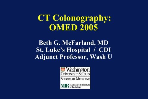
CT Colonography:<strong>OMED</strong> 2005Beth G. McFarland, MDSt. Luke’s Hospital / CDIAdjunct Professor, Wash U
Achievements and challenges3D PVR (perspective VR)3D transparency/ edge-enhanced
ObjectivesI. Technique advances» CT protocol (1DCT - 64DCT)» 2D MPR – 3D image displays» Patient factors- bowel prep / insufflationII.Standardization of reporting: C-RADSIII. ACR status: local coverage decisions
Evolution of 3D advancements** Acquisition AND Image display1998, 1DCT5 x 8 x 2 mm400images2003, 16DCT0.75 (1.0 EST) x15 mm x 1 mm800 +images
CTC Protocol• Two types of protocols evolving:1. CTC <strong>for</strong> patients at high risk <strong>for</strong> colon ca-- evaluate colon + staging (IV contrast)2. CTC <strong>for</strong> patients at low risk-- evaluate colon + extra-colonic (no IVcontrast, low dose)
CT Dose issues• High contrast interface of COLONIC polypand air permits a decrease of mAs» Macari, et al, RAD 2001: 4DCT- 1 mm EST, 50 eff mAS = 5-7 mSV (similar to BE)» Iannaconne, et al, RAD 2003: 4DCT- 2.5 mm EST, 10 eff mAS = 1.8-2.4 mSV• Low contrast interface of EXTRACOLONICorgans (w/o IV contrast) can becompromised by a decrease of mAs
Colon phantom- 16DCT11 x 1 mm6mm11 x 3 mm0.75 mm detector, 1.0 EST, beam pitch 1.550 eff mAs
16D CT25 eff mAs100 eff mAs50 eff mAs160 eff mAs
Key patient factors(input)Bowel prep +Be<strong>for</strong>e CTAt CT
Dry Tagging - Barium• 50 patients - FT (mag citrate,barium, diet)•50 patients: non-FT (pol gylcol)•FT increased specificity (88%,14/16), compared to non-FT (77%,17/20)•FT had decreased discom<strong>for</strong>t andside effects/ better willingnesswith catharsisLefere P, Radiology 2002;224:393-403
Wet tagging - iodineBe<strong>for</strong>e subtractionAfter subtractionZalis M. et al, AJR 2001;176:646-648
Overview of large trials• Johnson et al, Gastro 03• Pickhardt et al, NEJM 03• Cotton et al, JAMA 04• Iannaconne et al, Gastro 04• Rockey et al, Lancet 05Stool TaggingNoYesNoYesNo
Recent tagging protocolsPickhardt et alIannaccone et al(NEJM 2003) (Gastro 2004)Diet Clear liquid Low fiberCatharsis Phospha soda 90 cc NONETagging Gastro 120 cc Gastro 200 ccBarium 2% 500 ccSubtraction YES NO2D – 3D 3D primary review 2D primary review
Bowel preparation• Diet» Clear liquids or low fiber (Nutra-prep, EZEM)• Catharsis» Phospha-soda (45 cc), or Mg citrate• Tagging (w/o subtraction)» Gastrograffin 60 cc + Barium 2% 250 cc» Barium 40% (Tagitol V)- 20 cc x 3» (Barium 2% 500 cc)Futurechanges**
Insufflation per rectum
CT Colonography(failed endo, BE)APRPO
SupineLeft lateraldecubitis40 yr. old male,incompletecolonoscopy
3D CT endoscopic display1DCT5 x 8 mm2 mm RI0.65mm 3Vitrea TM
Interactivity of 2D MPR and 3D CT2D Axial MPR2D Coronal MPR3D CT endoscopic
2D- 3D interactivity• 2D multiplanar re<strong>for</strong>mation» time-efficient evaluation of polyp detection /characterization» provides extra-luminal orientation» maintains source density data (HU)• 3D endoscopic PVR» can be used as primary review to detect- mayimprove surface area visualization» demonstrates overall morphology of lesion
Edge- enhanced (BE) roadmap
C-RADS:Structured reporting- images/textVirtual colonoscopy workinggroupconsensus statement(Radiology 2005, in press)
C-RADS:Structured reporting• Image quality per segment:- distention, fluid- stool, tortuosity• Colorectal findings- size/location of lesions- 2D and 3D images of each
C-RADS:Structured reporting• Possible surveillance intervals- > 1 cm : colonoscopy- 6-9 mm: colonoscopy vs. follow• Extra-colonic findings:- to discern significant lesions(eg, AAA, adenopathy)
AxialReporting of findingsPol 1= HF polypSagProneAxial1 cm HF polyp(P=265, 634, -279)Supine
CoronalPol 2= sigmoid polypAxial5 mm sigmoid polyp(P= 371,561,-421)
Edge-enhanced CT overviewLPOPol 1Pol 2RPOAP view
Polyp size measurement• Polyp size is significant <strong>for</strong> clinicalmanagement and stratification of resultsduring validation» < 5 mm, 5-9 mm, > 10mm• CLINICAL VARIABILITY exists• CTC can provide both 2D and 3D tools <strong>for</strong>linear and volumetric (in development)measurements
Change of morphologyof pedunculated 12 mm polypSupineProne
Case 913.4Sigmoid polypPolyp size measurement:**** measure long axis of polyphead w/o stalkstalk14 mm CT= 9, 11, 11, 11 mm
CPT codingReimbursement
ACR report of CPT codesEffective as of July 2004:• Two category III codes created <strong>for</strong> CTC» 0066T asymptomatic (screening)» 0067T symptomatic (diagnostic)
ACR report of CPT codesAs of July 2004:• Category III codes are <strong>for</strong> trackingpurposes (not FDA approved, no RVU’s)» potentially can be reimbursed at individuallevel based on local payor policies» local coverage decisions (LCD’s) can be made<strong>for</strong> extended prospective reimbursementBarish M. Billing and reimbursement of VC, Intl Sym of VC 04
Local coverage decisions (LCD’s)• Can help promote localreimbursements <strong>for</strong> specific patientcohorts• ACR offers central networking andcoordination <strong>for</strong> physicians and thirdparty payers» CAC (Carrier Advisory Committee)» MCN (Managed Care Network)
Clinical vignettes <strong>for</strong> CTCreimbursement• Patients who have had incomplete colonoscopy» CAC approved in 9 states• Patients at risk <strong>for</strong> colonoscopy» pending• Frail / debilitated / elderly» pending• Screening in underserved areas» Madison, WI (Pickhardt)
Flat T3 lesion at colonoscopymissedat CTCColonoscopyCTCin retrospect
Missed flat T3 lesionat CTCpronesupine3D fly-through
Advanced rectal lesionmissedat prior colonoscopy
Missedrectal calocal stagingdistant staging
Per<strong>for</strong>mance• Effective multi-disciplinarycollaborations• Proper selection of patientcohortsPractice• Shared medical decisionmaking with patients• Ongoing Q/A
The 6 th International Symposium onVirtual ColonoscopyOctober 17-18, 2005Boston, MAw w w . v i r t u a l c o l o n o s c o p y . n e tw w w . b u . e d u / c m e