The IC3D Classification of the Corneal Dystrophies - Cornea Society
The IC3D Classification of the Corneal Dystrophies - Cornea Society
The IC3D Classification of the Corneal Dystrophies - Cornea Society

Create successful ePaper yourself
Turn your PDF publications into a flip-book with our unique Google optimized e-Paper software.
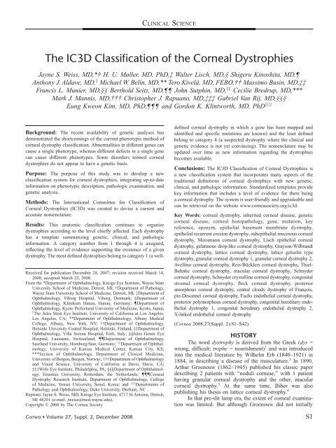
CLINICAL SCIENCE<strong>The</strong> <strong>IC3D</strong> <strong>Classification</strong> <strong>of</strong> <strong>the</strong> <strong><strong>Cornea</strong>l</strong> <strong>Dystrophies</strong>Jayne S. Weiss, MD,*† H. U. Møller, MD, PhD,‡ Walter Lisch, MD,§ Shigeru Kinoshita, MD,Anthony J. Aldave, MD, k Michael W. Belin, MD,** Tero Kivelä, MD, FEBO,†† Massimo Busin, MD,‡‡Francis L. Munier, MD,§§ Berthold Seitz, MD, John Sutphin, MD, kk Cecilie Bredrup, MD,***Mark J. Mannis, MD,††† Christopher J. Rapuano, MD,‡‡‡ Gabriel Van Rij, MD,§§§Eung Kweon Kim, MD, PhD, and Gordon K. Klintworth, MD, PhD kkkBackground: <strong>The</strong> recent availability <strong>of</strong> genetic analyses hasdemonstrated <strong>the</strong> shortcomings <strong>of</strong> <strong>the</strong> current phenotypic method <strong>of</strong>corneal dystrophy classification. Abnormalities in different genes cancause a single phenotype, whereas different defects in a single genecan cause different phenotypes. Some disorders termed cornealdystrophies do not appear to have a genetic basis.Purpose: <strong>The</strong> purpose <strong>of</strong> this study was to develop a newclassification system for corneal dystrophies, integrating up-to-dateinformation on phenotypic description, pathologic examination, andgenetic analysis.Methods: <strong>The</strong> International Committee for <strong>Classification</strong> <strong>of</strong><strong><strong>Cornea</strong>l</strong> <strong>Dystrophies</strong> (<strong>IC3D</strong>) was created to devise a current andaccurate nomenclature.Received for publication December 28, 2007; revision received March 14,2008; accepted March 22, 2008.From <strong>the</strong> *Department <strong>of</strong> Ophthalmology, Kresge Eye Institute, Wayne StateUniversity School <strong>of</strong> Medicine, Detroit, MI; †Department <strong>of</strong> Pathology,Wayne State University School <strong>of</strong> Medicine, Detroit, MI; ‡Department <strong>of</strong>Ophthalmology, Viborg Hospital, Viborg, Denmark; §Department <strong>of</strong>Ophthalmology, Klinikum Hanau, Hanau, Germany; {Department <strong>of</strong>Ophthalmology, Kyoto Prefectural University <strong>of</strong> Medicine, Kyoto, Japan;k <strong>The</strong> Jules Stein Eye Institute, University <strong>of</strong> California at Los Angeles,Los Angeles, CA; **Department <strong>of</strong> Ophthalmology, Albany MedicalCollege, Albany, New York, NY; ††Department <strong>of</strong> Ophthalmology,Helsinki University Central Hospital, Helsinki, Finland; ‡‡Department <strong>of</strong>Ophthalmology, Villa Serena Hospital, Forlí, Italy; §§Jules Gonin EyeHospital, Lausanne, Switzerland; {{Department <strong>of</strong> Ophthalmology,Saarland University, Homburg/Saar, Germany; kk Department <strong>of</strong> Ophthalmology,University <strong>of</strong> Kansas Medical Center, Kansas City, KS;***Section <strong>of</strong> Ophthalmology, Department <strong>of</strong> Clinical Medicine,University <strong>of</strong> Bergen, Bergen, Norway; †††Department <strong>of</strong> Ophthalmologyand Visual Science, University <strong>of</strong> California at Davis, Davis, CA;‡‡‡Wills Eye Institute, Philadelphia, PA; §§§Department <strong>of</strong> Ophthalmology,Erasmus University, Rotterdam, <strong>the</strong> Ne<strong>the</strong>rlands; {{{<strong><strong>Cornea</strong>l</strong>Dystrophy Research Institute, Department <strong>of</strong> Ophthalmology, College<strong>of</strong> Medicine, Yonsei University, Seoul, Korea; and kkk Departments <strong>of</strong>Pathology and Ophthalmology, Duke University, Durham, NC .Reprints: Jayne S. Weiss, MD, Kresge Eye Institute, 4717 St Antoine, Detroit,MI 48201 (e-mail: jweiss@med.wayne.edu).Copyright Ó 2008 by <strong>The</strong> <strong>Cornea</strong> <strong>Society</strong><strong>Cornea</strong> Volume 27, Suppl. 2, December 2008Results: This anatomic classification continues to organizedystrophies according to <strong>the</strong> level chiefly affected. Each dystrophyhas a template summarizing genetic, clinical, and pathologicinformation. A category number from 1 through 4 is assigned,reflecting <strong>the</strong> level <strong>of</strong> evidence supporting <strong>the</strong> existence <strong>of</strong> a givendystrophy. <strong>The</strong> most defined dystrophies belong to category 1 (a welldefinedcorneal dystrophy in which a gene has been mapped andidentified and specific mutations are known) and <strong>the</strong> least definedbelong to category 4 (a suspected dystrophy where <strong>the</strong> clinical andgenetic evidence is not yet convincing). <strong>The</strong> nomenclature may beupdated over time as new information regarding <strong>the</strong> dystrophiesbecomes available.Conclusions: <strong>The</strong> <strong>IC3D</strong> <strong>Classification</strong> <strong>of</strong> <strong><strong>Cornea</strong>l</strong> <strong>Dystrophies</strong> isa new classification system that incorporates many aspects <strong>of</strong> <strong>the</strong>traditional definitions <strong>of</strong> corneal dystrophies with new genetic,clinical, and pathologic information. Standardized templates providekey information that includes a level <strong>of</strong> evidence for <strong>the</strong>re beinga corneal dystrophy. <strong>The</strong> system is user-friendly and upgradeable andcan be retrieved on <strong>the</strong> website www.corneasociety.org/ic3d.Key Words: corneal dystrophy, inherited corneal disease, geneticcorneal disease, corneal histopathology, gene, mutation, keyreference, eponym, epi<strong>the</strong>lial basement membrane dystrophy,epi<strong>the</strong>lial recurrent erosion dystrophy, subepi<strong>the</strong>lial mucinous cornealdystrophy, Meesmann corneal dystrophy, Lisch epi<strong>the</strong>lial cornealdystrophy, gelatinous drop-like corneal dystrophy, Grayson-Wilbrandtcorneal dystrophy, lattice corneal dystrophy, lattice gelsolin typedystrophy, granular corneal dystrophy 1, granular corneal dystrophy 2,Avellino corneal dystrophy, Reis-Bücklers corneal dystrophy, Thiel-Behnke corneal dystrophy, macular corneal dystrophy, Schnydercorneal dystrophy, Schnyder crystalline corneal dystrophy, congenitalstromal corneal dystrophy, fleck corneal dystrophy, posterioramorphous corneal dystrophy, central cloudy dystrophy <strong>of</strong> Francxois,pre-Descemet corneal dystrophy, Fuchs endo<strong>the</strong>lial corneal dystrophy,posterior polymorphous corneal dystrophy, congenital hereditary endo<strong>the</strong>lialdystrophy 1, congenital hereditary endo<strong>the</strong>lial dystrophy 2,X-linked endo<strong>the</strong>lial corneal dystrophy(<strong>Cornea</strong> 2008;27(Suppl. 2):S1–S42)HISTORY<strong>The</strong> word dystrophy is derived from <strong>the</strong> Greek (dys =wrong, difficult; trophe = nourishment) 1 and was introducedinto <strong>the</strong> medical literature by Wilhelm Erb (1840–1921) in1884, in describing a disease <strong>of</strong> <strong>the</strong> musculature. 2 In 1890,Arthur Groenouw (1862–1945) published his classic paperdescribing 2 patients with ‘‘noduli corneae,’’ with 1 patienthaving granular corneal dystrophy and <strong>the</strong> o<strong>the</strong>r, macularcorneal dystrophy. 3 At <strong>the</strong> same time, Biber was alsopublishing his <strong>the</strong>sis on lattice corneal dystrophy. 4In that pre-slit lamp era, <strong>the</strong> extent <strong>of</strong> corneal examinationwas limited. But although Groenouw did not initiallyS1
Weiss et al <strong>Cornea</strong> Volume 27, Suppl. 2, December 2008appreciate <strong>the</strong> differences between granular and macular dystrophyor recognize <strong>the</strong> familial predisposition, <strong>the</strong> 2 diseasessubsequently became known as corneal dystrophies. 5 Fuchs 6used <strong>the</strong> word dystrophy to refer to ophthalmologic diseaseand postulated that dystrophic tissues resulted from lack <strong>of</strong>nourishment, hormones, blood, and nerve supply. Uhth<strong>of</strong>f 7 andlater Yoshida 8 continued to use <strong>the</strong> term in <strong>the</strong>ir publications.CORNEAL DYSTROPHY DEFINITIONAlthough many definitions <strong>of</strong> <strong>the</strong> word ‘‘dystrophy’’have appeared in <strong>the</strong> medical literature, 1 <strong>the</strong> term is mostcommonly used to describe an inherited disorder affectingcells, tissues, or organs, alone or in combination. Inophthalmology, <strong>the</strong> term ‘‘corneal dystrophy’’ has been usedto refer to a group <strong>of</strong> inherited corneal diseases that are typicallybilateral, symmetric, slowly progressive, and without relationshipto environmental or systemic factors. 9 As knowledge hasincreased, exceptions to each <strong>of</strong> <strong>the</strong>se definitions have beennoted. Thus, most patients with epi<strong>the</strong>lial basement membranedystrophy do not have a hereditary pattern. Some patients withposterior polymorphous corneal dystrophy only manifestunilateral changes. In macular dystrophy, <strong>the</strong> level <strong>of</strong> antigenicserum keratan sulfate correlates with <strong>the</strong> immunophenotypes <strong>of</strong><strong>the</strong> disease, indicating that systemic abnormalities are integral to<strong>the</strong> development <strong>of</strong> <strong>the</strong> characteristic corneal changes. Likewise,<strong>the</strong>re are a number <strong>of</strong> hereditary, bilateral diseases <strong>of</strong> <strong>the</strong>cornea, such as cornea plana, which have not been traditionallyclassified as corneal dystrophies and may be alternativelyaccommodated among <strong>the</strong> congenital anomalies affecting <strong>the</strong>cornea.Consequently, experience has demonstrated that <strong>the</strong>separation <strong>of</strong> entities into <strong>the</strong> category called cornealdystrophies may have more historical than practical meaning.<strong>The</strong>re remains no consensus as to <strong>the</strong> precise definition <strong>of</strong>corneal dystrophy, but, according to custom, we have chosento primarily deal with entities formerly called cornealdystrophies.CORNEAL DYSTROPHY LITERATUREBücklers, 10 whose name was later attached to Reis–Bücklers corneal dystrophy (RBCD), published <strong>the</strong> firstclassification <strong>of</strong> <strong>the</strong> corneal dystrophies when he described<strong>the</strong> differences between granular, lattice, and macular cornealdystrophies. Although <strong>the</strong> dystrophies can be classifiedaccording to genetic pattern, severity, histopathologic features,or biochemical characteristics, <strong>the</strong> most commonly usedclassification system has been anatomically based. 9 <strong>The</strong>dystrophies are typically classified by level <strong>of</strong> <strong>the</strong> cornea thatis involved, which separates <strong>the</strong>se entities into epi<strong>the</strong>lial andsubepi<strong>the</strong>lial, Bowman layer, stromal, Descemet membrane,and endo<strong>the</strong>lial dystrophies. 11–14SHORTCOMINGS OF CORNEALDYSTROPHY CLASSIFICATIONCritical review <strong>of</strong> <strong>the</strong> corneal dystrophy literature revealsnumerous apparent misconceptions and errors. For example,many publications emphasize <strong>the</strong> necessity <strong>of</strong> demonstratingS2corneal crystals to make <strong>the</strong> diagnosis <strong>of</strong> Schnyder crystallinecorneal dystrophy (SCD). 15,16 However, examination <strong>of</strong> largepedigrees <strong>of</strong> patients with SCD demonstrates that only 50% <strong>of</strong>affected patients actually have corneal crystals. 17 Never<strong>the</strong>less,publications over <strong>the</strong> past decades erroneously emphasize thatcrystals are necessary for <strong>the</strong> diagnosis <strong>of</strong> SCD. 18<strong>The</strong> direct consequence is that in some patients withSCD who lack stromal crystals, <strong>the</strong> diagnosis may be delayedfor decades. 17 Once established in textbooks, it is exceedinglydifficult to purge incorrect information about rare diseases.Many myths are perpetuated because very few ophthalmologistshave seen a substantial number <strong>of</strong> <strong>the</strong> unusual cornealdystrophies.Ano<strong>the</strong>r difficulty in <strong>the</strong> literature is <strong>the</strong> tendency toplace too much emphasis on a new or rare observation ra<strong>the</strong>rthan wait for a full analysis <strong>of</strong> a new disorder. For example,some <strong>of</strong> <strong>the</strong> early papers describing <strong>the</strong> ultrastructure <strong>of</strong> RBCDhad actually analyzed tissue from patients with Thiel–Behnkecorneal dystrophy (TBCD). 19 In a publication, <strong>the</strong> knownentity <strong>of</strong> RBCD was renamed as an unusual variant <strong>of</strong> granulardystrophy. 20,21 <strong>The</strong>se inconsistencies in <strong>the</strong> literature haveconfounded our understanding <strong>of</strong> precise findings in specificcorneal dystrophies.DOES EVERY SINGLE DYSTROPHYACTUALLY EXIST?Before <strong>the</strong> 1970s, new corneal dystrophies wereidentified and characterized almost exclusively by <strong>the</strong>irclinical appearance aided, in some cases, by light microscopichistopathology. In some cases, <strong>the</strong> description <strong>of</strong> a dystrophywas based on a report <strong>of</strong> a single family. 22,23In o<strong>the</strong>r cases, a new dystrophy could be misclassified asa variant <strong>of</strong> a previously described dystrophy. For years, <strong>the</strong>dystrophy <strong>of</strong> Waardenburg and Jonkers 24 appeared inreferences and textbooks. In actuality, <strong>the</strong>se patients hadThiel–Behnke corneal dystrophy. 25,26Often, it is impossible to ei<strong>the</strong>r confirm or exclude everycorneal dystrophy that has made its way into textbooks as anindependent entity. Moreover, misunderstandings that becameprevalent have <strong>of</strong>ten persisted long after <strong>the</strong>y could be resolved.For example, what did Reis and Bücklersactuallyseewhen<strong>the</strong>ydescribed what is now called Reis–Bücklers corneal dystrophy?22,23 <strong>The</strong> original pedigree is lost to follow up, and <strong>the</strong>irclinical description is sketchy concerning <strong>the</strong> specific signs andsymptoms. Never<strong>the</strong>less, we still presume that <strong>the</strong> entity <strong>the</strong>ydescribed is probably what is now established as Reis–Bücklersdystrophy (RBCD), but <strong>the</strong> original patients could have hadwhat is now called TBCD.Before honeycomb-shaped corneal dystrophy wasdescribed by Thiel and Behnke in 1967, 25 and even afterwards,patients affected with this dystrophy were instead reported asexamples <strong>of</strong> RBCD. 19 It took more than 30 years before <strong>the</strong>literature had separated <strong>the</strong>se 2 dystrophies. On <strong>the</strong> o<strong>the</strong>rhand, Grayson and Wilbrandt 27 described a family witha Bowman layer dystrophy <strong>the</strong>y initially reported as RBCD butactually provided insufficient evidence to determine definitivelywhe<strong>the</strong>r <strong>the</strong> unique findings, subsequently calledGrayson–Wilbrandt corneal dystrophy (GWCD), indicatedq 2008 <strong>The</strong> <strong>Cornea</strong> <strong>Society</strong>
<strong>Cornea</strong> Volume 27, Suppl. 2, December 2008<strong>IC3D</strong> <strong>Classification</strong> <strong>of</strong> <strong>the</strong> <strong><strong>Cornea</strong>l</strong> <strong>Dystrophies</strong>a distinct entity or a variant <strong>of</strong> a different Bowman layerdystrophy.Although <strong>the</strong> original publication on central cloudydystrophy <strong>of</strong> Francxois 28 described a hereditary cornealopacification, <strong>the</strong>re have been only a few o<strong>the</strong>r publicationsthat have described an entire family with this disease. 29,30 Botharticles were written before <strong>the</strong> advent <strong>of</strong> genotyping so nogenetic information is available. Central cloudy dystrophy <strong>of</strong>Francxois appears clinically indistinguishable from <strong>the</strong>degenerative condition, posterior crocodile shagreen. 31 It isnot possible to determine whe<strong>the</strong>r previous publicationsdescribing an individual patient with central cloudy dystrophy<strong>of</strong> Francxois were actually describing patients with posteriorcrocodile shagreen. 32 In <strong>the</strong> absence <strong>of</strong> additional affectedpedigrees or genetic studies confirming inheritance, it ispossible that central cloudy dystrophy <strong>of</strong> Francxois andposterior crocodile shagreen are <strong>the</strong> same entity. Withoutgenotypic information, it may be impossible to determinewhe<strong>the</strong>r rare or newly described dystrophies are actuallyunique diseases or represent phenotypic variations <strong>of</strong> previouslydescribed entities.GENETICS<strong>The</strong> development <strong>of</strong> genotypic analyses has revolutionizedour knowledge <strong>of</strong> <strong>the</strong> corneal dystrophies and fur<strong>the</strong>relucidated additional inaccuracies in <strong>the</strong> dystrophy nomenclature.<strong>The</strong> genetic characterization <strong>of</strong> corneal dystrophiesrevealed both genetic heterogeneity, that is, different genes(KRT3 and KRT12) causing a single dystrophy phenotype(Meesmann dystrophy), and phenotypic heterogeneity witha single gene (TGFBI) causing different allelic dystrophyphenotypes (RBCD, TBCD, granular type 1, granular type 2,and lattice type 1). Consequently, by enhancing our understanding<strong>of</strong> <strong>the</strong> dystrophies, newer genetic information hasmade <strong>the</strong> phenotypic classification system archaic.CURRENT CLASSIFICATION OF THECORNEAL DYSTROPHIES<strong>The</strong> knowledge base has exploded since <strong>the</strong> first descriptions<strong>of</strong> granular, macular, and lattice dystrophies over acentury ago. Not only has <strong>the</strong> word dystrophy lost importancebut also <strong>the</strong> distinctive name <strong>of</strong> many <strong>of</strong> <strong>the</strong> individualdystrophies has become less meaningful. <strong>The</strong> basis <strong>of</strong> <strong>the</strong>nomenclature system seems to be more historic than scientific.As <strong>the</strong> classification system <strong>of</strong> <strong>the</strong>se disorders has takenon historic implications, it has been proposed that <strong>the</strong>seconditions be classified ‘‘under <strong>the</strong> rubric <strong>of</strong> inherited cornealdiseases,’’ although acknowledging that ‘‘<strong>the</strong> popular designation<strong>of</strong> corneal dystrophy will probably keep its place. 33 ’’RECLASSIFICATION OF THE NOMENCLATUREIN OTHER MEDICAL SPECIALTIESOphthalmology is not <strong>the</strong> only medical field that hasdiscovered that <strong>the</strong> nomenclature <strong>of</strong> certain diseases has becomearchaic. Rapid advances in genotyping have challenged <strong>the</strong>nomenclature <strong>of</strong> o<strong>the</strong>r diseases in o<strong>the</strong>r specialties. Some <strong>of</strong><strong>the</strong>se specialties have met <strong>the</strong> challenge by devising newnomenclature systems. In 2001, <strong>the</strong> European Academy <strong>of</strong>q 2008 <strong>The</strong> <strong>Cornea</strong> <strong>Society</strong>Allergy and Clinical Immunology published a position paperproposing a new nomenclature in allergy after discussion with‘‘many pediatricians in Europe for several years. 34 ’’ One <strong>of</strong> <strong>the</strong>authors wrote that he ‘‘set up a reference panel <strong>of</strong> pediatricianswithin different areas <strong>of</strong> pediatrics and at intervals I asked <strong>the</strong>mfor <strong>the</strong>ir opinion on <strong>the</strong> proposal.’’ Subsequent articles in thisfield have underscored <strong>the</strong> importance on nomenclature <strong>of</strong>atopy, atopic disease, and allergy on classifying <strong>the</strong> individualpatient diseases and directing future <strong>the</strong>rapy. 35<strong>The</strong> disconnect between <strong>the</strong> language <strong>of</strong> basic scientistsand <strong>the</strong> language <strong>of</strong> clinicians has also presented challenges in<strong>the</strong> muscular dystrophy nomenclature. Dubovitz 36 wrote abouthis concern regarding a ‘‘major problem, within <strong>the</strong> field <strong>of</strong><strong>the</strong>rapy for muscular dystrophy, that has arisen from inappropriatenomenclature,’’ namely that it ‘‘. has hada negative impact on <strong>the</strong> whole field.’’ Klein wrote that‘‘From a historical perspective, 2 golden ages have shaped <strong>the</strong>current and evolving classification schemes: 1. <strong>the</strong> definition<strong>of</strong> clinical pathological entities in <strong>the</strong> early twentieth century;and 2. <strong>the</strong> application <strong>of</strong> molecular neurogenetics in <strong>the</strong> past10–15 years.’’ He concluded that <strong>the</strong> shortcoming <strong>of</strong> <strong>the</strong>current classification systems resulted not only because <strong>of</strong> <strong>the</strong>complex nature <strong>of</strong> <strong>the</strong> disorders but also that ‘‘modernclassification schemes was based on clinical, pathologic andgenetic/molecular criteria . attempt to integrate all threelevels’’ and although ‘‘genetic classifications are now widelyused . expert clinical diagnosis remains an important step incorrect diagnosis and classification.’’ 37 <strong>The</strong> author proposedclassification schemes based on clinical features, geneticfeatures, and molecular mechanisms or protein functions.THE FORMATION OF THE INTERNATIONALCOMMITTEE FOR CLASSIFICATION OFCORNEAL DYSTROPHIES (<strong>IC3D</strong>)In April 2005 at <strong>the</strong> World <strong>Cornea</strong> Congress meeting,<strong>the</strong> session on corneal dystrophies clearly elucidated thatnomenclature problems vexed not only SCD but also manyo<strong>the</strong>r dystrophies. That evening, J.S.W. approached <strong>the</strong> o<strong>the</strong>rmembers <strong>of</strong> <strong>the</strong> board <strong>of</strong> directors <strong>of</strong> <strong>the</strong> <strong>Cornea</strong> <strong>Society</strong> torequest <strong>the</strong>ir support for <strong>the</strong> creation <strong>of</strong> an internationalcommittee to revise <strong>the</strong> corneal dystrophy nomenclature. <strong>The</strong>goal was <strong>the</strong> recruitment <strong>of</strong> an international panel <strong>of</strong> interestedworld experts possessing firsthand experience with <strong>the</strong>clinical, genetic, and histopathologic findings <strong>of</strong> all <strong>the</strong>corneal dystrophies. In this way, <strong>the</strong> literature could becritically evaluated to distill <strong>the</strong> facts and recognize and <strong>the</strong>nremove outdated inaccurate information. With <strong>the</strong> support <strong>of</strong><strong>the</strong> <strong>Cornea</strong> <strong>Society</strong> President (M.W.B.), international ophthalmologicsocieties were contacted representing 5 continents torecruit representation <strong>of</strong> corneal specialists, ophthalmicpathologists, and geneticists for this collaborative effort.<strong>The</strong> International Committee for <strong>Classification</strong> <strong>of</strong><strong><strong>Cornea</strong>l</strong> <strong>Dystrophies</strong> (<strong>IC3D</strong>) held its first meeting in Chicagoin October 2005 at <strong>the</strong> American Academy <strong>of</strong> Ophthalmology,followed by meetings in San Paulo in February 2006 at <strong>the</strong>World Ophthalmology Congress, in Ft. Lauderdale inMay 2006 at <strong>the</strong> Association for Research in Vision andOphthalmology, in Las Vegas in October 2006 at <strong>the</strong> AmericanS3
Weiss et al <strong>Cornea</strong> Volume 27, Suppl. 2, December 2008Academy <strong>of</strong> Ophthalmology, and in San Diego in April 2007at <strong>the</strong> Association <strong>of</strong> Cataract and Refractive Surgeons. Inbetween, thousands <strong>of</strong> e-mails provided online discussion tomove <strong>the</strong> project forward.CHARACTERISTICS OF THENEW NOMENCLATUREAt <strong>the</strong> initial meeting, <strong>the</strong> group discussed <strong>the</strong> necessarycharacteristics <strong>of</strong> a new nomenclature that definitely wouldimprove accuracy, would be more informative, and could be easyto use, so that it truly could replace <strong>the</strong> nomenclature that hadbeen used for over a century—a gargantuan task, <strong>the</strong> successfulness<strong>of</strong> which only time will tell. <strong>The</strong> new nomenclaturehad to reflect current clinical, pathologic, and genetic knowledge,be easily adaptable to advances in understanding from <strong>the</strong>continued discovery <strong>of</strong> new genes and mutations and be linked to<strong>the</strong> old nomenclature for ease <strong>of</strong> use.THE <strong>IC3D</strong> TEMPLATES<strong>The</strong> development <strong>of</strong> a series <strong>of</strong> templates, which wouldassemble accurate and up-to-date information about eachdystrophy and facilitate <strong>the</strong> development and maintenance <strong>of</strong>a revised nomenclature, was undertaken. Each dystrophytemplate was a brief summary <strong>of</strong> <strong>the</strong> current genetic, clinical,and pathologic information about <strong>the</strong> disease and includedrepresentative clinical images. This approach also <strong>of</strong>fered <strong>the</strong>opportunity to correct errors in <strong>the</strong> literature and ‘‘set <strong>the</strong>record straight.’’ Published information was reviewed by allmembers <strong>of</strong> <strong>the</strong> committee, particularly those who hadexperience with a particular dystrophy. Although <strong>the</strong>re weresome dystrophies with which no member <strong>of</strong> <strong>the</strong> committee hadpersonal experience, such dystrophies were exceedingly rareand sometimes had only 1 case report in <strong>the</strong> literature. <strong>The</strong>process, <strong>the</strong>refore, was found to be very effective.CLASSIFICATION AND THE EVOLUTION OF ACORNEAL DYSTROPHY<strong>The</strong> largest challenge to <strong>the</strong> committee was how todevise a classification that would be flexible enough t<strong>of</strong>acilitate <strong>the</strong> expansion <strong>of</strong> knowledge from o<strong>the</strong>r sources,including genotyping. Evidence for <strong>the</strong> existence <strong>of</strong> a cornealdystrophy starts with <strong>the</strong> identification <strong>of</strong> a clinical phenotypeand may proceed to <strong>the</strong> characterization <strong>of</strong> <strong>the</strong> causative genemutation. When a corneal dystrophy is first described, <strong>the</strong>reis usually a predictable chain <strong>of</strong> events. Initially, an entity isidentified and characterized clinically. With corneal disordersthat impair vision severely enough to warrant keratoplasty,tissue evaluations <strong>of</strong> <strong>the</strong> diseased cornea lead to <strong>the</strong>establishment <strong>of</strong> distinct clinicopathologic entities. Even in<strong>the</strong> absence <strong>of</strong> tissue evaluations, <strong>the</strong> next phase involvesgenetic linkage studies that lead to <strong>the</strong> mapping <strong>of</strong> <strong>the</strong>chromosomal locus <strong>of</strong> <strong>the</strong> disorder, especially if <strong>the</strong> conditionhas a simple Mendelian inheritance pattern. This task is muchmore tedious and time consuming when more than 1 gene isinvolved or if <strong>the</strong>re is an interaction between genetic andenvironmental factors. Gene mapping is followed in duecourse by <strong>the</strong> identification <strong>of</strong> <strong>the</strong> relevant gene and particularmutations that are responsible for different phenotypical formsS4<strong>of</strong> <strong>the</strong> disorder. Eventually, identification <strong>of</strong> <strong>the</strong> gene productprovides a better understanding <strong>of</strong> <strong>the</strong> mechanism <strong>of</strong> <strong>the</strong>disorder and may present some <strong>the</strong>rapeutic possibilities.To indicate <strong>the</strong> level <strong>of</strong> evidence supporting <strong>the</strong>existence <strong>of</strong> a given dystrophy, <strong>the</strong> <strong>IC3D</strong> committee developeda series <strong>of</strong> descriptive, evidential categories as follows:CategoriesCategory 1: A well-defined corneal dystrophy in which <strong>the</strong>gene has been mapped and identified and specificmutations are known.Category 2: A well-defined corneal dystrophy that has beenmapped to 1 or more specific chromosomal loci, but <strong>the</strong>gene(s) remains to be identified.Category 3: A well-defined corneal dystrophy in which <strong>the</strong>disorder has not yet been mapped to a chromosomallocus.Category 4: This category is reserved for a suspected new, orpreviously documented, corneal dystrophy, although <strong>the</strong>evidence for it, being a distinct entity, is not yet convincing.<strong>The</strong> category assigned to a specific corneal dystrophycan be expected to change over time as knowledge progressivelyadvances. Eventually, all valid corneal dystrophiesshould attain <strong>the</strong> classification <strong>of</strong> category 1; macular cornealdystrophy is an example <strong>of</strong> a category 1 dystrophy.Conversely, over time and with fur<strong>the</strong>r information, someentities that are category 4 may be shown not to be distinctentities and may be removed. For example, ‘‘Central Discoid<strong><strong>Cornea</strong>l</strong> Dystrophy’’ 38 (CDCD), a category 4 dystrophy wasfound to be indistinguishable phenotypically from SCD sinecrystals. Consequently, <strong>the</strong> <strong>IC3D</strong> committee fur<strong>the</strong>r revieweda case report <strong>of</strong> CDCD to determine whe<strong>the</strong>r this was a uniquedystrophy or a variant <strong>of</strong> SCD. When <strong>the</strong> causative gene forSCD was found to be UBIAD1, 39,40 genetic testing <strong>of</strong> <strong>the</strong>proband with CDCD could be performed. Interestingly,<strong>the</strong> CDCD proband did demonstrate a unique mutation in<strong>the</strong> UBIAD1 gene (personal correspondence J.S.W.), whichwas not found in 100 control individuals. With a mutationin <strong>the</strong> UBIAD1 gene and corneal histopathology, whichdemonstrated stromal vacuoles consistent with dissolved lipid,it seemed that CDCD was actually SCD. Consequently, thiscategory 4 dystrophy was removed and CDCD was re-classifiedas SCD. This case clearly illustrates <strong>the</strong> importance and utility <strong>of</strong><strong>the</strong> <strong>IC3D</strong> classification system. If an entity is initially categorizedas a level 4 dystrophy, new information can be used to determinewhe<strong>the</strong>r <strong>the</strong> entity is indeed new or unique or is perhaps a variant<strong>of</strong> a previously described disease.THE NEW CLASSIFICATION SYSTEMOur proposed corneal dystrophy classification system isanatomically based, with dystrophies classified according to<strong>the</strong> layer chiefly affected (www.corneasociety.org/ic3d). Thus,<strong>the</strong>y are epi<strong>the</strong>lial and subepi<strong>the</strong>lial, Bowman layer, stromaland those affecting Descemet membrane and <strong>the</strong> endo<strong>the</strong>lium.<strong>The</strong> majority <strong>of</strong> <strong>the</strong> dystrophy names are identical or similar tothose in <strong>the</strong> current nomenclature. However, dystrophies witha known common genetic basis, that is, TGFBI dystrophies,have been grouped toge<strong>the</strong>r.q 2008 <strong>The</strong> <strong>Cornea</strong> <strong>Society</strong>
<strong>Cornea</strong> Volume 27, Suppl. 2, December 2008<strong>IC3D</strong> <strong>Classification</strong> <strong>of</strong> <strong>the</strong> <strong><strong>Cornea</strong>l</strong> <strong>Dystrophies</strong>Each template provides <strong>the</strong> key genetic, clinical andhistopathologic features that are characteristic for thatdystrophy. Each is assigned a level <strong>of</strong> evidence category <strong>of</strong>1, 2, 3, or 4, depending on <strong>the</strong> amount <strong>of</strong> clinical and geneticinformation available. A more detailed description <strong>of</strong> geneticmutations is included in <strong>the</strong> Appendix.REFERENCES1. Warburg M, Møller HU. Dystrophy: a revised definition. J Med Genet.1989;26:769–771.2. Erb W. Ueber die ‘‘juvenile Form’’ der progressiven Muskelatrophie undihre Beziehungen der sogenannten Pseudohypertrophie der Muskeln.Dtsch Arch Klin Med. 1884;34:467–519.3. Groenouw A. Knötchenförmige Hornhauttrübungen (Noduli corneae).Arch Augenheilkd. 1890;21:281–289.4. Biber H. Ueber einige seltene Hornhautekrankungen:die oberflächlichegittrige Keratitis [Inaugural dissertation]. A Diggelmann Zürich, ed; 1890.5. Møller HU. Granular corneal dystrophy Groenouw type I. Clinical andgenetic aspects. Acta Ophthalmol (Copenh). 1991; 69(suppl 198):1–40.6. Fuchs E. Dystrophia epi<strong>the</strong>lialis corneae. Albrecht Von Graefes Arch ClinExp Ophthalmol. 1910;76:478–508.7. Uhth<strong>of</strong>f W. Ein Fall von doppelseitiger zentraler, punktförmiger,supepi<strong>the</strong>lialer knötchenförmiger Keratitis, Groenouw mit anatomischemBefunde. Klin Monatsbl Augenheilkd. 1915;54:377–383.8. Yoshida Y. Über eine neue Art der Dystrophia corneae mit histologischemBefunde. Albrecht Von Graefes Arch Clin Exp Ophthalmol. 1924;114:91–100.9. American Academy <strong>of</strong> Ophthalmology. External diseases and cornea. In:Sutphin JE, ed. Basic and Clinical Sciences Course 2007–2008. SanFrancisco, CA: American Academy <strong>of</strong> Ophthalmology; 2007:305–329.10. Bücklers M. Die erblichen Hornhautdystrophie. Klin Monatsbl Augenheilkd.1938;Beiheft 3:1–135.11. Duke-Elder S, Leigh AG. <strong><strong>Cornea</strong>l</strong> dystrophies. In: Duke-Elder S, ed.System <strong>of</strong> Ophthalmology. Vol 8. Part 2. London, England: Kimpton;1965:864–867.12. Waring GO, Rodrigues MM, Laibson PR. <strong><strong>Cornea</strong>l</strong> dystrophies. I.<strong>Dystrophies</strong> <strong>of</strong> <strong>the</strong> epi<strong>the</strong>lium, Bowman’s layer and stroma. SurvOphthalmol. 1978;23:71–122.13. Klein D, Franceschetti A. Heredo-familiäre Hornhautdystrophie. In: BeckerPD, ed. Humangenetik. Vol 4. Stuttgart, Germany: Georg Thieme; 1964;80–81.14. Klintworth GK. <strong>The</strong> molecular genetics <strong>of</strong> <strong>the</strong> corneal dystrophies—current status. Front Biosci. 2003;8:687–713.15. Weiss JS. Schnyder’s dystrophy <strong>of</strong> <strong>the</strong> cornea. A Swede-Finn connection.<strong>Cornea</strong>. 1992;11:93–100.16. Weiss JS. Schnyder crystalline dystrophy sine crystals. Recommendationfor a revision <strong>of</strong> nomenclature. Ophthalmology. 1996;103:465–473.17. Weiss JS. Visual morbidity in 34 families with Schnyder’s crystallinecorneal dystrophy. Trans Am Ophthamol Soc. 2007;105:616–648.18. McCarthy MM, Innis S, Dubord P, et al. Panstromal Schnyder cornealdystrophy. A clinical pathologic report with quantitative analysis <strong>of</strong>corneal lipid composition. Ophthalmology. 1994;101:895–901.19. Kanai A, Kaufman HE, Polack FM. Electron microscopic study <strong>of</strong> Reis-Bücklers dystrophy. Ann Ophthalmol. 1973;5:953–962.20. Haddad R, Font RL, Fine BS. Unusual superficial variant <strong>of</strong> granulardystrophy <strong>of</strong> <strong>the</strong> cornea. Am J Ophthalmol. 1977;83:213–218.21. Møller HU. Interfamilial variability and intra-familial similarities <strong>of</strong> granularcorneal dystrophy Groenouw type I with respect to biomicroscopicalappearance and symptomatology. Acta Ophthalmol. 1989;67:669–677.22. Reis W. Familiäre, fleckige Hornhautentartung. Dtsch Med Wochenschr.1917;43:575.23. Bücklers M. Über eine weitere familiäre Hornhautdystrophie (Reis). KlinMonatsbl Augenkeilkd. 1949;114:386–397.24. Waardenburg PJ, Jonkers GH. A specific type <strong>of</strong> dominant progressivedystrophy <strong>of</strong> <strong>the</strong> cornea, developing after birth. Acta Ophthalmol(Copenh). 1961;39:919–923.25. Thiel H-J, Behnke H. Eine bisher unbekannte subepi<strong>the</strong>liale hereditäreHornhautdystrophie. Klin Monatsbl Augenheilkd. 1967;150:862–874.26. Wittebol-Post D, Van Schooneveld MJ, Pels E. <strong>The</strong> corneal dystrophy <strong>of</strong>Waardenburg and Jonkers. Ophthalmic Paediatr Genet. 1989;10:249–255.27. Grayson M, Wilbrandt H. Dystrophy <strong>of</strong> <strong>the</strong> anterior limiting membrane <strong>of</strong><strong>the</strong> cornea (Reis-Bücklers type). Am J Ophthalmol. 1966;61:345–349.q 2008 <strong>The</strong> <strong>Cornea</strong> <strong>Society</strong>28. Francxois J. Une nouvelle dystrophie hérédo-familiale de la cornée. J GenetHum. 1956;5:189–196.29. Strachan IM. Cloudy central corneal dystrophy <strong>of</strong> Francxois. Five cases in<strong>the</strong> same family. Br J Ophthalmol. 1969;53:192–194.30. Bramsen T, Ehlers N, Baggesen LH. Central cloudy corneal dystrophy <strong>of</strong>Francxois. Acta Ophthalmol (Copenh). 1976;54:221–226.31. Meyer JC, Quantock AJ, Thonar EJ, et al. Characterization <strong>of</strong> a centralcorneal cloudiness sharing features <strong>of</strong> posterior crocodile shagreen andcentral cloud dystrophy <strong>of</strong> Francxois. <strong>Cornea</strong>. 1996;15:347–354.32. Karp CL, Scott IU, Green WR, et al. Central cloudy corneal dystrophy <strong>of</strong>Francxois. A clinicopathologic study. Arch Ophthalmol. 1997;115:1058–1062.33. Klintworth GK. Genetic disorders <strong>of</strong> <strong>the</strong> cornea: from research to practicaldiagnostic testing. Graefes Arch Clin Exp Ophthalmol. 2005;33:231–232.34. Johansson G, Hourihane JO, Bousquet J, et al. A revised nomenclature forallergy. An EAACI position statement from <strong>the</strong> EAACI nomenclature taskforce. Allergy. 2001;56:813–824.35. Dreborg S. <strong>The</strong> implications <strong>of</strong> nomenclature. Ann Allergy AsthmaImmunol. 2002;89:S83–S85.36. Dubovitz V. Current and future <strong>the</strong>rapy in muscular dystrophy; need fora common language between basic scientists and clinicians. Acta Myol.2004;23:V–IX.37. Klein C. Movement disorders: classification. J Inherit Metab Dis. 2005;28:425–439.38. Aldave AJ, Edward DP, Park AJ, et al. Central discoid corneal dystrophy.<strong>Cornea</strong>. 2002; 21:739–744.39. Weiss JS, Kruth HS, Kuivaniemi H, et al. Mutations in <strong>the</strong> UBIAD1 geneon chromosome short arm 1, region 36 cause Schnyder crystalline cornealdystrophy. Invest Ophthalmol Vis Sci. 2007;48:5007–5012.40. Orr A, Sube MP, Marcadier, et al. Mutations in <strong>the</strong> UBIAD1 geneencoding a potential prenyltransferase are causal for Schnyder crystallinecorneal dystrophy. PLoS ONE. 2007;2:e685.THE <strong>IC3D</strong> CLASSIFICATION (C = CATEGORY)Epi<strong>the</strong>lial and Subepi<strong>the</strong>lial <strong>Dystrophies</strong>1. Epi<strong>the</strong>lial basement membrane dystrophy (EBMD)—majority degenerative, some C12. Epi<strong>the</strong>lial recurrent erosion dystrophy (ERED) C4, (Smolandiensisvariant) C33. Subepi<strong>the</strong>lial mucinous corneal dystrophy (SMCD) C44. Mutation in keratin genes: Meesmann corneal dystrophy(MECD) C15. Lisch epi<strong>the</strong>lial corneal dystrophy (LECD) C26. Gelatinous drop-like corneal dystrophy (GDLD) C1Bowman Layer <strong>Dystrophies</strong>1. Reis–Bücklers corneal dystrophy (RBCD)—Granular cornealdystrophy type 3 C12. Thiel–Behnke corneal dystrophy (TBCD) C1, potentialvariant C23. Grayson –Wilbrandt corneal dystrophy (GWCD) C4Stromal <strong>Dystrophies</strong>1. TGFBI corneal dystrophiesA. Lattice corneal dystrophya. Lattice corneal dystrophy, TGFBI type (LCD): Classiclattice corneal dystrophy (LCD1) C1, variants (III, IIIA,I/IIIA, and IV) are C1b. Lattice corneal dystrophy, gelsolin type (LCD2) C1 (Thisis not a true corneal dystrophy but is included here forease <strong>of</strong> differential diagnosis)B. Granular corneal dystrophy C1a. Granular corneal dystrophy, type 1 (classic) (GCD1) C1b. Granular corneal dystrophy, type 2 (granular-lattice)(GCD2) C1S5
Weiss et al <strong>Cornea</strong> Volume 27, Suppl. 2, December 2008c. Granular corneal dystrophy, type 3 (RBCD) = Reis–Bücklers C12. Macular corneal dystrophy (MCD) C13. Schnyder corneal dystrophy (SCD) C14. Congenital stromal corneal dystrophy (CSCD) C15. Fleck corneal dystrophy (FCD) C16. Posterior amorphous corneal dystrophy (PACD) C37. Central cloudy dystrophy <strong>of</strong> Francxois (CCDF) C48. Pre-Descemet corneal dystrophy (PDCD) C4Descemet Membrane andEndo<strong>the</strong>lial <strong>Dystrophies</strong>1. Fuchs endo<strong>the</strong>lial corneal dystrophy (FECD) C1, C2, or C32. Posterior polymorphous corneal dystrophy (PPCD) C1 or C23. Congenital hereditary endo<strong>the</strong>lial dystrophy 1 (CHED1) C24. Congenital hereditary endo<strong>the</strong>lial dystrophy 2 (CHED2) C15. X-linked endo<strong>the</strong>lial corneal dystrophy (XECD) C2TABLE 1. <strong>The</strong> <strong>IC3D</strong> <strong>Classification</strong>—Abbreviations andMIM NumberMIMAbbreviation<strong>IC3D</strong>Abbreviation MIM #Epi<strong>the</strong>lial basementmembrane dystrophy EBMD EBMD 121820Epi<strong>the</strong>lial recurrent erosiondystrophy None ERED 122400Subepi<strong>the</strong>lial mucinous CD None SMCD NoneMeesmann CD None MECD 122100Lisch epi<strong>the</strong>lial CD None LECD NoneGelatinous drop-like CD GDLD, CDGDL GDLD 204870Reis–Bücklers CD CDB1, CDRB, RBCD RBCD 608470Thiel–Behnke CD CDB2, CDTB TBCD 602082Grayson –Wilbrandt CD None GWCD NoneClassic Lattice CD CDL1 LCD1 122200Lattice CD, Meretoja type None LCD2 105120Granular CD, type 1 CGDD1 GCD1 121900Granular CD, type 2(granular–lattice) CDA, ACD GCD2 607541Macular CD MCDC1 MCD 217800Schnyder CD None SCD 121800Congenital stromal CD CSCD CSCD 610048Fleck CD None FCD 121850Posterior amorphous CD None PACD NoneCentral cloudy dystrophy <strong>of</strong>Francxois None CCDF 217600Pre-Descemet CD None PDCD NoneFuchs endo<strong>the</strong>lial CD FECD1 FECD 136800Posterior polymorphous CD PPCD1 PPCD 122000Congenital hereditaryendo<strong>the</strong>lial dystrophy 1 CHED1 CHED1 121700Congenital hereditaryendo<strong>the</strong>lial dystrophy 2 CHED2 CHED2 217700X-linked endo<strong>the</strong>lial CD None XECD NoneS6Online MIM (McKusick VA et al. http://www.ncbi.nlm.nih.gov/sites/entrez)CD, corneal dystrophy; MIM, Mendelian Inheritance in Man.EPITHELIAL ANDSUBEPITHELIAL DYSTROPHIESEpi<strong>the</strong>lial Basement MembraneDystrophy (EBMD)MIM #121820Alternative Names, EponymsMap-dot-fingerprint dystrophy.Cogan microcystic epi<strong>the</strong>lial dystrophy.Anterior basement membrane dystrophy.InheritanceMost cases have no inheritance documented. Many areconsidered to be degenerative or secondary to trauma. Familialcases have been reported.Genetic Locus5q31.GeneTGFBI in <strong>the</strong> minority <strong>of</strong> cases.OnsetPresent in adult life. Rarely seen in children.Signs (Fig. 1)Maps: Irregular islands <strong>of</strong> thickened, gray, hazy epi<strong>the</strong>liumwith scalloped, circumscribed borders, particularly affecting<strong>the</strong> central or paracentral cornea. Isolated or combinedwith o<strong>the</strong>r signs.Dots (Cogan): Irregular round, oval or comma-shaped,non-staining, putty-gray opacities. Clustered like an archipelagoin <strong>the</strong> central cornea. Typically combined with o<strong>the</strong>r signs,especially with maps.Fingerprint lines: Parallel, curvilinear lines, usually paracentral.Best seen in retro illumination. Isolated or combinedwith o<strong>the</strong>r signs, especially maps.Bleb pattern (Bron): A subepi<strong>the</strong>lial pattern like pebbledglass, best seen by retro-illumination. Isolated or combinedwith o<strong>the</strong>r signs.Poor adhesion <strong>of</strong> basal epi<strong>the</strong>lial cells to abnormal basallaminar material is thought predisposition to recurrent erosions.SymptomsAsymptomatic or recurrent erosions with pain, lacrimation,and blurred vision. Except for <strong>the</strong> bleb pattern, on-axis lesionsmay also cause blurred vision due to irregular astigmatism.CourseLocation and degree <strong>of</strong> pathology can fluctuate withtime.Light MicroscopyMaps: Sheets <strong>of</strong> intraepi<strong>the</strong>lial, multilamellar, basallaminar material.Fingerprint lines: Rib-like intraepi<strong>the</strong>lial extensions <strong>of</strong>basal laminar material.Dots: Intraepi<strong>the</strong>lial pseudocyst containing cytoplasmicdebris.Bleb pattern: Irregular, subepi<strong>the</strong>lial accumulation <strong>of</strong>a fibrillogranular material.q 2008 <strong>The</strong> <strong>Cornea</strong> <strong>Society</strong>
<strong>Cornea</strong> Volume 27, Suppl. 2, December 2008<strong>IC3D</strong> <strong>Classification</strong> <strong>of</strong> <strong>the</strong> <strong><strong>Cornea</strong>l</strong> <strong>Dystrophies</strong>FIGURE 1. Epi<strong>the</strong>lial basement membrane dystrophy. A, Map-like changes. B, Intraepi<strong>the</strong>lial dot opacities underlying map-likefigures. C, Fingerprint lines viewed in retroillumination.Transmission Electron MicroscopyMap: Thick epi<strong>the</strong>lial basement membrane that extendsinto <strong>the</strong> epi<strong>the</strong>lium as multilamellar, 2- to 6-nm-thick sheets.Fingerprint line: Fine fibrillogranular substance in additionto basement membrane. <strong>The</strong> fibrils are about 17 nm indiameter and <strong>the</strong> granular material about 8 nm.Dot: Intraepi<strong>the</strong>lial pseudocyst contains degeneratingcells with pyknotic nuclei and cytoplasmic debris.Bleb pattern: <strong>The</strong> anterior surface <strong>of</strong> this material formsdiscrete mounds, which dent <strong>the</strong> overlying basal epi<strong>the</strong>lialcells. May mimic cysts clinically but no cysts presenthistologically.Confocal MicroscopyMap-fingerprint-dot: Intraepi<strong>the</strong>lial basement membrane,which appears separated from normal basal epi<strong>the</strong>lialcells. Droplet-shaped configuration in <strong>the</strong> epi<strong>the</strong>lium. Ringlikestructure in <strong>the</strong> basal epi<strong>the</strong>lium.CategoryMost cases are sporadic and may be degenerative.Category 1 in a minority <strong>of</strong> cases.REFERENCES1. Boutboul S, Black GCM, Moore JE, et al. A subset <strong>of</strong> patients wi<strong>the</strong>pi<strong>the</strong>lial basement membrane corneal dystrophy have mutations inTGFBI/BIGH3. Hum Mutat. 2006;27:553–557.2. Bron AJ, Brown NA. Some superficial corneal disorders. Trans OphthalmolSoc UK. 1971;91:13–29.3. Bron AJ, Tripathi RC. Cystic disorders <strong>of</strong> <strong>the</strong> corneal epi<strong>the</strong>lium II.Pathogenesis. Br J Ophthalmol. 1973;57:361–375.4. Cogan DG, Donaldson DD, Kuwabara T, et al. Microcystic dystrophy <strong>of</strong><strong>the</strong> corneal epi<strong>the</strong>lium. Trans Am Ophthalmol Soc. 1964;62:213–225.5. Guerry D. Fingerprint-like lines in <strong>the</strong> cornea. Am J Ophthalmol. 1950;33:724–726.6. Laibson PR, Krachmer JH. Familial occurrence <strong>of</strong> dot (microcystic), map,fingerprint dystrophy <strong>of</strong> <strong>the</strong> cornea. Invest Ophthalmol Vis Sci. 1975;14:397–399.7. Laibson PR. Microcystic corneal dystrophy. Trans Am Ophthalmol Soc.1976;74:488–531.8. Lisch W, Lisch C. Die epi<strong>the</strong>liale Hornhautbasalmembrandystrophie. KlinMonatsbl Augenheilkd. 1983;183:251–255.9. Munier FL, Korvatska E, Djemai A, et al. Kerato-epi<strong>the</strong>lin mutations infour 5q31-linked corneal dystrophies. Nat Genet. 1997;15:247–251.10. Rodrigues MM, Fine BS, Laibson PR, et al. Disorders <strong>of</strong> <strong>the</strong> cornealepi<strong>the</strong>lium. A clinicopathologic study <strong>of</strong> dot, geographic, and fingerprintpatterns. Arch Ophthalmol. 1974;92:475–482.q 2008 <strong>The</strong> <strong>Cornea</strong> <strong>Society</strong>11. Vogt A. Lehrbuch und Atlas der Spaltlampenmikroskopie des lebendenAuges (1. Teil). Berlin, Germany: Springer; 1930:119–121.Epi<strong>the</strong>lial Recurrent Erosion Dystrophy (ERED)MIM #122400Alternative Names, Eponyms<strong><strong>Cornea</strong>l</strong> erosions, recurring hereditary (Franceschetti).VariantsDystrophia Smolandiensis.InheritanceAutosomal dominant.Genetic LocusUnknown.GeneUnknown; COL8A2, TGFBI, GSN, KRT3 and KRT12excluded in Smolandiensis variant.OnsetFirst decade <strong>of</strong> life.Signs (Fig. 2)Recurrent corneal erosions appear typically at 4 –6 years<strong>of</strong> age but occasionally as early as 8 months <strong>of</strong> age. <strong>The</strong>y areFIGURE 2. Epi<strong>the</strong>lial recurrent erosion corneal dystrophy(Smolandiensis variant). <strong>The</strong> right eye <strong>of</strong> a 41-year -old femalewith a central keloid-like opacification found in half <strong>of</strong> <strong>the</strong>affected family members.S7
Weiss et al <strong>Cornea</strong> Volume 27, Suppl. 2, December 2008precipitated by minimal trauma or are spontaneous. <strong>The</strong>cornea may show subepi<strong>the</strong>lial haze or blebs between attacks.In <strong>the</strong> Smolandiensis variant, half <strong>of</strong> <strong>the</strong> patients develop singleto a few permanent central subepi<strong>the</strong>lial corneal opacities,which appear at as early as 7 years <strong>of</strong> age. <strong>The</strong>se vary fromsubepi<strong>the</strong>lial fibrosis to protruding keloid-like nodules.SymptomsMost patients have attacks <strong>of</strong> redness, photophobia,epiphora, and ocular pain. Some experience a burningsensation and report sensitive eyes for years. Exposure tosunlight or draught, dust and smoke and lack <strong>of</strong> sleep canprecipitate attacks. In <strong>the</strong> Smolandiensis variant, a quarter <strong>of</strong>patients eventually need corneal grafts at mean age <strong>of</strong> 44 years.<strong>The</strong> opacities recur within 15 months in <strong>the</strong> graft periphery,but <strong>the</strong> central graft can remain clear for many years.CourseAttacks generally decline in frequency and intensity andcease by <strong>the</strong> age <strong>of</strong> 50 years. In <strong>the</strong> Smolandiensis variant,central subepi<strong>the</strong>lial opacities will progress.Light MicroscopyNo changes consistent with ei<strong>the</strong>r EBMD or known dystrophy<strong>of</strong> Bowman layer are reported for <strong>the</strong> Smolandiensis variant.Transmission Electron MicroscopyNot reported.Confocal MicroscopyNot reported.Category4, 3 (Smolandiensis variant).OnsetFirst decade <strong>of</strong> life.Signs (Fig. 3)Bilateral subepi<strong>the</strong>lial opacities and haze, most densecentrally, involving <strong>the</strong> entire cornea.SymptomsPainful episodes <strong>of</strong> recurrent corneal erosions, whichdecrease during adolescence (only 1 publication <strong>of</strong> a singlefamily).CourseProgressive loss <strong>of</strong> vision in adolescence.Light MicroscopySubepi<strong>the</strong>lial band <strong>of</strong> eosinophilic, periodic acid–Schiff–positive, Alcian blue–positive, hyaluronidase-sensitivematerial is present anterior to Bowman layer.Transmission Electron MicroscopySubepi<strong>the</strong>lial deposits <strong>of</strong> fine fibrillar material.ImmunohistochemistryCombination <strong>of</strong> chondroitin-4-sulfate and dermatansulfate.Confocal MicroscopyNot reported.Category4.REFERENCES1. Feder RS, Jay M, Yue BY, et al. Subepi<strong>the</strong>lial mucinous corneal dystrophy.Clinical and pathological correlations. Arch Ophthalmol. 1993;111:1106–1114.REFERENCES1. Hammar B, Björck E, Lagerstedt K, et al. A new corneal disease withrecurrent erosive episodes and autosomal dominant inheritance. ActaOphthalmol Scand. In press.2. Franceschetti A. Hereditaere rezidivierende Erosion der Hornhaut. ZAugenheilk. 1928;66:309–316.3. Valle O. Hereditary recurring corneal erosions: a family study, withspecial reference to Fuchs’ dystrophy. Acta Ophthalmol. 1967;45:829–836.4. Wales HJ. A family history <strong>of</strong> corneal erosions. Trans Ophthalmol Soc NZ.1956;8:77–78 .Subepi<strong>the</strong>lial Mucinous <strong><strong>Cornea</strong>l</strong>Dystrophy (SMCD)MIM: None.Alternative Names, EponymsNone.InheritanceAutosomal dominant.Genetic LocusUnknown.GeneUnknown.S8FIGURE 3. Subepi<strong>the</strong>lial mucinous corneal dystrophy. Subepi<strong>the</strong>lialopacities and haze involving <strong>the</strong> entire cornea; <strong>the</strong>se aremost dense toward <strong>the</strong> center (broad oblique and slit views).Mutations in Keratin Genes: Meesmann<strong><strong>Cornea</strong>l</strong> Dystrophy (MECD)MIM #122100Alternative Names, EponymsJuvenile hereditary epi<strong>the</strong>lial dystrophy.q 2008 <strong>The</strong> <strong>Cornea</strong> <strong>Society</strong>
<strong>Cornea</strong> Volume 27, Suppl. 2, December 2008<strong>IC3D</strong> <strong>Classification</strong> <strong>of</strong> <strong>the</strong> <strong><strong>Cornea</strong>l</strong> <strong>Dystrophies</strong>VariantStocker–Holt variant.InheritanceAutosomal dominant.Genetic LociLocus 12q13 (KRT3).Locus 17q12 (KRT12) Stocker–Holt variant.GenesKeratin K3 (KRT3).Keratin K12 (KRT12) Stocker–Holt variant.OnsetEarly childhood.Signs (Fig. 4)Multiple, tiny epi<strong>the</strong>lial vesicles extend to <strong>the</strong> limbusand are most numerous in <strong>the</strong> interpalpebral area with clearsurrounding epi<strong>the</strong>lium. Whorled and wedge-shaped epi<strong>the</strong>lialpatterns have been reported. <strong>The</strong> cornea may be slightlythinned and corneal sensation may be reduced.Indirect illumination shows varying diffuse grayopacities in different patterns, which may have a distinctborder. Areas <strong>of</strong> <strong>the</strong> central or peripheral cornea may beunaffected. <strong>The</strong> gray opacities appear as transparent cysts onindirect illumination. Coalescence <strong>of</strong> several cysts may resultin refractile linear opacities with intervening clear cornea.Stocker–Holt Variant<strong>The</strong> entire cornea demonstrates fine, grayish punctateepi<strong>the</strong>lial opacities that stain with fluorescein and fine linearopacities that may appear in a whorl pattern.CourseSlowly progressive.SymptomsPatients are typically asymptomatic or may have mildvisual reduction, although some patients complain <strong>of</strong> glare andlight sensitivity. Recurrent painful punctiform epi<strong>the</strong>lialerosions may occur. Rarely, blurred vision results fromcorneal irregularity and scarring.Stocker–Holt VariantPatients demonstrate more severe signs and symptomswith earlier onset compared with classic Meesmann cornealdystrophy.Light Microscopy<strong>The</strong> epi<strong>the</strong>lium always demonstrates intraepi<strong>the</strong>lialcysts. Cysts are filled with periodic acid–Schiff–positivecellular debris, which fluoresces. <strong>The</strong> epi<strong>the</strong>lium may bethickened and disorganized. Thickened multilaminar basementmembrane with projections into <strong>the</strong> basal epi<strong>the</strong>lium.Stocker–Holt VariantVariably thickened epi<strong>the</strong>lium with vacuolated cells andevidence <strong>of</strong> degeneration. Variably thickened basementmembrane extending into <strong>the</strong> epi<strong>the</strong>lium. Normal Bowmanlayer and stroma.Transmission Electron MicroscopyIntracytoplasmic ‘‘peculiar substance’’ represents a focalcollection <strong>of</strong> fibrogranular material surrounded by tangles <strong>of</strong>cytoplasmic filaments. Cystic round and well-delineatedlesions (10–50 mm across). Some lesions with reflectivepoints in <strong>the</strong> cytoplasm probably correspond to cell nuclei.Stocker–Holt VariantNot reported.Confocal MicroscopyHyporeflective areas in <strong>the</strong> basal epi<strong>the</strong>lium rangingfrom 40 to 150 mm in diameter, with potential reflective spotsinside.Stocker–Holt VariantNot reported.Category1, including Stocker–Holt VariantFIGURE 4. Meesmann corneal dystrophy. A, Multiple solitary microcysts that are most prominent in <strong>the</strong> interpalpebral region are seenin retroillumination. B, Diffuse gray opacity with broad oblique illumination, and multiple solitary microcysts in retroillumination.q 2008 <strong>The</strong> <strong>Cornea</strong> <strong>Society</strong>S9
Weiss et al <strong>Cornea</strong> Volume 27, Suppl. 2, December 2008REFERENCES1. Behnke H, Thiel HJ. Über die hereditäre epi<strong>the</strong>ldystrophie der Horhaut(Typ Meesman-Wilke) in Schleswig-Holstein. Klin Monatsbl Augenheilkd.1965;147:662–672.2. Burns RP. Meesmann’s corneal dystrophy. Trans Am Ophthalmol Soc.1968;66:530–635.3. Fine BS, Yan<strong>of</strong>f M, Pitts E, et al. Meesmann’s epi<strong>the</strong>lial dystrophy <strong>of</strong> <strong>the</strong>cornea. Am J Ophthalmol. 1977;83:633–642.4. Meesmann A. Über eine bisher nicht beschriebene dominant vererbteDystrophia epi<strong>the</strong>lialis corneae. Ber Zusammenkunft Dtsch OphthalmolGes. 1938;52:154–158.5. Stocker FW, Holt LB. A rare form <strong>of</strong> hereditary epi<strong>the</strong>lial dystrophy <strong>of</strong> <strong>the</strong>cornea: a genetic, clinical and pathologic study. Trans Am Ophthalmol Soc.1954;52:133–144.6. Thiel HJ, Behnke H. On <strong>the</strong> extent <strong>of</strong> variation <strong>of</strong> hereditary epi<strong>the</strong>lialcorneal dystrophy (Meesmann-Wilke type). Ophthalmologica. 1968;155:81–86.7. Tuft S, Bron AJ. Imaging <strong>the</strong> microstructural abnormalities <strong>of</strong> Meesmanncorneal dystrophy by in vivo confocal microscopy. <strong>Cornea</strong>. 2006; 25:868–870.8. Wittebol-Post D, Van-Bijsterveld OP, Delleman JW. Meesmann’s epi<strong>the</strong>lialdystrophy <strong>of</strong> <strong>the</strong> cornea. Biometrics and a hypo<strong>the</strong>sis. Ophthalmologica.1987;194:44–49.Lisch Epi<strong>the</strong>lial <strong><strong>Cornea</strong>l</strong> Dystrophy (LECD)MIM: None.Genetic locusXp 22.3.GeneUnknown.Alternative Names, EponymsBand-shaped and whorled microcystic dystrophy <strong>of</strong> <strong>the</strong>corneal epi<strong>the</strong>lium.InheritanceX-chromosomal dominant.OnsetChildhood.Signs (Fig. 5)Direct illumination shows localized gray opacities indifferent patterns: whorl-like, radial, band shaped, flame/fea<strong>the</strong>ry shaped, and club shaped. Indirect illuminationdemonstrates multiple, densely crowded clear cysts. <strong>The</strong>surrounding epi<strong>the</strong>lium is clear. Similar degree <strong>of</strong> opacitiesobserved in men and women.SymptomsAsymptomatic or blurred vision if <strong>the</strong> pupillary zone isinvolved.CourseSlow progression <strong>of</strong> opacities with possible deteriorationin vision.Light MicroscopyDiffuse cytoplasmic vacuolization <strong>of</strong> all cells in <strong>the</strong>affected area.Transmission Electron MicroscopyExtensive vacuolization <strong>of</strong> <strong>the</strong> cytoplasm <strong>of</strong> <strong>the</strong> affectedcorneal epi<strong>the</strong>lium. <strong>The</strong> vacuoles are ei<strong>the</strong>r optically empty orcontain weakly osmiophilic, partly homogenous, and partlylamellar material eventually due to collapsing and coalescing<strong>of</strong> vacuoles.ImmunohistochemistryScattered staining on Ki67 immunohistochemistryindicates no evidence <strong>of</strong> increased mitotic activity.Confocal MicroscopyMany solitary dark and well-demarcated lesions(50–100 mm) with round and oval configuration. Somelesions demonstrate central reflective points, which probablycorrespond to <strong>the</strong> cell nuclei.Category2.REFERENCES1. Alvarez-Fischer M, Alvarez de Toledo J, Barraquer RI. Lisch cornealdystrophy. <strong>Cornea</strong>. 2005;24:494–495.2. Butros S, Lang GK, Alvarez de Toledo J, et al. Die verschiedenenTrübungsmuster der Lisch-Hornhaut-dystrophie. Klin Monatsbl Augenheilkd.2006;223:837–840.3. Charles NC, Young JA, Kunar A, et al. Band-shaped and whorledmicrocystic dystrophy <strong>of</strong> <strong>the</strong> corneal epi<strong>the</strong>lium. Ophthalmology. 2000;107:1761–1764.4. Lisch W, Steuhl KP, Lisch C, et al. A new, band-shaped and whorledmicrocystic dystrophy <strong>of</strong> <strong>the</strong> corneal epi<strong>the</strong>lium. Am J Ophthalmol. 1992;114:35–44.5. Lisch W, Büttner A, Offner F, et al. Lisch corneal dystrophy is geneticallydistinct from Meesmann corneal dystrophy and maps to Xp22.3. Am JOphthalmol. 2000;130:461–468.6. Robin SB, Epstein RJ, Kornmehl EW. Band-shaped, whorled microcysticcorneal dystrophy. Am J Ophthalmol. 1994;117:543–544.7. Rohrbach JM, Grüb M, Szurman P. Einseitige, rezidivierende Trübung desHornhautepi<strong>the</strong>ls. Ophthalmologe. 2007;104:72–74.FIGURE 5. Lisch epi<strong>the</strong>lial corneal dystrophy. A, Localized, whorl-like gray opacity on direct illumination. B, Sclerotic scatterdemonstrating localized whorl-like gray opacity. C, Retroillumination demonstrating crowded microcysts.S10q 2008 <strong>The</strong> <strong>Cornea</strong> <strong>Society</strong>
<strong>Cornea</strong> Volume 27, Suppl. 2, December 2008<strong>IC3D</strong> <strong>Classification</strong> <strong>of</strong> <strong>the</strong> <strong><strong>Cornea</strong>l</strong> <strong>Dystrophies</strong>Gelatinous Drop-Like <strong><strong>Cornea</strong>l</strong>Dystrophy (GDLD)MIM #204870.Alternative Names, EponymsSubepi<strong>the</strong>lial amyloidosis.Primary familial amyloidosis (Grayson).Genetic Locus1p32.GeneTumor-associated calcium signal transducer 2 (TACSTD2,previously M1S1).InheritanceAutosomal recessive.OnsetFirst to second decade.Signs (Fig. 6)Initially, <strong>the</strong> subepi<strong>the</strong>lial lesions may appear similar toband-shaped keratopathy or <strong>the</strong>re may be groups <strong>of</strong> smallmultiple nodules, that is, mulberry configuration. <strong>The</strong>selesions demonstrate late staining with fluorescein, indicatingextremely hyperpermeable corneal epi<strong>the</strong>lium. Superficialvascularization is frequently seen. In later life, patients mayalso develop stromal opacification or develop larger nodularlesions, that is, kumquat-like lesions.SymptomsSignificant decrease in vision, photophobia, irritation,redness, and tearing.CourseProgression <strong>of</strong> protruding subepi<strong>the</strong>lial deposits andstromal opacity. Almost all patients develop recurrence aftersuperficial keratectomy, lamellar keratoplasty, or penetratingkeratoplasty, typically within a few years.Light MicroscopySubepi<strong>the</strong>lial and stromal amyloid deposits.Transmission Electron MicroscopyDisruption <strong>of</strong> epi<strong>the</strong>lial tight junctions in <strong>the</strong> superficialepi<strong>the</strong>lium. Amyloid is noted in <strong>the</strong> basal epi<strong>the</strong>lial layer.Confocal MicroscopyNot reported.Category1.REFERENCES1. Ide T, Nishida K, Maeda N, et al. A spectrum <strong>of</strong> clinical manifestations <strong>of</strong>gelatinous drop-like corneal dystrophy in Japan. Am J Ophthalmol. 2004;137:1081–1084.2. Kinoshita S, Nishida K, Dota A, et al. Epi<strong>the</strong>lial barrier function andultrastructure <strong>of</strong> gelatinous drop-like corneal dystrophy. <strong>Cornea</strong>. 2000;19:551–555.3. Klintworth GK, Valnickova Z, Kielar RA, et al. Familial subepi<strong>the</strong>lialcorneal amyloidosis —a lact<strong>of</strong>errin-related amyloidosis. Invest OphthalmolVis Sci. 1997;38:2756–2763.4. Nakaizumi GA. A rare case <strong>of</strong> corneal dystrophy. Acta Soc OphthalmolJpn. 1914;18:949–950.5. Ren Z, Lin PY, Klintworth GK, et al. Allelic and locus heterogeneity inautosomal recessive gelatinous drop-like corneal dystrophy. Hum Genet.2002;110:568–577.6. Tsujikawa M, Kurahashi H, Tanaka T, et al. Identification <strong>of</strong> <strong>the</strong> generesponsible for gelatinous drop-like corneal dystrophy. Nat Genet. 1999;21:420–423.7. Yoshida S, Kumano Y, Yoshida A, et al. Two bro<strong>the</strong>rs with gelatinous droplikedystrophy at different stages <strong>of</strong> <strong>the</strong> disease: role <strong>of</strong> mutational analysis.Am J Ophthalmol. 2002;133:830–832.BOWMAN LAYER DYSTROPHIESReis–Bücklers <strong><strong>Cornea</strong>l</strong> Dystrophy (RBCD)MIM #608470Alternative Names, Eponyms<strong><strong>Cornea</strong>l</strong> Dystrophy <strong>of</strong> Bowman layer, type I (CDB I).Geographic corneal dystrophy (Weidle).Superficial granular corneal dystrophy.Atypical granular corneal dystrophy.Granular corneal dystrophy, type 3.Anterior limiting membrane dystrophy, type I (ALMD I).Genetic Locus5q31.GeneTGFBI.InheritanceAutosomal dominant.OnsetChildhood.FIGURE 6. Gelatinous drop-like corneal dystrophy. A, Mulberry type. B, Band keratopathy type. C, Kumquat-like type.q 2008 <strong>The</strong> <strong>Cornea</strong> <strong>Society</strong>S11
Weiss et al <strong>Cornea</strong> Volume 27, Suppl. 2, December 2008Signs (Fig. 7)Confluent irregular and coarse geographic-like opacitieswith varying densities develop at <strong>the</strong> level <strong>of</strong> Bowman layerand superficial stroma, initially separated from one ano<strong>the</strong>r.Opacities may extend to <strong>the</strong> limbus and deeper stroma withtime. Can be confused with TBCD.SymptomsVision is impaired from childhood. Recurrent cornealerosions cause ocular discomfort and pain in <strong>the</strong> first decadebut may become less severe from <strong>the</strong> end <strong>of</strong> <strong>the</strong> second decade.Erosions are typically more frequent and severe than in TBCD.CourseSlowly progressive deterioration <strong>of</strong> vision. Recurrentcorneal erosions may resolve with time. Similar but frequentlymore aggressive course than TBCD but may not be able todistinguish in an individual case.Light MicroscopyBowman layer is replaced by a sheet-like connectivetissue layer with granular Masson trichrome–red deposits,which in advanced cases can extend to subepi<strong>the</strong>lial stroma.Transmission Electron MicroscopySubepi<strong>the</strong>lial electron-dense, rod-shaped bodies identicalto those in GCD1, but not <strong>the</strong> curly fibers <strong>of</strong> TBCD, areobserved on electron microscopy. Electron microscopy isnecessary for definitive histopathologic diagnosis to distinguishfrom TBCD.Confocal MicroscopyDistinct deposits are found in <strong>the</strong> epi<strong>the</strong>lium andBowman layer. <strong>The</strong> deposits in <strong>the</strong> basal epi<strong>the</strong>lial cell layershow extremely high reflectivity from small granular materialwithout any shadows. Bowman layer is replaced by highlyreflective irregular material, even more reflective than inTBCD (5q31). Fine diffuse deposits may be noted in <strong>the</strong>anterior stroma.ImmunohistochemistryRod-shaped bodies are immunopositive for transforminggrowth factor beta–induced protein (keratoepi<strong>the</strong>lin).Category1.REFERENCES1. Bücklers M.Über eine weitere familiäre Hornhaut-dystrophie (Reis). KlinMonatsbl Augenkeilkd. 1949;114:386–397.2. Kobayashi A, Sugiyama K. In vivo laser confocal microscopy findings forBowman’s layer dystrophies (Thiel-Behnke and Reis-Bücklers cornealdystrophies). Ophthalmology. 2007;114:69–75.3. Konishi M, Yamada M, Nakamura Y, et al. Immunohistology <strong>of</strong> keratoepi<strong>the</strong>linin corneal stromal dystrophies associated with R124 mutations<strong>of</strong> <strong>the</strong> BIGH3 gene. Curr Eye Res. 2000;21:891–896.4. Küchle M, Green WR, Völcker HE, et al. Reevaluation <strong>of</strong> cornealdystrophies <strong>of</strong> Bowman’s layer and <strong>the</strong> anterior stroma (Reis-Bücklersand Thiel-Behnke types): a light and electron microscopic study <strong>of</strong> eightcorneas and a review <strong>of</strong> <strong>the</strong> literature. <strong>Cornea</strong>. 1995;14:333–354.5. Munier FL, Korvatska E, Djemai A, et al. Kerato-epi<strong>the</strong>lin mutations infour 5q31-linked corneal dystrophies. Nat Genet. 1997;15:247–251.6. Reis W. Familiäre, fleckige Hornhautentartung. Dtsch Med Wochenschr.1917;43:575.7. Ridgway AE, Akhtar S, Munier FL, et al. Ultrastructural and molecularanalysis <strong>of</strong> Bowman’s layer corneal dystrophies: an epi<strong>the</strong>lial origin?Invest Ophthalmol Vis Sci. 2000;41:3286–3292.8. Small KW, Mullen L, Barletta J, et al. Mapping <strong>of</strong> Reis-Bücklers cornealdystrophy to chromosome 5q. Am J Ophthalmol. 1996;121:384–390.9. Stone EM, Ma<strong>the</strong>rs WD, Rosenwasser GO, et al. Three autosomaldominant corneal dystrophies map to chromosome 5q. Nat Genet. 1994;6:47–51.10. Streeten BW, Qi Y, Klintworth GK, et al. Immunolocalization <strong>of</strong> betaig-h3 protein in 5q31-linked corneal dystrophies and normal corneas.Arch Ophthalmol. 1999;117:67–75.11. Weidle EG. Klinische und feingewebliche Abgrenzung der Reis-Bücklers‘schen Hornhaut-dystrophie. Klin Monatsbl Augenheilkd.1989;194:217–226.12. Wittbol-Post D, Pels E. <strong>The</strong> dystrophy described by Reis and Bücklers.Ophthalmologica. 1989;199:1–9.Thiel–Behnke <strong><strong>Cornea</strong>l</strong> Dystrophy (TBCD)MIM #602082Alternative Names, Eponyms<strong><strong>Cornea</strong>l</strong> dystrophy <strong>of</strong> Bowman layer, type II (CDB2).Honeycomb-shaped corneal dystrophy.Anterior limiting membrane dystrophy, type II.Curly fibers corneal dystrophy.Waardenburg–Jonkers corneal dystrophy.Genetic Loci5q31.10q24.Gene5q31: TGFBI.10q24: Unknown.FIGURE 7. Reis–Bücklers corneal dystrophy. A, Coarse geographic opacity <strong>of</strong> <strong>the</strong> superficial cornea. B, Broad oblique illuminationdemonstrating dense, reticular, superficial opacity. C, Slit lamp view demonstrating irregularities in Bowman layer.S12q 2008 <strong>The</strong> <strong>Cornea</strong> <strong>Society</strong>
<strong>Cornea</strong> Volume 27, Suppl. 2, December 2008<strong>IC3D</strong> <strong>Classification</strong> <strong>of</strong> <strong>the</strong> <strong><strong>Cornea</strong>l</strong> <strong>Dystrophies</strong>InheritanceAutosomal dominant.OnsetChildhood.Signs (Fig. 8)Symmetrical subepi<strong>the</strong>lial reticular (honeycomb) opacitieswith peripheral cornea typically uninvolved. Variety <strong>of</strong>opacification patterns may make it impossible to distinguishfrom RBCD in early or individual cases. Opacities canprogress to deep stromal layers and corneal periphery.SymptomsRecurrent corneal erosions cause ocular discomfort andpain in <strong>the</strong> first and second decade. Gradual visual impairmentdevelops later. Erosions are less frequent, and <strong>the</strong> onset <strong>of</strong>visual impairment is later than in RBCD.CourseSlowly progressive deterioration <strong>of</strong> vision fromincreasing corneal opacification. Recurrent corneal erosionsmay resolve with time. Similar but frequently less aggressivecourse than RBCD but may not be able to distinguish in anindividual case.Light MicroscopyIrregular thickening <strong>of</strong> <strong>the</strong> epi<strong>the</strong>lial layer to allow forridges and furrows <strong>of</strong> underlying stroma, with focal absences<strong>of</strong> epi<strong>the</strong>lial basement membrane. Bowman layer is replacedby a fibrocellular layer between epi<strong>the</strong>lium and stroma witha pathognomonic wavy saw-too<strong>the</strong>d pattern.Transmission Electron MicroscopyPresence <strong>of</strong> curly collagen fibers with a diameter <strong>of</strong>9–15 nm is pathognomonic and distinguishes this dystrophyfrom RBCD.Confocal MicroscopyDistinct deposits are found in <strong>the</strong> epi<strong>the</strong>lium andBowman layer. <strong>The</strong> deposits in <strong>the</strong> basal epi<strong>the</strong>lial cell layershow homogeneous reflectivity with round edges accompanyingdark shadows. Bowman layer is replaced with reflectiveirregular material that is less reflective than in RBCD.ImmunohistochemistryCurly fibers are immunopositive for transforminggrowth factor beta–induced protein (keratoepi<strong>the</strong>lin) in TBCD(5q31).Category1. (TGFBI).2. (10q24).REFERENCES1. Kobayashi A, Sugiyama K. In vivo laser confocal microscopy findingsfor Bowman’s layer dystrophies (Thiel-Behnke and Reis-Bücklerscorneal dystrophies). Ophthalmology. 2007;114:69–75.2. Küchle M, Green WR, Völcker HE, et al. Reevaluation <strong>of</strong> cornealdystrophies <strong>of</strong> Bowman’s layer and <strong>the</strong> anterior stroma (Reis-Bücklers andThiel-Behnke types): a light and electron microscopic study <strong>of</strong> eightcorneas and a review <strong>of</strong> <strong>the</strong> literature. <strong>Cornea</strong>. 1995;14:333–354.3. Lohse E, Stock EL, Jones JC, et al. Reis-Bücklers corneal dystrophy.Immun<strong>of</strong>luorescent and electron microscopic studies. <strong>Cornea</strong>. 1989;8:200–209.4. Munier FL, Korvatska E, Djemai A, et al. Keratoepi<strong>the</strong>lin mutations infour 5q31-linked corneal dystrophies. Nat Genet. 1997;15:247–251.5. Ridgway AE, Akhtar S, Munier FL, et al. Ultrastructural and molecularanalysis <strong>of</strong> Bowman’s layer corneal dystrophies: an epi<strong>the</strong>lial origin?Invest Ophthalmol Vis Sci. 2000;41:3286–3292.6. Streeten BW, Qi Y, Klintworth GK, et al. Immunolocalization <strong>of</strong> beta igh3protein in 5q31-linked corneal dystrophies and normal corneas. ArchOphthalmol. 1999;117:67–75.7. Thiel HJ, Behnke H. Eine bisher unbekannte subepi<strong>the</strong>liale hereditäreHornhautdystrophie. Klin Monatsbl Augenheilkd. 1967;150:862–874.8. Weidle EG. Die wabenförmige Hornhautdystrophie (Thiel-Behnke)Neubewertung und Abgrenzung gegenüber der Reis-BücklerschenHornhautdystrophie. Klin Monatsbl Augenheilkd. 1999;214:125–135.9. Wittebol-Post D, Van Schooneveld MJ. <strong>The</strong> corneal dystrophy <strong>of</strong>Waardenburg and Jonkers. Ophthalmic Paediatr Genet. 1989;10:249–255.10. Yee RW, Sullivan LS, Lai HT, et al. Linkage mapping <strong>of</strong> Thiel-Behnkecorneal dystrophy (CDB2) to chromosome 10q23-q24. Genomics. 1997;46:152–154.Grayson–Wilbrandt <strong><strong>Cornea</strong>l</strong>Dystrophy (GWCD)MIM: None.Alternative Names, EponymsNone.Genetic LocusUnknown.GeneUnknown.InheritanceAutosomal dominant.OnsetFirst to second decade.FIGURE 8. Thiel–Behnke cornealdystrophy. A, Reticular honeycombpattern <strong>of</strong> Thiel–Behnke with geneticconfirmation<strong>of</strong>Arg555GininTGFBI.B, Superficial opacification in advanceddisease.q 2008 <strong>The</strong> <strong>Cornea</strong> <strong>Society</strong>S13
Weiss et al <strong>Cornea</strong> Volume 27, Suppl. 2, December 2008Signs (Fig. 9)Bowman layer demonstrates variable patterns <strong>of</strong>opacification from diffuse mottling to diffuse gray-whiteopacities, which extend anteriorly into <strong>the</strong> epi<strong>the</strong>lium. <strong>The</strong>cornea between <strong>the</strong> deposits is clear. Refractile bodies aredescribed in corneal stroma.SymptomsDecreased to normal visual acuity. Recurrent cornealerosions are less severe than in RBCD and TBCD.CourseProgressive.Light MicroscopyHomogeneous eosin-staining material between Bowmanlayer and <strong>the</strong> epi<strong>the</strong>lium, which does not stain with Alcian blueor Masson trichrome stains but is positive for Periodic acid –Schiff.Transmission Electron MicroscopyNot reported.Confocal MicroscopyNot reported.Category4.Note: <strong>The</strong>re is only 1 publication describing a single family.<strong>The</strong> report does not allow definitive diagnosis or exclusion <strong>of</strong>FIGURE 9. Grayson–Wilbrandt corneal dystrophy. A, Irregularlyshaped opacities scattered throughout <strong>the</strong> entire cornealsurface best seen in diffuse illumination. B, Irregular opacitiesfrom Bowman layer extending into and involving <strong>the</strong>epi<strong>the</strong>lium with prominent corneal nerves ( images reprintedwith permission from <strong>the</strong> American Journal <strong>of</strong> Ophthalmology1966;61:345–349).S14<strong>the</strong> <strong>the</strong>ory that this dystrophy may have been a dystrophy <strong>of</strong>Bowman layer or a variant <strong>of</strong> EBMD.REFERENCES1. Grayson M, Wilbrandt H. Dystrophy <strong>of</strong> <strong>the</strong> anterior limiting membrane <strong>of</strong><strong>the</strong> cornea (Reis-Bücklers type). Am J Ophthalmol. 1966;61:345–349.STROMAL DYSTROPHIESTGFBI <strong>Dystrophies</strong>Lattice <strong><strong>Cornea</strong>l</strong> Dystrophy, TGFBI Type (LCD): ClassicLattice <strong><strong>Cornea</strong>l</strong> Dystrophy (LCD1) and VariantsMIM #122200.Alternative Names, EponymsClassic LCD.LCD, type 1.Biber-Haab-Dimmer.Genetic Locus5q31.GeneTGFBIInheritanceAutosomal dominant.OnsetFirst decade.Signs (Fig. 10)Thin branching refractile lines and/or subepi<strong>the</strong>lial,whitish, ovoid dots usually appear by <strong>the</strong> end <strong>of</strong> <strong>the</strong> firstdecade. <strong>The</strong> lines start centrally and more superficially,spreading centrifugally and deeply, but leaving <strong>the</strong> peripheral1 mm, and Descemet membrane and endo<strong>the</strong>lium clear.A diffuse stromal, ground-glass haze usually develops later,accompanied by recurrent erosions. <strong>The</strong> number <strong>of</strong> lattice linesmay differ between <strong>the</strong> 2 eyes (unilateral cases are described),and <strong>the</strong> dystrophy may be difficult to diagnose in someyounger patients.SymptomsOcular discomfort, pain, and visual impairment,sometimes starting as early as in <strong>the</strong> first decade <strong>of</strong> life.Recurrent erosions are frequent. Visual impairment within <strong>the</strong>fourth decade.CourseProgressive, <strong>of</strong>ten leading to keratoplasty within <strong>the</strong>fourth decade <strong>of</strong> life.Light MicroscopyEpi<strong>the</strong>lial atrophy and disruption with degeneration <strong>of</strong>basal epi<strong>the</strong>lial cells; focal thinning or absence <strong>of</strong> Bowmanlayer, progressively increasing with age; eosinophilic layerbetween epi<strong>the</strong>lial basement membrane and Bowman layer;and stromal deposition <strong>of</strong> amyloid substance distorts <strong>the</strong>architecture <strong>of</strong> corneal lamellae. Amyloid deposits havecharacteristic staining. Deposits stain positive with Congored. Green birefringence is visible with a polarizing filter andq 2008 <strong>The</strong> <strong>Cornea</strong> <strong>Society</strong>
<strong>Cornea</strong> Volume 27, Suppl. 2, December 2008<strong>IC3D</strong> <strong>Classification</strong> <strong>of</strong> <strong>the</strong> <strong><strong>Cornea</strong>l</strong> <strong>Dystrophies</strong>FIGURE 10. Lattice corneal dystrophy, TGFBI type (classic lattice). A, Early lattice corneal dystrophy (LCD1) with dots and latticelines in retroillumination with genetic confirmation <strong>of</strong> Arg124Cys in TGFBI. B, Magnified view <strong>of</strong> lattice lines and dots in LCD1. C,Central opacification in advanced LCD1.red-green dichroism when a green filter is added with thisstain. Metachromasia is noted with crystal violet andfluorescence is noted with use <strong>of</strong> thi<strong>of</strong>lavin T staining.Transmission Electron MicroscopyExtracellular masses <strong>of</strong> fine, electron-dense, randomlyaligned fibrils with a diameter <strong>of</strong> 8–10 nm. <strong>The</strong>re are fewerkeratocytes in <strong>the</strong> areas <strong>of</strong> amyloid deposition: Some aredegenerated with cytoplasmic vacuolization, whereas o<strong>the</strong>rsappear metabolically active. Descemet membrane and endo<strong>the</strong>liumare normal.Confocal MicroscopyLinear and branching structures in <strong>the</strong> stroma withchanging reflectivity and poorly demarcated margins. Linesmust be differentiated from o<strong>the</strong>r similar images (ie, fungi).Category1.Note: Historically, multiple subtypes <strong>of</strong> lattice were created on<strong>the</strong> basis <strong>of</strong> phenotypic and genotypic variations. <strong>The</strong> LCDvariants are caused by more than 2 dozen distinct heterozygousamyloidogenic mutations, nearly all <strong>of</strong> which are located in <strong>the</strong>fourth FAS1 domain <strong>of</strong> TGFBI. LCD variants (type IIIA, I/IIIA,IV, and polymorphic amyloidosis) have a delayed onsetcompared with classic LCD (LCD, type 1). <strong>The</strong> lattice linesmay be larger, with a limbus to limbus ropy appearance (typeIIIA), thinner (type I/IIIA), or even absent (polymorphicamyloidosis), although one has to keep in mind that <strong>the</strong> latticepattern is very much dependent on age. <strong><strong>Cornea</strong>l</strong> erosions area typical presenting sign <strong>of</strong> LCD, type IIIA and I/IIIA, but arevirtually absent in LCD, type IV and polymorphic cornealamyloidosis. This erosive semiology likely reflects <strong>the</strong> anteriorto posterior (type IIIA and I/IIIA) or posterior to anterior(type IV) progression <strong>of</strong> <strong>the</strong> dystrophy.REFERENCES1. Biber H. Ueber einige seltene Hornhautkrankungen:die oberflächlichegittrige Keratitis [Inaugural dissertation]. Zurich; 1890.2. Chiou AG, Beuermann RW, Kaufman SC, et al. Confocal microscopy inlattice corneal dystrophy. Graefes Arch Clin Exp Ophthalmol. 1999;237:7697–7701.q 2008 <strong>The</strong> <strong>Cornea</strong> <strong>Society</strong>3. Dighiero P, Drunat S, Ellies P, et al. A new mutation (A546T) <strong>of</strong> <strong>the</strong>big-h3 gene responsible for a French lattice corneal dystrophy type IIIA.Am J Ophthalmol. 2000;129:248–251.4. Dighiero P, Niel F, Ellies P, et al. Histologic phenotype-genotypecorrelation <strong>of</strong> corneal dystrophies associated with eight distinct mutationsin <strong>the</strong> TGFBI gene. Ophthalmology. 2001;108:818–823.5. Ellies P, Renard G, Valleix S, et al. Clinical outcome <strong>of</strong> eight BIGH3-linked corneal dystrophies. Ophthalmology. 2002;109:793–797.6. Fujiki K, Hotta Y, Nakayasu K, et al. A new L527R mutation <strong>of</strong> <strong>the</strong>betaIGH3 gene in patients with lattice corneal dystrophy with deepstromal opacities. Hum Genet. 1998;103:286–289.7. Funayama T, Mashima Y, Kawashima M, et al. Lattice corneal dystrophytype III in patients with a homozygous L527R mutation in <strong>the</strong> TGFBIgene. Jpn J Ophthalmol. 2006;50:62–64.8. Hida T, Proia AD, Kigasawa K, et al. Histopathologic and immunochemicalfeatures <strong>of</strong> lattice corneal dystrophy type III. Am J Ophthalmol.1987;104:249–254.9. Hotta Y, Fujiki K, Ono K, et al. Arg124Cys mutation <strong>of</strong> <strong>the</strong> betaig-h3 genein a Japanese family with lattice corneal dystrophy type I. Jpn JOphthalmol. 1998;42:450–455.10. Kannabiran C, Klintworth GK. TGFBI gene mutations in cornealdystrophies. Hum Mut. 2006;27:615–625.11. Kawamoto K, Morishige N, Yamada N, et al. Delayed corneal epi<strong>the</strong>lialwound healing after penetrating keratoplasty in individuals with latticecorneal dystrophy. Am J Ophthalmol. 2006;142:173–174.12. Klintworth GB, Bao W, Afshari NA, et al. Two mutations in <strong>the</strong> TGFBI(BIGH3) gene associated with lattice corneal dystrophy in an extensivelystudied family. Invest Ophthalmol Vis Sci. 2004;45:1382–1388.13. Munier FL, Korvatska E, Djemai A, et al. Keratoepi<strong>the</strong>lin mutations infour 5q31-linked corneal dystrophies. Nat Genet. 1997;15:247–251.14. Munier FL, Frueh BE, O<strong>the</strong>nin-Girard P, et al. BIGH3 mutationspectrum in corneal dystrophies. Invest Ophthalmol Vis Sci. 2002;43:949–954.15. Nakagawa AS, Fujiki K, Enomoto Y, et al. Case <strong>of</strong> late onset and isolatedlattice corneal dystrophy with Asn544Ser (N544S) mutation <strong>of</strong> transforminggrowth factor beta-induced (TGFBI, BIGH3) gene. NipponGanka Gakkai Zasshi. 2004;108:618–620.16. Schmitt-Bernard CF, Guittard C, Arnaud B, et al. BIGH3 exon 14mutations lead to intermediate type I/IIIA <strong>of</strong> lattice corneal dystrophies.Invest Ophthalmol Vis Sci. 2000;41:1302–1308.17. Seitz B, Weidle E, Naumann GOH. Einseitige gittrige stromale HornhautdystrophieTyp III (Hida). Klin Monatsbl Augenheilkd. 1993;203:279–285.18. Snead DR, Ma<strong>the</strong>ws BN. Differences in amyloid deposition in primaryand recurrent corneal lattice dystrophy type 1. <strong>Cornea</strong>. 2002;21:308–311.19. Stewart H, Black GCM, Donnai D, et al. A mutation within exon 14 <strong>of</strong> <strong>the</strong>TGFBI (BIGH3) gene on chromosome 5q31 causes an asymmetric, lateonsetform <strong>of</strong> lattice corneal dystrophy. Ophthalmology 1999;106:964–970.20. Stock EL, Feder RS, O’Grady RB, et al. Lattice corneal dystrophy typeIIIA. Clinical and histopathologic correlations. Arch Ophthalmol. 1991;109:354–358.S15
Weiss et al <strong>Cornea</strong> Volume 27, Suppl. 2, December 200821. Stix B, Leber M, Bingemer P, et al. Hereditary lattice corneal dystrophy isassociated with corneal amyloid deposits enclosing C-terminal fragments<strong>of</strong> keratoepi<strong>the</strong>lin. Invest Ophthalmol Vis Sci. 2005;46:1133–1139.22. Suesskind D, Auw-Haedrich C, Schorderet DF, et al. Keratoepi<strong>the</strong>lin insecondary corneal amyloidosis. Graefes Arch Clin Experiment Ophthalmol.2006;244:725–731.23. Tian X, Fujiki K, Wang W, et al. Novel mutation (V505D) <strong>of</strong> <strong>the</strong> TGFBIgene found in a Chinese family with lattice corneal dystrophy, type I. Jpn JOphthalmol. 2005;49:84–88.24. Tsujikawa K, Tsujikawa M, Yamamoto S, et al. Allelic homogeneity dueto a founder mutation in Japanese patients with lattice corneal dystrophytype IIIA. Am J Med Genet. 2002;113:20–22.25. Yamada N, Chikama TI, Morishige N, et al. Homozygous mutation(L527R) <strong>of</strong> TGFBI in an individual with lattice corneal dystrophy. Br JOphthalmol. 2005;89:771–773.26. Yamamoto S, Okada M, Tsujikawa M, et al. A kerato-epi<strong>the</strong>lin (betaig-h3)mutation in lattice corneal dystrophy type IIIA. Am J Hum Genet. 1998;62:719–722.27. Yamamoto S, Okada M, Tsujikawa M, et al. <strong>The</strong> spectrum <strong>of</strong> beta ig-h3gene mutations in Japanese patients with corneal dystrophy. <strong>Cornea</strong>.2000;19:S21–S23.Lattice <strong><strong>Cornea</strong>l</strong> Dystrophy, Gelsolin Type(LCD2) (see note below)MIM #105120Genetic Locus9q34.GeneGelsolin GSN (See note below).Alternative Names, EponymsPart <strong>of</strong>Familial amyloidosis, Finnish (FAF).Meretoja syndrome.Amyloidosis V.Familial amyloidotic polyneuropathy IV (FAP-IV).InheritanceAutosomal dominant.OnsetThird to fourth decade.Signs (Fig. 11)Lattice lines, more peripheral and less numerous thanthose <strong>of</strong> lattice dystrophy, type I, appear in <strong>the</strong> corneal stroma,spreading centripetally from <strong>the</strong> limbus. <strong>The</strong> central cornea isrelatively spared. Pronounced dermatochalasis is typical andlagophthalmos common later in life. Risk <strong>of</strong> open angleglaucoma may be increased.Systemic signsCranial neuropathy, manifesting as facial paresis, bulbarpalsy, and laxity <strong>of</strong> <strong>the</strong> facial skin. Gradual onset <strong>of</strong> facialdrooping, causing eyebrows to fall over eyes, lagophthalmos,drooping <strong>of</strong> lower lip with drooling. Peripheral polyneuropathyaffects mainly senses <strong>of</strong> vibration and touch. Carpaltunnel syndrome. Autonomic disturbance includes orthostatichypotension, cardiac conduction abnormalities, and dysfunction<strong>of</strong> perspiration.SymptomsOcular: <strong><strong>Cornea</strong>l</strong> sensitivity is reduced or absent. Visualacuity is usually normal until <strong>the</strong> sixth decade because <strong>the</strong>dystrophy progresses from <strong>the</strong> peripheral to central cornea.Dry eye symptoms are frequent, and corneal erosions mayoccur late in life.CourseSlowly progressive, <strong>the</strong> majority are in good health stillin <strong>the</strong> seventh decade.VariantIn rare homozygotes, <strong>the</strong> systemic component is severe,manifesting with nephrotic syndrome and renal failure fromheavy glomerular amyloid deposits.Light MicroscopyAmyloid is deposited in <strong>the</strong> cornea in lattice lines, asa discontinuous band under Bowman layer and within <strong>the</strong>sclera. Streak-like deposits are seen between corneal lamellae,especially in <strong>the</strong> limbal cornea.ImmunohistochemistryDeposition <strong>of</strong> mutated gelsolin is detected in <strong>the</strong>conjunctiva, in <strong>the</strong> sclera, in <strong>the</strong> stroma <strong>of</strong> <strong>the</strong> ciliary body,along <strong>the</strong> choriocapillaris, in <strong>the</strong> perineurium <strong>of</strong> ciliary nerves,in <strong>the</strong> walls <strong>of</strong> ciliary vessels, and in <strong>the</strong> optic nerve.Extraocularly, amyloid is found in arterial walls, peripheralnerves and glomeruli.FIGURE 11. Lattice corneal dystrophy,gelsolin type (Meretoja). A,Diffuse lattice lines <strong>of</strong> <strong>the</strong> stroma.B, Typical facies <strong>of</strong> <strong>the</strong> Meretojasyndrome.S16q 2008 <strong>The</strong> <strong>Cornea</strong> <strong>Society</strong>
<strong>Cornea</strong> Volume 27, Suppl. 2, December 2008<strong>IC3D</strong> <strong>Classification</strong> <strong>of</strong> <strong>the</strong> <strong><strong>Cornea</strong>l</strong> <strong>Dystrophies</strong>Confocal MicroscopyProminent deposits, presumably amyloid, are seencontiguous to basal epi<strong>the</strong>lial cells and stromal nerves. Inseverely affected corneas, sub-basal and stromal nerves arereduced or absent. Anterior stroma shows fibrosis andabnormal extracellular matrix. Thick anterior and midstromalfilaments corresponding to lattice lines and thin undulatingstructures are visible.Category1.Note: This is not a true corneal dystrophy but is listed herebecause it can be confused with true lattice dystrophies, whichin turn may delay diagnosis <strong>of</strong> <strong>the</strong> underlying systemicamyloidosis for many years, especially in populations in whichthis type <strong>of</strong> familial amyloidosis is rare.REFERENCES1. Levy E, Haltia M, Fernandez-Madrid I, et al. Mutation in gelsolin gene inFinnish hereditary amyloidosis. J Exp Med. 1990;172:1865–1867.2. Kiuru S. Gelsolin-related familial amyloidosis, Finnish type (FAF), and itsvariants found worldwide. Amyloid. 1998;5:55–66.3. Kivelä T, Tarkkanen A, McLean I, et al. Immunohistochemical analysis <strong>of</strong>lattice corneal dystrophies types I and II. Br J Ophthalmol. 1993;77:799–804.4. Kivelä T, Tarkkanen A, Frangione B, et al. Ocular amyloid deposition infamilial amyloidosis, Finnish: an analysis <strong>of</strong> native and variant gelsolin inMeretoja’s syndrome. Invest Ophthalmol Vis Sci. 1994;35:3759–3769.5. Meretoja J. Inherited Systemic Amyloidosis with Lattice <strong><strong>Cornea</strong>l</strong> Dystrophy[MD <strong>the</strong>sis]. Helsinki, University <strong>of</strong> Helsinki; 1973.6. Rosenberg ME, Tervo TM, Gallar J, et al. <strong><strong>Cornea</strong>l</strong> morphology andsensitivity in lattice dystrophy type II (familial amyloidosis, Finnish type).Invest Ophthalmol Vis Sci. 2001;42:634–641.Granular <strong><strong>Cornea</strong>l</strong> Dystrophy, Type 1(Classic) (GCD1)MIM #121900.Alternative Names, Eponyms<strong><strong>Cornea</strong>l</strong> dystrophy Groenouw type I.Genetic Locus5q31.GeneTGFBIInheritanceAutosomal dominant.OnsetChildhood, may be seen as early as 2 years <strong>of</strong> age.Signs (Fig. 12)Slit lamp examination reveals well-defined granules thatappear white on direct illumination. On retroillumination,<strong>the</strong>se granules are composed <strong>of</strong> extremely small, translucentdots with <strong>the</strong> appearance <strong>of</strong> vacuoles, glassy splinters, orcrushed bread crumbs. Opacities do not extend to <strong>the</strong> limbus.In children, <strong>the</strong>re may be a vortex pattern <strong>of</strong> brownish granulessuperficial to Bowman layer. In later life, granules may extendinto <strong>the</strong> deeper stroma down to Descemet membrane. Homozygoteshave more severe manifestations.SymptomsGlare and photophobia are early symptoms. Visualacuity decreases as opacification progresses with age.Recurrent erosions are seen frequently. Homozygote has moresevere symptoms.CourseAs <strong>the</strong> condition progresses, <strong>the</strong> opacities become moreconfluent in <strong>the</strong> superficial cornea, resulting in a significantreduction <strong>of</strong> visual acuity.Light MicroscopyMultiple stromal deposits may extend from deepepi<strong>the</strong>lium to Descemet membrane. <strong>The</strong> hyaline opacitiesstain with Masson trichrome.Transmission Electron MicroscopyRod-shaped bodies are found, which are similar inappearance to those in RBCD.ImmunohistochemistryAbnormal deposits react with antibodies to transforminggrowth factor beta–induced protein (keratoepi<strong>the</strong>lin).Confocal MicroscopyHyper-reflective opacities.Category1.FIGURE 12. Granular corneal dystrophy, type 1. A, Discrete and confluent, axially distributed anterior stromal deposits. B, Diffusegranular opacities in an adult. C, Early subepi<strong>the</strong>lial verticillate opacity in a 6-year old.q 2008 <strong>The</strong> <strong>Cornea</strong> <strong>Society</strong>S17
Weiss et al <strong>Cornea</strong> Volume 27, Suppl. 2, December 2008REFERENCES1. Bücklers M. Die erblichen Hornhautdystrophie. Klin Monatsbl Augenheilkd.1938;Beiheft 3:1–135.2. Eiberg E, Møller HU, Berendt I, et al. Assignment <strong>of</strong> granular cornealdystrophy Groenouw type I (CDGG1) to Chromosome 5q. Eur J HumGenet. 1994;2:132–138.3. Groenouw A. Knötchenförmige Hornhauttrübungen (Noduli corneae).Arch Augenheilkd. 1890;21:281–289.4. Jones ST, Zimmerman LE. Histopathologic differentiation <strong>of</strong> granular,macular and lattice dystrophies <strong>of</strong> <strong>the</strong> cornea. Am J Ophthalmol. 1961;51:394–410.5. Matsuo N, Fujiwara H, Ofuchi Y. Electron and light microscopicobservations <strong>of</strong> a case <strong>of</strong> Groenouw’s nodular corneal dystrophy. FoliaOphthalmol Jpn. 1967;18:436–447.6. Møller HU. Granular corneal dystrophy Groenouw type I. Clinical andgenetic aspects. Acta Ophthalmol. 1991;69 (suppl 198):1–40.7. Munier FL, Korvatska E, Djemai A, et al. Kerato-epi<strong>the</strong>lin mutations infour 5q31-linked corneal dystrophies. Nat Genet. 1997;15:247–251.8. Seitz B, Behrens A, Langenbucher A, et al: Morphometric analysis <strong>of</strong>deposits in granular and lattice corneal dystrophy — histopathologicimplications for photo<strong>the</strong>rapeutic keratectomy. <strong>Cornea</strong>. 2004;23:380–385.9. Stone E M, Ma<strong>the</strong>rs WD, Rosenwasser GOD, et al. Three autosomaldominant corneal dystrophies map to chromosome 5q. Nat Genet. 1994;6:47–51.10. Weidle EG, Lisch W. Die verschiedenen Trübungsformen der bröckeligenHornhaut-dystrophie. Klin Monatsbl Augenheilkd. 1984;185:167–173.11. Wittebol-Post D, van der Want JJL, van Bijsterveld OP.Granular dystrophy<strong>of</strong> <strong>the</strong> cornea (Groenouw’s type I). Ophthalmologica. 1987;195:169–177.Granular <strong><strong>Cornea</strong>l</strong> Dystrophy, Type 2(Granular-Lattice) (GCD2)MIM #607541.Alternative Names, EponymsCombined granular–lattice corneal dystrophy.Avellino corneal dystrophy.For close to 100 years, this entity was considered a mildvariety <strong>of</strong> granular corneal dystrophy (Groenouw type I).Bücklers, as early as 1938, described a large family withillustrative pictures <strong>of</strong> this phenotype. Fifty years later, Weidlepublished <strong>the</strong> same patients and subdivided granular dystrophyaccording to subtle differences <strong>of</strong> clinical appearance. In 1988,Folberg et al described <strong>the</strong> histopathology <strong>of</strong> deposition <strong>of</strong>both amyloid and hyaline deposits in <strong>the</strong>se patients. In 1992,<strong>the</strong> clinical findings <strong>of</strong> <strong>the</strong>se patients were published. <strong>The</strong> combinedgranular corneal dystrophy–LCD was now called Avellinodystrophy. Avellino, which is <strong>the</strong> Italian district <strong>of</strong> <strong>the</strong> progenitor<strong>of</strong> <strong>the</strong> pedigree, became <strong>the</strong> popular name that appears in mostmodern textbooks to describe <strong>the</strong> granular-lattice findings.Genetic Locus5q31.GeneTGFBIInheritanceAutosomal dominant.OnsetHomozygous patients have earlier onset with dystrophydiagnosed, as early as 3 years <strong>of</strong> age, compared withheterozygote patients, who may be diagnosed as early as <strong>the</strong>age <strong>of</strong> 8 years. Most <strong>of</strong>ten, GCD2 is diagnosed during teens orduring early adulthood.Signs (Fig. 13)Initial slit lamp signs are subtle superficial stromal tinywhitish dots. In <strong>the</strong> next stage, rings or stellate-shapedFIGURE 13. Granular corneal dystrophy, type 2 (granular–lattice). A, Icicle and star-shaped stromal opacities among disk-shapedopacities in a heterozygote with histopathologic confirmation <strong>of</strong> granular corneal dystrophy, type 2 (GCD2), and geneticconfirmation <strong>of</strong> R124H mutation. B, Finger-like stars and disks in diffuse and retroillumination. C, Seventeen-year old with fewwhite dots and a family history <strong>of</strong> GCD2. D, Homozygote with denser and confluent opacities.S18q 2008 <strong>The</strong> <strong>Cornea</strong> <strong>Society</strong>
<strong>Cornea</strong> Volume 27, Suppl. 2, December 2008<strong>IC3D</strong> <strong>Classification</strong> <strong>of</strong> <strong>the</strong> <strong><strong>Cornea</strong>l</strong> <strong>Dystrophies</strong>snowflake stromal opacities appear between <strong>the</strong> superficialstroma and <strong>the</strong> mid stroma. Some patients may alsodemonstrate lattice lines in deeper cornea. Typically, <strong>the</strong>selines are located deeper than <strong>the</strong> snowflake stromal opacity. In<strong>the</strong> final stage, <strong>the</strong>re is a more superficial, translucent flattenedbreadcrumb opacity, which may coalesce in <strong>the</strong> anteriorstroma. Some patients only manifest multiple white dots.Patients with GCD2 have fewer opacities than those withGCD1. Homozygote patients initially demonstrate numeroussmall dots in <strong>the</strong> superficial cornea in early childhood. Byadulthood, <strong>the</strong>re are larger, very dense subepi<strong>the</strong>lial irregularlyshaped opacities, which may become deeper with time.SymptomsVision decreases with age as <strong>the</strong> central visual axisbecomes affected. Pain may accompany mild corneal erosions.CourseSlowly progressive. Homozygotes demonstrate morerapid progression.Light Microscopy<strong><strong>Cornea</strong>l</strong> opacities extend from <strong>the</strong> basal epi<strong>the</strong>lium to<strong>the</strong> deep stroma. Although <strong>the</strong>re is deposition <strong>of</strong> both typicalGCD1 deposits and amyloid; individual opacities stain wi<strong>the</strong>i<strong>the</strong>r Masson trichrome or Congo red. Homozygotes demonstratemore severe findings.Transmission Electron MicroscopyAnterior stromal rod-shaped, very electron-dense depositsare similar to <strong>the</strong> deposits noted in GCD1. On highermagnification, <strong>the</strong> rod-shaped deposit is composed <strong>of</strong> extracellularmasses <strong>of</strong> fine, electron-dense, highly aligned fibrils.An extremely common ultrastructural finding is <strong>the</strong>presence <strong>of</strong> randomly aligned fibrils <strong>of</strong> amyloid (see LCD1template).Homozygotes demonstrate more severe findings.Confocal MicroscopyFindings are a combination <strong>of</strong> GCD1 and LCD.Reflective, breadcrumb-like round deposits with well-delineatedborders or highly reflective, irregular trapezoidaldeposits are present in <strong>the</strong> anterior stroma (similar to GCD1).Linear and branching deposits with changing reflectivity areobserved (similar to LCD).Category1.Note: Injury to <strong>the</strong> central cornea results in exacerbation <strong>of</strong> <strong>the</strong>corneal dystrophy with increased opacification. LASIK iscontraindicated in this dystrophy.REFERENCES1. Bücklers M. Die erblichen Hornhautdystrophie. Klin Monatsbl Augenheilkd.1938; Beiheft 3:1–135.2. Folberg R, Alfonso E, Croxatto JO, et al. Clinically atypical granularcorneal dystrophy with pathologic features <strong>of</strong> lattice-like amyloiddeposits: a study <strong>of</strong> three families. Ophthalmology. 1988;95:46–51.3. Holland EJ, Daya SM, Stone EM, et al. Avellino corneal dystrophy. Clinicalmanifestations and natural history. Ophthalmology. 1992;99:1564–1568.4. Jones ST, Zimmerman LE. Histopathologic differentiation <strong>of</strong> granular,macular and lattice dystrophies <strong>of</strong> <strong>the</strong> cornea. Am J Ophthalmol. 1961;51:394–410.q 2008 <strong>The</strong> <strong>Cornea</strong> <strong>Society</strong>5. Jun RM, Tchah H, Kim TI, et al. Avellino corneal dystrophy after LASIK.Ophthalmology. 2004;111:463–468.6. Lee JH, Chung SH, Stulting RD, et al. Effects <strong>of</strong> corneal neovascularizationon <strong>the</strong> manifestations <strong>of</strong> Avellino corneal dystrophy(granular corneal dystrophy type II). <strong>Cornea</strong> 2006;25:914–918.7. Matsuo N, Fujiwara H, Ofuchi Y. Electron and light microscopicobservations <strong>of</strong> a case <strong>of</strong> Groenouw’s nodular corneal dystrophy. FoliaOphthalmol Jpn. 1967;18:436–447.8. Moon JW, Kim SW, Kim T, et al. Homozygous granular corneal dystrophytype II (Avellino corneal dystrophy): natural history and progression aftertreatment. <strong>Cornea</strong>. 2007;26:1095–1100.9. Munier FL, Korvatska E, Djemai A, et al. Kerato-epi<strong>the</strong>lin mutations infour 5q31-linked corneal dystrophies. Nat Genet. 1997;15:247–251.10. Roh MI, Chung SH, Stulting RD, et al. Preserved peripheral cornealclarity after surgical trauma in patients with Avellino corneal dystrophy.<strong>Cornea</strong>. 2006;25:497–498.11. Roh MI, Grossniklaus HE, Chung SH, et al. Avellino corneal dystrophyexacerbated after LASIK: scanning electron microscopic findings.<strong>Cornea</strong>. 2006;25:306–311.12. Weidle EG. Granular corneal dystrophy: two variants. In: Ferraz deOlivera LN, ed. Ophthalmology Today. Amsterdam, <strong>The</strong> Ne<strong>the</strong>rlands:Elsevier; 1988:617–619.Macular <strong><strong>Cornea</strong>l</strong> Dystrophy (MCD)MIM #217800.Alternative Names, EponymsGroenouw corneal dystrophy type II.Fehr spotted dystrophy.Genetic Locus16q22.GeneCarbohydrate sulfotransferase 6 gene—CHST6.InheritanceAutosomal recessive.OnsetChildhood.Signs (Fig. 14)Initially, diffuse stromal haze extending to <strong>the</strong> limbus;later, superficial, central, elevated, irregular whitish opacities(macules) develop and give <strong>the</strong> condition its name. Unlikegranular dystrophy, <strong>the</strong>re are no clear areas between cornealopacities. <strong>The</strong>re are also more posterior peripheral whitelesions. <strong>The</strong> cornea is thinner than normal in early disease. In<strong>the</strong> advanced stage, <strong>the</strong> corneal endo<strong>the</strong>lium is affected andDescemet membrane develops guttate excrescenses. In addition,<strong>the</strong> stroma thickens from <strong>the</strong> inbibition <strong>of</strong> water fromendo<strong>the</strong>lial decompensation.SymptomsSevere visual impairment occurs between 10 and30 years <strong>of</strong> age. Reduction <strong>of</strong> corneal sensitivity. Photophobia.Painful attacks can sometimes occur due to recurrent erosions.CourseSlowly progressive.Light MicroscopyGlycosaminoglycans (GAGs) accumulate intracellularlyand extracellularly in <strong>the</strong> corneal stroma, corneal endo<strong>the</strong>lium,S19
Weiss et al <strong>Cornea</strong> Volume 27, Suppl. 2, December 2008FIGURE 14. Macular corneal dystrophy. A, Early macular corneal dystrophy with few central opacities. B, Slit-lamp photograph <strong>of</strong>advanced macular dystrophy with stromal opacities at multiple levels and diffuse stromal haze. C, More advanced maculardystrophy at higher magnification revealing more numerous and diffuse corneal opacities and stromal haze.and Descemet membrane (stain positively with Hale colloidaliron or Alcian blue). Guttae are commonly present on Descemetmembrane.Transmission Electron MicroscopyKeratocytes and endo<strong>the</strong>lial cells stain positively forGAGs and contain vacuoles and lamellar bodies. <strong>The</strong>extracellular matrix contains clumps <strong>of</strong> fibrillogranularmaterial that stain positively for GAGs.Confocal MicroscopyBlurred limited accumulations <strong>of</strong> light reflectivematerial are located in <strong>the</strong> anterior part <strong>of</strong> <strong>the</strong> corneal stroma.Additional Findings<strong>The</strong>re are 3 variants <strong>of</strong> macular corneal dystrophy, whichare based on <strong>the</strong> immunoreactivity <strong>of</strong> <strong>the</strong> macular deposits.<strong>The</strong>se variants are indistinguishable from each o<strong>the</strong>r clinically.<strong>The</strong> immunophenotype <strong>of</strong> macular corneal dystrophydetermines <strong>the</strong> reactivity <strong>of</strong> <strong>the</strong> abnormal deposits with anantibody that is specific for <strong>the</strong> sulfated epitopes on antigenickeratan sulfate (AgKS).<strong>The</strong> serum AgKS correlates with <strong>the</strong> immunophenotypesin <strong>the</strong> corneal tissue.Macular corneal dystrophy type I: No AgKS reactivityin <strong>the</strong> cornea or in <strong>the</strong> serum.Macular corneal dystrophy type IA: Keratocytesmanifest AgKS reactivity but <strong>the</strong> extracellular material doesnot. Serum lacks AgKS.Macular corneal dystrophy type II: All <strong>the</strong> abnormalaccumulations react positively with AgKS and <strong>the</strong> serum hasnormal or lower levels <strong>of</strong> AgKS.Category1.REFERENCES1. Akama TO, Nishida K, Nakayama J, et al. Macular corneal dystrophy type Iand type II are caused by distinct mutations in a new sulphotransferasegene. Nat Genet. 2000;26:237–241.2. Groenouw A. Knötchenförmige Hornhauttrübungen (Noduli corneae).Arch Augenheilkd. 1890;21:281–289.3. Klintworth GK, Smith CF, Bowling BL. CHST6 mutations in NorthAmerican subjects with macular corneal dystrophy: a comprehensivemolecular genetic review. Mol Vis. 2006;12:159–176.S204. Klintworth GK, Vogel FS. Macular corneal dystrophy: an inherited acidmucopolysaccharide storage disease <strong>of</strong> <strong>the</strong> corneal fibroblast. Am J Pathol.1964;45:565–586.5. Szentmáry N, Seitz B, Langenbucher A, et al. Histologic and ultrastructuralchanges in corneas with granular and macular dystrophy after excimer laserphoto<strong>the</strong>rapeutic keratectomy. <strong>Cornea</strong>. 2006;25:257–263.6. Vance JM, Jonasson F, Lennon F, et al. Linkage <strong>of</strong> a gene for macularcorneal dystrophy to chromosome 16. Am J Hum Genet. 1996;58:757–762.Schnyder <strong><strong>Cornea</strong>l</strong> Dystrophy (SCD)MIM #21800.Alternative Names, EponymsSchnyder crystalline corneal dystrophy (SCCD).Schnyder crystalline dystrophy sine crystals.Hereditary crystalline stromal dystrophy <strong>of</strong> Schnyder.Crystalline stromal dystrophy.Central stromal crystalline corneal dystrophy.<strong><strong>Cornea</strong>l</strong> crystalline dystrophy <strong>of</strong> Schnyder.Schnyder corneal crystalline dystrophy.Genetic Locus1p36.GeneUbiA prenyltransferase domain containing 1—UBIAD1.InheritanceAutosomal dominant.OnsetMaybe as early as childhood, but diagnosis usually madeby <strong>the</strong> second or third decade. Diagnosis may be fur<strong>the</strong>rdelayed in patients who have <strong>the</strong> acrystalline form <strong>of</strong> <strong>the</strong>disease.Signs (Fig. 15)<strong><strong>Cornea</strong>l</strong> changes are predictable on <strong>the</strong> basis <strong>of</strong> age.Patients aged 23 years or younger have central corneal hazeand/or subepi<strong>the</strong>lial crystals. Between 23 and 38 years <strong>of</strong> age,arcus lipoides is noted. After age 38, mid-peripheralpanstromal haze develops causing <strong>the</strong> entire cornea to appearhazy. Despite <strong>the</strong> name, only 50% <strong>of</strong> patients demonstrateq 2008 <strong>The</strong> <strong>Cornea</strong> <strong>Society</strong>
<strong>Cornea</strong> Volume 27, Suppl. 2, December 2008<strong>IC3D</strong> <strong>Classification</strong> <strong>of</strong> <strong>the</strong> <strong><strong>Cornea</strong>l</strong> <strong>Dystrophies</strong>FIGURE 15. Schnyder corneal dystrophy (SCD). A, Central stromal opacity in early SCD without crystals. B, Central subepi<strong>the</strong>lialcrystals in early SCD with crystals. C, Central ring-like opacity, prominent peripheral arcus lipoides, and moderate mid-peripheralhaze in a middle-aged individual with non crystalline Schnyder. D, Central dense opacity, peripheral arcus lipoides, and prominentmid-peripheral haze. E, Advanced SCD with dense corneal opacification, subepi<strong>the</strong>lial crystals, and peripheral arcus lipoides.corneal crystals. Crystals may be unilateral, may rarelyregress, and can occur late in <strong>the</strong> disease.SymptomsVisual acuity decreases with age. Complaints <strong>of</strong> glareincrease with age. Although scotopic vision may beremarkably good (considering <strong>the</strong> slit lamp appearance),photopic vision may be disproportionately decreased. <strong><strong>Cornea</strong>l</strong>sensation decreases with age. Both affected and unaffectedmembers <strong>of</strong> <strong>the</strong> pedigrees may have hyperlipoproteinemia(type IIa, III, or IV).CourseSlowly progressive, although majority <strong>of</strong> patients olderthan 50 years may require keratoplasty for decreased photopicvision.Light MicroscopyAbnormal deposition <strong>of</strong> intra- and extracellular esterifiedand unesterified phospholipids and cholesterol in basalepi<strong>the</strong>lial cells, Bowman layer, and stroma. Organic solventsand resins can dissolve lipids. Consequently, to process <strong>the</strong>corneal specimen to allow special lipid stains such as oil red Oor Sudan black to be performed, <strong>the</strong> ophthalmologist shouldinform <strong>the</strong> pathologist before placing corneal specimen infixative that lipid stains are requested.Transmission Electron MicroscopyAbnormal accumulation <strong>of</strong> intracellular and extracellularesterified and unesterified phospholipids and cholesterolq 2008 <strong>The</strong> <strong>Cornea</strong> <strong>Society</strong>are deposited in epi<strong>the</strong>lium, in Bowman layer, and throughout<strong>the</strong> stroma. Endo<strong>the</strong>lial lipid has rarely been reported.Confocal MicroscopyIntracellular and extracellular highly reflective depositsmay lead to eventual disruption <strong>of</strong> <strong>the</strong> basal epi<strong>the</strong>lial/subepi<strong>the</strong>lialnerve plexus.Category1.Note: Although Schnyder crystalline corneal dystrophy hasbeen <strong>the</strong> more commonly used name for this entity, this namehas led to confusion in diagnosis because only 50% <strong>of</strong> <strong>the</strong>patients have crystals. Consequently, <strong>the</strong> name Schnydercorneal dystrophy should be <strong>the</strong> preferred name. If <strong>the</strong>ophthalmologist does not suspect Schnyder corneal dystrophywhen performing penetrating keratoplasty, <strong>the</strong> opportunity toperform lipid stains may be lost if <strong>the</strong> corneal specimen is notpreserved correctly and lipid is dissolved. In addition, <strong>the</strong>rehas been 1 published report <strong>of</strong> positive Congo red staining,suggesting amyloid deposition in a corneal specimen froma patient with Schnyder corneal dystrophy. More recently,a patient with an entity previously called central discoid cornealdystrophy was found to have a mutation in <strong>the</strong> UBIAD1gene, which causes Schnyder corneal dystrophy. Although <strong>the</strong>corneal pathology demonstrated GAGs, <strong>the</strong> phenotype wasidentical to Schnyder corneal dystrophy sine crystals and <strong>the</strong>genotype demonstrated <strong>the</strong> UBIAD1 mutation, and <strong>the</strong>re wasautosomal dominant inheritance. <strong>The</strong> entity called centralS21
Weiss et al <strong>Cornea</strong> Volume 27, Suppl. 2, December 2008discoid corneal dystrophy is actually Schnyder corneal dystrophy,although GAGs were found on histopathology.REFERENCES1. Aldave AJ, Edward DP, Park AJ, et al. Central discoid corneal dystrophy.<strong>Cornea</strong>. 2002;21:739–744.2. Delleman JW, Winkelman JE. Degeneratio corneae cristallinea hereditaria.A clinical, genetical and histological study. Ophthalmologica. 1968;155:409–4263. Eiferman RA, Rodrigues MM, Laibson PR, et al. Schnyder crystallinedystrophy associated with amyloid deposition. Metab Pediatr SystOphthalmol. 1979;3:15.4. Gaynor PM, Zhang WY, Weiss JS, et al. Accumulation <strong>of</strong> HDLapolipoproteins accompanies abnormal cholesterol accumulation inSchnyder’s corneal dystrophy. Arterioscler Thromb Vasc Biol. 1996;16:993–999.5. Lisch W, Weidle EG, Lisch C, et al. Schnyder’s dystrophy. Progression andmetabolism. Ophthalmic Paediatr Genet. 1986;7:45–56.6. Orr A, Sube MP, Marcadier, et al. Mutations in <strong>the</strong> UBIAD1 geneencoding a potential prenyltransferase are causal for Schnyder crystallinecorneal dystrophy. PLoS ONE. 2007:2:e685.7. Pameijer JK. Über eine fremdartige familiäre oberflächliche Hornhautveränderung.Klin Monatsbl Augenheilkd. 1935;95:516–517.8. Schnyder WF. Mitteilung über einen neuen Typus von familiärerHornhauterkrankung. Schweiz Med Wschr. 1929;10:559–571.9. Schnyder WF. Scheibenförmige Kristalleinlagerungen in der Hornhautmitteals Erbleiben. Klin Monatsbl Augenheilkd. 1939;103:494–502.10. Vesaluoma MH, Linna TU, Sankila EM, et al. In vivo confocalmicroscopy <strong>of</strong> a family with Schnyder crystalline corneal dystrophy.Ophthalmology. 1999;106:944–951.11. van Went JM, Wibaut F. En zeldzame erfelijke hoornvliessandoening.Niederl Tijdschr Geneesks. 1924;68:2996–2997.12. Weiss JS, Rodrigues MM, Kruth HS, et al. Panstromal Schnyder’s cornealdystrophy. Ultrastructural and histochemical studies. Ophthalmology.1992;99:1072–1081.13. Weiss JS. Schnyder’s dystrophy <strong>of</strong> <strong>the</strong> cornea. A Swede-Finn connection.<strong>Cornea</strong>. 1992;11:93–101.14. Weiss JS. Schnyder crystalline dystrophy sine crystals. Recommendationfor a revision <strong>of</strong> nomenclature. Ophthalmology. 1996;103:465–473.15. Weiss JS. Visual morbidity in thirty-four families with Schnyder’s cornealdystrophy. Trans Am Soc Ophthamol. 2007;105:1–33.16. Weiss JS, Kruth HS, Kuivaniemi H, et al. Mutations in <strong>the</strong> UBIAD1 geneon chromosome short arm 1, region 36 cause Schnyder crystalline cornealdystrophy. Invest Ophthalmol Vis Sci. 2007;48:5007–5012.Congenital Stromal <strong><strong>Cornea</strong>l</strong> Dystrophy (CSCD)MIM #610048.Alternative Names, EponymsCongenital hereditary stromal dystrophy.Congenital stromal dystrophy <strong>of</strong> <strong>the</strong> cornea.Genetic Locus12q21.33.GeneDecorin—DCN.InheritanceAutosomal dominant.OnsetCongenital.Signs (Fig. 16)Diffuse, bilateral, corneal clouding with flake-like,whitish stromal opacities throughout <strong>the</strong> stroma. <strong>The</strong> changesare equally pronounced in all areas <strong>of</strong> <strong>the</strong> cornea. <strong>The</strong>re are noS22FIGURE 16. Congenital stromal corneal dystrophy: diffusebilateral clouding with flake-like opacities throughout <strong>the</strong>stroma.signs <strong>of</strong> vascularization or staining with fluorescein. Pachymetrydemonstrates increased thickness.SymptomsModerate to severe visual loss.CourseNonprogressive or slowly progressive.Light Microscopy<strong>The</strong> stromal lamellae are separated from each o<strong>the</strong>r ina regular manner, may have areas <strong>of</strong> deposition <strong>of</strong> amorphousmaterial.Transmission Electron MicroscopyAbnormal lamellar layers consisting <strong>of</strong> thin filamentsrandomly arranged in an electron-lucent ground substanceseparate lamellae <strong>of</strong> normal appearance. <strong>The</strong> changes can beseen at all levels <strong>of</strong> <strong>the</strong> stroma. <strong>The</strong> collagen fibril diameter inall lamellae is roughly half that <strong>of</strong> normal collagen fibrils. <strong>The</strong>abnormal layers are broader in <strong>the</strong> posterior stroma. <strong>The</strong>keratocytes and endo<strong>the</strong>lium are normal, although absence <strong>of</strong><strong>the</strong> anterior banded zone <strong>of</strong> Descemet membrane has beenreported.Confocal MicroscopyEpi<strong>the</strong>lial cells appear normal. Increased reflectivityfrom <strong>the</strong> anterior stroma prevents fur<strong>the</strong>r studies.Category1.REFERENCES1. Bredrup C, Knappskog PM, Majewski J, et al. Congenital stromaldystrophy <strong>of</strong> <strong>the</strong> cornea caused by a mutation in <strong>the</strong> decorin gene. InvestOphthalmol Vis Sci. 2005;46:420–426.2. Desvignes P, Vigo. A propos d’un cas de dystrophie cornéenneparenchymateuse familiale à hérédité dominante. Bull Soc Ophtalmol Fr.1955;4:220–225.q 2008 <strong>The</strong> <strong>Cornea</strong> <strong>Society</strong>
<strong>Cornea</strong> Volume 27, Suppl. 2, December 2008<strong>IC3D</strong> <strong>Classification</strong> <strong>of</strong> <strong>the</strong> <strong><strong>Cornea</strong>l</strong> <strong>Dystrophies</strong>3. Odland M. Dystrophia corneae parenchymatosa congenita. A clinical,morphological and histochemical examination. Acta Ophthalmol (Copenh).1968;46:477–485.4. Pouliquen Y, Lacombe E, Schreinzer C et al. La dystrophie congénitalehéréditaire du stroma cornéen de Turpin. J Fr Ophtalmol. 1979;2:115–125.5. Turpin R, Tisserand M, Sérane J. Opacités cornéennes héréditaires etcongénitales réparties sur trois générations et atteignant deux jumellesmonozygotes. Arch Ophtalmol. 1939;3:109–111.6. Van Ginderdeuren R, De Vos R, Casteels I, et al. Report <strong>of</strong> a new familywith dominant congenital hereditary stromal dystrophy <strong>of</strong> <strong>the</strong> cornea.<strong>Cornea</strong>. 2002;21:118–120.7. Witschel H, Fine BS, Grützner P et al. Congenital hereditary stromaldystrophy <strong>of</strong> <strong>the</strong> cornea. Arch Ophthalmol. 1978;96:1043–1051.Fleck <strong><strong>Cornea</strong>l</strong> Dystrophy (FCD)MIM #121850.Alternative Names, EponymsFrancxois-Neetens speckled corneal dystrophy.Gene Locus2q35.GenePhosphatidylinositol-3-phosphate/phosphatidylinositol5-Kinase type III—PIP5K3.InheritanceAutosomal dominant.OnsetCongenital.Signs (Fig. 17)Distinctive appearance, with ‘‘small, translucent, discoidopacities’’ or ‘‘discrete, flat, gray-white, dandruff-like (sometimesring-shaped)’’ opacities scattered sparsely throughoutany level <strong>of</strong> <strong>the</strong> o<strong>the</strong>rwise clear stroma. Flecks may be presentup to <strong>the</strong> limbus. Epi<strong>the</strong>lium, Bowman layer, Descemetmembrane, and <strong>the</strong> endo<strong>the</strong>lium are not involved. <strong>The</strong>re maybe asymmetric or unilateral corneal involvement.SymptomsAsymptomatic.CourseNonprogressive.Light MicroscopySwollen, vacuolated keratocytes, which contain GAGand complex lipids (excess GAG stains with Alcian blue andcolloidal iron; lipids are demonstrated by Sudan black and oilred O).Transmission Electron MicroscopySome keratocytes show membrane-based inclusionswith delicate granular material.FIGURE 17. Fleck corneal dystrophy:Dandruff-like opacities seen in 2 differentpatients throughout <strong>the</strong> stromausing (A) broad oblique illuminationand indirect illumination, and (B) atvarying depths in <strong>the</strong> slit-lamp photograph.q 2008 <strong>The</strong> <strong>Cornea</strong> <strong>Society</strong>S23
Weiss et al <strong>Cornea</strong> Volume 27, Suppl. 2, December 2008Confocal MicroscopyAccumulation <strong>of</strong> pathologic material in stromal cells andinclusions in <strong>the</strong> basal nerves.Category1.REFERENCES1. Assi A, Ebenezer N, Ficker L. <strong><strong>Cornea</strong>l</strong> fleck dystrophy in an English family.Br J Ophthalmol. 1999;83:1407–1408.2. Gillespie F, Covelli B. Fleck (mouchetée) dystrophy <strong>of</strong> <strong>the</strong> cornea. Report<strong>of</strong> a family. South Med J. 1963;56:1265–1267.3. Holopainen JM, Moilanen JA, Tervo TM. In vivo confocal microscopy <strong>of</strong>Fleck dystrophy and pre- Descemet’s membrane corneal dystrophy.<strong>Cornea</strong>. 2003;22:160–163.4. Jiao X, Munier FL, Schorderet DF, et al. Genetic linkage <strong>of</strong> Francxois-Neetens fleck (mouchetee) corneal dystrophy to chromosome 2q35. HumGenet. 2003;112:593–599.5. Li S, Tiab L, Jiao X, et al. Mutations in PIP5K3 are associated withFrancxois-Neetens mouchetee fleck corneal dystrophy. Am J Hum Genet.2005;77:54–63.6. Patten JT, Hyndiuk RA, Donaldson DD, et al. Fleck (Mouchetee) dystrophy<strong>of</strong> <strong>the</strong> cornea. Ann Ophthalmol. 1976;8:25–32.7. Purcell JJ Jr, Krachmer JH, Weingeist TA. Fleck corneal dystrophy. ArchOphthalmol. 1977;95:440–444.Posterior Amorphous <strong><strong>Cornea</strong>l</strong>Dystrophy (PACD)MIM: None.Alternative Names, EponymsPosterior amorphous stromal dystrophy.GeneUnknown.InheritanceAutosomal dominant.OnsetOften occurs in <strong>the</strong> first decade <strong>of</strong> life; it has been notedas early as 16 weeks, suggesting a congenital nature.Signs (Fig. 18)PACD presents as diffuse gray-white, sheet-like opacitiesthat can involve any layer <strong>of</strong> <strong>the</strong> stroma but are mostprominent posteriorly. <strong>The</strong> lesions can be centroperipheral,extending to <strong>the</strong> limbus, or peripheral, <strong>the</strong> latter with lesspronounced findings and symptoms. <strong>The</strong>re are <strong>of</strong>ten transparentstromal breaks in <strong>the</strong> opacification. <strong><strong>Cornea</strong>l</strong> thinning toas low as 380 mm, a flattened corneal topography (,41.00 D)and hyperopia are present particularly in <strong>the</strong> centroperipheralform. Descemet membrane and endo<strong>the</strong>lium may be indentedby <strong>the</strong> opacities and focal endo<strong>the</strong>lial abnormalities have beenobserved. Prominent Schwalbe line, fine iris processes,pupillary remnants, iridocorneal adhesions, corectopia, pseudopolycoria,and anterior stromal tags have been reported,particularly in patients with a centroperipheral pattern. Noassociation with glaucoma is noted.Symptoms<strong>The</strong> visual acuity is mildly affected, usually better than20/40.S24FIGURE 18. Posterior amorphous corneal dystrophy: centraldeep stromal/pre-Descemet opacity with some degree <strong>of</strong>peripheral extension interrupted by a clear ring in <strong>the</strong> midperipheralcornea.CourseNone or slowly progressive. Usually no treatment isneeded, although sometimes penetrating keratoplasty is required.Light MicroscopyIrregular stromal architecture just anterior to a thinDescemet membrane and focal attenuation <strong>of</strong> endo<strong>the</strong>lial cells.Transmission Electron Microscopy<strong>The</strong>re are abnormally oriented collagen fibers andabnormal keratocytes with disorganization <strong>of</strong> <strong>the</strong> posteriorstromal lamellae. A fibrillar layer resembling stromal collagenfibers interrupts Descemet membrane. <strong>The</strong>se findings are notpathognomonic <strong>of</strong> this dystrophy and may be found in o<strong>the</strong>rabnormalities. In a patient with more pronounced changes,additional subepi<strong>the</strong>lial deposits and a thick collagenous layerposterior to Descemet membrane were present.Confocal MicroscopyMicr<strong>of</strong>olds and a hyper-reflective layer in <strong>the</strong> posteriorstroma are present.Category3.Note: <strong>The</strong> possible congenital onset, lack <strong>of</strong> progression, andassociation with iris abnormalities have raised <strong>the</strong> questionwhe<strong>the</strong>r this may in fact be a mesodermal dysgenesis ra<strong>the</strong>rthan a corneal dystrophy.REFERENCES1. Carpel EF, Sigelman RJ, Doughman DJ. Posterior amorphous cornealdystrophy. Am J Ophthalmol. 1977;83:629–632.2. Dunn SP, Krachmer JH, Ching SS. New findings in posterior amorphouscorneal dystrophy. Arch Ophthalmol. 1984;102:236–239.3. Erdem U, Muftuoglu O, Hurmeric V. In vivo confocal microscopy findingsin a patient with posterior amorphous corneal dystrophy. Clin ExpOphthalmol. 2007;35:99–102.q 2008 <strong>The</strong> <strong>Cornea</strong> <strong>Society</strong>
<strong>Cornea</strong> Volume 27, Suppl. 2, December 2008<strong>IC3D</strong> <strong>Classification</strong> <strong>of</strong> <strong>the</strong> <strong><strong>Cornea</strong>l</strong> <strong>Dystrophies</strong>4. Johnson AT, Folberg R, Vrabec MP, et al. <strong>The</strong> pathology <strong>of</strong> posterioramorphous corneal dystrophy. Ophthalmology. 1990;97:104–109.5. Moshegov CN, Hoe WK, Wiffen SJ, et al. Posterior amorphous cornealdystrophy. A new pedigree with phenotypic variation. Ophthalmology.1996;103:474–478.Central Cloudy Dystrophy <strong>of</strong> Francxois (CCDF)MIM #217600.Alternative Names, EponymsNone.Gene/Genetic LocusNone.InheritanceUnknown. Autosomal dominant inheritance is reportedin a few articles describing <strong>the</strong> entity. This entity may bephenotypically indistinguishable from posterior crocodileshagreen, which is a corneal degeneration.OnsetFirst decade (youngest affected patient was 8 years old).Signs (Fig. 19)Fortuitous finding <strong>of</strong> cloudy central polygonal orrounded stromal opacities that fade anteriorly and peripherallyand are surrounded by clear tissue. <strong>The</strong> changes are verysimilar to Vogt posterior crocodile shagreen.SymptomsMostly asymptomatic.CourseNonprogressive.Light MicroscopyNo description in familial cases. Faint undulatingappearance <strong>of</strong> <strong>the</strong> deep stroma and positive staining for GAGs.Transmission Electron MicroscopyNo description in familial cases. One publicationdescribed an elderly patient with no familial history. <strong><strong>Cornea</strong>l</strong>pathology revealed extracellular vacuoles, some <strong>of</strong> whichcontained fibrillogranular material and electron-dense deposits.Endo<strong>the</strong>lial vacuoles with fibrillogranular material. A sawtoo<strong>the</strong>dlamellar pattern has been reported.Confocal MicroscopyNo description in familial cases. In 2 unrelated patients,<strong>the</strong>re were small highly refractile granules and deposits in <strong>the</strong>anterior stroma. Multiple dark striae in <strong>the</strong> extracellular matrixwith increased intensity in <strong>the</strong> posterior stroma, which isadjacent to <strong>the</strong> corneal endo<strong>the</strong>lial layer.Category4.Note: Many <strong>of</strong> <strong>the</strong> publications referenced did not providedocumentation that <strong>the</strong> corneal disease was familial. Consequently,it is entirely possible that <strong>the</strong>se cases <strong>of</strong> CCDF wereactually posterior crocodile shagreen.REFERENCES1. Bramsen T, Ehlers N, Baggesen LH. Central cloudy corneal dystrophy <strong>of</strong>Francxois. Acta Ophthalmol (Copenh). 1976;54:221–226.2. Francxois J. Une nouvelle dystrophie hérédo-familiale de la cornée. J GenetHum. 1956;5:189–196.3. Karp CL, Scott IU, Green WR, et al. Central cloudy corneal dystrophy <strong>of</strong>Francxois. A clinicopathologic study. Arch Ophthalmol. 1997;115:1058–1062.4. Kobayashi A, Sugiyama K, Huang AJ. In vivo confocal microscopy inpatients with central cloudy dystrophy <strong>of</strong> Francxois. Arch Ophthalmol. 2004;122:1676–1679.5. Meyer JC, Quantock AJ, Thonar EJ, et al. Characterization <strong>of</strong> a centralcorneal cloudiness sharing features <strong>of</strong> posterior crocodile shagreen andcentral cloudy dystrophy <strong>of</strong> Francxois. <strong>Cornea</strong>. 1996;15:347–354.6. Strachan IM. Cloudy central corneal dystrophy <strong>of</strong> Francxois. Five cases in<strong>the</strong> same family. Br J Ophthalmol. 1969;53:192–194.7. Zaidi A, McLeod SD. Laser in situ keratomileusis in a patient withpresumed central cloudy corneal dystrophy <strong>of</strong> Francxois. Am J Ophthalmol.2005;139:376–377.Pre-Descemet <strong><strong>Cornea</strong>l</strong> Dystrophy (PDCD)MIM: None.Alternative Names, EponymsNone.GeneUnknown.InheritancePre-Descemet dystrophy is not a well-defined entity.Although <strong>the</strong>re is no definite pattern <strong>of</strong> inheritance, it has beendescribed in families over 2–4 generations. <strong>The</strong> subtypepunctiform and polychromatic pre-Descemet dystrophyFIGURE 19. Central cloudy dystrophy<strong>of</strong> Francois. A, Axially distributed,polygonal gray-white stromalopacities separated by linear areas <strong>of</strong>clear cornea. B, Broad beam slit lampphotograph demonstrating centralstromal opacities with linear clearareas and ‘‘cracked ice’’ appearance.q 2008 <strong>The</strong> <strong>Cornea</strong> <strong>Society</strong>S25
Weiss et al <strong>Cornea</strong> Volume 27, Suppl. 2, December 2008reported to be autosomal dominant in 1 pedigree mayrepresent a specific dystrophy.OnsetUsually after 30 years <strong>of</strong> age but has been found inchildren as young as 3 years (punctiform and polychromaticpre-Descemet dystrophy).Signs (Fig. 20)Pre-Descemet dystrophy has several subgroups; many <strong>of</strong><strong>the</strong>se may represent sporadic, age-related degenerative, andsecondary changes. <strong>The</strong>re are focal, fine, gray opacities in <strong>the</strong>deep stroma immediately anterior to Descemet membrane witha variety <strong>of</strong> shapes. Larger lesions occur. <strong>The</strong> opacities may becentral, annular, or diffuse. In <strong>the</strong> subtype, punctiform andpolychromatic pre-Descemet dystrophy, <strong>the</strong> changes are moreuniform and polychromatic. <strong>The</strong> rest <strong>of</strong> <strong>the</strong> cornea is normal.Similar opacities have been noted in association with o<strong>the</strong>rocular and systemic diseases, such as pseudoxanthomaelasticum, X-linked and recessive ichthyosis, keratoconus,PPCD, EBMD, and CCDF.Symptoms<strong>The</strong> vision is usually unaffected and <strong>the</strong> patients areasymptomatic.CoursePunctiform and polychromatic pre-Descemet dystrophyis nonprogressive. O<strong>the</strong>r forms show progression.Light MicroscopyHistopathologic studies are not consistent. Normalcornea except enlarged keratocytes in <strong>the</strong> posterior stromawith vacuoles and intracytoplasmic inclusions containinglipid-like material has been described.Transmission Electron MicroscopyMembrane-bound intracellular vacuoles containing electron-densematerial suggestive <strong>of</strong> secondary lysosomes andinclusions consistent with lip<strong>of</strong>uscin-like lipoprotein suggestinga degenerative process. No extracellular deposits noted.Confocal MicroscopyHyper-reflective dots located anterior to Descemet membrane;in 1 case reported to be present throughout <strong>the</strong> stroma.Category4.Note: Similar deep corneal opacities are frequently seen inpatients with ichthyosis and in carriers <strong>of</strong> X-linked ichthyosis(MIM #308100). It is unclear whe<strong>the</strong>r pre-Descemet dystrophyis a hereditary or a degenerative disorder.REFERENCES1. Curran RE, Kenyon KR, Green WR. Pre-Descemet’s membrane cornealdystrophy. Am J Ophthalmol. 1974;77:7110–7116.2. Fernandez-Sasso D, Acosta JE, Malbran E. Punctiform and polychromaticpre-Descemet’s dominant corneal dystrophy. Br J Ophthalmol. 1979;63:336–338.3. Grayson M, Wilbrandt M. Pre-Descemet dystrophy. Am J Ophthalmol.1967;64:227.4. Kempster RC, Hirst LW, de la Cruz Z, et al. Clinicopathologic study <strong>of</strong> <strong>the</strong>cornea in X-linked ichthyosis. Arch Ophthalmol. 1997;115:409–415.5. Lisch W, Weidle EG. Die posteriore kristalline Hornhautdystrophie. KlinMonatsbl Augenheilkd. 1984;185:128–131.6. Yassa NH, Font RL, Fine BH. <strong><strong>Cornea</strong>l</strong> immunoglobulin deposition in <strong>the</strong>posterior stroma. A case report including immunohistochemical andultrastructural observations. Arch Ophthalmol 1987;105:99–103.7. Ye YF, Yao YF, Zhou P, et al. In vivo confocal microscopy <strong>of</strong> pre-Descemet’s membrane corneal dystrophy. Clin Exp Ophthalmol. 2006;34:614–616.DESCEMET MEMBRANE ANDENDOTHELIAL DYSTROPHIESFuchs Endo<strong>the</strong>lial <strong><strong>Cornea</strong>l</strong> Dystrophy (FECD)MIM #136800.Alternative Names, EponymsEndoepi<strong>the</strong>lial corneal dystrophy.InheritanceCases without known inheritance are most common.Some cases with autosomal dominant inheritance reported.FIGURE 20. Pre-Descemet cornealdystrophy: punctate opacities anteriorto Descemet membrane demonstratedwith indirect illuminationand slit lamp beam.S26q 2008 <strong>The</strong> <strong>Cornea</strong> <strong>Society</strong>
<strong>Cornea</strong> Volume 27, Suppl. 2, December 2008<strong>IC3D</strong> <strong>Classification</strong> <strong>of</strong> <strong>the</strong> <strong><strong>Cornea</strong>l</strong> <strong>Dystrophies</strong>Genetic LocusFuchs endo<strong>the</strong>lial corneal dystrophy 13pTel –13q12.13,15q, 18q21.2 –q21.32.Early-onset variant Fuchs endo<strong>the</strong>lial corneal dystrophy1p34.3 – p32GeneNone.Early-onset variant collagen type VIII, Alpha 2—COL8A2.OnsetCases without known heredity as early as fifth decade.Fuchs endo<strong>the</strong>lial corneal dystrophy fourth decade and later.Early-onset variant FECD first decade. Most cases begin in<strong>the</strong> fourth decade or later but <strong>the</strong> early variant starts in <strong>the</strong> firstdecade.Signs (Fig. 21)<strong>Cornea</strong> guttata accompanied by stromal edema: centralbeaten metal-like endo<strong>the</strong>lial changes with or without pigmentdusting. <strong><strong>Cornea</strong>l</strong> guttae in adult-onset Fuchs endo<strong>the</strong>lialcorneal dystrophy are larger than those seen in early-onsetFuchs endo<strong>the</strong>lial corneal dystrophy. Stromal edema due toendo<strong>the</strong>lial decompensation. Intra- and interepi<strong>the</strong>lial edema(epi<strong>the</strong>lial bullae); bullous keratopathy. Subepi<strong>the</strong>lial fibrousscarring and peripheral superficial vascularization may occurin longstanding cases from chronic edema.SymptomsIntermittent reduced vision from epi<strong>the</strong>lial/stromaledema. Visual acuity worse in <strong>the</strong> morning due to increasedepi<strong>the</strong>lial/stromal edema. Pain, photophobia, and epiphora dueto epi<strong>the</strong>lial erosions resulting from burst epi<strong>the</strong>lial bullae.Progressive visual loss.CourseProgressive.Light MicroscopyDiffuse thickening and lamination <strong>of</strong> Descemet membrane.Sparse and atrophic endo<strong>the</strong>lial cells, hyaline excrescenceson thickened Descemet membrane (guttae). Guttae becomeburied or confluent or may be absent. Degeneration, thinning,and reduction <strong>of</strong> endo<strong>the</strong>lial cells. Increasing waviness <strong>of</strong> <strong>the</strong>stromal collagen lamellae. Thickening <strong>of</strong> Descemet membraneis noted.Transmission Electron MicroscopyMultiple layers <strong>of</strong> basement membrane–like material on<strong>the</strong> posterior part <strong>of</strong> Descemet membrane. Degeneration <strong>of</strong>endo<strong>the</strong>lial cells. Stromal thickening with severe disorganizationand disruption <strong>of</strong> <strong>the</strong> lamellar pattern.Confocal MicroscopyPolymegathism and pleomorphism <strong>of</strong> <strong>the</strong> endo<strong>the</strong>lialcells. Early-onset variant Fuchs endo<strong>the</strong>lial corneal dystrophyhas smaller guttae than typical Fuchs endo<strong>the</strong>lial cornealdystrophy.Category3 Fuchs endo<strong>the</strong>lial corneal dystrophy in patients withno known inheritance.2 Fuchs endo<strong>the</strong>lial corneal dystrophy with knowngenetic loci but gene not yet localized.1 Early-onset Fuchs endo<strong>the</strong>lial corneal dystrophy.REFERENCES1. Fuchs E. Dystrophia epi<strong>the</strong>lialis corneae. Albrecht von Graefes Arch ClinExp Ophthalmol. 1910;76:478–508.2. Fuchs E. Erkrankung der Hornhaut durch Schädigung von hinten. Albrechtvon Graefes Arch Clin Exp Ophthalmol. 1917;92:145–236.3. Gottsch JD, Zhang C, Sundin OH, et al. Fuchs corneal dystrophy: aberrantcollagen distribution in an L450W mutant <strong>of</strong> <strong>the</strong> COL8A2 gene. InvestOphthalmol Vis Sci. 2005;46:4504–4511.4. Hogan MJ, Wood J, Fine M. Fuchs’ endo<strong>the</strong>lial dystrophy <strong>of</strong> <strong>the</strong> cornea. AmJ Ophthalmol. 1974;78:363–383.FIGURE 21. Fuchs endo<strong>the</strong>lial cornealdystrophy. A, Central guttaeviewed in retroillumination and in<strong>the</strong> slit beam. B, <strong>Cornea</strong> guttaeas seen in specular reflection. C,Advanced stromal edema. D, Advancedendo<strong>the</strong>lial decompensationwith epi<strong>the</strong>lial microcystic and bullousedema.q 2008 <strong>The</strong> <strong>Cornea</strong> <strong>Society</strong>S27
Weiss et al <strong>Cornea</strong> Volume 27, Suppl. 2, December 20085. Krachmer JH, Purcell JJ Jr, Joung CW, et al. <strong><strong>Cornea</strong>l</strong> endo<strong>the</strong>lial dystrophy.A study <strong>of</strong> 64 families. Arch Ophthalmol. 1978;96:2036–2039.6. Sundin OH, Jun A, Broman KW, et al. Linkage <strong>of</strong> late-onset Fuchs cornealdystrophy to a novel locus at 13ptel-13q12.13. Invest Ophthalmol Vis Sci.2006;47:140–145.7. Sundin OH, Broman KW, Chang HH, et al. A common locus for late-onsetFuchs corneal dystrophy maps to 18q21.2-q21.32. Invest Ophthalmol VisSci. 2006;47:3912–3920.8. Waring GO III, Rodrigues MM, Laibson PR. <strong><strong>Cornea</strong>l</strong> dystrophies. II.Endo<strong>the</strong>lial dystrophies. Surv Ophthalmol. 1978;23:147–168.9. Zhang C, Bell WR, Sundin OH, et al. Immunohistochemistry and electronmicroscopy <strong>of</strong> early-onset Fuchs corneal dystrophy in three cases with <strong>the</strong> sameL450W COL8A2 mutation. Trans Am Ophthalmol Soc. 2006;104:85–97.Posterior Polymorphous <strong><strong>Cornea</strong>l</strong>Dystrophy (PPCD)MIM PPCD1 #122000, PPCD2 #609140, PPCD3#609141.Alternative Names, EponymsPosterior polymorphous dystrophy (PPMD).Schlichting dystrophy.InheritanceAutosomal dominant.Isolated unilateral cases, with similar phenotype but noheredity.Genetic LocusPPCD 1—20p11.2–q11.2.PPCD 2—1p34.3–p32.3.PPCD 3—10p11.2.GenePPCD 1—unknown.PPCD 2—collagen type VIII alpha 2, COL8A2PPCD 3—two-handed zinc-finger homeodomain transcriptionfactor 8—ZEB1.OnsetEarly childhood.Signs (Fig. 22)Often asymmetric. Deep corneal lesions <strong>of</strong> various shapesincluding nodular, vesicular (isolated, in clusters, or confluent)and blister-like lesions. ‘‘Railroad tracks’’ appearance (multipleand isolated). Varying gray tissue at <strong>the</strong> level <strong>of</strong> Descemetmembrane. Rarely stromal and epi<strong>the</strong>lial edema ranging toground-glass, milky appearance due to endo<strong>the</strong>lial decompensation.Peripheral iridocorneal adhesions in about 25% <strong>of</strong> cases.In about 15% <strong>of</strong> cases, intraocular pressure (IOP) was elevated.Rarely, secondary subepi<strong>the</strong>lial band keratopathy.SymptomsEndo<strong>the</strong>lial alterations <strong>of</strong>ten asymptomatic. Rarely extensiveand progressive visual impairment due to stromal clouding.CourseRarely congenital corneal clouding. Endo<strong>the</strong>lial changes<strong>of</strong>ten unchanged over years. Possible slow progression <strong>of</strong> polymorphicvesicles and greater thickness <strong>of</strong> Descemet membraneover years occasionally causing endo<strong>the</strong>lial decompensation.Light MicroscopyDescemet membrane with multiple layers <strong>of</strong> collagen onits posterior surface manifesting focal fusiform or nodularexcrescences.Transmission Electron MicroscopyExtreme thinning or absence <strong>of</strong> <strong>the</strong> posterior nonbandedlayer <strong>of</strong> Descemet membrane. Two types <strong>of</strong> collagenous tissueposterior to Descemet membrane form layers up to 25 nmthick. Multilayered epi<strong>the</strong>lial-like cells with microcilia anddesmosomes.Confocal MicroscopyVesicular lesions: Rounded dark areas with some celldetail apparent in <strong>the</strong> middle giving a doughnut-like appearance.Multilayered nests <strong>of</strong> cells. Railroad track: band-likedark area with irregular edges enclosing some smaller lightercells resembling epi<strong>the</strong>lium-like cells. Polymegathism <strong>of</strong> <strong>the</strong>endo<strong>the</strong>lium.FIGURE 22. Posterior polymorphouscorneal dystrophy. A, Endo<strong>the</strong>lialplaque-like lesions. B, Irregular crater-likefigures on Descemet membraneviewed with specularreflection. C, Railroad track opacitiesas seen in broad oblique illuminationand retroillumination.S28q 2008 <strong>The</strong> <strong>Cornea</strong> <strong>Society</strong>
<strong>Cornea</strong> Volume 27, Suppl. 2, December 2008<strong>IC3D</strong> <strong>Classification</strong> <strong>of</strong> <strong>the</strong> <strong><strong>Cornea</strong>l</strong> <strong>Dystrophies</strong>ImmunohistochemistryPPCD 1: Positive with anti-CK7 antibodies.CategoryPPCD 1—2.PPCD 2—1.PPCD 3—1.REFERENCES1. Aldave AJ, Yellore VS, Principe AH, et al. Candidate gene screening forposterior polymorphous dystrophy. <strong>Cornea</strong>. 2005;24:151–155.2. Cibis GW, Krachmer JA, Phelps CD, et al. <strong>The</strong> clinical spectrum <strong>of</strong>posterior polymorphous dystrophy. Arch Ophthalmol. 1977;95:1529–1537.3. Heon E, Ma<strong>the</strong>rs WD, Alward WL, et al. Linkage <strong>of</strong> posterior polymorphouscorneal dystrophy to 20q11. Hum Mol Genet. 1995;4:485–488.4. Koeppe L. Klinische Beobachtungen mit der Nernstspaltlampe und demHornhautmikroskop. Albr Graefe Arch Klin Exp Ophthalmol. 1916;91:363–379.5. Krafchak CM, Pawar H, Moroi SE, et al. Mutations in TCF8 cause posteriorpolymorphous corneal dystrophy and ectopic expression <strong>of</strong> COL4A3by corneal endo<strong>the</strong>lial cells. Am J Hum Genet. 2005;77:694–708.6. Patel DV, Grupcheva CN, McGhee CNJ. In vivo microscopy <strong>of</strong> posteriorpolymorphous dystrophy. <strong>Cornea</strong>. 2005;24:550–554.7. Rodrigues MM, Waring GO, Laibson PR, et al. Endo<strong>the</strong>lial alterations incongenital corneal dystrophies. Am J Ophthalmol. 1975;80:678–689.8. Shimizu S, Krafchak C, Fuse N, et al. A locus for posterior polymorphouscorneal dystrophy (PPCD3) maps to chromosome 10. Am J Med Genet.2004;130:372–377.9. Schlichting H. Blasen-und dellenförmige Endo<strong>the</strong>ldystrophie der Hornhaut.Klin Monatsbl Augenkeilkd. 1941;107:425–435.Congenital Hereditary Endo<strong>the</strong>lial Dystrophy 1(CHED1)MIM #121700.Alternative Names, EponymsNone.Genetic Locus20p 11.2–q11.2 (pericentromeric region).GeneUnknown.InheritanceAutosomal dominant.OnsetFirst or second year, occasionally congenital.Signs (Fig. 23A)Often asymmetric. <strong><strong>Cornea</strong>l</strong> clouding ranging from a diffusehaze to a ground-glass, milky appearance with occasional focalgray spots. Thickening <strong>of</strong> <strong>the</strong> cornea (can be 2–3 times normalthickness). Rarely subepi<strong>the</strong>lial band keratopathy. Asymptomaticpatients have only endo<strong>the</strong>lial changes in form <strong>of</strong> moon crater–like appearance and peau d’orange texture. Rarely elevated IOP.Symptoms<strong><strong>Cornea</strong>l</strong> clouding with blurred vision, photophobia, andtearing. Worsening <strong>of</strong> vision in <strong>the</strong> morning. Exclusively peaud’orange–like endo<strong>the</strong>lial alterations with no or little objectivereduction <strong>of</strong> vision.CourseProgression <strong>of</strong> corneal clouding over 1–10 years. Slowprogression <strong>of</strong> endo<strong>the</strong>lial alterations with <strong>the</strong> possibility <strong>of</strong>endo<strong>the</strong>lial decompensation over a prolonged period.Light MicroscopyDiffuse thickening and lamination <strong>of</strong> Descemet membrane.Sparse and atrophic endo<strong>the</strong>lial cells. Parts <strong>of</strong> <strong>the</strong> endo<strong>the</strong>lium arereplaced by keratin containing stratified squamous epi<strong>the</strong>lium.Transmission Electron MicroscopyMultiple layers <strong>of</strong> basement membrane–like material on<strong>the</strong> posterior part <strong>of</strong> Descemet membrane. Degeneration <strong>of</strong>endo<strong>the</strong>lial cells with many vacuoles. Stromal thickening withsevere disorganization and disruption <strong>of</strong> <strong>the</strong> lamellar pattern.Confocal MicroscopyNot reported.Category2.REFERENCES1. Cibis GW, Krachmer JA, Phelps CD, et al. <strong>The</strong> clinical spectrum <strong>of</strong>posterior polymorphous dystrophy. Arch Ophthalmol. 1977;95:1529–1537.2. Judisch GF, Maumenee IH. Clinical differences <strong>of</strong> recessive congenitalhereditary endo<strong>the</strong>lial dystrophy and dominant hereditary endo<strong>the</strong>lialdystrophy. Am J Ophthalmol. 1978;85:606–612.3. Levenson JE, Chandler JW, Kaufman HE. Affected asymptomatic relativesin congenital hereditary endo<strong>the</strong>lial dystrophy. Am J Ophthalmol. 1973;76:967–971.4. McCartney AC, Kirkness CM. Comparison between posterior polymorphousdystrophy and congenital hereditary endo<strong>the</strong>lial dystrophy <strong>of</strong> <strong>the</strong>cornea. Eye. 1988;2:63–70.5. Pearce WG, Tripathi RC, Morgan G. Congenital endo<strong>the</strong>lial cornealdystrophy. Clinical, pathological, and genetic study. Br J Ophthalmol.1969;53:577–591.6. Toma NMG, Ebenezer ND, Inglehearn CF, et al. Linkage <strong>of</strong> congenitalhereditary endo<strong>the</strong>lial dystrophy to chromosome 20. Hum Mol Genet.1995;4:2395–2398.FIGURE 23. Congenital hereditaryendo<strong>the</strong>lial dystrophy. A, CHED1—Milky appearance <strong>of</strong> cornea withdiffuse illumination. B, CHED2—Slitbeam photograph demonstratingdiffuse stromal thickening in ahomozygote individual with SLC4A11mutations.q 2008 <strong>The</strong> <strong>Cornea</strong> <strong>Society</strong>S29
Weiss et al <strong>Cornea</strong> Volume 27, Suppl. 2, December 2008Congenital Hereditary Endo<strong>the</strong>lialDystrophy 2 (CHED2)MIM #217700.Alternative Names, EponymsMaumenee corneal dystrophy.Genetic Locus20p13 (telomeric portion).GeneSolute carrier family 4, sodium borate transporter,member 11—SLC4A11.InheritanceAutosomal recessive.OnsetCongenital.Signs (Fig. 23B)Often asymmetric. More common and severe than CHED1.<strong><strong>Cornea</strong>l</strong> clouding ranging from a diffuse haze to ground-glass,milky appearance with occasional focal gray spots. Thickening<strong>of</strong> <strong>the</strong> cornea (can be 2–3 times normal thickness). Rarelysecondary subepi<strong>the</strong>lial band keratopathy. Rarely elevated IOP.Symptoms<strong><strong>Cornea</strong>l</strong> clouding with blurred vision <strong>of</strong>ten accompaniedby nystagmus. Minimal to no tearing or photophobia.CourseRelatively stationary.Light MicroscopyDiffuse thickening and lamination <strong>of</strong> Descemet membrane.Sparse and atrophic endo<strong>the</strong>lial cells.Transmission Electron MicroscopyMultiple layers <strong>of</strong> basement membrane–like material on<strong>the</strong> posterior part <strong>of</strong> Descemet membrane. Degeneration <strong>of</strong>endo<strong>the</strong>lial cells with many vacuoles. Stromal thickening withsevere disorganization and disruption <strong>of</strong> <strong>the</strong> lamellar pattern.Confocal MicroscopyNot reported.ImmunohistochemistryDistribution <strong>of</strong> collagen types I and III–V, and lamininwithin <strong>the</strong> posterior collagenous layer <strong>of</strong> Descemet membrane.SLC4A11 encodes Bicarbonate transporter-related protein-1(BTR1). BTR1 mutants remain in cytoplasm, whereas wildtypeBTR1 localizes mostly to <strong>the</strong> plasma membrane.Category1.4. Kirkness CM, McCartney A, Rice NS, et al. Congenital hereditary cornealedema <strong>of</strong> Maumenee: its clinical features, management, and pathology. Br JOphthalmol. 1987;71:130–144.5. Maumenee AE. Congenital hereditary corneal dystrophy. Am J Ophthalmol.1960;50:1114–1124.6. Sekundo W, Marshall GE, Lee WR, et al. Immuno-electron labelling <strong>of</strong>matrix components in congenital hereditary endo<strong>the</strong>lial dystrophy. GraefesArch Clin Exp Ophthalmol. 1994;232:337–346.7. Vithana EN, Morgan P, Sundavesan P, et al. Mutations in sodium-boratecotransporter SLC4A11 cause recessive congenital hereditary endo<strong>the</strong>lialdystrophy (CHED 2). Nat Genet. 2006;38:755–757.X-linked Endo<strong>the</strong>lial <strong><strong>Cornea</strong>l</strong>Dystrophy (XECD)MIM: None.Alternative Names, EponymsNone.Genetic LocusXq25.GeneUnknown.InheritanceX-chromosomal dominant.OnsetCongenital.REFERENCES1. Callaghan M, Hand CK, Kennedy SM, et al. Homozygosity mapping andlinkage analysis demonstrate that autosomal recessive congenital hereditaryendo<strong>the</strong>lial dystrophy (CHED) and autosomal dominant CHED aregenetically distinct. Br J Ophthalmol. 1999;83:115–119.2. Jiao X, Sultana A, Garg P, et al. Autosomal recessive corneal endo<strong>the</strong>lialdystrophy (CHED2) is associated with mutations in SLC4A11. J MedGenet. 2007;44:64–68.3. Kenyon KR, Antine B. <strong>The</strong> pathogenesis <strong>of</strong> congenital hereditaryendo<strong>the</strong>lial dystrophy <strong>of</strong> <strong>the</strong> cornea. Am J Ophthalmol. 1971;72:787–795.S30FIGURE 24. X-linked endo<strong>the</strong>lial corneal dystrophy.Seven-year-old boy with milk glass appearance <strong>of</strong> <strong>the</strong> cornea.q 2008 <strong>The</strong> <strong>Cornea</strong> <strong>Society</strong>
<strong>Cornea</strong> Volume 27, Suppl. 2, December 2008<strong>IC3D</strong> <strong>Classification</strong> <strong>of</strong> <strong>the</strong> <strong><strong>Cornea</strong>l</strong> <strong>Dystrophies</strong>Signs (Fig. 24)MalesCongenital clouding ranging from a diffuse haze toa ground-glass, milky appearance. Possible nystagmus.Only moon crater–like endo<strong>the</strong>lial changes.Secondary subepi<strong>the</strong>lial band keratopathy combinedwith moon crater–like endo<strong>the</strong>lial changes.FemalesOnly moon crater–like endo<strong>the</strong>lial changes.SymptomsMales: Often blurred vision.Females: Asymptomatic.CourseMales: Progressive. Females: Nonprogressive.Light MicroscopyMoon crater endo<strong>the</strong>lial changes and subepi<strong>the</strong>lial bandkeratopathy. Irregular thinning <strong>of</strong> <strong>the</strong> epi<strong>the</strong>lium and Bowmanlamella. Anterior stroma with irregularlyarrangedcollagenlamellae.Irregular thickening <strong>of</strong> Descemet membrane with small excavationsand pits. Loss <strong>of</strong> endo<strong>the</strong>lial cells or atypical appearance.Transmission Electron MicroscopyMoon crater endo<strong>the</strong>lial changes and subepi<strong>the</strong>lial bandkeratopathy. Subepi<strong>the</strong>lial accumulations <strong>of</strong> an amorphousgranular material. Irregular thinning <strong>of</strong> Bowman layer (up to0.5 mm) with many interruptions and gaps. Thickening <strong>of</strong>Descemet membrane (20 –35 mm) consisting <strong>of</strong> an abnormalanterior and posterior banded zone. Complete absence <strong>of</strong> <strong>the</strong>posterior nonbanded zone. Discontinuous endo<strong>the</strong>lial layerwith partly normal and partly degenerative appearing cells. Noevidence <strong>of</strong> desmosome-like adherent junctions between <strong>the</strong>cells or ton<strong>of</strong>ilament bundles within <strong>the</strong> cytoplasm.Confocal MicroscopyNot reported.Category2.REFERENCES1. Lisch W. Primäre bandförmige Hornhautdegeneration und ihre Assoziationmit anderen erblichen Hornhautveränderungen. Klin Monatsbl Augenheilk.1976;169:717–727.2. Schmid E, Lisch W, Philipp W, et al. A new, X-linked endo<strong>the</strong>lial cornealdystrophy. Am J Ophthalmol. 2006;141:478–487.RECOMMENDATIONS OF THE <strong>IC3D</strong>-THEESTABLISHMENT OF ACCEPTED CRITERIA FORPUBLICATION OF POTENTIAL NEW ORVARIANT CORNEAL DYSTROPHIESWe have attempted to present a revision <strong>of</strong> cornealdystrophy nomenclature that is accurate, is easy to use, and canbe updated with new discoveries. For over a century, <strong>the</strong>corneal dystrophy nomenclature has been confounded by <strong>the</strong>reports <strong>of</strong> new corneal dystrophies or corneal dystrophyvariants with inadequate factual substantiation. Sometimes,<strong>the</strong>se ‘‘new’’ diseases have been variants <strong>of</strong> previouslydescribed dystrophies. However, <strong>the</strong> advent <strong>of</strong> genetic testingq 2008 <strong>The</strong> <strong>Cornea</strong> <strong>Society</strong>has provided <strong>the</strong> opportunity to obtain genetic information toaccurately substantiate whe<strong>the</strong>r or not a corneal dystrophy isactually new. Ophthalmologists should adopt a more scientificapproach to <strong>the</strong> field <strong>of</strong> genetic corneal disease by bothdetailed characterization <strong>of</strong> phenotypic changes and obtaininggenetic testing when indicated. We hope <strong>the</strong> <strong>IC3D</strong> nomenclatureclassification will effectively endorse a more scientificand objective criteria for determining whe<strong>the</strong>r a ‘‘new’’ cornealdystrophy or dystrophy variant has indeed been discovered.We urge authors and reviewers alike that more stringentcriteria must be met before publication <strong>of</strong> <strong>the</strong>se entities.ACKNOWLEDGMENTS<strong>The</strong> members <strong>of</strong> <strong>IC3D</strong> Committee are as follows:J.S.W.,MD(Chair),M.W.B.,MD(Vice-Chair).Membership—Asia: E. K. K., MD, PhD, and S. K., MD; Australia: RasikVajpayee, MS; Europe: C. B., MD, Tony Bron, MD, M. B., MD,T.K.,MD,W.L.,MD,H.U.M.,MD,PhD,F.L.M.,MD,B. S., MD, and G. V. R., MD; and North America: A. J.A., MD,M .W. B., MD, J. S., MD, G. K. K., MD, PhD, M. J.M., MD, andC. R., MD, and J. S. W., MD. We would like to acknowledge <strong>the</strong>generous financial support <strong>of</strong> <strong>the</strong> <strong>Cornea</strong> <strong>Society</strong> without whichthis work would not have been possible. We appreciate <strong>the</strong>support <strong>of</strong> <strong>the</strong> Eye Defects Research Foundation and Research toPrevent Blindness. We also like to acknowledge <strong>the</strong> photographiccontributions <strong>of</strong> <strong>the</strong> members <strong>of</strong> <strong>the</strong> <strong>IC3D</strong>, Robert Feder, MD,Dienke Wittebol-Post, MD, and Jay Krachmer, MD.APPENDIXTable <strong>of</strong> Genes and Mutations Associated with<strong>the</strong> <strong><strong>Cornea</strong>l</strong> <strong>Dystrophies</strong><strong>The</strong> tables are grouped into four categories: Epi<strong>the</strong>lial and subepi<strong>the</strong>lial dystrophies Bowman layer dystrophies Stromal dystrophies Descemet membrane and Endo<strong>the</strong>lial dystrophiesEach table is organized into columns: Gene (locus) —<strong>The</strong> abbreviation and chromosomal locationfor each gene are provided. RefSeq ( reference sequence)—<strong>The</strong> reference sequenceused to determine <strong>the</strong> nucleotide and amino acid position <strong>of</strong>each mutation is listed. Exon—<strong>The</strong> exon in which each mutation is located is given. Nucleotide change—Each mutation is described at <strong>the</strong> nucleotidelevel. All nucleotide changes are numbered according to<strong>the</strong> Human Genome Variation <strong>Society</strong> (HGVS) mutationnomenclature system, in which nucleotide 1 is <strong>the</strong> A <strong>of</strong> <strong>the</strong>ATG-translation initiation codon. AA change ( amino acid change) —Each mutation is givenat <strong>the</strong> amino acid level. <strong>The</strong> HGVS mutation nomenclaturesystem is used, employing <strong>the</strong> three letter amino acid abbreviations,with <strong>the</strong> translation initiator methionine numbered as +1. Original—Each mutation, as originally reported, is listed toallow <strong>the</strong> reader to correlate <strong>the</strong> mutations listed in <strong>the</strong>nucleotide and amino acid change columns with <strong>the</strong>nomenclature utilized by <strong>the</strong> original authors. Reference—References are provided for each reported mutation.S31
Weiss et al <strong>Cornea</strong> Volume 27, Suppl. 2, December 2008Epi<strong>the</strong>lial <strong>Dystrophies</strong>Epi<strong>the</strong>lial Basement Membrane Dystrophy (EBMD)Gene (Locus) Refseq Exon Nucleotide Change AA Change Original ReferenceTGFBI (5q31) NM_000358 11 c.1526T.G p.Leu509Arg L509R 116 c.1998G.C p.Arg666Ser R666S 1Meesmann <strong><strong>Cornea</strong>l</strong> Dystrophy (MECD)Gene (Locus) Refseq Exon Nucleotide Change AA Change Original ReferenceKRT3 (12q13) NM_057088 7 c.1508G.C p.Arg503Pro R503P 2c.1525G.A p.Glu509Lys E509K 3KRT12 (17q12) NM_000223 1 c.386T.C p.Met129Thr M129T 4, 5c.389A.C p.Gln130Pro Q130P 6c.394C.G p.Leu132Val p.Leu132Val 7c.399T.G p.Asn133Lys N133K 8c.403A.G p.Arg135Gly Arg135Gly 9c.404G.T p.Arg135Ile Arg135Ile 9c.404G.C p.Arg135Thr R135T 3, 4c.405A.C p.Arg135Ser Arg135Ser 10c.409G.C p.Ala137Pro Ala137Pro 11c.419T.G p.Leu140Arg Leu140Arg 9c.427G.C p.Val143Leu V143L 36 c.1171_1197dup p.Lle391_Leu399dup 1222ins27 10c.1276A.G p. Ile 426Val I426V 12c.1277T.G p. Ile 426Ser I426S 5c.1285T.G* p.Tyr429Asp Tyr429Asp 9c.1286A.G p.Tyr429Cys Y429C 2*Reported as c.4046T.G ( GenBank accession number AF137286).Gelatinous Drop-Like <strong><strong>Cornea</strong>l</strong> Dystrophy (GDLD)Gene (Locus) Refseq Exon Nucleotide Change AA Change Original ReferenceTACSTD2 (M1S1) (1p32) NM_002353 1 c.2T.G p.Met1Arg M1R 13c.198C.A p.Cys66X C66X 14c.250A.T p.Lys84X K84X 15c.322T.C p.Cys108Arg C108R 15c.341T.G p.Phe114Cys F114C 14c.352C.T p.Gln118X Q118X 16–22c.352C.G p.Gln118Glu Q118E 13c.355T.A p.Cys119Ser C119S 13c.493_494insp.Gly165AlafsX15 8-bp ins 13CCACCGCCc.509C.A p.Ser170X S170X 22c.519dupC p.Ala174ArgfsX43 520insC 23c.551A.G p.Tyr184Cys Y184C 16c.557T.C p.Leu186Pro L186P 14, 24c.564delC p.Lys189SerfsX82 870delC 13c.581T.A p.Val194Glu V194E 13c.619C.T p.Gln207X Q207X 22c. 632delA p.Gln211ArgfsX60 632delA 22c.653delA p.Asp218ValfsX53 c.653delA 25c.679G.A p.Glu227Lys E227K 14c.772_783delp.Lle258X772 to 783del26ATCTATTACCTGinsT(ATCTATTACCTG) + 772insTc.811delA p.Lys271SerfsX26 1117delA 13S32q 2008 <strong>The</strong> <strong>Cornea</strong> <strong>Society</strong>
<strong>Cornea</strong> Volume 27, Suppl. 2, December 2008<strong>IC3D</strong> <strong>Classification</strong> <strong>of</strong> <strong>the</strong> <strong><strong>Cornea</strong>l</strong> <strong>Dystrophies</strong>Bowman Layer <strong>Dystrophies</strong>Reis–Bücklers <strong><strong>Cornea</strong>l</strong> Dystrophy (RBCD) = Granular <strong><strong>Cornea</strong>l</strong> Dystrophy, type 3 (see TGFBI corneal dystrophies)Thiel–Behnke <strong><strong>Cornea</strong>l</strong> Dystrophy (TBCD) (see TGFBI corneal dystrophies)Stromal <strong>Dystrophies</strong>Lattice <strong><strong>Cornea</strong>l</strong> Dystrophy, Gelsolin Type (LCD2)Gene (Locus) Refseq Exon Nucleotide Change AA Change Original ReferenceGSN (9q34) NM_000177 4 c.654G.A* p.Asp187Asn* G 654 !A 654 27–32c.654G.T* p.Asp187Tyr* G 654 !T 654 33–36*Nucleotide and codon numbering system used by <strong>the</strong> authors who first reported mutations in gelsolin gene, 28 with <strong>the</strong> amino acid numbering starting at <strong>the</strong> 28th translated residueAla, preceded by a 27-residue signal peptide. If <strong>the</strong> initiation Met is designated codon +1, <strong>the</strong> mutations would be documented as c.640G.A (p.Asp214Asn) and c.640G.T(p.Asp214Pyr).TGFBI <strong><strong>Cornea</strong>l</strong> <strong>Dystrophies</strong>Dystrophy Gene (Locus) Refseq ExonNucleotideChange AA Change Original ReferenceReis–Bücklers = granular corneal dystrophy type 3 (RBCD) TGFBI(5q31) NM_000358 4 c.371G.T p.Arg124Leu p.Arg124Leu 37–40Thiel–Behnke corneal dystrophy (TBCD) 12 c.1664G.A p.Arg555Gln p.Arg555Gln 37, 38Classic Lattice corneal dystrophy (LCD1) 4 c.370C.T p.Arg124Cys p.Arg124Cys 37, 41Granular corneal dystrophy, type 1 (classic) (GCD1) 12 c.1663C.T p.Arg555Trp p.Arg555Trp 37Granular corneal dystrophy, type 2 (granular-lattice) (GCD2) 4 c.371G.A p.Arg124His p.Arg124His 37, 40Granular <strong><strong>Cornea</strong>l</strong> Dystrophy—Variants<strong>Classification</strong> Gene (Locus) Refseq Exon Nucleotide Change AA Change Original ReferenceVariant GCD TGFBI(5q31) NM_000358 4 c.337G.A p.Val113Ile Val113Ile 42Variant GCD c.367G.C p.Asp123His D123H 43, 44Variant GCD c.370C.A p.Arg124Ser R124S 45, 46Variant GCDc.371G.T & c.373_378delACGGAGp.Arg124Leup.Thr125_Glu126delR124L andDeltaT125–DeltaE12647, 48Lattice <strong><strong>Cornea</strong>l</strong> Dystrophy—Variants<strong>Classification</strong> Gene (Locus) Refseq Exon Nucleotide Change AA Change Original ReferenceVariant LCD TGFBI (5q31) NM_000358 11 c.1501C.A p.Pro501Thr Pro501Thr 19, 20, 49–52Variant LCD c.1514T.A p.Val505Asp V501D 53Variant LCD 12 c.1553T.C p.Leu518Pro p.Leu518Pro 20, 54–57Variant LCD c.1580T.G p.Leu527Arg L527R 20, 54, 55, 58–63Variant LCD c.1612A.C p.Thr538Pro Thr538Pro 64Variant LCD c.1613C.G p.Thr538Arg T538R 45Variant LCD c.1616T.A p.Val539Asp Val539Asp 65Variant LCD c.1618_1620delTTT p.Phe540del DF540 45, 66Variant LCD c.1619T.C Phe540Ser Phe540Ser 67Variant LCD c.1631A.G p.Asn544Ser N544S 49, 61, 68Variant LCD c.1636G.A p.Ala546Thr A546T 47, 69, 70Variant LCD c.1637C.A p.Ala546Asp A546D 71–75Variant LCD c.1640T.C p.Phe547Ser F547S 76Variant LCD c.1652C.A p.Pro551Gln P551Q 71–73Variant LCD 13 c.1706T.G p.Leu569Arg Leu569Arg 77Variant LCD c.1714_1716delCAC p.His572del His572del 78Variant LCD c.1715A.G p.His572Arg H572R 79Variant LCD c.1781G.T p.Gly594Val Gly594Val 65(continued on next page)q 2008 <strong>The</strong> <strong>Cornea</strong> <strong>Society</strong>S33
Weiss et al <strong>Cornea</strong> Volume 27, Suppl. 2, December 2008(continued) Lattice <strong><strong>Cornea</strong>l</strong> Dystrophy—Variants<strong>Classification</strong> Gene (Locus) Refseq Exon Nucleotide Change AA Change Original ReferenceVariant LCD 14 c.1903T.A p.Met619Ly Met619Lys 80Variant LCD c.1864A.C p.Asn622His A!C transition at 81nucleotide 1911Variant LCD c.1866T.A p.Asn622Lys N622K(A) 45Variant LCD c.1866T.G p.Asn622Lys N622K(G) 45Variant CBD I& Variant LCD c.1868G.A p.Gly623Asp G623D 27, 45, 82Variant LCDc.1870_1875delGTGGTC p.Val624_Val625del Val624-Val625del 65Variant LCD c.1874T.A p.Val625Asp V625D 83Variant LCD c.1877A.G p.His626Arg H626R 45–47, 65, 84Variant LCD c.1877A.C p.His626Pro H626P 45Variant LCD c.1879delG p.Val627SerfsX44 V627S 45Variant LCD c.1886_1894dup p.Thr629_Asn630insAsnValPro NVP629-630ins 85Variant LCD c.1892T.A p.Val631Asp V631D 45Macular <strong><strong>Cornea</strong>l</strong> Dystrophy (MCD)Gene(Locus) Refseq ExonCHST6(16q22)NucleotideChangeAAChange Original ReferenceNM_021615 1 c.1A.T p.M1? p.M1? 86c.6G.A p.Trp2X Trp2Ter 87c.7C.A p.Leu3Met Leu3Met 87c.15_16delCG p.Val6LeufsX102 delCG707-708 88c.16_40del25p.Val6_Leu14.c.16_40del, Val6fs, R5fs;87, 89SerfsX56c.708-732del, R5fsc.15_16insp.Val6MetfsX106c.15_16ins90ATGCTGTGCGATGCTGTGCG, V6fsc.44T.C p.Leu15Pro 736T.C, L15P 91c.51delG p.Gln18ArgfsX52 c.51delG, Gln18fs 92c.52C.T p.Gln18X c.744C.T, Q18X 89c.65T.G p.Leu22Arg Leu22Arg 88c.91C.T p.Pro31Ser 783C.T, P31S 93c.94_100delp.Ser32GlnfsX36 c.786-792del, P31fs 89TCGTCCCc.124C.T p.His42Tyr His42Tyr 88c.137T.C p.Leu46Pro c.137T.C, Leu46Pro 92c.148C.A* p.Arg50Cys Arg50Cys 94c.148C.T p.Arg50Cys C840T, Arg50Cys 88c.149G.T p.Arg50Leu Arg50Leu 88c.152C.T p.Ser51Leu C844T, S51L; Ser51Leu 95c.155G.A p.Gly52Asp c.847G.A, G52D 89c.158C.T p.Ser53Leu Ser53Leu 88, 89c.161C.T p.Ser54Phe Ser54Phe 87c.166_167p.Val56Arg Val56Arg 87delGTinsAGc.172C.T p.Gln58X 864C.T, Q58X 91c.176T.C p.Leu59Pro T868C, L59P 96c.180delC p.Phe60LeufsX10 c.180delC, Phe60fs;87, 89c.872delC, F60fsc.182A.C p.Asn61Thr 874A.C, N61T 91c.189C.G p.His63Gln c.189C.G, His63Gln 92c.196G.T p.Val66Phe Val66Phe 97c.196G.C p.Val66Leu G888C, V66L 96S34q 2008 <strong>The</strong> <strong>Cornea</strong> <strong>Society</strong>
<strong>Cornea</strong> Volume 27, Suppl. 2, December 2008<strong>IC3D</strong> <strong>Classification</strong> <strong>of</strong> <strong>the</strong> <strong><strong>Cornea</strong>l</strong> <strong>Dystrophies</strong>(continued) Macular <strong><strong>Cornea</strong>l</strong> Dystrophy (MCD)Gene(Locus) Refseq ExonNucleotideChangeAAChange Original Referencec.198delC p.Phe67SerfsX3 delC890; c.890delC, V66fs 88, 89c.202T.C p.Tyr68His 894T.C, Y68H 91c.209T.A p.Met70Leu 891T.A, M70L 91C214C.T p.Pro72Ser 906C.T, P72S, Pro72Ser 93, 95c.217G.A p.Ala73Thr Ala73Thr 87c.217G.C p.Ala73Pro c.217G.C, Ala73Pro 92c.226G.A p.Val76Met G918A, V76M 96c.231G.C p.Trp77Cys c.923G.C 95c.231G.A p.Trp77X c.231G.A, Trp77X 92c.244C.T p.Gln82X 936C.T, Q82X, Gln82Stop 91, 98c.271_273p.Ala91SerfsX17 962_965delGCTinsA 91delGCTinsAc.274G.C p.Val92Leu c.274G.C, Val92Leu 92c.277C.A p.Arg93Ser c.969C.A 95c.278G.A p.Arg93His Arg93His 88c.290G.C p.Arg97Pro Arg97Pro 88c.293_294p.Ser98Trp c.985C.G, c.986C.G, S98W 89delCCinsGGc.293_294p.Ser98Leu Ser98Leu 87delCCinsTGc.304T.G p.Cys102Gly 996T.G, Cys102Gly 91, 95c.305G.A p.Cys102Tyr Cys102Tyr 88c.310A.G p.Met104Val Met104Val 95c.320T.C p.Phe107Ser c.1012T.C, F107S 89c.329A.G p.Tyr110Cys Tyr110Cys 95c.340C.T p.Arg114Cys c.340C.T, Arg114Cys 92c.363C.G p.Phe121Leu c.1055C.G, F121L 89c.364dupC p.Gln122Pr<strong>of</strong>sX100 1055-1056insC 91c.365A.C p.Gln122Pro Gln122Pro 95c.369G.A p.Trp123X c.369G.A, Trp123X;87, 89c.1061G.A, W123Xc.369_375p.Ser126GlyfsX98 1067-1068ins(GGCCGTG) 98dupGGCCGTGc.379C.T p.Arg127Cys Arg127Cys 88c.383C.T p.Ala128Val p.A128V 90c.391T.C p.Ser131Pro c.391T.C, Ser131Pro;87, 911083T.C, S131Pc.392C.T p.Ser131Leu c.392C.T, Ser131Leu 92c.413_414p.Pro139PhefsX243 2T insertion after 1106T,94dupTTframeshift after 137Ac.418C.T p.Arg140X C1110T, R140X, Arg140end 99, 100c.455T.C p.Leu152Pro 1147T.C, L152P 91c.459C.A p.Cys153X c.459C.A, Cys153X;87, 89c.1151C.A, C153Xc.484C.G p.Arg162Gly p.Arg162Gly 92c.494G.A p.Cys165Tyr p.C165Y 86c.494_495delGCinsCT p.Cys165Ser c.494G.C, c.495C.T,87Cys165Serc.495C.G p.Cys165Trp Cys165Trp 87c.497G.C p.Arg166Pro 1189G.C, R166P 91c.500C.T p.Ser167Phe Ser167Phe 87c.518T . C p.Leu173Pro p.Leu173Pro 101c.521A.G p.Lys174Arg A1213G, K174R 94c.529C.T p.Arg177Cys c.529C.T, Arg177Cys 92(continued on next page)q 2008 <strong>The</strong> <strong>Cornea</strong> <strong>Society</strong>S35
Weiss et al <strong>Cornea</strong> Volume 27, Suppl. 2, December 2008(continued) Macular <strong><strong>Cornea</strong>l</strong> Dystrophy (MCD)Gene(Locus) Refseq ExonNucleotideChangeAAChange Original Referencec.530G.A p.Arg177His R177H 102c.533T.G p.Phe178Cys Phe178Cys 87c.545delA p.Gln182ArgfsX199 c.545delA, Gln182fs;delA1237 87, 88c.573dupC p.Ala192ArgfsX30 c.573_574insC, Ala192fs 92c.578T.C p.Leu193Pro Leu193Pro 87c.581_586p.Asn194_Arg196ACCTAC 1273 GGT 88delACCTACinsGGT delinsArgCysc.585_587p.Arg196_Lle197insArg c.1279insACG, R195-196ins 89dupACGc.593T.A p.Val198Glu c.1285T.A, V198E 103c.599T.G p.Leu200Arg 1291T.G, L200R;Leu200Arg; 91, 99T1291G, L200Rc.604C.A p.Arg202Ser c.1296C.A, R202S 89, 91c.607G.A p.Asp203Asn c.607G.A, Asp203Asn 92c.609C.A p.Asp203Glu C1301A, D203E 94c.611C.A p.Pro204Gln P204Q;c.1303C.A, P204G; 89, 91, 1021303C.A, P204Qc.611C.G p.Pro204Arg Pro204Arg 87c.612_614p.Arg205TrpfsX176 GCG 1304 AT 88delGCGinsATc.614G.A p.Arg205Gln Arg205Gln 88c.614G.T p.Arg205Leu R205L 102c.616G.A p.Ala206Thr Ala206Thr 88c.617C.T p.Ala206Val 1309C.T, A206V 93c.629C.T p.Ser210Phe c.1321C.T, S210F 89c.631C.T p.Arg211Trp C1323T, 1323C.T, R211W 94, 102c.632G.A p.Arg211Gln Arg211Gln 98c.649G.A p.Ala217Thr A217T 102c.656_657insCTG p.Ala219_Arg220insTrp c.656_657insCTG,87, 89Ala219_Arg220insTrp;c.1348insCTG, W219-220insc.661G.T p.Asp221Tyr c.661G.T, Asp221Tyr;D221Y 87, 89c.663C.G p.Asp221Glu c.663C.G, Asp221Glu;87, 89c.1355C.G, D221Ec.668G.A p.Gly223Asp Gly223Asp 100c.682_683p.Thr228Aspc.682A.G, 683C.A,92delACinsGAThr228Aspc.696G.A p.Trp232X G1388A, W232X 96c.738C.G p.Cys246Trp c.738C.G, Cys246Trp 92c.740delG p.Arg247LeufsX134 c.740delG, Arg247fs 92c.744C.G p.Ser248Arg c.744C.G, Ser248Arg 92c.746A.C p.His249Pro His249Pro 88c.803A.G p.Tyr268Cys A1495G, Y268C 96c.814C.A p.Arg272Ser Arg272Ser 87c.815G.A p.Arg272His c.815G.A, Arg272His 92c.820G.A p.Glu274Lys Glu274Lys 88, 94c.827T.C p.Leu276Pro Leu276Pro, c.1519T.C;87, 99L276P, T1519Cc.925G.T p.Gly309X c.1617G.T, G309X 89c.985G.C p.Val329Leu c.985G.C, p.V329L 90c.991C.T p.Gln331X Gln331X 95c.993G.T p.Gln331His Gln331His 100c.1000C.T p.Arg334Cys Arg334Cys 87c.1001G.A p.Arg334His c.1693G.A, Arg334Cys 97c.1002_1012delinsTTG p.His335CysfsX27 His335fs 87S36q 2008 <strong>The</strong> <strong>Cornea</strong> <strong>Society</strong>
<strong>Cornea</strong> Volume 27, Suppl. 2, December 2008<strong>IC3D</strong> <strong>Classification</strong> <strong>of</strong> <strong>the</strong> <strong><strong>Cornea</strong>l</strong> <strong>Dystrophies</strong>(continued) Macular <strong><strong>Cornea</strong>l</strong> Dystrophy (MCD)Gene(Locus) Refseq ExonNucleotideChangeAAChange Original Referencec.1039G.T p.Glu347X c.1731G.T, E347X 89c.1046G.A p.Cys349Tyr c.1046G.A, Cys349Tyr 92c.1047C.G p.Cys349Trp c.1047C.G, Cys349Trp 92c.1052_1059p.Gln354ValfsX30c.1744_175195dupCTGCGCTGdupGTGCGCTGc.1056_1078del23 p.Ala352AlafsX5;del1748-1770 88p.Leu353CysfsX4c.1072T.G p.Tyr358Asp T1764G, Y358D 99delORF Absent Protein delORF 88*c.148C.A translates to p.Arg50Ser. Authors reported Arg50Cys as amino acid change, which would mean that nucleotide change is actually c.148C.T.Schnyder <strong><strong>Cornea</strong>l</strong> Dystrophy (SCD)Gene (Locus) Refseq Exon Nucleotide Change AA Change Original ReferenceUBIAD1 (1p36) NM_013319 1 c.305A.G p.Asn102Ser p.Asn102Ser 104–107c.335A.G p.Asp112Gly p.Asp112Gly 104c.353A.G p.Asp118Gly p.Asp118Gly 107c.355A.G p. Arg119Gly p. Arg119Gly 104, 106c.361C.G p.Leu121Val p.Leu121Val 106, 107c.511T.C p.Ser171Pro p.Ser171Pro 107c.524C.T p.Thr175Ile p.Thr175Ile 104, 107c.529G.A p.Gly177Arg p.Gly177Arg 105, 107c.556G.A p.Gly186Arg p.Gly186Arg 1072 c.695A.G p.Asn232Ser p.Asn232Ser 104c.708C.G p.Asp236Glu p.Asp236Glu 107Congenital Stromal <strong><strong>Cornea</strong>l</strong> Dystrophy (CSCD)Gene (Locus) Refseq Exon Nucleotide Change AA Change Original ReferenceDCN (12q22) NM_133503 8 c.941delC p.Pro314HisfsX14 p.Pro314fsX14 1088 c.967delT p.Ser323LeufsX5 p.S323fsX5 109Fleck <strong><strong>Cornea</strong>l</strong> Dystrophy (FCD)Gene (Locus) Refseq Exon Nucleotide Change AA Change Original ReferencePIP5K3 (2q35) NM_015040 17 c.2098delA p.Asn701ThrfsX7 2256delA 11017 c.2116_2117delCT p.Leu706ValfsX6 2274delCT 110Intron 20 c.3619 -1G.C p.Val1207AlafsX11 IVS19-1G!C,110intron 1920 c.2551C.T p.Arg851X R851X 11020 c.2962C.T p.Gln988X Q988X 11020 c.3088G.T p.Glu1030X E1030X 11020 c.3112C.T p.Arg1038X R1038X 11020 c.3308A.G p.Lys1103Arg K1103R 110Descemet membrane and Endo<strong>the</strong>lial <strong>Dystrophies</strong>Early-Onset Variant <strong>of</strong> Fuchs Endo<strong>the</strong>lial <strong><strong>Cornea</strong>l</strong> Dystrophy (FECD)Gene (Locus) Refseq Exon Nucleotide Change AA Change Original ReferenceCOL8A2 (1p34.3-1p32) NM_005202 2 c.1349T.G p.Leu450Trp L450W 1112 c.1363C.A p.Gln455Lys gln455lys 112q 2008 <strong>The</strong> <strong>Cornea</strong> <strong>Society</strong>S37
Weiss et al <strong>Cornea</strong> Volume 27, Suppl. 2, December 2008Posterior Polymorphous <strong><strong>Cornea</strong>l</strong> Dystrophy 3 (PPCD3)Gene (Locus) Refseq Exon Nucleotide Change AA Change Original ReferenceTCF8(10p11–10q11) NM_030751 1 c.2T.G p.Met1Arg Met1Arg 113c.34C.T p.Gln12X Gln12X 1135 c.640C.T p.Gln214X Gln214X 1137 c.929dupA p.Cys311ValfsX25 c.953_954insA 113c.973C.T p.Arg325X Arg325X 113c.1124delT p.Phe375SerfsX31 p.F375fs 114c.1332_1335delCAAT p. Ile444MetfsX48 c.1332_1335delCAAT 115c.1348C.T p.Gln450X c.1350C!T 115c.1387_1390delCCTT p.Pro463_Leu464 p.P463fs 114.TrpfsX29c.1482dupA p.Glu495ArgfsX10 c.1506dupA 113c.1568delA p.Val526X c.1592delA 113c.1576dupG p.Val526GlyfsX3 c.1578_1579insG 115c.2157C.G p.Tyr719X p.Y719X 114c.2182G.T p.Glu728X c.2184G!T 115c.2324dupA p.Glu776GlyfsX44 c.2324_2325dupA 1149 c.2916_2917delTG p.Gly973ValfsX14 c.2916_2917delTG 115c.2988_2989delAG p.Glu997AlafsX7 c.3012_3013delAG 113Congenital Hereditary Endo<strong>the</strong>lial Dystrophy 2 (CHED2)Gene (Locus) Refseq Exon Nucleotide Change AA Change Original ReferenceSLC4A11(20p11.2-20q11.2) NM_032034 2 c.140delA p.Tyr47SerfsX69 Tyr47SerfsX69 116c.246_247delTTinsA p.Phe84LeufsX32 p.Arg82ArgfsX33 1173 c.306delC p.Gly103ValfsX13 c.[306delC]+[?] 116c.334C.T p.Arg112X Arg112X 1164 c.353_356delAGAA p.Lys118ThrfsX12 353_356delAGAA 118c.473_480p.Arg158Pr<strong>of</strong>sX4 p.Arg158Pr<strong>of</strong>sX4; 116, 119delGCTTCGCCArg158GlnfsX45 c.618_619delAG p.Val208AlafsX38 Val208AlafsX38 116c.625C.T p.Arg209Trp Arg209Trp 116c.637T.C p.Ser213Pro p.Ser213Pro 119c.638C.T p.Ser213Leu Ser213Leu 1166 c.695G.A p.Ser232Asn p.Ser232Asn 120c.697C.T p.Arg233Cys Arg233Cys 1167 c.859_862p.Glu287Pr<strong>of</strong>sX21 E287fsX21 121delGAGAinsCCTc.878_889del12 p.Glu293_Glu296del Glu293_Glu296del 116c.985A.T p.Arg329X p.Arg329X 120IVS-7 c.996 + 26C_+44Cdel19 Unknown Unknown 116IVS-8 c.1091-1G.C Unknown Unknown 1169 c.1202C.A Thr401Lys Thr401Lys 11610 c.1253G.A p.Gly418Asp Gly418Asp 116c.1317_1322del6ins8 p.Leu440ValfsX6 Leu440ValfsX6 11611 c.1378_1381p.Tyr460_Ala461 p.Tyr460_Ala461119delTACGinsAdelinsThrdelinsThrc.1391G.A p.Gly464Asp G464D 118c.1418T.G p.Leu473Arg Leu473Arg 116c.1463G.A p.Arg488Lys p.Arg488Lys 11912 c.1466C.T p.Ser489Leu S489L 116, 11813 c.1704_1705delCT p.Ser569ArgfsX177 p.His568HisfsX177 117c.1751C.A p.Thr584Lys Thr584Lys 11614 c.1813C.T p.Arg605X p.Arg605X 116–118c.1894G.T p.Glu632X p.Glu632X 116, 117S38q 2008 <strong>The</strong> <strong>Cornea</strong> <strong>Society</strong>
<strong>Cornea</strong> Volume 27, Suppl. 2, December 2008<strong>IC3D</strong> <strong>Classification</strong> <strong>of</strong> <strong>the</strong> <strong><strong>Cornea</strong>l</strong> <strong>Dystrophies</strong>(continued) Congenital Hereditary Endo<strong>the</strong>lial Dystrophy 2 (CHED2)Gene (Locus) Refseq Exon Nucleotide Change AA Change Original Reference15 c.2014_2016delTTC or p.Phe672del or F672del or F673del 121c.2017_2019delTTC p.Phe673delIVS 15 c.2067 -6_-16 delinsInactivation <strong>of</strong> splice IVS15 –6_-16delins 118GGCCGGCCGGacceptor siteGGCCGGCCGG16 c.2233_2240p.Ile748MetfsX5 p.Thr747ThrfsX6 119dupTATGACAC17 c.2263C.T p.Arg755Trp Arg755Trp 116c.2264G.A (g.9044G.A) p.Arg755Gln p.Arg755Gln 117, 118c.2318C.T p.Pro773Leu Pro773Leu 116c.2389_2391delGAT p.Asp797del Asp797del 116c.2407C.T p.Gln803X Gln803X 116c.2411G.A p.Arg804His p.Arg804His 117c.2420delTinsGG p.Leu807ArgfsX71 p.Leu807ArgfsX71 117c.2423_2454del p.Leu808ArgfsX110 p.Leu808ArgfsX110 11918 c.2470G.A p.Val824Met p.Val824Met 116, 119c.2498C.T p.Thr833Met p.Thr833Met 117c.2528T.C p.Leu843Pro p.Leu843Pro 119c.2566A.G p.Met856Val p.Met856Val 119c.2606G.A p.Arg869His p.Arg869His 117c.2605C.T p.Arg869Cys R869C 116, 11819 c.2623C.T p.Arg875X Arg875X 116REFERENCES1. Boutboul S, Black GC, Moore JE, et al. A subset <strong>of</strong> patients wi<strong>the</strong>pi<strong>the</strong>lial basement membrane corneal dystrophy have mutations inTGFBI/BIGH3. Hum Mutat. 2006;27:553–557.2. Chen YT, Tseng SH, Chao SC. Novel mutations in <strong>the</strong> helix terminationmotif <strong>of</strong> keratin 3 and keratin 12 in 2 Taiwanese families with Meesmanncorneal dystrophy. <strong>Cornea</strong>. 2005;24:928–932.3. Irvine AD, Corden LD, Swensson O, et al. Mutations in cornea-specifickeratin K3 or K12 genes cause Meesmann’s corneal dystrophy. NatGenet. 1997;16:184–187.4. Corden LD, Swensson O, Swensson B, et al. Molecular genetics <strong>of</strong>Meesmann’s corneal dystrophy: ancestral and novel mutations in keratin12 (K12) and complete sequence <strong>of</strong> <strong>the</strong> human KRT12 gene. Exp EyeRes. 2000;70:41–49.5. Nichini O, Manzi V, Munier FL, et al. Meesmann corneal dystrophy(MECD): report <strong>of</strong> 2 families and a novel mutation in <strong>the</strong> corneaspecific keratin 12 (KRT12) gene. Ophthalmic Genet. 2005;26:169–173.6. Corden LD, Swensson O, Swensson B, et al. A novel keratin 12 mutationin a German kindred with Meesmann’s corneal dystrophy. Br JOphthalmol. 2000;84:527–530.7. Aldave AJ. <strong>The</strong> clinical utility <strong>of</strong> genetic analysis in <strong>the</strong> diagnosis andmanagement <strong>of</strong> inherited corneal disorders. Contemp Ophthalmol. 2005;4:1–10.8. Irvine AD, Coleman CM, Moore JE, et al. A novel mutation in KRT12associated with Meesmann’s epi<strong>the</strong>lial corneal dystrophy. Br JOphthalmol. 2002;86:729–732.9. Nishida K, Honma Y, Dota A, et al. Isolation and chromosomallocalization <strong>of</strong> a cornea-specific human keratin 12 gene and detection <strong>of</strong>four mutations in Meesmann corneal epi<strong>the</strong>lial dystrophy. Am J HumGenet. 1997;61:1268–1275.10. Yoon MK, Warren JF, Holsclaw DS, et al. A novel arginine substitutionmutation in 1A domain and a novel 27 bp insertion mutation in 2Bdomain <strong>of</strong> keratin 12 gene associated with Meesmann’s cornealdystrophy. Br J Ophthalmol. 2004;88:752–756.11. Takahashi K, Murakami A, Okisaka S, et al. Heterozygous Ala137Promutation in keratin 12 gene found in Japanese with Meesmann’s cornealdystrophy. Jpn J Ophthalmol. 2002;46:673–674.q 2008 <strong>The</strong> <strong>Cornea</strong> <strong>Society</strong>12. Coleman CM, Hannush S, Covello SP, et al. A novel mutation in <strong>the</strong>helix termination motif <strong>of</strong> keratin K12 in a US family with Meesmanncorneal dystrophy. Am J Ophthalmol. 1999;128:687–691.13. Ren Z, Lin PY, Klintworth GK, et al. Allelic and locus heterogeneity inautosomal recessive gelatinous drop-like corneal dystrophy. Hum Genet.2002;110:568–577.14. Alavi A, Elahi E, Tehrani MH, et al. Four mutations (three novel, onefounder) in TACSTD2 among Iranian GDLD patients. Invest OphthalmolVis Sci. 2007;48:4490–4497.15. Murakami A, Kimura S, Fujiki K, et al. Mutations in <strong>the</strong> membranecomponent, chromosome 1, surface marker 1 (M1S1) gene in gelatinousdrop-like corneal dystrophy. Jpn J Ophthalmol. 2004;48:317–320.16. Tian X, Fujiki K, Li Q, et al. Compound heterozygous mutations <strong>of</strong>M1S1 gene in gelatinous droplike corneal dystrophy. Am J Ophthalmol.2004;137:567–569.17. Yoshida S, Kumano Y, Yoshida A, et al. Two bro<strong>the</strong>rs with gelatinousdrop-like dystrophy at different stages <strong>of</strong> <strong>the</strong> disease: role <strong>of</strong> mutationalanalysis. Am J Ophthalmol. 2002;133:830–832.18. Yajima T, Fujiki K, Murakami A, et al. [Gelatinous drop-like cornealdystrophy: mutation analysis <strong>of</strong> membrane component, chromosome 1,surface marker 1]. Nippon Ganka Gakkai Zasshi. 2002;106:265–272.19. Ha NT, Fujiki K, Hotta Y, et al. Q118X mutation <strong>of</strong> M1S1 gene causedgelatinous drop-like corneal dystrophy: <strong>the</strong> P501T <strong>of</strong> BIGH3 gene foundin a family with gelatinous drop-like corneal dystrophy. Am JOphthalmol. 2000;130:119–120.20. Fujiki K, Nakayasu K, Kanai A. <strong><strong>Cornea</strong>l</strong> dystrophies in Japan. J HumGenet. 2001;46:431–435.21. Tsujikawa M, Tsujikawa K, Maeda N, et al. Rapid detection <strong>of</strong> M1S1mutations by <strong>the</strong> protein truncation test. Invest Ophthalmol Vis Sci. 2000;41:2466–2468.22. Tsujikawa M, Kurahashi H, Tanaka T, et al. Identification <strong>of</strong> <strong>the</strong> generesponsible for gelatinous drop-like corneal dystrophy. Nat Genet. 1999;21:420–423.23. Tasa G, Kals J, Muru K, et al. A novel mutation in <strong>the</strong> M1S1 generesponsible for gelatinous droplike corneal dystrophy. Invest OphthalmolVis Sci. 2001;42:2762–2764.S39
Weiss et al <strong>Cornea</strong> Volume 27, Suppl. 2, December 200824. Taniguchi Y, Tsujikawa M, Hibino S, et al. A novel missense mutation ina Japanese patient with gelatinous droplike corneal dystrophy. Am JOphthalmol. 2005;139:186–188.25. Mark<strong>of</strong>f A, Bogdanova N, Uhlig CE, et al. A novel TACSTD2 genemutation in a Turkish family with a gelatinous drop-like cornealdystrophy. Mol Vis. 2006;12:1473–1476.26. Ha NT, Chau HM, Cung le X, et al. A novel mutation <strong>of</strong> M1S1 genefound in a Vietnamese patient with gelatinous droplike cornealdystrophy. Am J Ophthalmol. 2003;135:390–393.27. Afshari NA, Mullally JE, Afshari MA, et al. Survey <strong>of</strong> patients withgranular, lattice, avellino, and Reis-Bucklers corneal dystrophies formutations in <strong>the</strong> BIGH3 and gelsolin genes. Arch Ophthalmol. 2001;119:16–22.28. Kwiatkowski DJ, Stossel TP, Orkin SH, et al. Plasma and cytoplasmicgelsolins are encoded by a single gene and contain a duplicated actinbindingdomain. Nature. 1986;323:455–458.29. Levy E, Haltia M, Fernandez-Madrid I, et al. Mutation in gelsolin gene inFinnish hereditary amyloidosis. J Exp Med. 1990;172:1865–1867.30. Maury CP, Kere J, Tolvanen R, et al. Finnish hereditary amyloidosis iscaused by a single nucleotide substitution in <strong>the</strong> gelsolin gene. FEBSLett. 1990;276:75–77.31. Paunio T, Sunada Y, Kiuru S, et al. Haplotype analysis in gelsolin-relatedamyloidosis reveals independent origin <strong>of</strong> identical mutation (G654A) <strong>of</strong>gelsolin in Finland and Japan. Hum Mutat. 1995;6:60–65.32. Stewart HS, Parveen R, Ridgway AE, et al. Late onset lattice cornealdystrophy with systemic familial amyloidosis, amyloidosis V, in anEnglish family. Br J Ophthalmol. 2000;84:390–394.33. Boysen G, Galassi G, Kamieniecka Z, et al. Familial amyloidosis withcranial neuropathy and corneal lattice dystrophy. J Neurol NeurosurgPsychiatry. 1979;42:1020–1030.34. Chastan N, Baert-Desurmont S, Saugier-Veber P, et al. Cardiacconduction alterations in a French family with amyloidosis <strong>of</strong> <strong>the</strong>Finnish type with <strong>the</strong> p. Asp187Tyr mutation in <strong>the</strong> GSN gene. MuscleNerve. 2006;33:113–119.35. de la Chapelle A, Tolvanen R, Boysen G, et al. Gelsolin-derived familialamyloidosis caused by asparagine or tyrosine substitution for asparticacid at residue 187. Nat Genet. 1992;2:157–160.36. Maury CP, Liljestrom M, Boysen G, et al. Danish type gelsolin relatedamyloidosis: 654G-T mutation is associated with a disease pathogeneticallyand clinically similar to that caused by <strong>the</strong> 654G-A mutation(familial amyloidosis <strong>of</strong> <strong>the</strong> Finnish type). J Clin Pathol. 2000;53:95–99.37. Munier FL, Korvatska E, Djemai A, et al. Kerato-epi<strong>the</strong>lin mutations infour 5q31-linked corneal dystrophies. Nat Genet. 1997;15:247–251.38. Ridgway AE, Akhtar S, Munier FL, et al. Ultrastructural and molecularanalysis <strong>of</strong> Bowman’s layer corneal dystrophies: an epi<strong>the</strong>lial origin?Invest Ophthalmol Vis Sci. 2000;41:3286–3292.39. Mashima Y, Nakamura Y, Noda K, et al. A novel mutation at codon 124(R124L) in <strong>the</strong> BIGH3 gene is associated with a superficial variant <strong>of</strong>granular corneal dystrophy. Arch Ophthalmol. 1999;117:90–93.40. Konishi M, Yamada M, Nakamura Y, et al. Immunohistology <strong>of</strong> keratoepi<strong>the</strong>linin corneal stromal dystrophies associated with R124 mutations<strong>of</strong> <strong>the</strong> BIGH3 gene. Curr Eye Res. 2000;21:891–896.41. Hotta Y, Fujiki K, Ono K, et al. Arg124Cys mutation <strong>of</strong> <strong>the</strong> betaig-h3bene in a Japanese family with lattice corneal dystrophy type I. Jpn JOphthalmol. 1998;42:450–455.42. Zenteno JC, Ramirez-Miranda A, Santacruz-Valdes C, et al. Expanding<strong>the</strong> mutational spectrum in TGFBI-linked corneal dystrophies:identification <strong>of</strong> a novel and unusual mutation (Val113Ile) in a familywith granular dystrophy. Mol Vis. 2006;12:331–335.43. Ha NT, Cung le X, Chau HM, et al. A novel mutation <strong>of</strong> <strong>the</strong> TGFBI genefound in a Vietnamese family with atypical granular corneal dystrophy.Jpn J Ophthalmol. 2003;47:246–248.44. Cung le X, Ha NT, Chau HM, et al. Mutation analysis <strong>of</strong> <strong>the</strong> TGFBI genein Vietnamese with granular and Avellino corneal dystrophy. Jpn JOphthalmol. 2004;48:12–16.45. Munier FL, Frueh BE, O<strong>the</strong>nin-Girard P, et al. BIGH3 mutationspectrum in corneal dystrophies. Invest Ophthalmol Vis Sci. 2002;43:949–954.46. Stewart HS, Ridgway AE, Dixon MJ, et al. Heterogeneity in granularcorneal dystrophy: identification <strong>of</strong> three causative mutations in <strong>the</strong>TGFBI (BIGH3) gene-lessons for corneal amyloidogenesis. Hum Mutat.1999;14:126–132.S4047. Dighiero P, Niel F, Ellies P, et al. Histologic phenotype-genotypecorrelation <strong>of</strong> corneal dystrophies associated with eight distinctmutations in <strong>the</strong> TGFBI gene. Ophthalmology. 2001;108:818–823.48. Dighiero P, Drunat S, D’Hermies F, et al. A novel variant <strong>of</strong> granularcorneal dystrophy caused by association <strong>of</strong> 2 mutations in <strong>the</strong> TGFBIgene-R124L and DeltaT125-DeltaE126. Arch Ophthalmol. 2000;118:814–818.49. Mashima Y, Yamamoto S, Inoue Y, et al. Association <strong>of</strong> autosomaldominantly inherited corneal dystrophies with BIGH3 gene mutations inJapan. Am J Ophthalmol. 2000;130:516–517.50. Kawasaki S, Nishida K, Quantock AJ, et al. Amyloid and Pro501 Thrmutated(beta)ig-h3 gene product colocalize in lattice corneal dystrophytype IIIA. Am J Ophthalmol. 1999;127:456–458.51. Tsujikawa K, Tsujikawa M, Yamamoto S, et al. Allelic homogeneity dueto a founder mutation in Japanese patients with lattice corneal dystrophytype IIIA. Am J Med Genet. 2002;113:20–22.52. Yamamoto S, Okada M, Tsujikawa M, et al. A kerato-epi<strong>the</strong>lin (betaigh3)mutation in lattice corneal dystrophy type IIIA. Am J Hum Genet.1998;62:719–722.53. Tian X, Fujiki K, Wang W, et al. Novel mutation (V505D) <strong>of</strong> <strong>the</strong> TGFBIgene found in a Chinese family with lattice corneal dystrophy, type I. JpnJ Ophthalmol. 2005;49:84–88.54. Fujiki K, Hotta Y, Nakayasu K, et al. Six different mutations <strong>of</strong> TGFBI(betaig-h3, keratoepi<strong>the</strong>lin) gene found in Japanese corneal dystrophies.<strong>Cornea</strong>. 2000;19:842–845.55. Hirano K, Nakamura M, Yamamoto N, et al. [Geographical feature <strong>of</strong>lattice corneal dystrophy patients in Aichi Prefecture: an analysis <strong>of</strong> <strong>the</strong>TGFBI gene]. Nippon Ganka Gakkai Zasshi. 2002;106:352–359.56. Endo S, Nguyen TH, Fujiki K, et al. Leu518Pro mutation <strong>of</strong> <strong>the</strong> beta igh3gene causes lattice corneal dystrophy type I. Am J Ophthalmol. 1999;128:104–106.57. Hirano K, Hotta Y, Fujiki K, et al. <strong><strong>Cornea</strong>l</strong> amyloidosis caused byLeu518Pro mutation <strong>of</strong> betaig-h3 gene. Br J Ophthalmol. 2000;84:583–585.58. Hirano K, Hotta Y, Nakamura M, et al. Late-onset form <strong>of</strong> lattice cornealdystrophy caused by leu527Arg mutation <strong>of</strong> <strong>the</strong> TGFBI gene. <strong>Cornea</strong>.2001;20:525–529.59. Fujiki K, Hotta Y, Nakayasu K, et al. A new L527R mutation <strong>of</strong> <strong>the</strong>betaIGH3 gene in patients with lattice corneal dystrophy with deepstromal opacities. Hum Genet. 1998;103:286–289.60. Funayama T, Mashima Y, Kawashima M, et al. Lattice cornealdystrophy type III in patients with a homozygous L527R mutation in <strong>the</strong>TGFBI gene. Jpn J Ophthalmol. 2006;50:62–64.61. Kawashima M, Yamada M, Funayama T, et al. [Six cases <strong>of</strong> late-onsetlattice corneal dystrophy associated with gene mutations induced by <strong>the</strong>transforming growth factor-beta]. Nippon Ganka Gakkai Zasshi. 2005;109:93–100.62. Nakagawa E, Sakimoto T, Inada N, et al. [Histopathological study <strong>of</strong>lattice corneal dystrophy with L 527 R mutation <strong>of</strong> transforming growthfactor-beta induced gene]. Nippon Ganka Gakkai Zasshi. 2004;108:118–123.63. Yamada N, Chikama TI, Morishige N, et al. Homozygous mutation(L527R) <strong>of</strong> TGFBI in an individual with lattice corneal dystrophy. Br JOphthalmol. 2005;89:771–773.64. Yu P, Gu Y, Yang Y, et al. A clinical and molecular-genetic analysis <strong>of</strong>Chinese patients with lattice corneal dystrophy and novel Thr538Promutation in <strong>the</strong> TGFBI (BIGH3) gene. J Genet. 2006;85:73–76.65. Chakravarthi SV, Kannabiran C, Sridhar MS, et al. TGFBI genemutations causing lattice and granular corneal dystrophies in Indianpatients. Invest Ophthalmol Vis Sci. 2005;46:121–125.66. Rozzo C, Fossarello M, Galleri G, et al. A common beta ig-h3 genemutation (delta f540) in a large cohort <strong>of</strong> Sardinian Reis Bucklerscorneal dystrophy patients. Mutations in brief no. 180 [Online]. HumMutat. 1998;12:215–216.67. Stix B, Leber M, Bingemer P, et al. Hereditary lattice corneal dystrophyis associated with corneal amyloid deposits enclosing C-terminalfragments <strong>of</strong> keratoepi<strong>the</strong>lin. Invest Ophthalmol Vis Sci. 2005;46:1133–1139.68. Nakagawa Asahina S, Fujiki K, Enomoto Y, et al. [Case <strong>of</strong> late onset andisolated lattice corneal dystrophy with Asn544Ser (N544S) mutation <strong>of</strong>transforming growth factor beta-induced (TGFBI, BIGH3) gene].Nippon Ganka Gakkai Zasshi. 2004;108:618–620.q 2008 <strong>The</strong> <strong>Cornea</strong> <strong>Society</strong>
<strong>Cornea</strong> Volume 27, Suppl. 2, December 2008<strong>IC3D</strong> <strong>Classification</strong> <strong>of</strong> <strong>the</strong> <strong><strong>Cornea</strong>l</strong> <strong>Dystrophies</strong>69. Solari HP, Ventura MP, Perez AB, et al. TGFBI gene mutations inBrazilian patients with corneal dystrophy. Eye. 2007;21:587–590.70. Dighiero P, Drunat S, Ellies P, et al. A new mutation (A546T) <strong>of</strong> <strong>the</strong>betaig-h3 gene responsible for a French lattice corneal dystrophy typeIIIA. Am J Ophthalmol. 2000;129:248–251.71. Klintworth GK, Bao W, Afshari NA. Two mutations in <strong>the</strong> TGFBI(BIGH3) gene associated with lattice corneal dystrophy in an extensivelystudied family. Invest Ophthalmol Vis Sci. 2004;45:1382–1388.72. Aldave AJ, Gutmark JG, Yellore VS, et al. Lattice corneal dystrophyassociated with <strong>the</strong> Ala546Asp and Pro551Gln missense changes in <strong>the</strong>TGFBI gene. Am J Ophthalmol. 2004;138:772–781.73. Aldave AJ, Yellore VS, Self CA, et al. <strong>The</strong> usefulness <strong>of</strong> buccal swabs formutation screening in patients with suspected corneal dystrophies.Ophthalmology. 2004;111:1407–1409.74. Eifrig DE Jr, Afshari NA, Buchanan HW 4th, et al. Polymorphic cornealamyloidosis: a disorder due to a novel mutation in <strong>the</strong> transforminggrowth factor beta-induced (BIGH3) gene. Ophthalmology. 2004;111:1108–1114.75. Correa-Gomez V, Villalvazo-Cordero L, Zenteno JC. <strong>The</strong> TGFBIA546D mutation causes an atypical type <strong>of</strong> lattice corneal dystrophy.Mol Vis. 2007;13:1695–1700.76. Takacs L, Losonczy G, Matesz K, et al. TGFBI (BIGH3) gene mutationsin Hungary—report <strong>of</strong> <strong>the</strong> novel F547S mutation associated withpolymorphic corneal amyloidosis. Mol Vis. 2007;13:1976–1983.77. Warren JF, Abbott RL, Yoon MK, et al. A new mutation (Leu569Arg)within exon 13 <strong>of</strong> <strong>the</strong> TGFBI (BIGH3) gene causes lattice cornealdystrophy type I. Am J Ophthalmol. 2003;136:872–878.78. Aldave AJ, Rayner SA, Kim BT, et al. Unilateral lattice cornealdystrophy associated with <strong>the</strong> novel His572del mutation in <strong>the</strong> TGFBIgene. Mol Vis. 2006;12:142–146.79. Atchaneeyasakul LO, Appukuttan B, Pingsuthiwong S, et al. A novelH572R mutation in <strong>the</strong> transforming growth factor-beta-induced gene ina Thai family with lattice corneal dystrophy type I. Jpn J Ophthalmol.2006;50:403–408.80. Aldave AJ, Yellore VS, Sonmez B, et al. A novel variant <strong>of</strong> combinedgranular-lattice corneal dystrophy associated with <strong>the</strong> Met619Lysmutation in <strong>the</strong> TGFBI gene. Arch Ophthalmol. In press.81. Stewart H, Black GC, Donnai D, et al. A mutation within exon 14 <strong>of</strong> <strong>the</strong>TGFBI (BIGH3) gene on chromosome 5q31 causes an asymmetric, lateonsetform <strong>of</strong> lattice corneal dystrophy. Ophthalmology. 1999;106:964–970.82. Aldave AJ, Rayner SA, King JA, et al. A unique corneal dystrophy <strong>of</strong>Bowman’s layer and stroma associated with <strong>the</strong> Gly623Asp mutation in<strong>the</strong> transforming growth factor beta-induced (TGFBI) gene. Ophthalmology.2005;112:1017–1022.83. Tian X, Fujiki K, Zhang Y, et al. A novel variant lattice corneal dystrophycaused by association <strong>of</strong> mutation (V625D) in TGFBI gene. Am JOphthalmol. 2007;144:473–475.84. Chau HM, Ha NT, Cung LX, et al. H626R and R124C mutations <strong>of</strong> <strong>the</strong>TGFBI (BIGH3) gene caused lattice corneal dystrophy in Vietnamesepeople. Br J Ophthalmol. 2003;87:686–689.85. Schmitt-Bernard CF, Guittard C, Arnaud B, et al. BIGH3 exon 14mutations lead to intermediate type I/IIIA <strong>of</strong> lattice corneal dystrophies.Invest Ophthalmol Vis Sci. 2000;41:1302–1308.86. Liskova P, Veraitch B, Jirsova K, et al. Sequencing <strong>of</strong> <strong>the</strong> CHST6 gene inCzech macular corneal dystrophy patients supports <strong>the</strong> evidence <strong>of</strong>a founder mutation. Br J Ophthalmol. 2007.87. Sultana A, Sridhar MS, Klintworth GK, et al. Allelic heterogeneity <strong>of</strong> <strong>the</strong>carbohydrate sulfotransferase-6 gene in patients with macular cornealdystrophy. Clin Genet. 2005;68:454–460.88. Warren JF, Aldave AJ, Srinivasan M, et al. Novel mutations in <strong>the</strong>CHST6 gene associated with macular corneal dystrophy in sou<strong>the</strong>rnIndia. Arch Ophthalmol. 2003;121:1608–1612.89. Sultana A, Sridhar MS, Jagannathan A, et al. Novel mutations <strong>of</strong> <strong>the</strong>carbohydrate sulfotransferase-6 (CHST6) gene causing macular cornealdystrophy in India. Mol Vis. 2003;9:730–734.90. Liu NP, Smith CF, Bowling BL, et al. Macular corneal dystrophy types Iand II are caused by distinct mutations in <strong>the</strong> CHST6 gene in Iceland.Mol Vis. 2006;12:1148–1152.91. Niel F, Ellies P, Dighiero P, et al. Truncating mutations in <strong>the</strong>carbohydrate sulfotransferase 6 gene (CHST6) result in macular cornealdystrophy. Invest Ophthalmol Vis Sci. 2003;44:2949–2953.q 2008 <strong>The</strong> <strong>Cornea</strong> <strong>Society</strong>92. Klintworth GK, Smith CF, Bowling BL. CHST6 mutations in NorthAmerican subjects with macular corneal dystrophy: a comprehensivemolecular genetic review. Mol Vis. 2006;12:159–176.93. El-Ashry MF, Abd El-Aziz MM, Wilkins S, et al. Identification <strong>of</strong> novelmutations in <strong>the</strong> carbohydrate sulfotransferase gene (CHST6) causingmacular corneal dystrophy. Invest Ophthalmol Vis Sci. 2002;43:377–382.94. Akama TO, Nishida K, Nakayama J, et al. Macular corneal dystrophytype I and type II are caused by distinct mutations in a newsulphotransferase gene. Nat Genet. 2000;26:237–241.95. Aldave AJ, Yellore VS, Thonar EJ, et al. Novel mutations in <strong>the</strong>carbohydrate sulfotransferase gene (CHST6) in American patients withmacular corneal dystrophy. Am J Ophthalmol. 2004;137:465–473.96. Ha NT, Chau HM, Cung le X, et al. Mutation analysis <strong>of</strong> <strong>the</strong>carbohydrate sulfotransferase gene in Vietnamese with macular cornealdystrophy. Invest Ophthalmol Vis Sci. 2003;44:3310–3316.97. Gulias-Canizo R, Castaneda-Diez R, Gomez-Leal A, et al. [<strong><strong>Cornea</strong>l</strong>macular dystrophy: clinical, histopathologic and ultrastructural features].Arch Soc Esp Oftalmol. 2006;81:315–320.98. Ha NT, Chau HM, Cung le X, et al. Identification <strong>of</strong> novel mutations <strong>of</strong><strong>the</strong> CHST6 gene in Vietnamese families affected with macular cornealdystrophy in two generations. <strong>Cornea</strong>. 2003;22:508–511.99. El-Ashry MF, Abd El-Aziz MM, Shalaby O, et al. Novel CHST6nonsense and missense mutations responsible for macular cornealdystrophy. Am J Ophthalmol. 2005;139:192–193.100. Liu NP, Bao W, Smith CF, et al. Different mutations in carbohydratesulfotransferase 6 (CHST6) gene cause macular corneal dystrophy typesI and II in a single sibship. Am J Ophthalmol. 2005;139:1118–1120.101. Yellore VS, Sonmez B, Chen MC, et al. An unusual presentation <strong>of</strong>macular corneal dystrophy associated with uniparental isodisomy anda novel Leu173Pro mutation. Ophthalmic Genet. 2007;28:169–174.102. Iida-Hasegawa N, Furuhata A, Hayatsu H, et al. Mutations in <strong>the</strong> CHST6gene in patients with macular corneal dystrophy: immunohistochemicalevidence <strong>of</strong> heterogeneity. Invest Ophthalmol Vis Sci. 2003;44:3272–3277.103. Abbruzzese C, Kuhn U, Molina F, et al. Novel mutations in <strong>the</strong> CHST6gene causing macular corneal dystrophy. Clin Genet. 2004;65:120–125.104. Orr A, Dube MP, Marcadier J, et al. Mutations in <strong>the</strong> UBIAD1 gene,encoding a potential prenyltransferase, are causal for Schnydercrystalline corneal dystrophy. PLoS ONE. 2007;2:e685.105. Weiss JS, Kruth HS, Kuivaniemi H, et al. Mutations in <strong>the</strong> UBIAD1 geneon chromosome short arm 1, region 36, cause Schnyder crystallinecorneal dystrophy. Invest Ophthalmol Vis Sci. 2007;48:5007–5012.106. Yellore VS, Khan MA, Bourla N, et al. Identification <strong>of</strong> mutations inUBIAD1 following exclusion <strong>of</strong> coding mutations in <strong>the</strong> chromosome1p36 locus for Schnyder crystalline corneal dystrophy. Mol Vis. 2007;13:1777–1782.107. Weiss JW, Kruth HS, Kuivaniemi H, et al. Genetic analysis <strong>of</strong> 14 familieswith Schnyder crystalline corneal dystrophy reveals clues to UBIAD1protein function. Am J Med Genet. 2008;146:271–283.108. Rodahl E, Van Ginderdeuren R, Knappskog PM, et al. A second decorinframe shift mutation in a family with congenital stromal cornealdystrophy. Am J Ophthalmol. 2006;142:520–521.109. Bredrup C, Knappskog PM, Majewski J, et al. Congenital stromaldystrophy <strong>of</strong> <strong>the</strong> cornea caused by a mutation in <strong>the</strong> decorin gene. InvestOphthalmol Vis Sci. 2005;46:420–426.110. Li S, Tiab L, Jiao X, et al. Mutations in PIP5K3 are associated withFrancois-Neetens mouchetee fleck corneal dystrophy. Am J Hum Genet.2005;77:54–63.111. Gottsch JD, Sundin OH, Liu SH, et al. Inheritance <strong>of</strong> a novel COL8A2mutation defines a distinct early-onset subtype <strong>of</strong> Fuchs cornealdystrophy. Invest Ophthalmol Vis Sci. 2005;46:1934–1939.112. Biswas S, Munier FL, Yardley J, et al. Missense mutations in COL8A2,<strong>the</strong> gene encoding <strong>the</strong> alpha2 chain <strong>of</strong> type VIII collagen, cause tw<strong>of</strong>orms <strong>of</strong> corneal endo<strong>the</strong>lial dystrophy. Hum Mol Genet. 2001;10:2415–2423.113. Aldave AJ, Yellore VS, Yu F, et al. Posterior polymorphous cornealdystrophy is associated with TCF8 gene mutations and abdominalhernia. Am J Med Genet A. 2007;143:2549–2556.114. Liskova P, Tuft SJ, Gwilliam R, et al. Novel mutations in <strong>the</strong> ZEB1 geneidentified in Czech and British patients with posterior polymorphouscorneal dystrophy. Hum Mutat. 2007;28:638.115. Krafchak CM, Pawar H, Moroi SE, et al. Mutations in TCF8 causeposterior polymorphous corneal dystrophy and ectopic expression <strong>of</strong>COL4A3 by corneal endo<strong>the</strong>lial cells. Am J Hum Genet. 2005;77:694–708.S41
Weiss et al <strong>Cornea</strong> Volume 27, Suppl. 2, December 2008116. Sultana A, Garg P, Ramamurthy B, et al. Mutational spectrum <strong>of</strong> <strong>the</strong>SLC4A11 gene in autosomal recessive congenital hereditary endo<strong>the</strong>lialdystrophy. Mol Vis. 2007;13:1327–1332.117. Jiao X, Sultana A, Garg P, et al. Autosomal recessive corneal endo<strong>the</strong>lialdystrophy (CHED2) is associated with mutations in SLC4A11. J MedGenet. 2007;44:64–68.118. Vithana EN, Morgan P, Sundaresan P, et al. Mutations in sodiumboratecotransporter SLC4A11 cause recessive congenital hereditaryendo<strong>the</strong>lial dystrophy (CHED2). Nat Genet. 2006;38:755–757.119. Desir J, Moya G, Reish O, et al. Borate transporter SLC4A11 mutationscause both Harboyan syndrome and non-syndromic corneal endo<strong>the</strong>lialdystrophy. J Med Genet. 2007;44:322–326.120. Aldave AJ, Yellore VS, Bourla N, et al. Autosomal recessive CHEDassociated with novel compound heterozygous mutations in SLC4A11.<strong>Cornea</strong>. 2007;26:896–900.121. Kumar A, Bhattacharjee S, Prakash DR, et al. Genetic analysis <strong>of</strong>two Indian families affected with congenital hereditary endo<strong>the</strong>lialdystrophy: two novel mutations in SLC4A11. Mol Vis. 2007;13:39–46.S42q 2008 <strong>The</strong> <strong>Cornea</strong> <strong>Society</strong>