Toxic epidermal necrolysis and Steven Johnson syndrome: 11-years ...
Toxic epidermal necrolysis and Steven Johnson syndrome: 11-years ...
Toxic epidermal necrolysis and Steven Johnson syndrome: 11-years ...

Create successful ePaper yourself
Turn your PDF publications into a flip-book with our unique Google optimized e-Paper software.
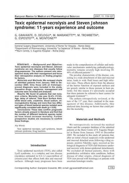
European Review for Medical <strong>and</strong> Pharmacological Sciences 2007; <strong>11</strong>: <strong>11</strong>9-127<br />
<strong>Toxic</strong> <strong>epidermal</strong> <strong>necrolysis</strong> <strong>and</strong> <strong>Steven</strong> <strong>Johnson</strong><br />
<strong>syndrome</strong>: <strong>11</strong>-<strong>years</strong> experience <strong>and</strong> outcome<br />
G. GRAVANTE, D. DELOGU*, M. MARIANETTI**, M. TROMBETTA*,<br />
G. ESPOSITO**, A. MONTONE**<br />
General Surgery Department, University of Rome Tor Vergata – Rome (Italy)<br />
*Department of Pharmacology, University “La Sapienza” of Rome – Rome (Italy)<br />
**Burn Centre, S. Eugenio Hospital – Rome (Italy)<br />
Abstract. – Background <strong>and</strong> Objective:<br />
<strong>Toxic</strong> <strong>epidermal</strong> <strong>necrolysis</strong> <strong>and</strong> <strong>Steven</strong> <strong>Johnson</strong><br />
<strong>syndrome</strong> are rare diseases that usually follow<br />
drug-exposures. The authors present one retrospective<br />
study with their management <strong>and</strong> focus<br />
their retrospective analysis on finding prognostic<br />
factors.<br />
Materials <strong>and</strong> Methods: We reviewed charts<br />
of admitted patients from January 1995 to December<br />
2005. Only those with an histologicproved<br />
diagnosis were included in the study.<br />
Causative drugs, symptoms, management <strong>and</strong><br />
outcome were recorded <strong>and</strong> analysed.<br />
Results: We found 32 patients that met inclusion<br />
criteria. Mortality rate was 34.4% (<strong>11</strong>/32).<br />
Age, delay of referral, Total Burn Surface Area,<br />
white blood cells, creatinine, blood sodium, immunoglobulins<br />
therapy <strong>and</strong> more than two different<br />
types of blood bacterial species isolated were<br />
significantly correlated with death (p < 0.05).<br />
Conclusions: These data confirm prognostic<br />
factors already present in literature <strong>and</strong> find that<br />
the number of different bacterial species isolated<br />
from blood increase mortality. Further<br />
prospective studies are necessary to confirm<br />
these findings.<br />
Key Words:<br />
<strong>Toxic</strong> <strong>epidermal</strong> <strong>necrolysis</strong>, Lyell <strong>syndrome</strong>, <strong>Steven</strong><br />
<strong>Johnson</strong> <strong>syndrome</strong>, Drug reactions.<br />
Introduction<br />
<strong>Toxic</strong> <strong>epidermal</strong> <strong>necrolysis</strong> (TEN), also called<br />
Lyell <strong>syndrome</strong>, is a complex <strong>and</strong> rare disease<br />
(incidence estimated in 0.4-1.2 cases/million of<br />
citizens/year) that affects both skin <strong>and</strong> mucosal<br />
surfaces. Although many progresses have been<br />
made in the comprehension of cellular <strong>and</strong> molecular<br />
mechanisms underlying pathophysiology,<br />
mortality remains severely high involving 30-<br />
49% of affected patients 1-3 .<br />
The peculiar characteristic of the disease, consisting<br />
in a wide detachment of skin <strong>and</strong> mucosal<br />
areas, leads to wide fluid losses <strong>and</strong> high infection<br />
rates. These effects derive from the absence<br />
of the barrier function of exposed surfaces <strong>and</strong><br />
are grossly similar to those present in burn patients.<br />
For this reason it’s universally accepted<br />
that these patients be referred to burn centres for<br />
adequate treatment 4-<strong>11</strong> .<br />
The authors retrospectively reviewed, at the<br />
turn of the <strong>11</strong> th year, their caseload in the management<br />
of this disease. Additionally, they<br />
analysed data focusing on factors that could predict<br />
the mortality.<br />
Materials <strong>and</strong> Methods<br />
We retrospectively reviewed the medical<br />
charts <strong>and</strong> the computer database of the admitted<br />
patients at the Burn Centre of S. Eugenio Hospital<br />
in Rome from January 1995 to December<br />
2005. We included in this study all patients that,<br />
after an initial clinical suspicion (wide <strong>epidermal</strong><br />
<strong>and</strong> mucosal detached areas following a drug exposure,<br />
positive Nikolsky sign), received an histological<br />
diagnosis of TEN (full thickness <strong>epidermal</strong><br />
necrosis with dermo-<strong>epidermal</strong> detachment).<br />
We excluded all burned patients, those with<br />
postinfective exfoliative skin conditions, bullous<br />
disease of other origins, clinical diagnosis of<br />
TEN (not proved with histology) or patients with<br />
uncertain histologic results.<br />
Corresponding Author: Gianpiero Gravante, MD; e-mail: ggravante@hotmail.com<br />
<strong>11</strong>9
We recorded patients’ demographics, delay of<br />
referral, comorbid conditions, precipitating event<br />
(event that led to drugs being administered), clinical<br />
<strong>and</strong> haematological parameters (fever, Total<br />
Body Surface Area of detached skin at first visit<br />
– TBSA –, white blood cells, haemoglobin,<br />
platelets, blood urea nitrogen, creatinine, sodium,<br />
potassium, transaminases), isolation of bacteria<br />
during the hospitalization (from wounds, blood<br />
<strong>and</strong> other sites), treatment adopted, length of<br />
hospitalization <strong>and</strong> outcome (death). Hospitilization<br />
was defined as the hospital stay only at the<br />
authors burn center <strong>and</strong> not days prior to transfer<br />
when the patient was hospitalized elsewhere.<br />
Drug/s involved were determined from patient’s<br />
or parents’ history considering all those administered<br />
during the previous 8 weeks before symptoms<br />
appearance (risk period).<br />
Management Protocol<br />
We developed our protocol over the <strong>years</strong>. Initial<br />
management consisted in the avoidance of all<br />
possible drugs <strong>and</strong> in extreme care in reintroducing<br />
only those necessary. A careful history was<br />
taken to establish the initiating drug <strong>and</strong> the precipitating<br />
event that rendered drug assumption<br />
necessary. Physical examination assessed TBSA.<br />
Baseline laboratory tests, chest x-ray films, <strong>and</strong><br />
skin biopsies were systematically obtained. Sepsis<br />
was defined from the presence of clinical<br />
(fever) <strong>and</strong> haematological signs (leukocytosis,<br />
positive blood cultures) <strong>and</strong> wound infections<br />
from erythema, pus discharge <strong>and</strong> positive<br />
wound cultures. Samples from wounds <strong>and</strong> blood<br />
were collected for cultures <strong>and</strong> sent for examination<br />
only if clinical <strong>and</strong> haematological signs of<br />
infections (i.e. fever or leucocytosis) were present.<br />
Serial pictures were taken in every patient<br />
for monitoring <strong>and</strong> records.<br />
Patient were recovered in intensive care-isolated<br />
rooms. Barrier garments <strong>and</strong> frequent h<strong>and</strong><br />
washings were used by health care personnel<br />
coming in <strong>and</strong> out of the isolation room. Heparin<br />
was routinely introduced for prophylaxis of<br />
thromboembolic events. Supportive therapies<br />
were adopted according to intensive care protocols<br />
as required. When clinical conditions ameliorated<br />
<strong>and</strong> did not require intensive supportive<br />
treatments, patients were transferred in normal<br />
non-isolated rooms.<br />
A Foley catheter was inserted for urine output<br />
monitoring. A femoral catheter for liquids replacement<br />
<strong>and</strong> a nasogastric feeding tube for enteral<br />
nutrition were routinely inserted until 1997.<br />
120<br />
G. Gravante, D. Delogu, M. Marianetti, M. Trombetta, G. Esposito, A. Montone<br />
In 1997, 2 patients suffered of gastrointestinal<br />
hemorrhage during nasogastric tube insertion <strong>and</strong><br />
one patient died of fatal pulmonary embolism after<br />
femoral thrombosis. After these episodes the<br />
nourishment was administered by total parenteral<br />
nutrition (low osmolarity formulas through a peripheral<br />
vein) in the early phase of the disease<br />
<strong>and</strong> by oral supplements later. Basal energy expenditure<br />
<strong>and</strong> metabolic requirements were calculated<br />
from the Harris-Benedict’s formula. No<br />
fluid resuscitation regimens specific of thermal<br />
burns were used but fluids were administered according<br />
to urine output <strong>and</strong> estimated insensible<br />
loss. Pain control was usually served best with a<br />
combination of non steroidal anti-inflammatory<br />
drugs (NSAIDs) <strong>and</strong>/or opioid analgesics.<br />
NSAIDs were used only if not suspected of determining<br />
TEN <strong>and</strong> if the patient did not refer<br />
any specific allergies. Gastric ulcer prophylaxis<br />
was prescribed in the form of proton-pump inhibitors.<br />
Local therapy consisted in a “no touch” approach<br />
to avoid any kind of skin damage. Local<br />
disinfection was not practised in wounds without<br />
clinical signs of infections (erythema, pus discharge)<br />
to limit skin detachments. All exposed<br />
areas were medicated only with an esterification<br />
product of the hyaluronic acid (Jaloskin, Gerke<br />
Pharm GmbH, Grevenbroich, Germany) until<br />
spontaneous complete coverage was achieved.<br />
No other topical treatments were used.<br />
All patients were put in an air-fluidised bed<br />
(Clinitron TM ) until autonomous mobilization was<br />
available. An ophthalmologist was usually early<br />
consulted for eye care <strong>and</strong> local lubricants initiated<br />
to prevent conjunctival-corneal adhesions<br />
(Xanthopterin 2-3 times daily). Chest <strong>and</strong> legs<br />
physical therapy was introduced as soon as possible<br />
to prevent pneumonia <strong>and</strong> deep venous<br />
thrombosis.<br />
In 1999 we introduced intravenous immunoglobulins<br />
in routine therapy <strong>and</strong> all patients,<br />
starting on February received the same<br />
dose <strong>and</strong> the same duration of treatment (400<br />
mg/kg for 5 days). No patient received steroids.<br />
Statistical Analysis<br />
All data analysis were performed using the<br />
Statistical Package for the Social Sciences<br />
Windows version 13.0 (SPSS, Chicago, Illinois,<br />
USA). Descriptive statistics used for continuous<br />
variables, after confirmation of normal<br />
distribution (histograms, Q-Q plots, Skewness<br />
<strong>and</strong> Kurtosis, Kolmogorov/Smirnov <strong>and</strong>
Shapiro Wilk testings), were mean <strong>and</strong> st<strong>and</strong>ard<br />
deviation. Descriptive statistics used for<br />
discrete variables consisted in frequencies report.<br />
Survival analysis was performed with the<br />
multivariate model of the Cox regression<br />
analysis. All p values were considered significant<br />
if < 0.05.<br />
Results<br />
We admitted 35 patients with an initial clinical<br />
diagnosis of toxic <strong>epidermal</strong> <strong>necrolysis</strong>, 32 of<br />
them received an histologic confirmation <strong>and</strong><br />
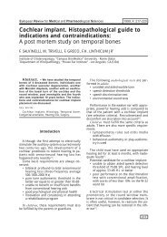
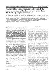
were analysed in this study (Table I). Year distribution<br />
seemed to decrease from 1997 (Figure 1).<br />
Initial care was usually administered at peripheral<br />
hospitals or from general practitioners <strong>and</strong> patients<br />
were referred to our centre for the progressive<br />
worsening of the cutaneous or systemic situation.<br />
Mean delay for referral in our institution<br />
was 10 days from the symptoms’ appearance<br />
(st<strong>and</strong>ard deviation 10.6, range 0-60).<br />
There were 21 women (65.6% of patients)<br />
<strong>and</strong> <strong>11</strong> men (34.4%). Mean age at diagnosis<br />
was 44.8 <strong>years</strong> (st<strong>and</strong>ard deviation 20.23,<br />
range 4-94). The most frequent comorbid conditions<br />
present were neoplastic diseases (10<br />
patients – 31.3%), followed by infections (5<br />
patients – 15.6%) <strong>and</strong> recent surgery, within a<br />
week to the onset of TEN (4 patients – 12.6%).<br />
The most frequent drugs administered at the<br />
time of TEN appearance were antibiotics (13<br />
patients – 40.6% – including also antiparasitic<br />
<strong>and</strong> antihelmintic drugs) followed by anticonvulsivants<br />
(10 patients – 31.3%) <strong>and</strong> nonsteroidal<br />
antinflammatory drugs (5 patients –<br />
15.6%) (Table II).<br />
A descriptive statistics of patients is summarized<br />
in Table III. <strong>Steven</strong>-<strong>Johnson</strong> <strong>syndrome</strong><br />
(SJS – defined as less than 10% of TBSA) was<br />
present in one patient <strong>and</strong> undetermined SJS-<br />
TEN overlap form (from 10 to 30% of TBSA)<br />
in 7 patients 12 . These forms together accounted<br />
for 25% of cases (8 patients). Wound cultures<br />
resulted positive for bacterial growth in 25 patients<br />
throughout the hospitalization (78.1%)<br />
while blood cultures in 19 patients (59.4%).<br />
Staphylococci accounted for 56% of wound infections<br />
<strong>and</strong> 48% of blood sepsis. Mean length<br />
of hospitalization was 17 days (st<strong>and</strong>ard deviation<br />
9, range 3-40). There were <strong>11</strong> deaths<br />
(34.4% of patients). Two of them occurred in<br />
<strong>Toxic</strong> <strong>epidermal</strong> <strong>necrolysis</strong><br />
<strong>Steven</strong> <strong>Johnson</strong> Syndrome <strong>and</strong> undetermined<br />
SJS-TEN overlap cases (2/8, 25%), 9 of the in<br />
TEN patients (9/24, 37.5%).<br />
Survival Analysis<br />
We examined as possible factors influencing<br />
death the sex, age, delay for referral to our centre<br />
since symptoms appearance, TBSA, white blood<br />
cells, creatinine, blood sodium <strong>and</strong> potassium,<br />
immunoglobulins (IgV’s) therapy <strong>and</strong> the number<br />
of bacterial species isolated from patients’<br />
blood throughout the hospitalization. Both delay<br />
of referral <strong>and</strong> number of bacteria were divided<br />
in two groups: for the delay in ≤ 4 days vs. > 4;<br />
for isolated bacteria in ≤ 2 vs. > 2. Cox regression<br />
analysis demonstrated that age, delay of referral,<br />
TBSA, white blood cells, creatinine, blood<br />
sodium, IgV’s therapy <strong>and</strong> more than two different<br />
types of blood bacteria isolated were significantly<br />
correlated with death (p < 0.05). In particular,<br />
the delay of referral increased mortality<br />
probability of 416.5 times, the absence of IgV’s<br />
therapy of 67.5 times <strong>and</strong> more than 2 types of<br />
bacteria 84 times (Table IV).<br />
Discussion<br />
<strong>Toxic</strong> <strong>epidermal</strong> <strong>necrolysis</strong> (also called Lyell<br />
<strong>syndrome</strong>) is a rare <strong>and</strong> severe adverse drug-induced<br />
skin/mucosal disease. Antibiotics, anticonvulsivants<br />
<strong>and</strong> non-steroidal antiinflammatory<br />
are the most commonly involved drugs 12 . Its rarity<br />
poses important problems for research, based<br />
mainly on retrospective studies, case series or<br />
case reports from different burn institutions 13 .<br />
Mortality is still high 1-3 <strong>and</strong> prognostic factors<br />
have been summarized in the SCORTEN<br />
score 14,15 .<br />
We recorded in our centre a high mortality<br />
rate (34% of patients) similar to most literature<br />
data 1-3,16-19 . The statistical analysis confirmed 3 of<br />
the 7 SCORTEN prognostic factors (age, delay<br />
of referral <strong>and</strong> TBSA, creatinine/blood urea nitrogen).<br />
Cancer was present in <strong>11</strong> of 32 patients<br />
(34.7%). Mortality in these patients was 36.4%<br />
(4/<strong>11</strong>), not significantly different from those not<br />
affected (33,3% – 7/21) <strong>and</strong> overall mortality<br />
(34,4% – <strong>11</strong>/32). We couldn’t specifically investigate<br />
the presence of tachycardia at admission as<br />
well as blood glucose or bicarbonate because<br />
charts of early recovered patients (1995-97) were<br />
missing about these data. We included in the<br />
121
Table I. Demographics <strong>and</strong> clinical characteristics of recovered patients. Drug 1 is referred to the newly introduced drug, “drug 2” to those already assumed.<br />
122<br />
G. Gravante, D. Delogu, M. Marianetti, M. Trombetta, G. Esposito, A. Montone<br />
Drugs<br />
Burn Event administered<br />
centre that led in the Length Dead<br />
delay of to drug Probably 8 weeks Duration of hospita- (D)<br />
Patient referral admini- involved preceding of fever Wound Blood lization or Alive<br />
no. Year Sex Age (days) stration drug TEN TBSA (days) culture culture Other (days) (A)<br />
1 1995 F 37 10 Hemorrhagic Deflazacort – 100 20 Pseud. Aeruginosa Negative Pseud. Aeruginosa 10 D<br />
stroke Staph. Aureus (sputum-lungs)<br />
2 1996 F 49 4 Gram- abscess Cefam<strong>and</strong>ole Carbamazepine 15 <strong>11</strong> – Acinetobacter Acinetobacter 9 D<br />
after brain Phenobarbital Baumanii Baumanii<br />
menyngioma surgery Staph. Aureus E. Coli (liquor)<br />
3 1997 F 21 12 – Pyrantel Rifaximin 20 30 Pseud. Aeruginosa Enterococcus Staph. Aureus 22 A<br />
pamoate Staph. Aureus Faecalis (oral)<br />
Diphteroids Pseud. Aeruginosa<br />
Staph. Aureus (central venous<br />
Staph. Coag.- catheter, vagina)<br />
Enterococcus<br />
Faecalis,<br />
E. Coli (vagina)<br />
4 1997 F 94 8 Flu-like symptoms Ketoprofen – 13 2 Staph. Aureus Negative – 16 A<br />
5 1997 F 19 9 Tension headache Nimesulide, – 70 26 – Staph. Aureus – 40 A<br />
Paracetamol, Pseud. Aeruginosa<br />
Aspirin-<br />
6 1997 F 4 3 – Diphtheria- Clotrimazole 26 2 Staph. Coag.- Negative Staph. Coag-. 12 A<br />
pertussis- Azythromycin Acinetobacter<br />
tetanus vaccine Baumanii<br />
(urinary catheter)<br />
7 1997 M 43 8 Leishmaniasis Amphotericin Ciprofloxacin, 90 3 – Staph. Coag- – 3 D<br />
(HBV,HCV Trimethroprim/<br />
<strong>and</strong> HIV+) Sulfa-methoxazole,<br />
Allopurinol,<br />
Itraconazole<br />
Paracetamol<br />
8 1997 M 31 7 Systemic Ketoprofen Carbamazepine 35 3 Staph. Aureus Staph. Coag- Staph. Aureus 15 A<br />
erythematosus (conjunctivae)<br />
lupus<br />
9 1997 M 63 7 Hemorrhagic Phenobarbital – 93.5 5 – Enterococcus – <strong>11</strong> D<br />
stroke Faecalis<br />
Citrobacter Freundii<br />
Staph. Coag<br />
10 1997 F 69 15 Brain astrocytoma Phenobarbital – 45 4 – – 7 A
<strong>Toxic</strong> <strong>epidermal</strong> <strong>necrolysis</strong><br />
<strong>11</strong> 1998 F 66 5 Pleural mesothelioma Ceftriaxone Epidoxorubicin 90 0 Staph. Aureus Staph. Aureus – 17 A<br />
Interferon Staph. Coag.-<br />
Bromazepam Strep. Viridans<br />
12 1998 F 36 10 Sun exposure Isothipendyl – 90 6 Staph. Coag.- Negative – 12 A<br />
(gel)<br />
13 1998 F 52 60 Cutaneous mycosis Terbinafine – 95 — Acinetobacter Acinetobacter Pseud. Aeruginosa 5 A<br />
Baumanii Baumanii Staph. Aureus<br />
Pseud. Aeruginosa Pseud. Aeruginosa Proteus Mirabilis<br />
Staph. Aureus Staph. Aureus (central venous<br />
Proteus Mirabilis catheter)<br />
Enterococcus<br />
Faecalis<br />
14 1998 F 27 5 Post-transplant Meropenem – 21 5 Staph. Coag.- Staph. Coag.- – 7 A<br />
chronic renal failure Enterococcus<br />
Kaposi’s sarcoma Faecalis<br />
15 1998 F 44 5 Glioblastoma – – 80 10 Kl. Pneumoniae Pseud. Aeruginosa Pseud. Aeruginosa 30 A<br />
Enterobacter (mouth)<br />
Aerogenes<br />
Enterococcus<br />
Faecalis<br />
Staph. Coag.-<br />
16 1999 M 55 5 Non-Hodgkin Fluconazole, – 100 19 Staph. Aureus, Staph. Aureus, Staph. Aureus, 19 A<br />
lymphoma (HIV+) Acyclovir, Acinetobacter Acinetobacter Acinetobacter<br />
Trimethroprim/ Baumanii, Baumanii, Baumanii,<br />
Sulfa- Staph. Coag- Staph. Coag- Pseud. Aeruginosa<br />
methoxazole, (central venous<br />
Allopurinol catheter)<br />
17 1999 F 54 7 Non-Hodgkin Fluconazole – 100 2 Staph.Aureus Staph. Aureus Staph. Coag.- 9 D<br />
lymphoma Pseud. Aeruginosa Pseud. Aeruginosa (pleural essudate)<br />
Cutaneous mycosis Acinetobacter<br />
Baumanii<br />
Staph. Coag.-<br />
Pseud. Fluorescens<br />
18 2000 M 62 6 Cirrhosis (HCV+) Allopurinol – 39 0 Enterococcus – – 28 D<br />
Chronic Heart Failure Faecalis<br />
Staph. Hominis<br />
19 2000 M 58 8 Brain astrocytoma Phenobarbital Ciprofloxacin 38 17 Staph. Aureus Staph. Aureus – 32 A<br />
Vancomycin<br />
20 2000 F <strong>11</strong> 8 Sun exposure Isothipendyl Dimethindene 83 0 Staph. Coag.- Negative – 14 A<br />
(gel) Rokitamycin<br />
21 2000 F 30 0 – – – 100 0 Staph. Coag.- Negative – 8 A<br />
22 2001 F 43 8 Flu-like symptoms Bromhexine Dextromethorphan 100 0 Diphteroids Diphteroids – 14 A<br />
Staph. Coag.- Staph. Aureus<br />
Continued<br />
123
124<br />
G. Gravante, D. Delogu, M. Marianetti, M. Trombetta, G. Esposito, A. Montone<br />
Table I (Continued). Demographics <strong>and</strong> clinical characteristics of recovered patients. Drug 1 is referred to the newly introduced drug, “drug 2” to those already assumed.<br />
Drugs<br />
Burn Event administered<br />
centre that led in the Length Dead<br />
delay of to drug Probably 8 weeks Duration of hospita- (D)<br />
Patient referral admini- involved preceding of fever Wound Blood lization or Alive<br />
no. Year Sex Age (days) stration drug TEN TBSA (days) culture culture Other (days) (A)<br />
23 2001 F 24 5 Corneal transplant Deflazacort Cefazolin 34 4 – Negative – 25 A<br />
24 2002 F 74 3 Hemorrhagic stroke Lansoprazole Phenytoin 100 7 Staph. Aureus Staph. Aureus – 21 D<br />
Phenobarbital Enterococcus<br />
Faecalis<br />
25 2002 F 58 6 Prosthesis removal – – 22 0 Providencia Stuartii Diphteroids – 16 D<br />
Enterococcus<br />
Faecalis<br />
C<strong>and</strong>ida<br />
26 2002 M 19 30 Systemic Carbamazepine Azathioprine 100 0 – – – 6 D<br />
erythematosus lupus<br />
27 2003 M 44 6 Acute renal failure Furosemide Carvedilol 6 0 Staph. Aureus Negative – 18 A<br />
28 2003 M 62 8 Brain astrocytoma Sertraline – 22 8 Staph. Coag.- Negative – 19 A<br />
29 2004 F 66 19 Abdominal operation Iron <strong>and</strong> folate – 54 15 Staph. Aureus Staph. Aureus – 27 A<br />
supplements<br />
30 2004 M 51 9 – Cefaclor Rosuvastatin 87 1 Staph. Coag.- – – 12 D<br />
31 2004 M 26 6 Epilepsy Carbamazepine – 35 0 – Negative – 34 A<br />
32 2005 F 44 15 Idiopathic Metronidazole Piperacillin 50 7 Acinetobacter Acinetobacter – 13 D<br />
myelofibrosis Baumanii, Baumanii<br />
Staph. Aureus
Number of cases<br />
8<br />
6<br />
4<br />
2<br />
0<br />
study both TEN, SJS patients (1 case) <strong>and</strong> overlapped<br />
forms (7 cases) because, at the time of recovery,<br />
all of them had wide <strong>epidermal</strong> <strong>and</strong> mucosal<br />
detached areas following drug exposures,<br />
positive Nikolsky signs <strong>and</strong> same histopathologic<br />
appearance of the skin. Specific analysis revealed<br />
prognostic differences among TEN (42.1% of<br />
mortality) vs. other conditions (25% of mortality).<br />
These findings were also confirmed by the<br />
Cox regression analysis that found TBSA as a<br />
factor influencing death.<br />
A lack of association with mortality was observed<br />
for the type of drug theoretically incriminated<br />
of TEN’s occurrence or with the total number<br />
of drugs assumed before hospitalization (data<br />
not shown). With particular regard to comorbidities,<br />
the statistical analysis did not show a higher<br />
mortality if patients were affected by one, two or<br />
more comorbidities, neither the analysis by specific<br />
diseases (i.e. tumors, strokes, infections)<br />
showed a correlation with mortality. We found an<br />
interesting prevalence of malignancies in our<br />
TEN patients (ten of them – 31.3% – were also<br />
affected by tumours). All oncologic patients in<br />
general received several drugs possibly involved<br />
in TEN pathogenesis (antibiotics, gout-preventing<br />
drugs, non steroidal anti-inflammatory<br />
drugs). Additionally, brain cancer patients (5 patients)<br />
assumed anticonvulsivants to prevent<br />
epileptic crisis.<br />
<strong>Toxic</strong> <strong>epidermal</strong> <strong>necrolysis</strong><br />
1997 1996 1998 2000 2002 2004 2006 0 20 40 60 80 1 00<br />
Year<br />
TBSA (%)<br />
Figure 1. Left panel: number of patients from 1995 to 2005. Right panel: TBSA distribution of patients.<br />
Number of cases<br />
12<br />
10<br />
8<br />
6<br />
4<br />
2<br />
0<br />
Delay of referral to the burn centre was confirmed<br />
as an important prognostic factor at the<br />
Cox regression multivariate analysis, contrarily<br />
to other authors 15 , <strong>and</strong> had the greatest influ-<br />
Table II. Demographics <strong>and</strong> clinical characteristics of recovered<br />
patients. Drug 1 is referred to the newly introduced<br />
drug, “drug 2” to those already assumed.<br />
Comorbidities/ N. Percentage<br />
drug cases (%)<br />
Tumor 10 31.3<br />
Infection 5 15.6<br />
Surgery 4 12.5<br />
Stroke 3 9.4<br />
Flu or muscular pain 3 9.4<br />
Renal failure 2 6.3<br />
Sun burn 2 6.3<br />
Viral hepatitis 2 6.3<br />
HIV 2 6.3<br />
SLE 2 6.3<br />
Epilepsy 1 3.1<br />
Antibiotics 13 40.6<br />
Anticonvulsivants 10 31.3<br />
Nonsteroidal 5 15.6<br />
antinflammatory drugs<br />
Systemic corticosteroids 2 6.3<br />
Allopurinol 3 9.4<br />
Sunscreen protective 2 6.3<br />
creams<br />
Others 7 21.9<br />
125
ence on mortality. If greater than 4 days it increased<br />
the risk of death of 416 times. This<br />
could be derived from the delay in introducing<br />
specific immunoglobulin-blocking therapy <strong>and</strong><br />
specific support therapy. Furthermore, patients<br />
126<br />
G. Gravante, D. Delogu, M. Marianetti, M. Trombetta, G. Esposito, A. Montone<br />
Table III. Descriptive statistics of 32 patients.<br />
Variable Patients (n = 32)<br />
Age 45 ± 20<br />
Sex (male number) <strong>11</strong>/32 (34.4%)<br />
Delay of referral 10 ± <strong>11</strong><br />
Number of drugs already on 1.3 ± 0.9<br />
board when TEN started<br />
Comorbidities (see Table III) 24/32 (75%)<br />
TBSA (%) 62 ± 34<br />
Presence of fever at 22/32 (68.8%)<br />
hospitalization<br />
Duration of fever 7 ± 8<br />
White blood cells (/mL) 8131 ± 6200<br />
Hemoglobin (g/dL) <strong>11</strong>.2 ± 2.6<br />
Platelets (/mL × 1000) 225 ± 137<br />
Blood Urea Nitrogen (mg/dL) 63 ± 56<br />
Creatinine (mg/dL) 1.6 ± 2.2<br />
Sodium (mmol/L) 136 ± 8<br />
Potassium (mmol/L) 4.2 ± 1.0<br />
GOT (U/L) 54 ± 63<br />
GPT (U/L) 56 ± 73<br />
IgV’s therapy 17/32 (53.1%)<br />
Wound culture positive 25/32 (78.1%)<br />
Different bacteria isolated 1.3 ± 1.0<br />
from wound cultures<br />
throughout hospitalization<br />
Sepsis 18/32 (56,2%)<br />
Different bacteria isolated from 1.2 ± 1.5<br />
blood cultures throughout<br />
hospitalization<br />
Length of hospitalization 17 ± 9<br />
Duration of disease 27 ± 12<br />
Table IV. Cox regression analysis of survival. Exp (B): hazard risk.<br />
referred late were probably more ill than those<br />
referred early. In other words, the late referral<br />
delayed introduction of immunoglobulin therapy<br />
<strong>and</strong> represented a failure in management at<br />
the preliminary hospital with worse patients<br />
outcomes in our center.<br />
Immunoglobulins (IgV’s), administered since<br />
1999, seemed to have a positive effect on mortality<br />
even in patients with high delays of referral.<br />
Although the effect of IgV’s therapy on <strong>Steven</strong><br />
<strong>Johnson</strong> Syndrome <strong>and</strong> TEN is not well established<br />
<strong>and</strong> our study was retrospective <strong>and</strong> not<br />
prospective, the absence of IgV’s therapy increased<br />
the risk of death of 67.5 times. A theoretic<br />
interpretation of this can derive from the persistence<br />
<strong>and</strong> activation of CD95 system (the main<br />
system inhibited by IgV’s) 20-21 even days later<br />
from the initial stimulus. However, since IgV’s<br />
was given to patients after 1999, <strong>and</strong> not to patients<br />
before, differences could be attributable to<br />
better care, more experience in TEN management<br />
<strong>and</strong> refinements in TEN protocol that occurred<br />
over time rather than a specific effect of<br />
immunoglobulins. For this reason, we confirm<br />
that the debate about the efficacy of IgV’s in<br />
TEN patients is still not over <strong>and</strong> further studies<br />
are required to address this question.<br />
Our data confirmed that septicaemia is an important<br />
mortality factor. Furthermore, we found<br />
that different bacterial species were present <strong>and</strong><br />
isolated from patients blood, simultaneously or<br />
metachronously throughout the hospitalization.<br />
The correlation analysis of these data showed a<br />
great increase in mortality after 3 or more different<br />
bacterial species were isolated during hospitalization.<br />
When isolated bacterial species were 3<br />
or more, mortality increased of 84 times. Al-<br />
95.0% CI for Exp (B)<br />
Hazard ratio Sig. (p) Lower Upper<br />
Sex 0.788 0.888 0.028 21.831<br />
Age 1.<strong>11</strong>8 0.042 1.004 1.244<br />
Delay of referral 416.513 0.010 4.325 40109.530<br />
Comorbidities (see Table III) 18.293 0.205 0.204 1640.991<br />
TBSA 1.069 0.021 1.010 1.131<br />
White blood cells 0.999 0.029 0.998 1.000<br />
Creatinine 2.205 0.047 1.012 4.808<br />
Blood sodium 0.714 0.020 0.538 0.949<br />
Blood potassium 0.164 0.085 0.021 1.283<br />
IgV’s therapy –67.491 0.007 3.128 1456.278<br />
> 2 blood bacteria isolated 83.994 0.044 1.121 6295.833
though the design of our study (retrospective<br />
analysis, a small population size hospitalized in<br />
one burn center, a long delay of referral from<br />
other institutions) does not allow drawing any<br />
firm conclusion, this data, if confirmed by further<br />
studies, could be used as prognostic markers during<br />
patient’s hospitalization.<br />
In conclusion, although much has been discovered<br />
about the etiology <strong>and</strong> pathogenesis of toxic<br />
<strong>epidermal</strong> <strong>necrolysis</strong> <strong>and</strong> new treatments have<br />
become available in the last <strong>years</strong>, mortality is<br />
still high. The analysis of our data shows that<br />
age, TBSA, white blood cells, creatinine, IgV’s<br />
therapy <strong>and</strong> more than two different types of<br />
blood bacteria isolated during hospitalization<br />
were significantly correlated with death (p <<br />
0.05). Further larger studies are necessary to confirm<br />
these findings.<br />
References<br />
1) CHAVE TA, MORTIMER NJ, SLADDEN MJ, HALL AP,<br />
HUTCHINSON PE. <strong>Toxic</strong> <strong>epidermal</strong> <strong>necrolysis</strong>: current<br />
evidence, practical management <strong>and</strong> future<br />
directions. Br J Dermatol 2005; 153: 241-253.<br />
2) BECKER DS. <strong>Toxic</strong> <strong>epidermal</strong> <strong>necrolysis</strong>. Lancet<br />
1998; 351: 1417-1420.<br />
3) DUCIC I, SHALOM A, RISING W. Outcome of patients<br />
with toxic <strong>epidermal</strong> <strong>necrolysis</strong> <strong>syndrome</strong> revisited.<br />
Plast Reconstr Surg 2002; <strong>11</strong>0, 768.<br />
4) HEIMBACH DM, ENGRAV LH, MARVIN JA, HARNAR TJ,<br />
GRUBE BJ. <strong>Toxic</strong> <strong>epidermal</strong> <strong>necrolysis</strong>. A step forward<br />
in treatment. JAMA 1987; 257: 2171-2175.<br />
5) ZOLTIE N, VERLENDE P, O'NEILL TJ, MCKENZIE AW.<br />
Lyell's <strong>syndrome</strong> on a burns unit. Burns 1994; 20:<br />
368-370.<br />
6) GREEN D, LAW E, STILL JM. An approach to the<br />
management of toxic <strong>epidermal</strong> <strong>necrolysis</strong> in a<br />
burn centre. Burns 1993; 19: 4<strong>11</strong>-414.<br />
7) PALMIERI TL, GREENHALGH DG, SAFFLE JR, SPENCE RJ,<br />
PECK MD, JENG JC, MOZINGO DW, YOWLER CJ, et al.<br />
A multicenter review of toxic <strong>epidermal</strong> <strong>necrolysis</strong><br />
treated in U.S. burn centers at the end of the<br />
twentieth century. J Burn Care Rehabil 2002; 23:<br />
87-96.<br />
8) HETTIARATCHY S, MOLONEY D, CLARKE J. Patients with<br />
acute skin loss: are they best managed on a<br />
burns unit? Ann R Coll Surg Engl 2001; 83: 26-<br />
29.<br />
<strong>Toxic</strong> <strong>epidermal</strong> <strong>necrolysis</strong><br />
9) KHOO AK, FOO CL. <strong>Toxic</strong> <strong>epidermal</strong> <strong>necrolysis</strong> in a<br />
burns centre: a 6-year review. Burns 1996; 22:<br />
275-278.<br />
10) YING S, HO W, CHAN HH. <strong>Toxic</strong> <strong>epidermal</strong> <strong>necrolysis</strong>:<br />
10 <strong>years</strong> experience of a burns centre in<br />
Hong Kong. Burns 2001; 27: 372-375.<br />
<strong>11</strong>) KELEMEN JJ 3RD, CIOFFI WG, MCMANUS WF, MASON<br />
AD Jr, PRUITT BA Jr. Burn center care for patients<br />
with toxic <strong>epidermal</strong> <strong>necrolysis</strong>. J Am Coll Surg<br />
1995; 180: 273-278.<br />
12) ROUJEAU JC, KELLY JP, NALDI L, RZANY B, STERN RS,<br />
ANDERSON T, AUQUIER A, BASTUJI-GARIN S, CORREIA O,<br />
LOCATI F, et al. Medication use <strong>and</strong> the risk of<br />
<strong>Steven</strong>s-<strong>Johnson</strong> <strong>syndrome</strong> or toxic <strong>epidermal</strong><br />
<strong>necrolysis</strong>. N Engl J Med 1995; 333: 1600-1607.<br />
13) RZANY B, CORREIA O, KELLY JP, NALDI L, AUQUIER A,<br />
STERN R. Risk of <strong>Steven</strong>s-<strong>Johnson</strong> <strong>syndrome</strong> <strong>and</strong><br />
toxic <strong>epidermal</strong> <strong>necrolysis</strong> during first weeks of<br />
antiepileptic therapy: a case-control study. Study<br />
Group of the International Case Control Study on<br />
Severe Cutaneous Adverse Reactions. Lancet<br />
1999; 353: 2190-2194.<br />
14) BASTUJI-GARIN S, FOUCHARD N, BERTOCCHI M, ROUJEAU<br />
JC, REVUZ J, WOLKENSTEIN P. SCORTEN: a severityof-illness<br />
score for toxic <strong>epidermal</strong> <strong>necrolysis</strong>. J<br />
Invest Dermatol 2000; <strong>11</strong>5: 149-153.<br />
15) GUEGAN S, BASTUJI-GARIN S, POSZEPCZYNSKA-GUIGNE E,<br />
ROUJEAU JC, REVUZ J. Performance of the<br />
16)<br />
SCORTEN during the first five days of hospitalization<br />
to predict the prognosis of <strong>epidermal</strong><br />
<strong>necrolysis</strong>. J Invest Dermatol 2006; 126: 272-276.<br />
CHAN HL. Observations of drug-induced toxic <strong>epidermal</strong><br />
<strong>necrolysis</strong> in Singapore. J Am Acad Dermatol<br />
1984; 10: 973-978.<br />
17) RONJEAU JC, GUILLAUNE JC, FABRE JC. <strong>Toxic</strong> <strong>epidermal</strong><br />
<strong>necrolysis</strong> (Lyell <strong>syndrome</strong>), incidence <strong>and</strong><br />
drug etiology in France. Arch Dermatol 1990;<br />
126: 37-42.<br />
18) NALDI L, LOCATI L, MARCHESI L. Incidence of toxic<br />
<strong>epidermal</strong> <strong>necrolysis</strong> in Italy. Arch Dermatol 1990;<br />
126: <strong>11</strong>03-<strong>11</strong>04.<br />
19) SCHOPF E, STUHMER A, RZANY B. <strong>Toxic</strong> <strong>epidermal</strong><br />
<strong>necrolysis</strong> <strong>and</strong> <strong>Steven</strong>-<strong>Johnson</strong> <strong>syndrome</strong>; an<br />
epidemiological study from West Germany. Arch<br />
Dermatol 1990; 127: 839-847.<br />
20) KIM PS, GOLDFARB IW, GAISFORD JC. <strong>Steven</strong>-<strong>Johnson</strong><br />
<strong>syndrome</strong> <strong>and</strong> toxic <strong>epidermal</strong> <strong>necrolysis</strong>: a<br />
pathophysiologic review with recommendations<br />
for a treatment protocol. J Burn Care Rehabil<br />
1983; 4: 91-100.<br />
21) VIARD I, WEHRLI P, B ULLANI R, SCHNEIDER P, H OLLER N,<br />
SALOMON D, HUNZIKER T, SAURAT JH, TSCHOPP J,<br />
FRENCH LE. Inhibition of toxic <strong>epidermal</strong> <strong>necrolysis</strong><br />
by blockade of CD95 with human intravenous immunoglobulin.<br />
Science 1998; 282: 490-493.<br />
127