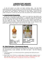
A. Collecting DataEach student should collect a known volume of water (or sediment, as the case maybe) from your choice of aquatic ecosystem/microhabitat on campus or near your home.Place your sample in a covered, plastic container <strong>and</strong> label it with:a. your nameb. your lab sectionc. the type of ecosystem (freshwater? Brackish canal? Lake Osceola? Be asspecific as possible)d. type of sample (water column? Water surface? Sediment?)e. Date <strong>and</strong> time of day the sample was collected.To observe your organsms, draw up one cc (= one milliliter (mL)) of sample. (Again: besure everything is properly labeled so that your samples won’t be misidentified.) Tocount organisms in your sample:1. Place two drops of your sample on a convex microscope slide2. Drop a coverslip onto the liquid3. Place one drop of methyl cellulose at the edge of the coverslip, <strong>and</strong> allow todiffuse under the coverslip. (This will slow down any rapidly swimmingmicroorganisms)4. STARTING ON LOW POWER, observe your sample under the microscope.5. Begin at one corner of the coverslip, <strong>and</strong> gradually work your way across <strong>and</strong>down, in a zig-zag pattern, until you have observed the entire coverslip field.6. Whenever you find a motile organism (protist or animal), stop <strong>and</strong> identify it ascompletely as you can by using the Identification Guide following this section.Online sources <strong>and</strong> the Photo Atlases in lab can also be useful.7. Record your results on the appropriate tally sheets provided.8. Write down the characteristics that allowed you to identify your organism in eachcase.B. Organism Identification GuideSince this is your first introduction to some of the vast diversity of Life, we don'texpect you to be able to identify with any great precision the many organisms we hopeyou will see. However, this guide should help you narrow down the identification of theliving organisms in your sample.Feel free to use online resources for identification. Once you have identified anorganism as a diatom, for example, a Google image search might well yield a possibleidentity for your organism. (Be careful, though. When it comes to protists, it sometimestakes a real expert to tell them apart. We’ll be happy if you learn to tell a flagellate froma ciliate at this stage of the game.)A broad overview of the types of organisms you might encounter can be found at theseh<strong>and</strong>y sites:http://tinyurl.com/tmhzhttp://www.microscopyu.com/moviegallery/pondscum/If you find something you can't identify, ask your instructor for help.systematics-22
1. ProtistsThese are the simplest of the eukaryotic organisms, <strong>and</strong> they are a very diverseassemblage now assigned to several different c<strong>and</strong>idate kingdoms once subsumedunder the now-defunct taxon "Protista." The types you are likely to see today will bevery small <strong>and</strong> usually highly motile. To see them well, you'll probably have to usemethyl cellulose to slow them down. Most common in daytime samples will be diatoms<strong>and</strong> small flagellates. But the occasional ciliate or amoeba will show up.2. Animalia, Porifera - The SpongesThe sponges are the simplest of animals, <strong>and</strong> they are found in both freshwater <strong>and</strong>marine habitats. They are characterized by an amorphous body shape with nodistinguishable head or tail end. Lacking true tissues, these animals have an array ofdiversified cell types, each of which performs a specific function. But you will not likelyto be able to see individual cells in a sponge under the microscope. It will just look likean amorphous blob with slightly greater degree of organization than pond sludge. Youcan find a variety of freshwater sponge images as http://tinyurl.com/porif3. Animalia, Cnidaria - Radially Symmetrical DiploblastsFound in both freshwater <strong>and</strong> marine habitats, these animals are radiallysymmetrical (i.e., the body is divisible into identical "pie shaped" wedges) <strong>and</strong> have twotrue tissue layers (endoderm <strong>and</strong> ectoderm). The most common ones in freshwater willby hydras, <strong>and</strong> in marine habitats you may find small medusae, the free-swimming“jellyfish” form cnidarians sometimes take. Check out some images here:http://tinyurl.com/pondhydrahttp://tinyurl.com/fwmedusa4. Animalia, Platyhelminthes - The FlatwormsIf the body is dorsoventrally flattened (i.e., flattened from "top" to "bottom") <strong>and</strong> thereis a distinct head end that guides the animal's movements, there's a good chance you'relooking at a flatworm. (If you're not sure, call the instructor for a positive I.D.) Theseanimals have three true tissue layers (endoderm, ectoderm <strong>and</strong> mesodermalmesenchyme) <strong>and</strong> simple organ systems. View a flatworm in action athttp://www.youtube.com/watch?v=_jjzQrR5PLQ5. Animalia, Rotifera - The Wheel AnimalculesThese tiny animals are no bigger than a large protist, yet they have three true tissuelayers <strong>and</strong> complex organ systems. They feed by means of a cephalic (head end)corona of cilia which beats food particles from the water into the mouth. They also usethe corona for swimming; it pulls the animal through the water like a little propeller whenit decides to weigh anchor (pull up its sticky pedal disk) <strong>and</strong> move.Check out the dramatic video of rotifers athttp://www.youtube.com/watch?v=YF8OJt_pujc And yes, that’s the rotifers singing.6. Animalia, Nematoda - The RoundwormsThese worms are very thin, symmetrical, <strong>and</strong> tapered at both ends. There is noevidence of body segmentation, <strong>and</strong> they move with a characteristic sinusoidal wavemotion unique to this phylum. This is because the body wall has only longitudinalmuscles, another characteristic unique to this phylum. Nothing else moves quite like anematode, <strong>and</strong> you can see them swimming athttp://www.youtube.com/watch?v=SpgjnXEFadgsystematics-23