alpha-1-antitrypsin deficiency in children: liver disease is not ...
alpha-1-antitrypsin deficiency in children: liver disease is not ...
alpha-1-antitrypsin deficiency in children: liver disease is not ...
- No tags were found...
You also want an ePaper? Increase the reach of your titles
YUMPU automatically turns print PDFs into web optimized ePapers that Google loves.
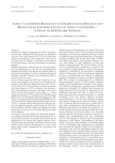
December 7, 2005EUROPEAN JOURNAL OF MEDICAL RESEARCH 509Eur J Med Res (2005) 10: 509-514 © I. Holzapfel Publ<strong>is</strong>hers 2005ALPHA-1-ANTITRYPSIN DEFICIENCY IN CHILDREN: LIVER DISEASE IS NOTREFLECTED BY LOW SERUM LEVELS OF ALPHA-1-ANTITRYPSIN –A STUDY ON 48 PEDIATRIC PATIENTST. Lang 1 , M. Mühlbauer 1 , M. Strobelt 1 , S. Weid<strong>in</strong>ger 2 , H. B. Hadorn 11 Children´s Hospital, Dr. v. Haunersches K<strong>in</strong>derspital,University of Munich, Munich, Germany2 Bavarian Red Cross Transfusion Service, Regensburg, GermanyAbstractBackground: Alpha-1-<strong>antitryps<strong>in</strong></strong> (α1-AT) <strong>is</strong> an importantprotease <strong>in</strong>hibitor. The phe<strong>not</strong>ypes are characterizedby a low total serum α1-AT or by an abnormalprote<strong>in</strong> accumulat<strong>in</strong>g <strong>in</strong> the hepatocytes. The aim ofour study was to exam<strong>in</strong>e a correlation of total serumα1-AT, phe<strong>not</strong>ype, and <strong>liver</strong> <strong>in</strong>volvement <strong>in</strong> pediatricpatients.Methods: 48 patients, deficient for α1-AT were <strong>in</strong>cluded.The phe<strong>not</strong>ypes for α1-AT were determ<strong>in</strong>ed by<strong>is</strong>oelectric focus<strong>in</strong>g. Liver <strong>d<strong>is</strong>ease</strong> was def<strong>in</strong>ed either aselevated transam<strong>in</strong>ases or/and as elevated conjugatedbilirub<strong>in</strong> and γGT. Patients were reexam<strong>in</strong>ed after amean <strong>in</strong>terval of 2 years.Results: Homozygous α1-AD was found <strong>in</strong> 12 patients,heterozygous <strong>in</strong> 24 patients. In 12 <strong>children</strong> rarevariants of α1-AD were diagnosed. Serum α1-AT levelsless than 60% of normal were found <strong>in</strong> all patientswith homozygous, <strong>in</strong> 37% of patients with heterozygousα1-<strong>antitryps<strong>in</strong></strong> <strong>deficiency</strong> (α1-AD), and <strong>in</strong> patientswith the homozygous variant PiM palermo . Liver<strong>d<strong>is</strong>ease</strong> was found <strong>in</strong> 8/12 patients with the phe<strong>not</strong>ypePiZZ and <strong>in</strong> 15/24 patients with heterozygous α1-AD.Three of 4 patients with the phe<strong>not</strong>ype PiMQ0 had severe<strong>liver</strong> <strong>d<strong>is</strong>ease</strong> despite normal serum levels for α1-AT. In 11 patients with heterozygous α1-AD <strong>liver</strong> <strong>d<strong>is</strong>ease</strong>was apparent despite normal serum α1-AT levels.In two patients with the variant type Mpalermo serumlevels were as low as 11% of normal without any signsof <strong>liver</strong> <strong>d<strong>is</strong>ease</strong>.Conclusions: Our data clearly show that <strong>in</strong> the diagnosticworkup of neonatal cholestas<strong>is</strong> measurement of totalserum α1-AT does <strong>not</strong> exclude <strong>liver</strong> <strong>d<strong>is</strong>ease</strong> due toabnormal α1-AT variants. We suggest analys<strong>is</strong> of α1-AT-phe<strong>not</strong>ype by <strong>is</strong>oelectric focuss<strong>in</strong>g <strong>in</strong> patients withunknown <strong>liver</strong> <strong>d<strong>is</strong>ease</strong>. Heterozygous or rare varianttypes might rema<strong>in</strong> undiagnosed by measur<strong>in</strong>g totalα1-AT only.Key words: α1-<strong>antitryps<strong>in</strong></strong> <strong>deficiency</strong>, <strong>children</strong>, <strong>liver</strong>INTRODUCTIONAlpha-1-<strong>antitryps<strong>in</strong></strong> or protease <strong>in</strong>hibitor (Pi) <strong>is</strong> a s<strong>in</strong>glecha<strong>in</strong> 52 kD glycoprote<strong>in</strong> cons<strong>is</strong>t<strong>in</strong>g of 394 am<strong>in</strong>oacids The prote<strong>in</strong> <strong>is</strong> encoded by a gene located on thed<strong>is</strong>tal long arm of chromosome 14 (14q31-32.2) [17].Structural variants of α1-AT <strong>in</strong> humans are classifiedaccord<strong>in</strong>g to the protease <strong>in</strong>hibitor phe<strong>not</strong>ype systemas def<strong>in</strong>ed by agarose gel electrophores<strong>is</strong> or <strong>is</strong>oelectricfocus<strong>in</strong>g of plasma [15]. To date more than 75 differentvariants are known, some of them lead<strong>in</strong>g to severe<strong>liver</strong> <strong>d<strong>is</strong>ease</strong> <strong>in</strong> early childhood or early pulmonaryemphysema [16, 21]. Several variants of α1-AT are associated with a reduction <strong>in</strong> serum α1-ATconcentrations, called <strong>deficiency</strong> variants. The genetic<strong>deficiency</strong> <strong>is</strong> <strong>in</strong>herited <strong>in</strong> an autosomal co-dom<strong>in</strong>antfashion. Homozygous PiZ α1-<strong>antitryps<strong>in</strong></strong> <strong>deficiency</strong>has been associated with <strong>liver</strong> <strong>d<strong>is</strong>ease</strong> <strong>in</strong> childhood,lead<strong>in</strong>g to endstage <strong>liver</strong> <strong>d<strong>is</strong>ease</strong> <strong>in</strong> a subset of patients[10]. Th<strong>is</strong> variant <strong>is</strong> characterized by a d<strong>is</strong>t<strong>in</strong>ctiveh<strong>is</strong>topathological feature of PAS positive <strong>in</strong>clusionbodies <strong>in</strong> the endoplasmatic reticulum of the hepatocytes[23]. In a prospective screen<strong>in</strong>g study of 200.000newborns <strong>in</strong> Sweden, 127 <strong>children</strong> were identified asbe<strong>in</strong>g deficient for α1-AT due to homozygous α1-<strong>antitryps<strong>in</strong></strong> <strong>deficiency</strong> PiZ [19]. A subset of 14 patientspresented with neonatal cholestas<strong>is</strong>, 3 of themdeveloped severe <strong>liver</strong> <strong>d<strong>is</strong>ease</strong>. However α1-<strong>antitryps<strong>in</strong></strong><strong>deficiency</strong> <strong>is</strong> still the most frequent metabolic<strong>liver</strong> <strong>d<strong>is</strong>ease</strong> lead<strong>in</strong>g to end stage <strong>liver</strong> <strong>d<strong>is</strong>ease</strong>. About10% of patients with the homozygous variant PiZ require<strong>liver</strong> transplantation dur<strong>in</strong>g childhood [6]. An<strong>in</strong>creased prevalence of neonatal <strong>liver</strong> <strong>d<strong>is</strong>ease</strong> <strong>is</strong> reported<strong>in</strong> patients with heterozygous PiMZ α1-<strong>antitryps<strong>in</strong></strong><strong>deficiency</strong> [1]. However only a very few adultpatients with the heterozygous form are reported requir<strong>in</strong>g<strong>liver</strong> transplantation [9]. The mechan<strong>is</strong>mfor <strong>liver</strong> cell damage <strong>in</strong> patients with α1-<strong>antitryps<strong>in</strong></strong><strong>deficiency</strong> <strong>is</strong> still unclear. Accumulation of α1-<strong>antitryps<strong>in</strong></strong><strong>in</strong> the <strong>liver</strong> cell <strong>is</strong> thought to be directly relatedto <strong>liver</strong> cell damage [14]. Experiments <strong>in</strong> transgenicmice carry<strong>in</strong>g the mutant Z allele of the human α1-<strong>antitryps<strong>in</strong></strong> gene showed h<strong>is</strong>tological approved <strong>liver</strong>cell damage <strong>in</strong> these animals despite normal serumconcentrations of α1-<strong>antitryps<strong>in</strong></strong>. It <strong>is</strong>, therefore, likelythat <strong>liver</strong> <strong>in</strong>jury can<strong>not</strong> be caused by low serum concentrationsof the prote<strong>in</strong> but by a <strong>in</strong>tracellular accumulationof the abnormal α1-<strong>antitryps<strong>in</strong></strong> gene product[4]. The cl<strong>in</strong>ical consequence of these pathophysiologicalmodels <strong>is</strong> that total serum concentrations ofα1-<strong>antitryps<strong>in</strong></strong> can<strong>not</strong> reveal enough <strong>in</strong>formation
510 EUROPEAN JOURNAL OF MEDICAL RESEARCHDecember 7, 2005about the r<strong>is</strong>k and the outcome of <strong>liver</strong> <strong>d<strong>is</strong>ease</strong> for the<strong>in</strong>dividual patient.On the bas<strong>is</strong> of these observations we hypothesized:Determ<strong>in</strong>ation of total serum α1-AT <strong>in</strong> the diagnosticworkup of neonatal cholestas<strong>is</strong> <strong>is</strong> an <strong>in</strong>sufficienttool of detect<strong>in</strong>g α1-<strong>antitryps<strong>in</strong></strong> <strong>deficiency</strong> andrare variants of the prote<strong>in</strong>.Detection of rare variants of α1-AT co<strong>in</strong>cid<strong>in</strong>g with<strong>liver</strong> <strong>d<strong>is</strong>ease</strong> can only be detected by determ<strong>in</strong>ation ofthe α1-AT phe<strong>not</strong>ype us<strong>in</strong>g <strong>is</strong>oelectric focus<strong>in</strong>g.We, therefore, reevaluated 48 <strong>children</strong> with differenttypes of α1-AT <strong>deficiency</strong> <strong>in</strong>clud<strong>in</strong>g total serum α1-AT, α1-AT phe<strong>not</strong>ype, appearance of <strong>liver</strong> <strong>d<strong>is</strong>ease</strong> bymeasur<strong>in</strong>g serum levels of AST, ALT, γGT, GLDH,bilirub<strong>in</strong>, and coagulation time at the time of diagnos<strong>is</strong>and after an observation period of 1-18 years (mean 2years).PATIENTS AND METHODSOur study <strong>in</strong>cluded 48 pediatric patients, 27 girls and21 boys, aged between 1 week and 16 years at the timeof diagnos<strong>is</strong> of α1-<strong>antitryps<strong>in</strong></strong> <strong>deficiency</strong>. Diagnos<strong>is</strong> ofα1-<strong>antitryps<strong>in</strong></strong> <strong>deficiency</strong> was proven by <strong>is</strong>oelectric focus<strong>in</strong>g(IEF) of the prote<strong>in</strong> <strong>in</strong> all patients. Analys<strong>is</strong> ofα1-<strong>antitryps<strong>in</strong></strong> was performed <strong>in</strong> serum samples byus<strong>in</strong>g the IEF technique followed by immunopr<strong>in</strong>t<strong>in</strong>g[15, 22]. Patients were grouped <strong>in</strong>to three subsets:patients homozygous for α1-<strong>antitryps<strong>in</strong></strong> <strong>deficiency</strong>(PiZ, PiM Palermo ), patients heterozygous for α1-<strong>antitryps<strong>in</strong></strong><strong>deficiency</strong> (PiMZ, PiMS, PiSZ, PiMP, PiVZ,PiMM procida , PiMQ0), and a patient with a normal rarevariant of the prote<strong>in</strong> (PiMP donauwörth ).We retrospectively analyzed the prevalence of <strong>liver</strong><strong>d<strong>is</strong>ease</strong> <strong>in</strong> each group at the time of diagnos<strong>is</strong>. Liver<strong>d<strong>is</strong>ease</strong> was def<strong>in</strong>ed as evidence of pathological <strong>liver</strong>function tests. Pathological elevation of transam<strong>in</strong>ases(ALT > 40 U/l, AST > 45 U/l) and/or elevation ofγGT (newborn: γGT >120 U/l, <strong>in</strong>fants and <strong>children</strong>:γGT > 45 U/l), conjugated bilirub<strong>in</strong> (> 1,5 mg/dl),and alkal<strong>in</strong>e phosphatase (> 350 U/l) were regarded ashepatit<strong>is</strong> or cholestas<strong>is</strong>, respectively. Serum concentrationsof α1-<strong>antitryps<strong>in</strong></strong> were determ<strong>in</strong>ed <strong>in</strong> each <strong>in</strong>dividualpatient. Liver biopsies were performed <strong>in</strong> patientswith pers<strong>is</strong>tent pathological <strong>liver</strong> function testsonly.All patients underwent a cl<strong>in</strong>ical and laboratoryreevaluation after a mean period of 2 years after be<strong>in</strong>gdiagnosed as deficient for α1-<strong>antitryps<strong>in</strong></strong>. Appearanceof <strong>liver</strong> <strong>d<strong>is</strong>ease</strong> was aga<strong>in</strong> def<strong>in</strong>ed by pathological elevationof <strong>liver</strong> function tests as described above.Serum concentrations of α1-<strong>antitryps<strong>in</strong></strong> were measured<strong>in</strong> all patients at the time of reevaluation.We compared the three subsets of patients regard<strong>in</strong>gappearance of <strong>liver</strong> <strong>d<strong>is</strong>ease</strong>, total serum concentrationsof α1-<strong>antitryps<strong>in</strong></strong>, cl<strong>in</strong>ical course of the patients,especially development of cirrhos<strong>is</strong>.Differences between goups were analysed us<strong>in</strong>g theWilcoxon-Rank Sum test.RESULTSAbnormal variants of the α1-<strong>antitryps<strong>in</strong></strong> were detected<strong>in</strong> all patients by <strong>is</strong>oelectric focus<strong>in</strong>g. Analys<strong>is</strong> ofserum α1-<strong>antitryps<strong>in</strong></strong> and <strong>is</strong>oelectric focus<strong>in</strong>g of theprote<strong>in</strong> was performed because of appearance of <strong>liver</strong><strong>d<strong>is</strong>ease</strong> <strong>in</strong> 50 %, cl<strong>in</strong>ical signs of airway obstruction <strong>in</strong>18 %, family h<strong>is</strong>tory of α1-<strong>antitryps<strong>in</strong></strong> <strong>deficiency</strong> <strong>in</strong>10%. In 12% of the patients α1-<strong>antitryps<strong>in</strong></strong> <strong>deficiency</strong>was detected dur<strong>in</strong>g a rout<strong>in</strong>e follow up.Homozygous α1-<strong>antitryps<strong>in</strong></strong> <strong>deficiency</strong> was diagnosed<strong>in</strong> 12 patients (PiZZ), heterozygous α1-<strong>antitryps<strong>in</strong></strong><strong>deficiency</strong> <strong>in</strong> 28 patients (PiMZ n = 17, PiMS n= 7, PiSZ n = 2, PiVZ n = 1, PiMP n = 1), rarevariants of the prote<strong>in</strong> were found <strong>in</strong> 8 patients(PiM(Q0) n = 4, PiMP donauwörth n = 1, PiMM procida n =1, PiM palermo n = 2), respectively. Low serum concentrationsof α1-<strong>antitryps<strong>in</strong></strong> were detected <strong>in</strong> all patientswith homozygous <strong>deficiency</strong>, however 36% of thepatients with heterozygous variants showed normalserum levels of α1-<strong>antitryps<strong>in</strong></strong>. Serum levels of α1-<strong>antitryps<strong>in</strong></strong>ranged between 78 and 100% <strong>in</strong> patients withthe rare variant M(Q0), between 9,5 and 12% <strong>in</strong> patientswith the variant PiM palermo , 10% <strong>in</strong> the phe<strong>not</strong>ypePiMM procida , 100% <strong>in</strong> the patient with the phe<strong>not</strong>ypePiMP donauwörth . Table 1 summarizes the results ofserum α1-<strong>antitryps<strong>in</strong></strong> accord<strong>in</strong>g to the phe<strong>not</strong>ypes.Table 1. Pi phe<strong>not</strong>ypes and serum concentrations of α1-<strong>antitryps<strong>in</strong></strong> <strong>in</strong> 48 pediatric patients with (values of serum α1-<strong>antitryps<strong>in</strong></strong>are given <strong>in</strong> mg/dl (a) and <strong>in</strong> percent of normal values for age (b)*Phe<strong>not</strong>ype Number of patients α1-AT <strong>in</strong> mg/dl α1-AT <strong>in</strong> %± SD of age related to normal valuehomozygous Z 12 34 24 ± 8M palermo 2 19 11 ± 4heterozygous MS 7 125 81 ± 11MZ 17 95 69 ± 9SZ 2 56 40 ± 7MP 1 115 88 ± 0VZ 1 60 46 ± 0MM procida 1 105 81 ± 0M(Q0) 4 190 94 ± 6variant MP donauwörth 1 200 100 ± 0* data are expressed as means.
December 7, 2005 EUROPEAN JOURNAL OF MEDICAL RESEARCH511At the time of diagnos<strong>is</strong> pathological elevation oftransam<strong>in</strong>ases was found <strong>in</strong> 2 patients and significantcholestas<strong>is</strong> <strong>in</strong> 6 patients with the phe<strong>not</strong>ype PiZZ,cholestas<strong>is</strong> was detected <strong>in</strong> one of the two patientswith the phe<strong>not</strong>ype PiSZ. 8 of 17 patients with thephe<strong>not</strong>ype PiMZ had biochemical evidence ofcholestas<strong>is</strong>. Transam<strong>in</strong>ases were elevated <strong>in</strong> 4 patientswith PiMZ and <strong>in</strong> two patients with the phe<strong>not</strong>ypePiMS. The patient with the rare phe<strong>not</strong>ype PiMPshowed no cl<strong>in</strong>ical signs of cholestas<strong>is</strong>. All patientswith the rare variant type M(Q0) were severely affectedby <strong>liver</strong> <strong>d<strong>is</strong>ease</strong> (severe neonatal hepatit<strong>is</strong> <strong>in</strong> 2 patients,neonatal cholestas<strong>is</strong> <strong>in</strong> 2 patients) despite serumlevels of α1-<strong>antitryps<strong>in</strong></strong> of 80% of normal range accord<strong>in</strong>gto patient’s age. One of them already had developedcirrhos<strong>is</strong> at the time of diagnos<strong>is</strong>. The patientwith the rare variant type MP donauwörth present<strong>in</strong>g witha normal serum level for α1-<strong>antitryps<strong>in</strong></strong> had h<strong>is</strong>tologicalevidence of cirrhos<strong>is</strong>. Two patients with the rarevariant M palermo <strong>in</strong> the homozygous state had no chemicalsigns of <strong>liver</strong> <strong>d<strong>is</strong>ease</strong> despite very low serum levelsof α1-<strong>antitryps<strong>in</strong></strong> (< 20% of normal range accord<strong>in</strong>gto patient’s age). The patient with the variant phe<strong>not</strong>ypeMM procida had normal serum levels of α1-<strong>antitryps<strong>in</strong></strong>and no cl<strong>in</strong>ical evidence of <strong>liver</strong> <strong>d<strong>is</strong>ease</strong>.CLINICAL COURSE OF THE PATIENTSThe cl<strong>in</strong>ical course of the patients was dependent onthe phe<strong>not</strong>ype of α1-<strong>antitryps<strong>in</strong></strong>.Eight of 14 patients with α1-<strong>antitryps<strong>in</strong></strong> <strong>deficiency</strong>(PiZZ or PiSZ) had cl<strong>in</strong>ical evidence of <strong>liver</strong> <strong>d<strong>is</strong>ease</strong> atthe time of diagnos<strong>is</strong>, <strong>in</strong> 6 patients neonatal cholestas<strong>is</strong>,<strong>in</strong> two patients cholestas<strong>is</strong> <strong>in</strong> <strong>in</strong>fancy (3 monthsand 6 months). Only 3 patients with <strong>liver</strong> <strong>d<strong>is</strong>ease</strong> dueto homozygous phe<strong>not</strong>ypes developed progressive <strong>liver</strong><strong>d<strong>is</strong>ease</strong> lead<strong>in</strong>g to cirrhos<strong>is</strong> with<strong>in</strong> 3 years. All ofthem had the phe<strong>not</strong>ype PiZ. Two of these patientsdied of chronic <strong>liver</strong> failure, one patient underwent <strong>liver</strong>transplantation and died of severe fungal <strong>in</strong>fection 2weeks after transplantation. Total serum α1-<strong>antitryps<strong>in</strong></strong>was similar <strong>in</strong> all patients with the homozygousform of the <strong>d<strong>is</strong>ease</strong> (range: 16 - 45% of the normalvalue corrected for age). There was no difference <strong>in</strong>the cl<strong>in</strong>ical outcome between patients with α1-<strong>antitryps<strong>in</strong></strong>levels less than 20% and more than 20% ofnormal. The results of the homozygous group of patientsare summarized <strong>in</strong> Table 2, demonstrat<strong>in</strong>g thecl<strong>in</strong>ical status of the patients at the time of diagnos<strong>is</strong>and at the time of reevaluation, <strong>in</strong>terest<strong>in</strong>gly 11 patientswith phe<strong>not</strong>ype PiZZ exhibited no signs of <strong>liver</strong><strong>d<strong>is</strong>ease</strong>.Eight of 24 patients with heterozygous α1-<strong>antitryps<strong>in</strong></strong><strong>deficiency</strong> (PiMZ and PiMS) had neonatalcholestas<strong>is</strong>. Neonatal hepatit<strong>is</strong> was observed <strong>in</strong> 2 patientswith the phe<strong>not</strong>ype PiMZ. As <strong>in</strong> patients withhomozygous α1-<strong>antitryps<strong>in</strong></strong> <strong>deficiency</strong> there was nodifference regard<strong>in</strong>g the manifestation of <strong>liver</strong> <strong>d<strong>is</strong>ease</strong><strong>in</strong> patients with nearly normal serum levels of α1-<strong>antitryps<strong>in</strong></strong>(80-95% of normal age related value), comparedto those with serum levels of less than 70%. Nocirrhos<strong>is</strong> was observed <strong>in</strong> the heterozygous group. Liverfunction tests (AST, ALT, γGT, bilirub<strong>in</strong>, alkal<strong>in</strong>ephosphatase) normalized <strong>in</strong> all patients but two with<strong>in</strong>the first 6 months after birth. Only two patients of th<strong>is</strong>group had slightly elevated <strong>liver</strong> function tests (ALT of50 U/l <strong>in</strong> one patient, γGT of 65 U/l <strong>in</strong> a<strong>not</strong>her patient)2 and 3 years after diagnos<strong>is</strong>, respectively. Theresults of the heterozygous group of patients are summarized<strong>in</strong> Table 3, demonstrat<strong>in</strong>g the cl<strong>in</strong>ical status ofthe patients at the time of diagnos<strong>is</strong> and at the time ofreevaluation.Table 2. Cl<strong>in</strong>ical status of patients with homozygous α1-AD at the time of diagnos<strong>is</strong> and at the time of reevaluation 1 – 3 yearsafter diagnos<strong>is</strong>.Healthy Cholestas<strong>is</strong> Hepatit<strong>is</strong> Zirrhos<strong>is</strong>At diagnos<strong>is</strong> At reevaluation At diagnos<strong>is</strong> At reevaluation At diagnos<strong>is</strong> At reevaluation At diagnos<strong>is</strong> At reevaluationPiZZ 4 9 7 0 1 0 0 2PiSZ 1 1 0 1 1 1 0 0PiM palermo 2 2 0 0 0 0 0 0MQ0 0 1 2 1 2 1 0 1MP donauwörth 0 0 0 0 0 0 1 1Table 3. Cl<strong>in</strong>ical status of patients with heterozygous α1-AD at the time of diagnos<strong>is</strong> and at the time of reevaluation 1 – 3 yearsafter diagnos<strong>is</strong>.Healthy Cholestas<strong>is</strong> Hepatit<strong>is</strong> Zirrhos<strong>is</strong>At diagnos<strong>is</strong> At reevaluation At diagnos<strong>is</strong> At reevaluation At diagnos<strong>is</strong> At reevaluation At diagnos<strong>is</strong> At reevaluationPiMZ 5 14 8 1 4 2 0 0PiMS 5 5 0 0 2 2 0 0PiMP 0 1 1 0 0 0 0 0PiVZ 1 1 0 0 0 0 0 0PiMM procida 1 1 0 0 0 0 0 0
512 EUROPEAN JOURNAL OF MEDICAL RESEARCHDecember 7, 2005The heterogeneous group of patients with rare variantsof the α1-<strong>antitryps<strong>in</strong></strong> prote<strong>in</strong> shows a broad spectrumof cl<strong>in</strong>ical manifestations. The patients with thephe<strong>not</strong>ypes PiMM procida , PiVZ, and PiM palermo showedno evidence of <strong>liver</strong> <strong>d<strong>is</strong>ease</strong> at the time of diagnos<strong>is</strong>, norat the time of reevaluation. Their serum levels of α1-<strong>antitryps<strong>in</strong></strong> however were below 30% <strong>in</strong> 5 patients, <strong>in</strong>two of them less than 80%. In contrast all patients withthe phe<strong>not</strong>ype PiM(Q0) and the patient with the phe<strong>not</strong>ypePiMP donauwörth had severe <strong>liver</strong> <strong>d<strong>is</strong>ease</strong> at the timeof diagnos<strong>is</strong> (neonatal cholestas<strong>is</strong> <strong>in</strong> 2 patients, neonatalhepatit<strong>is</strong> <strong>in</strong> 3 patients) despite normal levels of α1-<strong>antitryps<strong>in</strong></strong> <strong>in</strong> serum. Two patients with the phe<strong>not</strong>ypePiM(Q0) and the patient with the phe<strong>not</strong>ype PiMPdonauwörthdeveloped rapidly progressive cirrhos<strong>is</strong>. Onlyone out of these 4 patients had normal <strong>liver</strong> functiontests at the time of reevaluation. The results of thevariant groups of patients are summarized <strong>in</strong> Table 2,demonstrat<strong>in</strong>g the cl<strong>in</strong>ical status of the patients at thetime of diagnos<strong>is</strong> and at the time of reevaluation.Unknown environmental factors <strong>in</strong> addition to themodified genes may play a certa<strong>in</strong> role <strong>in</strong> the pathogenes<strong>is</strong>of <strong>liver</strong> <strong>d<strong>is</strong>ease</strong>.DISCUSSIONHomozygous α1-<strong>antitryps<strong>in</strong></strong> <strong>deficiency</strong> (phe<strong>not</strong>ypePiZZ) <strong>is</strong> associated with neonatal cholestas<strong>is</strong> and maylead to cirrhos<strong>is</strong> <strong>in</strong> up to 13% of the affected patients[16, 21]. The heterozygous form of the <strong>d<strong>is</strong>ease</strong> as thephe<strong>not</strong>ypes PiMZ and PiMS rarely co<strong>in</strong>cide with <strong>liver</strong><strong>d<strong>is</strong>ease</strong> but s<strong>in</strong>gle cases of cirrhos<strong>is</strong> are reported <strong>in</strong> theliterature [10, 14]. The homozygous as well as the heterozygousforms of the α1-<strong>antitryps<strong>in</strong></strong> <strong>deficiency</strong> areassociated with low serum levels of α1-<strong>antitryps<strong>in</strong></strong>. Inour series of 48 patients with α1-<strong>antitryps<strong>in</strong></strong> <strong>deficiency</strong><strong>in</strong>clud<strong>in</strong>g 10 patients with rare variants of the <strong>d<strong>is</strong>ease</strong>we found an all over prevalence of <strong>liver</strong> <strong>in</strong>volvement<strong>in</strong> 67% of the patients at the time of diagnos<strong>is</strong>. Thehigh prevalence of <strong>liver</strong> <strong>d<strong>is</strong>ease</strong> <strong>in</strong> our series might beexpla<strong>in</strong>ed by the design of the study. Patients were retrospectivelyevaluated and <strong>liver</strong> <strong>d<strong>is</strong>ease</strong> was the reasonof reduced α1-<strong>antitryps<strong>in</strong></strong> <strong>in</strong> 50 % of the patients.We detected <strong>liver</strong> <strong>in</strong>volvement <strong>in</strong> 58% of our patientswith the PiZ homozygous form of the <strong>d<strong>is</strong>ease</strong>,lead<strong>in</strong>g to end stage <strong>liver</strong> <strong>d<strong>is</strong>ease</strong> <strong>in</strong> 13%. Sveger et al.prospectively screened 200.000 newborns <strong>in</strong> Swedenfor PiZZ α1-<strong>antitryps<strong>in</strong></strong> <strong>deficiency</strong> [19]. They foundcl<strong>in</strong>ical signs of <strong>liver</strong> <strong>d<strong>is</strong>ease</strong> <strong>in</strong> 18% of their patientswith homozygous α1-<strong>antitryps<strong>in</strong></strong> <strong>deficiency</strong> lead<strong>in</strong>g tocirrhos<strong>is</strong> <strong>in</strong> 7%. As the patients <strong>in</strong> our study were referredto our cl<strong>in</strong>ic for diagnostical workup of <strong>liver</strong><strong>d<strong>is</strong>ease</strong> or respiratory abnormalities th<strong>is</strong> might expla<strong>in</strong>the higher prevalence of <strong>liver</strong> <strong>d<strong>is</strong>ease</strong> <strong>in</strong> our patients.However cl<strong>in</strong>ical outcome of our patients was similarto Sveger´s observations. Only 3 of 14 patients (PiZZor PiSZ) had cl<strong>in</strong>ical evidence of <strong>liver</strong> <strong>d<strong>is</strong>ease</strong> 1 - 2years after the diagnos<strong>is</strong> was establ<strong>is</strong>hed. In Sveger´sstudy 25% of the patients with PiZZ showed abnormal<strong>liver</strong> function tests 2 years after diagnos<strong>is</strong> [20]. Allof our patients with PiZZ had low serum levels of α1-<strong>antitryps<strong>in</strong></strong> accord<strong>in</strong>g to the literature. Two of our patientswith homozygous α1-<strong>antitryps<strong>in</strong></strong> <strong>deficiency</strong>were prematurely born <strong>in</strong>fants with neonatal seps<strong>is</strong>.Both <strong>children</strong> developed cirrhos<strong>is</strong>. In a<strong>not</strong>her child <strong>liver</strong>function tests deteriorated dur<strong>in</strong>g a gastro<strong>in</strong>test<strong>in</strong>al<strong>in</strong>fection due to Salmonella enteritid<strong>is</strong> at the age of 2years. Additional factors as parenteral nutrition and severesystemic <strong>in</strong>fections are known as r<strong>is</strong>k factors forthe development of severe <strong>liver</strong> <strong>d<strong>is</strong>ease</strong> <strong>in</strong> these <strong>children</strong>.Environmental factors result<strong>in</strong>g <strong>in</strong> an enhancedsynthes<strong>is</strong> of abnormal α1-<strong>antitryps<strong>in</strong></strong> lead to a higheraccumulation rate of the prote<strong>in</strong> <strong>in</strong> <strong>liver</strong> cells result<strong>in</strong>g<strong>in</strong> <strong>liver</strong> cell damage [12, 13]. These observations supportthe hypothes<strong>is</strong> that <strong>in</strong>tracellular accumulation ofthe mutant prote<strong>in</strong> <strong>in</strong> <strong>liver</strong> cells <strong>is</strong> responsible for <strong>liver</strong><strong>d<strong>is</strong>ease</strong> <strong>in</strong> homozygous α1-<strong>antitryps<strong>in</strong></strong> <strong>deficiency</strong> [11].In adults alcohol consumption and viral hepatit<strong>is</strong> also<strong>in</strong>crease the r<strong>is</strong>k of cirrhos<strong>is</strong> <strong>in</strong> patients with homozygousα1-<strong>antitryps<strong>in</strong></strong> <strong>deficiency</strong> despite normal <strong>liver</strong>function tests dur<strong>in</strong>g childhood [3].15 of 27 patients with heterozygous α1-<strong>antitryps<strong>in</strong></strong><strong>deficiency</strong> showed cl<strong>in</strong>ical evidence of <strong>liver</strong> <strong>in</strong>volvement<strong>in</strong> our series. In 12 of them <strong>liver</strong> function teststurned to normal with<strong>in</strong> 6 months after the time of diagnos<strong>is</strong>.Only 2 of them had slightly elevated transam<strong>in</strong>asesdur<strong>in</strong>g the observation period of 1-3 years. Th<strong>is</strong>co<strong>in</strong>cides with other reports <strong>in</strong> the literature.Heterozygous α1-<strong>antitryps<strong>in</strong></strong> <strong>deficiency</strong> <strong>is</strong> known asbe<strong>in</strong>g associated with an <strong>in</strong>creased r<strong>is</strong>k of neonatalcholestas<strong>is</strong> or neonatal hepatit<strong>is</strong> [1]. However cl<strong>in</strong>icaloutcome of th<strong>is</strong> group <strong>is</strong> different from patients withthe homozygous form of the <strong>d<strong>is</strong>ease</strong>. Only s<strong>in</strong>gle reportsof cirrhos<strong>is</strong> due to heterozygous α1-<strong>antitryps<strong>in</strong></strong><strong>deficiency</strong> ex<strong>is</strong>t [9]. As expla<strong>in</strong>ed previously additionalfactors are necessary to <strong>in</strong>crease the <strong>in</strong>tracellular accumulationof mutant prote<strong>in</strong> <strong>in</strong> <strong>liver</strong> cells. Factors as viralhepatit<strong>is</strong> and alcohol abuse are known to be associatedwith the development of cirrhos<strong>is</strong> even <strong>in</strong> patientswith heterozygous α1-<strong>antitryps<strong>in</strong></strong> <strong>deficiency</strong> [5,9]. In none of our patients cirrhos<strong>is</strong> was observed.However they still are at a higher r<strong>is</strong>k for <strong>liver</strong> <strong>d<strong>is</strong>ease</strong>compared to healthy controls. Eight of 17 <strong>children</strong>with heterozygous α1-<strong>antitryps<strong>in</strong></strong> <strong>deficiency</strong> and abnormal<strong>liver</strong> function tests were breast fed. Perlmutteret al. showed that breast feed<strong>in</strong>g <strong>is</strong> associated with alower r<strong>is</strong>k of mild neonatal cholestas<strong>is</strong> <strong>in</strong> <strong>children</strong> withheterozygous α1-<strong>antitryps<strong>in</strong></strong> <strong>deficiency</strong> [13]. The pathomechan<strong>is</strong>mof th<strong>is</strong> observation <strong>is</strong> still unclear. Liverfunction tests of the 4 patients returned to normalwhen breast feed<strong>in</strong>g was d<strong>is</strong>cont<strong>in</strong>ued.Serum levels of α1-<strong>antitryps<strong>in</strong></strong> ranged betweennormal and 65% of the normal levels adapted to thepatients´age. We did <strong>not</strong> observe any correlation betweenthe serum levels of α1-<strong>antitryps<strong>in</strong></strong> and the appearanceof pathological <strong>liver</strong> function tests <strong>in</strong> patientswith heterozygous α1-<strong>antitryps<strong>in</strong></strong> <strong>deficiency</strong>. Th<strong>is</strong> co<strong>in</strong>cideswith other reports <strong>in</strong> the literature [1, 19].The heterogeneous group of patients with rare mutantsof the α1-<strong>antitryps<strong>in</strong></strong> <strong>deficiency</strong> showed differentcl<strong>in</strong>ical outcome. The rare phe<strong>not</strong>ype PiM(Q0) wasfound <strong>in</strong> 4 patients as diagnosed by <strong>is</strong>oelectric focus<strong>in</strong>g<strong>in</strong> all patients. Two <strong>children</strong> were prematurely born(gestational age 28 weeks and 36 weeks, respectively).Both of them developed severe <strong>liver</strong> <strong>d<strong>is</strong>ease</strong> <strong>in</strong> theirfirst month of live and one child died of progressive<strong>liver</strong> <strong>d<strong>is</strong>ease</strong> with<strong>in</strong> 6 months. Their postnatal periodwas affected by several complications due to prematu-
December 7, 2005 EUROPEAN JOURNAL OF MEDICAL RESEARCH513rity, however, the reason for rapidly progressive <strong>liver</strong><strong>d<strong>is</strong>ease</strong> rema<strong>in</strong>s unclear. Liver biopsies were performed<strong>in</strong> 3 <strong>children</strong>, demonstrat<strong>in</strong>g cirrhos<strong>is</strong> <strong>in</strong> 1 caseand severe fatty degeneration of the <strong>liver</strong> <strong>in</strong> the othertwo cases. Only s<strong>in</strong>gle reports ex<strong>is</strong>t about th<strong>is</strong> rarevariant. It <strong>is</strong> characterized by a dim<strong>in</strong><strong>is</strong>hed band patternby <strong>is</strong>oelectric focus<strong>in</strong>g of the prote<strong>in</strong>. Ma<strong>in</strong>ly prematurepulmonary emphysema has been associatedwith different Pi(Q0) variants (PiQ0 Granite Falls,PiQ0 Bell<strong>in</strong>gham, PiQ0 Mattawa, PiQ0 Riedenburg,PiQ0Ludwigshafen). All of them were homozygousfor the null-allele result<strong>in</strong>g <strong>in</strong> low serum concentrationsof α1-<strong>antitryps<strong>in</strong></strong> [2, 7]. Our patients withPiM(Q0) were heterozygous for the null allele result<strong>in</strong>g<strong>in</strong> a slightly reduced concentration for α1-<strong>antitryps<strong>in</strong></strong>.The pathomechan<strong>is</strong>m <strong>in</strong> th<strong>is</strong> group <strong>is</strong> unclearbut <strong>is</strong> seems possible that an abnormal structured prote<strong>in</strong>might be responsible for <strong>liver</strong> cell damage.The rare variant PiMP donauwörth was found <strong>in</strong> one patientwith a normal serum concentration of α1-<strong>antitryps<strong>in</strong></strong>but severe early <strong>liver</strong> <strong>d<strong>is</strong>ease</strong>. H<strong>is</strong>tology revealedmicronodular cirrhos<strong>is</strong> <strong>in</strong> th<strong>is</strong> child. Th<strong>is</strong> patient developeda rapidly progressive cirrhos<strong>is</strong> dur<strong>in</strong>g the first3 years of live. Th<strong>is</strong> variant has also been found <strong>in</strong> thepatient´s hav<strong>in</strong>g normal <strong>liver</strong> function tests. An abnormalstructured prote<strong>in</strong> might have caused <strong>liver</strong> celldamage by accumulat<strong>in</strong>g <strong>in</strong> the <strong>liver</strong> cells. Howevernormal serum α1-<strong>antitryps<strong>in</strong></strong> stills rema<strong>in</strong>s unclear <strong>in</strong>th<strong>is</strong> patient [7].The lowest serum concentrations for α1-<strong>antitryps<strong>in</strong></strong>were found <strong>in</strong> 2 sibl<strong>in</strong>gs with the rare variantPiM palermo , first described <strong>in</strong> a Sicilian family [7, 18].None of them had abnormal <strong>liver</strong> function tests, norabnormal lung function tests. Th<strong>is</strong> co<strong>in</strong>cides with thereport about th<strong>is</strong> family. Possibly a normal prote<strong>in</strong>with a decreased synthes<strong>is</strong> rate might expla<strong>in</strong> the lowserum concentration and the lack of symptoms. Th<strong>is</strong>also supports the theory that ma<strong>in</strong>ly <strong>in</strong>tracellular accumulationof mutant prote<strong>in</strong>s <strong>in</strong> the <strong>liver</strong> cells seems tobe responsible for <strong>liver</strong> cell damage. Low serum concentrationsof normally function<strong>in</strong>g α1-<strong>antitryps<strong>in</strong></strong> do<strong>not</strong> correlate with <strong>liver</strong> <strong>d<strong>is</strong>ease</strong>. Low activity of theprotease <strong>in</strong>hibitor could be responsible for pulmonaryemphysema <strong>in</strong> some patients but <strong>is</strong> <strong>not</strong> necessarily associatedwith lung <strong>d<strong>is</strong>ease</strong>.In one patient we found the rare variant phe<strong>not</strong>ypePiVZ [8]. As <strong>in</strong> the patients with PiM palermo th<strong>is</strong> patientalso had very low serum concentrations of α1-<strong>antitryps<strong>in</strong></strong><strong>not</strong> associated with <strong>liver</strong> <strong>d<strong>is</strong>ease</strong>. There <strong>is</strong> <strong>not</strong>very much known about th<strong>is</strong> variant type which <strong>is</strong> regardedas heterozygous for α1-<strong>antitryps<strong>in</strong></strong> <strong>deficiency</strong>.Patients reported <strong>in</strong> the literature hav<strong>in</strong>g the allelePi*V do <strong>not</strong> have an <strong>in</strong>creased r<strong>is</strong>k for <strong>liver</strong> <strong>d<strong>is</strong>ease</strong>.However our female patient might be of a higher r<strong>is</strong>kfor <strong>liver</strong> <strong>d<strong>is</strong>ease</strong> as she <strong>is</strong> heterozygous for the Z-allele.In summary we found an <strong>in</strong>creased prevalence of<strong>liver</strong> <strong>d<strong>is</strong>ease</strong> <strong>in</strong> patients with homozygous and heterozygousα1-<strong>antitryps<strong>in</strong></strong> <strong>deficiency</strong>. However, progressionof the <strong>d<strong>is</strong>ease</strong> was only <strong>not</strong>ed <strong>in</strong> a subset ofpatients with the variant types PiZZ, PiM(Q0), andPiMPdonauwörth. The latter two phe<strong>not</strong>ypes were <strong>not</strong>associated with low serum levels of α1-<strong>antitryps<strong>in</strong></strong>.In the diagnostical workup of <strong>liver</strong> <strong>d<strong>is</strong>ease</strong> measur<strong>in</strong>gserum α1-<strong>antitryps<strong>in</strong></strong> might <strong>not</strong> provide enough<strong>in</strong>formation to rule out α1-<strong>antitryps<strong>in</strong></strong> <strong>deficiency</strong>. Rarevariants as shown <strong>in</strong> our study can have normal serumlevels of α1-<strong>antitryps<strong>in</strong></strong> but a structurally abnormalmutant prote<strong>in</strong> might cause <strong>liver</strong> cell damage by accumulat<strong>in</strong>g<strong>in</strong> <strong>liver</strong> cells. Phe<strong>not</strong>yp<strong>in</strong>g and <strong>in</strong> special casesDNA analys<strong>is</strong> should be performed <strong>in</strong> each s<strong>in</strong>gle patientwith unknown <strong>liver</strong> <strong>d<strong>is</strong>ease</strong> or neonatal cholestas<strong>is</strong>.In th<strong>is</strong> study less correlation between α1-<strong>antitryps<strong>in</strong></strong>levels <strong>in</strong> serum with the cl<strong>in</strong>ical phe<strong>not</strong>ype was found.We conclude, therefore, that analys<strong>is</strong> of the biochemicalphe<strong>not</strong>ype with appropriate methods <strong>is</strong> mandatory<strong>in</strong> detect<strong>in</strong>g <strong>d<strong>is</strong>ease</strong> caus<strong>in</strong>g variants.REFERENCES1. Aagenes O, Matlary A, Elgjo K, Munthe E, Fagerhol M:Neonatal cholestas<strong>is</strong> <strong>in</strong> α1-<strong>antitryps<strong>in</strong></strong> deficient <strong>children</strong>:cl<strong>in</strong>ical, genetic, h<strong>is</strong>tological and immunh<strong>is</strong>tochemicalf<strong>in</strong>d<strong>in</strong>gs. Acta Paediatr Scand 1972; 61: 632-642.2. Bamforth FJ, Kalsheker NA: α1-<strong>antitryps<strong>in</strong></strong> <strong>deficiency</strong>due to Pi null: cl<strong>in</strong>ical presentation and evidence for molecularheterogeneity. J Med Genet 1988; 25: 83-87.3. Crystal RG: α1-<strong>antitryps<strong>in</strong></strong> <strong>deficiency</strong>, emphysema, and<strong>liver</strong> <strong>d<strong>is</strong>ease</strong>: genetic bas<strong>is</strong> and strategies for therapy. JCl<strong>in</strong> Invest 1990; 95: 1343-1352.4. Dycaico MJ, Grant SGN, Felts K, Nichols WS, GellerSA, Hager JH, Pollard AJ, Kohler SW, Short HP, JirikFR, Hanahan D, Sorge JA: Neonatal hepatit<strong>is</strong> <strong>in</strong>duced byα1-<strong>antitryps<strong>in</strong></strong>: a transgenic mouse model. Science 1988;242: 1409-1415.5. Eriksson S: α1-<strong>antitryps<strong>in</strong></strong> <strong>deficiency</strong> and <strong>liver</strong> cirrhos<strong>is</strong> <strong>in</strong>adults: Acta Paediatr Scand 1987; 221: 461-467.6. Esquivel CO, Iwatsuki S, Gordon RD, Cox KL: Indicationsfor pediatric <strong>liver</strong> transplantation. J Pediatr 1987;111: 1039-1045.7. Faber JP, Poller W, Weid<strong>in</strong>ger S, Kirchgesser M, SchwaabR, Bidl<strong>in</strong>gmaier S, Olek K: Identification and DNA sequenceanalys<strong>is</strong> of 15 new α1-<strong>antitryps<strong>in</strong></strong> variants, <strong>in</strong>clud<strong>in</strong>gtwo Pi*Q alleles and one deficient Pi*M allele. Am JHum Genet 1994; 55: 1113-1121.8. Faber JP, Weid<strong>in</strong>ger S, Olek K: Sequence data of the raredeficient α1-<strong>antitryps<strong>in</strong></strong> variant PiZaugsburg. Am J HumGenet 1990, 46: 1158-1162.9. Hodges JE, Millward-Sadler GH, Barbat<strong>is</strong> C, Wright R:Heterozygous MZ α1-<strong>antitryps<strong>in</strong></strong> <strong>deficiency</strong> <strong>in</strong> adultswith chronic active hepatit<strong>is</strong> and cryptogenic cirrhos<strong>is</strong>. NEngl J Med 1981; 304: 357-360.10. Larsson C: Natural h<strong>is</strong>tory and life expectancy <strong>in</strong> severeα1-<strong>antitryps<strong>in</strong></strong> <strong>deficiency</strong>, PiZ. Acta Med Scand 1978;204: 345-351.11. Lomas DA, Evans DL, F<strong>in</strong>ch JT, Carrell RW. The mechan<strong>is</strong>mof Z α1-<strong>antitryps<strong>in</strong></strong> accumulation <strong>in</strong> the <strong>liver</strong>. Nature1992; 357: 605-607.12. Nebbia G, Hadchouel M, Odievre M. Early assessment ofevolution of <strong>liver</strong> <strong>d<strong>is</strong>ease</strong> associated with α1-<strong>antitryps<strong>in</strong></strong><strong>deficiency</strong> <strong>in</strong> childhood. J Pediatr 1983; 102:661-665.13. Perlmutter DH, Schles<strong>in</strong>ger MJ, Pierce JA: Synthes<strong>is</strong> ofstress prote<strong>in</strong>s <strong>in</strong> <strong>in</strong>creased <strong>in</strong> <strong>in</strong>dividuals with homozygousPiZZ α1-<strong>antitryps<strong>in</strong></strong> <strong>deficiency</strong> and <strong>liver</strong> <strong>d<strong>is</strong>ease</strong>. JCl<strong>in</strong> Invest 1988; 84: 1555-1561.14. Perlmutter DH: The cellular bas<strong>is</strong> for <strong>liver</strong> <strong>in</strong>jury <strong>in</strong> α1-<strong>antitryps<strong>in</strong></strong> <strong>deficiency</strong>. Hepatology 1991; 12: 172-185.15. Pierce JA, Eradio BG: Improved identification of <strong>antitryps<strong>in</strong></strong>phe<strong>not</strong>ypes through <strong>is</strong>oelectric focus<strong>in</strong>g withdithioerythritol. J Lab Cl<strong>in</strong> Med 1979; 94: 826-831.16. Psacharopoulos HAT, Mowat AP, Cook PJL: Outcomeof <strong>liver</strong> <strong>d<strong>is</strong>ease</strong> associated with α1-<strong>antitryps<strong>in</strong></strong> <strong>deficiency</strong>PiZ. Arch D<strong>is</strong> Child 1983; 58: 882-887.
514 EUROPEAN JOURNAL OF MEDICAL RESEARCHDecember 7, 200517. Rab<strong>in</strong> M, Watson M, Kidd V. Regional location of α1-antichymotryps<strong>in</strong>and α1-<strong>antitryps<strong>in</strong></strong> genes on human chromosome14. Somat Cell Mol Genet 1986; 12: 209-214.18. Sefton L, Kelsey G, Kearny P. A physical map of humanPI and AACT genes. Genomics 1990; 7: 382-388.19. Sveger T: Liver <strong>d<strong>is</strong>ease</strong> <strong>in</strong> α1-<strong>antitryps<strong>in</strong></strong> <strong>deficiency</strong> detectedby screen<strong>in</strong>g of 200,000 <strong>in</strong>fants. N Engl J Med1976; 294: 1216-1221.20. Sveger T: The natural h<strong>is</strong>tory of <strong>liver</strong> <strong>d<strong>is</strong>ease</strong> <strong>in</strong> α1-<strong>antitryps<strong>in</strong></strong>deficient <strong>children</strong>: Acta Paediatr Scand 1988; 77:847-851.21. Wall M, Moe E, E<strong>is</strong>enberg J: Long-term follow up of acohort of <strong>children</strong> with α1-<strong>antitryps<strong>in</strong></strong> <strong>deficiency</strong>. J Pediatr1990; 116: 248-251.22. Weid<strong>in</strong>ger S: Reliable phe<strong>not</strong>yp<strong>in</strong>g of α1-<strong>antitryps<strong>in</strong></strong> byhubrid <strong>is</strong>oelectric focus<strong>in</strong>g <strong>in</strong>an ultranarrow pH gradient.Electrophores<strong>is</strong> 1992; 13: 234-239.23. Yun<strong>is</strong> EJ, Agost<strong>in</strong>i RM, Glew RH. F<strong>in</strong>e structural observationsof the <strong>liver</strong> <strong>in</strong> α1-<strong>antitryps<strong>in</strong></strong> <strong>deficiency</strong>. Am JCl<strong>in</strong> Pathol 1976; 82: 265-286.Received: August 15, 2005 / Accepted: October 17, 2005Address for correspondence:Thomas Lang, M.D.Pediatric GastroenterologyChildren´s University HospitalDr.v.Haunersches K<strong>in</strong>derspitalL<strong>in</strong>dwurmstraße 4D-80337 Munich, GermanyPhone: +49-89 - 51602811FAX: +49-89 – 51607872e-mail: thomas.lang@med.uni-muenchen.de


