Physiologic and pathophysiologic consequences ... - Portal Neonatal
Physiologic and pathophysiologic consequences ... - Portal Neonatal
Physiologic and pathophysiologic consequences ... - Portal Neonatal
Create successful ePaper yourself
Turn your PDF publications into a flip-book with our unique Google optimized e-Paper software.
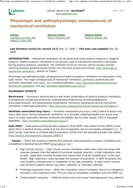
<strong>Physiologic</strong> <strong>and</strong> <strong>pathophysiologic</strong> <strong>consequences</strong> of mechanical ventilation http://www.uptodate.com/online/content/topic.do?topicKey=cc_medi/...<br />
Official reprint from UpToDate ®<br />
www.uptodate.com<br />
<strong>Physiologic</strong> <strong>and</strong> <strong>pathophysiologic</strong> <strong>consequences</strong> of<br />
mechanical ventilation<br />
Author<br />
Kenneth Lyn-Kew, MD<br />
Robert C Hyzy, MD<br />
Section Editor<br />
Polly E Parsons, MD<br />
Deputy Editor<br />
Kevin C Wilson, MD<br />
Last literature review for version 16.2: May 31, 2008 | This topic last updated: May 28,<br />
2008<br />
Print | Back<br />
INTRODUCTION — Mechanical ventilation can be performed using positive pressure or negative<br />
pressure. Positive pressure ventilation is the primary type of mechanical ventilation used today.<br />
During positive pressure ventilation, the ventilator forces air into the central airways <strong>and</strong> the<br />
resulting pressure gradient causes airflow into the small airways <strong>and</strong> alveoli. (See "Overview of<br />
mechanical ventilation", section on Types).<br />
<strong>Physiologic</strong> <strong>and</strong> <strong>pathophysiologic</strong> <strong>consequences</strong> of positive pressure ventilation are discussed in this<br />
topic review. Two major <strong>consequences</strong> of mechanical ventilation, pulmonary barotrauma <strong>and</strong><br />
ventilator-associated lung injury, are reviewed separately. (See "Pulmonary barotrauma during<br />
mechanical ventilation" <strong>and</strong> see "Ventilator-associated lung injury").<br />
PULMONARY EFFECTS<br />
Barotrauma — Pulmonary barotrauma is a well-know complication of positive pressure ventilation.<br />
Consequences include pneumothorax, subcutaneous emphysema, pneumomediastinum,<br />
pneumoperitoneum, <strong>and</strong> subcutaneous emphysema. Pulmonary barotrauma during mechanical<br />
ventilation is discussed separately. (See "Pulmonary barotrauma during mechanical ventilation").<br />
Ventilator-associated lung injury — Ventilator-associated lung injury (VALI) refers to acute lung<br />
injury that occurs during mechanical ventilation. It is clinically indistinguishable from acute lung<br />
injury or acute respiratory distress syndrome (ALI/ARDS) due to other causes. VALI is discussed<br />
separately. (See "Ventilator-associated lung injury").<br />
Auto-PEEP — Auto-positive end-expiratory pressure (Auto-PEEP, also called intrinsic PEEP) exists<br />
when there is positive airway pressure at the end of expiration due to incomplete exhalation [1] . In<br />
other words, inspiration is initiated before expiratory airflow from the preceding breath has ceased.<br />
(See "Positive end-expiratory pressure (PEEP)").<br />
Causes — There are numerous reasons that patients receiving positive pressure ventilation are<br />
susceptible to developing auto-PEEP (show table 1) [2] :<br />
High minute volume — High minute volume ventilation exists when there are large tidal<br />
volumes (greater than the patient's functional residual capacity), a high respiratory rate,<br />
or both. Large tidal volumes increase the volume that must be exhaled prior to the next<br />
breath. High respiratory rates decrease the duration of expiration. In both situations, the<br />
next breath is initiated prior to completion of the last exhalation. A high minute volume<br />
may be due to patient factors (eg, fever, infection) or ventilator settings.<br />
Prolonged inspiratory time — When the inspiratory time is increased, there is an obligatory<br />
decrease in the expiratory time. This can lead to incomplete exhalation <strong>and</strong> auto-PEEP [3] .<br />
Time-constant inequality — Patients whose lung units empty heterogeneously (eg, patients<br />
1 of 8 8/4/2008 6:27 PM
<strong>Physiologic</strong> <strong>and</strong> <strong>pathophysiologic</strong> <strong>consequences</strong> of mechanical ventilation http://www.uptodate.com/online/content/topic.do?topicKey=cc_medi/...<br />
with obstructive airways disease) are particularly susceptible to developing auto-PEEP<br />
during positive pressure ventilation, even at a relatively low minute ventilation.<br />
Expiratory flow resistance — Resistance to airflow (eg, narrow endotracheal tube,<br />
ventilator tubing) can cause auto-PEEP by impairing exhalation.<br />
Expiratory flow limitation (eg, obstructive airways disease) <strong>and</strong> altered respiratory system<br />
compliance (eg, expiratory muscle activity) similarly impede exhalation, causing<br />
auto-PEEP. Altered respiratory system compliance may also interfere with accurate<br />
measurement of auto-PEEP [4] .<br />
Detection — Auto-PEEP can be directly measured by applying an expiratory breath hold (usually<br />
0.5 to 1 second) <strong>and</strong> determining the airway pressure during the breath hold (show figure 1).<br />
Physical examination is effective at confirming auto-PEEP (high positive predictive value), but less<br />
useful for excluding auto-PEEP (lower negative predictive value) [5] .<br />
Consequences — Auto-PEEP exacerbates the hemodynamic effects of positive pressure<br />
ventilation (discussed below), increases the risk of pulmonary barotrauma, <strong>and</strong> makes it more difficult<br />
for the patient to trigger a ventilator-assisted breath (show figure 2). In addition, auto-PEEP can lead<br />
to incorrect estimation of the mean alveolar pressure <strong>and</strong> static lung compliance [6] .<br />
Treatment — Immediate intervention is necessary if auto-PEEP is detected (show table 2):<br />
Change ventilator settings — The ventilator settings should be changed in an effort to<br />
reduce or eliminate auto-PEEP. The most helpful maneuvers are those that increase the<br />
duration of expiration: increasing the inspiratory flow rate, decreasing the respiratory rate,<br />
or both. Decreasing the tidal volume or using applied PEEP to overcome auto-PEEP may<br />
also be helpful. The use of applied PEEP in this setting is discussed separately. (See<br />
"Positive end-expiratory pressure (PEEP)", section on Treatment).<br />
Reduce ventilatory dem<strong>and</strong> — Ventilatory dem<strong>and</strong> can be decreased by reducing<br />
carbohydrate intake, anxiety, pain, or fever. This may decrease the minute volume,<br />
thereby reducing auto-PEEP.<br />
Reduce expiratory flow resistance — Reduction of expiratory flow resistance by suctioning,<br />
administration of bronchodilators, <strong>and</strong> use of a wide endotracheal tube can reduce<br />
auto-PEEP.<br />
Heterogeneous ventilation — The distribution of positive pressure ventilation is never uniform<br />
because the amount of ventilation is a function of three factors that vary from region to region within<br />
the lungs: alveolar compliance, airway resistance, <strong>and</strong> dependency (upper versus lower lung zones).<br />
Compliant, non-dependent regions with minimal airway resistance will be best ventilated. In contrast,<br />
stiff, dependent regions with increased airway resistance will be least ventilated. The heterogeneity of<br />
ventilation is accentuated in patients who have airways disease, parenchymal lung disease, or both.<br />
<strong>Physiologic</strong> dead space — <strong>Physiologic</strong> dead space is the alveolar area that is not involved in gas<br />
exchange because of insufficient perfusion. Positive pressure ventilation tends to increase physiologic<br />
dead space by increasing ventilation in some regions without a corresponding increase of perfusion.<br />
<strong>Physiologic</strong> shunt — A physiologic shunt exists where there is blood flow through pulmonary<br />
parenchyma that is not involved in gas exchange because of insufficient ventilation. Patients with<br />
respiratory failure frequently have increased physiologic shunting due to focal atelectasis without a<br />
corresponding decrease in perfusion. The focal atelectasis develops because dependent lung is no<br />
longer pulled open due to decreased diaphragmatic contraction. Positive pressure ventilation can<br />
mitigate physiologic shunting by increasing the mean airway pressure, which helps maintain airway<br />
<strong>and</strong> alveolar patency. This is particularly true if positive end-expiratory pressure (PEEP) is added.<br />
(See "Positive end-expiratory pressure (PEEP)").<br />
Diaphragm — Positive pressure ventilation may lead to rapid disuse atrophy of diaphragmatic muscle<br />
2 of 8 8/4/2008 6:27 PM
<strong>Physiologic</strong> <strong>and</strong> <strong>pathophysiologic</strong> <strong>consequences</strong> of mechanical ventilation http://www.uptodate.com/online/content/topic.do?topicKey=cc_medi/...<br />
fibers. An observational study of 22 patients compared the size of diaphragmatic muscle fibers from<br />
patients who received positive pressure ventilation for more than 18 hours to those from patients<br />
who received positive pressure ventilation for fewer than three hours [7] . The mean cross sectional<br />
area of diaphragmatic muscle fibers was significantly smaller among those patients who received<br />
positive pressure ventilation for a longer duration. This relationship held for both fast twitch (1871<br />
versus 3949 micron2) <strong>and</strong> slow twitch (4725 versus 2025 micron2) muscle fibers.<br />
Respiratory muscles — Respiratory muscle atrophy can develop in patients undergoing positive<br />
pressure ventilation. Neuromuscular weakness in critically ill patients is discussed separately. (See<br />
"Neuromuscular weakness related to critical illness").<br />
Mucociliary motility — Positive pressure ventilation appears to impair mucociliary motility in the<br />
airways. In a series of 32 patients, bronchial mucus transport velocity was measured using<br />
technetium 99m-labeled albumin microspheres during the first three days of mechanical ventilation<br />
[8] . Bronchial mucus transport was frequently impaired <strong>and</strong> associated with retention of secretions<br />
<strong>and</strong> pneumonia.<br />
SYSTEMIC EFFECTS<br />
Hemodynamics — Positive pressure ventilation frequently decreases cardiac output, which may<br />
cause hypotension. There are several mechanisms that contribute to the fall in cardiac output:<br />
Decreased venous return — The amount of venous return is determined by the pressure<br />
gradient from the extrathoracic systemic veins to the right atrium. Intrathoracic <strong>and</strong> right<br />
atrial pressure increase during positive pressure ventilation, thereby reducing the gradient<br />
for venous return. This effect is accentuated by auto-PEEP, applied PEEP, or intravascular<br />
hypovolemia [9] .<br />
Reduced right ventricular output — Alveolar inflation during positive pressure ventilation<br />
compresses the pulmonary vascular bed. This increases pulmonary vascular resistance,<br />
thereby reducing right ventricular output.<br />
Reduced left ventricular output — Increased pulmonary vascular resistance can shift the<br />
interventricular septum to the left, impair diastolic filling of the left ventricle, <strong>and</strong> reduce<br />
left ventricular output.<br />
In contrast to these adverse effects, positive pressure ventilation may be beneficial in patients with<br />
left ventricular failure. Specifically, increased intrathoracic pressure can improve left ventricular<br />
performance by decreasing both venous return <strong>and</strong> left ventricular afterload [10] .<br />
These hemodynamic effects are the result of positive airway pressure being transmitted to the<br />
surrounding structures of the thorax. The extent to which this occurs varies according to chest wall<br />
<strong>and</strong> lung compliance. Transmission of airway pressure is greatest when there is low chest wall<br />
compliance (eg, fibrothorax) or high lung compliance (eg, emphysema); it is least when there is high<br />
chest wall compliance (eg, sternotomy) or low lung compliance (eg, ARDS, congestive heart failure).<br />
Monitoring — Another consequence of positive airway pressure being transmitted to surrounding<br />
intrathoracic structures is that hemodynamic measurements may be artificially elevated. PEEP plays a<br />
particularly prominent role because most hemodynamic measurements are performed at the end of<br />
expiration when PEEP is the primary source of positive airway pressure.<br />
The effect of positive pressure ventilation on hemodynamic measures has been best studied using the<br />
pulmonary capillary wedge pressure (PCWP). The PCWP is measured by a pulmonary artery catheter<br />
(Swan-Ganz catheter). When a patient is receiving positive pressure ventilation, the PCWP is<br />
artificially elevated <strong>and</strong> not reflective of the true transmural filling pressure.<br />
The true transmural filling pressure can be estimated by subtracting one-half of the PEEP level from<br />
the PCWP if the lung compliance is normal, or one-quarter of the PEEP level if lung compliance is<br />
3 of 8 8/4/2008 6:27 PM
<strong>Physiologic</strong> <strong>and</strong> <strong>pathophysiologic</strong> <strong>consequences</strong> of mechanical ventilation http://www.uptodate.com/online/content/topic.do?topicKey=cc_medi/...<br />
reduced [11] . As an example, for a patient with normal lung compliance who is receiving a PEEP of<br />
12 cmH2O <strong>and</strong> whose PCWP is measured as 18 mmHg, the true PCWP is estimated to be 12 mmHg.<br />
A more precise way to estimate the true transmural PCWP in patients requiring positive pressure<br />
ventilation utilizes the respiratory related variation of PCWP to estimate the transmission of alveolar<br />
pressure to the pulmonary vessels [12] . This measure is called the index of transmission:<br />
Index of transmission =<br />
(end inspiratory PCWP – end expiratory PCWP) / (plateau airway pressure – total PEEP)<br />
Measurement of the plateau airway pressure is described separately. (See "Pulmonary barotrauma<br />
during mechanical ventilation", section on Prevention).<br />
Once the index of transmission is calculated, the true PCWP can be estimated:<br />
Transmural PCWP =<br />
end-expiratory PCWP – (index of transmission x total PEEP)<br />
This estimate may be unreliable if the respiratory variation of the PCWP is greater than that of the<br />
pulmonary arterial pressure tracing (show figure 3) [13] . (See "Swan-Ganz catheterization:<br />
Interpretation of tracings").<br />
Gastrointestinal — Positive pressure ventilation for greater than 48 hours is a risk factor for<br />
clinically important GI bleeding due to stress ulceration. (See "Stress ulcer prophylaxis in the<br />
intensive care unit").<br />
Positive airway pressure (especially PEEP) is also associated with decreased splanchnic perfusion [14]<br />
. The mechanism underlying this association is unknown, but may be related to decreased cardiac<br />
output [15] . Decreased splanchnic perfusion manifests as elevated plasma aminotransferase <strong>and</strong><br />
lactate dehydrogenase levels.<br />
Other gastrointestinal complications seen in patients receiving positive pressure ventilation include<br />
erosive esophagitis, diarrhea, acalculous cholecystitis, <strong>and</strong> hypomotility [16,17] . It is uncertain<br />
whether these complications are due to mechanical ventilation or the critical illness. Hypomotility<br />
usually manifests as intolerance to enteral feeding. Correction of electrolytic abnormalities <strong>and</strong><br />
avoidance of drugs that adversely affect gastric motility (eg, opiates) can improve gastrointestinal<br />
motility.<br />
Renal — A fall in cardiac output induced by positive pressure ventilation can stimulate the reninangiotensin<br />
system, increase release of antidiuretic hormone, <strong>and</strong> reduce secretion of atrial<br />
natriuretic peptide. The result is fluid retention <strong>and</strong> edema.<br />
In addition, mechanical ventilation may contribute to acute renal failure. In a prospective cohort<br />
study of 29,269 critically ill patients, positive pressure ventilation was an independent risk factor for<br />
acute renal failure (OR 2.11, 95% CI, 1.58-2.82) [18] . The mechanism underlying this association is<br />
unknown, but it may also be related to decreased cardiac output.<br />
Central nervous system — Positive pressure ventilation increases intracranial pressure (ICP). This<br />
is probably the result of elevated intrathoracic pressure impairing cerebral venous outflow.<br />
Immune system — Positive pressure ventilation appears to induce inflammation. In a r<strong>and</strong>omized<br />
trial of 44 patients, patients who received positive pressure ventilation using large tidal volumes <strong>and</strong><br />
low PEEP had higher concentrations of inflammatory mediators in their blood <strong>and</strong> bronchoalveolar<br />
lavage fluid than patients who received a smaller tidal volumes <strong>and</strong> high PEEP [19] .<br />
Positive pressure ventilation may also promote translocation of tracheal bacteria into the<br />
bloodstream, according to one animal study [20] . Translocation was most pronounced during<br />
ventilation with large tidal volumes <strong>and</strong> low PEEP.<br />
4 of 8 8/4/2008 6:27 PM
<strong>Physiologic</strong> <strong>and</strong> <strong>pathophysiologic</strong> <strong>consequences</strong> of mechanical ventilation http://www.uptodate.com/online/content/topic.do?topicKey=cc_medi/...<br />
SUMMARY AND RECOMMENDATIONS<br />
1.<br />
2.<br />
3.<br />
4.<br />
5.<br />
6.<br />
7.<br />
8.<br />
9.<br />
10.<br />
11.<br />
12.<br />
13.<br />
14.<br />
Pulmonary effects of positive pressure ventilation include pulmonary barotrauma,<br />
ventilator-associated lung injury, intrinsic positive end expiratory pressure (auto-PEEP),<br />
heterogeneous ventilation, increased physiologic dead space, decreased physiologic<br />
shunting,diaphragmatic muscle atrophy, respiratory muscle weakness, <strong>and</strong> diminished<br />
mucociliary motility. (See "Pulmonary effects" above).<br />
Auto-PEEP exists when there is positive airway pressure at the end of expiration due to<br />
incomplete exhalation. It exacerbates the hemodynamic effects of positive pressure<br />
ventilation, increases the risk of pulmonary barotrauma, <strong>and</strong> makes it more difficult for the<br />
patient to trigger a ventilator-assisted breath. Detection of auto-PEEP should prompt<br />
immediate ventilator setting changes, efforts to reduce ventilatory dem<strong>and</strong>, <strong>and</strong> efforts to<br />
reduce expiratory flow resistance. (See "Auto-PEEP" above).<br />
Positive pressure ventilation may reduce cardiac output <strong>and</strong> impair hemodynamic<br />
monitoring. In addition, it is associated with gastrointestinal stress ulceration, decreased<br />
splanchnic perfusion, gastrointestinal hypomotility, fluid retention, acute renal failure,<br />
increased intracranial pressure, <strong>and</strong> inflammation. (See "Systemic effects" above).<br />
Use of UpToDate is subject to the Subscription <strong>and</strong> License Agreement.<br />
REFERENCES<br />
Pepe, PE, Marini, JJ. Occult positive end-expiratory pressure in mechanically ventilated<br />
patients with airflow obstruction: The auto-PEEP effect. Am Rev Respir Dis 1982; 126:166.<br />
Rossi, A, Polese, G, Br<strong>and</strong>i, G, et al. Intrinsic positive end-expiratory pressure (PEEPi).<br />
Intensive Care Med 1995; 21:522.<br />
Younes, M, Kun, J, Webster, K, Roberts, D. Response of ventilator-dependent patients to<br />
delayed opening of exhalation valve. Am J Respir Crit Care Med 2002; 166:21.<br />
Lessard, MR, Lofaso, F, Brochard, L. Expiratory muscle activity increases intrinsic positive<br />
end-expiratory pressure independently of dynamic hyperinflation in mechanically<br />
ventilated patients. Am J Respir Crit Care Med 1995; 151:562.<br />
Kress, JP, O'Connor, MF, Schmidt, GA. Clinical examination reliably detects intrinsic<br />
positive end-expiratory pressure in critically ill, mechanically ventilated patients. Am J<br />
Respir Crit Care Med 1999; 159:290.<br />
Valta, P, Corbeil, C, Chasse, M, et al. Mean airway pressure as an index of mean alveolar<br />
pressure. Am J Respir Crit Care Med 1996; 153:1825.<br />
Levine, S, Nguyen, T, Taylor, N, et al. Rapid disuse atrophy of diaphragm fibers in<br />
mechanically ventilated humans. N Engl J Med 2008; 358:1327.<br />
Konrad, F, Schreilber, T, Brecht-Kraus, D, et al. Mucociliary transport in ICU patients.<br />
Chest 1994; 105:237.<br />
Qvist, J, Pontoppidan, H, Wilson, RS, et al. Hemodynamic responses to mechanical<br />
ventilation with PEEP: the effect of hypervolemia. Anesthesiology 1975; 42:45.<br />
Bersten, A, Holt, A, Vedig, A, et al. Treatment of severe cardiogenic pulmonary edema with<br />
continuous positive airway pressure delivered by face mask. N Engl J Med 1991; 325:1825.<br />
Marini, JJ, O'Quin, R, Culver, BH, et al. Estimation of transmural cardiac pressures during<br />
ventilation with PEEP. J Appl Physiol 1982; 53:384.<br />
Teboul, JL, Pinsky, MR, Mercat, A, et al. Estimating cardiac filling pressure in mechanically<br />
ventilated patients with hyperinflation. Crit Care Med 2000; 28:3631.<br />
Teboul, JL, Besbes, M, Andrivet, P, et al. A bedside index assessing the reliability of<br />
pulmonary artery occlusion pressure measurements during mechanical ventilation with<br />
positive end-expiratory pressure. J Crit Care 1992; 7:22.<br />
De Backer, D. The effects of positive end-expiratory pressure on the splanchnic circulation.<br />
Intensive Care Med 2000; 26:361.<br />
5 of 8 8/4/2008 6:27 PM
<strong>Physiologic</strong> <strong>and</strong> <strong>pathophysiologic</strong> <strong>consequences</strong> of mechanical ventilation http://www.uptodate.com/online/content/topic.do?topicKey=cc_medi/...<br />
15.<br />
16.<br />
17.<br />
18.<br />
19.<br />
20.<br />
GRAPHICS<br />
Kiefer, P, Nunes, S, Kosonen, P, Takala, J. Effect of positive end-expiratory pressure on<br />
splanchnic perfusion in acute lung injury.Intensive Care Med 2000; 26:376.<br />
Dive, A, Moulart, M, Jonard, P, et al. Gastroduodenal motility in mechanically ventilated<br />
critically ill patients: A manometric study. Crit Care Med 1994; 22:441.<br />
Mutlu, GM, Mutlu, EA, Factor, P. GI complications in patients receiving mechanical<br />
ventilation. Chest 2001; 119:1222.<br />
Uchino, S, Kellum, JA, Bellomo, R, et al. Acute renal failure in critically ill patients: a<br />
multinational multicenter study. JAMA 2005; 294:813.<br />
Ranieri, VM, Suter, PM, Tortorella, C, et al. Effect of mechanical ventilation on<br />
inflammatory mediators in patients with acute respiratory distress syndrome: A<br />
r<strong>and</strong>omized controlled trial. JAMA 1999; 282:54.<br />
Nahum, A, Hoyt, J, Schmitz, L, et al. Effect of mechanical ventilation strategy on<br />
dissemination of intratracheally instilled Escherichia coli in dogs. Crit Care Med 1997;<br />
25:1733.<br />
Determinants of dynamic pulmonary hyperinflation <strong>and</strong> auto-PEEP<br />
Internal<br />
Respiratory mechanics<br />
Flow resistance<br />
Expiratory flow limitation<br />
Respiratory system compliance<br />
Breathing pattern<br />
Frequency of breathing<br />
TI/TTOT<br />
Tidal volume<br />
External<br />
Added flow resistance<br />
Fine bore endotracheal tube<br />
Ventilator tubing <strong>and</strong> devices<br />
Ventilator setting<br />
Frequency<br />
I:E ratio<br />
Inflation volume<br />
End-inspiratory pause<br />
TI: inspiratory time, TTOT: total cycle time.<br />
Adapted from Rossi, A, Polese, G, Br<strong>and</strong>i, G, et al, Intensive Care Med 1995; 21:522.<br />
Quantification of the auto-PEEP effect<br />
During mechanical ventilation of the patient with airflow obstruction, expiratory flow is too<br />
slow to allow complete deflation of the lung to its normal relaxed state before the ventilator<br />
delivers another breath. Slow flow continues until interrupted by the next inflation. Upper<br />
panel: Alveolar pressure remains positive at end exhalation (15 cmH2O) but is not<br />
measured by the ventilator manometer (0 cmH2O) located downstream from the site of<br />
increased airway resistance. Lower panel: Alveolar pressure at end-exhalation can be<br />
quantified by stopping flow transiently at the end of a set exhalation period, thereby<br />
allowing equilibration of pressures. Adapted from O'Quinn, R, Marini, JJ, Am Rev Respir Dis<br />
6 of 8 8/4/2008 6:27 PM
<strong>Physiologic</strong> <strong>and</strong> <strong>pathophysiologic</strong> <strong>consequences</strong> of mechanical ventilation http://www.uptodate.com/online/content/topic.do?topicKey=cc_medi/...<br />
1983; 128:319.<br />
Trigger threshold for auto-PEEP<br />
Effect of auto-PEEP on elevation of the triggering threshold in a mechanically<br />
ventilated patient with obstructive airways disease. This graphic representation<br />
shows airway pressure over time; the solid blue line demonstrates the circuit<br />
pressure as measured by the ventilator manometer, <strong>and</strong> the dashed red line is the<br />
alveolar pressure. In the absence of auto-PEEP, the sensitivity setting of -1 cm H20<br />
would be achieved by the patient making inspiratory efforts at the end of<br />
expiration, when airway pressure is at its minimum. In the presence of auto-PEEP,<br />
alveolar pressure remains positive. In this setting, the patient's inspiratory effort<br />
needs to decrease airway pressure not only by the -1 cm H20 sensitivity set on the<br />
machine, but also by the amount of positive alveolar pressure (auto-PEEP). In this<br />
graph, the patient's inspiratory efforts are insufficient to trigger the ventilator <strong>and</strong><br />
the patient is "locked out," being unable to get a breath because of an inability to<br />
overcome the elevated effective triggering threshold rendered by auto-PEEP.<br />
Adapted from Puritan-Bennett 1994, Form AA-1888.<br />
Treatment of auto-PEEP<br />
Changes in ventilator setting<br />
Increase expiratory duration<br />
Decrease respiratory rate<br />
Decrease tidal volume<br />
Reduction in the ventilatory dem<strong>and</strong><br />
Decrease carbohydrate intake<br />
Reduce dead space<br />
Reduce anxiety, pain, fever, shivering<br />
Reduction of total flow resistance<br />
Use of large bore endotracheal tubes<br />
Frequent suctioning<br />
Bronchodilators<br />
Application of external PEEP nearly up to the level of initial PEEPi<br />
Effect of PEEP on pulmonary hemodynamics<br />
7 of 8 8/4/2008 6:27 PM
<strong>Physiologic</strong> <strong>and</strong> <strong>pathophysiologic</strong> <strong>consequences</strong> of mechanical ventilation http://www.uptodate.com/online/content/topic.do?topicKey=cc_medi/...<br />
Pressure tracings from the same patient recorded at different levels of<br />
positive end-expiratory pressure (PEEP). The top panel shows 0 PEEP, the<br />
middle panel PEEP =15 cmH20, <strong>and</strong> the bottom panel PEEP = 20 cmH20.<br />
Pulmonary artery pressure (Ppa) is shown at the left of the tracing. The<br />
right side of the tracing shows wedge (pulmonary artery occlusion)<br />
pressure (PpaO). The red bars indicate the degree of respiratory (or<br />
respirophasic) variation exhibited at each level of PEEP in pulmonary<br />
artery pressure <strong>and</strong> wedge pressure. The ratio of respiratory variation in<br />
pulmonary artery pressure divided by respiratory variation in the wedge<br />
pressure was close to 1 when PEEP = 0 or 15 cmH20 PEEP. This increased<br />
to 2.3 at a level of 20 cmH20 PEEP. This suggests a shift from West zone<br />
three to a non-zone three condition, where airway pressure has exceeded<br />
intravascular pressure at the balloon occluded pulmonary artery catheter<br />
tip. The end-expiratory wedge pressure value during PEEP = 20 cmH20 is<br />
markedly higher than during PEEP = 15 cmH20 (18 versus 10 mmHg), a<br />
change that cannot solely be explained by the increase in PEEP. Redrawn<br />
from Teboul, JL, Besbes, M, Andrivet, P, et al, J Crit Care 1992; 7:22.<br />
© 2008 UpToDate, Inc. All rights reserved. | Subscription <strong>and</strong> License Agreement | Support Tag:<br />
[ecapp0603p.utd.com-189.124.212.67-D584DD1B49-6]<br />
Licensed to: Anna Carolina B. Dantas<br />
8 of 8 8/4/2008 6:27 PM


