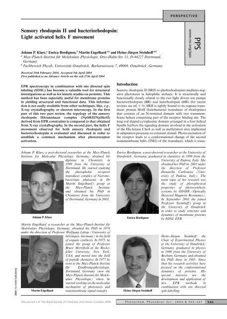
Sensory rhodopsin II and bacteriorhodopsin - Heinz-Jürgen ...
Sensory rhodopsin II and bacteriorhodopsin - Heinz-Jürgen ...
Sensory rhodopsin II and bacteriorhodopsin - Heinz-Jürgen ...

Create successful ePaper yourself
Turn your PDF publications into a flip-book with our unique Google optimized e-Paper software.
DOI: 10.1039/ b402656j<br />
<strong>Sensory</strong> <strong>rhodopsin</strong> <strong>II</strong> <strong>and</strong> bacterio<strong>rhodopsin</strong>:<br />
Light activated helix F movement<br />
Johann P. Klare, a Enrica Bordignon, b Martin Engelhard* a <strong>and</strong> <strong>Heinz</strong>-<strong>Jürgen</strong> Steinhoff* b<br />
a Max-Planck-Institut für Molekulare Physiologie, Otto-Hahn-Str. 11, D-44227 Dortmund,<br />
Germany<br />
b Fachbereich Physik, Universität Osnabrück, Barbarastrasse 7, 49069, Osnabrück, Germany<br />
Received 20th February 2004, Accepted 5th April 2004<br />
First published as an Advance Article on the web 27th April 2004<br />
EPR spectroscopy in combination with site directed spin<br />
labeling (SDSL) has become a valuable tool for structural<br />
investigations as well as for kinetic studies on proteins. This<br />
method has been especially useful for membrane proteins<br />
in yielding structural <strong>and</strong> functional data. This information<br />
is not easily available from other techniques, like, e.g.,<br />
X-ray crystallography or electron microscopy. In the first<br />
part of this two part review, the topology of the sensory<br />
<strong>rhodopsin</strong> <strong>II</strong>/transducer complex (NpSR<strong>II</strong>/NpHtr<strong>II</strong>)<br />
derived from EPR constraints is compared to that obtained<br />
from X-ray crystallography. In the second part, the helix F<br />
movement observed for both sensory <strong>rhodopsin</strong> <strong>and</strong><br />
bacterio<strong>rhodopsin</strong> is evaluated <strong>and</strong> discussed in order to<br />
establish a common mechanism after photoreceptor<br />
activation.<br />
Johann P. Klare, a post-doctoral researcher at the Max-Planck<br />
Institute for Molecular Physiology, Germany, obtained his<br />
diploma in Chemistry in<br />
1999 from the University of<br />
Dortmund. He started studying<br />
the photophobic receptor/<br />
transducer complex of Natronobacterium<br />
pharaonis in Dr<br />
Martin Engelhard’s group at<br />
the Max-Planck Institute<br />
<strong>and</strong> obtained his PhD in<br />
Chemistry from the University<br />
of Dortmund, Germany in 2003.<br />
Johann P. Klare<br />
Martin Engelhard, a researcher at the Max-Planck-Institut für<br />
Molekulare Physiologie, Germany, obtained his PhD in 1974<br />
under the direction of Professor Wolfgang Lüttge (University of<br />
Martin Engelhard<br />
Göttingen, Germany) in the field<br />
of organic synthesis. In 1975, he<br />
joined the group of Professor<br />
Bruce Merrifield at the Rockefeller<br />
University, New York,<br />
USA, <strong>and</strong> moved into the field<br />
of peptide chemistry. In 1977 he<br />
went to the Max-Planck-Institut<br />
für Ernährungsphysiologie,<br />
Dortmund, Germany (now the<br />
Max-Planck-Institut für Molekulare<br />
Physiologie) where he<br />
started working on the molecular<br />
mechanism of phototaxis <strong>and</strong><br />
transmembrane signal transfer. <strong>Heinz</strong>-<strong>Jürgen</strong> Steinhoff<br />
Introduction<br />
<strong>Sensory</strong> <strong>rhodopsin</strong> <strong>II</strong> (SR<strong>II</strong> or phobo<strong>rhodopsin</strong>) mediates negative<br />
phototaxis in halophilic archaea. It is structurally <strong>and</strong><br />
functionally closely related to the two light driven ion pumps<br />
bacterio<strong>rhodopsin</strong> (BR) <strong>and</strong> halo<strong>rhodopsin</strong> (HR) (for recent<br />
reviews see ref. 1–5). SR<strong>II</strong> is tightly bound to its cognate transducer<br />
protein Htr<strong>II</strong> (halobacterial transducer of <strong>rhodopsin</strong>s)<br />
that consists of an N-terminal domain with two transmembrane<br />
helices comprising part of the receptor binding site. The<br />
long rod shaped cytoplasmic domain arranged in a four helical<br />
bundle harbors the signaling domain involved in the activation<br />
of the His-kinase CheA as well as methylation sites implicated<br />
in adaptation processes to constant stimuli. Photo-excitation of<br />
the receptor leads to a conformational change of the second<br />
transmembrane helix (TM2) of the transducer, which is trans-<br />
Enrica Bordignon, a post-doctoral researcher at the University of<br />
Osnabrück, Germany, graduated in chemistry in 1999 from the<br />
University of Padova, Italy. She<br />
obtained her PhD in 2003 under<br />
the direction of Professor<br />
Donatella Carbonera (University<br />
of Padova, Italy). The<br />
main topic of her research was<br />
the study of photophysical<br />
properties of photosynthetic<br />
systems by ODMR (Optically<br />
Detected Magnetic Resonance).<br />
In September 2003 she joined<br />
Professor Steinhoff’s group at<br />
the University of Osnabrück<br />
in order to study structure <strong>and</strong><br />
dynamics of membrane proteins<br />
Enrica Bordignon by SDSL EPR.<br />
<strong>Heinz</strong>-<strong>Jürgen</strong> Steinhoff, the<br />
Chair of Experimental Physics<br />
at the University of Osnabrück,<br />
Germany, graduated in physics<br />
in 1980 from the University of<br />
Bochum, Germany, <strong>and</strong> obtained<br />
his PhD there in 1985. Since<br />
then his research activities have<br />
focused on the conformational<br />
dynamics of proteins. His<br />
special interests are the<br />
development <strong>and</strong> application of<br />
new EPR methods in<br />
combination with site directed<br />
spin labelling.<br />
This journal is © The Royal Society of Chemistry <strong>and</strong> Owner Societies 2004 Photochem. Photobiol. Sci., 2004, 3, 543–547<br />
543
mitted all the way down to the tip where CheA binds together<br />
with the adaptor protein CheW.<br />
Over the past years, several studies yielded detailed insight<br />
into the biophysical properties of NpSR<strong>II</strong> <strong>and</strong> its interaction<br />
with NpHtr<strong>II</strong>, thereby leading to the present model for signal<br />
generation <strong>and</strong> transfer. 2,6–18 The first structural information for<br />
a sensory <strong>rhodopsin</strong> was obtained for SR<strong>II</strong> from Natronobacterium<br />
pharaonis (NpSR<strong>II</strong>), showing the strong relation to<br />
the ion pumps BR <strong>and</strong> HR. 19,20 The recently published X-ray<br />
crystallographic structure of the complex formed between<br />
NpSR<strong>II</strong> <strong>and</strong> a C-terminal fragment of its cognate transducer<br />
NpHtr<strong>II</strong> revealed a tight interaction between the transducer<br />
transmembrane helices <strong>and</strong> the receptor helices F <strong>and</strong> G, 21 a<br />
result which was already anticipated from X-ray structural<br />
analysis of the receptor. 19,20<br />
A topological model for the complex was derived earlier<br />
using data from electron paramagnetic resonance (EPR)<br />
experiments. 17 EPR-spectroscopy in combination with site<br />
directed spin labeling (SDSL) is a well suited technique for<br />
structural investigations especially for membrane proteins. 22–24<br />
Nitroxide-scanning experiments can provide sequence correlated<br />
data about the mobility of the replaced residue as well as<br />
the accessibilities of paramagnetic reagents. The latter allows to<br />
differentiate between solvent exposed side chains, residues<br />
located in the hydrophobic region of a lipid bilayer, <strong>and</strong><br />
positions which are in the interior of the protein. Additionally,<br />
intra- <strong>and</strong> inter-molecular distances obtained using SDSL can<br />
provide long range constraints <strong>and</strong> relative orientations of<br />
secondary structure elements, which permit the construction<br />
of topological models of single proteins as well as protein<br />
complexes.<br />
In the following the NpSR<strong>II</strong>/NpHtr<strong>II</strong> structure derived from<br />
EPR data will be compared to that obtained by X-ray crystallography<br />
in order to discuss the current state <strong>and</strong> potentials of<br />
EPR spectroscopy in membrane protein structure determination.<br />
In a second part, the capability of EPR-spectroscopy in<br />
determining dynamic <strong>and</strong> kinetic parameters of protein conformational<br />
changes will be exemplified by describing the<br />
results on the helix F movement of both BR <strong>and</strong> NpSR<strong>II</strong>.<br />
NpSR<strong>II</strong>/NpHtr<strong>II</strong> structure: Comparing the EPR<br />
model with the X-ray structure<br />
EPR-model<br />
The SDSL method was applied to construct a topological<br />
model of the NpSR<strong>II</strong>/NpHtr<strong>II</strong> complex. In this study a<br />
C-terminal truncated transducer comprising the first 157<br />
amino acids was used. Both, NpSR<strong>II</strong> <strong>and</strong> NpHtr<strong>II</strong> carried a<br />
C-terminal His-tag for purification purposes. The proteins were<br />
mutated at the cytoplasmic side of their putative binding region<br />
(Fig. 1) <strong>and</strong> subsequently labeled with the nitroxide spin label<br />
MTS ((1-oxyl-2,2,5,5-tetramethylpyrroline-3-methyl)methanethiosulfonate)<br />
yielding the side chain R1. The experiments<br />
were carried out in dodecyl maltoside (DDM) in which NpSR<strong>II</strong><br />
<strong>and</strong> NpHtr<strong>II</strong> form a 1 : 1 complex as well as in membranes.<br />
Under these latter conditions the receptor transducer complex<br />
is present in a 2 : 2 stoichiometry. 18,25<br />
The reconstitution of the receptor with its transducer into<br />
polar lipids isolated from purple membrane resulted in a strong<br />
immobilization of the nitroxide side chains of the transducer<br />
compared to the DDM-solubilised samples. For V78R1 <strong>and</strong><br />
L82R1 the line widths were significantly larger than those for<br />
the other positions, indicating dipolar interactions between the<br />
spin labels of two adjacent TM2 helices. Measurements at low<br />
temperatures proved a considerable line broadening due to dipolar<br />
interaction for the spectra of V78R1 <strong>and</strong> L82R1. The<br />
inter-spin distances, obtained by fitting of EPR spectra, 26 were<br />
determined to be less than 12 Å. These results were explained<br />
by two TM2 helices of the 2 : 2 complex in close contact with<br />
544 Photochem. Photobiol. Sci., 2004, 3, 543– 547<br />
Fig. 1 Single site mutations in NpSR<strong>II</strong>-His <strong>and</strong> NpHtr<strong>II</strong>-157-His as<br />
discussed in the text. 17<br />
residues V78 <strong>and</strong> L82 located at the interface. More recent<br />
studies using gene fusion analysis on the SRI/HtrI system of<br />
H. salinarum demonstrated the same stoichiometry for this<br />
complex, 27 indicating that this arrangement might be a general<br />
feature of archaeal photo-receptor/ transducer complexes.<br />
Contrary to the close interaction between the two TM2 helices<br />
TM1 is further apart from its partner as only the spectrum of<br />
M22R1 displays a weak spin–spin interaction.<br />
To determine the topology of the NpSR<strong>II</strong>/NpHtr<strong>II</strong> reconstituted<br />
complex, distance measurements between TM1 or TM2<br />
<strong>and</strong> helix F or G were carried out. The positions were chosen at<br />
the cytoplasmic boundary of TM1 (21 to 25), TM2 (80, 81),<br />
helix F (157, 158), <strong>and</strong> helix G (210, 211). The inter-spin distances<br />
were used as constraints for modeling the topological<br />
arrangement of the NpHtr<strong>II</strong>/NpSR<strong>II</strong> complex. The strongest<br />
interactions, which correspond to the closest distance (
Fig. 2 (a) EPR model of the receptor/transducer complex transmembrane<br />
region 17 vs. (b) crystal structure. 21 View from the cytoplasmic<br />
side. Receptor helices are shown as red, transducer helices<br />
as green ribbons. Side chains replaced by Cys <strong>and</strong> subsequently<br />
labeled in the EPR study are shown in stick representation, colored<br />
according to the strength of the observed dipolar broadening of the<br />
EPR spectra (strong interaction, blue <strong>and</strong> red; weak interaction, cyan<br />
<strong>and</strong> orange).<br />
worthy that with the exception of Tyr199 the structure of<br />
NpSR<strong>II</strong> is indistinguishable from that in the complex. 19,20<br />
Direct comparison of the EPR-model with the crystal structure<br />
(see Fig. 2a <strong>and</strong> b) emphasizes the consistency of the SDSL<br />
model with that of the X-ray structure, especially, if the general<br />
topology, the stoichiometry, <strong>and</strong> the location <strong>and</strong> relative orientation<br />
of TM2 are considered. Notably, most of the side-chain<br />
orientations within the complex coincide quite well between<br />
X-ray structure <strong>and</strong> SDSL model, although in the latter model<br />
the BR instead of the NpSR<strong>II</strong> structure had to be taken<br />
(see above). A slight difference between the models is recognized<br />
concerning the distance of TM1 from the receptor, which<br />
is longer than predicted in the EPR model.<br />
Light induced conformational changes: comparison<br />
of NpSR<strong>II</strong> with BR<br />
<strong>Sensory</strong> <strong>rhodopsin</strong> <strong>II</strong><br />
On light excitation the NpSR<strong>II</strong> receptor reverts to the longlived<br />
intermediate M with a deprotonated Schiff base. This<br />
intermediate has been proposed to represent the signaling<br />
state. 30 To elucidate light induced conformational changes of<br />
the receptor as a possible key step in the signal transfer mechanism,<br />
EPR difference spectra 31 between photo-activated states<br />
<strong>and</strong> the initial state were determined for NpSR<strong>II</strong> variants with<br />
spin labels bound to the cytoplasmic ends of helices F <strong>and</strong><br />
G. 16,17 These spectra revealed considerable light-induced<br />
changes of the motional restrictions of only those spin label<br />
side chains, which are located between helices G <strong>and</strong> F of<br />
NpSR<strong>II</strong>. The shape of the difference spectra was found to be in<br />
agreement with the assumption of a transient mobilization of<br />
nitroxide side chains at positions 158, 159 <strong>and</strong> 211. Since the<br />
nitroxide side chains at G helix positions 212 <strong>and</strong> 213, which are<br />
oriented towards helices B <strong>and</strong> C, did not show any considerable<br />
mobility change during the NpSR<strong>II</strong> photocycle, a move-<br />
ment of helix G is unlikely. Hence it was concluded that the<br />
required transient increase of the accessible space for the reorientational<br />
motion of the spin label side chains at positions 158,<br />
159 <strong>and</strong> 211 had to result from an outward movement of helix<br />
F. This conclusion was further supported by the observation<br />
that in the presence of the transducer the transient difference<br />
spectrum determined for side chain position 158 is inverted: the<br />
accessible space for the reorientational motion of the nitroxide<br />
was decreased by the outward movement of helix F towards<br />
TM2 of the transducer. The kinetics of this conformational<br />
change has been followed at fixed B-field values where the difference<br />
spectrum shows local extremes. The time courses of the<br />
mobility changes yield clear evidence that the conformational<br />
change occurs with an apparent half time of 3 ms during the M<br />
state, which can be correlated to the M 1 to M 2 transition. 32 A<br />
recent FTIR study confirmed that protein conformational<br />
changes take place during the M 1 to M 2 transition. 9 The outward<br />
movement remains unchanged for about two orders of<br />
magnitude in time <strong>and</strong> reverts during the recovery of the initial<br />
state. These findings are in line with FTIR studies, which show<br />
that conformational changes occurring during the M state<br />
persist until the reformation of the parent state. 6,10,33<br />
Bacterio<strong>rhodopsin</strong><br />
The light induced isomerization of the retinal <strong>and</strong> rearrangements<br />
of the protein region <strong>and</strong> internal water molecules in its<br />
vicinity lead to the release of a proton from the Schiff base to<br />
the extracellular medium during the L to M transition of BR. A<br />
detailed analysis of the electron crystallographic data taken at<br />
various times after illumination shows a single large conformational<br />
change that is roughly coincident with formation of the<br />
M intermediate. 34 Upon comparison of projection difference<br />
maps, Subramaniam <strong>and</strong> Henderson 35 concluded that the<br />
structure of illuminated wild-type BR is identical to that of the<br />
D96G, F171C, F219L triple mutant. This mutant was then used<br />
to obtain a detailed picture of the state characterized by an<br />
open cytoplasmic channel in BR. According to these results an<br />
outward movement of helix F is evident starting from position<br />
182, whereas the changes in helix G are manifest starting from<br />
position 222. The top cytoplasmic end of helix F is displaced by<br />
≈3.5 Å away from the centre of the protein, whereas helix G is<br />
displaced by ≈2 Å towards a position between helix F <strong>and</strong> the<br />
centre of the protein <strong>and</strong> also becomes more ordered at its<br />
carboxy terminus. 35 The large tilt of helix F leads to a significant<br />
rearrangement of the EF loop, <strong>and</strong> to minor rearrangements<br />
of the uppermost residues in helix E. Other studies,<br />
aimed at determination of the structure of the open cytoplasmic<br />
channel conformation, are based on analysis of X-ray<br />
diffraction patterns of three-dimensional crystals of either<br />
wild-type BR 36 or the D96N mutant. 37 The conclusions of the<br />
X-ray crystallographic analyses disagree with each other <strong>and</strong><br />
with the findings of the electron diffraction studies regarding<br />
the nature of the main conformational change. Formation of<br />
the M intermediate was found to be associated with increased<br />
structural disorder in the crystal of the D96N mutant. Coordinates<br />
for the cytoplasmic sections of helices E <strong>and</strong> F, <strong>and</strong><br />
the EF loop, residues 154 to 175, could not be determined. 37<br />
Similarly, residues 223 to 228 in helix G could not be resolved.<br />
These regions correspond to the sites where the electron diffraction<br />
studies reveal the protein conformational changes. Coordinates<br />
determined from illuminated crystals of wild-type BR<br />
with an estimated occupancy of 35% of the M2 intermediate<br />
show also rearrangements in the vicinity of helices F <strong>and</strong><br />
G. 36 However, these coordinates report a smaller extent <strong>and</strong><br />
different direction of the helix F movement. Hence, the main<br />
structural change seems to be inhibited or muted in the<br />
three-dimensional crystals.<br />
During the subsequent transition from M2 to N the Schiff<br />
base is reprotonated with a proton released from Asp96. The<br />
Photochem. Photobiol. Sci., 2004, 3, 543– 547<br />
545
protein conformation of both intermediates, M 2 <strong>and</strong> N, were<br />
found to be very similar. 34 The subsequent reprotonation of<br />
Asp96 from the cytoplasm during the transition from N to O is<br />
accompanied by the recovery of the initial state conformation.<br />
A systematic investigation of a series of BR variants with<br />
spin label side chains introduced along the E-F loop <strong>and</strong> into<br />
the cytoplasmic ends of helices E <strong>and</strong> F, where only poorly<br />
resolved X-ray crystallographic data have been available,<br />
revealed a light induced transient increase of the motional freedom<br />
for the nitroxide side chains located between helices B, C<br />
<strong>and</strong> F. This result can be explained by a transient outward<br />
movement of helix F during the photocycle. 38 The extent of the<br />
observed helix movement was determined from inter-spin distances<br />
of stabilized intermediates. Cooling the BR sample to<br />
220 K under illumination accumulates the M intermediate to<br />
approximately 75%. 39 Inter-spin distance changes determined<br />
between spin labels bound to helices C <strong>and</strong> E, positions 101 <strong>and</strong><br />
159, C <strong>and</strong> F, positions 101 <strong>and</strong> 168, <strong>and</strong> C <strong>and</strong> G, positions<br />
100 <strong>and</strong> 226, revealed that the cytoplasmic end of helix F moves<br />
outwardly by about 1 to 2 Å. This transient helix tilt is accompanied<br />
by an inward movement of helix G of similar extent<br />
towards helix C. Thus, regarding the movement of helix F, the<br />
conformational change observed in NpSR<strong>II</strong> reveals considerable<br />
similarities to that observed for BR in purple membrane.<br />
However, no significant movement of helix G could be<br />
observed in NpSR<strong>II</strong> so far. It is interesting to note that the<br />
initial state structure of crystallized NpSR<strong>II</strong> shows the cytoplasmic<br />
end of helix G already shifted towards helices B <strong>and</strong> C,<br />
similar to what is proposed for helix G of M 2 in BR (see Fig. 3).<br />
Fig. 3 View onto the cytoplasmic side of BR 28 (top) <strong>and</strong> the<br />
asymmetric unit of the sensory <strong>rhodopsin</strong>/transducer complex,<br />
NpSR<strong>II</strong>/NpHtr<strong>II</strong> 21 (bottom). The schemes on the right depict the<br />
cytoplasmic end of the transmembrane helices constituting the complex<br />
(circles) <strong>and</strong> the conformational changes during the M 1 to M 2 transition<br />
for samples in purple membrane as determined by site directed spin<br />
labeling <strong>and</strong> EPR spectroscopy (grey <strong>and</strong> black arrows). Inter-spin<br />
distance data reveal an outward movement of the BR helix F by at least<br />
0.1 nm accompanied by an inward shift of the cytoplasmic terminus of<br />
helix G by 0.2 nm. The transient increase of the side chain mobility at<br />
positions 158, 159 <strong>and</strong> 211 indicate a similar outward movement of<br />
helix F in NpSR<strong>II</strong>. The lack of any considerable conformational<br />
changes in the vicinity of positions 212 <strong>and</strong> 213 makes large shifts of<br />
helix G unlikely. Measurements of inter-spin distance <strong>and</strong> distance<br />
changes between V78R1 <strong>and</strong> V78R1�, <strong>and</strong> between L82R1 <strong>and</strong> L82R1�<br />
in TM2 <strong>and</strong> TM2� (dotted), provide strong evidence that the tilt of<br />
helix F leads to a 20–30� rotation of TM2.<br />
Time constants of the rise <strong>and</strong> decay of the BR conformational<br />
change were determined from a superposition of exponentials<br />
fitted to the experimental EPR data. A comparison<br />
with the respective time constants for the photocycle intermedi-<br />
546 Photochem. Photobiol. Sci., 2004, 3, 543– 547<br />
ates of BR extracted from optical <strong>and</strong> FTIR spectroscopy have<br />
shown that the observed conformational change occurs in<br />
phase or prior to the reprotonation of the Schiff base depending<br />
on the nitroxide location. 39 The trigger for this conformational<br />
change must therefore be independent from the<br />
reprotonation of the Schiff base. In conclusion, the extent<br />
<strong>and</strong> direction of the helix movements determined by EPR<br />
spectroscopy in BR samples in purple membrane are in agreement<br />
with the results of electron crystallographic data. In addition,<br />
time resolved EPR data provided direct evidence for the<br />
occurrence of the conformational change during intermediate<br />
M, most probably during the M 1 to M 2 transition, quite similar<br />
to the observations on NpSR<strong>II</strong>.<br />
Transfer of the signal from NpSR<strong>II</strong> to NpHtr<strong>II</strong><br />
The tilt of helix F of the NpSR<strong>II</strong> receptor is transferred to the<br />
transducer as was shown by comparing the inter-residual distance<br />
changes observed between transducer/ transducer as well<br />
as receptor/ transducer during the photocycle. 17 Low temperature<br />
experiments (T = 170 K) with the receptor trapped in the<br />
M intermediate revealed decreases <strong>and</strong> increases of the dipolar<br />
interaction strengths between the two TM2 of the transducer<br />
dimer <strong>and</strong> between TM2 <strong>and</strong> helix F of NpSR<strong>II</strong>. Residues<br />
S158R1/K157R1 of helix F approached TM2-residues A80R1/<br />
T81R1 in the M state. Simultaneously, the strong dipolar interaction<br />
between the two V78R1 in neighboring TM2 helices was<br />
reduced significantly whereas the distance between the two<br />
L82R1 sites only one helical turn away did not change. This<br />
result clearly revealed a rearrangement of the two TM2 helices<br />
(see Fig. 3). From various possible mechanisms proposed to<br />
explain the signal transfer in chemo-receptors, 40,41 a clockwise<br />
rotation of TM2 by approximately 20–30� was found to be<br />
consistent with the experimental data. 17 The room temperature<br />
EPR signal changes recorded for V78R1 were compared with<br />
that of L159R1 which corresponds to an amino acid position<br />
on helix F oriented towards the inside of the receptor <strong>and</strong> with<br />
optical traces monitoring the depletion <strong>and</strong> reformation of the<br />
NpSR<strong>II</strong> parent state. The kinetic difference spectra recorded at<br />
room temperature represent features of a transient increase of<br />
the inter-spin distance for V78R1 <strong>and</strong> a transient mobilization<br />
for L159R1. Thus, the spectra detect events occurring at the<br />
level of the retinal chromophore, of helix F <strong>and</strong> of TM2, which<br />
allow to follow the signal transfer in the sequence retinal helix<br />
F TM2 <strong>and</strong> vice versa. From comparison to the optical traces,<br />
it is clear that the transducer signaling state is formed simultaneously<br />
with the receptor helix F movement, parallel the M1 to M2 transition. 42 With the reformation of the parent state, the<br />
reaction characteristic of the receptor seems to be decoupled<br />
from those of the transducer. The resetting movement of helix F<br />
into the original position seems to precede the recovery of TM2<br />
position, the latter one being delayed by approximately 200 ms.<br />
Concluding remarks<br />
In recent years EPR spectroscopy has been successfully applied<br />
to biological questions. In combination with site directed spin<br />
labeling its potentials for providing dynamic <strong>and</strong> kinetic data as<br />
well as structural information are especially useful for the<br />
elucidation of structure <strong>and</strong> function of membrane proteins.<br />
This class of proteins is notoriously difficult to crystallize <strong>and</strong><br />
therefore structural information is as yet scarcely available. The<br />
possibility to obtain dynamic <strong>and</strong> kinetic data on membrane<br />
processes will become decisive for the analysis of transmembrane<br />
signaling of eukaryotic <strong>and</strong> bacterial receptors.<br />
Acknowledgements<br />
The financial support of the Max Planck Society (M.E.) <strong>and</strong><br />
the Deutsche Forschungsgemeinschaft (M.E.; SFB 431/P18,<br />
E.B. <strong>and</strong> H.-J.S.) is gratefully acknowledged.
References<br />
1 U. Haupts, J. Tittor <strong>and</strong> D. Oesterhelt, General concept for ion<br />
translocation by halobacterial retinal proteins – the isomerization/<br />
switch/transfer (IST) model, Annu. Rev. Biophys. Biomol. Struct.,<br />
1999, 28, 367–399.<br />
2 G. Schäfer, M. Engelhard <strong>and</strong> V. Müller, Bioenergetics of the<br />
Archaea, Microbiol. Mol. Biol. Rev., 1999, 63, 570–620.<br />
3 E. M. L<strong>and</strong>au, E. Pebay-Peyroula <strong>and</strong> R. Neutze, Structural <strong>and</strong><br />
mechanistic insight from high resolution structures of archaeal<br />
<strong>rhodopsin</strong>s, FEBS Lett., 2003, 555, 51–56.<br />
4 J. L. Spudich, C.-S. Yang, K.-H. Jung <strong>and</strong> E. N. Spudich,<br />
Retinylidene proteins: Structures <strong>and</strong> functions from Archaea to<br />
humans, Annu. Rev Cell. Dev. Biol., 2000, 16, 365–392.<br />
5 J. L. Spudich <strong>and</strong> H. Luecke, <strong>Sensory</strong> <strong>rhodopsin</strong> <strong>II</strong>: functional<br />
insights from structure, Curr. Opin. Struct. Biol., 2002, 12, 540–546.<br />
6 V. Bergo, E. N. Spudich, J. L. Spudich <strong>and</strong> K. J. Rothschild,<br />
Conformational changes detected in a sensory <strong>rhodopsin</strong> <strong>II</strong>–<br />
transducer complex, J. Biol. Chem., 2003, 278, 36556–36562.<br />
7 Y. Furutani, M. Iwamoto, K. Shimono, N. Kamo <strong>and</strong> H. K<strong>and</strong>ori,<br />
FTIR spectroscopy of the M photointermediate in pharaonis<br />
phobo<strong>rhodopsin</strong>, Biophys. J., 2002, 83, 3482–3489.<br />
8 Y. Furutani, Y. Sudo, N. Kamo <strong>and</strong> H. K<strong>and</strong>ori, FTIR spectroscopy<br />
of the complex between pharaonis phobo<strong>rhodopsin</strong> <strong>and</strong> its<br />
transducer protein, Biochemistry, 2003, 42, 4837–4842.<br />
9 M. Hein, I. Radu, J. P. Klare, M. Engelhard <strong>and</strong> F. Siebert,<br />
Consequences of counterion mutation in sensory <strong>rhodopsin</strong> <strong>II</strong> of<br />
Natronobacterium pharaonis for photoreaction <strong>and</strong> receptor<br />
activation: An FTIR study, Biochemistry, 2004, 43, 995–1002.<br />
10 M. Hein, A. A. Wegener, M. Engelhard <strong>and</strong> F. Siebert, Timeresolved<br />
FTIR studies of sensory <strong>rhodopsin</strong> <strong>II</strong> (NpSR<strong>II</strong>) from<br />
Natronobacterium pharaonis: Implications for proton transport <strong>and</strong><br />
receptor activation, Biophys. J., 2003, 84, 1208–1217.<br />
11 S. Hippler-Mreyen, J. P. Klare, A. A. Wegener, R. Seidel,<br />
C. Herrmann, G. Schmies, G. Nagel, E. Bamberg <strong>and</strong> M. Engelhard,<br />
Probing the sensory <strong>rhodopsin</strong> <strong>II</strong> binding domain of its cognate<br />
transducer by calorimetry <strong>and</strong> electrophysiology, J. Mol. Biol., 2003,<br />
330, 1203–1213.<br />
12 Y. Sudo, M. Iwamoto, K. Shimono <strong>and</strong> N. Kamo, Association of<br />
pharaonis phobo<strong>rhodopsin</strong> with its cognate transducer decreases the<br />
photo-dependent reactivity by water-soluble reagents of azide <strong>and</strong><br />
hydroxylamineyw, Biochim. Biophys. Acta – Biomembranes, 2002,<br />
1558, 63–69.<br />
13 Y. Sudo, M. Iwamoto, K. Shimono <strong>and</strong> N. Kamo, Tyr-199 <strong>and</strong><br />
charged residues of pharaonis phobo<strong>rhodopsin</strong> are important for the<br />
Interaction with its transducer, Biophys. J., 2002, 83, 427–432.<br />
14 Y. Sudo, M. Iwamoto, K. Shimono, M. Sumi <strong>and</strong> N. Kamo, Photoinduced<br />
proton transport of pharaonis phobo<strong>rhodopsin</strong> (sensory<br />
<strong>rhodopsin</strong> <strong>II</strong>) is ceased by association with the transducer, Biophys.<br />
J., 2001, 80, 916–922.<br />
15 V. D. Trivedi <strong>and</strong> J. L. Spudich, Photostimulation of a sensory<br />
<strong>rhodopsin</strong> <strong>II</strong>/Htr<strong>II</strong>/Tsr fusion chimera activates CheA-autophosphorylation<br />
<strong>and</strong> CheY-phosphotransfer in vitro, Biochemistry,<br />
2003, 42, 13887–13892.<br />
16 A. A. Wegener, I. Chizhov, M. Engelhard <strong>and</strong> H.-J. Steinhoff, Timeresolved<br />
detection of transient movement of helix F in spin-labelled<br />
pharaonis sensory <strong>rhodopsin</strong> <strong>II</strong>, J. Mol. Biol., 2000, 301, 881–<br />
891.<br />
17 A. A. Wegener, J. P. Klare, M. Engelhard <strong>and</strong> H.-J. Steinhoff,<br />
Structural insights into the early steps of receptor–transducer signal<br />
transfer in archaeal phototaxis, EMBO J., 2001, 20, 5312–5319.<br />
18 C.-S. Yang <strong>and</strong> J. L. Spudich, Light-induced structural changes<br />
occur in the transmembrane helices of the Natronobacterium<br />
pharaonis Htr<strong>II</strong> transducer, Biochemistry, 2001, 40, 14207–14214.<br />
19 H. Luecke, B. Schobert, J. K. Lanyi, E. N. Spudich <strong>and</strong> J. L.<br />
Spudich, Crystal structure of sensory <strong>rhodopsin</strong> <strong>II</strong> at 2.4 Å: Insights<br />
into color tuning <strong>and</strong> transducer interaction, Science, 2001, 293,<br />
1499–1503.<br />
20 A. Royant, P. Nollert, K. Edman, R. Neutze, E. M. L<strong>and</strong>au,<br />
E. Pebay-Peyroula <strong>and</strong> J. Navarro, X-ray structure of sensory<br />
<strong>rhodopsin</strong> <strong>II</strong> at 2.1 Å resolution, Proc. Natl. Acad. Sci. USA, 2001,<br />
98, 10131–10136.<br />
21 V. I. Gordeliy, J. Labahn, R. Moukhametzianov, R. Efremov,<br />
J. Granzin, R. Schlesinger, G. Büldt, T. Savopol, A. J. Scheidig,<br />
J. P. Klare <strong>and</strong> M. Engelhard, Molecular basis of transmembrane<br />
signalling by sensory <strong>rhodopsin</strong> <strong>II</strong>–transducer complex, Nature,<br />
2002, 419, 484–487.<br />
22 D. L. Farrens, C. Altenbach, K. Yang, W. L. Hubbell <strong>and</strong> H. G.<br />
Khorana, Requirement of rigid-body motion of transmembrane<br />
helices for light activation of <strong>rhodopsin</strong>, Science, 1996, 274, 768–<br />
770.<br />
23 F. Bezanilla <strong>and</strong> E. Perozo, Force <strong>and</strong> voltage sensors in one<br />
structure, Adv. Protein Chem., 2003, 63, 211–241.<br />
24 C. Altenbach, J. Klein-Seetharaman, K. W. Cai, H. G. Khorana <strong>and</strong><br />
W. L. Hubbell, Estimation of inter-residue distances in spin labeled<br />
proteins at physiological temperatures: Experimental strategies <strong>and</strong><br />
practical limitations, Biochemistry, 2001, 40, 15493–15500.<br />
25 T. Arakawa, K. Shimono, S. Yamaguchi, S. Tuzi, Y. Sudo, N. Kamo<br />
<strong>and</strong> H. Saito, Dynamic structure of pharaonis phobo<strong>rhodopsin</strong><br />
(sensory <strong>rhodopsin</strong> <strong>II</strong>) <strong>and</strong> complex with a cognate truncated<br />
transducer as revealed by site-directed 13 C solid-state NMR, FEBS<br />
Lett., 2003, 536, 237–240.<br />
26 H.-J. Steinhoff, N. Radzwill, W. Thevis, V. Lenz, D. Br<strong>and</strong>enburg,<br />
A. Antson, G. Dodson <strong>and</strong> A. Wollmer, Determination of interspin<br />
distances between spin labels attached to insulin: comparison of<br />
electron paramagnetic resonance data with the X-ray structure,<br />
Biophys. J., 1997, 73, 3287–3298.<br />
27 X. Chen <strong>and</strong> J. L. Spudich, Demonstration of 2 : 2 stoichiometry in<br />
the functional SRI–HtrI signaling complex in Halobacterium<br />
membranes by gene fusion analysis, Biochemistry, 2002, 41, 3891–<br />
3896.<br />
28 L. O. Essen, R. Siegert, W. D. Lehmannn <strong>and</strong> D. Oesterhelt, Lipid<br />
patches in membrane protein oligomers – crystal structure of the<br />
bacterio<strong>rhodopsin</strong>-lipid complex, Proc. Natl. Acad. Sci. USA, 1998,<br />
95, 11673–11678.<br />
29 E. M. L<strong>and</strong>au <strong>and</strong> J. P. Rosenbusch, Lipidic cubic phases: A novel<br />
concept for the crystallization of membrane proteins, Proc. Natl.<br />
Acad. Sci. USA, 1996, 93, 14532–14535.<br />
30 B. Yan, T. Takahashi, R. Johnson <strong>and</strong> J. L. Spudich, Identification<br />
of signaling states of a sensory receptor by modulation of lifetimes<br />
of stimulus-induced conformations: The case of sensory <strong>rhodopsin</strong><br />
<strong>II</strong>, Biochemistry, 1991, 30, 10686–10692.<br />
31 H.-J. Steinhoff, R. Mollaaghababa, C. Altenbach, K. Hideg,<br />
M. Krebs, H. G. Khorana <strong>and</strong> W. L. Hubbell, Time resolved<br />
detection of structural changes during the photocycle of<br />
spin-labeled bacterio<strong>rhodopsin</strong>, Science, 1994, 266, 105–107.<br />
32 I. Chizhov, G. Schmies, R. Seidel, J. R. Sydor, B. Lüttenberg <strong>and</strong><br />
M. Engelhard, The photophobic receptor from Natronobacterium<br />
pharaonis: Temperature <strong>and</strong> pH dependencies of the photocycle of<br />
sensory <strong>rhodopsin</strong> <strong>II</strong>, Biophys. J., 1998, 75, 999–1009.<br />
33 V. Bergo, E. N. Spudich, K. L. Scott, J. L. Spudich <strong>and</strong> K. J.<br />
Rothschild, FTIR analysis of the S<strong>II</strong> 540 intermediate of sensory<br />
<strong>rhodopsin</strong> <strong>II</strong>: Asp73 is the Schiff base proton acceptor,<br />
Biochemistry, 2000, 39, 2823–2830.<br />
34 S. Subramaniam, I. Lindahl, P. Bullough, A. R. Faruqi, J. Tittor,<br />
D. Oesterhelt, L. Brown, J. K. Lanyi <strong>and</strong> R. Henderson, Protein<br />
conformational changes in the bacterio<strong>rhodopsin</strong> photocycle,<br />
J. Mol. Biol., 1999, 287, 145–161.<br />
35 S. Subramaniam <strong>and</strong> R. Henderson, Molecular mechanism of<br />
vectorial proton translocation by bacterio<strong>rhodopsin</strong>, Nature, 2000,<br />
406, 653–657.<br />
36 H. J. Sass, G. Büldt, R. Gessenich, D. Hehn, D. Neff, R. Schlesinger,<br />
J. Berendzen <strong>and</strong> P. Ormos, Structural alterations for proton<br />
translocation in the M state of wild-type bacterio<strong>rhodopsin</strong>, Nature,<br />
2000, 406, 649–653.<br />
37 H. Luecke, B. Schobert, H. T. Richter, J. P. Cartailler <strong>and</strong><br />
J. K. Lanyi, Structural changes in bacterio<strong>rhodopsin</strong> during ion<br />
transport at 2 angstrom resolution, Science, 1999, 286, 255–261.<br />
38 N. Radzwill, K. Gerwert <strong>and</strong> H.-J. Steinhoff, Time-resolved<br />
detection of transient movement of helices F <strong>and</strong> G in doubly<br />
spin-labeled bacterio<strong>rhodopsin</strong>, Biophys. J., 2001, 80, 2856–2866.<br />
39 T. Rink, M. Pfeiffer, D. Oesterhelt, K. Gerwert <strong>and</strong> H.-J. Steinhoff,<br />
Unraveling photoexcited conformational changes of bacterio<strong>rhodopsin</strong><br />
by time resolved electron paramagnetic resonance<br />
spectroscopy, Biophys. J., 2000, 78, 1519–1530.<br />
40 S. A. Chervitz <strong>and</strong> J. J. Falke, Molecular mechanism of transmembrane<br />
signaling by the aspartate receptor: A model, Proc. Natl.<br />
Acad. Sci. USA, 1996, 93, 2545–2550.<br />
41 K. M. Ottemann, W. Xiao, Y. K. Shin <strong>and</strong> D. E. J. Koshl<strong>and</strong>,<br />
A piston model for transmembrane signaling of the aspartate<br />
receptor, Science, 1999, 285, 1751–1754.<br />
42 J. P. Klare, Strukturelle und funktionelle Untersuchungen des<br />
photophoben Rezeptor/Transducer-Komplexes aus Natronobacterium<br />
pharaonis, Dissertation, Universität Dortmund, 2002.<br />
Photochem. Photobiol. Sci., 2004, 3, 543– 547<br />
547