Image processing and enhancement provided by commercial dental ...
Image processing and enhancement provided by commercial dental ...
Image processing and enhancement provided by commercial dental ...

You also want an ePaper? Increase the reach of your titles
YUMPU automatically turns print PDFs into web optimized ePapers that Google loves.
Dentomaxillofacial Radiology (2002) 31, 264 ± 272<br />
ã 2002 Nature Publishing Group. All rights reserved 0250 ± 832X/02 $25.00<br />
www.nature.com/dmfr<br />
TECHNICAL REPORT<br />
<strong>Image</strong> <strong>processing</strong> <strong>and</strong> <strong>enhancement</strong> <strong>provided</strong> <strong>by</strong> <strong>commercial</strong> <strong>dental</strong><br />
software programs<br />
TM Lehmann* ,1 , E Troeltsch 1 <strong>and</strong> K Spitzer 1<br />
1 Institute of Medical Informatics, Aachen University of Technology, Aachen, Germany<br />
Introduction<br />
Objectives: To identify <strong>and</strong> analyse methods/algorithms for image <strong>processing</strong> <strong>provided</strong> <strong>by</strong><br />
various <strong>commercial</strong> software programs used in direct digital <strong>dental</strong> imaging <strong>and</strong> to map them<br />
onto a st<strong>and</strong>ardized nomenclature.<br />
Methods: Twelve programs presented at the 28th International Dental-Show, March, 2001,<br />
Cologne, Germany <strong>and</strong> the Emago advanced software were included in this study. An arti®cial<br />
test image, comprised of gray scale ramps, step wedges, ®elds with Gaussian-distributed noise,<br />
<strong>and</strong> salt <strong>and</strong> pepper noise, was synthesized <strong>and</strong> imported to all programs to classify algorithms<br />
for display; linear, non-linear <strong>and</strong> histogram-based point <strong>processing</strong>; pseudo-coloration; linear<br />
<strong>and</strong> non-linear spatial ®ltering; frequency domain ®ltering; measurements; image analysis; <strong>and</strong><br />
annotations.<br />
Results: The 13 programs were found to possess a great variety of image <strong>processing</strong> <strong>and</strong><br />
<strong>enhancement</strong> facilities. All programs o€er gray-scale image display with interactive brightness<br />
<strong>and</strong> contrast adjustment <strong>and</strong> gray-scale inversion as well as calibration <strong>and</strong> length<br />
measurements. While Emago enables arbitrary spatial ®ltering with user-de®ned masks up to<br />
767 pixels in size, most programs sparsely include ®lters <strong>and</strong> tools for image analysis <strong>and</strong><br />
comparison. Moreover, the naming <strong>and</strong> implementation of <strong>provided</strong> functions di€er. Some<br />
functions inappropriately use st<strong>and</strong>ardized image <strong>processing</strong> terms to describe their operations.<br />
Conclusions: <strong>Image</strong> <strong>processing</strong> <strong>and</strong> <strong>enhancement</strong> functions are rarely incorporated in<br />
<strong>commercial</strong> software for direct digital imaging in <strong>dental</strong> radiology. Until now, comparison of<br />
software was limited <strong>by</strong> the arbitrary naming used in each system. St<strong>and</strong>ardized terminology<br />
<strong>and</strong> increased functionality of image <strong>processing</strong> should be o€ered to the <strong>dental</strong> profession.<br />
Dentomaxillofacial Radiology (2002) 31, 264 ± 272. doi:10.1038/sj.dmfr.4600707<br />
Keywords: radiography, <strong>dental</strong>, digital; radiographic image <strong>enhancement</strong>; image <strong>processing</strong>,<br />
computer-assisted; algorithms<br />
Direct digital <strong>dental</strong> imaging has been established over<br />
the past decade, <strong>and</strong> its usage <strong>by</strong> <strong>dental</strong> practitioners is<br />
steadily increasing. 1 Therefore, more companies are<br />
o€ering hardware <strong>and</strong> software for direct digital<br />
imaging. Usually, the systems rely on solid state<br />
sensors, such as charge coupled devices (CCD) or<br />
complementary metal oxide semiconductors (CMOS),<br />
<strong>and</strong>/or storage phosphor plates. Sensitometric properties,<br />
resolution, <strong>and</strong> technical parameters of competing<br />
hardware concepts have been evaluated exhaustively<br />
*Correspondence to: TM Lehmann, Institute of Medical Informatics, 52057<br />
Aachen, Germany; E-mail: lehmann@computer.org<br />
Received 25 October 2001; revised 24 January 2002; accepted 26 April 2002<br />
<strong>and</strong> compared to intra-oral ®lms, as well as other<br />
digital systems. 2<br />
Digital acquisition of radiographs enables digital<br />
image <strong>enhancement</strong> <strong>and</strong> <strong>processing</strong>. Several studies<br />
have shown that digital contrast <strong>enhancement</strong> <strong>and</strong><br />
®ltering may increase diagnostic accuracy for the<br />
detection of lesions <strong>and</strong> estimation of lesion depth. 3,4<br />
For instance, there is evidence that certain digital<br />
image <strong>enhancement</strong>s of direct digitally acquired radiographic<br />
images may improve accuracy of detection <strong>and</strong><br />
quanti®cation of carious lesions. 5 However, other<br />
authors observed in their studies the contrary, i.e.<br />
deterioration of accuracy <strong>by</strong> digital image <strong>enhancement</strong><br />
of directly digitally acquired radiographs. 6 Applied to<br />
secondary digitized radiographs of high quality, basic
digital manipulations may also fail to result in more<br />
valid measurements demonstrating statistical significance.<br />
7,8 In conclusion, it is undisputed that diagnostic<br />
impact depends on the task at h<strong>and</strong>, the quality of<br />
source data, <strong>and</strong> the kind of image <strong>processing</strong> applied.<br />
However, this does not imply that for any given task<br />
there is some image <strong>processing</strong> operation improving<br />
diagnostic e ciency.<br />
Nonetheless all systems for direct digital imaging<br />
o€er some type of image manipulation methods.<br />
Embedded in the menu structure <strong>by</strong> ingenious<br />
identi®ers <strong>and</strong>/or accessible <strong>by</strong> buttons with descriptive<br />
symbols, a great variety of linear <strong>and</strong> non-linear<br />
techniques in the spatial or frequency domain are<br />
<strong>provided</strong> to modify one image or a combination of two<br />
images. However, a st<strong>and</strong>ardized nomenclature for<br />
such tools with respect to all programs is missing. The<br />
same image <strong>processing</strong> technique may have di€erent<br />
names, <strong>and</strong>, even more crucial, functions with common<br />
names may have di€erent e€ects on the image.<br />
The goal of this work was to analyse <strong>and</strong> compare<br />
the procedures of image <strong>processing</strong> <strong>and</strong> <strong>enhancement</strong><br />
<strong>provided</strong> <strong>by</strong> certain <strong>commercial</strong> software systems for<br />
direct digital <strong>dental</strong> imaging <strong>and</strong> to map them onto a<br />
st<strong>and</strong>ardized nomenclature.<br />
Material <strong>and</strong> methods<br />
Selection of programs<br />
In 2001, a total of 63 companies, including manufacturers<br />
<strong>and</strong> providers, registered at the Cologne<br />
international <strong>dental</strong> exhibition using the keyword `Xray<br />
technology'. Based on this pre-selection, 12<br />
software systems were identi®ed for this study.<br />
Although not displayed at the meeting, the Emago<br />
software program was also included because of its<br />
frequent use in academic research, particularly for<br />
digital subtraction radiography. 9<br />
Table 1 lists all programs, versions, <strong>and</strong> providers.<br />
(In general, we found the web-site to be more helpful<br />
because it often links directly to the manufacturer's<br />
home page.) The hardware <strong>and</strong> software requirements<br />
were obtained from the providers' or manufacturers'<br />
descriptions. Most programs require a personal<br />
computer (PC) with Pentium processor <strong>and</strong> at least<br />
64 mega<strong>by</strong>te (MB) r<strong>and</strong>om access memory for the<br />
client operating on Windows or Linux <strong>and</strong> recommend<br />
at least 4 giga<strong>by</strong>te (GB) of free disk space for storing<br />
the images. While some programs o€er a 8006600<br />
pixel mode, the majority uses 10246768 pixel displays<br />
in high or true color modes, which are 16 bit or 24 bit<br />
color depth, respectively.<br />
Terminology<br />
<strong>Image</strong> <strong>enhancement</strong> <strong>and</strong> ®ltering Analoui 10,11 recently<br />
<strong>provided</strong> an overview of underlying concepts along<br />
with algorithms commonly used for radiographic image<br />
Dental image <strong>processing</strong><br />
TM Lehmann et al<br />
<strong>enhancement</strong> in <strong>dental</strong> radiology. 10,11 Linear <strong>and</strong> nonlinear<br />
point <strong>processing</strong>, the histogram-based approach,<br />
as well as spatial <strong>and</strong> frequency domain ®lters were<br />
de®ned <strong>and</strong> applied to <strong>dental</strong> radiographs. With<br />
respect to these image-<strong>processing</strong> techniques, we refer<br />
to the terminology introduced in the review.<br />
In addition, some of the programs o€er special ®lters<br />
such as thresholding, gradient ®ltering, <strong>and</strong> relief<br />
imaging. In thresholding, a binary image is obtained <strong>by</strong><br />
turning all pixels to black or white if their gray value is<br />
below or above the chosen threshold, respectively. A<br />
gradient ®lter computes the ®rst derivative of the gray<br />
values in the image <strong>and</strong> displays them in white on a black<br />
background, where the gray value in the gradient image<br />
denotes the strength of the edge. Hence, a gradient ®lter<br />
results in a contour image. Similarly, a relief image is<br />
obtained if the derivative is displayed on a gray<br />
background, <strong>and</strong> positive or negative gradients are<br />
coded in white or black, respectively. In several<br />
programs, this ®lter is also called `3demboss'. In<br />
addition, the three-dimensional (3D) embossing is<br />
frequently overlaid on the original image to maintain<br />
the initial gray distribution of the radiographs, resulting<br />
in some kind of image sharpening.<br />
<strong>Image</strong> display Several programs o€er special facilities<br />
for display <strong>and</strong> image measurements or analysis. A<br />
one-dimensional (1D) line pro®le graphically plots the<br />
pixel values, which are read along a straight line in the<br />
image. Using interpolation, the line can be oriented<br />
arbitrarily on the discrete pixel grid. Beside this 1D <strong>and</strong><br />
the normal two-dimensional (2D) display of a radiograph,<br />
a 3D surface model is obtained when pixel gray<br />
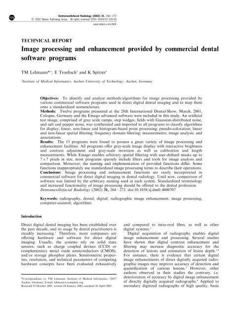
values are interpreted as a certain altitude in a<br />
l<strong>and</strong>scape formed from the image (Figure 1).<br />
Pseudo-coloring Since human perception of gray<br />
values is limited, coloring images might enhance small<br />
local contrast. Arbitrary colors are usually obtained<br />
following the edges of the red, green, blue (RGB) cube.<br />
Clark <strong>and</strong> Leonhard recommended pseudo-coloring<br />
with constant brightness, 12 while Lehmann, Kaser <strong>and</strong><br />
Repges proposed a simple parametric equation for<br />
pseudo-coloring gray-scale images keeping their original<br />
brightness progression. 13 The latter approach can<br />
be reduced to one color (e.g., brown in Trophy) or two<br />
colors (e.g., blue/green in Digora or red/yellow in<br />
Dimaxis). Furthermore, a certain range of equal gray<br />
scales can be shown with one, two, or three colors.<br />
Measurements <strong>and</strong> image analysis Length <strong>and</strong> area<br />
measurements require calibration, i.e., a certain wellknown<br />
distance within the image (e.g., the main axis of<br />
an implant) is marked <strong>and</strong> measured <strong>by</strong> the user.<br />
However, e€ects of di€erent foreshortening in cone<br />
beam projection, which results from di€erent object to<br />
sensor plane distances within one image, are neglected<br />
<strong>by</strong> all programs. To assist the comparison of two or<br />
more radiographs from the same <strong>dental</strong> region of the<br />
same patient, which have been serially acquired,<br />
265<br />
Dentomaxillofacial Radiology
266<br />
Table 1 Software systems<br />
Hardware <strong>and</strong> software requirements Features<br />
System Company or provider Operating Supported Variable<br />
software Version Name Address Web-page CPU RAM HD space Video system file formats paths<br />
.<br />
BMP<br />
TGA<br />
Win 95/98<br />
Win NT<br />
www.Schicktech.com Pentium 64 MB 1 GB 8006600<br />
8 Bit<br />
31-00 47th Avenue<br />
Long Isl<strong>and</strong> City,<br />
CDR 2.0 Demo Schick Technologies,<br />
Inc.<br />
Dentomaxillofacial Radiology<br />
TIFF<br />
.<br />
BMP<br />
JPG<br />
TIFF<br />
Win 2000<br />
Win NT<br />
1MB<br />
256 MB 6.4 GB 10246768<br />
24 Bit<br />
4MB<br />
Pentium<br />
300 MHz<br />
www.instrumentarium.fi/<br />
imaging<br />
.<br />
DICOM<br />
TIFF<br />
Win 2000<br />
Win NT<br />
ww.ic-med.de Pentium 16 MB 1 GB 8006600<br />
16 Bit<br />
Ð<br />
JFIF<br />
JPG<br />
TIFF<br />
Win 95/98<br />
Win 2000<br />
Win NT4<br />
2 GB 10246768<br />
24 Bit<br />
70 Hz<br />
www.soredex.com Pentium II C: 64 MB<br />
S: 128 MB<br />
Ð<br />
BMP<br />
DICOM<br />
JPEG<br />
Win 95/98<br />
Win NT<br />
10246768<br />
4MB<br />
C: 35 MB<br />
S: 20 GB<br />
C: 64 MB<br />
S: 128 MB<br />
www.planmeca.de C: 133 MHz<br />
S: 266 MHz<br />
NY 11101 USA<br />
Cliniview R 3.00 Instrumentarium Po Box 20<br />
Imaging<br />
Nahekelantie 20<br />
FIN-04301 Tuusla<br />
Finl<strong>and</strong><br />
Dexis 3.1.4 IC Med GmbH Walther Rathenau Str. 4<br />
D-06116 Halle/Saale<br />
Germany<br />
Digora 2.1 Rev 1 Orion Corporation PO Box 79<br />
Demo Sorodex<br />
NilsiaÈ nkatu 10-14<br />
FIN-00511 Helsinki<br />
Finl<strong>and</strong><br />
Dimaxis 2.4.1 Planmeca GmbH MuÈ hlfelderstr. 66<br />
D-82211 Hersching<br />
Germany<br />
Dental image <strong>processing</strong><br />
TM Lehmann et al<br />
TIFF<br />
.<br />
BMP<br />
TGA<br />
TIFF<br />
Win 3.1<br />
Win 95/98<br />
Win NT<br />
Pentium II 32 MB 1 GB 10246768<br />
16 Bit<br />
www.radiology.acta.ne/<br />
emago/emago.html<br />
c/o ACTA Oral Radiology<br />
Louwesweg 1<br />
NL-1066 EA Amsterdam<br />
3.2 Stichting Oral<br />
Diagnostic Systems<br />
Emago<br />
advanced<br />
.<br />
DICOM<br />
TIFF<br />
Win NT<br />
Win 2000<br />
10246768<br />
24 Bit<br />
Win 2000)<br />
16 MB 1 GB 10246768<br />
16 Bit<br />
www.friadent.de Pentium II 128 MB<br />
(256 MB for<br />
BMP .<br />
Linux<br />
Win 98 SE<br />
Win 2000<br />
Win ME<br />
Linux<br />
RedHat 7.2<br />
www.fimet.fi Pentium<br />
120 MHz<br />
.<br />
64 MB 4 GB 10246768<br />
24 Bit<br />
www.rapp-informatik.de Pentium III<br />
800 MHz<br />
The Netherl<strong>and</strong>s<br />
Friacom 2.4.183 Friadent GmbH Steinzeugstr.50<br />
Dental<br />
D-68229 Mannheim<br />
Office<br />
Germany<br />
IOX 2.2.0.10 Fimet OY Teollisuustie 6<br />
<strong>Image</strong><br />
FIN-07230 MonninkylaÈ<br />
Viewer<br />
Finl<strong>and</strong><br />
multiXray 1.0 Q Rapp Informatik RosenbuÈ hlstr. 24<br />
Systeme GmbH D-89182 Bernstadt/Ulm<br />
BMP<br />
DICOM<br />
GIF<br />
JPEG<br />
TGA<br />
TIFF<br />
.<br />
Win 95/98 BMP<br />
GIF<br />
JPEG<br />
8006600<br />
16 Bit<br />
32 MB 20 MB<br />
(without<br />
images)<br />
www.visiplex.de Pentium<br />
133 MHz<br />
Von-Braun-Str. 25<br />
D-52511 Geilenkirchen<br />
Germany<br />
Proimage 3.1.0 Dent-X/Visiplex<br />
Medical Systems<br />
GmbH<br />
TIFF<br />
Sidexis 5.3 Sinora Dental Fabrikstr. 31<br />
www.sinora.de Pentium 64 MB 4 GB 8006600 Win 98 JPEG .<br />
Siemens GmbH D-64625 Bensheim<br />
166 MHz<br />
8 Bit Win 2000 TIFF<br />
Germany<br />
4MB Win NT<br />
Trophy 4.1 h Trophy Radiologie Gerbereistr. 7<br />
www.trophy-digital.de Pentium 128 MB 8006600 Win 95/98 JPEG .<br />
Demo GmbH<br />
D-77649 Kehl-Kork<br />
400 MHz<br />
16 Bit Win 2000 TIFF<br />
Germany<br />
Pentium<br />
4MB Win NT<br />
VixWin 1.4 Demo Dentsply Gendex Hamburg Innovation Park www.gendex.de 133 MHz C: 64 MB 4 GB 8006600 Win 95/98 JFIF Ð<br />
2000<br />
Dental Systeme Albert Einstein Ring 13<br />
S: 128 MB<br />
24 Bit Win 2000 JPEG<br />
D-22761 Hamburg<br />
2MB Win NT 4 TIFF<br />
Germany<br />
CPU, central <strong>processing</strong> unit; C, client; S, server; BMP, Windows Bitmap; DICOM, Digital Imaging <strong>and</strong> Communications in Medicine; GIF, Graphics Interchange Format; HD, hard disk;<br />
JFIF, JPEG File Interchange Format; JPEG, Joint Photographic Experts Group; RAM, r<strong>and</strong>om access memory; TGA, Truevision Targa; TIFF, Tagged <strong>Image</strong> File Format
a b<br />
contrast adjustment <strong>and</strong> geometry registration are<br />
<strong>provided</strong> to enable subtraction 14 or color addition. 15<br />
Annotation <strong>and</strong> miscellaneous Annotations of radiographs<br />
with lines or arrows, delimited regions, <strong>and</strong> text<br />
are useful to indicate a certain region of interest (ROI).<br />
After appropriate calibration, annotations can also be<br />
used for implant planning. Furthermore, pixel manipulation<br />
can be applied to alter the original<br />
radiograph. Such functions include brushes <strong>and</strong><br />
pencils in any width, gray scale, or color. We also<br />
noted the ROI features that allow the application of a<br />
spatial ®lter to only a certain window within the image,<br />
as well as important functions that allow for selective<br />
`undo' <strong>and</strong> `reset' to be performed.<br />
Test image<br />
Linear <strong>and</strong> shift invariant ®lters were identi®ed <strong>by</strong> the<br />
result obtained from ®ltering a single bright spot on a<br />
dark background regardless of whether such ®lters<br />
were implemented in the spatial or the frequency<br />
domain. In addition, non-linear ®lters, such as the<br />
median ®lter, can be identi®ed <strong>by</strong> inspecting the<br />
response to salt <strong>and</strong> pepper noise, also referred to as<br />
shot noise or spot noise. Histogram spreading is easily<br />
distinguished from histogram equalization <strong>by</strong> noting<br />
whether the initial image occupies the full range of<br />
gray scales.<br />
Based on this fundamental coherence, a 5126400<br />
pixel sized test image with 8 bit gray-value depth was<br />
designed <strong>and</strong> synthesized using the Khoros Pro 2.2<br />
system (Khoral Inc., Albuquerque, NM, USA).<br />
Basically, the image is composed of four stripes<br />
(Figure 1). The upper stripe contains four ®elds:<br />
Gaussian-distributed noise with mean 128 <strong>and</strong><br />
variance 1 (histogram stretched); black squares of<br />
increasing size on a white ground; the same structure<br />
vertically ¯ipped <strong>and</strong> gray scale inverted; <strong>and</strong> salt <strong>and</strong><br />
pepper noise on a gray background where the spots<br />
Dental image <strong>processing</strong><br />
TM Lehmann et al<br />
Figure 1 The arti®cial test image (a) is used to determine the algorithms of image <strong>processing</strong>. The 3D surface model (b) was obtained with the<br />
Trophy software system. <strong>Image</strong> gray values are interpreted as surface lines, which has been named ISO-surfaces <strong>by</strong> the International<br />
Organization of St<strong>and</strong>ardization. Note that this visualization emphasizes the logarithmic nature of the step wedge ascending in the lower part of<br />
the image<br />
have an incidence of 5% for each of black <strong>and</strong> white.<br />
The middle two stripes show continuous gray<br />
progression from black to white <strong>and</strong> vice versa,<br />
respectively. The lower stripe contains 32 steps of<br />
gray values (0, 30, 56, 80, 99, 119, 135, 149, 161, 172,<br />
182, 191, 198, 205, 211, 216, 220, 225, 228, 231, 234,<br />
237, 239, 241, 242, 244, 245, 246, 247, 248, 249 <strong>and</strong><br />
250) imitating the logarithmic response of X-ray<br />
imaging of an aluminum wedge of constant step<br />
height. In the foreground, two concentric white circles<br />
with a centered cross are drawn to simplify navigation<br />
in the zoomed image.<br />
Installation <strong>and</strong> testing<br />
Linux (RedHat 7.2), Windows 98 <strong>and</strong> Windows NT<br />
4.0 operating systems were installed on a common PC<br />
computer. The computer hosts a Winner 2000 AVI<br />
graphics card (Elsa GmbH, Aachen, Germany)<br />
connected to a 21'' color monitor with 11556864<br />
pixel resolution (Elsa GmbH, Aachen, Germany). All<br />
<strong>dental</strong> software was installed in the single user mode<br />
on a 4 GB hard disk drive with 128 MB r<strong>and</strong>om access<br />
memory. The test image was loaded to all programs<br />
preferably in the tagged image ®le format (TIFF) using<br />
the import/export functions. Since some demo versions<br />
had disabled import or export functions (e.g., Emago),<br />
the test image was manually copied over existing<br />
images of demo patients on the hard disk.<br />
Results<br />
Installation <strong>and</strong> software features<br />
The multiXray software operates on Linux only. The<br />
IOX <strong>Image</strong> Viewer <strong>and</strong> the Pro<strong>Image</strong> software<br />
currently do not support Windows NT; hence, these<br />
programs were installed on Windows 98, while all<br />
others ran on Windows NT (Table 1). The Digora,<br />
Dimaxis, <strong>and</strong> VixWin programs did not o€er user-<br />
267<br />
Dentomaxillofacial Radiology
268<br />
determined installation paths but required installation<br />
on the system C drive, which might be inconvenient.<br />
To import <strong>and</strong> export images, we determined the<br />
supported ®le formats from the manufacturers'<br />
descriptions. Only Dexis, Dimaxis, Friacom, <strong>and</strong><br />
multiXray <strong>provided</strong> an interface for the digital<br />
imaging <strong>and</strong> communication in medicine (DICOM)<br />
®le format. Note that the ability to read or write this<br />
®le format does not mean DICOM compliance or<br />
conformance, since that is intended to mean that the<br />
device containing the software could be plugged into a<br />
DICOM network, or read a piece of DICOM media,<br />
<strong>and</strong> neither is the case. The IOX <strong>Image</strong> Viewer imports<br />
only 8 bit ®les in 12006800 pixel resolution l<strong>and</strong>scape<br />
format using the Windows bitmap format (BMP),<br />
while all others support TIFF images with arbitrary<br />
image size <strong>and</strong> resolution. However, as a consequence<br />
of the various existing versions of TIFF, the format in<br />
use might di€er within the applications resulting in<br />
incompatibilities. For example, Friacom displayed a<br />
black bar above the imported image, which might be a<br />
result of the variable length of the TIFF header, <strong>and</strong><br />
Dimaxis showed the test image split in two vertical<br />
parts showing the left half of the image displayed to<br />
the right of the right half.<br />
<strong>Image</strong> <strong>enhancement</strong> <strong>and</strong> analysis<br />
Table 2 summarizes the overall results. However, this<br />
table indicates only the fact that a function is <strong>provided</strong><br />
<strong>by</strong> the software. As indicated below large di€erences<br />
were found with respect to their implementation <strong>and</strong><br />
usefulness.<br />
<strong>Image</strong> display Of course, all programs can display<br />
images. While half of the programs o€er 1D line<br />
pro®les, only the Trophy software can display an<br />
image in 3D surface view. While Cliniview is capable<br />
of reducing images in size, it can not magnify them.<br />
Dimaxis, Emago, IOX <strong>Image</strong> Viewer, multiXray,<br />
Sidexis, <strong>and</strong> Pro<strong>Image</strong> can interpolate images to any<br />
size, while all others restrict zooming to integer<br />
multiples of two <strong>by</strong> pixel doubling. Friacom does<br />
not o€er tools for rotation or ¯ipping, which are<br />
required for intra-oral imaging in portrait or l<strong>and</strong>scape<br />
mode.<br />
Point <strong>processing</strong> All programs enable interactive<br />
contrast <strong>and</strong> brightness adjustment, as well as image<br />
inversion. Only half incorporate more sophisticated<br />
image transforms, such as gamma correction. CDR,<br />
Friacom, multiXray, <strong>and</strong> Sidexis do not display the<br />
histogram of a radiograph. Direct thresholding is<br />
possible with Dexis <strong>and</strong> Emago only. Using multi-<br />
Xray, thresholding is obtained indirectly <strong>by</strong> interactively<br />
forming the corresponding mapping curve, 10<br />
which is quite cumbersome. However, Dexis is the<br />
only software that does not o€er any automatic<br />
contrast <strong>enhancement</strong>. Although the technical terms<br />
are well de®ned in the case of point <strong>processing</strong>, the<br />
Dentomaxillofacial Radiology<br />
Dental image <strong>processing</strong><br />
TM Lehmann et al<br />
function named equalization within the CDR software,<br />
in fact, performs a histogram spreading or stretching<br />
(clipping). The implementation in Friacom is `buggy' in<br />
such a way that white (gray value 255) is switched to<br />
black (gray value 0) in any histogram transform. CDR,<br />
Cliniview, Dimaxis, Friacom, multiXray, <strong>and</strong> Trophy<br />
feature local adaptive histogram <strong>enhancement</strong>. The<br />
Cliniview, Friacom, <strong>and</strong> IOX <strong>Image</strong> Viewer programs<br />
do not o€er any pseudo-coloration.<br />
Spatial <strong>and</strong> frequency ®ltering The Emago software<br />
allows spatial ®ltering with user-de®ned masks up to<br />
767 pixels in size. Therefore, one can de®ne all<br />
common masks for noise reduction, unsharp masking,<br />
<strong>and</strong> gradient or relief computation. Contrarily,<br />
Friacom does not o€er any ®lter at all.<br />
Only Dimaxis, Emago, multiXray, Proimage, <strong>and</strong><br />
Sidexis o€er a non-linear median ®lter, which is<br />
capable of `removing' salt <strong>and</strong> pepper noise or dead<br />
pixels in CCD arrays. Furthermore, Sidexis comes with<br />
a so-called `black point ®lter', which replaces black<br />
pixels with the mean of their eight neighbors but leaves<br />
all other pixels unchanged. Hence, this ®lter removes<br />
dead pixels but avoids the smoothing incorporated <strong>by</strong><br />
a median ®lter. Likewise, Trophy's `advanced sharpness<br />
®lter' emphasizes the contrast of bright structures<br />
(assumed to be bone or teeth) more than that of dark<br />
structures (assumed to be background). Trophy also<br />
comes with an inverse Wiener ®lter specially designed<br />
to enhance intra-oral images acquired with the RVG-5<br />
sensor.<br />
Measurements <strong>and</strong> image analysis All programs<br />
measure distances <strong>and</strong> lengths in the images after<br />
calibration. However, angles could not be determined<br />
with Dexis, IOX multiXray, Pro<strong>Image</strong>, or Trophy.<br />
Areas can be measured only with Emago, Sidexis, <strong>and</strong><br />
VixWin. Dexis <strong>and</strong> Emago o€er a feature for<br />
comparing serially acquired images from the same<br />
patient's <strong>dental</strong> region. While Dexis performs a color<br />
addition without geometrical registration <strong>and</strong> contrast<br />
adjustment, Emago incorporates all tools required for<br />
digital subtraction studies. However, all Emago<br />
procedures are performed manually.<br />
Annotations <strong>and</strong> miscellaneous About half of the<br />
programs allow the annotation of radiographs. Using<br />
the Friacom software, annotations are not restricted to<br />
text <strong>and</strong> lines; instead, the dentist can access a large<br />
database of implant shapes for computer assisted<br />
implant planning. Dimaxis, Friacom, multiXray, <strong>and</strong><br />
Sidexis o€er a region of interest where image<br />
<strong>processing</strong> <strong>and</strong> <strong>enhancement</strong> is performed exclusively,<br />
while all other parts of the image remain unchanged.<br />
The Pro<strong>Image</strong> software o€ers advanced tools for pixel<br />
modi®cation, which are usually found in common<br />
programs like Paint (Microsoft Corp., Redmond, WA,<br />
USA), PaintShopPro (Jasc Software Inc., Eden Prairie,<br />
MN, USA), or PhotoShop (Adobe Systems Inc., San<br />
Jose, CA, USA). System functions like undo <strong>and</strong> reset
Table 2 <strong>Image</strong> <strong>processing</strong> <strong>and</strong> <strong>enhancement</strong><br />
Friacom IOX<br />
<strong>Image</strong> <strong>processing</strong> Emago <strong>dental</strong> image VixWin<br />
<strong>and</strong> <strong>enhancement</strong> CDR Cliniview Dexis Digora Dimaxis advanced office viewer multiXray Proimage Sidexis Trophy 2000<br />
<strong>Image</strong> display<br />
Line profile (1D) . Ð Ð Ð . . Ð Ð Ð Ð . . .<br />
<strong>Image</strong> (2D) . . . . . . . . . . . . .<br />
Surface model (3D) Ð Ð Ð Ð Ð Ð Ð Ð Ð Ð Ð . Ð<br />
Zoom . Ð . . . . . . . . . . .<br />
Mirroring (flipping) . . Ð . . . Ð . . . Ð . .<br />
Rotation . . . . . . Ð . . . . . .<br />
Linear point <strong>processing</strong><br />
Brightness adjustment . . . . . . . . . . . . .<br />
Contrast adjustment . . . . . . . . . . . . .<br />
Gray scale inversion . . . . . . . . . . . . .<br />
Non-linear point <strong>processing</strong><br />
Gamma correction Ð . Ð . Ð . Ð . . Ð Ð . .<br />
Thresholding, binarization Ð Ð . Ð Ð . Ð Ð . Ð Ð Ð Ð<br />
Histogram-based point <strong>processing</strong><br />
Display a histogram Ð . . . . . Ð . Ð . Ð . .<br />
Stretching (clipping) . Ð Ð . . . . . . . . . .<br />
Equalization Ð . Ð . . . . . . . Ð Ð Ð<br />
Local adaptive <strong>enhancement</strong> (AHE) . . Ð Ð . Ð . Ð . Ð Ð . Ð<br />
Dental image <strong>processing</strong><br />
TM Lehmann et al<br />
Pseudocoloration<br />
Arbitrary colors . Ð Ð Ð Ð Ð Ð Ð Ð . Ð . Ð<br />
Constant brightness Ð Ð Ð Ð . Ð Ð Ð . Ð . Ð Ð<br />
Increasing brightness Ð Ð . . . Ð Ð Ð Ð Ð Ð . .<br />
Equal gray value coloration . Ð Ð Ð Ð . Ð Ð Ð . Ð . .<br />
Linear spatial filtering<br />
Noise reduction . . Ð Ð Ð . Ð Ð . . . Ð .<br />
Unsharp masking . Ð . . Ð . Ð . . Ð . Ð .<br />
Gradient filter Ð Ð Ð Ð . . Ð Ð . Ð Ð Ð Ð<br />
Relief Ð Ð . . . Ð Ð . . . . .<br />
Relief overlay Ð . Ð . Ð . Ð Ð Ð Ð Ð Ð Ð<br />
User-specific filter Ð Ð Ð Ð Ð . Ð Ð Ð Ð Ð Ð Ð<br />
Non-linear spatial filtering<br />
Median filter Ð Ð Ð Ð . . Ð Ð . . . Ð Ð<br />
Local adaptive filter Ð Ð Ð Ð Ð Ð Ð Ð Ð Ð . . Ð<br />
Frequency domain filtering<br />
St<strong>and</strong>ard filtering Ð Ð Ð Ð Ð Ð Ð Ð Ð Ð Ð Ð Ð<br />
Inverse (Wiener) filtering Ð Ð Ð Ð Ð Ð Ð Ð Ð Ð Ð . Ð<br />
Measurements<br />
Calibration . . . . . . . . . . . . .<br />
Length . . . . . . . . . . . . .<br />
Angle . . . . . . . Ð Ð Ð . Ð .<br />
Area Ð Ð Ð Ð Ð . Ð Ð Ð Ð . Ð .<br />
continued<br />
269<br />
Dentomaxillofacial Radiology
270<br />
Table 2 continued<br />
Friarcom IOX<br />
<strong>Image</strong> <strong>processing</strong> Emago <strong>dental</strong> image VixWin<br />
<strong>and</strong> <strong>enhancement</strong> CDR Cliniview Dexis Digora Dimaxis advanced office viewer multiXray Proimage Sidexis Trophy 2000<br />
Dentomaxillofacial Radiology<br />
<strong>Image</strong> analysis<br />
Contrast adjustment Ð Ð Ð Ð Ð . Ð Ð Ð Ð Ð Ð Ð<br />
Geometry registration Ð Ð Ð Ð Ð . Ð Ð Ð Ð Ð Ð Ð<br />
Subtraction Ð Ð Ð Ð Ð . Ð Ð Ð Ð Ð Ð Ð<br />
Color addition Ð Ð . Ð Ð . Ð Ð Ð Ð Ð Ð Ð<br />
Annotations<br />
Lines, arrows . . Ð Ð . Ð . Ð Ð . Ð . .<br />
Areas (circle, ellipse, rectangle) Ð Ð Ð Ð . Ð Ð Ð Ð . Ð . .<br />
Text . . Ð Ð . Ð . Ð . . Ð . .<br />
Implants Ð Ð Ð Ð Ð Ð . Ð Ð Ð Ð Ð Ð<br />
Dental image <strong>processing</strong><br />
TM Lehmann et al<br />
Miscellaneous<br />
Region of interest Ð Ð Ð Ð . Ð . Ð . Ð . Ð Ð<br />
Pixel manipulation Ð Ð Ð Ð Ð Ð Ð Ð Ð . Ð Ð Ð<br />
Undo function . Ð . . . Ð Ð Ð Ð Ð . . .<br />
Reset function Ð . Ð Ð Ð . . . . . . Ð .<br />
Figure 2 Histograms <strong>provided</strong> <strong>by</strong> Cliniview, Dexis, Digora,<br />
Dimaxis, Emago, IOX, Proimage, Trophy, <strong>and</strong> VixWin are shown<br />
in parts (a) to (i), respectively<br />
become essential when applying any ®lter to an image;<br />
surprisingly, only Sidexis <strong>and</strong> VixWin o€er both.<br />
Discussion<br />
The use of digital radiography is steadily increasing in<br />
<strong>dental</strong> radiology. A great variety of computer software<br />
is <strong>commercial</strong>ly available providing several functions<br />
for image <strong>processing</strong> <strong>and</strong> <strong>enhancement</strong>. Although<br />
technical terms <strong>and</strong> names of ®lters are well<br />
st<strong>and</strong>ardized, fancy names <strong>and</strong> symbols in <strong>dental</strong><br />
software hide the underlying algorithms. Hence,<br />
dentists can neither compare the functionality of<br />
di€erent software nor control the results of ®lters<br />
applied to assist or improve diagnosis. The results of<br />
this investigation elucidate the functionality <strong>and</strong><br />
di€erences of 13 software programs. While all<br />
programs o€er su cient tools for point <strong>processing</strong>,<br />
spatial ®ltering is generally underrepresented. Only a
small minority of programs supports serial studies <strong>and</strong><br />
comparison of images. Except the Emago program,<br />
advanced image <strong>processing</strong> for automated registration<br />
<strong>and</strong> subtraction is not yet o€ered.<br />
The ®ndings in Table 2 are rather general. In some<br />
programs, not all functions are applicable to all<br />
images. Of course, the restriction of inverse Wiener<br />
®ltering to images acquired with the corresponding<br />
sensor seems appropriate, but, for example, there is no<br />
reason to disable pseudo-coloring or spatial ®ltering<br />
for imported images.<br />
Resulting from di€erent details of implementation,<br />
the o€ered functions may vary signi®cantly with<br />
respect to user friendliness <strong>and</strong> usefulness. For<br />
instance, Figure 2 shows the histograms of the same<br />
intra-oral radiograph computed <strong>by</strong> nine of the software<br />
programs under investigation. Typically, image regions,<br />
where the primary beam is unattenuated <strong>by</strong> the teeth<br />
or their supporting structures, may be comprised of<br />
adequate ¯uence to cause sensor saturation (i.e., signal<br />
clipping). Hence, each image contains a rather large<br />
number of black pixels. Therefore, a linear plot of the<br />
histogram reduces to a high bar on the left, as obtained<br />
<strong>by</strong> Digora, Dimaxis, Emago, <strong>and</strong> Proimage. VixWin<br />
optimally scales the histogram <strong>and</strong> hence, its visualization<br />
of the histogram has the most impact. Trophy<br />
colors the histogram with respect to the gray levels<br />
resulting in partly low contrast of the curve to the<br />
background.<br />
In some programs, ®lters can only be turned on <strong>and</strong><br />
o€, but di€erent ®lters cannot be combined or<br />
repetitively performed. A large collection of tools<br />
may not necessarily result in a useful program. 16 On<br />
the one h<strong>and</strong>, usage <strong>and</strong> h<strong>and</strong>ling must be intuitive <strong>and</strong><br />
easy. For instance, `playing' with the Emago's user<br />
de®ned ®lter is impeded <strong>by</strong> a missing undo function.<br />
One needs to leave the menu for general reset <strong>and</strong> then<br />
return to modify the ®lter function, which is a<br />
cumbersome procedure. On the other h<strong>and</strong>, the<br />
diagnostic value of some software features already<br />
o€ered still needs clinical evaluation.<br />
The human eye has learned to analyse structures of<br />
pictures in a unique way. Psychophysical experiments<br />
References<br />
1. Wenzel A, Moystad A. Experience of Norwegian general <strong>dental</strong><br />
practitioners with solid state <strong>and</strong> storage phosphor detectors.<br />
Dentomaxillofac Radiol 2001; 30: 203 ± 208.<br />
2. Araki K, Endo A, Okano T. An objective comparison of four<br />
intra-oral radiographic systems: sensitometric properties <strong>and</strong><br />
resolution. Dentomaxillofac Radiol 2000; 29: 76 ± 80.<br />
3. Wenzel A. Computer-aided image manipulation of intraoral<br />
radiographs to enhance diagnosis in <strong>dental</strong> practice: a review. Int<br />
Dent J 1993; 43: 99 ± 108.<br />
4. Wenzel A. Digital radiography <strong>and</strong> caries diagnosis. Dentomaxillofac<br />
Radiol 1998; 27: 3±11.<br />
5. Moystad A, Svanaes DB, Risnes S, Larheim TA, GroÈ ndahl HG.<br />
Detection of approximal caries with a storage phosphor system.<br />
A comparison of enhanced digital images with <strong>dental</strong> x-ray ®lm.<br />
Dentomaxillofac Radiol 1996; 25: 202 ± 206.<br />
Dental image <strong>processing</strong><br />
TM Lehmann et al<br />
indicate that photographic <strong>and</strong> radiographic images<br />
with enhanced edges are often more pleasing to the<br />
human visual system than the original pictures. But the<br />
tuning of an image with respect to a certain person<br />
might not provide the best view for other readers. In<br />
conclusion, one is attempted to assume that various<br />
methods for image <strong>enhancement</strong> are required to serve<br />
individual dem<strong>and</strong>s. However, st<strong>and</strong>ardized nomenclature<br />
must be used when those functions are<br />
incorporated in <strong>commercial</strong> <strong>dental</strong> software programs.<br />
Therefore, users should perform appropriate tests to<br />
assure themselves that image operations are doing what<br />
they are supposed to do. More speci®cally, users<br />
should assure themselves that they exactly know what<br />
an image operation is doing to their data.<br />
Four programs can already import or export the<br />
DICOM ®le format. However, we did not analyse in<br />
this study whether this feature really o€ers an easy<br />
exchange of data. It is well known that the DICOM ®le<br />
format contains several trapdoors. Nonetheless, since<br />
the interchange of images will surely become easier in<br />
the future <strong>and</strong> the choice of a certain sensor technology<br />
will not necessarily determine the choice of software,<br />
manufacturers will de®nitely need to improve the<br />
quantity <strong>and</strong> quality of tools they provide for image<br />
exchange, <strong>enhancement</strong>, <strong>processing</strong>, <strong>and</strong> analysis. A<br />
good guideline for complete functionality can be<br />
adopted from NIH <strong>Image</strong> (National Institutes of<br />
Health, Bethesda, MD, USA) or <strong>Image</strong>J (National<br />
Institutes of Health, Bethesda, MD, USA), which are<br />
public domain image <strong>processing</strong> <strong>and</strong> analysis programs<br />
specially designed for medicine. In any case, manufactures<br />
have to acknowledge the technical nomenclature,<br />
which has been established for <strong>dental</strong> image<br />
<strong>processing</strong>. 10,11<br />
In conclusion, image <strong>processing</strong> <strong>and</strong> <strong>enhancement</strong><br />
functions are rarely incorporated in <strong>commercial</strong> software<br />
for direct digital imaging in <strong>dental</strong> radiology.<br />
Until now, comparison of software was limited <strong>by</strong> the<br />
arbitrary naming used in each system. In future,<br />
st<strong>and</strong>ardized terminology <strong>and</strong> increased functionality<br />
of image <strong>processing</strong> should be o€ered to the <strong>dental</strong><br />
profession.<br />
6. Tyndall DA, Ludlow JB, Platin E, Nair M. A comparison of<br />
Kodak Ektaspeed Plus ®lm <strong>and</strong> the Simens Sidexis digital<br />
imaging system for caries detection using receiver operating<br />
characteristics analysis. Oral Surg Oral Med Oral Pathol Oral<br />
Radiol Endod 1998; 85: 113 ± 118.<br />
7. Eickholz P, Riess T, Lenhard M, Hassfeld S, Staehle HJ. Digital<br />
radiography of interproximal bone loss; validity of di€erent<br />
®lters. J Clin Periodontol 1999; 26: 294 ± 300.<br />
8. Eickholz P, Kolb I, Lenhard M, Hassfeld S, Staehle HJ. Digital<br />
radiography of interproximal caries: e€ect of di€erent ®lters.<br />
Caries Res 1999; 33: 234 ± 241.<br />
9. Heo MS, Lee SS, Lee KH, Choi HM, Choi SC, Park TW.<br />
Quantitative analysis of apical root resorption <strong>by</strong> means of<br />
digital subtraction radiography. Oral Surg Oral Med Oral Phatol<br />
Oral Radiol Endod 2001; 91: 369 ± 373.<br />
271<br />
Dentomaxillofacial Radiology
272<br />
10. Analoui M. Radiographic image <strong>enhancement</strong>. Part I: spatial<br />
domain techniques. Dentomaxillofac Radiol 2001; 30: 1±9.<br />
11. Analoui M. Radiographic digital image <strong>enhancement</strong>. Part II:<br />
transform domain techniques. Dentomaxillofac Radiol 2001; 30:<br />
65 ± 77.<br />
12. Clark FJJ, Leonhard JK. Proposal for a st<strong>and</strong>ardized<br />
continuous pseudo-color spectrum with optimal visual contrast<br />
<strong>and</strong> resolution. Proceedings of the 3rd International Conference<br />
on <strong>Image</strong> Processing <strong>and</strong> its Applications, Warwick, UK, 1989;<br />
687 ± 691.<br />
13. Lehmann TM, Kaser A, Repges R. A simple parametric<br />
equation for pseudocoloring grey scale images keeping their<br />
original brightness progression. <strong>Image</strong> <strong>and</strong> Vision Computing<br />
1997; 15: 251 ± 257.<br />
Dentomaxillofacial Radiology<br />
Dental image <strong>processing</strong><br />
TM Lehmann et al<br />
14. Lehmann TM, GroÈ ndahl HG, Benn D. Computer-based<br />
registration for digital subtraction in <strong>dental</strong> radiology.<br />
Dentomaxillofac Radiol 2000; 29: 323 ± 346.<br />
15. Wel<strong>and</strong>er U, Eklund I, Tronje G, McDavid WD, Dove SB,<br />
MaÈ hler R et al. Color coding of radiographic changes over time<br />
<strong>by</strong> means of image addition. Oral Surg Oral Med Oral Pathol<br />
1994; 78: 531 ± 538.<br />
16. Mol A. Digital quantitative radiology: tools <strong>and</strong> toys.<br />
Dentomaxillofac Radiol 1999; 28: 328 ± 329.