APT Practice Parameter for Standard Polysomnography APT ...
APT Practice Parameter for Standard Polysomnography APT ...
APT Practice Parameter for Standard Polysomnography APT ...
- No tags were found...

You also want an ePaper? Increase the reach of your titles
YUMPU automatically turns print PDFs into web optimized ePapers that Google loves.
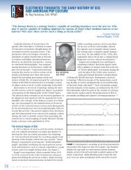
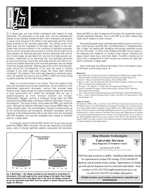
Publication of the Association of Polysomnographic Technologists • 2006, Volume 15, Number 4 • www.aptweb.org2). A dipole layer can have infinite orientations with respect to scalpelectrodes. The electrodes on the scalp “see” only the potentials andpolarity of the potential pointed at them. Each orientation will producea unique result because of the effect on the solid angle (see Fig. 3) thedipole presents to the recording electrodes. The surface area of thedipole layer and the orientation of the layer with respect to the electrodeshave profound effects on the recording of electrical potentials.Due to volume conduction (the process of current flow through the tissuesbetween the electrical generator and the electrode) and summationof solid angles we see data from sources of at least several centimeters,independent of our electrode size, and potentially generatedby many local sources. Due to the solid angle theorem and volume conductionthe closest electrode to the neuronal generator may not alwaysrecord the largest potential 5 . Differing placement of the recording electrodearound the circumference of the area will result in markedchanges in the solid angle even though the event itself remainsunchanged. The polarity of the event also depends on electrode placementnot whether the event is due to EPSP or IPSP, the <strong>for</strong>mer beinga positive potential and the latter being negative.Sleep is a normal function of our brains. There are regions of thebrain and brainstem that promote wakefulness. As the influence of “thewakefulness generators decreases, neurons that promote sleepbecome active. Sleep ensues as a light transitional stage and becomesa more synchronized <strong>for</strong>m (within the bandwidth that we view inpolysomnography) as more neuronal networks are involved.Transmission between neurons is enhanced during wake and REMwhereas during NREM sleep a blocking of afferent in<strong>for</strong>mation is seen inthe thalamus 1 . The brains activity, during wake and REM, are nearly thesame. Although the afferent in<strong>for</strong>mation stops during NREM sleep, thecortex remains active. The corticothalamic conection remains active asdo the corticocortical communications. Brainstem stimulation and theresponse of the thalamocortical cells on the other hand are associatedwith EEG activation and neuronal excitability that creates an activatedstate vs. a sleep state.In conclusion what is it that the EEG shows me? As you know we candetermine NREM, REM, and wake. We can also determine normal EEG,being a lack of clinically significant patterns associated with disorders.Abnormal EEG can also be determined but does not necessarily mean aclinically significant disorder. This is why MRI is an often utilized diagnostictool in relation to brain function.Electrophysiologists study potentials generated by just one neuron oreven small groups recorded with microelectrodes or mesoelectrodes.We, in sleep, are dealing with oscillating macroscopic potentials recordedfrom the scalp 6 . To name a few illnesses that EEG may be utilized inthe diagnosis and treatment of: strokes, brain tumors, infectious diseases,severe head injury, and brain death. The EEG is merely one ofmany tools in assessment of brain function but remains the gold standardin evaluation of sleep state.Now I shall block the afferent in<strong>for</strong>mation from my brainstem reticular<strong>for</strong>mation and let sleep ensue. ★References:1. Mircea Steriade. Principles and <strong>Practice</strong> of Sleep Medicine 2006 Elsevier Chapter 9 BrainElectrical Activity and Sensory Processing During Waking and Sleep States:1012. Duane E. Haines. Fundamental Neuroscience, Second Edition:5083. Bruce J. Fisch. Fisch & Spehlmann’s EEG Primer Basic Principles of Digital and AnalogEEG, Third Revised and Enlarged Edition: 4-94. R Cooper, J.W. Osselton, J.C. Shaw. EEG Technology Second Edition: 8-135. Volume Conduction Principles in Clinical Neurosurgery. February 2005. VeterinaryNeurology and Neurosurgery.http://www.neurovet.org/Electrophysiology/VolumeConduction/VolCondPartABcite.htm6. Paul L. Nunez, Ramesh Srinivasan Electric Fields of the Brain The Neurophysics of EEGSecond Edition: 3-4About the AuthorWill Eckhardt, RPSGT, CRT, is a member of the <strong>APT</strong> Board of Directors and serves as the<strong>APT</strong> Board Liaison <strong>for</strong> the <strong>APT</strong> <strong>Standard</strong>s and Guidelines Committee. He is a board memberof the New England Polysomnographic Society (NEPS) and is NEPS Education CommitteeChair. His full time position is with Sleep HealthCenters where he is the Director of Education.Eckhardt also is a faculty member at Northern Essex Community College where he teachesin the polysomnography program and is a member of the advisory board. He is a member ofthe A2Zzz Magazine editorial board and a recipient of the <strong>APT</strong> Dr. Allen DeVilbiss LiteraryAward in 2004. He is also a member of the American Academy of Sleep Medicine Committeeon Polysomnographic Technologists Issues.Sleep Disorder TechnologistsUniversity ServicesSleep Diagnostic & Treatment CentersLocations in PA & NJLansdale, NE & South Phila, Pottstown, Warrington, West Chester, PA & Voorhees NJFig. 3 Solid Angle — The voltage recorded by each electrode is proportional tothe product of the solid angle and the actual voltage of the dipole. Even thoughthe cross-sectional area of the dipole layer is the same the voltages measuredby the two electrodes would differ from one another in amount because the solidangles are different.Full/ Part-time positions available. Qualified individuals shouldbe experienced in routine PSG testing, CPAP and BIPAPtitrations and nocturnal seizure testing. Opportunities <strong>for</strong> furthergrowth and development exist <strong>for</strong> motivated individuals. Sendresume to (610) 344-7922,employment@uservices.com.Or call (610) 344-9921 <strong>for</strong> further in<strong>for</strong>mation.Multiple locations, good working environment, competitive pay.www.uservices.com29