A quantitative cell migration assay using ThinCert ... - Greiner Bio One
A quantitative cell migration assay using ThinCert ... - Greiner Bio One
A quantitative cell migration assay using ThinCert ... - Greiner Bio One

Create successful ePaper yourself
Turn your PDF publications into a flip-book with our unique Google optimized e-Paper software.
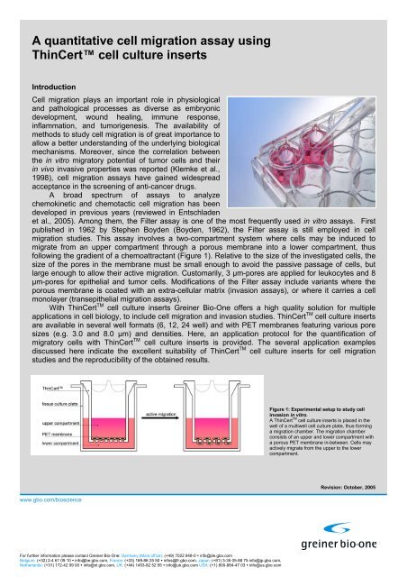
A <strong>quantitative</strong> <strong>cell</strong> <strong>migration</strong> <strong>assay</strong> <strong>using</strong><strong>ThinCert</strong> <strong>cell</strong> culture insertsIntroductionCell <strong>migration</strong> plays an important role in physiologicaland pathological processes as diverse as embryonicdevelopment, wound healing, immune response,inflammation, and tumorigenesis. The availability ofmethods to study <strong>cell</strong> <strong>migration</strong> is of great importance toallow a better understanding of the underlying biologicalmechanisms. Moreover, since the correlation betweenthe in vitro migratory potential of tumor <strong>cell</strong>s and theirin vivo invasive properties was reported (Klemke et al.,1998), <strong>cell</strong> <strong>migration</strong> <strong>assay</strong>s have gained widespreadacceptance in the screening of anti-cancer drugs.A broad spectrum of <strong>assay</strong>s to analyzechemokinetic and chemotactic <strong>cell</strong> <strong>migration</strong> has beendeveloped in previous years (reviewed in Entschladenet al., 2005). Among them, the Filter <strong>assay</strong> is one of the most frequently used in vitro <strong>assay</strong>s. Firstpublished in 1962 by Stephen Boyden (Boyden, 1962), the Filter <strong>assay</strong> is still employed in <strong>cell</strong><strong>migration</strong> studies. This <strong>assay</strong> involves a two-compartment system where <strong>cell</strong>s may be induced tomigrate from an upper compartment through a porous membrane into a lower compartment, thusfollowing the gradient of a chemoattractant (Figure 1). Relative to the size of the investigated <strong>cell</strong>s, thesize of the pores in the membrane must be small enough to avoid the passive passage of <strong>cell</strong>s, butlarge enough to allow their active <strong>migration</strong>. Customarily, 3 µm-pores are applied for leukocytes and 8µm-pores for epithelial and tumor <strong>cell</strong>s. Modifications of the Filter <strong>assay</strong> include variants where theporous membrane is coated with an extra-<strong>cell</strong>ular matrix (invasion <strong>assay</strong>s), or where it carries a <strong>cell</strong>monolayer (transepithelial <strong>migration</strong> <strong>assay</strong>s).With <strong>ThinCert</strong> TM <strong>cell</strong> culture inserts <strong>Greiner</strong> <strong>Bio</strong>-<strong>One</strong> offers a high quality solution for multipleapplications in <strong>cell</strong> biology, to include <strong>cell</strong> <strong>migration</strong> and invasion studies. <strong>ThinCert</strong> TM <strong>cell</strong> culture insertsare available in several well formats (6, 12, 24 well) and with PET membranes featuring various poresizes (e.g. 3.0 and 8.0 µm) and densities. Here, an application protocol for the quantification ofmigratory <strong>cell</strong>s with <strong>ThinCert</strong> TM <strong>cell</strong> culture inserts is provided. The several application examplesdiscussed here indicate the ex<strong>cell</strong>ent suitability of <strong>ThinCert</strong> TM <strong>cell</strong> culture inserts for <strong>cell</strong> <strong>migration</strong>studies and the reproducibility of the obtained results.Figure 1: Experimental setup to study <strong>cell</strong>invasion in vitro.A <strong>ThinCert</strong> TM <strong>cell</strong> culture inserts is placed in thewell of a multiwell <strong>cell</strong> culture plate, thus forminga <strong>migration</strong> chamber. The <strong>migration</strong> chamberconsists of an upper and lower compartment witha porous PET membrane in-between. Cells mayactively migrate from the upper to the lowercompartment.www.gbo.com/bioscienceRevision: October, 2005For further information please contact <strong>Greiner</strong> <strong>Bio</strong>-<strong>One</strong>: Germany (Main office): (+49) 7022 948-0 • info@de.gbo.comBelgium: (+32) 2-4 61 09 10 • info@be.gbo.com, France: (+33) 169-86 25 50 • infos@fr.gbo.com, Japan: (+81) 3-35 05-88 75 info@jp.gbo.com,Netherlands: (+31) 172-42 09 00 • info@nl.gbo.com, UK: (+44) 1453-82 52 55 • info@uk.gbo.com USA: (+1) 800-884-47 03 • info@us.gbo.com
Principle of the Assay<strong>ThinCert</strong> TM <strong>cell</strong> culture inserts are placed in a multiwell <strong>cell</strong> culture plate, thereby forming twocompartments: the upper compartment of the insert and the lower compartment of the plate well. Bothcompartments form the <strong>migration</strong> chamber, separated by the porous PET membrane (figure 1). Cellssown on the PET membrane in serum-free medium may be induced to actively migrate through thePET membrane into the lower compartment with media containing serum or another chemoattractant.Adherent <strong>cell</strong>s remain attached to the under-side of the PET membrane. After fluorescently labelling all<strong>cell</strong>s with Calcein-AM, the migratory <strong>cell</strong>s are detached from the underside of the PET membrane.Finally, the migratory <strong>cell</strong>s are quantified in a standard fluorescence microplate reader. An overview onthe individual steps of the <strong>migration</strong> <strong>assay</strong> is given in figure 2.1. Prepare <strong>cell</strong> cultures according to standard <strong>cell</strong> culture procedures2. Starve <strong>cell</strong>s overnight in serum-free medium with 0.2 % BSA3. Harvest <strong>cell</strong>s and wash them twice in PBS4. Resuspend <strong>cell</strong>s in serum-free medium with 0.2 % BSA to an appropriate final <strong>cell</strong> concentration (e.g. 10 6 /ml)5. Place 24 well <strong>ThinCert</strong>TM <strong>cell</strong> culture inserts in the wells of a CELLSTAR ® 24 well <strong>cell</strong> culture plate6. Add 600 µl culture medium with or without chemoattractant to each well of the <strong>cell</strong> culture plate7. Add 200 µl <strong>cell</strong> suspension to each <strong>cell</strong> culture insert8. Incubate the <strong>cell</strong> culture plate for 3-24 h in a <strong>cell</strong> culture incubator at 37°C and 5 % CO 29. Remove the <strong>cell</strong> culture medium from each well of the <strong>cell</strong> culture plate and replace it by 450 µl serum-free culturemedium with 8 µM Calcein-AM10. Incubate for 45 min in a <strong>cell</strong> culture incubator at 37°C and 5 % CO 211. Remove the culture medium from the <strong>cell</strong> culture inserts12. Transfer the <strong>cell</strong> culture inserts into a freshly prepared 24 well <strong>cell</strong> culture plate containing 500 µl prewarmed Trypsin-EDTA per well13. Incubate for 10 min in a <strong>cell</strong> culture incubator at 37°C and 5 % CO 2 , agitate the plate from time to time14. Discard the <strong>cell</strong> culture inserts and transfer 200 µl of the Trypsin-EDTA solution (now containing the migratory <strong>cell</strong>s)from each well into a black flat bottom 96 well plate15. Read fluorescence in a fluorescence plate reader at an excitation wavelength of 485 nm and an emission wavelengthof 520 nmFigure 2: Short protocol for a <strong>quantitative</strong> <strong>migration</strong> <strong>assay</strong> with adherent <strong>cell</strong>s.The individual steps are schematically illustrated.www.gbo.com/bioscienceFor further information please contact <strong>Greiner</strong> <strong>Bio</strong>-<strong>One</strong>: Germany (Main office): (+49) 7022 948-0 • info@de.gbo.comBelgium: (+32) 2-4 61 09 10 • info@be.gbo.com, France: (+33) 169-86 25 50 • infos@fr.gbo.com, Japan: (+81) 3-35 05-88 75 info@jp.gbo.com,Netherlands: (+31) 172-42 09 00 • info@nl.gbo.com, UK: (+44) 1453-82 52 55 • info@uk.gbo.com USA: (+1) 800-884-47 03 • info@us.gbo.com
MaterialItem Manufacturer Cat.-No.DMEM medium <strong>Bio</strong>chrom AG F0435L-Alanyl-L-Glutamine <strong>Bio</strong>chrom AG K0302RPMI medium <strong>Bio</strong>chrom AG F1295Trypsin/EDTA solution (0.05%/0.02%) <strong>Bio</strong>chrom AG L2143Black 96 well polystyrene strip plate <strong>Greiner</strong> <strong>Bio</strong>-<strong>One</strong> GmbH 756 076CELLSTAR ® 24 well <strong>cell</strong> culture plate <strong>Greiner</strong> <strong>Bio</strong>-<strong>One</strong> GmbH 662 160<strong>ThinCert</strong> 24 well <strong>cell</strong> culture insert with 8 µm pores <strong>Greiner</strong> <strong>Bio</strong>-<strong>One</strong> GmbH 662 638Fetal calf serum Invitrogen Life Technologies 10 270-106Calcein-AM solution (4mM in anhydrous DMSO) Sigma C1359-100ULBovine serum albumin (30%) Sigma A7284-10MLMethods 1Cell cultures were prepared and maintained according to standard <strong>cell</strong> culture procedures. The <strong>cell</strong>lines HT1080, HeLa and NIH3T3 were cultured in DMEM medium with 10% fetal calf serum (FCS). Thenight before the <strong>migration</strong> experiment the <strong>cell</strong>s were deprived in serum-free DMEM medium containing0.2 % bovine serum albumin. Cells were harvested, washed twice in PBS, and resuspended in serumfreeDMEM medium with 0.2 % BSA to obtain an appropriate final concentration (e.g. 10 6 <strong>cell</strong>s/ml). 24well <strong>ThinCert</strong> TM <strong>cell</strong> culture inserts with 8 µm pores and translucent PET membranes were placed inthe wells of a CELLSTAR ® 24 well <strong>cell</strong> culture plate. 600 µl of serum-free DMEM medium with 0.2 %BSA and varying concentrations of FCS was added to each well of the <strong>cell</strong> culture plate (lowercompartment). 200 µl of the <strong>cell</strong> suspension was added to each <strong>cell</strong> culture insert, and the plate withinserts was incubated for 20 h in an incubator at 37°C and 5 % CO 2 . Subsequently, the <strong>cell</strong> culturemedium was removed from each well of the <strong>cell</strong> culture plate and replaced with 450 µl DMEM mediumcontaining 0.2 % BSA and 8 µM Calcein-AM. The plate with inserts was incubated for 45 min in a <strong>cell</strong>culture incubator at 37°C and 5 % CO 2 . Thereafter, the culture medium was removed from the<strong>ThinCert</strong> TM <strong>cell</strong> culture inserts 2 , and the <strong>ThinCert</strong> TM <strong>cell</strong> culture inserts were transferred in the wells of afreshly prepared 24 well plate containing 500 µl Trypsin-EDTA per well. This plate was incubated for 10min in a <strong>cell</strong> culture incubator at 37°C and 5 % CO 2 with sporadic agitation. The <strong>ThinCert</strong> TM <strong>cell</strong> cultureinserts were discarded, and 200 µl of the Trypsin-EDTA solution (now containing the detachedmigratory <strong>cell</strong>s) was transferred from each well of the 24 well plate into a new well of a flat-bottomblack 96 well plate. Finally, the migratory <strong>cell</strong>s were quantified in the black 96 well plate with afluorescence plate reader (Tecan Safire) at an excitation wavelength of 485 nm and an emissionwavelength of 520 nm.1The following procedure is optimized for adherent <strong>cell</strong>s only and may be adapted to suit other <strong>cell</strong> types.2Phenol red in the culture medium may interfere with the subsequent fluorescence reading. The culture medium must therefore be removedfrom the <strong>cell</strong> culture insert.www.gbo.com/bioscienceFor further information please contact <strong>Greiner</strong> <strong>Bio</strong>-<strong>One</strong>: Germany (Main office): (+49) 7022 948-0 • info@de.gbo.comBelgium: (+32) 2-4 61 09 10 • info@be.gbo.com, France: (+33) 169-86 25 50 • infos@fr.gbo.com, Japan: (+81) 3-35 05-88 75 info@jp.gbo.com,Netherlands: (+31) 172-42 09 00 • info@nl.gbo.com, UK: (+44) 1453-82 52 55 • info@uk.gbo.com USA: (+1) 800-884-47 03 • info@us.gbo.com
Results and DiscussionMigrations of different <strong>cell</strong> types along gradients of fetal calf serum (FCS) were analyzed afterapplication of the above-mentioned protocol. When 2×10 5 HT1080 <strong>cell</strong>s were sown onto the <strong>ThinCert</strong> TMmembrane, a concentration-dependent <strong>migration</strong> along different gradients of FCS could be observed.Similar high <strong>migration</strong> rates were observed along the 0 % → 5 % and 0 % → 10 %-FCS gradients,whereas weaker gradients (0 % → 0.5 % FCS and 0 % → 2 % FCS) yielded lower <strong>migration</strong> rates(figure 3).Furthermore, the <strong>migration</strong> rates of HeLa and NIH3T3 <strong>cell</strong>s were assessed in the absence orpresence of FCS gradients. The presence of 0 % → 10 % FCS gradients yielded a 10 and 20 - foldincrease in the number of migratory HeLa and NIH3T3 <strong>cell</strong>s, respectively, as compared to the controlexperiments without serum (figure 4).Figure 3: Migration of HT1080 <strong>cell</strong>s over 20 h in responseto varying concentrations of fetal calf serum. Each data pointrepresents the average of four single experiments. Error bars indicatethe standard deviation. RFU: relative fluorescence units.Figure 4: Migration of HeLa and NIH3T3 <strong>cell</strong>s through 8 µmpores over 20 h in the absence and presence of fetal calf serum.RFU: relative fluorescence units.Throughout a wide range of <strong>cell</strong> populations (25×10 4 to 300×10 4 <strong>cell</strong>s/<strong>migration</strong> chamber), the numberof migratory HT1080 <strong>cell</strong>s displayed a linear correlation with the total number of <strong>cell</strong>s in the <strong>migration</strong>chamber (figure 5). It is recommended that <strong>cell</strong> concentrations to be applied in a <strong>cell</strong> <strong>migration</strong> <strong>assay</strong>be selected from within this range.Finally, two different production lots of <strong>ThinCert</strong> TM <strong>cell</strong> culture inserts were used to perform <strong>cell</strong><strong>migration</strong> <strong>assay</strong>s with HT1080 <strong>cell</strong>s along 0 % → 10 %-FCS gradients. A high reproducibility of resultswas observed, from among both individual <strong>ThinCert</strong> TM <strong>cell</strong> culture inserts as well as two different lots ofmanufacture used to conduct the experiments (figure 6).Figure 5: Migration of HT1080 <strong>cell</strong>s over 20 h in response to 10 %fetal calf serum. The number of migratory <strong>cell</strong>s (relative fluorescenceunits, RFU) correlates linearly with the total amount of <strong>cell</strong>s in the <strong>cell</strong>culture insert. Each data point represents the average of four singleexperiments. Error bars indicate the standard deviation.Figure 6: Reproducibility of <strong>migration</strong> experiments with <strong>ThinCert</strong> TM<strong>cell</strong> culture inserts. Migration experiments were performed <strong>using</strong>2x10 5 HT1080 <strong>cell</strong>s per <strong>cell</strong> culture insert and 10 % fetal calf serum.Only little variation of the amount of migratory <strong>cell</strong>s was observedamong individual experiments and the two different lots of inserts.www.gbo.com/bioscienceFor further information please contact <strong>Greiner</strong> <strong>Bio</strong>-<strong>One</strong>: Germany (Main office): (+49) 7022 948-0 • info@de.gbo.comBelgium: (+32) 2-4 61 09 10 • info@be.gbo.com, France: (+33) 169-86 25 50 • infos@fr.gbo.com, Japan: (+81) 3-35 05-88 75 info@jp.gbo.com,Netherlands: (+31) 172-42 09 00 • info@nl.gbo.com, UK: (+44) 1453-82 52 55 • info@uk.gbo.com USA: (+1) 800-884-47 03 • info@us.gbo.com
ConclusionThe examples described here illustrate the ex<strong>cell</strong>ent suitability of <strong>ThinCert</strong> TM <strong>cell</strong> culture inserts for <strong>cell</strong><strong>migration</strong> <strong>assay</strong>s. The adapted protocol allows for both ease of quantification of the migratory <strong>cell</strong>s andex<strong>cell</strong>ent reproducibility of the obtained results. This protocol provides a ready-to-use solution for <strong>cell</strong><strong>migration</strong> studies, in addition to serving as a basis for use in advanced applications such astransepithelial <strong>migration</strong> or in vitro <strong>cell</strong> invasion studies.ReferencesBoyden S. (1962) The chemotactic effect of mixtures of antibody and antigen on polymorphonuclearleucocytes. J Exp Med. Mar 1;115:453-66.Entschladen F., Drell T.L. 4th, Lang K., Masur K., Palm D., Bastian P., Niggemann B., Zaenker K.S.(2005) Analysis methods of human <strong>cell</strong> <strong>migration</strong>. Exp Cell Res. Jul 15;307(2):418-26.Klemke R.L., Leng J., Molander R., Brooks P.C., Vuori K., Cheresh D.A. (1998) CAS/Crk couplingserves as a "molecular switch" for induction of <strong>cell</strong> <strong>migration</strong>. J Cell <strong>Bio</strong>l. Feb 23;140(4):961-72.www.gbo.com/bioscienceFor further information please contact <strong>Greiner</strong> <strong>Bio</strong>-<strong>One</strong>: Germany (Main office): (+49) 7022 948-0 • info@de.gbo.comBelgium: (+32) 2-4 61 09 10 • info@be.gbo.com, France: (+33) 169-86 25 50 • infos@fr.gbo.com, Japan: (+81) 3-35 05-88 75 info@jp.gbo.com,Netherlands: (+31) 172-42 09 00 • info@nl.gbo.com, UK: (+44) 1453-82 52 55 • info@uk.gbo.com USA: (+1) 800-884-47 03 • info@us.gbo.com