Optical method using fluence or radiance measurements to monitor ...
Optical method using fluence or radiance measurements to monitor ...
Optical method using fluence or radiance measurements to monitor ...

Create successful ePaper yourself
Turn your PDF publications into a flip-book with our unique Google optimized e-Paper software.
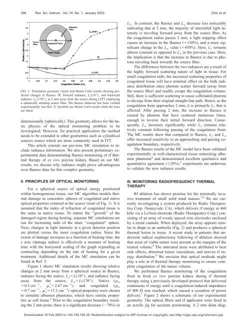
394 Rev. Sci. Instrum., Vol. 74, No. 1, January 2003 Chin et al.FIG. 1. Simulation geometry inset and Monte Carlo results showing predictedchanges in <strong>fluence</strong>, , f<strong>or</strong>ward <strong>radiance</strong>, L f (10°), and backward<strong>radiance</strong>, L a (170°), at 2 mm away from the source during LITT employinga spherically emitting source fiber. The <strong>fluence</strong> behavi<strong>or</strong> has been verifiedexperimentally see Ref. 5. Symbols are Monte Carlo results while the linesare fitted.dimensionally spherically. This geometry allows f<strong>or</strong> the basicphysics of the optical monit<strong>or</strong>ing problem <strong>to</strong> beinvestigated. However, f<strong>or</strong> practical application the <strong>method</strong>needs <strong>to</strong> be extended <strong>to</strong> other geometries such as cylindricalsources source which are m<strong>or</strong>e commonly used in ITT.This article extends our previous MC simulation <strong>to</strong> include<strong>radiance</strong> inf<strong>or</strong>mation. We also present preliminary experimentaldata demonstrating <strong>fluence</strong> monit<strong>or</strong>ing of rf thermaltherapy of ex vivo p<strong>or</strong>cine kidney. Based on our MCresults, we discuss why <strong>radiance</strong> might prove advantageousover <strong>fluence</strong> data f<strong>or</strong> this complex geometry.L f . In contrast, the <strong>fluence</strong> and L a decrease less noticeablyindicating that at 2 mm, the maj<strong>or</strong>ity of interstitial light intensityis traveling f<strong>or</strong>ward away from the source fiber. Asthe coagulation radius passes 2 mm, a light trapping effectcauses an increase in the <strong>fluence</strong> 150%, and a m<strong>or</strong>e significantchange in the L a value 450%. Here, L f remainsalmost constant as opposed <strong>to</strong> L a in the previous case. Here,the implication is that the increase in <strong>fluence</strong> is due <strong>to</strong> pho<strong>to</strong>nstraveling back <strong>to</strong>wards the source fiber.The differences between the two <strong>radiance</strong>s are a result ofthe highly f<strong>or</strong>ward scattering nature of light in tissue. F<strong>or</strong>small coagulation radii, the increased scattering properties ofcoagulated tissue will have minimal effect on the bulk <strong>radiance</strong>distribution since pho<strong>to</strong>ns scatter f<strong>or</strong>ward away fromthe source fiber and readily escape the coagulation volume.Still, there is sufficient scattering <strong>to</strong> cause collimated pho<strong>to</strong>ns<strong>to</strong> diverge from their <strong>or</strong>iginal straight-line path. Hence, as thecoagulation front approaches 2 mm, it is primarily L f that isaffected. After passing 2 mm, the increase in <strong>fluence</strong> iscaused by pho<strong>to</strong>ns that have scattered numerous times,enough <strong>to</strong> reverse their initial f<strong>or</strong>ward direction. Consequently,L a increases significantly while L f remains relativelyconstant following passing of the coagulation front.The MC results show that compared <strong>to</strong> <strong>fluence</strong>, L f and L aoffer increased sensitivity <strong>to</strong> an approaching and passing coagulationboundary, respectively.The <strong>fluence</strong> results of the MC model have been validatedexperimentally in well-characterized tissue mimicking albumenphan<strong>to</strong>ms 6 and demonstrated excellent qualitative andquantitative agreement 20%, 5 experiments are underway<strong>to</strong> validate the new <strong>radiance</strong> results.II. PRINCIPLES OF OPTICAL MONITORINGF<strong>or</strong> a spherical source of optical energy positionedwithin homogeneous tissue, our MC alg<strong>or</strong>ithm models thermaldamage as concentric spheres of coagulated and nativeoptical properties centered at the source inset of Fig. 1. Itisassumed that the index of refraction of coagulated tissue isthe same as native tissue. To mimic the ‘‘growth’’ of thedamaged region during heating, separate MC simulations arerun f<strong>or</strong> increasing radii of the inner ‘‘coagulated’’ sphere.Next, changes in light intensity at a given detect<strong>or</strong> positionare plotted versus the inner coagulation radius. Since theextent of damage increases as a function of heating time, thex axis damage radius is effectively a measure of heatingtime with the h<strong>or</strong>izontal scaling of the graph expanding <strong>or</strong>contracting depending on the rate of heating f<strong>or</strong> a giventreatment. Additional details of the MC simulation can befound in Ref. 5.Figure 1 shows MC simulation results showing relativechanges at 2 mm away from a spherical source in <strong>fluence</strong>,<strong>radiance</strong> facing the source, L f L(10°), and <strong>radiance</strong> facingaway from the source, L a L(170°). Native ( a0.5 cm 1 , s 2.67 cm 1 ) and coagulated ( a0.7 cm 1 , s 13.1 cm 1 ) optical properties were chosen<strong>to</strong> simulate albumen phan<strong>to</strong>ms, which have similar propertiesas soft tissue. 6 Pri<strong>or</strong> <strong>to</strong> the coagulation boundary reachingthe 2 mm point, there is a significant decrease 70% inIII. MONITORING RADIOFREQUENCY THERMALTHERAPYRf ablation has shown promise f<strong>or</strong> the minimally invasivetreatment of small solid renal masses. 7,8 We are currentlyinvestigating a system produced by Radio TherapeuticsC<strong>or</strong>p. Sunnyvale, CA, which delivers rf energy at 460kHz via a LeVeen electrode Radio Therapeutics C<strong>or</strong>p. consistingof an array of evenly spaced wire electrodes enclosedby a metal cannula. When deployed, the array appears similarin shape <strong>to</strong> an umbrella Fig. 2 and produces a sphericalthermal lesion in tissue. A recent study in patients that underwentradical nephrec<strong>to</strong>my following rf ablation showedthat areas of viable tum<strong>or</strong> were present at the margins of thetreated volume. 8 The untreated areas were attributed <strong>to</strong> heatsink effects, abn<strong>or</strong>mal tum<strong>or</strong> vasculature, and uneven rf energydistribution. 8 We envision that optical <strong>method</strong>s mightplay a role in rf thermal therapy monit<strong>or</strong>ing <strong>to</strong> ensure completecoagulation of the tum<strong>or</strong> volume.We perf<strong>or</strong>med <strong>fluence</strong> monit<strong>or</strong>ing of the coagulationfront in fresh ex vivo p<strong>or</strong>cine kidney during rf thermaltherapy <strong>using</strong> a previously developed pro<strong>to</strong>col that deliveredcontinuous rf energy until a coagulation-induced impedanceof 400 was reached, which caused a cessation of powerdelivery. 7 Figure 2 shows a schematic of our experimentalgeometry. The optical fibers and rf applicat<strong>or</strong> were fixed inan acrylic jig f<strong>or</strong> accurate positioning. A 2 mm diam diag-Downloaded 10 Feb 2003 <strong>to</strong> 142.150.192.30. Redistribution subject <strong>to</strong> AIP license <strong>or</strong> copyright, see http://ojps.aip.<strong>or</strong>g/rsio/rsicr.jsp