Two human cases of tick bite caused by Ixodes nipponensis
Two human cases of tick bite caused by Ixodes nipponensis
Two human cases of tick bite caused by Ixodes nipponensis

Create successful ePaper yourself
Turn your PDF publications into a flip-book with our unique Google optimized e-Paper software.
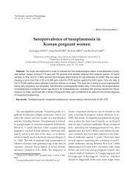
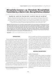
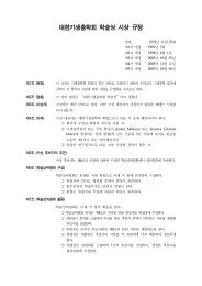
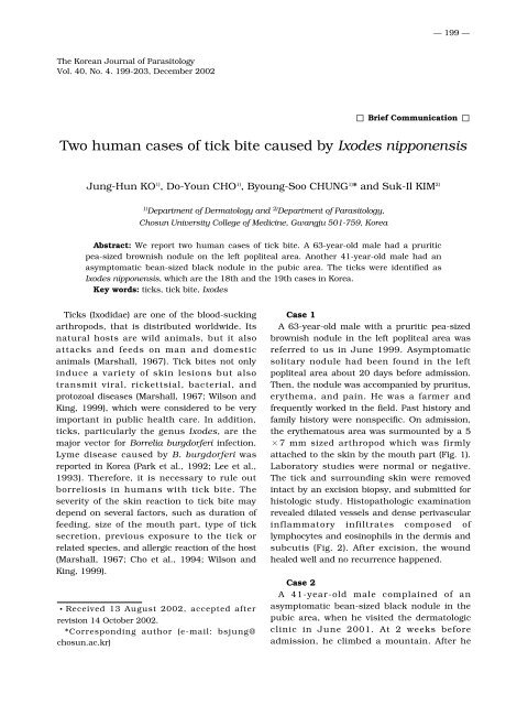
— 199 —The Korean Journal <strong>of</strong> ParasitologyVol. 40, No. 4. 199-203, December 2002 Brief Communication <strong>Two</strong> <strong>human</strong> <strong>cases</strong> <strong>of</strong> <strong>tick</strong> <strong>bite</strong> <strong>caused</strong> <strong>by</strong> <strong>Ixodes</strong> <strong>nipponensis</strong>Jung-Hun KO 1) , Do-Youn CHO 1) , Byoung-Soo CHUNG 1) * and Suk-Il KIM 2)1) Department <strong>of</strong> Dermatology and 2) Department <strong>of</strong> Parasitology,Chosun University College <strong>of</strong> Medicine, Gwangju 501-759, KoreaAbstract: We report two <strong>human</strong> <strong>cases</strong> <strong>of</strong> <strong>tick</strong> <strong>bite</strong>. A 63-year-old male had a pruriticpea-sized brownish nodule on the left popliteal area. Another 41-year-old male had anasymptomatic bean-sized black nodule in the pubic area. The <strong>tick</strong>s were identified as<strong>Ixodes</strong> <strong>nipponensis</strong>, which are the 18th and the 19th <strong>cases</strong> in Korea.Key words: <strong>tick</strong>s, <strong>tick</strong> <strong>bite</strong>, <strong>Ixodes</strong>Ticks (Ixodidae) are one <strong>of</strong> the blood-suckingarthropods, that is distributed worldwide. Itsnatural hosts are wild animals, but it alsoattacks and feeds on man and domesticanimals (Marshall, 1967). Tick <strong>bite</strong>s not onlyinduce a variety <strong>of</strong> skin lesions but alsotransmit viral, rickettsial, bacterial, andprotozoal diseases (Marshall, 1967; Wilson andKing, 1999), which were considered to be veryimportant in public health care. In addition,<strong>tick</strong>s, particularly the genus <strong>Ixodes</strong>, are themajor vector for Borrelia burgdorferi infection.Lyme disease <strong>caused</strong> <strong>by</strong> B. burgdorferi wasreported in Korea (Park et al., 1992; Lee et al.,1993). Therefore, it is necessary to rule outborreliosis in <strong>human</strong>s with <strong>tick</strong> <strong>bite</strong>. Theseverity <strong>of</strong> the skin reaction to <strong>tick</strong> <strong>bite</strong> maydepend on several factors, such as duration <strong>of</strong>feeding, size <strong>of</strong> the mouth part, type <strong>of</strong> <strong>tick</strong>secretion, previous exposure to the <strong>tick</strong> orrelated species, and allergic reaction <strong>of</strong> the host(Marshall, 1967; Cho et al., 1994; Wilson andKing, 1999).Received 13 August 2002, accepted afterrevision 14 October 2002.*Corresponding author (e-mail: bsjung@chosun.ac.kr)Case 1A 63-year-old male with a pruritic pea-sizedbrownish nodule in the left popliteal area wasreferred to us in June 1999. Asymptomaticsolitary nodule had been found in the leftpopliteal area about 20 days before admission.Then, the nodule was accompanied <strong>by</strong> pruritus,erythema, and pain. He was a farmer andfrequently worked in the field. Past history andfamily history were nonspecific. On admission,the erythematous area was surmounted <strong>by</strong> a 5×7 mm sized arthropod which was firmlyattached to the skin <strong>by</strong> the mouth part (Fig. 1).Laboratory studies were normal or negative.The <strong>tick</strong> and surrounding skin were removedintact <strong>by</strong> an excision biopsy, and submitted forhistologic study. Histopathologic examinationrevealed dilated vessels and dense perivascularinflammatory infiltrates composed <strong>of</strong>lymphocytes and eosinophils in the dermis andsubcutis (Fig. 2). After excision, the woundhealed well and no recurrence happened.Case 2A 41-year-old male complained <strong>of</strong> anasymptomatic bean-sized black nodule in thepubic area, when he visited the dermatologicclinic in June 2001. At 2 weeks beforeadmission, he climbed a mountain. After he
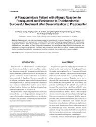
— 200 —Fig. 3. A bean-sized <strong>tick</strong> was firmly attached onthe skin <strong>of</strong> pubic region in the case 2. The <strong>tick</strong>was engorged with blood meal.Fig. 1. A pea-sized light brown <strong>tick</strong> was firmlyattached on the skin <strong>of</strong> left popliteal area in thecase 1.Fig. 2. Biopsy specimen from the case 1 showsdense inflammatory infiltrates composed <strong>of</strong>lymphocytes and eosinophils in the dermis andsubcutis (H&E stain, ×200).came back home, he found an arthropod on theback and removed it <strong>by</strong> himself. He also felt amildly anesthetic sensation in the pubic area 2days later and found a black colored nodule.The nodule became bigger, therefore, hethought that it might be a tumor. Past historyand family history were nonspecific. Onadmission, the nodule was a black arthropod, 7×8 mm in size, firmly attached to the skin <strong>by</strong>the mouth part (Fig. 3). It was easily removed<strong>by</strong> heat, and all signs and symptomsdisappeared.The <strong>tick</strong> from the case 1 was oval in shape, 5×7 mm in size, and light brown in color (Fig.4A and 4B). Another <strong>tick</strong> from the case 2 wasoval, 7×8 mm in size, and black in color (Fig.5). The capitulum <strong>of</strong> anterior portion <strong>of</strong> thebody, the scutum <strong>of</strong> dorsal portion, spicularplate, genital aperture, anus, and 4 pairs <strong>of</strong>legs <strong>of</strong> ventral portion were observed. Spicularplate was ovoid and located on the ventrolateralsurface posterior to the coxa IV. Genitalopening was straight and located on the level <strong>of</strong>the coxa IV. Anus was round and located onthe posterior portion, and horseshoe-shapedanal groove was extended to posterior margin <strong>of</strong>the body, surrounding the anus (Fig. 5).Internal spur <strong>of</strong> the coxa I was long, but notoverlaid on the edge <strong>of</strong> the coxa II in the case 1(Fig. 6A) and the case 2 (Fig. 6B). Thesecharacteristics were typical features <strong>of</strong> I.nipponesis (Lee et al., 1989; Cho et al., 1995;Ryu et al., 1998).Thirty-one <strong>cases</strong> <strong>of</strong> <strong>tick</strong> <strong>bite</strong> have beenreported in Korea up to the present (Yun et al.,2001). Seventeen <strong>cases</strong> were identified with I.<strong>nipponensis</strong> (Noh and Cho, 1983; Lee et al.,
— 201 —Fig. 4. <strong>Ixodes</strong> <strong>nipponensis</strong> after removal with dorsal view (A) and ventral view (B) in the case 1. Scalebars = 1 mm.Fig. 5. The <strong>tick</strong> removed from pubic area in thecase 2. The whole body with ventral view. InvertedU-shaped anal groove (arrow) encircles the anus.A scale bar = 1 mm.1989; Paik et al., 1989; Chang et al., 1991; Choet al., 1991; Cho et al., 1994, 1995; Chu et al.,1997; Ryu et al., 1998; Chae et al., 2000; Yunet al., 2001), and 1 case with I. ovatus (Changet al., 1991), 1 case with I. monospinosus (Choet al., 1999), 1 case with I. persulcatus (Im etal., 1998), 1 case with Haemaphysalis flava(Yoon et al., 1996), and 1 case withHaemaphysalis longicornis (Rho et al., 1999)have been reported. The remainder were with<strong>Ixodes</strong> as the genus level. Our <strong>cases</strong> are the18th and the 19th <strong>cases</strong> with I. <strong>nipponensis</strong>.Ticks are usually 1-9 mm long beforeengorgement, but they reach 2 cm in lengthafter feeding (Marshall, 1967). Ticks feed for 7-12 days for a full engorgement and then drop<strong>of</strong>f the hosts to continue their life cycle (Wilsonand King, 1999; Odom et al., 2000; Yun et al.,2001). Some <strong>tick</strong>s may feed for 30 days orlonger (Yoon et al., 1996; Chu et al., 1997). Thecase 1 fed over 20 days and the case 2 for 14days. During this time, the patients sufferedfrom fever, chill, headache, abdominal pain,and vomiting. After removal <strong>of</strong> the engorged<strong>tick</strong>s, the above symptoms disappeared within12 to 36 hours (Odom et al., 2000).The most frequent location <strong>of</strong> <strong>tick</strong> <strong>bite</strong>s isknown to be perianal area, abdomen, scrotum,
— 202 —Fig. 6. Internal spur (arrow) <strong>of</strong> coxa I, much longer than external spur, does not overlay on the edge <strong>of</strong>coxa II in the <strong>tick</strong>s from the case 1 (A) and the case 2 (B). This is one <strong>of</strong> the characteristics <strong>of</strong> <strong>Ixodes</strong><strong>nipponensis</strong>.extremities, and scalp (Cho et al., 1994; Yun etal., 2001). In our <strong>cases</strong>, the locations were onthe leg and the pubic area. Tick <strong>bite</strong>s occurmostly in the spring and summer. Our <strong>cases</strong>were detected in June.Dermatoses <strong>by</strong> the <strong>tick</strong> <strong>bite</strong> include papularurticaria, subcutaneous hemorrhage, focalnecrosis, peladoid alopecia, secondary bacterialinfection, erythema chronicum migrans,acrodermatitis chronica atrophicans, andlymphocytoma cutis. Neurologic manifestationsdue to <strong>tick</strong> saliva are pyrexia and paralysis(Marshall, 1967; Patterson et al., 1979). Ourcase 2 patient felt mild anesthesia in the pubiclesion. Due to very slow penetration <strong>of</strong> the skin<strong>by</strong> the insect’s hypostome and the fact that theinsect’s saliva is both anticoagulant andanesthetic, the patient hardly feels pain oritching sensation and it may take several daysto find the <strong>tick</strong> on the body surface (Winer andStrakosch, 1941).Winer and Strakosch (1941) divided thehistological findings <strong>of</strong> <strong>tick</strong> <strong>bite</strong> into threestages with reference to duration: acute (amonth or less), subacute (several months), andchronic (a year or more) stage. In the acutestage, dilated blood vessels and perivascularinflammatory infilitrates composed <strong>of</strong>lymphocytes and eosinophils in the superficialdermis are observed. In the subacute stage,extensive perivascular inflammatory infilitratescomposed <strong>of</strong> lymphocytes, neutrophils,fibroblasts, and eosinophils are prominent. Inthe chronic stage, fibrous tissue proliferation, afew Langhans’ giant cells, lymphocytes, andeosinophils deposited between fibrous tissuesare observed. In the case 1, histologicmanifestations were dilated vessels andperivascular infiltrates <strong>of</strong> eosinophils andlymphocytes in the dermis and subcutis,indicating an acute stage <strong>of</strong> <strong>tick</strong> <strong>bite</strong>.For the treatment, applications <strong>of</strong> gasoline,benzene, chlor<strong>of</strong>orm, ether, s<strong>of</strong>t paraffine, andheat are effective. In addition, suffocating the<strong>tick</strong> with petrolatum is recommended. Abruptpulling or squeezing <strong>of</strong> the <strong>tick</strong> is discouraged,because mouth part may be left behind theskin and secretion <strong>of</strong> salivary toxins may be
— 203 —increased to aggravate inflammation or foreignbody reaction. For this reason, punch biopsy orsurgical excision is very effective, particularlywhen the tenacious <strong>tick</strong> cannot be easilyextracted <strong>by</strong> other means (Winer andStrakosch, 1941; Patterson et al., 1979; Cho etal., 1994; Wilson and King, 1999). In our <strong>cases</strong>,we removed <strong>tick</strong>s <strong>by</strong> excision biopsy and heatto prevent saliva release and transmission <strong>of</strong>any microorganism during <strong>tick</strong> manipulation.Regular follow-up was carried out, but norecurrence or other disorder was observed.REFERENCESChae KS, Gang H, Lee DW, et al. (2000) Tick <strong>bite</strong>s.Korean J Dermatol 38: 111-116 (in Korean).Chang HS, Hur SG, Lee SC, Chun IK, Kim YP(1991) <strong>Two</strong> <strong>cases</strong> <strong>of</strong> <strong>tick</strong> <strong>bite</strong>s <strong>caused</strong> <strong>by</strong><strong>Ixodes</strong> ovatus and <strong>Ixodes</strong> <strong>nipponensis</strong>. KoreanJ Dermatol 29: 647-652 (in Korean).Cho NJ, Bang DS, Cho BK, Oh YJ, Lee WK (1991)<strong>Two</strong> <strong>cases</strong> <strong>of</strong> <strong>tick</strong> <strong>bite</strong>s <strong>caused</strong> <strong>by</strong> <strong>Ixodes</strong><strong>nipponensis</strong>. Korean J Dermatol 29: 533-537(in Korean).Cho BK, Kang H, Bang D, Kim SN, Hwang S, SongES (1994) Tick <strong>bite</strong>s in Korea. Int J Dermatol33: 552-555.Cho BK, Nam HW, Cho SY, Lee WK (1995) A case<strong>of</strong> <strong>tick</strong> <strong>bite</strong> <strong>by</strong> a spontaneously retreated<strong>Ixodes</strong> <strong>nipponensis</strong>. Korean J Parasitol 33:239-242.Cho CG, Son SW, Kim AR, et al. (1999) A Case <strong>of</strong>Tick <strong>bite</strong> Caused <strong>by</strong> <strong>Ixodes</strong> monospinosus.Korean J Dermatol 37: 390-394 (in Korean).Chu JP, Cho YJ, Jeong GS, Ko BM (1997) A case<strong>of</strong> <strong>tick</strong> infestation in chest wall <strong>by</strong> <strong>Ixodes</strong><strong>nipponensis</strong>. Infection 29: 53-56 (in Korean).Im K, Lee IY, Lee WJ (1998) A <strong>human</strong> case <strong>of</strong> <strong>tick</strong><strong>bite</strong> <strong>by</strong> <strong>Ixodes</strong> persulcatus. Korean J Parasitol36: 63-65.Lee SH, Chai JY, Kho WG, Hong SJ, Chung YD(1989) A <strong>human</strong> case <strong>of</strong> <strong>tick</strong> <strong>bite</strong> <strong>by</strong> <strong>Ixodes</strong><strong>nipponensis</strong> on the scalp. Korean J Parasitol27: 67-69 (in Korean).Lee MG, Chung KY, Choi YS, Cho SN (1993) Lymedisease. Korean J Dermatol 31: 601-605 (inKorean).Marshall J (1967) Ticks and the <strong>human</strong> skin.Dermatologica 135: 60-65.Noh YT, Cho BK (1983) A taxonomical study on<strong>tick</strong>s in Korea (VI). <strong>Ixodes</strong> <strong>nipponensis</strong> Kitaokaand Saito. Modern Medicine 26: 89-90.Odom RB, James WD, Berger TG (2000) Andrews’Diseases <strong>of</strong> the skin. 9th ed. pp 561-563Philadelphia: WB Saunders.Paik SC, Oh YJ, Kim SY, Cho BK, Houh W (1989)A case <strong>of</strong> <strong>tick</strong> <strong>bite</strong> <strong>caused</strong> <strong>by</strong> <strong>Ixodes</strong><strong>nipponensis</strong>. Korean J Dermatol 27: 83-88 (inKorean).Park KH, Lee SH, Won WJ, Chang WH (1992)Isolation <strong>of</strong> Borrelia burgdorferi, the causativeagent <strong>of</strong> Lyme disease, from <strong>Ixodes</strong> <strong>tick</strong>s inKorea. J Korean Soc Microbiol 27: 307-312 (inKorean).Patterson JW, Fitzwater JE, Connell J (1979)Localized <strong>tick</strong> <strong>bite</strong> reaction. Cutis 24: 168-172.Roh DK, Jang IG, Cho BK, Son SJ, Lee IY, Lee WK(1999) Tick <strong>bite</strong> <strong>by</strong> Haemaphysalis longicornisNeumann: Laboratory observation <strong>of</strong> thecausative <strong>tick</strong>. Korean J Dermatol 37: 631-636(in Korean).Ryu JS, Lee JU, Ahn MH, Min DY, Ree HI (1998) A<strong>human</strong> case <strong>of</strong> <strong>tick</strong> <strong>bite</strong> <strong>by</strong> <strong>Ixodes</strong> <strong>nipponensis</strong>.Korean J Parasitol 36: 59-61.Winer LH, Strakosch EA (1941) Tick <strong>bite</strong>s-Dermacentor variabilis (SAY). J Invest Dermatol4: 249-258.Wilson DC, King LE (1999) Arthropod <strong>bite</strong>s andstings. Fitzpatrick’s Dermatology in generalmedicine, 5th ed. pp 2687-2688, Philadelphia:WB Saunders.Yoon SY, Kim KH, Suhr KB, et al. (1996) A case <strong>of</strong><strong>tick</strong> <strong>bite</strong> <strong>caused</strong> <strong>by</strong> Haemaphysalis flava.Korean J Dermatol 34: 326-330 (in Korean).Yun SK, Ko GB, Chon TH (2001) Tick <strong>bite</strong>: Report<strong>of</strong> a case & review <strong>of</strong> Korean <strong>cases</strong>. Korean JDermatol 39: 891-895 (in Korean).