A Biomechanical Analysis of the Causes of Traumatic ... - NACDL
A Biomechanical Analysis of the Causes of Traumatic ... - NACDL
A Biomechanical Analysis of the Causes of Traumatic ... - NACDL

You also want an ePaper? Increase the reach of your titles
YUMPU automatically turns print PDFs into web optimized ePapers that Google loves.
REVIEWA <strong>Biomechanical</strong> <strong>Analysis</strong> <strong>of</strong> <strong>the</strong> <strong>Causes</strong> <strong>of</strong> <strong>Traumatic</strong> BrainInjury in Infants and ChildrenWerner Goldsmith, PhD* and John Plunkett, MD†Abstract: There is significant disagreement among medical pr<strong>of</strong>essionalsregarding <strong>the</strong> mechanisms for infant brain injury. Thisdisagreement is due in part to <strong>the</strong> failure by some to acknowledgeand incorporate known biomechanical data and models into hypo<strong>the</strong>sesregarding causes. A proper biomechanical understanding <strong>of</strong> <strong>the</strong>mechanisms <strong>of</strong> traumatic brain injury (TBI) challenges many publishedand testified assumptions regarding TBI in infants andchildren.This paper analyzes <strong>the</strong> biomechanical relationship between <strong>the</strong>causes <strong>of</strong> TBI in infants and children, and <strong>the</strong>ir physiologicalconsequences. Loading characteristics, injury parameters and criteria,scaling, failure characteristics, differences between infants andadults, and impact due to falls are described and discussed in <strong>the</strong>context <strong>of</strong> <strong>the</strong> laws <strong>of</strong> mechanics. Recent studies are critiqued withreference to <strong>the</strong>ir contribution to an understanding <strong>of</strong> brain injurymechanisms. Finally, methods for improving our currently incompleteknowledge <strong>of</strong> infant head injuries, and <strong>the</strong>ir mechanisms,consequences and tolerances are proposed. There is an urgent needfor close collaboration between physicians and biomechanicians toobjectively and scientifically evaluate infant head injuries to fur<strong>the</strong>rdefine <strong>the</strong>ir mechanical bases, and to assist in <strong>the</strong>ir diagnosis andtreatment.Key Words: <strong>Traumatic</strong> Infant Head Injury, Shaken BabySyndrome (SBS), Impact, Impulsive Loading(Am J Forensic Med Pathol 2004;25: 89–100)Biomechanicians and physicians evaluate trauma in fundamentallydifferent ways. A biomechanician constructs oraccepts a particular system, obtains its physical and geometriccharacteristics, applies a specified and quantifiable input(load), and <strong>the</strong>n determines <strong>the</strong> output using experimental,From <strong>the</strong> *Graduate School, Departments <strong>of</strong> Mechanical Engineering andBioengineering, University <strong>of</strong> California, Berkeley, California; and <strong>the</strong>†Department <strong>of</strong> Laboratory and Medical Education, Regina MedicalCenter, Hastings, Minnesota.Reprints: John Plunkett, MD, Laboratory and Medical Education Director,Regina Medical Center, 1175 Nininger Road, Hastings, MN 55033.E-mail: plunkettj@reginamedical.org.Copyright © 2004 by Lippincott Williams & WilkinsISSN: 0195-7910/04/2502-0089DOI: 10.1097/01.paf.0000127407.28071.63analytical, and numerical techniques. A physician sees <strong>the</strong>end product <strong>of</strong> signs and symptoms, and relies primarily ifnot exclusively on experience and observational case materialto diagnose and treat. A biomechanician traces a continuouspath from cause to effect using <strong>the</strong> laws <strong>of</strong> nature, tries todetermine <strong>the</strong> specific mechanism <strong>of</strong> an injury, and attemptsto ei<strong>the</strong>r establish or eliminate an ultimate mechanical cause.A physician treats <strong>the</strong> physiological alterations due to aninjury, and does not usually need to validate <strong>the</strong> diagnosiswith a controlled experiment prior to making a decision toinitiate treatment.Recent advances in <strong>the</strong> quantitative characterization <strong>of</strong><strong>the</strong> material and structural properties <strong>of</strong> <strong>the</strong> developing infantbrain and skull permit <strong>the</strong> biomechanician to study <strong>the</strong> differencesbetween traumatic brain injury in infants andadults. 1–6 The findings suggest that understanding <strong>the</strong> uniqueage-dependant properties <strong>of</strong> <strong>the</strong> developing brain and skull iscritical for determining both impulsive and impact loadingthresholds. New and continued research, including pediatricinjury models, will play a vital role in evaluating infanttraumatic head injury. 7 Collaboration between biomechaniciansand physicians will provide a foundation for developingimproved clinical, diagnostic, preventive, <strong>the</strong>rapeutic, andrehabilitative strategies to address <strong>the</strong> unique problems <strong>of</strong>pediatric head injury.BiomechanicsBiomechanics is <strong>the</strong> application <strong>of</strong> <strong>the</strong> science <strong>of</strong> mechanicsto biologic systems, whe<strong>the</strong>r plant or animal. 8,9Mechanics is defined as <strong>the</strong> consequences <strong>of</strong> <strong>the</strong> application<strong>of</strong> forces and couples (a set <strong>of</strong> equal, parallel and oppositelydirected forces) to one object or a system <strong>of</strong> objects (solid,liquid or gas). A solid object may be considered analyticallyas a particle (a point in space that has mass, and that resistsmotion); as a group or system <strong>of</strong> such particles; or as a singlesolid body that may be ei<strong>the</strong>r rigid or deformable.Skull and brain motion and <strong>the</strong> forces causing <strong>the</strong>m aregoverned by Newton’s Second Law, Force mass timesacceleration (F ma). The displacement <strong>of</strong> <strong>the</strong> head (as anobject with mass) can be described as planar or as occurringin space as defined by motion in one or all 3 <strong>of</strong> <strong>the</strong> X-, Y-, andZ-axes. The displacements are governed by Newton’s LawThe American Journal <strong>of</strong> Forensic Medicine and Pathology • Volume 25, Number 2, June 2004 89
The American Journal <strong>of</strong> Forensic Medicine and Pathology • Volume 25, Number 2, June 2004TBI in Infants and ChildrenFIGURE 2. Impact loading (a) Witha fixed object (b) With a movingobject.Tolerance Curve (WSTC), is based on a number <strong>of</strong> assumptions,including <strong>the</strong> equivalence <strong>of</strong> linear skull fracture and<strong>the</strong> onset <strong>of</strong> concussion, and <strong>the</strong> direct extrapolation <strong>of</strong> <strong>the</strong>results from cadavers to living humans. The WSTC ando<strong>the</strong>rs 35 do not differentiate between age, sex, or size, andwere expected to apply to infants and children as well asadults. However, this extrapolation is almost certainly invalidbecause <strong>the</strong> cerebral mechanical properties and even scaledgeometry <strong>of</strong> infants is quite different from that <strong>of</strong> adults. 1–7Regardless <strong>of</strong> <strong>the</strong> utility <strong>of</strong> <strong>the</strong> HIC for defining limits<strong>of</strong> translational acceleration, o<strong>the</strong>r criteria have been proposed.36 A separate criterion is also needed for rotationalacceleration and for a combination <strong>of</strong> rotation and translationoccurring simultaneously (as is almost always <strong>the</strong> case forbiologic systems). 37Stürtz established linear acceleration head tolerancelevels for children by reconstructing pedestrian-automobileFIGURE 3. Contact phenomenon and wave propagation inimpact to <strong>the</strong> head.impacts. 38,39 He determined that 83g was <strong>the</strong> limit for <strong>the</strong>anterior/posterior acceleration when <strong>the</strong> acceleration lastedmore than 3 milliseconds. He also concluded that a peakacceleration <strong>of</strong> 70g was associated with completely reversibleinjury, and that 110g was associated with 25% irreversibleinjury. These limits correspond to HIC values <strong>of</strong> 350 and 600,respectively, for a 6-year-old child dummy 40 ).The bi<strong>of</strong>idelity and motion <strong>of</strong> <strong>the</strong> Child Restraint-AirBag Interaction (CRABI) anthropometric dummy 41 havebeen studied by subjecting <strong>the</strong> dummy to linear and angularacceleration. 42 The author suggested limiting HIC values for<strong>the</strong> 6-month-old infant <strong>of</strong> 121 with an acceleration duration <strong>of</strong>22 milliseconds (a peak tolerance <strong>of</strong> 32g), if <strong>the</strong> values arescaled from adult dummy values <strong>of</strong> HIC 1000, an accelerationduration <strong>of</strong> 15 milliseconds, and a peak acceleration<strong>of</strong> 85 g. However, <strong>the</strong> CRABI dummy may not provideadequate bi<strong>of</strong>idelity at impact levels producing significantinjury. 42 Experiments using o<strong>the</strong>r surrogates were somewhatinconclusive, but indicated no significant difference in tolerancelevels between children and adults, 43,44 with values in<strong>the</strong> range <strong>of</strong> those found by Stürtz. 39Gennarelli and Thibault established angular accelerationtolerances by subjecting baboons to severe rotationalacceleration replicating automotive impacts. 13,17,32 These experimentswere designed to isolate <strong>the</strong> rotational component<strong>of</strong> brain acceleration caused by an impact, not to study“whiplash”. The measured angular acceleration correspondingto <strong>the</strong> observed injuries, from concussion to SDH to DAI,were extrapolated to humans using scaling relations discussedbelow. These extrapolations were also used for <strong>the</strong> first <strong>of</strong>only 3 published scientific experiments conducted to measure<strong>the</strong> acceleration levels produced by shaking and by impact,and to predict <strong>the</strong> likely outcome in an infant. 45 The results(Fig. 4) plot angular acceleration as a function <strong>of</strong> <strong>the</strong> changein angular velocity, with injury thresholds scaled from primateexperiments to a 500 g brain mass. Mean peak tangentialaccelerations for “shaking” were 9.3 g, while those for“impact” were 428 g, 46 times greater. These impulsive© 2004 Lippincott Williams & Wilkins 91
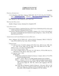
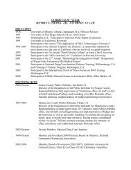
Goldsmith and Plunkett The American Journal <strong>of</strong> Forensic Medicine and Pathology • Volume 25, Number 2, June 2004FIGURE 4. Angular acceleration as afunction <strong>of</strong> angular velocity scaled toa 500 g brain mass, showing thresholds<strong>of</strong> concussion, SDH and DAI forboth shaking and impact. 44 (Reproducedwith permission from DuhaimeA-C, Gennarelli TA, ThibaultLE, et al. The shaken baby syndrome:a clinical, pathologic andbiomechanical study. J Neurosurg.1987;66:409–415.)loading and impact experiments were repeated with an infantdummy that had a deformable skull. 46,47 In <strong>the</strong>se studies, <strong>the</strong>ratio <strong>of</strong> peak impact acceleration to impulsive loading accelerationwas reduced from 46 to 39, as expected with adeformable skull model.The low load level from shaking was substantiated byexperiments in our Berkeley laboratory using an anthropometricdummy shaken by a variety <strong>of</strong> individuals. “Shaking”produced frequencies from 3–5 Hz, with associated totaldisplacements from 102–152 mm (4–6 in). If <strong>the</strong> excursionand frequency are modeled as ”simple harmonic motion“, 48 arepetitive movement at a uniform rate from an equilibriumposition with an amplitude, A, half (51–76 mm 2–3 in) <strong>of</strong><strong>the</strong> total displacement, <strong>the</strong> motion approximates an actual“shaking”. Using this model, <strong>the</strong> maximum linear acceleration,a max, A 2 , where is <strong>the</strong> angular velocity, was 7.7 g,slightly less than that found in <strong>the</strong> previous tests. 45–47 When<strong>the</strong> actual digital camera data ra<strong>the</strong>r than <strong>the</strong> model wereanalyzed, <strong>the</strong> result was 15 g, higher than <strong>the</strong> model because<strong>the</strong> real-time motion is not that <strong>of</strong> a rigid-body.Two sets <strong>of</strong> whiplash tests on anes<strong>the</strong>tized baboons,31,49 modeling a rear-end vehicular collision, causedacute SDH and spinal cord injuries with angular velocities92above 500 rad/s and angular accelerations <strong>of</strong> more than 10krad/s 2 . (10 krad/s 2 is 10,000 ft/s 2 , or 155 g’s, at a radius <strong>of</strong>1 foot.) These values are typical <strong>of</strong> head accelerations undervehicular impact conditions. The results <strong>of</strong> <strong>the</strong>se experimentshave been cited incorrectly 50–52 to support <strong>the</strong> mechanism forShaken Baby Syndrome (SBS), <strong>the</strong> authors apparently notunderstanding that <strong>the</strong> results replicated <strong>the</strong> effects <strong>of</strong> a 48.3km/h (30 mph) vehicular impact, not a 9.3 g “shake”.Scaling and impulsive loadingDifferent sized brains do not have <strong>the</strong> same injurythresholds. However, it is possible to convert or scale braininjury thresholds associated with impulsive loading frommany animals to humans. These species relationships arebased on an empirically verified equation, derived from<strong>the</strong>ory, where <strong>the</strong> ratio <strong>of</strong> <strong>the</strong> accelerations is equal to <strong>the</strong>inverse <strong>of</strong> <strong>the</strong> brain mass ratio raised to <strong>the</strong> 1/3 power forlinear acceleration and <strong>the</strong> 2/3 power for angular acceleration(Fig. 5). 53,54 Scaling assumes complete material property,structural, and geometric similarity between <strong>the</strong> various systemscompared. The head <strong>of</strong> an infant is smaller and geometricallyunlike that <strong>of</strong> an adult. 55–57 The structural properties <strong>of</strong><strong>the</strong> infant skull (ease <strong>of</strong> deformation and decreased threshold© 2004 Lippincott Williams & Wilkins
The American Journal <strong>of</strong> Forensic Medicine and Pathology • Volume 25, Number 2, June 2004TBI in Infants and ChildrenFIGURE 5. Concussion tolerance as a function <strong>of</strong> angularacceleration and brain mass. 52 (Modified and reproduced withpermission from Ommaya AK, Yarnell P, Hirsch AE, et al.Scaling <strong>of</strong> experimental data for cerebral concussion in subhumanprimates to concussive threshold for man. In: Proc 11 thStapp Car Crash Conf New York: SAE; 1967: 47–52. SAE Paper670906.)to fracture), and <strong>the</strong> mechanical properties <strong>of</strong> <strong>the</strong> infant brainare also different from those <strong>of</strong> an older child oradult. 5,46,58,59 These actual and potential differences betweenadult and infant brain structural and mechanical propertiesmean that impact-induced acceleration tolerances for nonhumanadult primates should not be scaled to human infants.Deformation and failure characteristics <strong>of</strong>objectsA SDH is usually caused by rupture <strong>of</strong> <strong>the</strong> parasagittalbridging veins. The mechanical and failure behavior <strong>of</strong> <strong>the</strong>sevessels have been determined by load tests that provide ei<strong>the</strong>rforce-deformation or stress-strain relations up to rupture.Unidirectional stress is force per unit area, F/A, whiledeformation (strain) may be through extension, compression,or “shear”. Ma<strong>the</strong>matically, uniaxial strain ( L/Lo)where L is <strong>the</strong> change in length and L o is <strong>the</strong> originallength. 8,48The behavior <strong>of</strong> all solid materials under loading conditionsis characterized ei<strong>the</strong>r by a relationship between stressand strain (and possibly time), a constitutive equation, or bya load-deformation curve, a structural designation. Solidmaterial behavior is characterized as elastic, or time-independentand reversible, when a substance subjected to a forceimmediately returns to its original shape upon unloadingalong <strong>the</strong> same path as <strong>the</strong> loading curve. The behavior isviscoelastic when internal energy dissipation significantlydelays <strong>the</strong> return <strong>of</strong> <strong>the</strong> deformation to its original state uponunloading and/or <strong>the</strong> unloading path is significantly below <strong>the</strong>loading curve. It is plastic when a substance (such as clay)retains a permanent deformation after load removal. Mostbiologic tissues, including brain, are viscoelastic and <strong>the</strong>irbehavior depends on <strong>the</strong> rate <strong>of</strong> load application. Only teeth,nails, and adult bone have nearly elastic behavior beforefailure.The critical load is that where mechanical (structural)failure is produced, although physiological (functional) disruptionmay occur at lower levels. The critical value is <strong>the</strong>ultimate strain, u , corresponding to an ultimate stress, u ,where <strong>the</strong>re is total mechanical disruption (fracture <strong>of</strong> <strong>the</strong>skull or rupture <strong>of</strong> <strong>the</strong> bridging veins). The likelihood <strong>of</strong>bridging vein rupture also depends on <strong>the</strong> orientation <strong>of</strong> <strong>the</strong>vessels to <strong>the</strong> superior sagittal sinus. Veins anterior to <strong>the</strong>midfrontal pole drain posteriorly to join <strong>the</strong> sinus in <strong>the</strong> samedirection as its flow, and are subject to maximum extensionstrain only with a frontal impact. Vessels at <strong>the</strong> midfrontalpole enter <strong>the</strong> sinus perpendicular to it. Veins posterior to <strong>the</strong>midfrontal pole drain anteriorly into <strong>the</strong> sinus, in <strong>the</strong> directionapposite its flow, and experience maximum extension strainwith an occipital impact. 10,11,60,61An example <strong>of</strong> a set <strong>of</strong> nonlinearly elastic relationships<strong>of</strong> vessels under slow loading conditions is shown inFigure 6. 62,63Differences in adult and infant skull propertiesand <strong>the</strong> response to impact and impulsiveloadsThe mechanical characteristics <strong>of</strong> <strong>the</strong> infant and adultskull are not <strong>the</strong> same. The adult skull is only slightlydeformable prior to fracture. The infant skull is not rigid, andshould be considered as a segmented unit <strong>of</strong> loosely associatedcurved plates with interspersed s<strong>of</strong>t membranes (suturesand fontanelles). 55,64FIGURE 6. Quasi-static stress versus stretch behavior for variousfresh cortical arteries and veins. 59 (Reproduced with permissionfrom Monson KL, Goldsmith W, Barbaro N, et al. Staticand dynamic mechanical and failure properties <strong>of</strong> humancerebral blood vessels. In: Crashworthiness, Occupant Protectionand Biomechanics in Transportation Systems. New York, ASME;2000. AMD 246/BED 49:255–65.)© 2004 Lippincott Williams & Wilkins 93
Goldsmith and Plunkett The American Journal <strong>of</strong> Forensic Medicine and Pathology • Volume 25, Number 2, June 2004Thibault and Margulies 5,6,65,66 compared <strong>the</strong> mechanicalproperties <strong>of</strong> <strong>the</strong> skulls <strong>of</strong> human infants and adults tothose <strong>of</strong> infant and adult pigs. They found significant variationin <strong>the</strong> failure stress <strong>of</strong> <strong>the</strong> skull <strong>of</strong> <strong>the</strong> neonate, <strong>the</strong> youngchild, and <strong>the</strong> adult, with an increase in stress to fracture <strong>of</strong>more than a factor <strong>of</strong> 10 from <strong>the</strong> newborn to <strong>the</strong> adult (Fig.7). The variation in <strong>the</strong> elastic modulus, E, showed a 10-foldincrease (Fig. 8), supporting nearly identical conclusionsfrom earlier tests. 40,67,68The Consumer Products Safety Commission (CPSC) 56has published a compendium on <strong>the</strong> geometric properties <strong>of</strong><strong>the</strong> infant head. An earlier work indicated substantial differencein <strong>the</strong> vasculature <strong>of</strong> infants and adults. 57 The neonateshad thinner and fewer vessels than <strong>the</strong> mature cortex, 69 <strong>the</strong>blood/brain barrier was incompletely developed, and <strong>the</strong>infant blood cerebral blood flow and oxygen consumptionwere more than twice that in adults.These structural differences cause <strong>the</strong> injury mechanismfor an infant to be fundamentally different from that <strong>of</strong>an older child or adult. Impact loading <strong>of</strong> <strong>the</strong> compliant infantskull/brain unit produces potentially damaging levels <strong>of</strong> strainwithin <strong>the</strong> entire structure. Deformation, not impact-inducedangular acceleration, is <strong>the</strong> critical factor. Nonimpact loading(“shaking”) may result in strains <strong>of</strong> <strong>the</strong> neurovascular structures,but <strong>the</strong>re is no associated skull deformation. <strong>Biomechanical</strong>studies have demonstrated that <strong>the</strong> head accelerationsgenerated by shaking are below <strong>the</strong> thresholds forFIGURE 7. Skull failure stress for <strong>the</strong> neonate, young child andadult. 8 (Reproduced with permission from Ommaya AK,Goldsmith W, Thibault L. Biomechanics and neuropathology<strong>of</strong> adult and pediatric head injury. Br J Neurosurg.2002;16: 220–242.)94FIGURE 8. Elastic modulus <strong>of</strong> human cranial bone versus age.(Created by Kirk L. Thibault, PhD, from data in <strong>the</strong> studiesreferenced.)traumatic DAI, SDH, and even concussion. If one could“shake” an infant or a child, or an adult for that matter,vigorously enough to cause traumatic DAI or SDH, <strong>the</strong>re willbe significant structural neck damage, including <strong>the</strong> craniocervicaljunction (F.A. Bandak, personal communication).70–71 Shaking simply does not generate accelerationsthat exceed any known injury tolerance values for traumaticbrain injury.The material and structural properties <strong>of</strong> <strong>the</strong> skull andbrain are age-dependant. The transition from <strong>the</strong> predominatelydeformation-mediated impact response in an infant toa predominately acceleration (impulsive)-mediated responsein an older child or adult is a continuum. It is not possible,with today’s data, to state a specific age at which <strong>the</strong> finalchange occurs. In vivo and surrogate biomechanical experimentsto fur<strong>the</strong>r define <strong>the</strong> parameters <strong>of</strong> age-dependencymust be assisted by various imaging technologies includingMRI, a careful neurologic examination for signs <strong>of</strong> cervicalspinal cord or brain stem injury in a child who is living, anda complete and careful postmortem examination includingposterior neck dissection if death occurs.<strong>Analysis</strong> <strong>of</strong> impacts due to fallsThe linear impulse-momentum relation derived fromNewton’s Second law applied over time can be used toanalyze an impact caused by a fall. The relationship statesthat <strong>the</strong> area under <strong>the</strong> force-time (F-t) curve is equal to <strong>the</strong>change <strong>of</strong> linear momentum; in practice, <strong>the</strong> product <strong>of</strong> mass,m, times <strong>the</strong> change <strong>of</strong> velocity, v. 19,48 For contact events,<strong>the</strong> maximum force generated can be closely approximatedby <strong>the</strong> expression F max 2mv/, where is <strong>the</strong> duration <strong>of</strong>contact. 72 If a 0.91 m (3 ft) tall child falls by backwardrotation about <strong>the</strong> soles <strong>of</strong> his/her feet, <strong>the</strong> translationalimpact velocity is 4.24 m/s (13.9 ft/s) at <strong>the</strong> occiput, and <strong>the</strong>angular impact velocity is 4.63 rad/s. Our Berkeley laboratory© 2004 Lippincott Williams & Wilkins
The American Journal <strong>of</strong> Forensic Medicine and Pathology • Volume 25, Number 2, June 2004TBI in Infants and Childrendetermined that contact duration is 3–7 milliseconds bydropping a weight-adjusted cadaver skull onto <strong>the</strong> edge <strong>of</strong> ahardwood stair step. If only <strong>the</strong> impact <strong>of</strong> <strong>the</strong> head produces<strong>the</strong> contact force (<strong>the</strong> weight <strong>of</strong> <strong>the</strong> body does not contribute),<strong>the</strong> contact time is taken as 5 milliseconds and <strong>the</strong> headweighs 22.4 Newtons (N) (5.3 lb), 20% <strong>of</strong> <strong>the</strong> body weight <strong>of</strong>112 N (26.4 lb), <strong>the</strong> peak force is 4070 N (915 lb) from <strong>the</strong>linear impulse-momentum relation, assuming a triangularforce pr<strong>of</strong>ile. This is a peak acceleration <strong>of</strong> 173 g, nearlysixteen times greater than would be obtained by shaking andtwice as great as <strong>the</strong> tolerance limit suggested by Stürtz. 39These linear acceleration values approach those obtainedfrom <strong>the</strong> previously described actual experiments; 45–47 <strong>the</strong>small differences are most likely due to variation in surfacecharacteristics or a conservative assumption for <strong>the</strong> contacttime in <strong>the</strong> above analysis.There has been sworn testimony in courts <strong>of</strong> law byexpert witnesses who state that trauma caused by shaking isequivalent to a fall from a two-story (or higher) window onto<strong>the</strong> pavement. 73 If such a fall is head first with <strong>the</strong> body in avertical or near vertical position at impact, <strong>the</strong> forces on <strong>the</strong>head can be calculated from <strong>the</strong> linear impulse-momentumlaw. Assuming that <strong>the</strong> weight <strong>of</strong> <strong>the</strong> child is 66.7 N (15 lb),that <strong>the</strong> fall height is 5.63 m (18 ft), and that <strong>the</strong> contactduration is a very conservative 10 milliseconds, <strong>the</strong> impactvelocity will be 10.37 m/s (34 ft/s), and <strong>the</strong> peak contact force14,190 N (3167 lb). This exceeds by at least an order <strong>of</strong>magnitude <strong>the</strong> force that can be exerted by shaking, and atleast 3 times that considered from <strong>the</strong> data <strong>of</strong> Gurdjian 30 as<strong>the</strong> human tolerance limit. The analogy <strong>of</strong> “shaking” injuryto that from a two-story fall is not justifiable.It has also been stated that falls from low heights do notproduce significant injury or death. 74–76 However, Mohan etal 77 analyzed 30 falls in children under <strong>the</strong> age <strong>of</strong> 10, andreconstructed 6 <strong>of</strong> <strong>the</strong>m using <strong>the</strong> MVMA Two-DimensionalCrash Victim Simulator computer model. The authors concludedthat falls as low as 2 meters may cause serious injuryor death.Plunkett documented 18 fatal falls from heights <strong>of</strong> 0.6to3m(2–10 ft). 24 One <strong>of</strong> <strong>the</strong>se falls was coincidentallyvideotaped, permitting an approximate analysis <strong>of</strong> <strong>the</strong> contactloads. A 23-month-old girl who weighed 129 N (29 lb) andwas 0.838 m (33 in) tall fell from a play structure onto a 10mm (0.39 in) thick carpet without foam backing covering aconcrete floor. She had lost her grip on a rail when she wasin a nearly horizontal position approximately 1.07 m (42 in)above <strong>the</strong> ground. She struck <strong>the</strong> ground first with heroutstretched hands and <strong>the</strong>n with her right upper frontalforehead, followed by her right shoulder (Fig. 9). Althoughalert immediately after <strong>the</strong> fall, she became unconsciouswithin approximately 5 minutes. She was taken to a tertiarycare hospital where a 100 mL right-sided SDH was immediatelyevacuated. The attending medical staff documentedFIGURE 9. Idealized contact position and loads in <strong>the</strong> fall <strong>of</strong> a29 lb (12.9K) child from a height <strong>of</strong> 42 in (1.07 m).extensive bilateral retinal hemorrhage (RH) following <strong>the</strong>surgery. Postoperatively, she developed refractory cerebraledema and was removed from life support after 1.5 days. Anautopsy showed a small residual right SDH, cerebral edemawith herniation, and a 40 45 mm (1.58 1.77 in) rightfrontal scalp contusion. There was no skull fracture.Her unsupported weight caused rotation about her feetuntil her body was at an angle <strong>of</strong> approximately 45° from <strong>the</strong>horizontal, with her head and neck inclined at approximately30° to <strong>the</strong> horizontal. Her feet <strong>the</strong>n disengaged <strong>the</strong> playstructure and she fell freely with her center <strong>of</strong> gravity dropping0.483 m (19 in) until she struck <strong>the</strong> carpet. If <strong>the</strong> moment<strong>of</strong> inertia (<strong>the</strong> property <strong>of</strong> an object to resist rotation) taken at<strong>the</strong> center <strong>of</strong> mass, G, is idealized as a uniform cylinder witha radius <strong>of</strong> 101.6 mm (4 in), her moment <strong>of</strong> inertia about herfeet is 3.116 N-m-s 2 (27.57 lb-in-s 2 ). 48 The impact velocitydue to rotation, using a number <strong>of</strong> simplifying assumptions, is4.36 m/s (14.3 ft/s) whose vertical component is 3.78 m/s(12.4 ft/s). The concurrent velocity due to <strong>the</strong> vertical motion<strong>of</strong> <strong>the</strong> center <strong>of</strong> gravity in <strong>the</strong> fall is 3.05 m/s (10 ft/s), so that<strong>the</strong> impact velocity <strong>of</strong> <strong>the</strong> head is 6.83 m/s (22.4 ft/s)(compared with 4.57 m/s 15 ft/s if <strong>the</strong> body remainedentirely horizontal). This calculation ignores <strong>the</strong> brakingeffect <strong>of</strong> <strong>the</strong> outstretched arms, so that <strong>the</strong> likely impact speed<strong>of</strong> <strong>the</strong> head was <strong>of</strong> <strong>the</strong> order <strong>of</strong> 4.57–6.10 m/s (15–20 ft/s).Since <strong>the</strong> skull, scalp and carpet were not available tomeasure compliance, <strong>the</strong> contact duration will be conservativelyassumed to be 10 milliseconds. The child’s weightdistribution will be assumed to be 107 N (24 lb) for torso,© 2004 Lippincott Williams & Wilkins 95
Goldsmith and Plunkett The American Journal <strong>of</strong> Forensic Medicine and Pathology • Volume 25, Number 2, June 20049. Goldsmith W. The state <strong>of</strong> head injury biomechanics – past, presentand future. Part 1. Crit Rev Biomed Engng. 2001;29:441–600.10. Lõwenhielm P. Dynamic properties <strong>of</strong> parasagittal bridging veins. ZRechtsmed. 1974;74:55–62.11. Lee MH, Haut RC. Insensitivity and tensile breakdown properties <strong>of</strong><strong>the</strong> human parasagittal bridging veins to strain rate dependence inbiomechanics <strong>of</strong> subdural hematoma. J Biom. 1989;22:532–542.12. Ommaya AK, Thibault LE, Bandak F. Mechanisms <strong>of</strong> head injury. IntJ Impact Eng. 1994;15:525–560.13. Ommaya AK, Corrao P, Letcher PS. Head injury in <strong>the</strong> chimpanzee:biodynamics <strong>of</strong> traumatic unconsciousness. J Neurosurg. 1963;39:152–166.14. Prange MT, Margulies SS. Regional, directional and age-dependentproperties <strong>of</strong> <strong>the</strong> brain using physical models to determine corticalstrains in <strong>the</strong> head during dynamic loading. In: Proc Ann Intern ConfIEEE Engng in Med and Bio Soc. New York: IEEE. 1989;3:816–817.15. Arbogast KB, Margulies SS. Regional differences in mechanical properties<strong>of</strong> <strong>the</strong> porcine central nervous system. In: Proc 42 nd Stapp CarCrash Conf 1997. Warrendale, PA: Society <strong>of</strong> Automotive Engineers.SAE Paper 973336:293–300.16. Kleiven S. Influence <strong>of</strong> impact direction on <strong>the</strong> human head in prediction<strong>of</strong> subdural hematoma. J Neurotr. 2003;20:365–379.17. Gennarelli TA, Thibault LE, Adams JH, et al. . Diffuse axonal injuryand traumatic coma in <strong>the</strong> primate. Ann Neurol. 1982;12:264–274.18. Smith SL, Andrus PK, Gleason DD, et al. Infant rat model <strong>of</strong> <strong>the</strong>shaken baby syndrome: preliminary characterization and evidence for<strong>the</strong> role <strong>of</strong> free radicals in cortical hemorrhaging and progressiveneuronal degeneration. J Neurotr. 1998;15:693–705.19. Goldsmith W. Impact: The Physical Theory and Behaviour <strong>of</strong> CollidingSolids. London: Edward Arnold Ltd.; 1960. Mineola, NY: DoverPublications; 2001.20. Ommaya AK, Grubb RL, Naumann RA. Coup and contre-coup injury:Observations on <strong>the</strong> mechanics <strong>of</strong> visible brain injuries in <strong>the</strong> RhesusMonkey. J Neurosurg. 1971;35:503–516.21. Gross AG. Impact thresholds and brain concussion. Aviation Med.1958;29:725–732.22. Lubock P, Goldsmith W. Experimental cavitation studies in a modelhead-neck system. J Biom. 1980;13:1041–1052.23. Nusholtz GS, Glascoe LG, Wylie EB. Cavitation during head impact.In: Occupant Protection and Injury Assessment in <strong>the</strong> AutomotiveCrash Environment, SP 1231. Warendale, PA: Society <strong>of</strong> AutomotiveEngineers; 1997:151:–623.24. Plunkett J. Fatal pediatric head injuries caused by short distance falls.Am J Forens Med Pathol. 2001;22:1–12.25. Goldsmith W. Fatal pediatric head injuries caused by short distancefalls (letter). Am J Forens Med Pathol 2001;22:334–336.26. Plunkett J. Fatal pediatric head injuries caused by short distance falls(letter). Am J Forens Med Pathol 2002;23:103–105.27. Hess RL, Weber K, Melvin JW. A review <strong>of</strong> research on head impacttolerance and injury criteria related to occupant protection. In: Headand Neck Injury Criteria: A Consensus Workshop. Washington, DC: US Government Printing Office; 1983. (DOT HS 806 434):183–194.28. American National Standards Institute. American National Standardfor Protective Headgear – for Bicyclists. Washington, DC: AmericanNational Standards Institute; April 1984. ANSI Z90.29. Snell Memorial Foundation. Standards for Protective Headgear forUse With Bicycles. Child Helmet. Addendum for CPS Requirements.North Highlands, CA: Snell Memorial Foundation; 1995.30. Goldsmith W. Meaningful concepts <strong>of</strong> head injury. In: Proc 1989International Research Council on <strong>the</strong> Biomechanics <strong>of</strong> Impact Conf.Bron, France: IRCOBI. 1989;1:–12.31. Ommaya AK, Hirsch AE. Tolerances for cerebral concussion fromhead impact and whiplash in primates. J Biom. 1971;4:13–21.32. Gennarelli TA, Thibault LE. Biomechanics <strong>of</strong> acute subdural hematoma.J Trauma. 1982;22:680–686.33. Gurdjian ES, Lissner HR, Patrick LM. Protection <strong>of</strong> <strong>the</strong> head and neckin sports. JAMA. 1962;182:509–512.34. Gurdjian ES. Impact Head Injury: Mechanistic, Clinical, and PreventiveCorrelations. Springfield, IL: CC Thomas; 1975.9835. Ono K, Kikushi A, Nakamura M, et al. Human head tolerance insagittal impact reliable estimation deduced from experimental headinjury using subhuman primates and human cadaver skulls. In: Proc24 th Stapp Car Crash Conf. Warrendale, PA: Society <strong>of</strong> AutomotiveEngineers; 1980:101:–160. SAE Paper 801303.36. Sturgess CE. A <strong>the</strong>rmomechanical <strong>the</strong>ory <strong>of</strong> impact trauma. In: ProcImechE J Automobile Div. London: Institution <strong>of</strong> Mechanical Engineers;2003: In press.37. Newman JA. A generalized model for brain injury threshold. In: Proc1986 International Research Council on <strong>the</strong> Biomechanics <strong>of</strong> ImpactConf. Bron, France: IRCOBI; 1986: 121:–131.38. Stürtz G. Correlation <strong>of</strong> dummy-loadings with real injuries <strong>of</strong> childrenby repetition tests. In: Proc V th International IRCOBI Conf. Bron,France: IRCOBI; 1980:194:–208.39. Stürtz G. <strong>Biomechanical</strong> data <strong>of</strong> children. In: Proc 24 th Stapp CarCrash Conf. Warrendale, PA: Society <strong>of</strong> Automotive Engineers; 1980.(SAE Paper 801313):525–59.40. Melvin JW. Injury assessment reference values for <strong>the</strong> CRABI-6 monthinfant dummy in a rear-facing infant restraint with airbag deployment.SAE J for Passenger Cars. Warrendale, PA: Society <strong>of</strong> AutomotiveEngineers; 1995. SAE Paper 950872:1553–64.41. Irwin A, Mertz HJ. <strong>Biomechanical</strong> basis for <strong>the</strong> CRABI and Hybrid IIIchild dummies. In: Proc 41 st Stapp Car Crash Conf. Warrendale, PA:Society <strong>of</strong> Automotive Engineers; 1995. (SAE Paper 973317):261–72.42. Klinich KD, Hulbert GM, Schneider LW. Estimating infant head injurycriteria and impact response using crash reconstruction and finiteelement modeling. Stapp Car Crash J. 2002;46: Paper O2S-32.43. Lucchini E, Weissner R. Difference between <strong>the</strong> kinematics and loadings<strong>of</strong> impacted adults and children: Results from dummy tests. In:Proc 1980 International Research Council on <strong>the</strong> Biomechanics <strong>of</strong>Impact Conf. Bron, France: IRCOBI; 1980:165:–78.44. Stcherbatcheff G, Tarrière C, Duclos P, et al. Simulation <strong>of</strong> collisionsbetween pedestrians and vehicles using adult and child dummies. In:Proc 19 th Stapp Car Crash Conf. Warrendale, PA: Society <strong>of</strong> AutomotiveEngineers; 1975. (SAE Paper 751167):147–53.45. Duhaime A-C, Gennarelli TA, Thibault LE, et al. The shaken babysyndrome: a clinical, pathological and biomechanical study. J Neurosurg.1987;66:409–415.46. Prange MT, Coats B, Raghupathi R, et al. Rotational loads duringinflicted and accidental infant head injury. J Neurotr 2001;Abst.D8;18:1142.47. Prange MT, Coats B, Duhaime A-C, et al. Anthropomorphic simulations<strong>of</strong> falls, shakes, and inflicted impacts in infants. J Neurosurg.2003;99:143–150.48. Berger SA, Goldsmith W, Lewis ER, eds. Introduction to Bioengineering.Oxford: Oxford University Press; 1996, 2000.49. Ommaya AK. Trauma to <strong>the</strong> nervous system. Ann Roy Coll Surg Engl.1966;39:317–347.50. Caffey J. The whiplash shaken infant syndrome: manual shaking by <strong>the</strong>extremities with whiplash-induced intracranial and intraocular bleedings,linked with residual permanent brain damage and mental retardation.Pediatrics. 1974;54:396–403.51. Guthkelch AN. Infantile subdural hematoma and its relationship towhiplash injuries. Brit Med J. 1971;2:430–431.52. Lonergan GJ, Baker AM, Morey MK, et al. Child abuse: Radiologicpathologiccorrelation. Radiographics. 2003;23:811–845.53. Ommaya AK, Yarnell P, Hirsch AE, et al. Scaling <strong>of</strong> experimental datafor cerebral concussion in sub-human primates to concussive thresholdfor man. In: Proc. 11 th Stapp Car Crash Conf. New York: Society <strong>of</strong>Automotive Engineers; 1967. (SAE Paper 670906):47–52.54. Ljung C. On scaling in head injury research. In: Proc V th InternIRCOBI Conf on <strong>the</strong> Biomechanics <strong>of</strong> Impacts. Bron, France: IRCOBI;Sept. 9–11, 1980; 209–217.55. Netter FH. Atlas <strong>of</strong> Human Anatomy. Summit, NJ: Ciba-Geigy Corp;1989.56. Schneider LW, Lehman RJ, Flug M, et al. Size and Shape <strong>of</strong> <strong>the</strong> HeadFrom Birth to Four Years. Final Report to <strong>the</strong> Consumer ProductsSafety Commission. Washington, DC: Univ <strong>of</strong> Michigan Transp ResInst 1986; UMTRI-86-2.© 2004 Lippincott Williams & Wilkins
The American Journal <strong>of</strong> Forensic Medicine and Pathology • Volume 25, Number 2, June 2004TBI in Infants and Children57. Stehbens WE. Pathology <strong>of</strong> <strong>the</strong> Cerebral Blood Vessels. St. Louis:Mosby;1972.58. Thibault KL, Margulies SS. Age-dependant material properties <strong>of</strong> <strong>the</strong>porcine cerebrum: Effect on pediatric inertial head injury criteria.J Biomech. 1998;31:1119–1126.59. Prange MT, Margulies SS. Regional, directional, and age-dependantproperties <strong>of</strong> <strong>the</strong> brain undergoing large deformations. Trans ASME.2002;124:244–252.60. Oka K, Rhoton AJ, Barry M, et al. Microsurgical anatomy <strong>of</strong> <strong>the</strong>superficial veins <strong>of</strong> <strong>the</strong> cerebrum. Neurosurg. 1985;17:711–748.61. Huang HM, Lee MC, Chiu WT, et al. Three-dimensional finite elementanalysis <strong>of</strong> subdural hematoma. J Trauma. 1999;47:538–544.62. Monson KL, Goldsmith W, Barbaro N, et al. Static and dynamicmechanical and failure properties <strong>of</strong> human cerebral blood vessels. In:Crashworthiness, Occupant Protection and Biomechanics in TransportationSystems. New York: ASME; 2000. AMD 246/BED 49:255–65.63. Monson KL, Goldsmith W, Barbaro N, et al. Axial mechanical properties<strong>of</strong> fresh human cerebral blood vessels. J Biom Engng. 2003;125:288–294.64. Sobatta J. Atlas <strong>of</strong> Human Anatomy 1927–1928. Edited from <strong>the</strong> 6 thGerman edition by JP McMurrich, 1 and 3. New York: GE Stechert;1927–28.65. Margulies SS, Thibault LE. An analytical model <strong>of</strong> traumatic diffusebrain injury. J Biom Eng. 1989;111:241–249.66. Thibault KL. Pediatric Head Injuries: <strong>the</strong> Influence <strong>of</strong> Brain and SkullProperties dissertation. Philadelphia: University <strong>of</strong> Pennsylvania;1997.67. Hubbard RP. Flexure <strong>of</strong> layered cranial bone. J Biomech. 1971;4:251–263.68. Runge CF, Yousses A, Thibault KL. Material properties <strong>of</strong> humaninfant skull and suture: experiments and numerical analysis. In: InjuryPrevention Through Biomechanics Symposium, National Center forInjury Prevention and Control. Detroit: Centers for Disease Control;1998. Report 1–1998.69. McMinn RMH, Hutchings RT, eds. Color Atlas <strong>of</strong> Human Anatomy.Chicago: Year Book Med Publ, Inc.; 1977.70. Duncan M. Laboratory note: On <strong>the</strong> tensile strength <strong>of</strong> <strong>the</strong> fresh adultfœtus. BMJ 1874;2:763–764.71. Mertz HJ, Patrick LM. Strength and response <strong>of</strong> <strong>the</strong> human neck. In:Proc <strong>of</strong> <strong>the</strong> 15 th Stapp Car Crash Conference. Warrendale, PA: Society<strong>of</strong> Automotive Engineers; 1971. SAE Paper 710855:2903–28.72. Goldsmith W, Kabo JM. Performance <strong>of</strong> baseball caps. Am J SportsMed. 1982;10:31–37.73. 2001 WL 1630083 (Colo. App.). Colorado Court <strong>of</strong> Appeals, Div. 1.No. 00CA0312.74. Chadwick DL, Chin S, Salerno C, et al. Deaths from falls in children:How far is fatal? J Trauma. 1991;31:1353–1355.75. Williams RA. Injuries in infants and small children resulting fromwitnessed and corroborated free falls. J Trauma. 1991;31:1350–1352.76. Lyons TJ, Oates RK. Falling out <strong>of</strong> bed: A relatively benign occurrence.Pediatrics. 1993;92:125–127.77. Mohan D, Bowman BM, Snyder RG, et al. A biomechanical analysis <strong>of</strong>head impact injuries to children. J Biom Engng. 1979;101:250–260.78. Zhang L, Bae J, Hardy WN, et al. Computational study <strong>of</strong> <strong>the</strong> contribution<strong>of</strong> <strong>the</strong> vasculature on <strong>the</strong> dynamic response <strong>of</strong> <strong>the</strong> brain. StappCar Crash J. 2002;46:145–163.79. Geddes JF, Hackshaw AK, Vowles GH, et al. Neuropathology <strong>of</strong>inflicted head injury in children. I. Patterns <strong>of</strong> brain damage. Brain.2001;124:1290–1298.80. Geddes JF, Vowles GH, Hackshaw AK, et al. Neuropathology <strong>of</strong>inflicted head injury in children, II: Microscopic brain injury in infants.Brain. 2001;124:1299–1306.81. Shannon B, Becker L. Mechanisms <strong>of</strong> brain injury in infantile childabuse. The Lancet. 2001;358:686–687.82. Wilkins B. Head injury – abuse or accident. Commentary by SunderlandR. Arch Dis Child. 1997;76:393–397.83. Greenwald MJ. The shaken baby syndrome. Semin Ophthalmol. 1990;5:202–215.84. Am Acad Ophthalmol. Shaken Baby Syndrome Resources. Availableat: http://www.aao.org/aao/education/library/shaken_baby.cfm-ocular.Date accessed: April 22, 2004.85. Morad Y, Kim YM, Armstrong DC, et al. Correlation between retinalabnormalities and intracranial abnormalities in <strong>the</strong> Shaken Baby Syndrome.Am J Ophthalmol. 2002;134:354–359.86. Aoki N, Masuzawa H. Infantile acute subdural hematoma: clinicalanalysis <strong>of</strong> 26 cases. J Neurosurg. 1984;51:273–280.87. Muller PJ, Deck JHN. Intraocular and optic nerve sheath hemorrhage incases <strong>of</strong> sudden intracranial hypertension. J Neurosurg. 1974;41:160–166.88. Tongue AC. The ophthalmologist’s role in diagnosing child abuse.Ophthalmol. 1991;98:1009–1010.89. Avery GB, Fletcher MA, McDonald MG. Neonatology, 4 th ed. Philadelphia:Lippincott; 1994: 1146.90. Edlow JA, Caplan LR. Avoiding pitfalls in <strong>the</strong> diagnosis <strong>of</strong> subarachnoidhemorrhage. NEJM. 2000;342:29–36.91. Tomasi LD, Rosman NP. Purtscher retinopathy in battered childsyndrome. Am J Dis Child. 1975;129:1335–1337.92. David DB, Mears T, Quinlan MP. Ocular complications associatedwith bungee jumping. Br J Ophthalmol. 1994;78:234–235.93. Bain BK, Talbot EM. Bungee jumping and intraocular hemorrhage.Br J Ophthalmol. 1994;78:236–237.94. Rooms L, Fitzgerald N, McClain KL. Hemophagocytic lymphohistiocytosismasquerading as child abuse: presentation <strong>of</strong> three cases andreview <strong>of</strong> central nervous system findings in hemophagocytic lymphohistiocytosis.Pediatr. 2003;111:e636–e640.95. Biousse V, Rucker JC, Vignal C, et al. Anemia and papilledema. Am JOphthalmol. 2003;135:437–446.96. Medele RJ, Stummer W, Mueller AJ, et al. Terson’s syndrome insubarachnoid hemorrhage and severe brain injury accompanied byacutely raised intracranial pressure. J Neurosurg 1998;88:851–854.97. Lantz PE, Sinal SH, Stanton CA, et al. Evidence-based case report.Perimacular retinal folds from childhood head trauma. BMJ. 2004;328:754–756.98. Bergstrom M, Ericson K, Levander B, et al. Computed tomography <strong>of</strong>cranial subdural and epidural hematomas: Variation <strong>of</strong> attenuationrelated to time and clinical events such as rebleeding. J Comp AssistTomogr. 1977;1:449–455.99. Thibault LE. Brain injury from <strong>the</strong> macro to <strong>the</strong> micro level and backagain: what have we learned to date? In: Proc 1993 Intern Conf onBiomechanics <strong>of</strong> Impacts. Bron, France: IRCOBI; 1993: 3:–25.100. Thibault LE, Gennarelli TA, Tipton HW, et al. The physiologicresponse <strong>of</strong> isolated nerve tissue to dynamic loading. ACEMB. 1974;16:176.101. Bain A, Meaney DF. Thresholds for mechanical injury to <strong>the</strong> in vivowhite matter. In: Proc 43 rd Stapp Car Crash Conf. Warrendale, PA:Society <strong>of</strong> Automotive Engineers; SAE 1999. Paper 99SC19.102. Barbee KA, Macarak EJ, Thibault LE. Strain measurement in culturedvascular smooth muscle cells subjected to mechanical deformation.Ann Biomed Engng. 1994;23:14–22.103. Ueno K, Melvin JW, Li L, et al. Development <strong>of</strong> a tissue level braininjury criteria by finite element analysis. In: Bandak FA, Eppinger RH,Ommaya AK, eds. <strong>Traumatic</strong> Brain Injury: Bioscience and Mechanics.Larchmont, NY, Mary Ann Liebert; 1996:155–66.104. Saatman KE, Thibault LE. Rapid elongation <strong>of</strong> a myelinated fiber: amodel <strong>of</strong> head injury. Proc Annual Intern Conf IEEE Engng in MedBiol Soc. New York: IEEE; 1989;5:1663–1664.105. Galbraith JA, Thibault LE, Matteson DR. Mechanical and electricalresponses <strong>of</strong> <strong>the</strong> giant squid axon to simple elongation. J Biom Engng.1993;115:15–22.106. Thibault LE, Gennarelli TA. Biomechanics <strong>of</strong> diffuse brain injury. In:Proc. 10 th Intern. Conf on Experimental Safety Vehicles Warrendale, PA:Society <strong>of</strong> Automotive Engineers PT 43; 1985. SAE Paper 856022.© 2004 Lippincott Williams & Wilkins 99
Goldsmith and Plunkett The American Journal <strong>of</strong> Forensic Medicine and Pathology • Volume 25, Number 2, June 2004Glossary1. Acceleration, a – The rate <strong>of</strong> change <strong>of</strong> velocity.2. Angular acceleration, – The rate <strong>of</strong> change <strong>of</strong> angular velocity. Theacceleration <strong>of</strong> a rigid body about a fixed axis may be resolved into twocomponents, tangential (a t r) and normal (a n r 2 ) to <strong>the</strong> radius <strong>of</strong>rotation, r.3. Angular velocity, - The rate <strong>of</strong> change <strong>of</strong> angular position. The linearvelocity associated with angular motion is normal to <strong>the</strong> radius <strong>of</strong> <strong>the</strong>motion (v r).4. Anthropometric – Accurately mimics actual human dimensions.5. Bi<strong>of</strong>idelic – Accurately mimics actual human properties.6. Coronal plane – Side-to-side plane.7. Constitutive equation – The relationship between stress and strain, and<strong>the</strong>ir rates, that defines <strong>the</strong> behavior <strong>of</strong> a solid under loading.8. Couple – A set <strong>of</strong> equal and oppositely directed but non-colinear forces.9. Density – Mass per unit volume.10. Displacement – The distance that an object moves.11. Elastic modulus – The ratio <strong>of</strong> stress to strain in a linear stress-straincurve.12. Failure – A stress or strain that does not allow a return to <strong>the</strong> normalphysical state.13. Finite Element Method – The numerical, approximate solution <strong>of</strong> problemsin mechanics by subdividing <strong>the</strong> spatial and temporal fields intosmall finite parts using <strong>the</strong> assigned properties <strong>of</strong> <strong>the</strong> system. Thevalidity <strong>of</strong> <strong>the</strong> solution depends on <strong>the</strong> validity <strong>of</strong> <strong>the</strong> input properties <strong>of</strong><strong>the</strong> system.14. Force, F – Mass times acceleration.15. Impulsive loading – Motion at a distance from an applied force.16. Impact – The collision <strong>of</strong> two solid objects resulting in demonstrabledeformation, and propagation <strong>of</strong> a signal away from <strong>the</strong> point <strong>of</strong> contact.17. Kilogram, kg – The unit <strong>of</strong> mass in <strong>the</strong> metric system.18. Linear acceleration – Acceleration along a straight line.19. Load – A force or moment that produces motion or deformation.20. Mass, m – The property <strong>of</strong> an object that resists a change in velocity.21. MVMA – Motor Vehicle Motion <strong>Analysis</strong>, a numerical method developedat <strong>the</strong> University <strong>of</strong> Michigan to examine <strong>the</strong> motion <strong>of</strong> vehicleoccupants subsequent to impact.22. Newton, N – The unit <strong>of</strong> force in <strong>the</strong> metric (kms) system. For purposes<strong>of</strong> this article, <strong>the</strong> Newton is used as an equivalent unit for mass to avoidconverting <strong>the</strong> English unit <strong>of</strong> force (Pound) to <strong>the</strong> English unit for mass(slug).23. Particle – A point in space that has mass.24. Pascal, Pa – The unit <strong>of</strong> stress, force/unit area, in <strong>the</strong> metric system. OnePa one N/m 2 ; 1kPa 1000Pa; 1 MPa 1,000,000 Pa.25. Pound, lb – The unit <strong>of</strong> force in <strong>the</strong> English (sfs) system.26. Quasi-static – Extremely slow rate <strong>of</strong> loading, for example, catchingone’s leg in an elevator door, or a finger in a vise.27. Radian, rad – The unit <strong>of</strong> angular displacement. A radian is <strong>the</strong> radius <strong>of</strong>a circle stretched over its circumference. There are 2 rad in a circle, andeach rad subtends an angle <strong>of</strong> 57.3 degrees. One krad 1000 rad.28. Sagittal plane – Anterior-posterior or posterior-anterior plane.29. Scaling – Extrapolating <strong>the</strong> results from one set <strong>of</strong> experiments toano<strong>the</strong>r system based on size.30. Slug, s – The English unit <strong>of</strong> mass. One slug weight (in pounds)/32.2at sea level.31. Stress, - Force per unit area.32. Strain, – Change in length per unit reference length. Strain may beelongation, compression, or shear.33. Ultimate stress – The stress at which a material fails structurally,associated with <strong>the</strong> largest load prior to failure.34. Velocity, v – The rate <strong>of</strong> change <strong>of</strong> displacement.100© 2004 Lippincott Williams & Wilkins