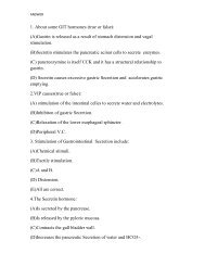
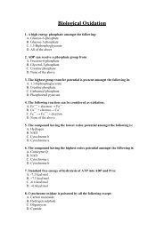
BIOLOGICAL OXIDATION
BIOLOGICAL OXIDATION
BIOLOGICAL OXIDATION

Create successful ePaper yourself
Turn your PDF publications into a flip-book with our unique Google optimized e-Paper software.
Redox potentialIt is the affinity of a substance to accept electrons i.e. it is thepotential for a substance to become reduced.Hydrogen has the lowest redox potential (-0.42 volt), whileoxygen has the highest redox potential (+0.82 volt). The redoxpotentials of all other substances lie between that of hydrogen andoxygen.Electrons are transferred from substances with low redoxpotential to substances with higher redox potential. This transferof electrons is an energy yielding process and the amount ofenergy liberated depends on the redox potential differencebetween the electron donor and acceptor.
1. Oxidases½AH 2AH 2O 2 O 2OxidaseUricaseAH 2 OAH 2 O 2
3. Anaerobic DehydrogenasesAH 2OxidizedCarrier 1ReducedCarrier 2Oxidized Carrier 3H 2 OAerobicAdehydrogenaseReducedCarrier 1OxidizedCarrier 2ReducedCarrier 3½ O 2
They can use artificial substrate (dye) as hydrogen acceeptor.OxidizedReducedCarrier 1Carrier 2OxidizedCarrier 3ReduceddyeAH 2AAerobicdehydrogenaseReducedcarrier 1OxidizedCarrier 2Reducedcarrier3Dye
•.Anaerobic dehydrogenases are further classified according to their coenzymes into:•NAD + linked anaerobic dehydrogenases e.g.a)Cytoplasmic glycerol-3-phosphate dehydrogenaseb)Isocitrate dehydrogenase.c)Malate dehydrogenase.d)β- Hydroxy acyl CoA dehydrogenase.e)β- Hydroxy butyrate dehydrogenase.•NADP linked anaerobic dehydrogenases e.g.a)Glucose-6-phosphate dehydrogenase.b)Malic enzyme.c)Cytoplasmic isocitrate dehydrogenase•FAD linked anaerobic dehydrogenases e.g.a)Succinate dehydrogenase.b)Mitochondrial glycerol-3-phosphate dehydrogenase.c)Acy1 CoA dehydrogenase.
•Cytochromes :-All cytochromes are anaerobic dehydrogenases except cytochrome oxidase(cyt a3), which is an oxidase and cytochrome P450 that is mono-oxygenase(hydroxylase).•Ubiquinol (coenzyme Q) dehydrogenase, which ispresent in the respiratory chain.
4. HydroperoxidasesThese enzymes use hydrogen peroxide (H 2O 2) as substrate changing it intowater to get rid of its harmful effects.They are further classified into peroxidases and catalases.•Peroxidases: These enzymes need a reduced substrate as hydrogen donorperoxidaseH2O2 + XH2 (reduced substrate) ----------→ X(oxidized substrate) + 2 H2OExample:-Glutathione peroxidase gets rid of H 2 O 2 from red cells to protect themfrom haemolysisGlutathione PeroxidaseH 2 O 2 + 2 G-S H -----------→ 2H 2 O + G-S-S-G
•Catalases: These enzymes act on 2 molecules of hydrogen peroxide; onemolecule is hydrogen donor & the other molecule is hydrogen accepetor.2H2O2 + catalase---------→ 2H2O + O2N.B. Hydrogen peroxide is continuously produced by the action of aerobicdehydrogenases and some oxidases. It is also produced by action ofsuperoxide dismutase on superoxide (O• 2). It is removed by the action ofperoxidases and catalases to protect cells against its harmful effects.
XH 2XAH 2APeroxidaseO 2 H2O2 2 H 2 OAerobic dehydrogenases H 2 Oand some oxidasesCatalase__Superoxide dismutaseO 2 + O 2H 2 O 2 O 2H 2 O 2Hydrogen peroxide metabolism
5. OxgenasesThese enzymes catalyze direct incorporation (addition) of oxygen intosubstrate.They are further classified into dioxygenases and monooxygenases.A. Dioxygenases (true oxgenases)These enzymes catalyze direct incorporation of two atoms of oxygen moleculeinto substrate e.g. tryptophan pyrrolase, homnogentisic acid dioxygenase,carotenase and β- hydroxy anthranilic acid dioxygenase .DioxygenaseA + O2 ---------→ AO2
B. Mono-oxygenases (pseudo-oxygenases; hydroxylases; mixedfunction oxygenases):AH + O 2 + XH 2------→A-OH + H 2 O + X
Fuctions of cytochrome P450Functions of microsomal cytochrome P4501-It is important for detoxication of xenobiotics by hydroxylation. e.g.insecticides,carcinogens,mutagens and drugs.2-It is also important for metabolism of some drugs by hydroxylation e.g.morphine, aminopyrine, benzpyrine and aniline.drug-H + O 2 + XH→2drug-OH + H 2 O + XFunction of mitochondrial cytochrome P4501- It has a role in biosynthesis of steroid hormones from cholesterol in adrenalcortex, testis, ovary and placenta by hydroxylation2-It has a role in biosynthesis of bile acids from cholesterol in the liver byhydroxylation at C26 by 26 hydroxylase.3- It is important for activation of vitamin D
Cytochrome P450It is a group of hydroxylases which are collectively referred to as cytochromeP450.They are so called because their reduced forms exhibit an intense absorptionband at wavelength 450 nm when complexed to carbon monoxide.They are conjugated protein containing haeme (haemoproteins).According to their intracellular localization they may be classified into:•Microsomal cytochrome P450.It is present mainly in the microsomes of liver cells. It represents about 14% ofthe microsomal fraction of liver cells.•Mitochondrial cytochrome P450.It is present in mitochondria of many tissues but it is particularly abundant inliver and steroidogenic tissues as adrenal cortex, testis, ovary, placenta andkidney.
Electron Transport ChainElectron transport chain is a chain of catalysts(enzymes & coenzymes )arranged in stepwise ofincreasing redox potential. It collects reducingequivalents (hydrogen atoms and electrons) fromsubstrates transferring it stepwise to be oxidized ina final reaction with oxygen to form water andenergy. It is also known as redox chain orrespiratory chainThe electron carrier are found within fourmembrane-bound enzyme-complexes, which areimbedded in the inner mitochondrial membrane
Components of the electron transport chainThe electron transport chain is formed of :-•Hydrogen and electron carriers.•Four membrane-bound enzyme complexesHydrogen and electron carriers of the electron transport chain1- NAD + :-It receives two hydrogen atoms (2H) from substractes as isocitrate,malate, β-hydroxy acy1 CoA and β-hydroxy butyrate.Its reduced form (NADH+H + ) passes its hydrogen to flavoproteincontaining FMN and iron sulfur protein (FeS).
2-FlavoproteinsFAD and FMN serve as hydrogen carriers, which are tightly boundto flavoproteins in a manner that prevents its reduced formreacting with oxygen directly.There are many types of flavoproteins that have a role in electrontransport chain Flavoprotein Fp1 containing FMN receives twohydrogen atoms from reduced NAD+ passing them to coenzymeQ.Flavoprotein Fp2 containing FAD receives two hydrogen atomsfrom substrates as succinate, acyl CoA passing them tocoenzyme Q.
3- Ubiquinone (Coenzyme Q) :-Ubiquinones are a group of compounds containing quinine ring but vary accordingto number of isoprene units at the side chain.It is a small molecule collecting reducing equivalents from the more fixedcomponent of the respiratory chain.Ubiquinone can carry two hydrogen atoms forming ubiquinol (reduced coenzyme Q)or one hydrogen atom forming semiquinone. So, it forms a bridge between flavoproteins,which can carry 2 hydrogen atoms, and cytochrome b.Reduced coenzyme Q passes the electrons to cytochrome b and releases 2H + intothe mitochondrial matrix.The oxidation of ubiquinol involves the successive action of 2 enzymes:1- Ubiquinol (coenzyme Q) dehydrogenase which transfers electrons to cytochromec. it needs cyt b, FeS protein and cyt c1 as coenzymes .2- Cytochrome oxidase, which transfers electrons from cyt c to oxygen. It needs cyt a ascoenzymes.
4- Cytochromes :-They are electron carrier transferring electrons from coenzyme Q tooxygen.They have given letter designation a, b and c according to their order ofdiscovery. All cytochromes are haemoproteins but they differ in redox potential.The haeme in cytochromes differs from that of haemoglobin as the ironatom oscillates between oxidation and reduction during the physiologicalaction of cytochromes, while the iron of haemoglobin remains in the reducedform during its physiological action.Cytochrome c is a water soluble, peripheral membrane protein. It isrelatively mobile.Cytochrome a & cyt a3 contains copper in addition to the haeme group.N.B. the mobile components of the electron transport chain includecoenzyme Q and cytochrome c. they collect reducing equivalents from the otherfixed components.
5-Iron sulfur protein :-It is an additional component found in the electron transport chain.It is also called FeS or none- haeme iron. It consists of a cluster ofcysteine residues which complex iron through covalent bonds withthe sulfur of cysteine.It is associated with the flavoproteins(FAD &FMN) and cytochrome b.The sulfur and iron are thought to take part in the oxidationreductionmechanism between flavoprotein and coenzyme Q asthe iron atom in these complexes oscillates between oxidation andreduction that allow them to either give up or accept electrons.
Enzyme Complexes of the Electron Transport Chain
The enzymes of the electron transport chain are organized in the innermitochondrial membrane in the form of four enzyme complexes.The four enzyme complexes of the electron transport chain are:Complex I: NADH dehydrogenase (NADH-uniquinone oxidoreductase)It is a flavoprotein that contains FMN as well as FeS protein as coenzymes.It transfers hydrogen atoms from NADH+H + to uniquinone.Complex II: Succinate dehydrogenase (succinate-uniquinoneoxidoreductase)It is a flavoprotein that contains FAD as well as FeS protein as coenzymes.It transfers hydrogen atoms from succinate to uniquinone .Complex III: Ubiquinol dehydrogenase (ubiquinol-cytochrome coxidoreductase).It transfers electrons from ubiquinol to cytochrome c using cyt b and cyt c1 ascoenzymes.Complex IV: Cytochrome oxidase (cytochrome-oxygen oxidoreductase)It transfers electrons from cytochrome c to oxygen.It needs cyt a and cyt a3 as coenzymes.
In addition to these four enzyme complexes, there is fifth complex(complex V) which is the ATP synthase that responsible for biosynthesisof ATP from ADP and inorganic phosphate.
Sequence of events in the electron transport chainThe hydrogen atoms produced from oxidation of substratescan enter the chain through FAD or NAD+.The hydrogen atoms are then successively transferred throughthe respiratory chain to oxygen to produce water and energy. thesequence of events that occurs in the electron transport chain willbe shown in the following diagram
Oxidative PhosphorylationIt means coupling of the electron transport in respiratory chain withphosphorylation of ADP to form ATP. It is a process by which the energy ofbiological oxidation is ultimately converted to the chemical energy of ATP.There are 3 sites of the chain that can give enough energy for ATP synthesis.These sites are:Site I between FMN and coenzyme Q at enzyme complex I.Site II between cyt b and cyt c1 at enzyme complex III.Site III between cyt a and cyt a3 at enzyme complex IV.The number of ATP generated depends on the site at which the substrate islinked to the respiratory chain:If the substrate is linked to chain through FAD, 2 ATP are formed foreach molecule oxidized. If the substrate is linked to chain throughNAD + , 3 ATP are formed for each molecule oxidized.
P/O ratioIt is ratio of the number of molecules of ADP convertedto ATPto the number of oxygen atoms utilized by respiratorychain.It is a measure to the efficiency of oxidativephosphorylation.It is 3/1 if NADH+H + is used and 2/1 if FADH 2 is used.
Mechanism of oxidative phosphorylationThere are 2 theories explaining this mechanism :-1- Chemical theory :-It suggests that there is a direct chemical coupling of oxidation andphosphorylation through high-energy intermediate compounds. This theory isnot accepted, as postulated high-energy intermediate compounds were neverfound.2- Chemiosomotic theory :-It suggest that the transfer of electrons through the electrons transport chaincauses protons to be translocated (pumped out) from the mitochondrial matrix tothe intermembrane space at the three sites of ATP production (I , III, IV) (i.e. itacts as a proton pump) resulting in an electrochemical potential difference acrossthe inner mitochondrial membrane. The electrical potential difference is dueto accumulation of the positively charged hydrogen ions outside themembrane and the chemical potential difference is due to the difference inpH, being more acidic outside the membrane .
In order for oxidative phosphorylation to proceed, twoprincipal conditions must be met. First, the innermitochondrial membrane must be physically intact sothat protons can only reenter the mitochondrion by aprocess coupled to ATP synthesis. Second, a highconcentration of protons must be developed on theoutside of the inner membrane.The 2 electrons from NADH generate a 6-proton gradient. Theenergy of the gradient is used to drive ATP synthesis as theprotons are transported back downElectrons return to the mitochondrion through theintegral membrane protein known as ATP synthase (orComplex V).
Inhibitors of oxidative phosphorylationThe inhibitors of oxidative phosphorylation areclassified into:I- Specific-site inhibitors that inhibit oxidation.II- Non specific-site inhibitors that inhibit phosphorylation
A- Specific-site inhibitorsThey block the oxidation process at specific sites on therespiratory chain (at one of the 3 sites of ATP production).1. Site 1 inhibitors :-These are substances that inhibit electron transport from reducedFMN to coenzyme Q.They include:1- Rotenone (insecticide and fish poisoning).2- Chloropromazine (tranquilizer).3- Barbiturates (hypnotic).4- Alkyl guanidine (hypotensive).
2- Site II inhibitors :-These are substances that inhibit electron transport from reduced cyt b to cyt c1.They include:1- Antimycin A (antibiotic).2- BAL (British Anti Lewisite). It is dithioglycerol, an antagonist for an old wargas.3- Dimercaprol.4- Phenformine (hypoglycemic).5- Napthoquinone.3- Site III inhibitors :-These are substances that inhibit electron transport from reduced cyt ba to cyt a3.They include:1- Cyanide.2- Carbon monoxide (CO).3- Hydrogen sulphide (H 2 S).4- Sodium azide.
B- Non specific-site inhibitors :-They function primarily by blocking phosphorylation, but theyprevent the whole process of oxidative phosphorylation e.g.• Oligomycin (antibiotic) that inhibits ATP synthase enzyme .• Atractyloside (herbicide) that inhibits ADP/ ATP transporterwhich is responsible for the transport of ADP into the mitochondriaand the transport ATP out of the mitochondria.
Inhibitors of Oxidative PhosphorylationName Function Site of ActionRotenone e – transport inhibitor Complex Ibarbiturates e – transport inhibitor Complex IAntimycin A e – transport inhibitor Complex IIICyanide e – transport inhibitor Complex IVCarbon Monoxide e – transport inhibitor Complex IVAzide e – transport inhibitor Complex IV2,4,-dinitrophenol Uncoupling agent transmembrane H + carrierPentachlorophenol Uncoupling agent transmembrane H + carrierOligomycin Inhibits ATP synthase ATP synthase
UncouplersThey are substances that dissociate oxidation fromphosphorylation leading to loss of the resulting energy as heat.There is normal oxygen consumption without ATP generation andP/O ratio becomes zero.Uncoupling agents act as lipophilic weak acids, associating withprotons on the exterior of mitochondria, passing through themembrane with the bound proton, and dissociating the proton onthe interior of the mitochondrion. These agents cause maximumrespiratory rates but the electron transport generates no ATP, sincethe translocated protons do not return to the interior through ATPsynthase.Example of the uncouplers include:2,4 dinitrophenol.Dinitrocrisol.Pentachlorophenol.Calcium injection.Thyroid hormones.
Brown Adipose Tissue and Heat GenerationThe uncoupling of proton flow releases the energy of theelectrochemical proton gradient as heat. This process is a normalphysiological function of brown adipose tissue. Brown adipose tissuegets its color from the high density of mitochondria in the individualadipose cells. Newborn babies contain brown fat in their neck andupper back .The process of thermogenesis in brown fat is initiated by the release offree fatty acids from the triglycerides stored in the adipose cells.The mitochondria in brown fat contain a protein called uncouplingprotein 1, UCP1 (also called thermogenein)which responsible for fattyacid oxidation & heat production in brown adipose tissue of mammalsin response to cold sensation . UCP1 acts as a channel in the innermitochondrial membrane to control the permeability of the membraneto protons
Oxidation of Cytoplasmic NADH+H +NADH+H + is continuously formed in the cytoplasm by glycolysisand it must be oxidized to regenerate cytoplasmic NAD + which isimportant for the process of glycolysis to proceed normally.In the absence of oxygen :-To regenerate NAD + under anaerobic conditions, two electrons aretransferred from each NADH+H + molecule to pyruvic acid forming lactic acid bythe action of the lactate dehydrogenase enzyme (LDH).LDHpyruvic acid + NADH+H + --→ lactic acid + NAD +Anaerobic bacteria and yeasts reduce pyruvic acid to ethanol and CO 2with oxidation of NADH+H + to NAD + by the alcohol dehydrogenaseenzyme, thereby regenerating NAD + .
•In the presence of oxygenThe inner mitochondrial membrane is impermeable to NADH+H + . Therefore,NADH+H + produced during glycolysis cannot pass directly into themitochondria to be oxidized. Two shuttles serve to transport reducingequivalent from NADH+H + in cytosol to the electron teansport chain inmitochondria; glycerol-3-phosphate shuttle and malate aspartate shuttle.1- Glycerol-3-phosphate shuttle:-
2- Malate aspartate shuttle
Energy from oxidation of cytosolic NADH+H +•Cytosolic NADH+H + is oxidized by lactatedehydrogenase in absence of oxygen and gives noenergy but serves to regenerate NAD + .•Glycerol-3-phosphate shuttle generates 2 ATP for everycytosolic NADH+H + molecule oxidized, as FADH 2bypasses the first phosphorylation site in the electrontransport chain.•Malate aspartate shuttle generates 3 ATP for everycytosolic NADH+H + molecule oxidized. So, it is moreefficient than the glycerol-3-phosphate shuttle.
Mitochondrial transport system:•ATP–ADP - transport: The inner mitochondrial membrane requiresspecialized carriers to transport ADP & P ifrom the cytosol into mitochondriawhere ATP can be resyntheasized. An adenine nucleotide carrier transportsone molecule of ADP from the cytosol into mitochondria & exporting oneATP from mitochondria to cytosol. This carrier protein is strongly inhibited byherbicides. The result is no ADP inside mitochondria so no ATP production.•Transport of reducing equivalents•Pyruvate transport system utilizes H + that neutralizes -ve charges ofPyruvate.•Carnitine transport system for long chain fatty acids.
ATPIt is adenosine triphosphate, a nucleotide formed of adenine, ribose and 3inorganic phosphates. It is a high energy phosphate compound .ATP production :-The energy needed for ATP synthesis is mostly obtained from:1- Oxidative phosphorylation :-in which the electrons produced by oxidation of foodstuffs are transferred through theelectron transport chain to react finally with oxygen and the produced energy is utilizedfor ATP synthesis.2- Substrate level phosphorylation:-ATP can be formed by transferring the high energy from substrates directly to ADP.ATP is formed at substrate levels from:1- 1,3 diphosphoglycerate by phosphoglycerate kinase (in glycolysis).2-Phosphoenol pyruvate by pyruvate kinase (in glycolysis).3-Succinyl CoA by succinate thiokinase (in citric acid cycle).4-Creatine phosphate by creatine kinase enzyme.5- ADP by myokinase (adenyl kinase) enzyme
Biological Importance of ATPIt is the source of energy for:•Biosynthetic reactions.•Muscle contraction.•Nerve conduction.•Active absorption and secretion.•Active transport across biological membranes.•Activation of monosaccharides, fatty acids andamino acids.•Formation of creatine phosphate, which is theenergy store in muscles.•Biosynthesis of cAMP.
Low energy bondsThey are compounds that contain low energy bonds.Low energy bond is that bond which on hydrolysis givesenergy less than 4000 calories.Examples of low energy bonds include:•Ester bonds in lipids.•Glycosidic bonds in carbohydrates.•Peptide bonds in proteins.•Phosphate ester bond in glucose 6 phosphate andfructose 6 phosphate.
High energy bondsThey are compound that contain one or morehigh-energy bonds.High energy bond is that bond which onhydrolysis gives energy more than 7000calories.Type of high energy bonds:High energy bonds may be classified into:•High energy phosphate bonds.•High energy sulfur bonds.
A- High energy phosphate bonds1- Pyrophosphate bond which occurs in ATP, GTP and ADP, GDP, CDPand UDP.2- Carboxyl- phosphate bond that occurs in 1,3 diphosphoglycerate.3- Enol- phosphate bond that occurs in phosphoenol pyruvic acid.4- Guanidine- phosphate that occurs in creatine phosphate and argininephosphate.B-High energy sulfur bonds.1- Thioester bonds present in acetyl CoA (active acetate) andsuccinyl CoA (activate succinate).2- Sulfur bond in active methionine (S-adenosyl methionine).3- Sulfur bond in active sulfate (PAPS, phosphor-adenosylphospho-sulfate).
THANK YOU