Omental and Retroperitoneal Hydatid Cyst: A Case Report
Omental and Retroperitoneal Hydatid Cyst: A Case Report
Omental and Retroperitoneal Hydatid Cyst: A Case Report

Create successful ePaper yourself
Turn your PDF publications into a flip-book with our unique Google optimized e-Paper software.
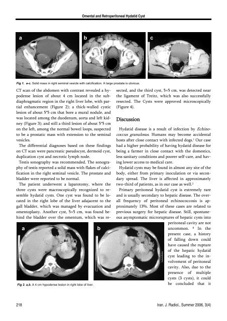
<strong>Omental</strong> <strong>and</strong> <strong>Retroperitoneal</strong> <strong>Hydatid</strong> <strong>Cyst</strong>a b cFig 1. a-c. Solid mass in right seminal vesicle with calcification. A large prostate is obvious.aFig 2. a,b. A 4 cm hypodense lesion in right lobe of liver.bCT scan of the abdomen with contrast revealed a hypodenselesion of about 4 cm located in the subdiaphragmaticregion in the right liver lobe, with partialenhancement (Figure 2); a thick-walled cysticlesion of about 5*5 cm that bore a mural nodule, <strong>and</strong>was located among the duodenum, aorta <strong>and</strong> left kidney(Figure 3); <strong>and</strong> still a third lesion of about 5*5 cmon the left, among the normal bowel loops, suspectedto be a prostatic mass with extension to the seminalvesicles.The differential diagnoses based on these findingson CT scan were pancreatic pseudocyst, dermoid cyst,duplication cyst <strong>and</strong> necrotic lymph node.Testis sonography was recommended. The sonographyof testis reported a solid mass with flakes of calcificationin the right seminal vesicle. The prostate <strong>and</strong>bladder were reported to be normal.The patient underwent a laparotomy, where thethree cysts were macroscopically recognized to resemblehydatid cysts. One cyst was found to be locatedin the right lobe of the liver adajacent to thegall bladder, which was managed by evacuation <strong>and</strong>omentoplasty. Another cyst, 5×5 cm, was found behindthe bladder over the omentum, which was resected,<strong>and</strong> the third cyst, 5×5 cm, was detected nearthe ligament of Treitz, which was also successfullyresected. The <strong>Cyst</strong>s were approved microscopically(Figure 4).Discussion<strong>Hydatid</strong> disease is a result of infection by Echinococcusgranulosus. Humans may become accidentalhosts after close contact with infected dogs. 1 Our casehad a higher probability of having hydatid disease forbeing a farmer in close contact with the domestics,less sanitary conditions <strong>and</strong> poorer self-care, <strong>and</strong> havinglower access to medical care.<strong>Hydatid</strong> cysts may be found in almost any site of thebody, either from primary inoculation or via secondaryspread. The liver is affected in approximatelytwo-third of patients, as in our case as well. 2Primary peritoneal hydatid cyst is extremely rare<strong>and</strong> is usually secondary to hepatic disease. The overallfrequency of peritoneal echinococcosis is approximately13%. Most of these cases are related toprevious surgery for hepatic disease. Still, spontaneousasymptomatic microruptures of hepatic cysts intoperitoneal cavity are notuncommon.8In thepresent case, a historyof falling down couldhave caused the ruptureof the hepatic hydatidcyst leading to the involvementof peritonealcavity. Also, due to thepresence of multiplecysts (3 cysts), it couldbe concluded that it218 Iran. J. Radiol., Summer 2006, 3(4)