Omental and Retroperitoneal Hydatid Cyst: A Case Report
Omental and Retroperitoneal Hydatid Cyst: A Case Report
Omental and Retroperitoneal Hydatid Cyst: A Case Report
Create successful ePaper yourself
Turn your PDF publications into a flip-book with our unique Google optimized e-Paper software.
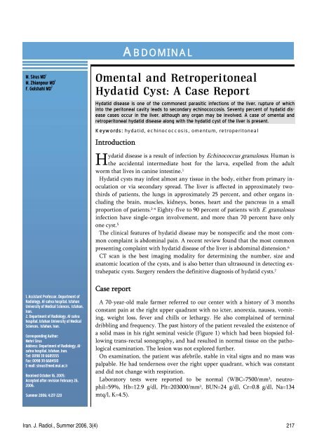
ABDOMINALM. Sirus MD 1M. Zhianpour MD 1F. Golshahi MD 21. Assistant Professor, Department ofRadiology, Al-zahra hospital, IsfahanUniversity of Medical Sciences, Isfahan,Iran.2. Department of Radiology, Al-zahrahospital, Isfahan University of MedicalSciences, Isfahan, Iran.Corresponding Author:Mehri SirusAddress: Department of Radiology, Alzahrahospital, Isfahan, Iran.Tel: 0098 311 6685555Fax: 0098 311 6684510E-mail: sirous@med.mui.ac.irReceived October 16, 2005;Accepted after revision February 26,2006.Summer 2006; 4:217-220<strong>Omental</strong> <strong>and</strong> <strong>Retroperitoneal</strong><strong>Hydatid</strong> <strong>Cyst</strong>: A <strong>Case</strong> <strong>Report</strong><strong>Hydatid</strong> disease is one of the commonest parasitic infections of the liver, rupture of whichinto the peritoneal cavity leads to secondary echinococcosis. Seventy percent of hydatid diseasecases occur in the liver, although any organ may be involved. A case of omental <strong>and</strong>retroperitoneal hydatid disease along with the hydatid cyst of the liver is present.Keywords: hydatid, echinococcosis, omentum, retroperitonealIntroduction<strong>Hydatid</strong> disease is a result of infection by Echinococcus granulosus. Human isthe accidental intermediate host for the larva, expelled from the adultworm that lives in canine intestine. 1<strong>Hydatid</strong> cysts may infest almost any tissue in the body, either from primary inoculationor via secondary spread. The liver is affected in approximately twothirdsof patients, the lungs in approximately 25 percent, <strong>and</strong> other organs includingthe brain, muscles, kidneys, bones, heart <strong>and</strong> the pancreas in a smallproportion of patients. 2-4 Eighty-five to 90 percent of patients with E. granulosusinfection have single-organ involvement, <strong>and</strong> more than 70 percent have onlyone cyst. 5The clinical features of hydatid disease may be nonspecific <strong>and</strong> the most commoncomplaint is abdominal pain. A recent review found that the most commonpresenting complaint with hydatid disease of the liver is abdominal distension. 6CT scan is the best imaging modality for determining the number, size <strong>and</strong>anatomic location of the cysts, <strong>and</strong> is also better than ultrasound in detecting extrahepaticcysts. Surgery renders the definitive diagnosis of hydatid cysts. 7<strong>Case</strong> reportA 70-year-old male farmer referred to our center with a history of 3 monthsconstant pain at the right upper quadrant with no icter, anorexia, nausea, vomiting,weight loss, fever <strong>and</strong> chills or lethargy. He also complained of terminaldribbling <strong>and</strong> frequency. The past history of the patient revealed the existence ofa solid mass in his right seminal vesicle (Figure 1) which had been biopsied followingtrans-rectal sonography, <strong>and</strong> had resulted in normal tissue on the pathologicalexamination. The lesion was not explored further.On examination, the patient was afebrile, stable in vital signs <strong>and</strong> no mass waspalpable. He had tenderness over the right upper quadrant, which was constant<strong>and</strong> did not change with respiration.Laboratory tests were reported to be normal (WBC=7500/mm 3 , neutrophil=59%,Hb=12.9 g/dl, Plt=203000/mm 3 , BUN=24 g/dl, Cr=0.8 g/dl, Na=134mtq/l, K=4.5).Iran. J. Radiol., Summer 2006, 3(4) 217
<strong>Omental</strong> <strong>and</strong> <strong>Retroperitoneal</strong> <strong>Hydatid</strong> <strong>Cyst</strong>a b cFig 1. a-c. Solid mass in right seminal vesicle with calcification. A large prostate is obvious.aFig 2. a,b. A 4 cm hypodense lesion in right lobe of liver.bCT scan of the abdomen with contrast revealed a hypodenselesion of about 4 cm located in the subdiaphragmaticregion in the right liver lobe, with partialenhancement (Figure 2); a thick-walled cysticlesion of about 5*5 cm that bore a mural nodule, <strong>and</strong>was located among the duodenum, aorta <strong>and</strong> left kidney(Figure 3); <strong>and</strong> still a third lesion of about 5*5 cmon the left, among the normal bowel loops, suspectedto be a prostatic mass with extension to the seminalvesicles.The differential diagnoses based on these findingson CT scan were pancreatic pseudocyst, dermoid cyst,duplication cyst <strong>and</strong> necrotic lymph node.Testis sonography was recommended. The sonographyof testis reported a solid mass with flakes of calcificationin the right seminal vesicle. The prostate <strong>and</strong>bladder were reported to be normal.The patient underwent a laparotomy, where thethree cysts were macroscopically recognized to resemblehydatid cysts. One cyst was found to be locatedin the right lobe of the liver adajacent to thegall bladder, which was managed by evacuation <strong>and</strong>omentoplasty. Another cyst, 5×5 cm, was found behindthe bladder over the omentum, which was resected,<strong>and</strong> the third cyst, 5×5 cm, was detected nearthe ligament of Treitz, which was also successfullyresected. The <strong>Cyst</strong>s were approved microscopically(Figure 4).Discussion<strong>Hydatid</strong> disease is a result of infection by Echinococcusgranulosus. Humans may become accidentalhosts after close contact with infected dogs. 1 Our casehad a higher probability of having hydatid disease forbeing a farmer in close contact with the domestics,less sanitary conditions <strong>and</strong> poorer self-care, <strong>and</strong> havinglower access to medical care.<strong>Hydatid</strong> cysts may be found in almost any site of thebody, either from primary inoculation or via secondaryspread. The liver is affected in approximatelytwo-third of patients, as in our case as well. 2Primary peritoneal hydatid cyst is extremely rare<strong>and</strong> is usually secondary to hepatic disease. The overallfrequency of peritoneal echinococcosis is approximately13%. Most of these cases are related toprevious surgery for hepatic disease. Still, spontaneousasymptomatic microruptures of hepatic cysts intoperitoneal cavity are notuncommon.8In thepresent case, a historyof falling down couldhave caused the ruptureof the hepatic hydatidcyst leading to the involvementof peritonealcavity. Also, due to thepresence of multiplecysts (3 cysts), it couldbe concluded that it218 Iran. J. Radiol., Summer 2006, 3(4)
Sirus et al.a b cFig 3. a-c. <strong>Cyst</strong>ic lesion, thick-walled with mural nodule.abcould have been secondary to shedding from onecyst, probably following a trauma, as the patients reportedan incident of falling from the horse back longago, the released daughter cysts could have formedthe other 2 cysts. Still, this hypothesis cannot be supportedby plausible evidence.E. granulosus infection of the liver frequently producesno symptoms. The right lobe is affected in 60 to85 percent of patients, as was in our case. Significantsymptoms are unusual before the cyst has reached atleast 10cm in diameter. 9 Peritoneal echinococcosisusually goes undetected until cysts are large enoughto produce symptoms. 8,10,11 In our case the patient hadright upper quadrant pain with a cyst that measured5×5 cm on CT scan.Isolated retroperitoneal hydatid cyst is rare <strong>and</strong> it isusually the result of spontaneous, traumatic or surgicalrupture of a primary hepatic cyst. 12,13In the present case, one of the cysts was retroperitoneal,among the left kidney, duodenum <strong>and</strong> abdominalaorta <strong>and</strong> the patient had a history of previoustrauma.The calcification <strong>and</strong> enlargement of the rightseminal vesicle with normal pathologic result in ourpatients could be due to a dead hydatid cyst. Althoughisolated hydatid cyst in a seminal vesicle hasnot been reported previously, in the present case thepossibility for the secondary involvement of seminalvesicles from abdominal hydatid cysts exists <strong>and</strong> thenormal biopsy can be the result of sampling from theadjacent normal tissue. 13 Since no further investigationof the lesion was done after the normal biopsyreport, its true etiology cannot be cleared.Routine laboratory tests, including complete bloodcounts <strong>and</strong> liver function tests, may be abnormal butare nonspecific <strong>and</strong> cannot make a diagnosis. In ourpatient the laboratory findings were all normal.CT scan is the imaging of choice for determiningthe number, size <strong>and</strong> anatomic location of cysts, <strong>and</strong>is also superior to ultrasound in detecting extrahepaticcysts. 7 Imaging findings in hydatid diseasedepend on the stage of the cyst growth. It means,whether the cyst is unilocular, contains daughtervesicles, contains daughter cysts, is partially calcified,or is completely calcified (dead), the imaging defers.So the range of differential diagnosis is wide <strong>and</strong>missed cases are common. In the present case, theunusual location of the cysts <strong>and</strong> the shape of the cystwith thick wall <strong>and</strong> mural nodule on CT, suggesteddifferential diagnoses of pancreatic pseudocyst or necroticlymph nodes probable.Therefore, it is to emphasize that in any cystic lesionin any anatomic location, hydatid cyst should beconsidered at least as a differential diagnosis especiallyin places that hydatid cyst is endemic.Fig 4. The Phatologic feature of the cystsIran. J. Radiol., Summer 2006, 3(4) 219
<strong>Omental</strong> <strong>and</strong> <strong>Retroperitoneal</strong> <strong>Hydatid</strong> <strong>Cyst</strong>References1. Grove DI. Worms in Australia. Med J Aust 1993; 159: 464–4652. Baden LR, Elliott DD. A 42-year-old woman with cough, fever, <strong>and</strong>abnormalities on thoracoabdominal computed tomography. N Engl JMed 2003; 348: 447-4553. Gelman R, Brook G, Green J, Ben-Itzhak O, Nakhoul F. Minimalchange glomerulonephritis associated with hydatid disease. ClinNephrol. 2000; 53:152-1554. Ali-Khan Z, Rausch RL. Demonstration of amyloid <strong>and</strong> immunecomplex deposits in renal <strong>and</strong> hepatic parenchyma of Alaskan alveolarhydatid disease patients. Ann Trop Med Parasitol. 1987; 81: 381-3925. Frider B, Larrieu E, Odriozola M. Long-term outcome of asymptomaticliver hydatidosis. J Hepatol 1999; 30: 228-2316. Al-Bassam A, Hassab H, Al-Olayet Y, Shadi M, Aal-Shami G, Aal-Rabeeah A et al. <strong>Hydatid</strong> disease of the liver in children. Ann TropPaediatr 1999; 19: 191–1967. Kervancioglu R, Bayram M, Elbeyli L. CT findings in pulmonaryhydatid disease. Acta Radiol 1999; 40:510-5148. Pedrosa I, Saiz A, Arrazola J, Ferreiros J, Pedrosa CS. <strong>Hydatid</strong> disease:radiologic <strong>and</strong> pathologic features <strong>and</strong> complications. Radiographics.2000; 20: 795-8179. Ammann RW, Eckert JC. Cestodes, Echinococcus. Gastroenterol ClinNorth Am 1996; 25: 655-68910. Kern, P, Bardonnet, K, Renner, E, Auer H, Pawlowski Z, AmmannRW et al. European echinococcosis registry: human alveolar echinococcosis.Emerg Infect Dis 2003; 9: 343-34911. Morar R, Feldman C. Pulmonary echinococcosis. Eur Respir J 2003;21: 1069-107712. Elton C, Lewis M, Jourdan MH. Unusual site of hydatid cyst: a casereport. Lancet 1999; 355: 213213. Engin G, Acunas B, Rozanes I. hydatid cyst with unusual localization.Eur Radiol 2000; 10: 1904-1912FFFFF220 Iran. J. Radiol., Summer 2006, 3(4)


