Issue 6 2007 - acpfg
Issue 6 2007 - acpfg
Issue 6 2007 - acpfg
- No tags were found...

You also want an ePaper? Increase the reach of your titles
YUMPU automatically turns print PDFs into web optimized ePapers that Google loves.
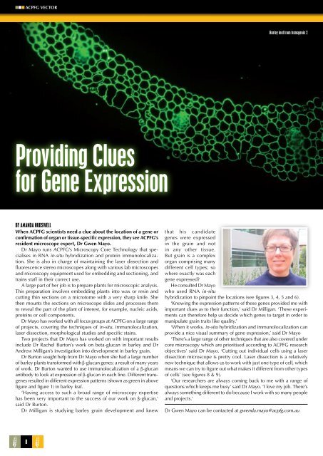
ACPFG VECTORBarley leaf from transgenic 2Providing Cluesfor Gene ExpressionBy Amanda HudswellWhen ACPFG scientists need a clue about the location of a gene orconfirmation of organ or tissue-specific expression, they see ACPFG’sresident microscope expert, Dr Gwen Mayo.Dr Mayo runs ACPFG’s Microscopy Core Technology that specialisesin RNA in-situ hybridization and protein immunolocalization.She is also in charge of maintaining the laser dissection andfluorescence stereo microscopes along with various lab microscopesand microscopy equipment used for embedding and sectioning, andtrains staff in their correct use.A large part of her job is to prepare plants for microscopic analysis.This preparation involves embedding plants into wax or resin andcutting thin sections on a microtome with a very sharp knife. Shethen mounts the sections on microscope slides and processes themto reveal the part of the plant of interest, for example, nucleic acids,proteins or cell components.Dr Mayo has worked with all focus groups at ACPFG on a large rangeof projects, covering the techniques of in-situ, immunolocalization,laser dissection, morphological studies and specific stains.Two projects that Dr Mayo has worked on with important resultsinclude Dr Rachel Burton’s work on beta-glucan in barley and DrAndrew Milligan’s investigation into development in barley grain.Dr Burton sought help from Dr Mayo when she had a large numberof barley plants transformed with β-glucan genes; a result of many yearsof work. Dr Burton wanted to use immunolocalization of a β-glucanantibody to look at expression of β-glucan in each line. Different transgenesresulted in different expression patterns (shown as green in abovefigure and figure 1) in barley leaf.‘Having access to such a broad range of microscopy expertisehas been very important to the success of our work on β-glucan,’said Dr Burton.Dr Milligan is studying barley grain development and knewthat his candidategenes were expressedin the grain and notin any other tissue.But grain is a complexorgan comprising manydifferent cell types; sowhere exactly was eachgene expressed?He consulted Dr Mayowho used RNA in-situhybridization to pinpoint the locations (see figures 3, 4, 5 and 6).‘Knowing the expression patterns of these genes provided me withimportant clues as to their function,’ said Dr Milligan. ‘These experimentscan therefore help us decide which genes to target in order tomanipulate grain traits like quality.’‘When it works, in-situ hybridization and immunolocalization canprovide a nice visual summary of gene expression,’ said Dr Mayo‘There’s a large range of other techniques that are also covered undercore microscopy which are prioritised according to ACPFG researchobjectives’ said Dr Mayo. ‘Cutting out individual cells using a laserdissection microscope is pretty cool. Laser dissection is a relativelynew technique that allows us to work with just one type of cell, whichmeans we can try to figure out what makes it different from other typesof cells’ (see figures 8 & 9).‘Our researchers are always coming back to me with a range ofquestions which keeps me busy’ said Dr Mayo. ‘I love my job. There’salways something different to do because I work with so many peopleand projects.’Dr Gwen Mayo can be contacted at gwenda.mayo@<strong>acpfg</strong>.com.au8