Ion Exchange of Proteins with Organic Solvents - LC column, HPLC ...
Ion Exchange of Proteins with Organic Solvents - LC column, HPLC ...
Ion Exchange of Proteins with Organic Solvents - LC column, HPLC ...
- No tags were found...

You also want an ePaper? Increase the reach of your titles
YUMPU automatically turns print PDFs into web optimized ePapers that Google loves.
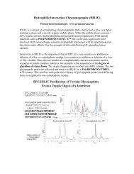
If a protein isn't normally found in an aqueous medium, such as a membrane protein, itmay not be possible to do IEX at all <strong>with</strong>out some organic solvent. The example belowconcerns Lung Surfactant Protein in an emulsion <strong>with</strong> 500 parts lipid. With 70%organic solvent in the mobile phase, hydrophilic interaction is superimposed on top <strong>of</strong> theelectrostatic effects. Under these conditions, lipid is not retained and elutes in the voidvolume. The protein is then eluted <strong>with</strong> a salt gradient. A number <strong>of</strong> variants are evident,corresponding to truncation sequences <strong>with</strong> deletions from either terminus.- data courtesy <strong>of</strong> Richard Hartwick (PharmAssist) -Hydrophilic interaction becomes significant if the mobile phase is > 60% organic, as inthe above example. These HILIC effects are superimposed on the electrostatic effects. Inthe following example, phosphorylation variants <strong>of</strong> Histone H1 are separated on aPolyCAT A <strong>column</strong>. Histones are basic proteins and are well-retained on a cationexchange<strong>column</strong>. With 0% ACN in the mobile phases [TOP], the more extensivelyphosphorylated variants elute early, since the negative charge <strong>of</strong> the phosphate repels thenegatively-charged stationary phase. Overall, resolution is poor. With 70% ACN in themobile phases [BOTTOM], resolution is excellent, <strong>with</strong> the order <strong>of</strong> elution inverted.Under these conditions the hydrophilic interaction <strong>with</strong> a phosphate group is greater thanits electrostatic repulsion, leading to a net increase in retention. With 40% ACN[MIDDLE], both forces are in balance, and all variant forms coelute.
Histone H4 acetylation and methylation variants: A cation-exchange <strong>column</strong> canseparate variants <strong>with</strong> acetylated Lys- residues, since they vary in the number <strong>of</strong> +charges. However, <strong>with</strong> hydrophilic interaction superimposed on the electrostatic effects,the <strong>column</strong> can also separate variants <strong>with</strong> the same number <strong>of</strong> acetylated Lys- residuesbut differing in the number <strong>of</strong> methylated Lys- residues. In the figure below, the morehighly acetylated forms are present in the smallest amounts and for the shortest time inthe cell cycle, but their presence is required if the cell is to enter mitosis. Theiridentification was only possible by adding this top-down separation step prior todigestion and bottom-up peptide identification. Otherwise, they would be masked by thefar more abundant, less highly acetylated variants.Histone H1.5 phosphorylation variants: HP<strong>LC</strong> is superior to electrophoresis forseparation <strong>of</strong> protein variants. The figure below shows the isolation <strong>of</strong> Histone H1.5 andthe separation <strong>of</strong> phosphorylation variants by HPCE.
When the same mixture is resolved by cation-exchange in the HILIC mode [BELOW],the resulting peaks are more than twice as sharp. Furthermore, the <strong>column</strong> is able todistinguish between positional variants, each bearing a single phosphate group but ondifferent Ser- residues. When the cell is in mitosis, more highly phosphorylated formsappear that are absent in interphase. Again, these forms are required for successful entry<strong>of</strong> the cell into mitosis. The schematic below shows the sequence <strong>of</strong>phosphorylation. The variants <strong>with</strong> 4 or 5 phosphates represent the first observation <strong>of</strong> ahistone phosphorylated at a Thr- residue. Again, these low-abundance forms could onlybe identified by first separating them prior to digestion from more abundant variants.
PolyCAT A and PolySULFOETHYL A are trademarks <strong>of</strong> Poly<strong>LC</strong> Inc.