Oligomeric properties and signal peptide binding by Escherichia coli ...
Oligomeric properties and signal peptide binding by Escherichia coli ...
Oligomeric properties and signal peptide binding by Escherichia coli ...

Create successful ePaper yourself
Turn your PDF publications into a flip-book with our unique Google optimized e-Paper software.
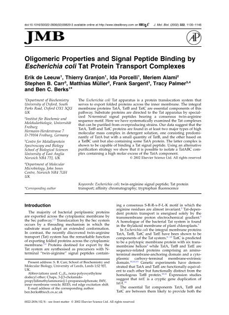
1138 <strong>Escherichia</strong> <strong>coli</strong> Tat Protein Transport ComplexesABCT- T+ S MSMFt NiT- T+ S MSMFt NiT- T+ S MSMFt Ni66352420TatB/CTatATatB/CTatATatB/CTatA1 2 3 4 5 6 71 2 3 4 5 6 71 2 3 4 5 6 7TatATatATatATatBTatBTatBTatCTatCTatC1 2 3 4 5 6 71 2 3 4 5 6 7 1 2 3 4 5 6 7Figure 2. Affinity purification of Tat complexes. TatABCDE were co-ordinately overproduced with a hexahistidinetag on (a) TatA (b) TatB or (c) TatC. The proteins present at each step of the purifications were analysed <strong>by</strong> SDS-PAGE <strong>and</strong> visualised either <strong>by</strong> Coomassie brilliant blue staining (upper panel) or immunoblotting using antiseradirected against TatA, TatB or TatC as indicated (lower panels). Fractions are (Tþ) whole cells induced for TatABCDEoverproduction; (S) high speed supernatant <strong>and</strong> (M) total membranes following subcellular fractionation of the cells;(SM) the Chaps-solubilized membrane supernatant; the (Ft) unbound <strong>and</strong> (Ni) pooled Tat-protein-containing boundfractions following application of the solubilized membrane extract to a Ni 2þ affinity column. Lane (T2) showswhole cells that were not induced for TatABCDE overproduction. The molecular masses (kDa) of st<strong>and</strong>ard proteinsare given to the left of the Coomassie blue-stained gels.proteins isolated <strong>by</strong> affinity chromatography werepurified away from contaminating proteins <strong>by</strong> gelpermeation chromatography <strong>and</strong> were found in apeak of apparent molecular mass .650 kDa(Figure 3(b)). Thus a TatB-specific purification ledto the isolation of a high molecular mass Tat(A)BCcomplex similar to that obtained when the hexahistidinetag was located on the TatC componentAFr. Nr. 8 9 10 11 12 13 14TatBTatCTatAB9 10 11 1213CTatBTatCTatA8 9 10 11 12 13TatBTatCTatAFr. Nr.2 4 6 8 10 12 14 16 18 20 22 24 4 6 8 10 12 14 16 18 20 22 24 4 6 8 10 12 14 16 18 20 22 24Vo 669 200 66443 150Vo 669 200 66443 150Vo 669 200 66443 150Figure 3. Gel filtration purification of Tat complexes following subunit-specific affinity chromatography. The boundfractions from the Ni 2þ affinity chromatography separations shown in Figure 2 were each individually pooled, concentrated<strong>and</strong> subject to size exclusion chromatography. The samples analysed are those in which the histidine tag was on(a) TatA, (b) TatB or (c) TatC. The 280 nm absorbance of the column eluant is plotted in each case. Only the fractionsanalysed <strong>by</strong> SDS-PAGE <strong>and</strong> Coomassie staining at the top of the Figure contain Tat proteins. The lane numbers oneach SDS-PAGE gel correlate to the fraction numbers of the elution profile. In addition the positions of the eluted fractionsare shown <strong>by</strong> the comb above the elution profile. The elution positions of water-soluble st<strong>and</strong>ard proteins areshown below the column profile <strong>and</strong> labelled with the protein mass (kDa). The st<strong>and</strong>ards used were (V 0 ) blue dextran,(669 kDa) thyroglobulin, (443 kDa) apoferritin, (200 kDa) alcohol dehydrogenase, (150 kDa) carbonic anhydrase <strong>and</strong>(66 kDa) bovine serum albumin.
<strong>Escherichia</strong> <strong>coli</strong> Tat Protein Transport Complexes 1139(above). Taken together the results of the TatB <strong>and</strong>TatC-specific purification suggest that TatB preferentiallyassociates with TatC <strong>and</strong>/or that the interactionof TatA with TatBC is destabilised duringthe affinity chromatography step.Affinity-tagging of the TatA proteinNext we investigated the nature of Tat complexesthat could be purified when a hexahistidineaffinity tag was placed at the carboxy terminus ofTatA. Membranes were prepared from a strainexpressing the modified tat operon on pFAT75AH.All the TatA protein in the detergent-solubilisedextract was bound to the affinity matrix(Figure 2(a), lane 7). In addition, the columnbound almost the entire complement of TatB <strong>and</strong>TatC (Figure 2(a), lane 7), indicating that these proteinsare found in complexes containing TatA. Gelpermeation chromatography of the pooled boundfractions from the Ni 2þ column indicated that theTat proteins were again found in complexes withan apparent molecular mass of greater than650 kDa (Figure 3(a)).The material isolated with the TatA-specific tagis unlikely to represent a single type of complexsince the Tat proteins did not elute at a constantratio from the Ni 2þ column (data not shown butanalogous to Figure 4(a), see below). Specifically,while the ratio of TatB to TatC in successive fractionswas constant the ratio of TatA to TatBCincreased in fractions eluting at higher imidazoleconcentrations. Such behaviour would be consistentwith, for example, a mixture of Tat(A)BC <strong>and</strong>TatA(B) complexes since these complexes containdifferent numbers of tag-bearing TatA molecules(<strong>and</strong> therefore differing affinities for Ni 2þ ) butwould be difficult to separate <strong>by</strong> gel permeationchromatography because they possess similarapparent molecular masses.We attempted to clarify the nature of the TatAcontainingcomplexes present in the detergentsolublisedextract using a double-tagging approachin which TatABC were co-overexpressed with ahexahistidine affinity tag fused to the carboxy terminusof TatA <strong>and</strong> a streptavidin affinity tagfused to the carboxy terminus of TatC (plasmidpFAT75AhisBCstrep). As a first purification stepthe soluble extract was subjected to Ni 2þ chromatography.The elution profile is similar to thatreported above for the TatA-tagged material withfractions of varying TatA to TatBC ratio (Figure4(a)). The fractions with a high TatA to TatBC ratiowere pooled <strong>and</strong> re-chromatographed on a Strep-Tactin affinity column to purify those complexesthat contained TatC. Most of the TatA proteinloaded onto the column did not bind <strong>and</strong> wasrecovered in the flow through fractions (Figure4(b), lane Ft). However, a small amount of TatAwas retained on the column <strong>and</strong> co-eluted togetherwith most of the TatB <strong>and</strong> TatC present in theloaded sample (Figure 4(b), lanes 3–6; note thatthese samples were TCA-precipitated <strong>and</strong> representFigure 4. Purification of a TatABC complex <strong>by</strong> adouble tagging strategy. TatABC were co-ordinatelyoverproduced with a hexahistidine tag on the carboxyterminus of TatA <strong>and</strong> a Strep tag at the carboxy terminusof TatC. Membranes from the overproducing strain weresolubilized in Chaps <strong>and</strong> subject to Ni 2þ affinity chromatography.(a) Fractions eluting from the Ni 2þ affinity columnwith an increasing imidazole gradient. Theproteins present in each successive fraction were analysed<strong>by</strong> SDS-PAGE <strong>and</strong> visualised <strong>by</strong> Coomassie brilliantblue staining. (b) The three right-h<strong>and</strong> fractionsfrom the Ni 2þ affinity column which contain a largeamount of TatA were pooled (lane L) <strong>and</strong> loaded on astreptactin column. Unbound proteins were retained(lane Ft). Bound material was then eluted with desthiobiotin<strong>and</strong> successive fractions collected (eluate, lanes1–6). The proteins present at each step were separated<strong>by</strong> SDS-PAGE <strong>and</strong> visualised <strong>by</strong> Coomassie brilliantblue staining (upper panel) or immunoblotting usingantisera directed against TatA, TatB or TatC as indicated(lower panels). The molecular masses (kDa) of st<strong>and</strong>ardproteins are given to the left of the Coomassie bluestainedgels.material that is 100 times concentrated relative tothe unbound fraction in lane Ft). Detailed characterizationof the TatABC material purified in thisexperiment was precluded <strong>by</strong> the low yield; theentire material purified from a four litre bacterialculture is loaded on the gel shown in the righth<strong>and</strong>panel of Figure 4(b). Nevertheless, it is apparentfrom the Coomassie blue staining intensities(cf. Figure 2(c), lane 7 <strong>and</strong> Figure 4(b), lane 3) thatthe TatABC complexes purified <strong>by</strong> the doubletaggingapproach have a considerably higher ratio
1140 <strong>Escherichia</strong> <strong>coli</strong> Tat Protein Transport ComplexesABFigure 5. Purification of a TatAcontainingcomplex from a strainT- T+ S M SM Ft Nilacking TatC. TatAB were co-ordinately66TatBoverproduced in a Dtat-ABCDDtatE background with a45hexahistidine tag on the carboxy29TatBTatAterminus of TatA. Membranes from24Fr. Nr. 10 11 12 13 14the overproducing strain were solubilizedin Chaps <strong>and</strong> subject to Ni20TatA2þaffinity chromatography. (a) The1 2 3 4 5 6 7proteins present at each step of thepurification were analysed <strong>by</strong> SDS-PAGE <strong>and</strong> visualised either <strong>by</strong> CoomassieTatAbrilliant blue staining(upper panel) or immunoblottingusing antisera directed againstTatBTatA or TatB as indicated (lowerFr. Nr. 4 6 8 10 12 14 16 18 20 22 24 261 2 3 4 5 6 7panels). Fractions are (Tþ) wholeVo cells induced for TatAB overproduction;(S) high speed supernatant669 200 66443 150<strong>and</strong> (M) total membranes followingsubcellular fractionation of thecells; (SM) the Chaps-solubilized membrane supernatant; the (Ft) unbound <strong>and</strong> (Ni) pooled Tat-protein-containingbound fractions following application of the solubilized membrane extract to a Ni 2þ affinity column. Lane (T2)shows whole cells that were not induced for TatAB overproduction. The molecular masses (kDa) of st<strong>and</strong>ard proteinsare given to the left of the Coomassie blue-stained gel. (b) Pooled fractions after the Ni 2þ affinity step were concentrated<strong>and</strong> further purified <strong>by</strong> gel permeation chromatography. The 280 nm absorbance of the column eluant is plotted.Proteins in the indicated peak fractions were separated <strong>by</strong> SDS-PAGE <strong>and</strong> visualised <strong>by</strong> Coomassie brilliant blue stainingwith the lane numbers on the SDS-PAGE gel at the top of the Figure corresponding to the fraction numbers of theelution profile. The positions of the fractions analysed <strong>by</strong> SDS-PAGE are also indicted <strong>by</strong> the comb above the elutionprofile. The elution positions of water-soluble st<strong>and</strong>ard proteins are shown below the column profile <strong>and</strong> labelledwith the protein mass (kDa). The identities of the marker proteins are given in the legend to Figure 3.of TatA to TatBC than the Tat(A)BC complexes purifiedusing only a TatC-specific step. Indeed thelevels of the TatB <strong>and</strong> TatC proteins are so low inthe material purified <strong>by</strong> double tagging that theyare only clearly detected <strong>by</strong> Coomassie blue stainingin the peak eluted fraction (Figure 4(b), lane3). This experiment demonstrates that the Tat systemis capable of assembling complexes containingall three essential Tat proteins but with very highTatA to TatBC ratios. It also shows that most ofthe histidine-tagged TatA protein present in thedetergent-solubilised extract is found in complexesthat do not contain TatC. In a control experimentwe found that if the solubilized extract wasinitially chromatographed on Strep-Tactin agaroserather than a Ni 2þ affinity column, then a Tat(A)BCcomplex similar to that purified using the constructwith a hexahistidine tag on TatC was isolated (datanot shown). Thus the type of tag on TatC does notinfluence the type of complex purified.Direct interaction between TatA <strong>and</strong> TatBIt was reported 16 that it is possible to purify ahigh molecular mass TatA(B) complex from astrain that is co-ordinately overexpressingtatABCDE. To determine whether TatC, -D, <strong>and</strong>/or-E are required for the formation of the TatA(B)complex we undertook purification of TatAcontainingspecies from a DtatABCDDtatE strainco-overexpressing tatA <strong>and</strong> tatB only. In order tofacilitate purification of TatA-containing complexesthe TatA protein was provided with a carboxyterminalhexahistidine tag.Following overexpression of plasmid pFAT75AhisB,membranes were solubilized <strong>by</strong> Chapsunder our st<strong>and</strong>ard conditions <strong>and</strong> proteins subjectedto chromatography on Ni 2þ affinity resin.The TatA protein was specifically bound to thecolumn <strong>and</strong> was co-eluted with a small portion ofthe TatB protein found in the initial extract (Figure5(a), lane 7). The majority of the TatB protein was,however, found in the unbound fraction (Figure5(a), lane 6; note that as in earlier Figures, the totalvolume of the pooled unbound fraction is threetimes that of the pooled bound fraction). Gel permeationchromatography shows that the TatA(B)complexes that bind to the Ni 2þ column have anapparent molecular mass in excess of 650 kDa.In conclusion, although an earlier study 18 proposedthat TatA <strong>and</strong> TatB only interact via TatC,our data indicate that TatA <strong>and</strong> TatB are capableof direct interaction when expressed <strong>and</strong> purifiedin the absence of TatC.Isolated TatA forms large oligomersSince the TatA(B) complex is composed predominantlyof TatA we examined whether TatAalone is capable of forming a high molecular masscomplex. TatA was purified from a DtatABCDDtatEstrain overproducing hexahistidine-tagged TatAfrom plasmid pFAT584 12 using the same solubilisation<strong>and</strong> Ni 2þ affinity protocol as the TatA(B)
<strong>Escherichia</strong> <strong>coli</strong> Tat Protein Transport Complexes 1141AB663524201424203520Fr.Nr.T- T+ MSMFt Ni1 2 3 4 5 6TatA8 9 10 11 12 13 14 15 16 17 18 19 20V 0 669 443200150TatATatAFigure 6. Purification of a TatA-containing complexfrom a strain lacking both TatB <strong>and</strong> TatC. TatA with ahexahistidine tag on the carboxy terminus was overproducedin a DtatABCDDtatE background. Membranesfrom the overproducing strain were solubilized inChaps <strong>and</strong> subject to Ni 2þ affinity chromatography. (a)The proteins present at each step of the purificationwere analysed <strong>by</strong> SDS-PAGE <strong>and</strong> visualised <strong>by</strong> Coomassiebrilliant blue staining. Fractions are (Tþ) whole cellsinduced for TatA, (M) total membranes following subcellularfractionation of the cells; (SM) the Chaps-solubilizedmembrane supernatant; the (Ft) unbound <strong>and</strong> (Ni)pooled Tat-protein-containing bound fractions followingapplication of the solubilized membrane extract to aNi 2þ affinity column. Lane (T2) shows whole cells thatwere not induced for TatA overproduction. (b) Characterizationof TatA complexes <strong>by</strong> gel permeation chromatography.Separations are shown for the pooledconcentrated fractions from the Ni 2þ affinity column separationperformed in (a) (upper panel) or for the pooledunbound (Ft) fraction from the Ni 2þ affinity column inFigure 2(c) (lower panel). The proteins present in theeluted fractions were analysed <strong>by</strong> SDS-PAGE <strong>and</strong> visualised<strong>by</strong> Coomassie brilliant blue staining. The molecularmasses in kDa of st<strong>and</strong>ard proteins are given to the leftof the Coomassie blue-stained gels. The elution positionsof water-soluble st<strong>and</strong>ard proteins are shown below thecolumn profile <strong>and</strong> labelled with the protein mass inkDa. The identities of the marker proteins are given inthe legend to Figure 3.purification described above. TatA was recoveredat high purity after Ni 2þ chromatography (Figure6(a), lane 6). However, the isolated TatA proteinwas found over a broad range of apparent molecularmasses as judged <strong>by</strong> gel permeation chromatographywith a major peak centred at approximately400 kDa <strong>and</strong> a minor peak below 65 kDa (Figure6(b), upper panel). Thus TatA expressed in theabsence of other Tat components is apparently66more heterogeneous with respect to oligomericstate or molecular conformation than TatAexpressed in their presence. Nevertheless, giventhat the mass of a TatA protomer is 9.6 kDa, it isclear that isolated TatA protein is still mainly presentas higher order oligomers.In the TatC-specific affinity purificationdescribed above the majority of the TatA moleculespresent in the initial soluble extract were notretained on the Ni 2þ affinity column (Figure 2(c),lane 6). Interestingly, gel permeation chromatographydemonstrates that the apparent molecularmass of the TatA in these flow-through fractions issimilar to that of the peak fractions of TatAexpressed in the absence of other Tat components(compare lower <strong>and</strong> upper panels in Figure 6(b)).This indicates that Tat(A)BC <strong>and</strong> high apparentmolecular mass TatA complexes are present simultaneouslyin the detergent-solubilised extract.Binding of twin-arginine <strong>signal</strong> <strong>peptide</strong>s <strong>by</strong>TatC-containing complexesIn the initial descriptions of E. <strong>coli</strong> Tat complexpurification 16,18 the functional integrity of the variouspreparations was not addressed. Here, weconsidered that an assay of the initial step of thetranslocation cycle, in which the substrate proteintwin-arginine <strong>signal</strong> <strong>peptide</strong> is recognised <strong>by</strong> oneor more components of the Tat translocationmachinery, might be tractable to in vitro analysis.We therefore decided to test whether our purifiedTat complexes contained a functional twin-arginine<strong>signal</strong> <strong>peptide</strong> <strong>binding</strong> site.We took a direct approach in which we monitoredchanges in the tryptophan fluorescence <strong>properties</strong>of the purified complexes upon addition of achemically synthesised Tat <strong>signal</strong> <strong>peptide</strong>. The fluorescence<strong>properties</strong> of tryptophan residues aresensitive, endogenous reporters of their localenvironment. TatA <strong>and</strong> TatB each contain a singletryptophan residue while TatC possesses twotryptophan side-chains. For these experiments theaffinity-tagged Tat complexes described abovewere purified in the detergent C 12 E 9 rather than inChaps in order to prevent minor backgroundabsorbance. The Tat protein complexes purified inC 12 E 9 were indistinguishable on the basis of proteincomposition, purity, or apparent molecular mass<strong>by</strong> gel permeation chromatography from those preparedin Chaps, with the exception that isolatedTatA had a less heterogeneous gel permeation profile(data not shown). The <strong>signal</strong> <strong>peptide</strong> used inthese experiments was that of the SufI protein thathad been used in the in vitro <strong>binding</strong> <strong>and</strong> transportexperiments described above (Figure 1). The isolatedSufI twin-arginine <strong>signal</strong> <strong>peptide</strong> has beenshown to competitively inhibit Tat transport invitro. 15 Thus the <strong>signal</strong> <strong>peptide</strong> alone is capable offunctional interaction with the E. <strong>coli</strong> Tat apparatusdespite the absence of its cognate passenger protein.As a control for the specificity of any interactionbetween the SufI <strong>signal</strong> <strong>peptide</strong> <strong>and</strong> the
1142 <strong>Escherichia</strong> <strong>coli</strong> Tat Protein Transport Complexesa352b352350350emission nm348346344emission nm348346344342342340340338295 300 305 310excitation nm338295 300 305 310excitation nmc352d352350350emission nm348346344emission nm348346344342342340340338295 300 305 310excitation nm338295 300 305 310excitation nmFigure 7. Interaction of purified Tat complexes with <strong>signal</strong> <strong>peptide</strong>s analysed <strong>by</strong> REES. Dependence of the positionof the tryptophan emission maximum on the excitation wavelength for Tat complexes purified using the detergentC 12 E 9 . The samples are (a) a Tat(A)BC complex analogous to the preparation shown in Figure 3(c); (b) a sample analogousto that shown in Figure 2(a) containing TatABC with a high molar excess of TatA; (c) a TatA only complex analogousto the preparation shown in Figure 6(b); (d) a TatA(B) complex analogous to that shown in Figure 5(b). Datashown are those for the isolated Tat complexes alone (V) or following addition of either the SufI <strong>signal</strong> <strong>peptide</strong> (B) orSufI <strong>signal</strong> <strong>peptide</strong> in which the twin arginine residues had been substituted <strong>by</strong> twin lysine residues (O). It was desirableto carry out the experiments under conditions where the concentration of <strong>peptide</strong>s exceeds the number of <strong>signal</strong><strong>peptide</strong> <strong>binding</strong> sites present. Since the number of <strong>signal</strong> <strong>peptide</strong> <strong>binding</strong> sites in each complex was not known, a <strong>peptide</strong>to protein molar ratio of 100 (based on a mass of 600 kDa for each complex) was employed. Samples were suspendedin a 500 ml final volume of 20 mM Mops (pH 7.2), 20 mM NaCl, 1 mM EDTA, 0.1% C 12 E 9 . Measurementswere carried out at 25 8C using a Perkin–Elmer LS50B fluorimeter. Each point was the average of at least four independentdeterminations <strong>and</strong> all spectra were corrected for the baseline spectrum of the water Raman peak contribution.purified Tat complexes we employed a second synthetic<strong>peptide</strong> in which the twin-arginine residuesof the consensus motif were conservativelyreplaced with two lysine residues. These substitutionshave previously been shown to preventTat-dependent targeting <strong>and</strong> transport of the SufIprecursor both in vivo 21 <strong>and</strong> in vitro. 6,15 Furthermore,<strong>and</strong> in contrast to the authentic “RR”-containing twin-arginine <strong>signal</strong> <strong>peptide</strong>, the“KK”-variant <strong>signal</strong> <strong>peptide</strong> does not competitivelyinhibit in vitro Tat transport, stronglysuggesting that it has a very low affinity for the <strong>signal</strong><strong>peptide</strong> <strong>binding</strong> site of the Tat system. 15None of the purified Tat complexes exhibitedsignificant changes in tryptophan fluorescenceintensity upon addition of the SufI <strong>signal</strong> <strong>peptide</strong>s(not shown). Instead, interactions were qualitativelymonitored using the so-called Red EdgeExcitation Shift (REES) effect which responds tochanges in the mobility of solvent molecules in theenvironment of the fluorophore. 22,23 A change intryptophan REES was detected for the Tat(A)BCcomplex (isolated via a hexahistidine tag on TatC)in the presence of the RR-containing <strong>signal</strong> <strong>peptide</strong>(Figure 7(a)). In contrast, the inactive KK-bearing<strong>signal</strong> <strong>peptide</strong> failed to elicit a detectable changein the Tat(A)BC tryptophan REES (Figure 7(a)). Weconclude that the RR <strong>signal</strong> <strong>peptide</strong> interacts withthe Tat(A)BC complex, causing a conformationalchange, while the KK <strong>signal</strong> <strong>peptide</strong> either fails tobind or fails to elicit the same change in tryptophanenvironment as the RR <strong>signal</strong> <strong>peptide</strong>. Thefact that <strong>signal</strong> <strong>peptide</strong> <strong>binding</strong> occurs in detergentsolution indicates that neither a membraneenvironment nor the protonmotive force isrequired for this interaction. This is consistent
<strong>Escherichia</strong> <strong>coli</strong> Tat Protein Transport Complexes 1143with recent reports of energy-independent precursor<strong>binding</strong> <strong>by</strong> the Tat systems of both bacteria15,24 – 27<strong>and</strong> thylakoids.A specific REES change in response to the RR,but not KK, <strong>signal</strong> <strong>peptide</strong> was also detected forTat proteins purified with a TatA-specific tag(Figure 7(b)), a preparation which is probably amixture of TatA(B), Tat(A)BC <strong>and</strong> TatABC complexes(above). However, no change in tryptophanREES was observed when either the TatA-only orTatA(B) complexes were exposed to either <strong>signal</strong><strong>peptide</strong> (Figure 7(c) <strong>and</strong> (d)).DiscussionHere, we report the purification <strong>by</strong> a double affinity-taggingmethod of a TatABC complex containinga high molar excess of the TatA protein. Thisdemonstrates for the first time that the three essentialTat components are capable of forming a complexin which the three proteins are present in aratio that reflects the high relative abundance ofTatA in the cytoplasmic membrane. 14,16 However,only a small proportion of the Tat proteins presentin the detergent-solublised extract were found insuch a complex. Instead, the major species purifiedunder the st<strong>and</strong>ardised expression, solubilisation,<strong>and</strong> chromatographic conditions utilised hereresemble the Tat(A)BC <strong>and</strong> TatA(B) complexesdescribed previously. 16,18 Tat(A)BC is based on aTatBC unit with some TatA present while TatA(B)is composed predominantly of TatA. Both types ofcomplex have a high apparent molecular mass<strong>and</strong> will therefore contain multiple copies of atleast the major constituent subunits. Several linesof evidence suggest that these major species reflectthe complexes formed <strong>by</strong> the Tat proteins in theresting cell membrane when substrate proteins<strong>and</strong> the protonmotive force are both absent. Tostart with, the same types of complex are isolatedif the purification protocol is varied. For example,the detergent C 12 E 9 can be substituted for Chapswithout affecting the species purified <strong>and</strong> othershave reported purification of a Tat(A)BC complexusing a different affinity tag, expression system<strong>and</strong> yet another detergent. 18 Thus these two, <strong>and</strong>only these two, types of complex are consistentlyisolated <strong>by</strong> different purification protocols. Indeed,both types of complex are apparently present simultaneouslyin the soluble extract since TatA isfound as a high molecular mass species followingremoval of Tat(A)BC <strong>by</strong> affinity chromatography(Figure 6(b)). The in vitro Tat transport data(Figure 1) strongly suggest that the major portionof the detergent-solublised Tat proteins are derivedfrom functional Tat complexes. Given that theminor TatABC complex is stable to purification(this work) then the TatA(B) <strong>and</strong> Tat(A)BC complexesare unlikely to be TatABC fragmentationproducts but rather distinct molecular species intheir own right. It should also be noted that the compositionsof the isolated complexes are consistentwith our current knowledge of the size <strong>and</strong> subunitinteractions in the E. <strong>coli</strong> Tat system: it has beenshown that TatA <strong>and</strong> TatB are present in high molecularmass complexes in detergent-solublisedextracts of membranes from wild-type E. <strong>coli</strong> cells, 28chemical crosslinking studies in wild-typemembranes 12 have demonstrated homo-oligomericinteractions for both TatA <strong>and</strong> TatB while there isalso evidence for direct interactions between TatB<strong>and</strong> TatC 11,18 as well as TatC self interactions. 17 Interestingly,we found that TatA expressed in theabsence of the other Tat proteins still forms high molecularmass complexes (Figure 6), reinforcing theview that TatA protomers predominantly form interactionswith other TatA protomers. 12Using intrinsic tryptophan fluorescence as ourspectroscopic probe we have been able to demonstrate<strong>binding</strong> of a twin-arginine <strong>signal</strong> <strong>peptide</strong> tothe Tat(A)BC complex (Figure 7(a)). This <strong>binding</strong>is apparently of a specific nature since a responsewas not detected with the KK variant <strong>signal</strong> <strong>peptide</strong>.This is the first report of a functional propertyassociated with any purified Tat component <strong>and</strong>provides further evidence for the physiologicalrelevance of this purified complex. A <strong>signal</strong> <strong>peptide</strong>-dependentchange in tryptophan fluorescence<strong>properties</strong> was only detected for those preparationsthat contain the TatC protein (Figure 7).This does not, however, allow us to conclude thatthe <strong>signal</strong> <strong>peptide</strong> binds to TatC since the particularfluorescence technique employed here onlydetects <strong>signal</strong> <strong>peptide</strong> <strong>binding</strong> indirectly throughchanges in tryptophan environment. Thus, to see afluorescence change the complex under studymust contain not only the subunit(s) that bind the<strong>signal</strong> <strong>peptide</strong> but also the subunit(s) containingthe conformation-sensitive tryptophan residues<strong>and</strong> any additional subunits that are required totransduce the effects of <strong>signal</strong> <strong>peptide</strong> <strong>binding</strong>into the conformational change that affects theresponsive tryptophan residues. All we can deduceis that TatC must carry out at least one of thesefunctions. Nevertheless our data do not contradictthe current view that Tat <strong>signal</strong> <strong>peptide</strong>s bind toTatB, TatC or both. 3,26,29The Tat systems of bacteria <strong>and</strong> plant chloroplastthylakoid membranes are homologous 7,13 <strong>and</strong>would therefore be expected to follow broadly thesame organisational <strong>and</strong> mechanistic principles.Blue native PAGE has been used to investigate theTat complexes found in resting, detergent-solubilisedthylakoids. 26 The major species are an approximately700 kDa complex containing TatC togetherwith the TatB homologue Hcf106, <strong>and</strong> separatehomo-oligomeric complexes containing the TatAequivalent, Tha4. Thus the types of Tat complexdetected in resting thylakoids are broadly consistentwith those deduced <strong>by</strong> protein purificationin the bacterium E. <strong>coli</strong>.In E. <strong>coli</strong> some TatB co-purifies with TatA whenco-expressed in the absence of TatC protein (Figure5). This demonstrates that TatA <strong>and</strong> TatB can interactdirectly. In this light it is interesting to note that
1144 <strong>Escherichia</strong> <strong>coli</strong> Tat Protein Transport Complexesin resting thylakoids not all the Hcf106 protein isassociated with TatC <strong>and</strong> that low yield crosslinkingcan be detected between Hcf106 <strong>and</strong> Tha4under certain conditions. 26Recently it was shown that Tha4 can be efficientlycrosslinked to both Hcf106 <strong>and</strong> TatC in thylakoidsbut only if a substrate protein <strong>and</strong> thetransmembrane protonmotive force are bothpresent. 30 These observations were taken to indicatethat an active translocation site is assembledfrom separate Hcf106-TatC <strong>and</strong> Tha4 components.If this model is correct then the E. <strong>coli</strong> TatABC complexpurified here could represent the assembledtranslocation site. The small proportion of Tat proteinsrecovered in this state in our experimentscan be rationalised <strong>by</strong> noting that the overproducedtransporter proteins are likely to be in vastexcess over the native Tat substrates <strong>and</strong> thereforeonly a low percentage of the transporters will beactively engaged in translocation at the time ofcell breakage. However, it is conceivable that theassembled thylakoid Tha4–Hcf106-TatC complexcould instead correspond to the E. <strong>coli</strong> Tat(A)BCcomplex, since this also contains TatA.When TatA was provided with a hexahistidinetag essentially all of the TatB <strong>and</strong> TatC proteinswere also retained on the Ni 2þ affinity column(Figure 2(a), lanes 6 <strong>and</strong> 7), indicating that allTatBC complexes bind at least one molecule ofTatA. This is a clear difference from the thylakoidTat system where Tha4 does not tightly associatewith Hcf106 or TatC in detergent solution underresting conditions. 26 This raises the possibility thatthe E. <strong>coli</strong> Tat translocation site is at least partiallypre-assembled relative to that found in restingthylakoids.Materials <strong>and</strong> MethodsBacterial strains <strong>and</strong> growth conditionsE. <strong>coli</strong> strain M15 (F 2 , lac, ara, gal, mtl ) 31 or strainDADE (MC4100 32 DtatABCDDtatE ) 19 both harbouringpREP4 (Kan R , lacI þ , Roche Molecular Biochemicals)were used for expression of tat genes cloned in pQE60.Strains were grown aerobically at 37 8C in Luria-Bertanimedium supplemented with appropriate antibiotics asdescribed. 10Plasmid constructionConstruction of plasmids pFAT75AH <strong>and</strong> pFAT75CHemployed plasmid pFAT210, which contains the multicloningsite <strong>and</strong> C-terminal hexahistidine tag codingsequence from plasmid pQE60 (Qiagen) as a PCR-generatedSal I/Cla I fragment in pBluescript II KS þ (Stratagene).The plasmid pFAT75AH expresses tatABCDEwith a sequence encoding a hexahistidine tag at the 3 0end of tatA (a tatA his allele) <strong>and</strong> was constructed as follows.A 200 bp Cla I-Sac II fragment covering the 5 0 endof the tatB gene was excised from plasmid pFAT15 10 <strong>and</strong>cloned into pFAT210 to give pFAT581. The tatA genelacking the stop codon was then amplified <strong>by</strong> PCRusing primers TATA5 10 <strong>and</strong> TACHIS (5 0 -GCGCAGATCT-CACCTGCTCTTTATCGTGGCGC-3 0 ), digested withEco RI <strong>and</strong> Bgl II <strong>and</strong> cloned into pFAT581 to givepFAT582. Finally, a 600 bp Eco RI-Sac II fragment coveringthe tatA his allele was transferred from pFAT582 ontopFAT75 16 that had been predigested with Eco RI <strong>and</strong>Sac II to give pFAT75AH. The plasmid pFAT75CHexpresses tatABCDE with a sequence encoding a carboxy-terminalhexahistidine tag at the 3 0 end of tatC (atatC his allele) <strong>and</strong> was constructed as follows. A 1200 bpfragment covering the 3 0 end of the tatB gene <strong>and</strong> all thetatC gene less the stop codon, was amplified withprimers TATCH1 (5 0 -GCGCGTCGACGCGTCGATGGAT-GAACTACGCCAG-3 0 <strong>and</strong> TATCH2 (5 0 -GCGCAGA TCT-CTTCAGTTTTTTCGCTTTCTGC-3 0 ), digested with Sal I<strong>and</strong> Bgl II, <strong>and</strong> cloned into pFAT210 to give pFAT211.The tatD <strong>and</strong> tatE genes on plasmid pFAT75 (Sargentet al. 16 ) were then amplified <strong>by</strong> PCR using primersTATCH3B (5 0 -CGCTGAAGCAGAAAT CGATAAAACT-GAAG-3 0 ) <strong>and</strong> TATE8 (5 0 -GCGCAAGCTTGGATGGAA-GTTAAGTAA TCC-3 0 ), digested with Cla I/Pst I, <strong>and</strong>then cloned into pFAT211 to give pFAT212. Finally, aSac II-Pst I fragment containing the tatC his allele wastransferred from pFAT212 onto pFAT75C that had beenpredigested with the same enzymes, yielding plasmidpFAT75CH.Plasmid pFAT575 expresses tatABCDE with asequence encoding an amino-terminal hexahistidine tagat the 5 0 end of the tatB gene <strong>and</strong> was constructed as follows.A primer TATBH1 (5 0 -GTGTAATCCGTGCATCAC-CATCACCATCACGTGTTTGATATCGGTTT TAGC-3 0 )covering the start of the tatB gene <strong>and</strong> including a hexahistidinecoding sequence was used with primerTATBH2 (5 0 -GGAACGCTTCATCGACTCCGCGGCCT-GGCG-3 0 ) to generate a 300 bp PCR product. This PCRproduct was used as a mega-primer together with primerTATA5 10 to generate a 600 bp PCR product that wasdigested with Eco RI <strong>and</strong> Sac II <strong>and</strong> then cloned intopFAT75 16 to give pFAT575.Plasmid pFAT75AhisBCstrep was constructed asfollows. The tatA his , tatB <strong>and</strong> tatC genes were amplified<strong>by</strong> PCR using primers QE1 <strong>and</strong> TATCSTREP (5 0 -GCGC-GGATCCTTATT TTTCAAACTGTGGGTGCGACCAAT-TCGATTCTTCAGTTTTTTCGCTTTCTGC-3 0 ) with pFA-T75AH as template. The product was digested withEco RI <strong>and</strong> Bam HI <strong>and</strong> cloned into pQE60 that had beenpreviously digested with the same enzymes.Plasmid pFAT75AhisB was constructed following PCRamplification of the tatA his <strong>and</strong> tatB genes from pFA-T75AH using primers QE1 (5 0 -GGCGTATCACGAGGCC-CTTTCG-3 0 ) <strong>and</strong> TATC2. 9 The product was digested withEco RI <strong>and</strong> Bam HI <strong>and</strong> cloned into pQE60 that had beenpreviously digested with the same enzymes.Plasmid pFAT584 expresses tatA his only <strong>and</strong> has beendescribed. 12In vitro <strong>binding</strong> <strong>and</strong> translocationTat wild-type IMVs were prepared from E. <strong>coli</strong> strainMC4100 <strong>and</strong> those for tatA his BCDE overexpression fromstrain M15 (pREP4) harbouring plasmid pFAT75AH.Coupled transcription/translation from a pBluescriptSKderivative of plasmid pNR14 21 encoding pre-SufI 15 orplasmid pDMB 33 encoding pro-OmpA were performedas described. 15 Reactions were incubated for 45 minutesat 37 8C. Where appropriate IMVs were added five minutesafter the start of the incubation to a final concentrationof one to two A 280 nm units ml 21 .
<strong>Escherichia</strong> <strong>coli</strong> Tat Protein Transport Complexes 1145For the <strong>binding</strong> experiments the reaction mixture wassubjected to floatation gradient centrifugation 34 adaptedas described. 20 Four fractions were sequentially withdrawnfrom the top of the gradient <strong>and</strong> subjected toTCA-precipitation. The pelleted material was also recovered<strong>and</strong> directly resuspended into SDS-PAGE loadingbuffer.For the translocation experiments, samples weredivided into two halves following the precursor synthesis<strong>and</strong> incubation reactions. One half was directly subjectedto TCA-precipitation. To the other half was added anequal volume of 1 mg ml 21 Proteinase K <strong>and</strong> this samplewas incubated for 20 minutes at 25 8C. The Proteinase Kdigestion was stopped <strong>by</strong> addition of an equal volume ofa 10% TCA solution. This sample was heated for ten minutesat 56 8C to inactivate the Proteinase K <strong>and</strong> then placedon ice for TCA-precipitation of the proteins.Purification of recombinant Tat complexesE. <strong>coli</strong> strain M15 was used for purifications involvingplasmids pFAT75AH, pFAT575, pFAT75CH or pFAT75AhisBCstrepwhile strain DADE was used for purificationsinvolving plasmids pFAT584 12 or pFAT75AhisB. Freshlytransformed cells were grown to mid-log phase <strong>and</strong>expression was induced <strong>by</strong> the addition of IPTG to afinal concentration of 2 mM. After three hours cellswere harvested <strong>by</strong> centrifugation (7000g; 20 minutes)<strong>and</strong> resuspended in 20 mM Mops (pH 7.2), 200 mMNaCl (buffer A) supplemented with 10 mgml 21 DNaseI<strong>and</strong> 50 mgml 21 lysozyme. Cells were disrupted <strong>by</strong> twopassages through a French pressure cell at 5000 psi inbuffer A supplemented with protease inhibitors (Completee,Roche Molecular Biochemicals) at the manufacturer’srecommended concentration. Crude membranefractions were obtained <strong>by</strong> differential centrifugation asdescribed. 10 These membrane fractions were solubilisedat a protein concentration of 5 mg ml 21 for one hour at4 8C in buffer A containing 50 mM final concentrationChaps. Insoluble material was removed <strong>by</strong> ultracentrifugationat 150,000g for 30 minutes <strong>and</strong> the cleared supernatantwas applied to a Ni 2þ -loaded 5 ml HiTrapChelating HP column (Amersham-Pharmacia Biotech)equilibrated in buffer A containing 5 mM Chaps <strong>and</strong>30 mM imidazole. The column was washed with 25 mlof buffer A containing 5 mM Chaps, 30 mM imidazole<strong>and</strong> then developed in 20 ml of the same buffer with alinear gradient of imidazole to a final concentration of750 mM. Fractions containing the purified Tat proteinsas judged <strong>by</strong> SDS-PAGE <strong>and</strong> Coomassie brilliant bluestaining were pooled <strong>and</strong> concentrated <strong>by</strong> ultrafiltration.In most cases the concentrated sample was then appliedto a superose-6 HR 10/30 gel filtration column (Amersham-PharmaciaBiotech) equilibrated in buffer A containing5 mM Chaps <strong>and</strong> 1 mM Na 2 EDTA. Alternatively,for the preparation employing pTatAhisBCstrep, thepooled <strong>and</strong> concentrated samples were supplementedwith 10 mgml 21 avidin <strong>and</strong> applied to a 1.7 ml gravityflow Streptactin–Sepharose column (Institut fürBioanalytik, Göttingen, Germany) that had been preequilibratedin 2 ml of buffer B (100 mM Tris (pH 8.0),150 mM NaCl, 1 mM Na 2 EDTA, 5 mM Chaps). Thecolumn was washed with 5 ml of buffer B <strong>and</strong> boundproteins eluted <strong>by</strong> 3 ml of buffer B containing 2.5 mMdesthiobiotin (Sigma).Tat proteins for use in the REES experiments werepurified in C 12 E 9 rather than Chaps. Membranes weredispersed at a final C 12 E 9 concentration of 1% (v/v) <strong>and</strong>0.1% (v/v) C 12 E 9 replaced Chaps in the column chromatographysteps.General analytical methodsSDS-PAGE <strong>and</strong> immunoblotting were carried out asdescribed 35,36 <strong>and</strong> immunoreactive b<strong>and</strong>s were visualisedwith the ECL detection system (Amersham-PharmaciaBiotech). Immunoaffinity purified anti-TatA <strong>and</strong> anti-TatB antibodies were prepared as described. 16 The anti-TatC antibody was raised against a synthetic <strong>peptide</strong>. 15The SufI <strong>signal</strong> <strong>peptide</strong>s used here were chemicallysynthesised <strong>by</strong> Affiniti Research Products Ltd. (Mamhead,UK) <strong>and</strong> had the sequences SLSRRQFIQA SGIALAAG-AVPLKASA (SufI-RR) <strong>and</strong> SLSKKQFIQASGIALAAGAV-PLKASA (SufI-KK). In these <strong>peptide</strong>s, alanine wassubstituted for the cysteine residue found at position 16in the native SufI <strong>signal</strong> <strong>peptide</strong> in order to avoid oxidationproblems during synthesis. This substitution hasno effect on the in vivo transport kinetics of the SufI protein(N. R. Stanley & T.P., unpublished results).AcknowledgementsWe thank Dr Christopher McDevitt for providinga sample of C 12 E 9 -purified Tat(A)BC complex<strong>and</strong> Jason Southgate <strong>and</strong> Verity Lyall for technicalassistance. This work was supported <strong>by</strong> the Commissionof the European Community through theprogramme “ExporteRRs”, <strong>by</strong> a grant from theWellcome Trust <strong>and</strong> <strong>by</strong> the Biotechnology <strong>and</strong> BiologicalSciences Research Council through grant88/P09634. I.P. is the recipient of a graduate studentshipfrom the Department of Biochemistry,University of Oxford. F.S. <strong>and</strong> T.P. are RoyalSociety University Research Fellows <strong>and</strong> B.C.B. isR. J. P. Williams Senior Research Fellow at WadhamCollege, Oxford.References1. Pugsley, A. P. (1993). The complete general-secretorypathway in Gram-negative bacteria. Microbiol. Rev.57, 50–108.2. Manting, E. H. & Driessen, A. J. (2000). <strong>Escherichia</strong><strong>coli</strong> translocase: the unravelling of a molecularmachine. Mol. Microbiol. 37, 226–238.3. Berks, B. C., Sargent, F. & Palmer, T. (2000). The Tatprotein export pathway. Mol. Microbiol. 35, 260–274.4. Sargent, F., Berks, B. C. & Palmer, T. (2002). Assemblyof membrane-bound respiratory complexes <strong>by</strong> theTat protein-transport system. Arch. Microbiol. 178,77–84.5. Berks, B. C. (1996). A common export pathway forproteins <strong>binding</strong> complex redox cofactors? Mol.Microbiol. 22, 393–404.6. Yahr, T. L. & Wickner, W. T. (2001). Functional reconstitutionof bacterial Tat translocation in vitro. EMBOJ. 20, 2472–2479.7. Mori, J. & Cline, K. (2001). Post-translational proteintranslocation into thylakoids <strong>by</strong> the Sec <strong>and</strong> DpHdependentpathways. Biochim. Biophys. Acta, 1541,80–90.
1146 <strong>Escherichia</strong> <strong>coli</strong> Tat Protein Transport Complexes8. Weiner, J. H., Bilous, P. T., Shaw, G. M., Lubitz, S. P.,Frost, L., Thomas, G. H. et al. (1998). A novel <strong>and</strong> ubiquitoussystem for membrane targeting <strong>and</strong>secretion of cofactor-containing proteins. Cell, 93,93–101.9. Bogsch, E., Sargent, F., Stanley, N. R., Berks, B. C.,Robinson, C. & Palmer, T. (1998). An essential componentof a novel bacterial protein export systemwith homologues in plastids <strong>and</strong> mitochondria.J. Biol. Chem. 273, 18003–18006.10. Sargent, F., Bogsch, E., Stanley, N. R., Wexler, M.,Robinson, C., Berks, B. C. & Palmer, T. (1998). Overlappingfunctions of components of a bacterial Secindependentprotein export pathway. EMBO J. 17,3640–3650.11. Sargent, F., Stanley, N. R., Berks, B. C. & Palmer, T.(1999). Sec-independent protein translocation in<strong>Escherichia</strong> <strong>coli</strong>: a distinct <strong>and</strong> pivotal role for theTatB protein. J. Biol Chem. 74, 36073–36082.12. de Leeuw, E., Porcelli, I., Sargent, F., Palmer, T. &Berks, B. C. (2001). Membrane interactions <strong>and</strong> selfassociationof the TatA <strong>and</strong> TatB components of thetwin-arginine translocation pathway. FEBS Letters,506, 143–148.13. Settles, A. M., Yonetani, A., Baron, A., Bush, D. R.,Cline, K. & Martienssen, R. (1997). Sec-independentprotein translocation <strong>by</strong> the maize Hcf106 protein.Science, 278, 1467–1470.14. Jack, R. L., Sargent, F., Berks, B. C., Sawers, G. &Palmer, T. (2001). Constitutive expression of the<strong>Escherichia</strong> <strong>coli</strong> tat genes indicates an important rolefor the twin-arginine translocase during aerobic <strong>and</strong>anaerobic growth. J. Bacteriol. 183, 1801–1804.15. Alami, M., Trescher, D., Wu, L.-F. & Müller, M.(2002). Separate analysis of Tat-specific membrane<strong>binding</strong><strong>and</strong> translocation in <strong>Escherichia</strong> <strong>coli</strong>. J. Biol.Chem. 277, 20499–20503.16. Sargent, F., Gohlke, U., de Leeuw, E., Stanley, N. R.,Palmer, T., Saibil, H. R. & Berks, B. C. (2001). Purifiedcomponents of the <strong>Escherichia</strong> <strong>coli</strong> Tat protein transportsystem form a double-layered ring structure.Eur. J. Biochem. 268, 3361–3367.17. Buchanan, G., de Leeuw, E., Stanley, N. R., Wexler,M., Berks, B. C., Sargent, F. & Palmer, T. (2002). Functionalcomplexity of the twin-arginine translocaseTatC component revealed <strong>by</strong> site-directed mutagenesis.Mol. Microbiol. 43, 1457–1470.18. Bolhuis, A., Mathers, J. E., Thomas, J. D., Barrett,C. M. & Robinson, C. (2001). TatB <strong>and</strong> TatC form afunctional <strong>and</strong> structural unit of the twin-argininetranslocase from <strong>Escherichia</strong> <strong>coli</strong>. J. Biol. Chem. 276,20213–20219.19. Wexler, M., Sargent, F., Jack, R. L., Stanley, N. R.,Bogsch, E. G., Robinson, C. et al. (2000). TatD is acytoplasmic protein with DNase activity: no requirementfor TatD family proteins in Sec-independentprotein export. J. Biol. Chem. 275, 16717–16722.20. Neumann-Haefelin, C., Schafer, U., Müller, M. &Koch, H. G. (2000). SRP-dependent co-translationaltargeting <strong>and</strong> SecA-dependent translocation analyzedas individual steps in the export of a bacterialprotein. EMBO J. 19, 6419–6426.21. Stanley, N. R., Palmer, T. & Berks, B. C. (2000). Thetwin-arginine consensus motif of Tat <strong>signal</strong> <strong>peptide</strong>sis involved in Sec-independent protein targeting in<strong>Escherichia</strong> <strong>coli</strong>. J. Biol. Chem. 275, 11591–11596.22. Lakowicz, J. R. & Keating-Nakamoto, S. (1984). Rededgeexcitation of fluorescence <strong>and</strong> dynamic <strong>properties</strong>of proteins <strong>and</strong> membranes. Biochemistry, 23,3013–3021.23. Demchenko, A. P. (2002). The red-edge effects: 30years of exploration. Luminescence, 17, 19–42.24. Ma, X. & Cline, K. (2000). Precursors bind to specificsites on thylakoid membranes prior to transport onthe delta pH protein translocation system. J. Biol.Chem. 275, 10016–10022.25. Musser, S. M. & Theg, S. M. (2000). Characterizationof the early steps of OE17 precursor transport <strong>by</strong> thethylakoid DpH/Tat machinery. Eur. J. Biochem. 167,2588–2598.26. Cline, K. & Mori, H. (2001). Thylakoid DpH-dependentprecursor proteins bind to a cpTatC–Hcf106complex before Tha4-dependent transport. J. Cell.Biol. 154, 719–729.27. Berghöfer, J. & Klösgen, R. B. (1999). Two distincttranslocation intermediates can be distinguishedduring protein transport <strong>by</strong> the TAT (DpH) pathwayacross the thylakoid membrane. FEBS Letters, 460,328–332.28. Bolhuis, A., Bogsch, E. G. & Robinson, C. (2000). Subunitinteractions in the twin-arginine translocasecomplex of <strong>Escherichia</strong> <strong>coli</strong>. FEBS Letters, 472, 88–92.29. Jongbloed, J. D., Martin, U., Antelmann, H., Hecker,M., Tjalsma, H., Venema, G. et al. (2000). TatC is aspecificity determinant for protein secretion via thetwin-arginine translocation pathway. J. Biol. Chem.275, 41350–41357.30. Mori, J. & Cline, K. (2002). A twin arginine <strong>signal</strong><strong>peptide</strong> <strong>and</strong> the pH gradient trigger reversibleassembly of the thylakoid DpH/Tat translocase.J. Cell. Biol. 157, 205–210.31. Villarejo, M. R. & Zabin, I. (1974). Beta-galactosidasefrom termination <strong>and</strong> deletion mutant strains.J. Bacteriol. 120, 466–474.32. Casadaban, M. J. & Cohen, S. N. (1979). Lactosegenes fused to exogenous promoters in one stepusing a Mu-lac bacteriophage: in vivo probe for transcriptionalcontrol sequences. Proc. Natl Acad. Sci.USA, 76, 4530–4533.33. Behrmann, M., Koch, H. G., Hengelage, T., Wieseler,B., Hoffschulte, H. K. & Müller, M. (1998). Requirementsfor the translocation of elongation-arrested,ribosome-associated OmpA across the plasma membraneof <strong>Escherichia</strong> <strong>coli</strong>. J. Biol. Chem. 273,13898–13904.34. Millmann, J. S. & Andrews, D. W. (1999). A sitespecific,membrane-dependent cleavage eventdefines the membrane <strong>binding</strong> domain of FtsY.J. Biol. Chem. 274, 33227–33234.35. Laemmli, U. K. (1970). Cleavage of structural headproteins during assembly of the head of bacteriophageT4. Nature, 227, 280–285.36. Towbin, H., Staehelin, T. & Gordon, J. (1979). Electrophoretictransfer of proteins from polyacrylamidegelsto nitrocellulose sheets: procedure <strong>and</strong> someapplications. Proc. Natl Acad. Sci. USA, 76,4350–4354.Edited <strong>by</strong> G. von Heijne(Received 1 July 2002; received in revised form 2 August 2002; accepted 2 August 2002)