IX2-DSU/BX-DSU - Olympus
IX2-DSU/BX-DSU - Olympus
IX2-DSU/BX-DSU - Olympus

You also want an ePaper? Increase the reach of your titles
YUMPU automatically turns print PDFs into web optimized ePapers that Google loves.
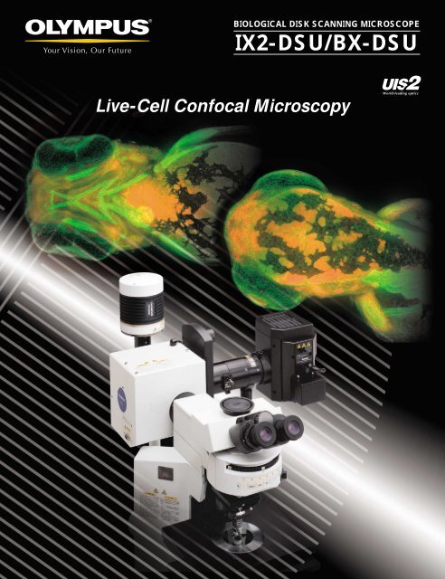
BIOLOGICAL DISK SCANNING MICROSCOPE<strong>IX2</strong>-<strong>DSU</strong>/<strong>BX</strong>-<strong>DSU</strong>Live-Cell Confocal Microscopy
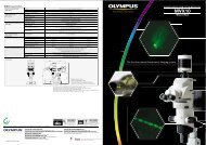
Confocal image oa cooled CCComparing 2-D images of cellular microtubule fragments taken withthe <strong>DSU</strong> (Disk Scan Unit) versus standard, widefield fluorescenceImages from the same section of PtK2 cell microtubule fragments: Image on the left was acquired using standard, widefieldfluorescence and image on the right was acquired using the <strong>DSU</strong> confocal. Note the excellent confocal effects of the <strong>DSU</strong> includingthe excellent optical sectioning and removal of out of focus light especially with thicker specimens.Photos courtesy of:OLYMPUS CORPORATION Scientific Equipment DivisionStandardWidefieldFluorescence<strong>DSU</strong> live-cellconfocal■ Acquiring confocal images using a cooled CCD cameraIn conventional fluorescence microscopy the inside structure of athick specimen cannot be observed clearly because of the significantcontribution of out of focus light from above and below thefocal plane. The disk scan unit makes it possible to reject this outof focus light by placing a rotating, slit disk in a confocal plane ofthe microscope. The result is a clear, continuous, optically crosssectionedimage that can imaged up to 30 f/p/s with a cooledCCD camera. Every image acquired by the camera has the <strong>DSU</strong>confocal effect alleviating the need for post image processing,thus improving data reliability for 3-D images.*See specifications to find which CCD cameras are recommended.■ Excellent cost-performanceBy utilizing white light sources, the <strong>DSU</strong> is more cost effectivethan laser based systems. You can use the white light source(xenon or mercury lamp) you already own, without modification.As your needs change, you can easily update the fluorescenceexcitation characteristics by adding new filter and dichromaticmirror sets to coincide with your new fluorochromes. A laserbased system often requires a new laser source for a new fluorochromes.The <strong>DSU</strong> system also supports DAPI excitation in the near UVwithout modification. Filter-mirror sets for GFP and DsRED areincluded.■ Principles of OperationFluorescence excitation light from a white light source (xenon or mercurylamp) is first filtered for the required wavelength and then reflected via adichromatic mirror. This reflected light passes through a unique, spinningslit confocal disk, (which is located in a conjugate position to the objective’sfocal plane), through the objective and focused onto the specimen.Emitted fluorescence light from the specimen is then collected by the objectiveand sent back through the confocal disk. The passing of focusedlight back through the disk produces the required confocal effects.Fluorescence emission light is then selected for wavelength by a filter andfocused on a cooled CCD camera to form visible images.C-CCDcameraFilter changer(emission side)FSFilter changer(excitation side)LampCooled CCDcameraFilter changer(emission side)DichromaticmirrorConfocaldiskFSLampFilter changer(excitation side)Confocal diskMirror cubeDichromaticmirrorCooled CCDcameraFilter changer(excitation side)DichromaticmirrorConfocal diskMirror cubeObjectiveMirrorcubeFilter changers are optional.Confocal diskSpecimen
3-D extended focus image of fruit fly brain3-D extended focus image obtained from a 230µm thick specimen via70 discrete z-sections at 2µm increments.Objective: UPlanApo20XPhoto courtesy of:Prof. Kei ItoDr. Takeshi AwasakiDepartment of Molecular BiologyTokyo University3-D extendedfocus image3-D image of swallowtail butterflyvisual central nerves3-D image obtained from a 300µm thick specimen via 90 discretez-sections at 2µm increments.Objective: UPlanApo20XPhoto courtesy of:Mitsuyo Kinoshita, Prof. Kentaro ArikawaLaboratory of Neuroethology, Graduate School of Integrated ScienceYokohama City University<strong>DSU</strong> 3-D imagebservation withD camera■ Easy operationA simple hand switch close to the operator's hand makes it easyto control such operations as inserting/removing the confocaldisk into the light path, exchanging cubes and opening/closingthe shutter. It is quick and easy to exchange between generalfluorescence observation and confocal observation.■ A choice of disks for different purposesFive different types of disks are available, with different slit widthsto suit different objectives and specimen thicknesses.Exchanging one disk for another is easy using the provided tools.In the case of the <strong>BX</strong>61WI, the best confocal effect is obtainedby selecting the appropriate disk to suit the objective in use.■ Advanced system performanceConfocal 3-D image stacks can be acquired easily using theInverted IX81 or Upright <strong>BX</strong>61 microscope platforms with theirbuilt-in, precise Z motor. Image acquisition software can be usedto control microscope, <strong>DSU</strong> and digital camera for a very easy touse complete solution to your imaging needs.<strong>BX</strong>-<strong>DSU</strong><strong>IX2</strong>-<strong>DSU</strong>