Cholangiocarcinoma in Experimental Hamsters with Long-standing ...
Cholangiocarcinoma in Experimental Hamsters with Long-standing ...
Cholangiocarcinoma in Experimental Hamsters with Long-standing ...
You also want an ePaper? Increase the reach of your titles
YUMPU automatically turns print PDFs into web optimized ePapers that Google loves.
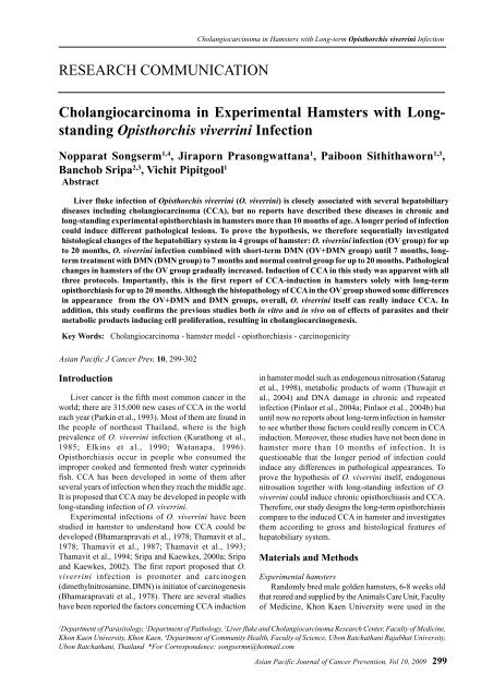
<strong>Cholangiocarc<strong>in</strong>oma</strong> <strong>in</strong> <strong>Hamsters</strong> <strong>with</strong> <strong>Long</strong>-term Opisthorchis viverr<strong>in</strong>i InfectionRESEARCH COMMUNICATION<strong>Cholangiocarc<strong>in</strong>oma</strong> <strong>in</strong> <strong>Experimental</strong> <strong>Hamsters</strong> <strong>with</strong> <strong>Long</strong>stand<strong>in</strong>gOpisthorchis viverr<strong>in</strong>i InfectionNopparat Songserm 1,4 , Jiraporn Prasongwattana 1 , Paiboon Sithithaworn 1,3 ,Banchob Sripa 2,3 , Vichit Pipitgool 1AbstractLiver fluke <strong>in</strong>fection of Opisthorchis viverr<strong>in</strong>i (O. viverr<strong>in</strong>i) is closely associated <strong>with</strong> several hepatobiliarydiseases <strong>in</strong>clud<strong>in</strong>g cholangiocarc<strong>in</strong>oma (CCA), but no reports have described these diseases <strong>in</strong> chronic andlong-stand<strong>in</strong>g experimental opisthorchiasis <strong>in</strong> hamsters more than 10 months of age. A longer period of <strong>in</strong>fectioncould <strong>in</strong>duce different pathological lesions. To prove the hypothesis, we therefore sequentially <strong>in</strong>vestigatedhistological changes of the hepatobiliary system <strong>in</strong> 4 groups of hamster: O. viverr<strong>in</strong>i <strong>in</strong>fection (OV group) for upto 20 months, O. viverr<strong>in</strong>i <strong>in</strong>fection comb<strong>in</strong>ed <strong>with</strong> short-term DMN (OV+DMN group) until 7 months, longtermtreatment <strong>with</strong> DMN (DMN group) to 7 months and normal control group for up to 20 months. Pathologicalchanges <strong>in</strong> hamsters of the OV group gradually <strong>in</strong>creased. Induction of CCA <strong>in</strong> this study was apparent <strong>with</strong> allthree protocols. Importantly, this is the first report of CCA-<strong>in</strong>duction <strong>in</strong> hamsters solely <strong>with</strong> long-termopisthorchiasis for up to 20 months. Although the histopathology of CCA <strong>in</strong> the OV group showed some differences<strong>in</strong> appearance from the OV+DMN and DMN groups, overall, O. viverr<strong>in</strong>i itself can really <strong>in</strong>duce CCA. Inaddition, this study confirms the previous studies both <strong>in</strong> vitro and <strong>in</strong> vivo on of effects of parasites and theirmetabolic products <strong>in</strong>duc<strong>in</strong>g cell proliferation, result<strong>in</strong>g <strong>in</strong> cholangiocarc<strong>in</strong>ogenesis.Key Words: <strong>Cholangiocarc<strong>in</strong>oma</strong> - hamster model - opisthorchiasis - carc<strong>in</strong>ogenicityAsian Pacific J Cancer Prev, 10, 299-302IntroductionLiver cancer is the fifth most common cancer <strong>in</strong> theworld; there are 315,000 new cases of CCA <strong>in</strong> the worldeach year (Park<strong>in</strong> et al., 1993). Most of them are found <strong>in</strong>the people of northeast Thailand, where is the highprevalence of O. viverr<strong>in</strong>i <strong>in</strong>fection (Kurathong et al.,1985; Elk<strong>in</strong>s et al., 1990; Watanapa, 1996).Opisthorchiasis occur <strong>in</strong> people who consumed theimproper cooked and fermented fresh water cypr<strong>in</strong>oidsfish. CCA has been developed <strong>in</strong> some of them afterseveral years of <strong>in</strong>fection when they reach the middle age.It is proposed that CCA may be developed <strong>in</strong> people <strong>with</strong>long-stand<strong>in</strong>g <strong>in</strong>fection of O. viverr<strong>in</strong>i.<strong>Experimental</strong> <strong>in</strong>fections of O. viverr<strong>in</strong>i have beenstudied <strong>in</strong> hamster to understand how CCA could bedeveloped (Bhamarapravati et al., 1978; Thamavit et al.,1978; Thamavit et al., 1987; Thamavit et al., 1993;Thamavit et al., 1994; Sripa and Kaewkes, 2000a; Sripaand Kaewkes, 2002). The first report proposed that O.viverr<strong>in</strong>i <strong>in</strong>fection is promoter and carc<strong>in</strong>ogen(dimethylnitrosam<strong>in</strong>e, DMN) is <strong>in</strong>itiator of carc<strong>in</strong>ogenesis(Bhamarapravati et al., 1978). There are several studieshave been reported the factors concern<strong>in</strong>g CCA <strong>in</strong>duction<strong>in</strong> hamster model such as endogenous nitrosation (Sataruget al., 1998), metabolic products of worm (Thuwajit etal., 2004) and DNA damage <strong>in</strong> chronic and repeated<strong>in</strong>fection (P<strong>in</strong>laor et al., 2004a; P<strong>in</strong>laor et al., 2004b) butuntil now no reports about long-term <strong>in</strong>fection <strong>in</strong> hamsterto see whether those factors could really concern <strong>in</strong> CCA<strong>in</strong>duction. Moreover, those studies have not been done <strong>in</strong>hamster more than 10 months of <strong>in</strong>fection. It isquestionable that the longer period of <strong>in</strong>fection could<strong>in</strong>duce any differences <strong>in</strong> pathological appearances. Toprove the hypothesis of O. viverr<strong>in</strong>i itself, endogenousnitrosation together <strong>with</strong> long-stand<strong>in</strong>g <strong>in</strong>fection of O.viverr<strong>in</strong>i could <strong>in</strong>duce chronic opisthorchiasis and CCA.Therefore, our study designs the long-term opisthorchiasiscompare to the <strong>in</strong>duced CCA <strong>in</strong> hamster and <strong>in</strong>vestigatesthem accord<strong>in</strong>g to gross and histological features ofhepatobiliary system.Materials and Methods<strong>Experimental</strong> hamstersRandomly bred male golden hamsters, 6-8 weeks oldthat reared and supplied by the Animals Care Unit, Facultyof Medic<strong>in</strong>e, Khon Kaen University were used <strong>in</strong> the1Department of Parasitology, 2 Department of Pathology, 3 Liver fluke and <strong>Cholangiocarc<strong>in</strong>oma</strong> Research Center, Faculty of Medic<strong>in</strong>e,Khon Kaen University, Khon Kaen, 4 Department of Community Health, Faculty of Science, Ubon Ratchathani Rajabhat University,Ubon Ratchathani, Thailand *For Correspondence: songsermn@hotmail.comAsian Pacific Journal of Cancer Prevention, Vol 10, 2009 299
Nopparat Songserm et alstudy. The protocol was approved by the Animal EthicCommittee of the Faculty of Medic<strong>in</strong>e, Khon KaenUniversity.O. viverr<strong>in</strong>i metacercaria and their <strong>in</strong>duction to hamsterCypr<strong>in</strong>oid fish from endemic area of Khon KaenProv<strong>in</strong>ce were collected, m<strong>in</strong>ced and digested <strong>with</strong> peps<strong>in</strong>.Metacercaria of O. viverr<strong>in</strong>i were selected and countedunder the stereomicroscope and after that they were fedon hamster via gastric tube.Carc<strong>in</strong>ogen adm<strong>in</strong>istrationDMN (Sigma, USA) was diluted <strong>in</strong> distilled waterunder biohazard hood. The f<strong>in</strong>al concentration of solutionwas 12.5 ppm, kept <strong>in</strong> a dark conta<strong>in</strong>er, and transferred tobrown bottles used only for hamsters <strong>with</strong> DMNadm<strong>in</strong>istration <strong>in</strong> a separate room.Short-term adm<strong>in</strong>istration: 12.5 ppm of DMN solutionwas supplied to hamster for 10 weeks, start<strong>in</strong>g from thetime of O. viverr<strong>in</strong>i <strong>in</strong>fection. After that they receivednormal dr<strong>in</strong>k<strong>in</strong>g water until sacrificed. <strong>Long</strong>-termadm<strong>in</strong>istration: hamsters received the same concentrationof DMN solution from the beg<strong>in</strong>n<strong>in</strong>g until the day ofsacrifice.Study design1. OV group: hamsters <strong>in</strong>fected <strong>with</strong> 50 metacercariaof O. viverr<strong>in</strong>i were divided <strong>in</strong>to seven groups. Each group(4-6 hamsters/group) was sacrificed accord<strong>in</strong>g to theschedule of 4th, 8th, 12th, 14th, 16th, 18th and 20th monthafter <strong>in</strong>fection (see Figure 1).2. OV+DMN group: hamsters treated <strong>with</strong> 50metacercaria of O. viverr<strong>in</strong>i comb<strong>in</strong>e <strong>with</strong> 12.5 ppm ofDMN for 10 weeks. There are 7 groups of hamsters; eachgroup (4-6 hamsters/group) was sacrificed accord<strong>in</strong>g tothe schedule of 1st, 2nd, 3rd, 4th, 5th, 6th and 7th monthafter treatment.3. DMN group: hamsters adm<strong>in</strong>istrated <strong>with</strong>cont<strong>in</strong>uously long-term of DMN were divided <strong>in</strong>to 2groups. Each group (4-6 hamsters/group) was sacrificedaccord<strong>in</strong>g to the schedule of 4th and 7th month aftertreatment.4. Control group: hamsters received neithermetacercaria nor DMN were reared <strong>in</strong> the same conditionas the experimental hamsters. There are 4 groups ofhamsters; each group (4-6 hamsters/group) was sacrificedControlDMNOV+DMNFigure 1. <strong>Experimental</strong> Procedure for CCA Induction<strong>in</strong> <strong>Hamsters</strong>. DMN, dimethylnitrosam<strong>in</strong>e; OV, Opisthorchisviverr<strong>in</strong>i; *, period of DMN treatment; arrows, sacrifice po<strong>in</strong>ts300**OV| | | | | | | | | | | | | |0 1 2 3 4 5 6 7 8 12 14 16 18 20Months of treatmentAsian Pacific Journal of Cancer Prevention, Vol 10, 2009accord<strong>in</strong>g to the schedule of 4th, 8th, 12th and 20th month.Blood was drawn from the orbital plexus of hamsters.After that, hamsters of each group were sacrificed underdeep anesthesia, blood was also drawn directly from theheart and the liver was removed. The livers were exam<strong>in</strong>edfor gross pathology and histopathology. Four lobes of theliver were studied for hepatic nodules, hepatic fibrosisand tumor mass. Later the livers were fixed <strong>in</strong> 10% bufferformal<strong>in</strong>, serially sliced <strong>in</strong> a 4-5 mm thickness, embedded<strong>in</strong> paraff<strong>in</strong>. Sections were cut at 4 µm thickness and sta<strong>in</strong>ed<strong>with</strong> hematoxyl<strong>in</strong>-eos<strong>in</strong>. Histopathological features of bileduct epithelium (e.g. <strong>in</strong>flammatory cell <strong>in</strong>filtration, bileduct dilatation, periductal fibrosis, bile duct epithelialhyperplasia, cholangiofibrosis, goblet cell metaplasia,dysplasia and CCA) and hepatic parenchyma (e.g.cirrhosis and hepatocellular carc<strong>in</strong>oma, HCC) werecarefully <strong>in</strong>vestigated under light microscope.ResultsPathological changes <strong>in</strong> hamsters <strong>with</strong> O. viverr<strong>in</strong>i<strong>in</strong>fection alone gradually <strong>in</strong>creased over time (Figure 2).Hepatic nodules and hemorrhagic spots <strong>in</strong> 8th, 12th, 14thand 18th month post-<strong>in</strong>fection (p.i.). However, hamstersof this group no showed tumor mass. <strong>Hamsters</strong> ofOV+DMN group showed tumor mass s<strong>in</strong>ce 3rd monthand severity was cont<strong>in</strong>uously <strong>in</strong>creased. <strong>Hamsters</strong> ofDMN group showed tumor mass <strong>in</strong> all hamsters of 7thmonth, but not <strong>in</strong> hamsters of 4th month after cont<strong>in</strong>uouslylong-term adm<strong>in</strong>istration of DMN.Histopathological f<strong>in</strong>d<strong>in</strong>gs of all experimental groupsPathological f<strong>in</strong>d<strong>in</strong>g ofhepatobiliary system(%)Pathological f<strong>in</strong>d<strong>in</strong>g ofhepatobiliary system(%)8006004002000OV group4 mo. 8 mo. 12mo. 14mo. 16mo. 18mo. 20mo.Monthsof <strong>in</strong>fectionNOR INF BDD PDF HYP CLF MET DYS CIR CCA HCC60040020004 mo. 7 mo.DMN groupMonthsof adm<strong>in</strong>istrationNOR INF BDD PDF HYP CLF MET DYS CIR CCA HCCFigure 2. Pathological F<strong>in</strong>d<strong>in</strong>gs for the OV and DMN<strong>Experimental</strong> Groups Accord<strong>in</strong>g to the Duration ofInfection/Treatment. NOR, normal appearance; INF,<strong>in</strong>flammatory cell <strong>in</strong>filtration; BDD, bile duct dilatation; PDF,periductal fibrosis; HYP, bile duct epithelial hyperplasia; CLF,cholangiofibrosis; MET, goblet cell metaplasia; DYS, dysplasia;CIR, cirrhosis; CCA, cholangiocarc<strong>in</strong>oma; and HCC,hepatocellular carc<strong>in</strong>oma
showed identical features of bile duct epithelium such as<strong>in</strong>flammatory cells <strong>in</strong>filtration, periductal fibrosis, bileduct epithelial hyperplasia, cholangiofibrosis. Cirrhosis,histopathological feature of hepatic parenchyma wasfound only <strong>in</strong> OV group. Importantly, dysplasia was alsofound only <strong>in</strong> OV group as early as 14th month p.i. , whilegoblet cell metaplasia was showed <strong>in</strong> only group ofcarc<strong>in</strong>ogenic-<strong>in</strong>duced hamsters (OV+DMN and DMNgroups).Although no gross appearance of tumor mass <strong>in</strong> OVgroup, histopathological features of CCA was found <strong>in</strong> 2of 9 hamsters <strong>in</strong> 14th and 20th month p.i., respectively.All hamsters of OV+DMN group from 3rd month p.i. andall hamsters of DMN group <strong>in</strong> 7th month after long-termtreatment showed CCA. In addition, 1 of 5 hamsters ofDMN group <strong>in</strong> 7th month showed CCA together <strong>with</strong>HCC. Interest<strong>in</strong>gly, histopathology of CCA <strong>in</strong> OV groupshowed some appearances different from OV+DMN andDMN groups.Intensity of malignant foci of CCA was quantitativeanalysis by histological f<strong>in</strong>d<strong>in</strong>gs. Approximately 1-10 fociof 0.06 cm per section were found <strong>in</strong> only right lobe liverof OV group at 14th and 20th month. For OV+DMNgroup, the malignant foci were found throughout the liver,mostly <strong>in</strong> right, left, medium and caudal lobes, respectivelyas early as 3rd month post treatment. Distributions ofmalignant foci <strong>with</strong> size of 0.1-1.53 cm were found morethan 10 foci per section. Similarly, malignant foci <strong>with</strong>0.125-1 cm were showed <strong>in</strong> all hamsters of DMN groupat 7th month after treatment, but mostly distribute <strong>in</strong> left,right, medium and caudal lobes, respectively. In addition,malignant foci were not found <strong>in</strong> normal control hamsters.However, the distributions of malignant foci were notstatistically significant among 3 liver lobes, except caudallobe <strong>in</strong> carc<strong>in</strong>ogenic-<strong>in</strong>duced groups (OV+DMN andDMN).DiscussionIn the present study CCA <strong>in</strong>duction was successful<strong>with</strong> all three protocols. Until now, no previous reportson CCA <strong>in</strong>duction <strong>in</strong> hamsters by long-term <strong>in</strong>fection ofO. viverr<strong>in</strong>i alone for up to 10 months of <strong>in</strong>fection and bycont<strong>in</strong>uous adm<strong>in</strong>istration of DMN alone more than 10weeks. Although O. viverr<strong>in</strong>i-<strong>in</strong>fected hamsters have beenreported at 9th month p.i., not showed at other time po<strong>in</strong>tof experiment (Thamavit et al., 1993; Sithithaworn et al.,2002).In this study, not all hamsters <strong>with</strong> O. viverr<strong>in</strong>i<strong>in</strong>fection alone develop to the CCA at the same time.Histopathological appearance of CCA was found <strong>in</strong> halfnumber of hamsters at 14th and 20th month p.i. Indeed,due to cancer <strong>in</strong>duction may be responsible to <strong>in</strong>dividualdifference of genetics that code for DNA repair<strong>in</strong>g,immune response and <strong>in</strong>flammation (Jaiswal et al., 2000).Although some hamsters did not showed histopathologyof CCA, all of them showed cholangiofibrosis, goblet cellmetaplasia and dysplasia. Accord<strong>in</strong>g to these criteria ofhistopathology, they were identified to be precancerouslesion of CCA (Thamavit et al., 1993). Differently, the<strong>in</strong>cidence of CCA <strong>in</strong> the group of carc<strong>in</strong>ogenic-<strong>in</strong>duced<strong>Cholangiocarc<strong>in</strong>oma</strong> <strong>in</strong> <strong>Hamsters</strong> <strong>with</strong> <strong>Long</strong>-term Opisthorchis viverr<strong>in</strong>i Infectionhamsters (OV+DMN and DMN groups) was 100% at thesame time. All hamsters of OV+DMN group from 3rdmonth and all of DMN group <strong>in</strong> 7th month have developedthe CCA. These results may derive from the differentmechanisms of carc<strong>in</strong>ogenesis among the two groups ofCCA-<strong>in</strong>duced hamsters, <strong>with</strong> or <strong>with</strong>out DMN. Moredetail is needed to be further study.There are many cancers are associated <strong>with</strong> <strong>in</strong>fectiousagents, e.g., virus (hepatitis B and C), bacteria(Helicobactor pylori), and parasite (O. viverr<strong>in</strong>i), whichcan promote carc<strong>in</strong>ogenesis through the <strong>in</strong>duction ofchronic <strong>in</strong>flammatory states (Park<strong>in</strong>, 2006). Accord<strong>in</strong>gto CCA, endogenous nitrosation, disability of DNArepair<strong>in</strong>g system and decrease <strong>in</strong> ability of tumorsuppressor function are the major causes of carc<strong>in</strong>ogenesis(Satarug et al., 1998; P<strong>in</strong>laor et al., 2004a). In the sameway <strong>with</strong> our experiment, O. viverr<strong>in</strong>i alone can <strong>in</strong>duceCCA <strong>in</strong> long-stand<strong>in</strong>g <strong>in</strong>fection. This occurrence mayderive from the actions of oxygen radicals, e.g., nitricoxide (NO) released from cytok<strong>in</strong>es that can <strong>in</strong>duceoxidative DNA damage to the <strong>in</strong>fected biliary epithelium(P<strong>in</strong>laor et al., 2003; P<strong>in</strong>laor et al., 2004a). Furthermore,repeated <strong>in</strong>fections <strong>with</strong> O. viverr<strong>in</strong>i contribute to ris<strong>in</strong>gDNA damage through <strong>in</strong>creased nitric oxide synthaseexpression <strong>in</strong> biliary epithelium (P<strong>in</strong>laor et al., 2004b).In summation, NO not only <strong>in</strong>duces DNA damage buthas been reported to mediate DNA repair <strong>in</strong>hibition(Jaiswal et al., 2000; Jaiswal et al., 2001).At present, the diagnosis of CCA can be elusive; it isoften not made until advanced disease is present and at astage when a curative surgical resection is not feasible.Attempts to solve these problems have been done <strong>in</strong> manyaspects of CCA both <strong>in</strong> man and experimental animals.The hamster has been described as the suitableexperimental animals due to the natural pathway of diseaseas same as <strong>in</strong> man. The O. viverr<strong>in</strong>i-<strong>in</strong>fected hamster wasused to exam<strong>in</strong>e the pathology of hepatobiliary system;early pathological changes consisted of <strong>in</strong>flammation,periductal fibrosis, and proliferative responses, <strong>in</strong>clud<strong>in</strong>gepithelial hyperplasia, goblet cell metaplasia, andadenomatous hyperplasia, may represent predispos<strong>in</strong>glesions that enhance susceptibility of DNA to carc<strong>in</strong>ogens(Bhamarapravati et al., 1978; Sripa and Kaewkes, 2000a).In chronic <strong>in</strong>fection, periportal and periductal fibrosis werethe most prom<strong>in</strong>ent histological features. However, the<strong>in</strong>flammatory responses are less severe <strong>in</strong> chronic<strong>in</strong>fection than the acute one suggest<strong>in</strong>g thatimmunomodulation may occur (Sripa and Kaewkes,2000a).There are several researches have been studied onpathogenesis of liver fluke-<strong>in</strong>duced CCA, both worm’sown and its metabolic products. Mechanical <strong>in</strong>jury fromfluke’s suckers hook on the biliary epithelium for feed<strong>in</strong>gand mov<strong>in</strong>g, result<strong>in</strong>g <strong>in</strong> tissue damage even early <strong>in</strong><strong>in</strong>fection (Bhamarapravati et al., 1978). As the flukematures, the lesion becomes more obvious and ulcerated.The metabolic products of liver fluke were secreted orexcreted from the tegument and excretory open<strong>in</strong>gs <strong>in</strong>tobile or culture medium <strong>in</strong> vitro (Wongratanacheew<strong>in</strong> etal., 1988; Sripa and Kaewkes, 2000b). The metabolicproducts themselves may be toxic to or <strong>in</strong>teract <strong>with</strong> theAsian Pacific Journal of Cancer Prevention, Vol 10, 2009 301
Nopparat Songserm et albiliary epithelium for <strong>in</strong>duc<strong>in</strong>g host immune responses(Har<strong>in</strong>asuta et al., 1984). There are some studies have beenattempted to describe how metabolic products of liverfluke <strong>in</strong>duced cell proliferation, but most of them wereperformed <strong>in</strong> vitro, e.g., mouse fibroblast (Thuwajit et al.,2004) and human CCA cell l<strong>in</strong>e (Sripa et al., 2005). For<strong>in</strong> vivo study, bile duct epithelial hyperplasia, as a form ofcell proliferation was found <strong>in</strong> hamsters <strong>with</strong>opisthorchiasis (Bhamarapravati et al., 1978; Sripa andKaewkes, 2000a), <strong>with</strong> presentation of O. viverr<strong>in</strong>iantigens <strong>in</strong> periductal of bile duct, macrophages,epithelioid cells, and giant cells of the granuloma, asdetected by immunohistochemistry (Sripa and Kaewkes,2000a).Although <strong>in</strong> vivo study <strong>in</strong> hamsters <strong>in</strong>dicated that O.viverr<strong>in</strong>i itself and its metabolic products can <strong>in</strong>duce cellproliferation, we here showed for the first time that longterm exposure to the parastite alone can really <strong>in</strong>duceCCA.ReferencesBhamarapravati N, Thamavit W, Vajrasthira, S (1978). Liverchanges <strong>in</strong> hamsters <strong>in</strong>fected <strong>with</strong> a liver fluke of man,Opisthorchis viverr<strong>in</strong>i. Am J Trop Med Hyg, 27, 787-94.Elk<strong>in</strong>s DB, Haswell-Elk<strong>in</strong>s MR, Mairiang E, et al (1990). Ahigh frequency of hepatobiliary disease and suspectedcholangiocarc<strong>in</strong>oma associated <strong>with</strong> heavy Opisthorchisviverr<strong>in</strong>i <strong>in</strong>fection <strong>in</strong> a small community <strong>in</strong> north-eastThailand. Trans R Soc Trop Med Hyg, 84, 715-9.Har<strong>in</strong>asuta T, Riganti M, Bunnag D (1984). Opisthorchisviverr<strong>in</strong>i <strong>in</strong>fection: pathogenesis and cl<strong>in</strong>ical features.Arzneimittelforschung, 34, 1167-9.Jaiswal M, LaRusso NF, Burgart LJ, Gores GJ (2000).Inflammatory cytok<strong>in</strong>es <strong>in</strong>duce DNA damage and <strong>in</strong>hibitDNA repair <strong>in</strong> cholangiocarc<strong>in</strong>oma cells by a nitric oxidedependentmechanism. Cancer Res, 60, 184-90.Jaiswal M, LaRusso NF, Shapiro RA, Billiar TR, Gores GJ(2001). Nitric oxide-mediated <strong>in</strong>hibition of DNA repairpotentiates oxidative DNA damage <strong>in</strong> cholangiocytes.Gastroenterology, 120, 190-9.Kurathong S, Lerdverasirikul P, Wongpaitoon V, et al (1985).Opisthorchis viverr<strong>in</strong>i <strong>in</strong>fection and cholangiocarc<strong>in</strong>oma. Aprospective, case-controlled study. Gastroenterology, 89,151-6.Park<strong>in</strong> DM (2006). The global health burden of <strong>in</strong>fectionassociatedcancers <strong>in</strong> the year 2002. Int J Cancer, 118, 3030-44.Park<strong>in</strong> DM, Oshima H, Srivatanakul P, Vatanasapt V (1993).<strong>Cholangiocarc<strong>in</strong>oma</strong>: epidemiology, mechanism ofcarc<strong>in</strong>ogenesis and prevention. Cancer EpidemiolBiomarkers Prev, 2, 537-44.P<strong>in</strong>laor S, Hiraku Y, Ma N, et al (2004a). Mechanism of NOmediatedoxidative and nitrative DNA damage <strong>in</strong> hamsters<strong>in</strong>fected <strong>with</strong> Opisthorchis viverr<strong>in</strong>i: a model of<strong>in</strong>flammation-mediated carc<strong>in</strong>ogenesis. Nitric Oxide, 11,175-83.P<strong>in</strong>laor S, Ma N, Hiraku Y, et al (2004b). Repeated <strong>in</strong>fection<strong>with</strong> Opisthorchis viverr<strong>in</strong>i <strong>in</strong>duces accumulation of 8-nitroguan<strong>in</strong>e and 8-oxo-7,8-dihydro-2'-deoxyguan<strong>in</strong>e <strong>in</strong> thebile duct of hamsters via <strong>in</strong>ducible nitric oxide synthase.Carc<strong>in</strong>ogenesis, 25, 1535-42.P<strong>in</strong>laor S, Yongvanit P, Hiraku Y, et al (2003). 8-nitroguan<strong>in</strong>eformation <strong>in</strong> the liver of hamsters <strong>in</strong>fected <strong>with</strong> Opisthorchisviverr<strong>in</strong>i. Biochem Biophys Res Commun, 309, 567-71.302Asian Pacific Journal of Cancer Prevention, Vol 10, 2009Satarug S, Haswell-Elk<strong>in</strong>s MR, Sithithaworn P, et al (1998).Relationships between the synthesis of N-nitrosodimethylam<strong>in</strong>e and immune responses to chronic<strong>in</strong>fection <strong>with</strong> the carc<strong>in</strong>ogenic parasite, Opisthorchisviverr<strong>in</strong>i, <strong>in</strong> men. Carc<strong>in</strong>ogenesis, 19, 485-91.Sithithaworn P, Ando K, Limviroj W, et al (2002). Expressionof tenasc<strong>in</strong> <strong>in</strong> bile duct cancer of hamster liver by comb<strong>in</strong>edtreatment of dimethylnitrosam<strong>in</strong>e <strong>with</strong> Opisthorchis viverr<strong>in</strong>i<strong>in</strong>fections. J Helm<strong>in</strong>thol, 76, 261-8.Sripa B, Kaewkes S (2000a). Localisation of parasite antigensand <strong>in</strong>flammatory responses <strong>in</strong> experimental opisthorchiasis.Int J Parasitol, 30, 735-40.Sripa B, Kaewkes S (2000b). Relationship between parasitespecificantibody responses and <strong>in</strong>tensity of Opisthorchisviverr<strong>in</strong>i <strong>in</strong>fection <strong>in</strong> hamsters. Parasite Immunol, 22, 139-45.Sripa B, Kaewkes S (2002). Gall bladder and extrahepatic bileduct changes <strong>in</strong> Opisthorchis viverr<strong>in</strong>i-<strong>in</strong>fected hamsters.Acta Trop, 83, 29-36.Sripa B, Leungwattanawanit S, Nitta T, et al (2005).Establishment and characterization of an opisthorchiasisassociatedcholangiocarc<strong>in</strong>oma cell l<strong>in</strong>e (KKU-100). WorldJ Gastroenterol, 11, 3392-7.Thamavit W, Bhamarapravati N, Sahaphong S, Vajrasthira S,Angsubhakorn S (1978). Effects of dimethylnitrosam<strong>in</strong>e on<strong>in</strong>duction of cholangiocarc<strong>in</strong>oma <strong>in</strong> Opisthorchis viverr<strong>in</strong>i<strong>in</strong>fectedSyrian golden hamsters. Cancer Res, 38, 4634-9.Thamavit W, Kongkanuntn R, Tiwawech D, Moore MA (1987).Level of Opisthorchis <strong>in</strong>festation and carc<strong>in</strong>ogen dosedependenceof cholangiocarc<strong>in</strong>oma <strong>in</strong>duction <strong>in</strong> Syriangolden hamsters. Virchows Arch B Cell Pathol Incl MolPathol, 54, 52-8.Thamavit W, Pairojkul C, Tiwawech D, Itoh M, Shirai T, Ito N(1993). Promotion of cholangiocarc<strong>in</strong>ogenesis <strong>in</strong> the hamsterliver by bile duct ligation after dimethylnitrosam<strong>in</strong>e<strong>in</strong>itiation. Carc<strong>in</strong>ogenesis, 14, 2415-7.Thamavit W, Pairojkul C, Tiwawech D, Shirai T, Ito N (1994).Strong promot<strong>in</strong>g effect of Opisthorchis viverr<strong>in</strong>i <strong>in</strong>fectionon dimethylnitrosam<strong>in</strong>e-<strong>in</strong>itiated hamster liver. Cancer Lett,78, 121-5.Thuwajit C, Thuwajit P, Kaewkes S, et al (2004). Increased cellproliferation of mouse fibroblast NIH-3T3 <strong>in</strong> vitro <strong>in</strong>ducedby excretory/secretory product(s) from Opisthorchisviverr<strong>in</strong>i. Parasitology, 129, 455-64.Watanapa P (1996). <strong>Cholangiocarc<strong>in</strong>oma</strong> <strong>in</strong> patients <strong>with</strong>opisthorchiasis. Br J Surg, 83, 1062-64.Wongratanacheew<strong>in</strong> S, Bunnag D, Vaeusorn N, Siris<strong>in</strong>ha S(1988). Characterization of humoral immune response <strong>in</strong>the serum and bile of patients <strong>with</strong> opisthorchiasis and itsapplication <strong>in</strong> immunodiagnosis. Am J Trop Med Hyg, 38,356-62.


