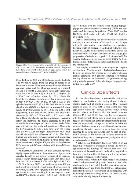
160 Chapter 15Figure 15-1. Treatment in progress <strong>with</strong> the cornea soaked<strong>with</strong> riboflavin and irradiated by the UV lamp. In the lowerright corner, one can appreciate the patient’s perspective ofthe UV lamp.µm using 3 mW/cm 2 irradiance (~5.4 J/cm 2 ) at the cornealfront surface. 14 Therefore, preoperative pachymetry isessential and must be measured in all cases. Usually, thecentral corneal thickness in keratoconus is not reducedto less than 400 µm. In corneas <strong>with</strong> less then 400 µm,the <strong>cross</strong>-<strong>linking</strong> treatment should be avoided using thesedosage parameters, and a hypotonic solution of riboflavinin physiological solution (<strong>with</strong>out Dextran T-500) shouldbe applied in order to swell the cornea before and duringthe irradiation.Considering the crystalline lens, the UVA dose of 0.65J/cm 2 (0.36 mW/cm 2 ) is far below a cataractogenous levelof 70 J/cm 2 . 15 In addition, lens damage is usually inducedby UVB light (wavelength range of 290 to 320 nm), whichhas a higher energy than UVA. Regarding retinal phototoxicity,UVA levels in the posterior segment are negligibleand comparable to ambient sun exposure.Surgical TechniqueThe procedure should be conducted under sterileconditions in the operating room. Topical anesthetic isapplied and the central 7 mm of the corneal epitheliumis removed using a blunt knife, or 30 s of application of20% alcohol. As a photosensitizer, riboflavin 0.1% solutionin Dextran T-500 20% solution is applied 15 minutesbefore the irradiation and every 2 to 3 minutes duringthe irradiation. Alternatively, a smaller area of epitheliummay be removed, provided that the time of application ofriboflavin is increased, and the presence of riboflavin inthe anterior chamber is confirmed by the presence of ayellow flare under the slit-lamp examination prior to theapplication of UVA light.After allowing riboflavin to permeate through the corneainto the anterior chamber, the irradiation is startedFigure 15-2. Fluorescence of the riboflavin under the UV lightindicates active treatment application.using a UV lamp (370 nm; Peschkemed, Huenenberg,Switzerland) at a 1-cm working distance for 30 minutesusing a 3 mW/cm 2 irradiance (~5.4 J/cm 2 ) (Figures 15-1and 15-2). After the treatment, an antibiotic eyedrop isapplied, and a bandage contact lens is fitted to the cornealsurface until reepithelialization.Indications and ClinicalResultsThe principle indication for the use of the collagen<strong>cross</strong>-<strong>linking</strong> of the cornea is to halt the procession ofectasia in diseases of the cornea, such as keratoconusand pellucid marginal degeneration. Collagen <strong>cross</strong>-<strong>linking</strong>may also be effective in the treatment and prophylaxisof iatrogenic keratectasia, resulting from laser insitu keratomileusis (LASIK). Beyond keratoectasia, thenew technique can also be used in treating corneal meltingconditions or infectious keratitis, as the <strong>cross</strong>-<strong>linking</strong>strengthens a collagenolytic cornea and UVA irradiationsterilizes the infectious agent.The first prospectively controlled clinical study thatincluded 23 eyes <strong>with</strong> moderate or advanced progressivekeratoconus showed collagen <strong>cross</strong>-<strong>linking</strong> as effective inhalting the progression of keratoconus over a period of upto 4 years. 15 In the study, a mean preoperative progressionof keratometry (max K) by 1.42 diopters (D) in 52%of eyes over a 6-month period was followed by a postoperativedecrease in 70% of eyes, revealing a reduction ofmean keratometry by 2.01 D. Moreover, the postoperativespherical equivalent (SEQ) was reduced by an average of1.14 D, whereas at the same time, 22% of the untreatedfellow control eyes had a postoperative progression ofkeratectasia by 1.48 D.Clinical studies carried out elsewhere and presentedat the 1st and 2nd International Congress of <strong>Corneal</strong>
<strong>Corneal</strong> Cross-Linking With Riboflavin and Ultraviolet Irradiation in Unstable Corneas 161Figure 15-3. Third postoperative day: right after the removal ofthe contact lens one can observe transient stromal and epithelialedema <strong>with</strong> a mild cotton-like hazy appearance <strong>with</strong>in thecorneal stroma. (Courtesy of T. Seiler, MD PhD.)Cross Linking in 2005 and 2006 showed similar findings.The prospective results from our group in Serbia for 38keratoconic eyes of 19 patients, where the more advancedeye was treated and the fellow eye served as a control,showed a 6-month postoperative statistically significantmean decrease in max K by 1.75 ± 1.24 D, SEQ by 1.94± 3.30 D, and refractive cylinder by 1.31 ± 1.96 in thetreated eyes, whereas the fellow eyes had a mean increasein max K by 0.24 ± 0.97 D, SEQ by 0.13 ± 1.00 D, andcylinder by 0.06 ± 0.83 (P < 0.01). Both the uncorrectedvisual acuity (UCVA) and best spectacle-corrected visualacuity (BSCVA) increased by 0.01 ± 0.12 and 0.04 ± 0.22,respectively, in the treated eyes, whereas the fellow eyesdecreased by 0.03 ± 0.23 and 0.02 ± 0.17, respectively,but <strong>with</strong>out statistically significant difference. Regardingsafety, the endothelial cell counts decreased by 64 ± 158c/mm 2 in the treated eyes and 20 ± 44 c/mm 2 in the felloweyes <strong>with</strong>out reaching statistical significance. Meanwhilethe IOP increased by 1.92 ± 2.22 mm Hg in the treatedeyes and 0.08 ± 2.25 mm Hg in the fellow eyes <strong>with</strong> a highstatistically significant difference (P