Small Animal - University of Minnesota
Small Animal - University of Minnesota
Small Animal - University of Minnesota
- No tags were found...

Create successful ePaper yourself
Turn your PDF publications into a flip-book with our unique Google optimized e-Paper software.
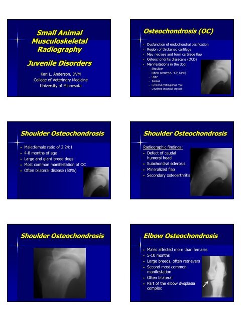
<strong>Small</strong> <strong>Animal</strong>MusculoskeletalRadiographyJuvenile DisordersKari L. Anderson, DVMCollege <strong>of</strong> Veterinary Medicine<strong>University</strong> <strong>of</strong> <strong>Minnesota</strong>Osteochondrosis (OC)• Dysfunction <strong>of</strong> endochondral ossification• Region <strong>of</strong> thickened cartilage• May necrose and form cartilage flap• Osteochondritis dissecans (OCD)• Manifestations in the dog– Shoulder– Elbow (condyle, FCP, UME)– Stifle– Tarsus– Retained cartilaginous core– Ununited anconeal processShoulder Osteochondrosis• Male:female ratio <strong>of</strong> 2.24:1• 4-88 months <strong>of</strong> age• Large and giant breed dogs• Most common manifestation <strong>of</strong> OC• Often bilateral disease (50%)Shoulder OsteochondrosisRadiographic findings:• Defect <strong>of</strong> caudalhumeral head• Subchondral sclerosis• Mineralized flap• Secondary osteoarthritisShoulder OsteochondrosisElbow Osteochondrosis• Males affected more than females• 5-10 months• Large breeds, <strong>of</strong>ten retrievers• Second most commonmanifestation• Often bilateral• Part <strong>of</strong> the elbow dysplasiacomplex
Elbow OsteochondrosisRadiographic findings:• Subchondral defect inmedial condyle• Subchondral sclerosis• Rarely see mineralizedflap• Craniolateral-caudomedial oblique• Secondary osteoarthritisStifle Osteochondrosis• Males affected more thanfemales• 5-77 months• Large breeds – Great Dane,Retrievers, Newfoundlands, , GSD• Least common manifestation• Often bilateralStifle OsteochondrosisRadiographic findings:• Commonly lateralfemoral condyle• Defect or flattening inarticular surface• Subchondral sclerosis• Intracapsular swelling• Mineralized flap• Secondary osteoarthritisTarsal Osteochondrosis• Males and females equallyaffected• 6-12 months• Large breeds – Rottweilers,Labradors• Third most commonmanifestation• 40% bilateralTarsal OsteochondrosisRadiographic findings:• Medial trochlear ridge mostcommon• DP and flexed lateral views• Widening <strong>of</strong> joint space• Flat or malshapenedtrochlear ridge• Subchondral sclerosis• Caudal mineralized flap• Secondary osteoarthritis• Tibiotarsal swellingRetained CartilaginousCore• OC <strong>of</strong> the distal ulnar physis• 6-12 months• Large and giant breeds – SaintBernard, Great Dane, Setters• Often incidental• May present with angular limbdeformity
Retained CartilaginousCoreRadiographic findings:• Cone shaped lucency indistal ulnar metaphysis• Smooth and symmetrical inasymptomatic patients• Irregular and asymmetricalin symptomatic patients• Usually bilaterallysymmetricRetained CartilaginousCoreCanine Hip Dysplasia• 5-12 months <strong>of</strong> age• All breeds affected, also cats• Underlying process is jointlaxity– Leads to abnormal hipdevelopment– Secondary osteoarthritis• May be unilateralRadiographic Evaluation<strong>of</strong> HipsExtended limb VD view:• Used by OFA• Limbs extended and parallel• Patellas superimposed onmidline• Femurs cross ischiatictuberosity equally• Wings <strong>of</strong> ilia and obturatorforamina symmetricRadiographic Evaluation<strong>of</strong> HipsPennHIPPennHIP:• Distraction/compressiontechnique• Quantitative measurement <strong>of</strong>passive hip laxity – DI• Probability <strong>of</strong> developing hipdysplasia directly related to DI• Three views: distraction,compression, extended limbVD
Radiographic Evaluation<strong>of</strong> HipsNordberg angle:• Measurement made fromextended limb VD – numberrepresents laxity• Normal ≥ 105º• Abnormal ≤ 90º• Borderline 90º to 105ºCanine Hip DysplasiaNormal Radiographic findings:• Deep cup-shapedacetabulum• Smooth circular margin <strong>of</strong>femoral head• >2/3 <strong>of</strong> head within dorsalrim• Parallel joint space• Narrow femoral neckCanine Hip DysplasiaCanine Hip DysplasiaAbnormal Radiographic findings:• Shallow acetabulum• Flattening <strong>of</strong> femoral head• Subluxation <strong>of</strong> hip• Thickened femoral necks• Subchondral sclerosis• Secondary osteoarthritisCanine Hip DysplasiaCanine Hip Dysplasia
Fragmented Medial CoronoidProcessUnunited AnconealProcess• Males affected twice as <strong>of</strong>ten• 5-12 months <strong>of</strong> age• Large breeds – GSD• Normal anconeal process fuses by5 months• May be bilateral (20-35%)Ununited AnconealProcessRadiographic findings:• Radiolucent lineseparating the anconealprocess from theolecranon after 20 wks• Line generally irregularand <strong>of</strong> variable width• Ends sclerotic, secondaryosteoarthritis• Flexed lateralOFA Evaluation <strong>of</strong> ElbowDysplasia• Must be 2 years <strong>of</strong> age• Extreme flexed lateral views• No grading for non-dysplasticelbows• Dysplastic classification:– Grade I – min bone formation (5 mm)Panosteitis• Males affected four times more<strong>of</strong>ten• 5-12 months• Large and giant breeds – GSD,Great Danes, Dobermans, Retrievers,Basset hounds• Self-limitinglimiting dz affecting longbones• Not an inflammatory processPanosteitisRadiographic findings:• Early blurring and accentuation <strong>of</strong>trabecular bone• Diminished cortico-medullarydefinition• Patchy, nodular medullaryopacities• Smooth periosteal new bone• Eventual normal remodeling
PanosteitisHypertrophic Osteodystrophy(HOD)• 2-66 months <strong>of</strong> age• Large and giant breeds – GreatDane, boxer, GSD, weimaraner• Self-limitinglimiting dz <strong>of</strong> metaphyses <strong>of</strong>long bones• Neutrophilic inflammatoryresponse with necrosis,hemorrhage, osteoclastsHypertrophic Osteodystrophy(HOD)Radiographic findings:• Radiolucent lines inmetaphyses, parallel andadjacent to physis• Metaphyseal flaring andsclerosis• Periosteal “cuffing”• S<strong>of</strong>t tissue swelling• Premature closure <strong>of</strong> physismay occurHypertrophic Osteodystrophy(HOD)Legg-CalvCalvé-PerthesDisease• AKA: aseptic necrosis <strong>of</strong> thefemoral head• 4-10 months <strong>of</strong> age• Toy and small breeds – Yorkies,Toy Poodles, Poms, , Chihuahua,terriers, Pug, Dachshund• Usually unilateralLegg-CalvCalvé-PerthesDiseaseRadiographic findings:• Radiolucencies insubchondral bone• Decreased opacity in femoralepiphysis and metaphysis• Flattening <strong>of</strong> head• Remodeling <strong>of</strong> acetabulum• Secondary osteoarthritis
Feline SyndromePatellar Luxation• Young and immature dogs• Toy and miniature breeds, but also largebreeds• Congenital or developmental deformities <strong>of</strong>rear limbs predispose to patellar luxation– Abnormal femoral neck angles– Lateral bowing <strong>of</strong> femur– Hypoplastic medial femoral condyle– Rotation <strong>of</strong> tibial tuberosity– Shallow trochlear groove• May be traumatic with concurrent cruciatedzPatellar LuxationPatellar LuxationRadiographic findings:• Often don’t t appreciate on radiographs• Include hip, femur, proximal tibia• May see patella superimposed overfemoral trochlea• May see secondary osteoarthritisGrowth Plate Injuries• Skeletally immature patients• Trauma is the most common cause,other causes include HOD, retainedcartilaginous core, chondrodysplasias• Any physis, but most clinicallysignificant in distal antebrachium• Secondary angular limb deformitiesGrowth Plate Injuries• Salter Harris fractures:– Type I: physeal– Type II: physeal-metaphyseal– Type III: physeal-epiphysealepiphyseal– Type IV: physeal-epiphysealepiphyseal-metaphyseal– Type V: compression physeal– Type VI: eccentric physeal impactionresulting in transphyseal bridging
Growth Plate InjuriesRadiographic findings:• Affected physis may or may notappear closed• May be affected entirely orpartially• Radiograph both limbs• Secondary angular limbdeformities and jointincongruenciesGrowth Plate InjuriesPremature closure <strong>of</strong> distal ulna:• Partial or complete physealclosure• Shortened ulna• Cranial and medial bowing <strong>of</strong>radius• Carpal valgus• Humero-ulnarulnar jointsubluxation• Secondary osteoarthritisGrowth Plate InjuriesGrowth Plate InjuriesPremature closure <strong>of</strong> distal radius:• Partial or complete closure <strong>of</strong> physis• Shortened radius• Humero-radial radial subluxation• May see fragmented medial coronoid• Secondary osteoarthritis