Hesperetin Induced G1-Phase Cell Cycle Arrest in Human Breast ...
Hesperetin Induced G1-Phase Cell Cycle Arrest in Human Breast ...
Hesperetin Induced G1-Phase Cell Cycle Arrest in Human Breast ...
- No tags were found...

Create successful ePaper yourself
Turn your PDF publications into a flip-book with our unique Google optimized e-Paper software.
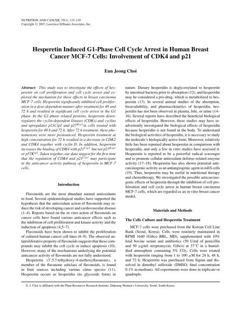
NUTRITION AND CANCER, 59(1), 115–119Copyright C○ 2007, Lawrence Erlbaum Associates, Inc.<strong>Hesperet<strong>in</strong></strong> <strong>Induced</strong> <strong>G1</strong>-<strong>Phase</strong> <strong>Cell</strong> <strong>Cycle</strong> <strong>Arrest</strong> <strong>in</strong> <strong>Human</strong> <strong>Breast</strong>Cancer MCF-7 <strong>Cell</strong>s: Involvement of CDK4 and p21Eun Jeong ChoiAbstract: This study was to <strong>in</strong>vestigate the effects of hesperet<strong>in</strong>on cell proliferation and cell cycle arrest and exploredthe mechanism for these effects <strong>in</strong> breast carc<strong>in</strong>omaMCF-7 cells. <strong>Hesperet<strong>in</strong></strong> significantly <strong>in</strong>hibited cell proliferation<strong>in</strong> a dose-dependent manner after treatment for 48 and72 h and resulted <strong>in</strong> significant cell cycle arrest <strong>in</strong> the <strong>G1</strong>phase. In the <strong>G1</strong>-phase related prote<strong>in</strong>s, hesperet<strong>in</strong> downregulatesthe cycl<strong>in</strong>-dependent k<strong>in</strong>ases (CDKs) and cycl<strong>in</strong>sand upregulates p21Cip1 and p27 Kip1 <strong>in</strong> cells treated withhesperet<strong>in</strong> for 48 h and 72 h. After 72 h treatment, these phenomenonswere more pronounced. <strong>Hesperet<strong>in</strong></strong> treatment athigh concentration for 72 h resulted <strong>in</strong> a decrease <strong>in</strong> CDK2and CDK4 together with cycl<strong>in</strong> D. In addition, hesperet<strong>in</strong><strong>in</strong>creases the b<strong>in</strong>d<strong>in</strong>g of CDK4 with p21 Cip1 but not p27 Kip1or p57K ip2 . Taken together, our data suggest for the first timethat the regulation of CDK4 and p21 Cip1 may participate<strong>in</strong> the anticancer activity pathway of hesperet<strong>in</strong> <strong>in</strong> MCF-7cells.IntroductionFlavonoids are the most abundant natural antioxidants<strong>in</strong> food. Several epidemiological studies have supported thehypothesis that the antioxidant action of flavonoids may reducethe risk of develop<strong>in</strong>g cancer and cardiovascular disease(1–4). Reports based on the <strong>in</strong> vitro action of flavonoids oncancer cells have found various anticancer effects such asthe <strong>in</strong>hibition of cell proliferation and k<strong>in</strong>ase activity and the<strong>in</strong>duction of apoptosis (4,5–7).Flavonoids have been shown to <strong>in</strong>hibit the proliferationof cultured human cancer cell l<strong>in</strong>es (8–9). The observed antiproliferativeproperty of flavonoids suggests that these compoundsmay <strong>in</strong>hibit the cell cycle or <strong>in</strong>duce apoptosis (10).However, many of the mechanisms underly<strong>in</strong>g the potentialanticancer activity of flavonoids are not fully understood.<strong>Hesperet<strong>in</strong></strong> (3 ′ ,5,7-trihydroxy-4-methoxyflavanone), amember of the flavanone subclass of flavonoids, is found<strong>in</strong> fruit sources <strong>in</strong>clud<strong>in</strong>g various citrus species (11).<strong>Hesperet<strong>in</strong></strong> occurs as hesperid<strong>in</strong> (its glycoside form) <strong>in</strong>nature. Dietary hesperid<strong>in</strong> is deglycosylated to hesperet<strong>in</strong>by <strong>in</strong>test<strong>in</strong>al bacteria prior to absorption (12), and hesperid<strong>in</strong>may be considered a pro-drug, which is metabolized to hesperet<strong>in</strong>(13). In several animal studies of the absorption,bioavailability, and pharmacok<strong>in</strong>etics of hesperid<strong>in</strong>, hesperid<strong>in</strong>has not been observed <strong>in</strong> plasma, bile, or ur<strong>in</strong>e (14–16). Several reports have described the beneficial biologicaleffects of hesperid<strong>in</strong>. However, these studies may have <strong>in</strong>advertently<strong>in</strong>vestigated the biological effects of hesperid<strong>in</strong>because hesperid<strong>in</strong> is not found <strong>in</strong> the body. To understandthe biological activities of hesperid<strong>in</strong>, it is necessary to studythe molecule’s biologically active form. Moreover, relativelylittle has been reported about hesperet<strong>in</strong> <strong>in</strong> comparison withhesperid<strong>in</strong>, and only a few <strong>in</strong> vitro studies have assessed it.<strong>Hesperet<strong>in</strong></strong> is reported to be a powerful radical scavengerand to promote cellular antioxidant defense-related enzymeactivity (17–18). <strong>Hesperet<strong>in</strong></strong> has also shown potential anticarc<strong>in</strong>ogenicactivity as an antiangiogenic agent <strong>in</strong> mES cells(19). Thus, hesperet<strong>in</strong> may be useful <strong>in</strong> nutritional therapyand chemotherapy. We <strong>in</strong>vestigated the possible anticarc<strong>in</strong>ogeniceffects of hesperet<strong>in</strong> through the <strong>in</strong>hibition of cell proliferationand cell cycle arrest <strong>in</strong> human breast carc<strong>in</strong>omaMCF-7 cells, which are regarded as an <strong>in</strong> vitro breast cancermodel.Materials and MethodsThe <strong>Cell</strong>s Culture and <strong>Hesperet<strong>in</strong></strong> TreatmentMCF-7 cells were purchased from the Korean <strong>Cell</strong> L<strong>in</strong>eBank (Seoul, Korea). <strong>Cell</strong>s were rout<strong>in</strong>ely ma<strong>in</strong>ta<strong>in</strong>ed <strong>in</strong>RPMI 1640 (Gibco BRL, MD), supplemented with 10%fetal bov<strong>in</strong>e serum and antibiotics (50 U/ml of penicill<strong>in</strong>and 50 µg/ml streptomyc<strong>in</strong>; Gibco) at 37 ◦ C <strong>in</strong> a humidifiedatmosphere conta<strong>in</strong><strong>in</strong>g 5% CO 2 . <strong>Cell</strong>s were treatedwith hesperet<strong>in</strong> rang<strong>in</strong>g from 1 to 100 µM for 24 h, 48 h,and 72 h. <strong>Hesperet<strong>in</strong></strong> was purchased from Sigma and dissolved<strong>in</strong> dimethyl sulfoxide (DMSO; f<strong>in</strong>al concentration0.1% <strong>in</strong> medium). All experiments were done <strong>in</strong> triplicate orquadruple.E. J. Choi is affiliated with the Plant Resources Research Institute, Duksung Women’s University, Seoul, South Korea.
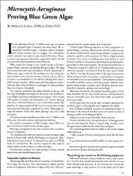
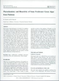
<strong>Cell</strong> Proliferation and <strong>Cell</strong> Death Assay<strong>Cell</strong> proliferation was determ<strong>in</strong>ed us<strong>in</strong>g the methyl thiazolyltetrazolium (MTT) assay. At 24 h, 48 h, and 72 h po<strong>in</strong>t,the cells exposed to hesperet<strong>in</strong> were added to MTT. DMSOwas added 4 h later to each well to dissolve the result<strong>in</strong>g formazancrystals, and then absorbance was recorded at 490 nm<strong>in</strong> a microplate reader SpectraMax Plus; Molecular Devices,CA).<strong>Cell</strong> <strong>Cycle</strong> Distribution<strong>Cell</strong>s were then harvested, washed with cold phosphatebuffered solution (PBS), and processed for cell cycle analysis.Briefly, the cells were fixed <strong>in</strong> absolute ethanol and storedat –20 ◦ C for later analysis. The fixed cells were centrifugedat 1,000 rpm and washed with cold PBS twice. RibonucleaseA (20 µg/ml f<strong>in</strong>al concentration) and propidium iodidesta<strong>in</strong><strong>in</strong>g solution (50 µg/ml f<strong>in</strong>al concentration) was addedto the cells and <strong>in</strong>cubated for 30 m<strong>in</strong> at 37 ◦ C <strong>in</strong> the dark.The cells were analyzed a FACS Calibur <strong>in</strong>strument (BDBiosciences, San Jose, CA) equipped with <strong>Cell</strong>Quest version3.3 software. ModFit LT version 3.1 trial cell cycle analysissoftware was used to determ<strong>in</strong>e the percentage of cells <strong>in</strong> thedifferent phases of the cell cycle.Immunoblott<strong>in</strong>g and Immunoprecipitation Assay<strong>Cell</strong>s were lysed <strong>in</strong> radio-immunoprecipitation assaybuffer (1% NP-40, 150 mM NaCl, 0.05% 4-chloro-2,5-dimethoxyamphetam<strong>in</strong>e, 1% sodium dodecyl sulfate (SDS),50mM Tris, pH 7.5) conta<strong>in</strong><strong>in</strong>g protease <strong>in</strong>hibitor for 1 hat 4 ◦ C. The supernatant was separated by centrifugation,and prote<strong>in</strong> concentration was determ<strong>in</strong>ed by Bradfordprote<strong>in</strong> assay kit II (Bio-rad Laboratories, CA). Prote<strong>in</strong>s(25 µg/well) denatured with sample buffer were separatedby 10% SDS-polyacrylamide gel. Prote<strong>in</strong>s were transferredonto nitrocellulose membranes (0.45 µm). The membraneswere blocked with a 1% bov<strong>in</strong>e serum album<strong>in</strong> solutionfor 3 h, washed twice with PBS conta<strong>in</strong><strong>in</strong>g 0.2% Tween-20, and <strong>in</strong>cubated with the primary antibody overnight at4 ◦ C. Antibodies aga<strong>in</strong>st cycl<strong>in</strong>-dependent k<strong>in</strong>ases (CDKs)CDK2, CDK4, CDK6, cycl<strong>in</strong> D, cycl<strong>in</strong> E, p16 INK4a , p18 INK4c ,p21 Cip1 , p27 Kip1 , p57 Kip2 , and β-act<strong>in</strong> were purchased fromSanta Cruz Biotechnology, Inc. (Santa Cruz, CA) and used toprobe the separate membranes. The next day, the immunoreactionwas cont<strong>in</strong>ued with the secondary goat antirabbithorseradish-peroxidase–conjugated antibody after wash<strong>in</strong>gfor 2 h at room temperature. The specific prote<strong>in</strong> bands weredetected by Opti-4CN Substrate kit (Bio-rad Laboratories).For CDK4-CDK <strong>in</strong>hibitors b<strong>in</strong>d<strong>in</strong>g assay, cells weretreated with vehicle or 100 µM of hesperet<strong>in</strong> for 48 h and72 h. CDK4 from cell lysates (250 µg prote<strong>in</strong>) was immunoprecipitatedus<strong>in</strong>g anti-CDK4 antibody (4 µg) and preclearedprote<strong>in</strong> A/G-plus agarose beads (Santa Cruz) for overnightat 4 ◦ C. The precipitates were washed with lysis buffer, dena-Figure 1. Effect of hesperet<strong>in</strong> on cell proliferation of MCF-7 cells. <strong>Cell</strong>swere exposed to either vehicle (0.1% dimethyl sulfoxide <strong>in</strong> medium) orhesperet<strong>in</strong> (1–100 µM) and <strong>in</strong>cubated for 24 h, 48 h, and 72 h. All dataare reported as the percentage change <strong>in</strong> comparison with the vehicle-onlygroup, which were arbitrarily assigned 100% viability. ∗ ,P
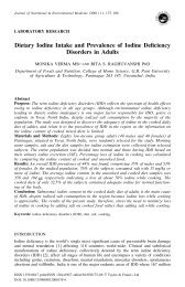
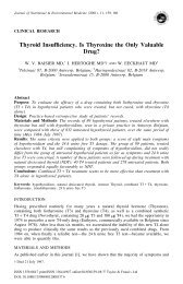
Figure 2. Effect of hesperet<strong>in</strong> on cell cycle distribution of MCF-7 cells. Values are expressed as percentage of the cell population <strong>in</strong> the <strong>G1</strong>, S, and <strong>G1</strong>/Mphase of cell cycle. <strong>Cell</strong>s were exposed to either vehicle (0.1% dimethyl sulfoxide <strong>in</strong> medium) or hesperet<strong>in</strong> (1–100 µM) and <strong>in</strong>cubated for 48 h and 72 h.∗ ,P
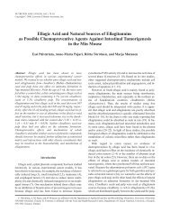
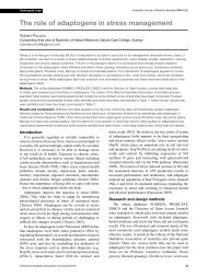
Figure 4. Effect of hesperet<strong>in</strong> on Bcl-2 and Bcl-2 associated x prote<strong>in</strong>(Bax) expression of MCF-7 cells. <strong>Cell</strong>s were exposed to either vehicle (0.1%dimethyl sulfoxide <strong>in</strong> medium) or hesperet<strong>in</strong> (1–100 µM) and <strong>in</strong>cubated for48 h.<strong>in</strong> mouse embryonic stem cells, suggest<strong>in</strong>g that its <strong>in</strong>hibitoryeffects may be associated with its prooxidant activity (19).Thus, hesperet<strong>in</strong> is a potential anticancer agent. Moreover,unlike other flavonoids that have prooxidant activity, nohesperet<strong>in</strong>-<strong>in</strong>duced cytotoxicity was observed, even at highconcentrations (100 µM). Thus, we <strong>in</strong>vestigated the anticancereffects of hesperet<strong>in</strong> on breast carc<strong>in</strong>oma MCF-7 cells<strong>in</strong> which hesperet<strong>in</strong> <strong>in</strong>hibits cell proliferation by caus<strong>in</strong>g cellcycle arrest.To assess whether the hesperet<strong>in</strong>-<strong>in</strong>duced <strong>in</strong>hibition of cellgrowth was mediated via alterations <strong>in</strong> cell cycle progression,cells were exposed to either vehicle (0.1% DMSO) or variousconcentrations of hesperet<strong>in</strong> (1, 5, 10, 50, 100 µM). <strong>Hesperet<strong>in</strong></strong>significantly <strong>in</strong>hibited cell proliferation <strong>in</strong> a dose- andtime-dependent manner. <strong>Hesperet<strong>in</strong></strong> can <strong>in</strong>hibit the proliferationof both estrogen receptor-negative MDA-MB-435 andestrogen receptor-positive MCF-7 human breast cancer cells<strong>in</strong> vitro<strong>in</strong> vitro (21–22). Our results supported these observations;the exposure of MCF-7 cells to hesperet<strong>in</strong> resulted<strong>in</strong> a significant <strong>in</strong>hibition of cell proliferation.We evaluated the mechanism by which hesperet<strong>in</strong> <strong>in</strong>hibitedcell proliferation us<strong>in</strong>g a cell cycle analysis. The eukaryoticcell cycle is controlled by catalytic complexes ofCDK/cycl<strong>in</strong> that coord<strong>in</strong>ate <strong>in</strong>ternal and external signals atseveral key checkpo<strong>in</strong>ts (23). <strong>G1</strong> arrest leads cells to undergorepair or to follow the apoptosis pathway (24). ActivatedCDK/cycl<strong>in</strong> complexes can be changed to an <strong>in</strong>active stateby the b<strong>in</strong>d<strong>in</strong>g of CDK <strong>in</strong>hibitory subunits (CKIs). The CKIscan be divided <strong>in</strong>to 2 classes. One class conta<strong>in</strong>s p21 Cip1 ,p27 Kip1 , p57K ip2 , and related prote<strong>in</strong>s, with a preference forCDK2 and CDK4/cycl<strong>in</strong> complexes; the other class conta<strong>in</strong>sthe INK4 family, <strong>in</strong>clud<strong>in</strong>g p16 INK4a , p15 INK4b , p18 INK4c , andp19 INK4d and closely related CKIs specific for CDK4 andCDK6/cycl<strong>in</strong> complexes (25–26).<strong>Hesperet<strong>in</strong></strong> <strong>in</strong>duced a decrease <strong>in</strong> cycl<strong>in</strong>D and CDK4, togetherwith an <strong>in</strong>crease <strong>in</strong> p21 Cip1 and p27 Kip1 associated withCDK4. CDK4 appears to play a key role <strong>in</strong> the <strong>G1</strong> cell cyclearrest observed <strong>in</strong> response to hesperet<strong>in</strong>.We assessed the effect of hesperet<strong>in</strong> on the <strong>in</strong>teractionbetween CDK4 and its <strong>in</strong>hibitors us<strong>in</strong>g immunoprecipitation.An <strong>in</strong>crease <strong>in</strong> the b<strong>in</strong>d<strong>in</strong>g of CDK4p21 Cip1 , but not ofCDK4–p27 Kip1 or CDK4–p57 Kip2 , was observed <strong>in</strong> MCF-7cells exposed to hesperet<strong>in</strong>. Additionally, hesperet<strong>in</strong> <strong>in</strong>duced<strong>G1</strong>-phase cell cycle arrest through the regulation of CDK4and p21 Cip1 , <strong>in</strong>dicat<strong>in</strong>g that the <strong>in</strong>hibition of cell proliferationand the contribution to the apoptotic process is a possiblemechanism by which hesperet<strong>in</strong> acts on cancer cells.Moreover, the <strong>G1</strong>-phase cell cycle arrest <strong>in</strong>duced by hesperet<strong>in</strong>resulted <strong>in</strong> apoptosis <strong>in</strong> MCF-7 cells. <strong>Hesperet<strong>in</strong></strong><strong>in</strong>duced a dose- and time-dependent decrease <strong>in</strong> Bcl-2 expressionand <strong>in</strong>crease <strong>in</strong> Bax expression <strong>in</strong> MCF-7 cells.Apoptosis is a multistep process important <strong>in</strong> controll<strong>in</strong>g cellnumber and proliferation as part of normal development;the Bax genes and members of the Bcl-2 family such asbcl-2 are <strong>in</strong>volved <strong>in</strong> the control of apoptotic pathways. Decreasedexpression of Bcl-2 and <strong>in</strong>creased expression of Baxare correlated with the response to anticancer compoundsfor chemotherapy <strong>in</strong> cancer cell l<strong>in</strong>es. In addition, the lossof Bcl-2 expression can promote the <strong>in</strong>duction of apoptosis(27–29). An anticancer drug that <strong>in</strong>itiates apoptosis may beuseful as a new category of chemotherapeutic agent.In conclusion, our results <strong>in</strong>dicate that hesperet<strong>in</strong> treatmentleads to the <strong>in</strong>hibition of cell proliferation, the <strong>in</strong>ductionof cell cycle arrest at the <strong>G1</strong> phase, and apoptosis. Additionally,our data suggest for the first time that the regulation ofCDK4 and p21 Cip1 may participate <strong>in</strong> the anticancer activityof hesperet<strong>in</strong> <strong>in</strong> MCF-7 cells. Therefore, we suggest thathesperet<strong>in</strong> is worthy of further study to assess its potentialas an anticancer drug; it rema<strong>in</strong>s to be determ<strong>in</strong>ed whetherhesperet<strong>in</strong> has anticarc<strong>in</strong>ogenic activity <strong>in</strong> vivo.Acknowledgments and NotesThis work was supported by the Korea Research Foundation Grant fundedby the Korean Government (MOEHRD, KRF–2006–311–F00127). Addresscorrespondence to EJ Choi, Ph.D., Plant Resource Research Institute,Duksung Women’s University, 419 Ssangmun-dong, Dobong-ku, 132–714,Seoul, South Korea. Phone: +82-2-901-8663. FAX: 82-2-901-8661. E-mail:ejchoi@duksung.ac.kr.Submitted 14 January 2007; accepted <strong>in</strong> f<strong>in</strong>al form 27 March 2007.References1. Wu AH, Ziegler RG, Horn-Ross PL, Nomura AM, West DW, et al.:Tofu and risk of breast cancer <strong>in</strong> Asian-Americans. Cancer EpidemiolBiomarkers Prev 5, 901–906, 1996.2. Middleton E Jr, Kandaswami C, and Theoharides TC: The effects ofplant flavonoids on mammalian cells: implications for <strong>in</strong>flammation,heart disease, and cancer. Pharmacol Rev 52, 673–751, 2000.3. Folts JD: Potential health benefits from the flavonoids <strong>in</strong> grape productson vascular disease. Adv Exp Med Biol 505, 95–111, 2002.4. Zheng Q, Hirose Y, Yoshimi N, Murakami A, Koshimizu K, et al.: Further<strong>in</strong>vestigation of the modify<strong>in</strong>g effect of various chemopreventiveagents on apoptosis and cell proliferation <strong>in</strong> human colon cancer cells.J Cancer Res Cl<strong>in</strong> Oncol 128, 539–546, 2002.5. B<strong>in</strong>nerup SJ and Sorensen J: Nitrate and nitrite microgradients <strong>in</strong> barleyrhizosphere as detected by a highly sensitive denitrification bioassay.Appl Environ Microbiol 58, 2375–2380, 1992.6. Tsuda H, Ohshima Y, Nomoto H, Fujita K, Matsuda E, et al.: Cancerprevention by natural compounds. Drug Metab Pharmacok<strong>in</strong>et 19,245–263, 2004.7. Kanadaswami C, Lee LT, Lee PP, Hwang JJ, Ke FC, et al.: The antitumoractivities of flavonoids. In Vivo 19, 895–909, 2005.118 Nutrition and Cancer 2007
8. S<strong>in</strong>gh RP and Agarwal R: Natural flavonoids target<strong>in</strong>g deregulated cellcycle progression <strong>in</strong> cancer cells. Curr Drug Targets 7, 345–354, 2006.9. Nichenametla SN, Taruscio TG, Barney DL, and Exon JH: A review ofthe effects and mechanisms of polyphenolics <strong>in</strong> cancer. Crit Rev FoodSci Nutr 46, 161–183, 2006.10. Casagrande F and Darbon JM: Effects of structurally related flavonoidson cell cycle progression of human melanoma cells: regulation ofcycl<strong>in</strong>-dependent k<strong>in</strong>ases CDK2 and CDK1. Biochem Pharmacol 61,1205–1215, 2001.11. Garg A, Garg S, Zaneveld LJ, and S<strong>in</strong>gla AK: Chemistry and pharmacologyof the Citrus bioflavonoid hesperid<strong>in</strong>. Phytother Res 15,655–669, 2001.12. Ameer B, We<strong>in</strong>traub RA, Johnson JV, Yost RA, and Rouseff RL: Flavanoneabsorption after nar<strong>in</strong>g<strong>in</strong>, hesperid<strong>in</strong>, and citrus adm<strong>in</strong>istration.Cl<strong>in</strong> Pharmacol Ther 60, 34–40, 1996.13. Lee NK, Choi SH, Park SH, Park EK, and Kim DH: Antiallergic activityof hesperid<strong>in</strong> is activated by <strong>in</strong>test<strong>in</strong>al microflora. Pharmacology 71,174–180, 2004.14. Matsumoto H, Ikoma Y, Sugiura M, Yano M, and Hasegawa Y: Identificationand quantification of the conjugated metabolites derived fromorally adm<strong>in</strong>istered hesperid<strong>in</strong> <strong>in</strong> rat plasma. J Agric Food Chem 52,6653–6659, 2004.15. Booth AN, Jones FT, and DeEds F: Metabolic fate of hesperid<strong>in</strong>, eriodictyol,homoeriodictyol, and diosm<strong>in</strong>. JBiolChem230, 661–668,1958.16. Hackett AM, Marsh I, Barrow A, and Griffiths LA: The biliary excretionof flavanones <strong>in</strong> the rat. Xenobiotica 9, 491–502, 1979.17. Kim JY, Jung KJ, Choi JS, and Chung HY: <strong>Hesperet<strong>in</strong></strong>: a potent antioxidantaga<strong>in</strong>st peroxynitrite. Free Radic Res 38, 761–769, 2004.18. Pollard SE, Whiteman M, and Spencer JP: Modulation of peroxynitrite<strong>in</strong>ducedfibroblast <strong>in</strong>jury by hesperet<strong>in</strong>: a role for <strong>in</strong>tracellular scaveng<strong>in</strong>gand modulation of ERK signall<strong>in</strong>g. Biochem Biophys Res Commun347, 916–923, 2006.19. Choi EJ, Kim GD, Chee KM, and Kim GH: The effects of hesperet<strong>in</strong>on vessel structure formation <strong>in</strong> mouse embryonic stem (mES) cells.Nutrition <strong>in</strong> press, 2006.20. S<strong>in</strong>gh RP and Agarwal R: Natural flavonoids target<strong>in</strong>g deregulated cellcycle progression <strong>in</strong> cancer cells. Curr Drug Targets 7, 345–354, 2006.21. So FV, Guthrie N, Chambers AF, and Carroll KK: Inhibition of proliferationof estrogen receptor-positive MCF-7 human breast cancer cellsby flavonoids <strong>in</strong> the presence and absence of excess estrogen. CancerLett 112, 127–133, 1997.22. Guthrie N and Carroll KK: Inhibition of mammary cancer by citrusflavonoids. Adv Exp Med Biol 439, 227–236, 1998.23. Grana X and Reddy EP: <strong>Cell</strong> cycle control <strong>in</strong> mammalian cells: roleof cycl<strong>in</strong>s, cycl<strong>in</strong> dependent k<strong>in</strong>ases (CDKs), growth suppressor genesand cycl<strong>in</strong>-dependent k<strong>in</strong>ase <strong>in</strong>hibitors (CKIs). Oncogene 11, 211–219,1995.24. Mantena SK, Sharma SD, and Katiyar SK: Berber<strong>in</strong>e, a natural product,<strong>in</strong>duces <strong>G1</strong>-phase cell cycle arrest and caspase-3-dependent apoptosis<strong>in</strong> human prostate carc<strong>in</strong>oma cells. Mol Cancer Ther 5, 296–308, 2006.25. Deng C, Zhang P, Harper JW, Elledge SJ, and Leder P: Mice lack<strong>in</strong>gp21CIP1/WAF1 undergo normal development, but are defective <strong>in</strong> <strong>G1</strong>checkpo<strong>in</strong>t control. <strong>Cell</strong> 82, 675–684, 1995.26. Sherr CJ and Roberts JM: CDK <strong>in</strong>hibitors: positive and negative regulatorsof <strong>G1</strong>-phase progression. Genes Dev 13, 1501–1512, 1999.27. Suzuki K, Kazui T, Yoshida M, Uno T, Kobayashi T, et al.: Drug<strong>in</strong>ducedapoptosis and p53, BCL-2 and BAX expression <strong>in</strong> breast cancertissues <strong>in</strong> vivo and <strong>in</strong> fibroblast cells <strong>in</strong> vitro. Jpn J Cl<strong>in</strong> Oncol 29, 323–331, 1999.28. Szostak MJ, Kaur P, Am<strong>in</strong> P, Jacobs SC, and Kyprianou N: Apoptosisand bcl-2 expression <strong>in</strong> prostate cancer: significance <strong>in</strong> cl<strong>in</strong>ical outcomeafter brachytherapy. JUrol165, 2126–2130, 2001.29. Gazzaniga P, Gradilone A, Vercillo R, Gand<strong>in</strong>i O, Silvestri I, et al.:Bcl-2/bax mRNA expression ratio as prognostic factor <strong>in</strong> low-gradeur<strong>in</strong>ary bladder cancer. Int J Cancer 22, 100–104, 1996.Vol. 59, No. 1 119