Mapping microalgal resources of Idukki District, Kerala in Western ...
Mapping microalgal resources of Idukki District, Kerala in Western ...
Mapping microalgal resources of Idukki District, Kerala in Western ...
You also want an ePaper? Increase the reach of your titles
YUMPU automatically turns print PDFs into web optimized ePapers that Google loves.
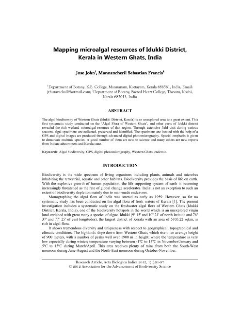
Acta Biologica Indica 2012, 1(1):91-97<strong>Mapp<strong>in</strong>g</strong> <strong>microalgal</strong> <strong>resources</strong> <strong>of</strong> <strong>Idukki</strong> <strong>District</strong>,<strong>Kerala</strong> <strong>in</strong> <strong>Western</strong> Ghats, IndiaJose John 1 , MannancherilSebastianFrancis 21 Department <strong>of</strong> Botany, K.E. College, Mannanam, Kottayam, <strong>Kerala</strong> 686561, India, Email:jthuravackal@hotmail.com; 2 Department <strong>of</strong> Botany, Sacred Heart College, Thevara, Kochi,<strong>Kerala</strong> 682013, IndiaABSTRACTThe algal biodiversity <strong>of</strong> <strong>Western</strong> Ghats (<strong>Idukki</strong> <strong>District</strong>, <strong>Kerala</strong>) is an unexplored area to a great extent. Thisfirst systematic study conducted on the ‘Algal Flora <strong>of</strong> <strong>Western</strong> Ghats’, and other parts <strong>of</strong> <strong>Idukki</strong> districtrevealed the rich wetland <strong>microalgal</strong> resource <strong>of</strong> that region. Through extensive field visit dur<strong>in</strong>g variousseasons, algal specimens are collected, preserved and identified. The specimens are located with the help <strong>of</strong> aGPS and digital images are produced through advanced digital photomicrography. Special emphasis is givento demarcate endemic species. A good number <strong>of</strong> them are new to science and many others are new reportsfrom Indian subcont<strong>in</strong>ent and <strong>Kerala</strong> state.Keywords: Algal biodiversity, GPS, digital photomicrography, <strong>Western</strong> Ghats, endemic.INTRODUCTIONBiodiversity is the wide spectrum <strong>of</strong> liv<strong>in</strong>g organisms <strong>in</strong>clud<strong>in</strong>g plants, animals and microbes<strong>in</strong>habit<strong>in</strong>g the terrestrial, aquatic and other habitats. Biodiversity provides the basis <strong>of</strong> life on earth.With the explosive growth <strong>of</strong> human population, the life support<strong>in</strong>g system <strong>of</strong> earth is becom<strong>in</strong>g<strong>in</strong>creas<strong>in</strong>gly threatened as the rate <strong>of</strong> global change accelerates. India is not an exception to such anextent <strong>of</strong> biodiversity depletion ma<strong>in</strong>ly due to man-made endeavors.Monograph<strong>in</strong>g the algal flora <strong>of</strong> India was started as early as 1959. However, so far nosystematic study has been conducted on the algal flora <strong>of</strong> fresh waters <strong>of</strong> <strong>Kerala</strong> [1]. The present<strong>in</strong>vestigation <strong>in</strong>cludes a systematic study on the freshwater algal flora <strong>of</strong> <strong>Western</strong> Ghats (<strong>Idukki</strong><strong>District</strong>, <strong>Kerala</strong>, India), one <strong>of</strong> the biodiversity hotspots <strong>in</strong> the world which is an unexplored virg<strong>in</strong>land enriched with great many a species <strong>of</strong> algae. <strong>Idukki</strong> (9º 15' and 10º 21' <strong>of</strong> north latitude and 76º37' and 77º 25' <strong>of</strong> east longitudes), the largest district <strong>of</strong> <strong>Kerala</strong> with an area <strong>of</strong> 5105.22 sqkm, isrich <strong>in</strong> algal flora.It shows tremendous diversity and uniqueness with respect to geographical, topographical andclimatic conditions. The highlands slope down from <strong>Western</strong> Ghats, which rise to an average height<strong>of</strong> 900 meters, with a number <strong>of</strong> peaks well over 1900 m <strong>in</strong> height, where the temperature is verylow especially dur<strong>in</strong>g w<strong>in</strong>ter; temperature vary<strong>in</strong>g between -1ºC to 15ºC <strong>in</strong> November/January and5ºC to 15ºC dur<strong>in</strong>g March/April. This area receives plenty <strong>of</strong> ra<strong>in</strong>s from both the South-Westmonsoon dur<strong>in</strong>g June-August and the North-East monsoon dur<strong>in</strong>g October-November.Research Article, Acta Biologica Indica 2012, 1(1):91-97© 2012 Association for the Advancement <strong>of</strong> Biodiversity SciencePr<strong>in</strong>ted <strong>in</strong> India; ISSN 2249-123491
Acta Biologica Indica 2012, 1(1):91-97MATERIALS AND METHODSThis study comprised <strong>of</strong> extensive field visit to different parts <strong>of</strong> the <strong>Western</strong> Ghats region <strong>of</strong> <strong>Idukki</strong>district dur<strong>in</strong>g various seasons. Algal specimens are collected, preserved and identified. Thespecimens are located with the help <strong>of</strong> a GPS. Special emphasis is given to demarcate endemicspecies. Digital images <strong>of</strong> various taxa are taken with the help <strong>of</strong> a digital camera attached to themicroscope and are transferred to the computer for further analysis.RESULTS AND DISCUSSIONThe state <strong>Kerala</strong> <strong>in</strong> general and <strong>Idukki</strong> district <strong>in</strong> particular is known for its luxuriant greenery,moderate climate and rich biodiversity. <strong>Idukki</strong> district with tropical ever green forest and richfreshwater <strong>resources</strong> is rich <strong>in</strong> <strong>microalgal</strong> diversity. Out <strong>of</strong> the eleven classes <strong>of</strong> algae, seven arerepresented <strong>in</strong> the present study. Out <strong>of</strong> the 33 taxa <strong>of</strong> algae recorded, 19 belong to Chlorophyceae,1 to Xanthophyceae, 4 to Bacillariophyceae, 1 to D<strong>in</strong>ophyceae, 5 to Euglen<strong>in</strong>eae, 2 toRhodophyceae and 1 to Cyanophyceae. Of these 3 are new reports (1 from Rhodophyceae and 2from Euglen<strong>in</strong>eae). Algal samples were collected with the help <strong>of</strong> plankton net, <strong>in</strong> the case <strong>of</strong>smaller forms and direct mass collection for larger ones. Collections were made from the surfacelevel, shallow bed, underside <strong>of</strong> rocks, mucilage masses attached to dripp<strong>in</strong>g rocks, tree trunks,streams, pools, rocky water bodies, ditches, canals, etc. The temperature and pH were noted. Thelocations <strong>of</strong> collections were measured with the help <strong>of</strong> GPS.Botryococcus protuberance W. et G.S. WestM.T. Philipose, 1967, p. 197, Fig. 109Colony irregular with 4-8-16 or more cells held together by a tough gelat<strong>in</strong>ous membrane.Frequently jo<strong>in</strong>ed together <strong>in</strong> compound colonies by long tough hyal<strong>in</strong>e strands <strong>of</strong> mucilage.Cells ovoid, obovoid or ovoid-cuneate with their <strong>in</strong>ner narrower ends embedded <strong>in</strong> theenvelope, the outer ends be<strong>in</strong>g not so enclosed and project<strong>in</strong>g out <strong>of</strong> the colony. Cells 9.5-11.5µm broad, 16.5-20.0µm long. Colonies 100-120µm <strong>in</strong> diameter [2].Found <strong>in</strong> site: Marayoor [N 09 53' 35.91"; E 77 12' 20.04"; pH 8.9; Temp 27ºC]Dimorphococcus lunatus A. BraunM.T. Philipose, 1967, p. 205, Fig. 115Colonies irregular. Cells <strong>in</strong> groups <strong>of</strong> four and arranged alternately <strong>in</strong> a zigzag fashion. Outercells <strong>of</strong> each group reniform or somewhat crescent shaped. Inner cells elongated-ovoid toellipsoid. Ends <strong>of</strong> cells rounded. Chloroplast a parietal plate nearly cover<strong>in</strong>g the entire cell wall<strong>in</strong> mature cells. Cells 4-15µm broad, 9-25µm long. Colonies up to 100µm <strong>in</strong> diameter [2].Found <strong>in</strong> site: Koduvely [N 09 57' 16.65"; E 76 46' 01.57"; pH 8.1; Temp 22ºC]Thorakomonas sabulosa Kors.M.O.P. Iyengar and T.V. Desikachary, 1981, p. 347, pl. 200, Figs. 1-4Cells ellipsoidal <strong>in</strong> front view, up to 16µm long, 14µm broad; lorica rough, chloroplast cupshaped;pyrenoid s<strong>in</strong>gle, axial; eyespot anterior; contractile vacuoles 2, anterior [3].Found <strong>in</strong> site: Iravikulam [N 10 09' 08.91"; E 77 01' 14.56"; pH 8.1; Temp 19ºC]Tetraedron trigonum (Naeg) Hansg. fa. gracile (Re<strong>in</strong>sch) De ToniM.T. Philipose, 1967, p. 142, Fig. 58aCells with more markedly concave sides than <strong>in</strong> the type. Cell membrane smooth. Cells 19-35µm <strong>in</strong> diameter, 6-8µm thick. Sp<strong>in</strong>es 6.2-7µm long[2].Found <strong>in</strong> site: Mulappuram [N 09 57' 17.06"; E 76 45' 59.75"; pH 7.3; Temp 22ºC]Glaucocystis nostoch<strong>in</strong>earum ItzigsohnM.T. Philipose, 1967, p. 188, Fig. 10192
Acta Biologica Indica 2012, 1(1):91-97Colonies <strong>of</strong> 2-8 (usually 4) cells enclosed with<strong>in</strong> the old mother cell wall. Cells oblong-ellipsoidand with a number (less than 20) <strong>of</strong> radiat<strong>in</strong>g chromatophore-like bodies <strong>in</strong>side, which arevermiform and blue green and belong to a member <strong>of</strong> the Chroococcales. Cells 10-18µm broad,15-30µm long. Colonies 26-51µm broad, 39-63µm long[2].Found <strong>in</strong> site: Karimannoor [N 10 03' 04.84"; E 76 52' 19.64"; pH 9.3; Temp 22ºC]Ecballocystis courtallensis Iyeng.M.T. Philipose, 1967, p. 168, Fig. 124Thallus microscopic form<strong>in</strong>g many celled branched dendroid colonies. The colony is attachedto the rocky substratum with the help <strong>of</strong> a mucilag<strong>in</strong>ous pad. Cells elongate, cyl<strong>in</strong>drical withrounded or slightly conical ends. Cells 45-49µm long and 8-9.5µm broad; chloroplast parietal;lamellated polar nodular thicken<strong>in</strong>gs seen <strong>in</strong> mature cells [2].Found <strong>in</strong> site: Iravikulam [N 10 09' 08.91"; E 77 01' 14.56"; pH 8.1; Temp 19ºC]Trentepohlia umbr<strong>in</strong>a (Kütz) BornetV. Krishnamurthy, 2000, p. 190Filaments form a th<strong>in</strong>, crustaceous and fairly granular layer, reddish-brown. Cells <strong>of</strong> prostratesystem rounded or ellipsoid, torulose, readily separat<strong>in</strong>g from one another and probably help<strong>in</strong>g<strong>in</strong> dispersal by w<strong>in</strong>d; 13-20µm <strong>in</strong> diameter, with smooth cell-wall. Sporangia stalked, term<strong>in</strong>alon poorly developed erect filaments [4].Found <strong>in</strong> site: Nedumkandam [N 10 08' 39.07"; E 77 06' 25.42"; pH 9.1; Temp17ºC]Temnogamatum tirupatiense IyengarM.S. Randhawa, 1959, p. 184, fig. 114 A,B.Vegetative cells 11-13µm broad and 12-16 times as long as broad; chloroplast narrow plateshapedwith 10-15 pyrenoids <strong>in</strong> a row. Filaments monoecious, conjugation unique <strong>in</strong> be<strong>in</strong>gbrought about between gametangia situated apart from each other <strong>in</strong> the same filament, throughthe coil<strong>in</strong>g <strong>of</strong> the <strong>in</strong>terven<strong>in</strong>g portion <strong>of</strong> the filament; gametangia 11-13µm × 12-15µm;zygospores about 40-45µm <strong>in</strong> diameter [5].Found <strong>in</strong> site: Vattavada [N 09 54' 41.27"; E 76 55' 00.47"; pH 7.8; Temp 19ºC]Netrium digitus (Ehrbg.) Itzigs. & RothePrescott, 1961, p. 8, pl. 1, fig. 5Cell body 147-153µm long, 44-45µm wide; chloroplast parietal with fibricate marg<strong>in</strong>s [6].Found <strong>in</strong> site: Kulappuram [N 09 57' 45.27"; E 76 42' 37.81"; pH 4.5; Temp 27ºC]Arthrodesmus curvatus Turner var. latus PrescottPrescott, 1961, p. 76, pl. 33, figs. 1-3Larger and much wider than the species, both with and without sp<strong>in</strong>es. Chloroplast furcate withone pyrenoid per semicell. Cells W. ssp. 31µm; W. csp. 62µm; 34.1µm long; isthmus 9.3µmwide [6].Found <strong>in</strong> site: Kaliyar [N 09 57' 17.04"; E 76 47' 03.21"; pH 7.6; Temp 26ºC]Stigeocloniun faciculare Kütz<strong>in</strong>gV. Krishnamurthy, 2000, p. 48Plants tufted, 1-5cm long, bright green and slimy; prostrate system palmelloid or rhizoidal;erect filaments dichotomously or alternately branched below, primary branches <strong>of</strong>ten oppositeabove; rarely 2-4 primary branches form<strong>in</strong>g a whorl on ma<strong>in</strong> axis; secondary branches mostlyalternate, form<strong>in</strong>g a fascicle on primary branches. Cells <strong>of</strong> the ma<strong>in</strong> axis cyl<strong>in</strong>drical, but may beslightly <strong>in</strong>flated; cells <strong>of</strong> ma<strong>in</strong> axes 6-18.5µm <strong>in</strong> diameter, 2-4-7 times as long. Chloroplast adense band [4].Found <strong>in</strong> site: Kodikulam [N 09 57' 21.63"; E 76 47' 47.76"; pH 6.1; Temp 24ºC]Mougeotia operculata TranseauM.S. Randhawa, 1959, p. 151, fig. 6993
Acta Biologica Indica 2012, 1(1):91-97Vegetative cells 18-21µm × 60-285µm; chloroplast with 4-8 pyrenoids, usually 4. Conjugationscalariform; zygospores <strong>in</strong> the conjugat<strong>in</strong>g tubes, compressed-spheroid, 27-30 × 21-27µm, witha prom<strong>in</strong>ent equatorial ridge and suture on the wall; spore wall pale yellow, shallowscorbiculate[5].Found <strong>in</strong> site: Udumbannoor [N 09 57' 06.76"; E 76 45' 31.77"; pH 7.4; Temp 21ºC]Spirogyra longata (Vauch.) Kutz.M.S. Randhawa 1959, p. 304, fig. 268Vegetative cells 19-27µm × 99-165µm; septa plane; chloroplasts 1, mak<strong>in</strong>g 6-8 turns.Conjugation scalariform and lateral; tube formed by both gametangia; female gametangiaenlarged up to 37µm <strong>in</strong> diameter; zygospore globose, 23-30µm × 44-66µm and ovoid,mesospore brown and smooth [5].Found <strong>in</strong> site: Vannappuram [N 09 55' 23.71"; E 76 47' 31.37"; pH 7.3; Temp 20ºC]Spirotaenia condensata BrebPrescott, 1961, p. 8, pl. 1, figs. 1, 2Vegetative cells large, cigar-shaped, 60-334µm long, 10-30.4µm diameter, spiral band-formchloroplasts [6].Found <strong>in</strong> site: Devikulam [N 09 59' 36.82"; E 77 02' 37.85"; pH 7.9; Temp 21ºC]Onychonema laeve Nordst var. latum West & WestPrescott, 1961, p. 121, pl. 60, fig. 13Cell body 15-22µm long, 17-25µm wide, constriction 7-8.6µm wide; semicells elongatedellipsoidal or broad hexagonal reniform, from both sides two short sp<strong>in</strong>es direct<strong>in</strong>g <strong>in</strong>ward;apex flat with two long projections to connect with adjo<strong>in</strong><strong>in</strong>g cell [6].Found <strong>in</strong> site: Kaloor [N 09 57' 16.65"; E 76 46' 01.57"; pH 8.1; Temp 22ºC]Pleurotaenum ovatum Nordst var. <strong>in</strong>termius MobiusPrescott, 1961, p. 17, pl. 6, figs. 3,4Cell body 212-307µm long, 79-95µm wide, isthmus 46-49µm wide [6].Found <strong>in</strong> site: Kulamavu [N 09 51' 16.29"; E 76 58' 19.20"; pH 8.9; Temp 22ºC]Staurastrum tohopekaligense Wolle var. trifurcatum West & WestPrescott, 1961, p. 114, pl. 48, fig. 2Cell body 39-60µm long, 29-90µm wide, isthmus 18 µm [6].Found <strong>in</strong> site: Pa<strong>in</strong>gottor [N 09 57' 16.65"; E 76 46' 01.57"; pH 8.1; Temp 22ºC]Triploceras gracile Bail var. undulatum Scott & PresH. Hirosh, et al, 1977Cell body 206-(310-360)-668µm long, (20-28)-53µm wide (<strong>in</strong>clud<strong>in</strong>g sp<strong>in</strong>es), L/W=10-15;semicells not swelled at base, slightly tapered at term<strong>in</strong>al, wavy lateral marg<strong>in</strong> with shortsp<strong>in</strong>es, 2 arm-like projections at each end, each projection equipped with two sp<strong>in</strong>es; cell wallsmooth; axial chloroplasts with radially-arranged lam<strong>in</strong>ae [7].Found <strong>in</strong> site: Ezhalloor [N 09 57' 17.06"; E 76 45' 59.75"; pH 7.3; Temp 22ºC]Vaucheria unc<strong>in</strong>ata KutzG.S.Venkataraman, 1961, p. 72, figs. 48a-fFilaments 42-76µm broad. Oogonia and antheridia on fruit<strong>in</strong>g branches which are trifurcate,furcations recurved, pendulous; oogonia spherical or compressed-spherica, two to seven, 135-175µm broad, 120-150µm long; antheridium short cyl<strong>in</strong>dric or saccate, occasionally circ<strong>in</strong>ate,antheridial wall dis<strong>in</strong>tegrat<strong>in</strong>g soon after open<strong>in</strong>g [8].Found <strong>in</strong> site: Koviloor [N 10 10' 54.12"; E 77 15' 33.49"; pH 9.4; Temp 20ºC]Ophiocytium parvulum (Perty) A. BraunL.H. Tiffany, 1952, p. 208, pl. 56, fig. 632Cells 3-15µm broad, without polar sp<strong>in</strong>es, curved or bent, <strong>of</strong>ten very long and much <strong>in</strong>tertw<strong>in</strong>ed[9].94
Acta Biologica Indica 2012, 1(1):91-97Found <strong>in</strong> site: Pa<strong>in</strong>igottor [N 09 57' 16.65"; E 76 46' 01.57"; pH 8.1; Temp 22ºC]Stauroneis agrestis PetKrammer and Lange-Bertalot, 1986, p. 247, fig. 90:21-22Length 19-28 µm. breadth 4-6µm. striae 27-34 <strong>in</strong> 10µm [10].Found <strong>in</strong> site: Viripara [N 10 14' 18.94"; E 77 11' 04.19"; pH 7.6; Temp 20ºC]P<strong>in</strong>nularia crucifera A. Cl. var. subcapitata A. Cl.Sarode and Kamat, 1984, p. 139, fig. 360Valves 42-52µm long, 7.5-7.8µm broad, l<strong>in</strong>ear with parallel marg<strong>in</strong>s and slightly capitate ends;raphe th<strong>in</strong> and straight with unilaterally bent central pores and curved term<strong>in</strong>al fissures; axialarea wide, l<strong>in</strong>ear; central area large, reach<strong>in</strong>g the marg<strong>in</strong>s; striae 12 <strong>in</strong> 10µm, almost parallel toone another or slightly radial [11].Found <strong>in</strong> site: Kudakkallu [N 10 05' 21.57"; E 77 03' 47.72"; pH 8.8; Temp 12ºC]P<strong>in</strong>nularia braunii (Grun.) Cleve var. amphicephala (Mayer) HustedtSarode and Kamat 1984, p. 136, fig. 351Valves 51.5-54µm long, 8-8.5µm broad, l<strong>in</strong>ear elliptical with slightly constricted, somewhatproduced, rounded capitate ends; raphe th<strong>in</strong> and straight; axial area somewhat narrow, l<strong>in</strong>earlanceolate; central area very wide, rhomboid, reach<strong>in</strong>g the marg<strong>in</strong>s; striae 10-11 <strong>in</strong> 10µm, radial<strong>in</strong> the middle and convergent at the ends [11].Found <strong>in</strong> site: Pothamedu [N 09 39' 26.65"; E 76 55' 22.91"; pH 8.4; Temp 20ºC]Gymnod<strong>in</strong>ium fuscum (Ehr.) Ste<strong>in</strong>G.W.Prescott, 1951, p. 426, fig. 23Cells large, ovoid, the epicone dome-shaped, the hypocone as broad as or broader then theepicone, narrowed, posteriorly to form an <strong>in</strong>verted cone with a slightly produced tip; transversefurrow slightly spiral; the longitud<strong>in</strong>al furrow extend<strong>in</strong>g about half way <strong>in</strong>to the hypocone, butscarcely at all <strong>in</strong>to the epicone; chromatophores numerous ovoid discs or rods, radiallyarranged; cells 55-60µm <strong>in</strong> diameter, 80-100µm long [6].Found <strong>in</strong> site: Mailakkombu [N 09 57' 06.76"; E 76 45' 31.77"; pH 7.4; Temp 21ºC]Astasia fustis Pr<strong>in</strong>gsheimWolowski, 1998, p. 45, figs. 158-161Cells 73.5-90.0µm long, 11.1-17.7µm wide, cyl<strong>in</strong>drical-elongated; each cell rounded at theanterior end, slightly taper<strong>in</strong>g and tail-like towards the posterior end. The pellicle f<strong>in</strong>ely andspirally striated. The nucleus rounded without constant position, translocated. Canal open<strong>in</strong>gapical. Flagellum up to 1/4 <strong>of</strong> the cell length. Paramylon gra<strong>in</strong>s abundant, ovate, usuallytranslocated. Movement by wave like expansion to anterior or posterior cell, slight and ratherconstant [12].Found <strong>in</strong> site: Mailakkombu [N 09 57' 21.25"; E 76 47' 31.56"; pH 8.4; Temp 24ºC]Phacus ankylonoton PochmannWolowski, 1998, p. 78, figs. 267-269Cell 33.0-39.6µm long, 10.5-17.0µm wide, oval with long fold at the back, slightly curvedcauda at the posterior end [12].Found <strong>in</strong> site: Cheenikuzhy [N 09 58' 23.14"; E 76 42' 47.17"; pH 5.2; Temp 28ºC]Trachelomonas hispida (Perty) Ste<strong>in</strong> var. hispida (Starmach)Wolowski, 1998, p. 56, figs. 186-187Lorica 22.5-30.0µm long, 12.5-25.2µm wide, elliptical or broadly elliptical, covered with sp<strong>in</strong>esand small pores [12].Found <strong>in</strong> site: Gaproad [N 09 53' 47.13"; E 77 11' 20.43"; pH 8.8; Temp 29ºC]Batrachospermum vagum (Roth) C. Agardh.T.V. Desikachary & V.N. Raja Rao 1980, p. 229.95
Acta Biologica Indica 2012, 1(1):91-97Plants large, 2-20cm high; colour olivaceous with a t<strong>in</strong>ge <strong>of</strong> blue green; pr<strong>of</strong>usely branched, thebranch<strong>in</strong>g somewhat irregular. The term<strong>in</strong>al portions <strong>of</strong> axes and branches blunt. Whorls <strong>of</strong>branchlets dist<strong>in</strong>ct only <strong>in</strong> the younger portions, term<strong>in</strong>al cells <strong>of</strong> branchlets prolonged <strong>in</strong>to longhairs. Sexual plants monoecious. Spermatangia term<strong>in</strong>al on branchlets, solitary or <strong>in</strong> clusters <strong>of</strong>2-4. Carpogonial branches 7-14 celled, straight, curved or contorted, cells <strong>of</strong> carpogonial branchshort, the lower ones bear<strong>in</strong>g short 2-5 celled lateral branches reach<strong>in</strong>g up to the level <strong>of</strong> thecarpogonium, which is symmetrical, term<strong>in</strong>al on the carpogonial branch and provided with an<strong>in</strong>verted skittle shaped trichogyne: the carpogonial branch usually aris<strong>in</strong>g from the basal cell <strong>of</strong>branchlets. Carposporophyte one, rarely two, <strong>in</strong> each whorl, rather large, compact andsubspherical: carposporangia term<strong>in</strong>al on the gonimoblast filaments.Found <strong>in</strong> site: Oorkad [N 10 10' 49.78"; E 77 15' 20.59"; pH 9.2; Temp 16ºC]Lyngbya birgei SmithT.V. Desikachary, 1959, p. 296Filaments straight, seldom coiled, free-float<strong>in</strong>g, 20-24µm broad; sheath firm, colourless, mostlyunlamellated; trichomes not constricted at the cross-walls, 18-23µm broad, ends rounded, notattenuated, not capitate, cells shorter than broad [13].Found <strong>in</strong> site: Valara [N 09 55' 23.62"; E 76 58' 07.33"; pH 9.3; Temp 22ºC]Cyl<strong>in</strong>drospermum majus Kütz<strong>in</strong>g ex Born et FlahT.V. Desikachary, 1959, p. 360, pl. 80, fig. 1Thallus expanded, mucilag<strong>in</strong>ous, blackish green; trichomes 4-5µm broad, constricted at thecross-walls, light blue-green; cells cyl<strong>in</strong>drical, 5-6µm long; heterocysts oblong, up to about10µm long; spores ellipsoidal, 10-15µm broad, 20-30µm long, epispore brownish with dist<strong>in</strong>ctpapillae [13].Found <strong>in</strong> site: Nettimedu [N 10 05' 47.78"; E 77 05' 59.00"; pH 10.8; Temp 04ºC]Audou<strong>in</strong>ella kurisukuthii sp. nov.Family: BatrachospermaceaeOrder: BatrachospermalesClass: RhodophyceaeThe thallus consists <strong>of</strong> a ma<strong>in</strong> axis (erect filament) and a basal rhizoidal system. The thalli aredensely tufted, attached to substratum by the rhizoidal system. The cells <strong>of</strong> the erect filamentsare cyl<strong>in</strong>drical with uniform thickness and measur<strong>in</strong>g 15-20µm <strong>in</strong> diameter and 35-40µm <strong>in</strong>length. Cells have a s<strong>in</strong>gle lobed parietal chromatophore, devoid <strong>of</strong> pyrenoid. Branch<strong>in</strong>g isusually alternate and rarely opposite. The branches usually arise just below the septa <strong>of</strong> cells <strong>of</strong>the ma<strong>in</strong> axis. S<strong>in</strong>gle type <strong>of</strong> monosporangia born at the distal end <strong>of</strong> one celled branch <strong>of</strong>limited growth and they occur alone. The mature sporangia are usually almost sphericalmeasur<strong>in</strong>g 14-15 µ m <strong>in</strong> diameter. The stalk <strong>of</strong> the sporangium is oblong with 16-18µm lengthand 7-8µm width.Found <strong>in</strong> site: Kurisukuthi [N 10 15' 27.52"; E 77 10' 04.61"; pH 7.3; Temp 21ºC]Trachelamonas koduveliensis sp. nov.Family: EuglenaceaeOrder: EuglenalesClass: Euglen<strong>in</strong>eaeTest cyl<strong>in</strong>drical, bottle shaped, broadly rounded posteriorly, truncate at the anterior end andabruptly narrowed to form a comparatively longer cyl<strong>in</strong>drical collar (6-7µm); wall smooth,thickened at the base <strong>of</strong> the collar; test 14-15µm broad at the middle, 12µm at the base; 36-38µm long.Found <strong>in</strong> site: Koduvely [N 09 58' 23.14"; E 76 42' 47.17"; pH 5.2; Temp 28ºC]Euglena kanthallorensis sp. nov.Family: Euglenaceae96
Acta Biologica Indica 2012, 1(1):91-97Order: EuglenalesClass: Euglen<strong>in</strong>eaeCells 43.4-78.5µm long, 11.0-20.5µm wide, fusiform; each cell slightly narrow<strong>in</strong>g at theanterior end, posterior end attenuated. Chloroplast s<strong>in</strong>gle, grass green, with undulated marg<strong>in</strong>s,seems detached from the pellicle. Eye spot prom<strong>in</strong>ent.Found <strong>in</strong> site: Kanthalloor [N 10 13' 32.77"; E 77 11' 36.13"; pH 8.1; Temp 20ºC]Although <strong>Kerala</strong> is known for its luxuriant greenery, moderate climate and rich biodiversity, itis under severe threat <strong>in</strong> the name <strong>of</strong> eco-tourism and various developmental activities. Be<strong>in</strong>g theprimary producers <strong>in</strong> the graz<strong>in</strong>g food cha<strong>in</strong>, microalgae hold the key for the productivity <strong>of</strong> allwater bodies. All animals liv<strong>in</strong>g <strong>in</strong> water are directly or <strong>in</strong>directly l<strong>in</strong>ked with microalgae <strong>in</strong> theirlife cycle. <strong>Mapp<strong>in</strong>g</strong> and enlist<strong>in</strong>g all these taxa is the prelim<strong>in</strong>ary step to understand their l<strong>in</strong>ks withother life forms. It would help to understand various possibilities <strong>of</strong> utiliz<strong>in</strong>g them as well as to takemeasures to withstand various anthropogenic pressures on their habitat.REFERENCES[1] Rao PSN, Gupta SL. In: Floristic diversity and conservation strategies <strong>in</strong> India, Mudgal V, Ajra PK (eds.),Botanical Survey <strong>of</strong> India, Culcutta, 1997.[2] Philipose MT. Chlorococcales. Indian Council <strong>of</strong> Agricultural Research, New Delhi, 1967.[3] Iyengar MOP, Desikachary TV. Volvocales. Indian Council <strong>of</strong> Agricultural Research, New Delhi, 1981.[4] Krishnamurthy V. Alage <strong>of</strong> India and Neighbour<strong>in</strong>g countries. Oxford and IBH Publish<strong>in</strong>g Co. Pvt. Ltd.,New Delhi, 2000.[5] Randhawa MS. Zygnemaceae. Indian Council <strong>of</strong> Agricultural Research, New Delhi, 1959.[6] Prescott GW. Algae <strong>of</strong> the western great lakes area. Cranbrook Institute <strong>of</strong> Science, New York, 1951.[7] Fritsch FE. The structure and reproduction <strong>of</strong> the Algae. Cambridge University Press, London, 1935.[8] Venkataraman GS. Vaucheriaceae. Indian Council <strong>of</strong> Agricultural Research, New Delhi, 1961.[9] Tiffany LH, Britton ME. The Algae <strong>of</strong> Ill<strong>in</strong>ois. Hafner Publish<strong>in</strong>g Co., New York, 1952.[10] Krammer K, Lange-Bertalot H. Süßwasserflora von Mitteleuropa - Bacillariophyceae, Vol. 1, GustavFisher Verlag, Stuttgart, 1986.[11] Sarode PT, Kamat ND. Freshwater diatoms <strong>of</strong> Maharashtra. Saikripa Prakashan, Aurangabad, 1984.[12] Wolowski K. Taxonomic and environmental studies on Euglenophytes <strong>of</strong> the Krakow - Czestochowaupland (South Poland). W. Szafer Institute <strong>of</strong> Botany, Polish Academy <strong>of</strong> Sciences, Krakow, 1998.[13] Desikachary TV. Cyanophyta. Indian Council <strong>of</strong> Agricultural Research, New Delhi, 1959.97


