PHARMACOLOGY - Laboratory of Dr. Roger L. Papke - University of ...
PHARMACOLOGY - Laboratory of Dr. Roger L. Papke - University of ...
PHARMACOLOGY - Laboratory of Dr. Roger L. Papke - University of ...
Create successful ePaper yourself
Turn your PDF publications into a flip-book with our unique Google optimized e-Paper software.
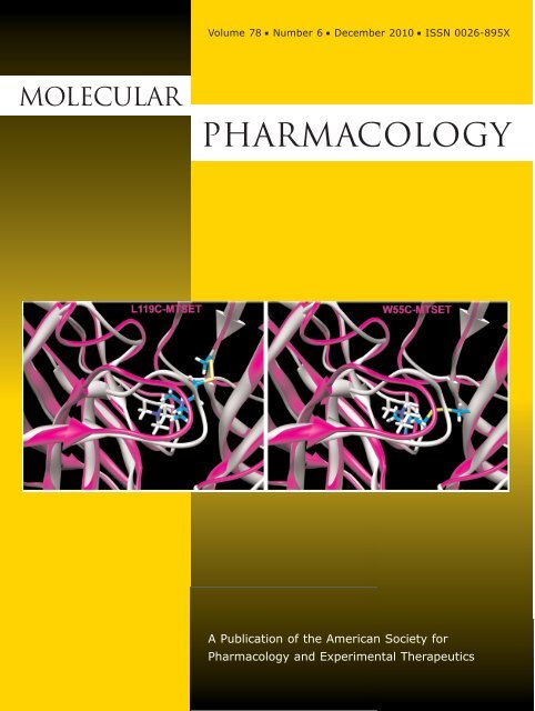
Volume 78 ■ Number 6 ■ December 2010 ■ ISSN 0026-895XMolecularPharmacologyA Publication <strong>of</strong> the American Society forPharmacology and Experimental Therapeutics
0026-895X/10/7806-1012–1025$20.00MOLECULAR <strong>PHARMACOLOGY</strong> Vol. 78, No. 6Copyright © 2010 The American Society for Pharmacology and Experimental Therapeutics 66662/3641795Mol Pharmacol 78:1012–1025, 2010Printed in U.S.A.Tethered Agonist Analogs as Site-Specific Probes for Domains<strong>of</strong> the Human 7 Nicotinic Acetylcholine Receptor thatDifferentially Regulate Activation and DesensitizationJingyi Wang, Nicole A. Horenstein, Clare Stokes, and <strong>Roger</strong> L. <strong>Papke</strong>Department <strong>of</strong> Chemistry, <strong>University</strong> <strong>of</strong> Florida, Gainesville, Florida (J.W., N.A.H.); and Department <strong>of</strong> Pharmacology andTherapeutics, <strong>University</strong> <strong>of</strong> Florida College <strong>of</strong> Medicine, Gainesville, Florida (C.S., R.L.P.)Received May 28, 2010; accepted September 2, 2010ABSTRACTHomomeric 7 nicotinic acetylcholine receptors represent animportant and complex pharmaceutical target. They can beactivated by structurally diverse agonists and are highly likely toenter and remain in desensitized states at rates determined bythe structures <strong>of</strong> the agonists. To identify structural elementsregulating this function, we introduced reactive cysteines intothe 7 ligand-binding domain allowing us to bind sulfhydrylreactive(SH) agonist analogs or control reagents onto specificpositions in the ligand binding domain. We identified four 7mutants (S36C, L38C, W55C, and L119C) in which the tethering<strong>of</strong> the SH reagents blocked further acetylcholine-evoked activation<strong>of</strong> the receptor. However, after selective reaction with SHagonist analogs, the type II allosteric modulator N-(5-chloro-2,4-dimethoxyphenyl)-N-(5-methyl-3-isoxazolyl-3-isoxazolyl)-urea (PNU-120596) could reactivate L119C and W55C mutantsand receptors with a reduced or modified C-loop. ModifiedS36C and L38C mutants were insensitive to reactivation byPNU-120596, whether they were reacted with agonist analogsor alternative SH reagents. Molecular modeling showed that inthe W55C and L119C mutants, the ammonium pharmacophore <strong>of</strong>the agonist analog methanethiosulfonate-ethyltrimethylammoniumwould be in a similar but nonidentical position underneaththe C-loop. The orientation assumed by the ligand tethered to119C was approximately 3-fold more sensitive to PNU-120596than the alternative pose at 55C. Our results support the hypothesisthat a single ligand can bind within the receptor in differentways and, depending on the specific binding pose, may variouslypromote activation or desensitization, or, alternatively, function asa competitive antagonist. This insight may provide a new approachfor drug development.IntroductionNicotinic acetylcholine receptors (nAChRs) are pentamericligand-gated ion channels, mainly expressed in muscle andneurons, and are also found in glial and non-neuronal tissues(Gahring and <strong>Roger</strong>s, 2005). In the brain, heteromeric 42-containing and homomeric 7 subtypes are the two majortypes <strong>of</strong> the nAChRs representing high-affinity binding sitesfor nicotine and -bungarotoxin, respectively (Clarke et al.,1986; Gotti et al., 2007). Although 42 receptors have beenstrongly associated with the cognitive and addicting effects <strong>of</strong>nicotine, 7 nAChRs have been implicated as influential inThis work was supported by the National Institutes <strong>of</strong> Health NationalInstitute <strong>of</strong> General Medical Sciences [Grant R01-GM057481].Article, publication date, and citation information can be found athttp://molpharm.aspetjournals.org.doi:10.1124/mol.110.066662.neuroprotection (Svensson and Nordberg, 1999), attentionaland cognitive enhancement (Young et al., 2004), and theregulation <strong>of</strong> inflammatory signaling (Wang et al., 2003; Giebelenet al., 2007; Pavlov et al., 2007).Electron microscopy studies <strong>of</strong> Torpedo californica nAChR(Unwin et al., 2002; Miyazawa et al., 2003) and more recentlyX-ray analyses <strong>of</strong> molluscan acetylcholine binding protein(AChBP) have provided high-resolution templates for homologymodels <strong>of</strong> the nAChR (Smit et al., 2001; Sixma and Smit,2003). Each nAChR subunit consists <strong>of</strong> an extracellular ligandbinding domain (LBD), a transmembrane domain consisting<strong>of</strong> four bundled -helices, and an intracellular domainwith poorly defined structure and function. In the LBD, agonistsare bounded by six loop structures, loops A, B, and Cfrom the subunit (primary face) and loops D, E, and F fromthe non- subunit (complementary face) (Sixma and Smit,2003). Conserved residues on these loops are important forABBREVIATIONS: nAChR, nicotinic acetylcholine receptor; ACh, acetylcholine; LBD, ligand-binding domain; PAM, positive allosteric modulator;PNU-120596, N-(5-chloro-2,4-dimethoxyphenyl)-N-(5-methyl-3-isoxazolyl)-urea; TMA, tetramethylammonium; Br-ACh, bromoacetylcholine;MTSET, methanethiosulfonate-ethyltrimethylammonium; MTSEA, (2-aminoethyl)methanethiosulfonate; QN-SH, 2-(quinuclidinium)ethyl methanethiosulfonate;EMTS, ethyl methanethiosulfonate; MTSACE, 2-(aminocarbonyl)ethyl methanethiosulfonate; DTT, 1,4-dithiothreitol; SH, sulfhydrylreactive;AChBP, acetylcholine binding protein; MS222, 3-aminobenzoic acid ethyl ester; DTT, dithiothreitol.1012
Activation and Desensitization by Tethered Agonist Analogs 1013agonist binding and/or activation on the receptor (Brejc et al.,2001).The nAChR are members <strong>of</strong> the large “cysteine-loop” superfamily<strong>of</strong> ligand-gated ion channels, which includes receptorsfor GABA, glycine, and serotonin (Millar and Gotti,2009). Ion channel activation is associated with one or more<strong>of</strong> the conformational states that can be induced by ligandbinding; although nonconducting, putatively desensitizedstates are more stable and predominate after the prolongedbinding <strong>of</strong> agonist. The 7 receptor has been proposed tomanifest a distinct form <strong>of</strong> rapid desensitization, which maybe facilitated by high levels <strong>of</strong> agonist binding-site occupancy(<strong>Papke</strong> et al., 2000). This form <strong>of</strong> desensitization can bedestabilized by mutations in the ion channel or with a positiveallosteric modulator (PAM) (Bertrand et al., 2008).The discovery <strong>of</strong> 7-selective PAMs such as N-(5-chloro-2,4-dimethoxyphenyl)-N-(5-methyl-3-isoxazolyl-3-isoxazolyl)-urea(PNU-120596) has both suggested new ways to target 7receptors therapeutically (Grønlien et al., 2007) and providednew tools to study them experimentally. In particular, wehave gained new insights into the unique desensitizationproperties <strong>of</strong> 7 receptors through the use <strong>of</strong> this type II PAM(Grønlien et al., 2007; <strong>Papke</strong> et al., 2009), which not onlyenhances apparent peak current during agonist applicationbut can also reactivate one or more desensitized states(<strong>Papke</strong> et al., 2009) by binding in an intrasubunit cavitylocated between the four -helical transmembrane domains(Young et al., 2008).The 7 nAChR is a challenging therapeutic target because<strong>of</strong> both the structural limitation associated with homologymodeling and its propensity to enter and remain in desensitizedstates in ways that can depend on the structure <strong>of</strong> theagonist (<strong>Papke</strong> et al., 2009). An important goal is to decipherthe complex structural interplay between the character anddisposition <strong>of</strong> ligands bound to the receptor and the affect <strong>of</strong>that binding on receptor activation and desensitization.Cysteine mutagenesis is a commonly used approach fordetermining the solvent accessibility and functional significance<strong>of</strong> specific residues in proteins such as ligand-gated ionchannels (Karlin and Akabas, 1998; Spura et al., 1999; Sullivanet al., 2002; Barron et al., 2009). Unique cysteines areintroduced by point mutations at a series <strong>of</strong> positions, andeach mutant is then reacted with a sulfhydryl-reactive (SH)reagent to determine by functional analyses whether thereaction produced a labeled receptor.In this article, we report a systematic study <strong>of</strong> the geometricand spatial requirements for how bound ligands mayactivate the 7 receptor and, in some cases, promote conversion<strong>of</strong> the receptor to PNU-120596-sensitive desensitizedstates. Site-specific cysteine mutants in the LBD <strong>of</strong> human7 were expressed in Xenopus laevis oocytes. The cysteinemutants and wild-type receptors with the C-loop disulfideCys190 to Cys191 reduced were reacted with a panel <strong>of</strong>SH-reactive agonist analogs and characterized in terms <strong>of</strong>initial channel activation, blockade <strong>of</strong> subsequent AChevokedresponses, and the induction <strong>of</strong> PNU-120596-sensitivedesensitization. Our experimental data provide a usefulapproach for integrating nAChR homology modeling, includingdocking and molecular dynamics simulations, with functionalanalysis <strong>of</strong> ligand-dependent conformational changes.Materials and Methods7 nAChR Clones and Site-Directed Mutants. The human 7clone was obtained from <strong>Dr</strong>. Jon Lindstrom (<strong>University</strong> <strong>of</strong> Pennsylvania,Philadelphia, PA). The human RIC-3 clone, obtained from <strong>Dr</strong>.Millet Treinin (Hebrew <strong>University</strong>, Jerusalem, Israel), was coinjectedwith the 7 constructs to improve the levels and speed <strong>of</strong>receptor expression. Amino acids are numbered as for human 7(vicinal C-loop cysteines at positions 190 and 191). Mutations wereintroduced using the QuikChange Site-Directed Mutagenesis kit(Stratagene, La Jolla, CA) following the manufacturer’s instructions.To obtain a cysteine-null background, a naturally occurring cysteinein 7 was mutated to serine (7 C116S). As reported previously(<strong>Papke</strong> et al., 2010), the pharmacology and macroscopic activationproperties <strong>of</strong> this cysteine-null receptor were indistinguishable fromthose <strong>of</strong> wild-type 7 in regard to the potency and relative efficacy <strong>of</strong>diverse agonists including ACh, tetramethylammonium (TMA),quinuclidine, and 3-(2-methoxy,4-hydroxy-benzylidene)anabasine.The mutant also was indistinguishable from wild-type in regard tothe rapid concentration-dependent desensitization characteristic <strong>of</strong>7 (<strong>Papke</strong> et al., 2010). The novel cysteine mutants used in theseexperiments were made in the LBD <strong>of</strong> 7 C116S. All mutations wereconfirmed with automated fluorescent sequencing. After linearizationand purification <strong>of</strong> cloned cDNA, RNA transcripts were preparedin vitro using the appropriate mMessage mMachine kit from AmbionInc. (Austin, TX).Expression in X. laevis Oocytes. Mature (9 cm) female X.laevis African frogs (Nasco, Ft. Atkinson, WI) were used as thesource <strong>of</strong> oocytes. Before surgery, frogs were anesthetized by layingthe animal in a 1.5g/l solution <strong>of</strong> 3-aminobenzoic acid ethyl ester(MS222; Sigma-Aldrich, St. Louis, MO) for 30 min. Oocytes wereremoved from an abdominal incision.To digest the follicular cell layer, harvested oocytes were treatedwith 1.25 mg/ml collagenase from Worthington Biochemical Cooperation(Freehold, NJ) for 2hatroom temperature in the Barth’ssolution without calcium (88 mM NaCl, 1 mM KCl, 2.38 mMNaHCO 3 , 0.82 mM MgSO 4 , 15 mM HEPES, pH 7.6, and 12 g/ltetracycline). After that, stage 5 oocytes were isolated and injectedwith 50 nl (5–20 ng) <strong>of</strong> appropriate subunit cRNAs. Recordings weremade 2 to 10 days after injection. The experimental response valueswere normalized to control ACh applications to avoid the variety <strong>of</strong>the absolute magnitude <strong>of</strong> the evoked current response over time.Chemicals. The methanethiosulfonate compounds ethyl methanethiosulfonate(EMTS), 2-(aminocarbonyl)ethyl methanethiosulfonate(MTSACE), 2-(quinuclidinium)ethyl methanethiosulfonate(QN-SH), (2-aminoethyl)methanethiosulfonate (MTSEA), and methanethiosulfonate-ethyltrimethylammonium(MTSET) (Fig. 2) werepurchased from Toronto Research Chemicals Inc. (North York, ON,Canada). PNU-120596 was obtained from Tocris Bioscience (Ellisville,MO), and all <strong>of</strong> the other chemicals for electrophysiology includingBromo-acetylcholine (Br-ACh) were from Sigma-Aldrich.EMTS, PNU-120596, and MTSACE stock solutions were made indimethyl sulfoxide monthly and freshly diluted in Ringer’s solutionevery day. Other SH reagent stock solutions were made daily inRinger’s solution and diluted.Electrophysiology. Experiments were conducted using Opus-Xpress 6000A (Molecular Devices, Sunnyvale, CA). OpusXpress is anintegrated system that provides automated impalement and voltageclamp <strong>of</strong> up to eight oocytes in parallel. Cells were automaticallybath-perfused with Ringer’s buffer, and both the voltage and currentelectrodes were filled with 3 M KCl. The agonist compounds weredelivered from a 96-well plate and applied via disposable tips toeliminate any possibility <strong>of</strong> cross-contamination. <strong>Dr</strong>ug applicationsalternated between ACh controls and experimental applications.Cells were voltage-clamped at a holding potential <strong>of</strong> 60 mV. Datawere collected at 50 Hz and filtered at 20 Hz. Flow rates were set at2 ml/min. Unless otherwise indicated, drug applications were 12 s induration followed by 181-s washout periods.
1014 Wang et al.Experimental Protocols and Data Analysis. Each oocyte receivedtwo initial control applications <strong>of</strong> ACh, a 60-s 1 mM SHreagent application (initial flow rate <strong>of</strong> 2 ml/min for 10 s, followed by0.5 ml/min for 50 s), a follow-up control application <strong>of</strong> ACh, then anapplication <strong>of</strong> 300 M PNU-120596, and another follow-up AChcontrol application. Control ACh was 60 M for 7 wild type, 7C116S, and 7 L119C; 100 ACh for 7 L38C; and 300 M for theother mutants. These concentrations were empirically determined togive robust reproducible responses with repeated ACh applicationsand reflected intrinsic differences in the ACh potency for the mutants(<strong>Papke</strong> et al., 2010).The peak amplitude and the net charge (<strong>Papke</strong> and Porter <strong>Papke</strong>,2002) <strong>of</strong> experimental responses were calculated relative to the average<strong>of</strong> the first two ACh control responses to normalize the dataand compensate for the varying levels <strong>of</strong> channel expression amongthe oocytes. PNU-120596 was capable <strong>of</strong> allowing agonists to induceenormous and sustained responses, which introduced large varianceinto the calculated net charge response. Therefore, peak responseswere compared after using PNU-120596, whereas treatments beforePNU-120596 were compared based on the net charge response. Inseparate experiments (data not shown), it was determined that one<strong>of</strong> the primary effects <strong>of</strong> PNU-120596 is to eliminate the normalseparation <strong>of</strong> peak current and net charge concentration-responserelationships seen with 7 nAChR (<strong>Papke</strong> and Porter <strong>Papke</strong>, 2002).This suggests that a primary effect <strong>of</strong> PNU-120596 is to reduce theunique agonist concentration-dependent form <strong>of</strong> desensitization thatwe hypothesize is promoted by binding <strong>of</strong> agonist at multiple sites.Means and S.E.M. values were calculated from the normalizedresponses <strong>of</strong> at least four oocytes for each experimental concentration.Individual oocytes were used for no more than one test concentrationbecause SH reagents are potentially able to form covalentbonds with the receptor. Whenever PNU-120596 was used, the cellswere discarded afterward; the bath was cleaned with ethanol andflushed with Ringer’s buffer for 20 min.The protocol for study <strong>of</strong> the reduced Cys190–Cys191 C-loop disulfidewas as above, except that after the initial ACh controls, the7 C116S nAChRs were treated with 1 mM dithiothreitol (DTT) for60 s (initial flow rate <strong>of</strong> 2 ml/min for 10 s followed by 0.5 ml/min for50 s) plus additional minutes, as indicated, via static bath (i.e.,stopping the flow <strong>of</strong> Ringer’s washthrough to retain the reagent inthe bath). Then, a follow-up ACh test was applied before a 60-streatment with an SH reagent, followed by ACh, PNU-120596, andanother ACh application. All <strong>of</strong> the responses were normalized to theaverage <strong>of</strong> the two initial ACh control applications and compared asdescribed above.SH reagent labeling kinetics experiments similarly involved incubatingreceptors for varied periods <strong>of</strong> reaction time before washout <strong>of</strong>reagent. The comparative concentration-response data betweenwild-type and C116S receptors for Br-ACh, MTSET, MTSEA, andQN-SH were collected from 100 M to 3 mM, using 300 M ACh ascontrol before and after each application <strong>of</strong> the reagent. Data wereanalyzed as described previously (<strong>Papke</strong> and Porter <strong>Papke</strong>, 2002).Molecular Modeling. A homology model for the human 7nAChR was created using the Aplysia californica AChBP structure2PGZ to select the cysteine mutant candidates (Hansen et al., 2005).A ClustalW (Higgins et al., 1996) alignment <strong>of</strong> the AChBP andhuman 7 sequence was generated and submitted to the SwissModel structure server. The resulting monomeric model was superimposedtwice on the A and B chains <strong>of</strong> the AChBP pentamericcrystal structure to generate a dimer model (Fig. 1A). The model wasthen examined for clashes, which subsequently resolved by variation<strong>of</strong> side chain rotamer or in combination with constrained minimizationusing the GROMOS force field resident in the Swiss-PdbViewer(version 4.0; http://spdbv.vital-it.ch/), followed by Amber 10 (http://ambermd.org/) molecular mechanics refinement with the bound2PGZ ligand (cocaine hydrochloride) included to prevent collapse <strong>of</strong>the LBD during structural optimization. Docking was performedwith the Dock 6.1 program (http://dock.compbio.ucsf.edu/DOCK_6/index.htm) with evaluation <strong>of</strong> dock scores based on a grid <strong>of</strong> 0.3-Åspacing.Homology models for MTSET- and MTSEA-labeled cysteine mutantsand energy minimization <strong>of</strong> these structures in AMBER requireddefinition <strong>of</strong> atoms and force field parameters, which wereobtained by computation on model-labeled cysteines. The labeledadduct formed between N-formyl cysteine carboxamide and MTSEAor MTSET was built and its structure minimized, and charges werecalculated with MOPAC and AM1 parameters in ANTECHAMBER.Force constants were identified for the structures with the generalizedAmber force field, and these values were transferred as requiredto allow recognition <strong>of</strong> the labeled residues <strong>of</strong> the protein model inthe xleap routine <strong>of</strong> Amber 10.Fig. 1. Cysteine mutations in the LBD <strong>of</strong> 7 nAChR. A, side view <strong>of</strong> thereceptor model as a dimer. The key elements <strong>of</strong> the 7 receptor weremodeled by using the 2BG9 template for the transmembrane domainfused to the ligand binding domain made from 2PGZ template. The plusface C-loop is shown in yellow, the M2 and M3 linker loop is shown inred-pink, the cysteine loop is colored green, and the different residuessubjected for mutation are colored variously. All <strong>of</strong> the cysteine mutantswere made in the background <strong>of</strong> C116S. Free cysteines at Cys190 andCys191 were generated at the same time by using DTT to reduce theC-loop disulfide bond. The view was made from the outside <strong>of</strong> the channelpore, and the four transmembrane helixes (M1 to M4) were lined clockwiseas shown, putting M2 helix toward the channel pore. The intracellulardomain is not shown. B, a close view <strong>of</strong> the predicted agonist bindingsite indicating the plus face and minus face <strong>of</strong> the receptor, colored in blueand light sea green, respectively.
Activation and Desensitization by Tethered Agonist Analogs 1015ResultsThe Location <strong>of</strong> Cysteine Mutants in the LBD. Toselect the cysteine mutants for labeling study, we dockedBr-ACh and QN-SH to the wild-type human 7 nAChRdimer. After docking, different poses within 5 kcal/mol differencefrom the best score were used to predict the potentialinteracting side chains that could be mutated to cysteine.This set included five on the complementary face: Ser36,Leu38, Trp55, Leu119, and Ile165 (Fig. 1B). We have demonstratedpreviously the functional competency <strong>of</strong> receptorswith cysteine mutations at these sites and determined thatcysteine mutations on the primary face <strong>of</strong> the LBD are lesswell tolerated because such mutants were either not wellexpressed or were poorly responsive to ACh (<strong>Papke</strong> et al.,2010). The ACh EC 50 values for wild-type (C116C), C116S,S36C, L38C, W55C, L119C, and I165C were 27 3 (Horensteinet al., 2008), 29.4 0.7 (<strong>Papke</strong> et al., 2010), 20 9, 24 5, 180 20, 30 4, and 60 26, M, respectively.To evaluate the labeling kinetics for different cysteine mutants,it is important to know the environment <strong>of</strong> the freecysteine. In general, ionic thiolate (solvent accessible) is amuch better nucleophile than nonionic thiolate (solvent exclusive)(Roberts et al., 1986). The environments <strong>of</strong> the fivecysteine mutation sites at positions Ser36, Leu38, Trp55,Leu119, and Ile165 were predicted by evaluating both the solvent-accessiblesurface and the solvent-exclusive surface in Chimera-1.4(http://www.cgl.ucsf.edu/chimera/download.html) forthe homology model built on the 2PGZ template. Both methodsranked the five amino acids consistently, in an order <strong>of</strong> Ile165 Trp55 Leu119 Ser36 Leu38, in regard to relative solventaccessibility.SH Reagents. The structures <strong>of</strong> the six SH reagents usedin these experiments are shown in Fig. 2 along with that <strong>of</strong>the type II 7-selective PAM PNU-120596, which was usedas a functional probe <strong>of</strong> the covalently modified receptors.Two <strong>of</strong> these agents, MTSET and Br-ACh, are agonist analogsthat have been used previously to characterize muscleand/or T. californica-type nAChR (Spura et al., 1999; Stewartet al., 2006). Because quinuclidine is an agonist for neuronalnAChR, we hypothesized that if posed in a favorable orientationin the 7 LBD, QN-SH might also function as tetheredagonist once covalently bound to the receptor. We usedMTSEA as a fourth cationic SH reagent to test for the potentialimportance <strong>of</strong> the positive charge placement in the LBDindependent <strong>of</strong> the other elements <strong>of</strong> the presumed pharmacophore(Horenstein et al., 2008). The noncationic agentsMTSACE and EMTS were used as controls for nonspecificeffects <strong>of</strong> the SH reactions.Intrinsic Agonist Activity <strong>of</strong> the Three Agonist Analogson Cysteine-Null and Wild-Type 7. As expected,neither MTSEA nor the noncationic SH reagents evokedcurrents when applied to cells expressing either wild-type 7or the cysteine-null 7 C116S. The affinity-alkylating agentBr-ACh was a potent and efficacious full agonist for wild-typeand C116S 7 (Fig. 3). MTSET, an affinity ligand used previouslyto study muscle-type and T. californica nAChR subtypes,was a low-potency weak partial agonist for both <strong>of</strong> the7 control receptors (Fig. 3). Although quinuclidine is aneffective agonist for 7 (Horenstein et al., 2008), QN-SH hadno apparent agonist activity above our limits <strong>of</strong> detection foreither wild-type or C116S 7 (data not shown).Experimental Design to Characterize the Effects <strong>of</strong>7 Receptor Covalent Modifications by SH Reagents.Figure 4A illustrates the docking <strong>of</strong> TMA in the LBD <strong>of</strong> 7.TMA is a full agonist <strong>of</strong> 7, so this pose represents thehypothetical minimal structure able to induce the array <strong>of</strong> 7functional states, including the active ion channel state andligand-bound, nonconducting states commonly associatedwith “desensitization,” <strong>of</strong> which one or more can be convertedinto a conducting state with the application <strong>of</strong> the PAMPNU-120596. Figure 4B shows the predicted placement <strong>of</strong>the affinity ligand MTSET covalently bound to a cysteine atthe location <strong>of</strong> Leu119. The model suggests that the agonistanalog will be most likely positioned in the same vicinity <strong>of</strong>the LBD as the docked TMA, but with important differences.Our experimental goal is to determine whether there is functionalityto MTSET or alternative agonist analogs tethered insuch orientations in regard to ion channel function or theinduction and stabilization <strong>of</strong> functionally relevant nonconductingstates.Figure 4C outlines the basic sequence <strong>of</strong> experiments to bepresented in the following sections along with proposed interpretations.After the initial characterization <strong>of</strong> the cysteinemutants in regard to ACh sensitivity (experiment 1),the mutants were treated with SH reagents (experiment 2).Experiment 2 first served to determine whether the mutantsretained the same pharmacological pr<strong>of</strong>ile as the controlreceptors (wild-type and C116S 7) in regard to transientactivation <strong>of</strong> the ion channel by the SH reagents. Next, weconducted additional ACh applications (experiment 3) tomake an initial determination <strong>of</strong> whether a covalent modification<strong>of</strong> the receptors occurred that resulted in perturbation<strong>of</strong> function. We anticipated two most likely outcomes: thatsubsequent ACh-evoked responses would remain unchanged(case I), or that they would be reduced, indicative <strong>of</strong> functionallysignificant modification <strong>of</strong> the receptor (case II). Case IFig. 2. The structures <strong>of</strong> PNU-120596and the six reagents: Br-ACh, MTSET,MTSEA, QN-SH, EMTS, and MTSACE.
1016 Wang et al.represents two possibilities: failure <strong>of</strong> the SH reagent to reactwith the receptor (case Ia), or failure <strong>of</strong> the modification toresult in a detectable change in function (case Ib). A thirdoutcome that might also have been anticipated is that receptorswould be modified in such a way as to have increasedsensitivity to ACh (case III), which is not illustrated in thefigure.An important caveat to results that appeared intermediatebetween those anticipated for cases I and II is that theremight be significant difference in the reaction rates for thevarious covalent modifications; therefore, in such cases, kineticstudies were also conducted (experiment 2A). Oncereceptors were determined to be conditionally induced into acovalently modified state that was unresponsive to furtheractivation by ACh, we hypothesized that (case IIb) the tetheredligand might be situated as a simple antagonist, eitheroccluding the access <strong>of</strong> the LBD to agonist or holding thereceptors in a nonactivatible state or (case IIa) that thetethered agonist might function like an agonist that, after atransient phase <strong>of</strong> activation, stabilizes the receptor in adesensitized conformation that might be reactivated by thetype II PAM PNU-120596. Experiment 4, the application <strong>of</strong>PNU-120596 was conducted to make this determination. Finally,experiment 5, a follow-up application <strong>of</strong> ACh, probesthe PNU-120596 potentiation <strong>of</strong> this ACh response as affectedby the sulfhydryl-reacted receptors.The Basic Effects <strong>of</strong> Cysteine Mutagenesis. In thehuman 7 nAChR, a single, nondisulfide bonded free cysteineis found at position 116, which, if left intact, couldcomplicate the interpretation <strong>of</strong> thiol-specific labeling studieswhen cysteine is introduced elsewhere in the receptor. Thereforethe 7 C116S mutant (<strong>Papke</strong> et al., 2010) was preparedand used as the background for all additional cysteine mutants.The mutants used for these studies were selected froma total <strong>of</strong> 44 7 LBD cysteine mutants reported elsewhere(<strong>Papke</strong> et al., 2010). The cysteine mutations at Ser36, Leu38,Trp55, Leu119, and Ile165 (Fig. 1B) were selected for furtherstudy in these experiments based on their strategic placementwithin the LBD and also because these cysteine mutationshad relatively little impact on receptor function beforeSH modifications. When control applications <strong>of</strong> AChwere made to oocytes expressing these mutants (experi-Fig. 3. Intrinsic agonist activity <strong>of</strong> SH reagentagonist analogs for wild-type 7 and7 C116S. Top, representative traces <strong>of</strong>wild-type 7 and 7 C116S to Br-ACh andMTSET displayed with ACh control responsesobtained from the same oocytes.Bottom, concentration-response curves forBr-ACh and MTSET, normalized to themaximal ACh-evoked responses. Each pointrepresents the average response <strong>of</strong> at leastfour oocytes ( S.E.M.).
Activation and Desensitization by Tethered Agonist Analogs 1017Fig. 4. A, the docked pose <strong>of</strong> TMA in the LBD <strong>of</strong> 7. B, the predicted placement <strong>of</strong> the affinity ligand MTSET covalently bound to a cysteine at the location<strong>of</strong> Leu119. C, experimental design. Schematic <strong>of</strong> the design and hypotheses related to the possible events in the labeling study with the SH reagents, afterthe initial characterization <strong>of</strong> functional cysteine mutants. Case I represents the lack <strong>of</strong> functional evidence that a covalent bond formed between the cysteinemutant and the SH reagent. This could indicate two possible conditions: case Ia, in which no covalent modification occurred to because <strong>of</strong> unfavorable reactionconditions; and case Ib, in which the tethered 7 receptor can still accommodate ACh in one or more <strong>of</strong> the five binding sites, either the same domain wherethe SH reagent is attached or other free sites. Case II represents the condition in which the SH reagent has become covalently attached, and upon furtherapplication <strong>of</strong> ACh, activation <strong>of</strong> the ion channel is not observed, because <strong>of</strong> either SH reagent competitively occupying the agonist binding site or inducingligand-bound nonconducting (i.e., desensitized) states. Receptors were subsequently challenged with the type II PAM PNU-120596 to determine whether theorientation <strong>of</strong> the reagent in the binding site was sufficiently agonist-like to induce desensitization that is reversible by PNU-120596 (case IIa) or whetherthe ligand is functionally indistinguishable from a tethered antagonist (case IIb).
1018 Wang et al.ment 1 in Fig. 4C), all gave robust responses measuring atleast 30% the net charge <strong>of</strong> oocytes injected with the C116Sreceptor (<strong>Papke</strong> et al., 2010) at the same time point afterinjection.Intrinsic Activity <strong>of</strong> the SH Reagents as Agonists forWild-Type and C116S 7 Receptors. Six SH reagentswere used for experimental modification <strong>of</strong> the cysteine mutants(Fig. 2), four <strong>of</strong> which we hypothesized would functionas agonist analogs. To confirm that hypothesis, they werecompared with ACh for their ability to activate wild-type andC116S 7 receptors expressed in oocytes. Our experiments confirmed(Fig. 3, bottom) that Br-ACh is a potent full agonist for7 control nAChR (EC 50 12–15 M), and MTSET was aweak partial agonist (I max 26%), with an EC 50 valuesgreater than 200 M for both control constructs (Fig. 3,bottom). The EC 50 values for MTSET were 292 62 and219 104 M for wild-type (C116C) and C116S, respectively.Although considered a possible agonist analog, QN-SH hadno apparent agonist activity above our limits <strong>of</strong> detection foreither wild-type or C116S 7. Likewise, the three other SHreagents (Fig. 2) showed no detectable agonist activity onwild-type or C116S 7 nAChR (data not shown).Fig. 5. Representative traces from voltage-clamp experiments, outlined in Fig. 4C, illustrating the effects <strong>of</strong> 60-s applications <strong>of</strong> SH reagents on C116Sand cysteine mutant 7 receptors and the subsequent effects <strong>of</strong> PNU-120596’s potentiation. The results are displayed in the order <strong>of</strong> experimentprotocol from left to the right with total time <strong>of</strong> 5 min between each step: ACh (12 s), SH reagent (1 mM MTSET or MTSEA, as indicated, for 60 s),ACh (12 s), PNU-120596 (300 M for 12 s), and ACh (12 s). Because some mutants showed reduced sensitivity to ACh, the concentration <strong>of</strong> AChapplications was 60 M for C116S and L119C 7 mutants, and 300 M for 7 S36C, W55C, and I165C. The drug applications are shown above theresponse curve as boldface black lines. Because <strong>of</strong> the magnitude <strong>of</strong> PNU-120596 effects, the ACh-evoked responses <strong>of</strong> I165C after PNU-120596application are shown at a less sensitive current scale, and likewise for the PNU-120596-evoked response <strong>of</strong> 7 L119C after MTSEA. Note thatalthough a 60-s application <strong>of</strong> MTSET produced a significant reduction in the subsequent ACh-evoked response <strong>of</strong> the S36C mutant, longerapplications were required to produce a maximal effect (Fig. 7).
Activation and Desensitization by Tethered Agonist Analogs 1019Intrinsic Activity <strong>of</strong> the SH Reagents on the 7 CysteineMutants. The presentation <strong>of</strong> a SH reagent to a cysteinemutant (experiment 2 in Fig. 4C) is predicted to resultin a covalent modification <strong>of</strong> the receptor, dependent on theaccessibility <strong>of</strong> the specific mutated residue. However, becauseseveral <strong>of</strong> our reagents are agonist analogs, covalentreaction with the receptor might be expected to be precededby, or coincident to, activation <strong>of</strong> the receptor. Therefore, wemeasured current stimulated by the presentation <strong>of</strong> the SHreagents. Not surprisingly, significant currents were onlydetected for Br-ACh and MTSET (for representative MTSETresponses, see Fig. 5), the two analogs that had the greatestagonist activity with the wild-type and C116S 7 receptors. Asummary <strong>of</strong> evoked responses stimulated by Br-ACh andMTSET is displayed in Fig. 6. Analysis <strong>of</strong> variance indicatedthat for all <strong>of</strong> the receptors tested, Br-ACh was equally asefficacious as ACh and that MTSET was significantly lessefficacious than either ACh or Br-ACh (p 0.001). Therewere no significant differences in the relative agonist efficaciesamong the various mutants. It is noteworthy that QN-SH, which showed minimal agonist property with wild-type7 or any <strong>of</strong> the mutants tested, did show significant responsesduring the application to 7 L119C nAChR at 1 mM(21% <strong>of</strong> the net-charge response relative to the ACh controls;data not shown).ACh-Mediated Responses Subsequent to the Application<strong>of</strong> SH Reagents. The ACh-evoked responses <strong>of</strong> C116Sand I165C 7 receptors were unchanged relative to their initialACh control responses by the application <strong>of</strong> any <strong>of</strong> the SHreagents (Fig. 5). Given the lack <strong>of</strong> free cysteines, it is unlikelythat there was any covalent modification <strong>of</strong> 7 C116S, and ifthere were any reactions with either the wild-type (C116C; datanot shown) or the I165C 7 receptors (Fig. 5), they seemed to befunctionally neutral, at least in regard to ACh activation (Fig.4C case I). In contrast, there were functional aftereffects identifiedin the other four receptors (W55C, L119C, S36C, andL38C) that contained mutations in the complementary face <strong>of</strong>the LBD. Representative traces are shown in Fig. 5, and asummary <strong>of</strong> the results is shown in Fig. 7.As shown in Fig. 7, not all <strong>of</strong> the reagents were equallyeffective at inhibiting the ACh-evoked responses <strong>of</strong> the cysteinemutants. Indeed, each mutant had a distinct pr<strong>of</strong>ile for the sixreagents. Reaction <strong>of</strong> the free cysteine at the Ser36 positionproduced decreases in the ACh response with all <strong>of</strong> the fivemethanethiosulfonate SH reagents but not the alternative reactiveagonist analog Br-ACh. However, treatments <strong>of</strong> 7 S36Cwith MTSACE, EMTS, QN-SH, and MTSEA produced onlypartial inhibition <strong>of</strong> 7 S36C ACh-evoked responses, whereasafter MTSET treatment, no significant ACh responses could bedetected. The 7 L38C mutant was very sensitive to MTSEAFig. 6. Activation pr<strong>of</strong>iles <strong>of</strong> Br-ACh andMTSET on wild-type and mutant human7 nAChRs. The responses shown in thegraph are net-charge responses obtainedduring the application <strong>of</strong> 1 mM Br-ACh orMTSET, normalized to the ACh maximalresponse determined previously (<strong>Papke</strong>et al., 2010). Data represent the averageresponses <strong>of</strong> at least four oocytes (S.E.M.).
1020 Wang et al.and showed 77% inhibition <strong>of</strong> ACh-evoked responses afterQN-SH. Only the positively charged SH reagents and Br-AChwere able to inhibit the 7 W55C mutant.As reported previously (<strong>Papke</strong> et al., 2010), the ACh-evokedresponses <strong>of</strong> 7 L119C were very efficiently blocked by all <strong>of</strong> thecationic SH reagent treatments. After 60-s treatments withQN-SH, MTSET, or MTSEA, ACh-evoked responses decreasedessentially to 0. A 60-s MTSACE application also blocked 75%<strong>of</strong> the 7 L119C ACh-evoked response. The 7 L119C mutantwas also sensitive to Br-ACh; however, a 3-min application wasrequired for the complete blocking effect (data not shown). It isnoteworthy that the ACh response <strong>of</strong> the EMTS-treated 7L119C receptor seemed enhanced relative to the response <strong>of</strong>that mutant to ACh before EMTS.Although MTSEA and QN-SH produced maximal effects withjust 60-s treatments, the agonist-like reagents Br-ACh andMTSET required treatments <strong>of</strong> up to 5 min for maximal effectson subsequent ACh responses (experiment 3a in Fig. 4C), andeven after 5-min treatment with MTSET, L38C remained responsiveto subsequent applications.As shown in Fig. 7, the L119C and W55C 7 mutants weresimilar in their sensitivity to Br-ACh block <strong>of</strong> ACh-evoked responses.A full 3-min incubation was required to achieve thiseffect. However, there were no significant effects <strong>of</strong> Br-AChapplications on the ACh-evoked responses <strong>of</strong> S36C or L38C 7mutants, even with Br-ACh applications <strong>of</strong> up to 7 min.Activation <strong>of</strong> SH-Modified 7 Mutants by PNU-120596.The type II PAM PNU-120596 does not produce significant activation<strong>of</strong> wild-type or C116S 7 receptors when applied alone(Table 1, Fig. 5). However, when ACh or other agonists are coappliedwith PNU-120596 or applied to receptors primed previouslywith PNU-120596 (<strong>Papke</strong> et al., 2009), agonist-evoked responsesare substantially increased (Table 1 and Fig. 5). The 7 W55C,I165C, and S36C mutants showed no significant response to applications<strong>of</strong> 300 M PNU-120596 alone in the absence <strong>of</strong> agonistor before treatments with SH reagents. After the application <strong>of</strong> SHreagents to wild-type, C116S, or I165C 7 receptors, there were noresponses to subsequent applications <strong>of</strong> PNU-120596 in the absence<strong>of</strong> agonist (representative traces in Fig. 5). It is noteworthythat the 7 L38C and L119C mutants, as well as C116S receptorspreviously treated with DTT, did show small but significant responsesto subsequent applications <strong>of</strong> PNU-120596 alone (Table1). Nonetheless, PNU-120596 was still a very effective potentiator<strong>of</strong> these receptors. Applications <strong>of</strong> 60 M ACh after priming with300 M PNU-120596 produced responses that were 9- to 31-foldlarger than the responses measured during the prior application <strong>of</strong>ACh alone (Table 1).In addition to priming and/or potentiating agonist-evokedresponses, PNU-120596 can be used to convert previously desensitizedreceptors into a conducting state (experiment 4, Fig.4C). Therefore, we hypothesized that if the treatment <strong>of</strong> thecysteine mutant 7 receptors with our SH reagents inhibitedsubsequent ACh-evoked responses because the receptors werebeing locked into a PNU-120596-sensitive form <strong>of</strong> desensitization,then they should be directly activatable by PNU-120596after SH treatments. We hypothesized that this activity woulddepend on both the agonist character <strong>of</strong> the reagent and thespecific orientation <strong>of</strong> the covalently bound reagent in the LBD.Fig. 7. ACh responses after SH reagentapplication. In initial experiments, theoocytes were treated with the SH reagentsat 1 mM for 60 s and washed withRinger’s buffer for 3 min before the application<strong>of</strong> ACh. This protocol producedonly partial effects for Br-ACh and forMTSET on subtypes other than 7 L119C(see Fig. 5). Therefore, to compensate forthe relatively slow reaction rates <strong>of</strong> Br-ACh and MTSET (on receptors other thanL119C), the applications <strong>of</strong> these SH reagentswere extended to 5 min, which wassufficient to produce optimal effects. Thenet charge responses <strong>of</strong> the post-SH reagentACh-evoked responses were normalizedto the average <strong>of</strong> the two pre-SHreagent ACh controls. A response <strong>of</strong> 1.0represents no detectable change made bySH reagents (reflecting case I in Fig. 4C),whereas a response <strong>of</strong> 0 represents completeblocking <strong>of</strong> subsequent ACh activationby covalently attached SH reagents(reflecting case II in Fig. 4C).
Activation and Desensitization by Tethered Agonist Analogs 1021Although such effects as seen with W55C and L119C (Fig. 5)most likely are due to exclusion <strong>of</strong> ACh from the covalentlymodified agonist binding sites, they might alternatively indicatethe conversion <strong>of</strong> receptors into PNU-120596-insensitivenonconducting states.The PNU-120596 response pr<strong>of</strong>iles <strong>of</strong> the different mutantsafter the SH reagent treatments are shown in Fig. 8, and somerepresentative traces are shown in Fig. 5. The S36C and L38Cmutants had no apparent PNU-120596-evoked responses afterapplication <strong>of</strong> any <strong>of</strong> the cationic SH reagents. L38C yielded aTABLE 1PNU-120596 effects: responses to PNU-120596 applied alone, andsubsequent potentiation <strong>of</strong> ACh-evoked responsesTo conduct these experiments, cells were typically used with 24 h <strong>of</strong> injection, whenthe control responses to 60 M were very low, otherwise the potentiated responsesbecame too large to maintain voltage clamp. Under these conditions, PNU-120596applications produced small stimulus artifact baseline deflections which, when normalizedto small initial controls, gave values for peak deflections <strong>of</strong> 10%, which wetake as our limit for detection. Values are peak currents relative to the peak currentsevoked by 60 M ACh before the application <strong>of</strong> 300 M PNU-120596.ReceptorResponse to PNU-120596Applied AloneACh Response afterApplication <strong>of</strong> PNU-120596%7 N.S. 3000 40C116S N.S. 2000 5001165C N.S. 1250 240S36C N.S. 1200 450L38C 35 9% 910 63W55C N.S. 1200 40L119C 54 4% 3100 460aC116S DTT 26 16% 1700 500N.S., no significant response.a These are the data for 7C116S receptors after a 60-s treatment with 1 mM DTTintended to reduce the vicinal disulfide in the C-loop.small PNU-120596-evoked response after treatment withEMTS; however, this PNU response was intrinsic to L38C (Table1) and did not depend on reaction with an SH reagent. Incontrast, W55C showed greatly enhanced responses to applications<strong>of</strong> PNU-120596 alone after treatment with Br-ACh,QN-SH, and MTSET but no PNU-120596 responses at allafter treatment with MTSACE or EMTS. These results suggestthat once reacted with the agonist analogs, the 7 W55Creceptors were at least partially converted into a PNU-120596-sensitive desensitized state. Consistent with the previousobservation that reactions with either MTSACE orEMTS failed to block the subsequent ACh-evoked responses<strong>of</strong> 7 W55C, these agents clearly did not induce PNU-120596-sensitive desensitization either. It is noteworthythat PNU-120596 did reactivate the MTSEA-treated 7W55C but less than it did after the true agonist analogs(Fig. 8). This observation may suggest that the core pharmacophorefor PNU-120596 reactivation may be simplerthan the core agonist structure for activation <strong>of</strong> the unmodifiedwild-type receptors.To varying degrees, application <strong>of</strong> PNU-120596 stimulatedresponses from 7 L119C receptors after treatments witheach <strong>of</strong> the SH reagents. Note that the PNU-120596-evokedresponse after EMTS treatment was relatively small, suggestingthat this reagent was ineffective at converting receptorsto the PNU-120596-sensitive desensitized state. Thisobservation provides further insight into what may be thecore pharmacophore for PNU-120596 reactivation, suggestingthe requirement for only a charged nitrogen or perhapsother cationic center.Fig. 8. Responses to the type II PAMPNU-120596 after SH reagent treatment.After SH reagent and ACh treatments,300 M PNU-120596 was applied for 12 s(see Fig. 5 for sample traces). Because <strong>of</strong>the prolonged duration <strong>of</strong> responses consequentand subsequent to the application<strong>of</strong> PNU-120596 (beyond our normalwindow for net-charge measurements),data for these experiments are presentedas peak current amplitudes, normalizedto the peak currents <strong>of</strong> the initial controls.The treatment <strong>of</strong> 7 W55C and 7L119C with some <strong>of</strong> the SH reagents inducedthe PNU-120596-sensitive desensitizedstate (case IIa in Fig. 4C), whereasthe same treatments <strong>of</strong> 7 S36C and 7L38C gave results as shown for case IIb inFig. 4C.
1022 Wang et al.PNU-120596 Potentiation <strong>of</strong> ACh Responses in SH-Reacted Receptors. Furthermore, although activation byPNU-120596 alone after SH treatments provides a probe forinduced desensitization, further applications <strong>of</strong> ACh to PNU-120596-primed SH-reacted receptors could also indicateforms <strong>of</strong> receptor blockade through decreases in the AChpotentiation. To compare the effects <strong>of</strong> SH reagent modifications<strong>of</strong> the cysteine mutants on ACh responses after treatmentwith PNU-120596, we made additional ACh applicationsafter the SH reagent and PNU-120596 applications.Responses were compared with baseline ACh-evoked responses,and this value was compared with the PNU-120596potentiation observed in the absence <strong>of</strong> SH reactions (Table1). Some representative traces are shown in Fig. 5, and theresults are summarized in Tables 2 and 3. The 7 S36Creceptors showed largely diminished post-PNU-120596 AChpeak current values, except after Br-ACh. The post-PNU-120596 ACh peak responses <strong>of</strong> 7 L38C were most stronglyaffected by QN-SH and MTSEA and were totally unaffectedby EMTS treatment. The PNU-120596-potentiated ACh responses<strong>of</strong> 7 W55C were most strongly affected by QN-SHand MTSEA, whereas MTSET, Br-ACh, and MTSACE hadintermediate effects. EMTS, which seemed to potentiate 7W55C and L119C ACh-evoked responses (Fig. 7), also increasedthe PNU-120596-potentiated ACh currents <strong>of</strong> L119C, suggestingthat EMTS, once reacted with L119C, may itself be anallosteric potentiator, possibly working in an additive or synergisticmanner with PNU-120596.The PNU-120596 potentiation <strong>of</strong> 7 L119C ACh responseswas strongly reduced by all <strong>of</strong> the reagents except for EMTSand Br-ACh, which produced only 30 and 75% reductions,respectively. In general, all <strong>of</strong> the SH reagent effects on thePNU-120596-potentiated ACh responses were consistentwith the inhibition <strong>of</strong> the control ACh-evoked responses, asshown in Fig. 7. One striking disparity was seen in the effects<strong>of</strong> MTSACE on 7 W55C. Whereas MTSACE did not block thecontrol ACh-evoked responses, it did decrease the PNU-120596potentiation. In addition, for the W55C 7 receptor, bothMTSET and MTSEA reduced the ACh response (Fig. 7), butafter MTSET, PNU-120596 greatly potentiated the ACh response,and after MTSEA, it greatly inhibited it (Tables 1–3).The Modification <strong>of</strong> the Reduced C-Loop and Potentiation<strong>of</strong> PNU-120596. To study the potential effects <strong>of</strong> SHmodifications to the C-loop, a key structure in the LBD (Fig.1), we attempted to reduce the vicinal disulfide at positions190 and 191 in the 7 C116S receptor by treatment with 1mM DTT. We found that when exposed to DTT for increasingperiods <strong>of</strong> time, the receptors became progressively less responsiveto activation by ACh, barely showing any responseafter 5 min <strong>of</strong> DTT treatment. It is noteworthy that evenwhen the receptors were no longer able to be activated byACh, the reduced receptors could be activated by Br-ACh orMTSET (Fig. 9). We then conducted a series <strong>of</strong> ACh andPNU-120596 applications to determine whether the agonistanalogs showed evidence <strong>of</strong> covalently modifying the receptorsand inducing PNU-120596-sensitive desensitization.After the treatment <strong>of</strong> the reduced receptors with MTSETor QN-SH, there was a small recovery <strong>of</strong> sensitivity to ACh,whereas after Br-ACh, the receptors remained unresponsiveto ACh (Fig. 9A). However, treatment <strong>of</strong> the reduced receptorswith each <strong>of</strong> the agonist analogs seemed to very effectivelyinduce the PNU-120596-sensitive desensitized state,because large currents were stimulated by the application <strong>of</strong>PNU-120596 alone (Fig. 9B).Modeling Modifications <strong>of</strong> the Agonist and the BindingSite. We modeled the potential effects <strong>of</strong> bound SHreagents by introducing non-natural amino acids into the 7homology model that would be equivalent to the covalentlymodified amino acid residues <strong>of</strong> the receptor (Fig. 10, A andB). For example, after MTSET reacted with the free cysteinein the LBD, a trimethylamino ethylthio group was attachedto the sulfur <strong>of</strong> the cysteine mutant. After molecular mechanicsminimization, the structures <strong>of</strong> the MTSET-modified mutants(S36C, W55C, and L119C) were superimposed. The sideview <strong>of</strong> the receptor (Fig. 10A) shows that MTSET-labeled36C is farther away from the C-loop, whereas MTSET-55Cand MTSET-119C place the ammonium pharmacophore furtherunderneath the C-loop. MTSET-labeled 36C can alsoplace the ammonium group <strong>of</strong> the label proximate to Glu189,possibly excluding the ammonium group from the LBD byelectrostatic attraction.DiscussionIt is unclear how structurally different agonists bind tonAChRs and initiate the channel gating or, alternatively, ionTABLE 2Effects <strong>of</strong> SH reagents on PNU-120596-primed ACh-evoked responsesData are expressed relative to initial ACh control responses. The durations <strong>of</strong> the SH reagent reactions were those required to give maximal effects in the absence <strong>of</strong>PNU-120596 treatments.Receptor MTSACE EMTS Br-ACh QN-SH MTSET MTSEA%S36C 40 20 210 40 810 210 130 40 23 5 8 1L38C 450 180 990 200 690 110 58 14 630 21 30 16W55C 460 70 1760 40 220 40 56 12 440 140 7 1L119C 160 100 2140 260 760 340 21 10 81 10 71 52TABLE 3The effects <strong>of</strong> SH reagents on the relative PNU-120596 potentiation <strong>of</strong> ACh responsesValues are the ratios <strong>of</strong> PNU-120596 potentiation in Table 1 (before SH treatments) and 1B (after SH treatments).Receptor MTSACE EMTS Br-ACh QN-SH MTSET MTSEAS36C 0.033 0.175 0.675 0.108 0.019 0.007L38C 0.495 1.09 0.758 0.063 0.692 0.033W55C 0.383 1.467 0.183 0.046 0.367 0.006L119C 0.052 0.690 0.246 0.007 0.026 0.023
Activation and Desensitization by Tethered Agonist Analogs 1023channel desensitization. We traditionally associate the efficacy<strong>of</strong> specific agents with their relative effectiveness forinducing the transitions <strong>of</strong> the receptors among multipleconformational states subsequent to binding in a single preferredconformation. However, we know from crystallography<strong>of</strong> the ACh-binding protein that specific ligands will stabilizethese ligand binding domain analogs in distinct low-energyconformations (Celie et al., 2004), which in native receptorsare likely to be associated with different conformationalstates. However, for low P open receptors such as 7, thelifetime <strong>of</strong> the open channel conformation is far too brief toexpect that particular ligand-bound conformational state tobe stable in a low-energy structure. Because <strong>of</strong> the transientlow-probability nature <strong>of</strong> the open state in an 7 receptorthat has not been primed by an allosteric modulator such asPNU-120596, it is reasonable to hypothesize that the ligand-boundstate that precedes ion channel opening isitself not stable and will be conformationally distinct fromthe more stable bound states that are most likely to inducedesensitization.In our studies, we used SH-reactive agonist analogs andthe PAM PNU-120596 to probe the 7 LBD for the functionalconsequences <strong>of</strong> tethering agonist-like molecules in specificorientations. Our results support the hypothesis that a singleligand can bind within the receptor in different ways and,depending on the specific binding pose, may variously promoteactivation or desensitization, or, alternatively, functionas a competitive antagonist.One limitation to the use <strong>of</strong> ACh responsiveness as a solereporter <strong>of</strong> covalent modification is that it provides little orno insight into the underlying basis for the loss <strong>of</strong> function,for example, whether the binding site is simply occluded orwhether the receptor is being held in specific nonfunctionalstates, such as those associated with desensitization. Thisdistinction could be <strong>of</strong> particular importance for examiningthe effects <strong>of</strong> SH reagents that may be agonist analogs. There-Fig. 9. The modification <strong>of</strong> and PNU-120596’s potentiation on 7 C116S afterDTT treatment. A, representative tracesillustrating the strong agonist activity <strong>of</strong>MTSET, QN-SH, and Br-ACh for 5-minDTT-treated 7 C116S, and the induction<strong>of</strong> a PNU-120596-sensitive desensitization.B, the SH reagent and PNU-120596activation <strong>of</strong> DTT-treated 7 C116S relativeto the average <strong>of</strong> two ACh controlsbefore DTT treatment. The data representthe average responses <strong>of</strong> at least fouroocytes ( S.E.M.).
Activation and Desensitization by Tethered Agonist Analogs 1025absence <strong>of</strong> PNU-120596, but, as suggested by the data in Fig. 8,the L119C-MTSET intermediate state is more readily convertedinto a stable conducting state.It is reasonable that an agonist-like molecule covalentlybound within the LBD <strong>of</strong> the nicotinic 7 receptors could havethe effect <strong>of</strong> stabilizing some <strong>of</strong> the same ligand-bound nonconducting(i.e., desensitized) states that predominate the statefunction <strong>of</strong> the native receptor in the prolonged presence <strong>of</strong>agonist. Our most basic concept <strong>of</strong> ligand-gated ion channelfunction is that the receptors have evolved to react rapidly andtransiently to an activating signal and then are prevented fromexcessive activation by the absorbing character <strong>of</strong> the desensitizedstates. However, although this model fits our perception <strong>of</strong>synaptic ion channels, it is unclear that it is applicable toreceptors such as 7 nAChR, which perform important functionsin non-neuronal cells, suggested in some cases to notrequire ion channel activation (de Jonge and Ulloa, 2007; vanMaanen et al., 2009). It has also been shown that some forms <strong>of</strong>7-mediated cytoprotection require long periods <strong>of</strong> treatmentwith concentrations <strong>of</strong> agonist lower than the threshold concentrationsrequired for transient activation <strong>of</strong> the ion channel (Liet al., 1999). In the same cells, strong transient activation <strong>of</strong> the7 ion channel is cytotoxic within seconds (Li et al., 1999).These alternative modes <strong>of</strong> signaling suggest that stable, ligand-bound,nonconducting states, such as those revealed bythe effects <strong>of</strong> PNU-120596, may represent additional functionalstates for the 7 receptor.In conclusion, our results support the hypothesis that asingle ligand may adopt multiple binding poses within theligand binding pocket and that the topology <strong>of</strong> resulting functionaland nonfunctional states depends on both the ligandand the way in which it binds. The nonstationary behavior <strong>of</strong>a ligand-gated ion channel after a jump in agonist concentrationmay therefore be due to the relative stability <strong>of</strong> variousbinding poses and not the relative probability <strong>of</strong> specificconformational transitions arising from a single bindingmode. Our data suggest that a new approach for drug developmentcan be implemented, taking advantage <strong>of</strong> developingstructure models and targeting ligands with more comprehensiveconsideration for where they bind, how they bind,and most importantly what is likely to happen next.AcknowledgmentsWe thank Lynda Cortes, Sara Braley, and Shehd Abdullah AbbasAl Rubaiy for technical assistance.ReferencesBarron SC, McLaughlin JT, See JA, Richards VL, and Rosenberg RL (2009) Anallosteric modulator <strong>of</strong> alpha7 nicotinic receptors, N-(5-chloro-2,4-dimethoxyphenyl)-N-(5-methyl-3-isoxazolyl)-urea(PNU-120596), causes conformationalchanges in the extracellular ligand binding domain similar to those caused byacetylcholine. Mol Pharmacol 76:253–263.Bertrand D, Bertrand S, Cassar S, Gubbins E, Li J, and Gopalakrishnan M (2008)Positive allosteric modulation <strong>of</strong> the alpha7 nicotinic acetylcholine receptor: ligandinteractions with distinct binding sites and evidence for a prominent role <strong>of</strong> theM2–M3 segment. Mol Pharmacol 74:1407–1416.Brejc K, van Dijk WJ, Klaassen RV, Schuurmans M, van Der Oost J, Smit AB, andSixma TK (2001) Crystal structure <strong>of</strong> an ACh-binding protein reveals the ligandbindingdomain <strong>of</strong> nicotinic receptors. Nature 411:269–276.Celie PH, van Rossum-Fikkert SE, van Dijk WJ, Brejc K, Smit AB, and Sixma TK(2004) Nicotine and carbamylcholine binding to nicotinic acetylcholine receptors asstudied in AChBP crystal structures. Neuron 41:907–914.Clarke PB, Hamill GS, Nadi NS, Jacobowitz DM, and Pert A (1986) 3 H-nicotine- and125 I-alpha-bungarotoxin-labeled nicotinic receptors in the interpeduncular nucleus<strong>of</strong> rats. II. Effects <strong>of</strong> habenular deafferentation. J Comp Neurol 251:407–413.de Jonge WJ and Ulloa L (2007) The alpha7 nicotinic acetylcholine receptor as apharmacological target for inflammation. Br J Pharmacol 151:915–929.Gahring LC and <strong>Roger</strong>s SW (2005) Neuronal nicotinic acetylcholine receptor expressionand function on nonneuronal cells. AAPS J 7:E885–E894.Giebelen IA, van Westerloo DJ, LaRosa GJ, de Vos AF, and van der Poll T (2007)Local stimulation <strong>of</strong> alpha7 cholinergic receptors inhibits LPS-induced TNF-alpharelease in the mouse lung. Shock 28:700–703.Gotti C, Moretti M, Gaimarri A, Zanardi A, Clementi F, and Zoli M (2007) Heterogeneityand complexity <strong>of</strong> native brain nicotinic receptors. Biochem Pharmacol74:1102–1111.Grønlien JH, Håkerud M, Ween H, Thorin-Hagene K, Briggs CA, Gopalakrishnan M,and Malysz J (2007) Distinct pr<strong>of</strong>iles <strong>of</strong> alpha7 nAChR positive allosteric modulationrevealed by structurally diverse chemotypes. Mol Pharmacol 72:715–724.Hansen SB, Sulzenbacher G, Huxford T, Marchot P, Taylor P, and Bourne Y(2005)Structures <strong>of</strong> Aplysia AChBP complexes with nicotinic agonists and antagonistsreveal distinctive binding interfaces and conformations. EMBO J 24:3635–3646.Higgins DG, Thompson JD, and Gibson TJ (1996) Using CLUSTAL for multiplesequence alignments. Methods Enzymol 266:383–402.Horenstein NA, Leonik FM, and <strong>Papke</strong> RL (2008) Multiple pharmacophores for theselective activation <strong>of</strong> nicotinic alpha7-type acetylcholine receptors. Mol Pharmacol74:1496–1511.Karlin A and Akabas MH (1998) Substituted-cysteine accessibility method. MethodsEnzymol 293:123–145.Li Y, <strong>Papke</strong> RL, He YJ, Millard WJ, and Meyer EM (1999) Characterization <strong>of</strong> theneuroprotective and toxic effects <strong>of</strong> 7 nicotinic receptor activation in PC12 cells.Brain Res 830:218–225.Millar NS and Gotti C (2009) Diversity <strong>of</strong> vertebrate nicotinic acetylcholine receptors.Neuropharmacology 56:237–246.Miyazawa A, Fujiyoshi Y, and Unwin N (2003) Structure and gating mechanism <strong>of</strong>the acetylcholine receptor pore. Nature 423:949–955.<strong>Papke</strong> RL, Kem WR, Soti F, López-Hernández GY, and Horenstein NA (2009)Activation and desensitization <strong>of</strong> nicotinic alpha7-type acetylcholine receptors bybenzylidene anabaseines and nicotine. J Pharmacol Exp Ther 329:791–807.<strong>Papke</strong> RL, Meyer E, Nutter T, and Uteshev VV (2000) alpha7 receptor-selectiveagonists and modes <strong>of</strong> alpha7 receptor activation. Eur J Pharmacol 393:179–195.<strong>Papke</strong> RL and Porter <strong>Papke</strong> JK (2002) Comparative pharmacology <strong>of</strong> rat and humanalpha7 nAChR conducted with net charge analysis. Br J Pharmacol 137:49–61.<strong>Papke</strong> RL, Stokes C, Williams DK, Wang J, and Horenstein NA (2010) Cysteineaccessibility analysis <strong>of</strong> the human alpha7 nicotinic acetylcholine receptor ligandbinding domain identifies L119 as a gatekeeper. J Neuropharmacol doi: 10.1016/j.neuropharm.2010.07.014.Pavlov VA, Ochani M, Yang LH, Gallowitsch-Puerta M, Ochani K, Lin X, Levi J,Parrish WR, Rosas-Ballina M, Czura CJ, Larosa GJ, Miller EJ, Tracey KJ, andAl-Abed Y (2007) Selective alpha7-nicotinic acetylcholine receptor agonist GTS-21improves survival in murine endotoxemia and severe sepsis. Crit Care Med 35:1139–1144.Roberts DD, Lewis SD, Ballou DP, Olson ST, and Shafer JA (1986) Reactivity <strong>of</strong>small thiolate anions and cysteine-25 in papain toward methyl methanethiosulfonate.Biochemistry 25:5595–5601.Sixma TK and Smit AB (2003) Acetylcholine binding protein (AChBP): a secretedglial protein that provides a high-resolution model for the extracellular domain <strong>of</strong>pentameric ligand-gated ion channels. Annu Rev Biophys Biomol Struct 21:21.Smit AB, Syed NI, Schaap D, van Minnen J, Klumperman J, Kits KS, Lodder H, vander Schors RC, van Elk R, Sorgedrager B, Brejc K, Sixma TK, and Geraerts WP(2001) A glia-derived acetylcholine-binding protein that modulates synaptic transmission.Nature 411:261–268.Spura A, Russin TS, Freedman ND, Grant M, McLaughlin JT, and Hawrot E (1999)Probing the agonist domain <strong>of</strong> the nicotinic acetylcholine receptor by cysteinescanning mutagenesis reveals residues in proximity to the alpha-bungarotoxinbinding site. Biochemistry 38:4912–4921.Stewart DS, Chiara DC, and Cohen JB (2006) Mapping the structural requirementsfor nicotinic acetylcholine receptor activation by using tethered alkyltrimethylammoniumagonists and antagonists. Biochemistry 45:10641–10653.Sullivan D, Chiara DC, and Cohen JB (2002) Mapping the agonist binding site <strong>of</strong> thenicotinic acetylcholine receptor by cysteine scanning mutagenesis: antagonist footprintand secondary structure prediction. Mol Pharmacol 61:463–472.Svensson AL and Nordberg A (1999) Beta-estradiol attenuate amyloid beta-peptidetoxicity via nicotinic receptors. Neuroreport 10:3485–3489.Unwin N, Miyazawa A, Li J, and Fujiyoshi Y (2002) Activation <strong>of</strong> the nicotinicacetylcholine receptor involves a switch in conformation <strong>of</strong> the alpha subunits. JMol Biol 319:1165–1176.van Maanen MA, <strong>Papke</strong> RL, Koepke J, Bevaart L, Clark R, Lamppu D, VervoordeldonkMJ, LaRosa GJ and Tak PP (2009) Therapeutic Effect <strong>of</strong> Stimulating theNicotinic Acetylcholine Receptor in the Collagen-Induced Model <strong>of</strong> RheumatoidArthritis: a Role for Ion Channel Activity and Penetration <strong>of</strong> the Central NervousSystem. Thesis, pp 77–97, <strong>University</strong> <strong>of</strong> Amsterdam, Amsterdam, Netherlands.Wang H, Yu M, Ochani M, Amella CA, Tanovic M, Susarla S, Li JH, Wang H, YangH, Ulloa L, Al-Abed Y, Czura CJ, and Tracey KJ (2003) Nicotinic acetylcholinereceptor alpha7 subunit is an essential regulator <strong>of</strong> inflammation. Nature 421:384–388.Young GT, Zwart R, Walker AS, Sher E, and Millar NS (2008) Potentiation <strong>of</strong> alpha7nicotinic acetylcholine receptors via an allosteric transmembrane site. Proc NatlAcad Sci USA 105:14686–14691.Young JW, Finlayson K, Spratt C, Marston HM, Crawford N, Kelly JS, and SharkeyJ (2004) Nicotine improves sustained attention in mice: evidence for involvement<strong>of</strong> the alpha7 nicotinic acetylcholine receptor. Neuropsychopharmacology 29:891–900.Address correspondence to: <strong>Dr</strong>. <strong>Roger</strong> L. <strong>Papke</strong>, Department <strong>of</strong> Pharmacologyand Therapeutics <strong>University</strong> <strong>of</strong> Florida, P.O. Box 100267, Gainesville,FL 32610-0267. E-mail: rlpapke@ufl.edu


