GENE TARGETING 1977–PRESENT - Nobelprize.org
GENE TARGETING 1977–PRESENT - Nobelprize.org
GENE TARGETING 1977–PRESENT - Nobelprize.org

You also want an ePaper? Increase the reach of your titles
YUMPU automatically turns print PDFs into web optimized ePapers that Google loves.
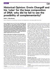
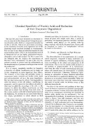
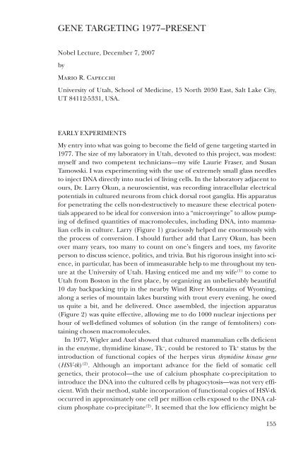
<strong>GENE</strong> <strong>TARGETING</strong> <strong>1977–PRESENT</strong>Nobel Lecture, December 7, 2007byMario R. CapecchiUniversity of Utah, School of Medicine, 15 North 2030 East, Salt Lake City,UT 84112-5331, USA.EARLY EXPERIMENTSMy entry into what was going to become the field of gene targeting started in1977. The size of my laboratory in Utah, devoted to this project, was modest:myself and two competent technicians—my wife Laurie Fraser, and SusanTamowski. I was experimenting with the use of extremely small glass needlesto inject DNA directly into nuclei of living cells. In the laboratory adjacent toours, Dr. Larry Okun, a neuroscientist, was recording intracellular electricalpotentials in cultured neurons from chick dorsal root ganglia. His apparatusfor penetrating the cells non-destructively to measure these electrical potentialsappeared to be ideal for conversion into a “microsyringe” to allow pumpingof defined quantities of macromolecules, including DNA, into mammaliancells in culture. Larry (Figure 1) graciously helped me enormously withthe process of conversion. I should further add that Larry Okun, has beenover many years, too many to count on one’s fingers and toes, my favoriteperson to discuss science, politics, and trivia. But his rigorous insight into science,in particular, has been of immeasurable help to me throughout my tenureat the University of Utah. Having enticed me and my wife (1) to come toUtah from Boston in the first place, by <strong>org</strong>anizing an unbelievably beautiful10 day backpacking trip in the nearby Wind River Mountains of Wyoming,along a series of mountain lakes bursting with trout every evening, he owedus quite a bit, and he delivered. Once assembled, the injection apparatus(Figure 2) was quite effective, allowing me to do 1000 nuclear injections perhour of well-defined volumes of solution (in the range of femtoliters) containingchosen macromolecules.In 1977, Wigler and Axel showed that cultured mammalian cells deficientin the enzyme, thymidine kinase, Tk – , could be restored to Tk + status by theintroduction of functional copies of the herpes virus thymidine kinase gene(HSV-tk) (2) . Although an important advance for the field of somatic cellgenetics, their protocol—the use of calcium phosphate co-precipitation tointroduce the DNA into the cultured cells by phagocytosis—was not very efficient.With their method, stable incorporation of functional copies of HSV-tkoccurred in approximately one cell per million cells exposed to the DNA calciumphosphate co-precipitate (2) . It seemed that the low efficiency might be155
Figure 1. A photograph of Lawrence M. Okun.Figure 2. A schematic of the apparatus I used to inject DNA into nuclei of mammalian cellsin culture. Micromanipulators are used to guide the needle, tip diameter 0.1 μm, containingthe DNA solution into nuclei of living cells while being viewed through a light microscope.156
a problem of delivery. Most of the DNA taken up by the cells did not appearto be delivered to the nucleus, where it could function, but instead was destinedfor lysosomes, where it was degraded. I sought to determine whether Icould introduce functional copies of the HSV-tk gene directly into nuclei ofcultured TK – cells using the microinjection apparatus described above. Thisprocedure proved to be extremely efficient; one cell in three that receivedthe DNA stably passed functional copies of the HSV-tk gene onto its daughtercells (3) . An immediate outcome of these experiments was that the high efficiencyof DNA transfer that we observed by microinjection made it practicalfor investigators to use the same methodology to generate transgenic micecontaining random insertions of exogenous DNA. This was accomplished byinjection of the desired DNA into nuclei of one-celled mouse zygotes withthe resulting embryos allowed to come to term after transfer to the uterusof foster mothers (4–8) . The generation of transgenic mice, in which chosenexogenous pieces of DNA have been randomly inserted within the mousegenome, has become a cottage industry.However, I found that the efficient transfer of functional HSV-tk genes intothe host cell genome required that the injected HSV-tk genes be linked to anadditional short viral DNA sequence (3) . It seemed plausible to me that highlyevolved viral genomes which, as part of their life cycle, resided in the host cellgenome, might contain bits of DNA sequence that enhanced their ability toestablish themselves within the host cell genome. I searched the genomes ofthe lytic simian virus, SV40, and the ASV retroviral provirus for the presenceof such sequences and found them (3) . When linked to the injected HSV-tkgene, these sequences increased the frequency with which TK + cells weregenerated by a factor of 100 over that produced by HSV-tk DNA injectedalone. I showed that this enhancement did not result from independentreplication of the injected HSV-tk DNA as an extra-chromosomal plasmid,but rather that the efficiency-enhancing sequences were either increasingthe frequency with which the exogenous DNA was inserting itself into thehost genome or increasing the probability that the HSV-tk gene, once integratedinto the host genome, was being expressed in the recipient cells. Thelatter turned out to be correct. These experiments were completed beforethe idea of gene-expression enhancers had emerged and contributed to thedefinition of these special DNA sequences (9) . Further, the emerging idea ofenhancers profoundly influenced our contributions to the development ofgene targeting vectors. Specifically, it alerted us to the importance of usingappropriate enhancers to mediate expression of newly introduced selectablegenes (used to select for successfully altered recipient cells), regardless of theinherent expression characteristics of the host chromosomal sites into whichwe were targeting those genes (10) .HOMOLOGOUS RECOMBINATIONAlthough the ability to introduce exogenous DNA randomly into the hostcell genome with very high efficiency by microinjection was itself extremely157
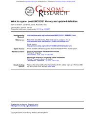
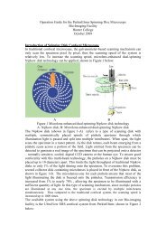
Figure 3. Formation of highly ordered head-to-tail DNA concatemers following introductionof multiple copies of the same DNA sequence into mammalian cell nuclei.useful, the observation that I found most fascinating from these early DNAinjectionexperiments was that, when multiple copies of the HSV-tk plasmidwere injected into a given cell, though many of them became randomly insertedinto the host cell’s genome, they would all be found at a single locus,as a highly ordered head-to-tail concatemer (see Figure 3) (11) . This was thekey observation and stimulus for the targeting project that followed.It was clear, however, that this project would now progress more rapidlywith the efforts of additional investigators. Fortunately, two very gifted postdoctoralfellows, Drs. Kim Folger Bruce and Eric Wong chose to join mygroup at this time (see Figure 4). It seemed that the highly-ordered concatemersof exogenous genes found at the insertion sites could not ariseby a random mechanism, but were likely generated either by replicationof the injected DNA before insertion (for example by a rolling circle-typemechanism of DNA replication) or by homologous recombination betweenthe co-injected HSV-tk plasmids. We proved that they were generated by homologousrecombination (11) . This conclusion was very significant because itdemonstrated that mammalian somatic cells contain an efficient enzymaticmachinery for mediating homologous recombination. The high efficiency ofthis machinery became evident from the observation that when more than100 HSV-tk plasmid molecules were injected per cell, they were all incorporatedinto a single, ordered, head-to-tail concatemer (11) . These experimentswere also the first demonstration of homologous recombination between cointroducedDNA molecules in cultured mammalian cells. From these resultsit was immediately apparent to me that if we could harness this efficient machineryto mediate homologous recombination between a newly introducedDNA molecule of our choice and the same DNA sequence in the recipient158
Figure 4. Photographs of Kim Folger Bruce and Eric Wong who worked in my laboratoryfrom 1981–1985 and 1983–1986 respectively.cell’s genome, we would have the ability to mutate any endogenous cellulargene in cultured cells, in any chosen way. It was thus these experiments thatprovided us the incentive for vigorous pursuit of gene targeting in mammaliancells. Interestingly, these experiments were done prior to our hearingthat gene targeting could be readily achieved in yeast (12) . The results derivedfrom the analysis of mechanisms of gene targeting in yeast did, however,influence our thinking during subsequent development of gene targeting inmammalian cells (13–15) .The next step in our quest for achieving mammalian gene targeting requiredour becoming more familiar with the homologous recombinationmachinery in mammalian cells, for example its substrate preferences andwhat were the most common reaction products resulting from homologousrecombination. At this time Dr. Kirk Thomas also joined my research groupas a postdoctoral fellow and became a critical contributor to our research(Figure 5). By examining homologous recombination between co-injectedDNA molecules, we learned, among other things, that linear DNA molecules,rather than circular or super-coiled molecules were a preferred substrate forhomologous recombination, that the efficiency of homologous recombinationwas cell-cycle dependent, showing a peak of activity in early S phase; andthat, although both reciprocal and non-reciprocal exchanges occurred, therewas a distinct bias towards the latter (16–18) . These results contributed substantiallyto our choice of experimental design for the next stage of our quest—the detection of homologous recombination between newly introduced,exogenous DNA molecules and their endogenous chromosomal homologsin recipient cells.159
Figure 5. Photograph of Kirk R. Thomas who was in my laboratory from 1983 to 2002 firstas a postdoctoral fellow and then as a Senior Scientist of the Howard Hughes MedicalInsitute.In 1980, I submitted a grant application to the National Institute of Healthproposing to test the feasibility of such gene targeting in mammalian cells.These experiments were emphatically discouraged by the reviewers on thegrounds that there might be only a vanishing small probability that the newlyintroduced DNA would ever find its matching sequence within the host cellgenome, a prerequisite for homologous recombination. Despite the rejection,I decided to put all of our effort into continuing this line of research.This was a big gamble on our part. Aware that the frequency of gene targetingto homologous sites was likely to be low and that the far more commoncompetitive reaction would be random insertion of the targeting vector intonon-homologous sites of the host cell genome, we proposed to use selectionto eliminate cells not containing the desired homologous recombinationevents. One first test of gene targeting (Figure 6) used, as the chromosomaltarget, DNA sequences that we had previously randomly inserted into thehost cell genome. Thus, the first step in this scheme required generatingcell lines containing random insertions of a defective neomycin-resistance gene(neo r ) that contained either a small deletion or a point mutation in that gene.In the second step, the targeting-vector DNA also contained a defective neo rgene, with a mutation that differed from the one present at the host cell targetsite. Homologous recombination, between the two defective neo r genes,one in the targeting vector and the second residing in the host cell chromosome,could generate a functional neo r from the two defective parts andrender the cells resistant to the drug G418, which is lethal to cells without a160
Figure 6. Regeneration of a functional neo r gene by gene targeting. The recipient cellcontains a defective neo r with a deletion mutation (del). The targeting vector contains a5’-point mutation (*). With a frequency of approximately 1 in 1000 cells receiving an injectionof the targeting vector containing the point mutation, the chromosomal copy of neo ris corrected with the information supplied by the targeting vector.functional neo r gene. Thus, successful gene targeting events would yield cellscapable of growth in medium containing G418.For the first step we generated recipient cell lines containing a singlecopy of the defective neo r gene, cell lines containing multiple copies of thedefective gene as a head-to-tail concatemer and, by inhibiting concatemerformation, even cell lines containing multiple defective neo r genes as singlecopies inserted in separate chromosomes. These different recipient cell linesallowed us to evaluate how the number and location of the target sites withina recipient cell’s genome influenced the targeting frequency. By 1984, wehad good evidence that gene targeting in cultured mammalian cells indeedoccurs (19) . At this time, I submitted another grant application to the sameNational Institute of Health study section that had rejected our earlier proposedgene targeting experiments. Their response was “We are glad that youdidn’t follow our advice.”To our delight, correction of the defective chromosomally-located neo rgenes by homologous recombination with our microinjected gene-targetingvector occurred at an absolute frequency of 1 per 1,000 injected cells (20) .This frequency was many orders of magnitude greater than the reversionfrequency of the individual neo r mutations by themselves. Furthermore, thefrequency was not only higher than we expected, particularly consideringthat the extent of DNA sequence homology between the targeting vector andthe target locus was less than 1,000 base pairs, but the relatively high targetingfrequency made it practical for us to examine a number of parametersinfluencing that frequency (20) . An important lesson learned from testing thedifferent recipient cell lines was that all of the chromosomal target positionsanalyzed seemed equally accessible to the homologous recombination machinery,indicating that a large fraction of the mouse genome was likely to bemanipulable through gene targeting (20) .At this time, Oliver Smithies and his colleagues reported their classicexperiment of targeting modification of the -globin locus in cultured mam-161
malian cells (21) . This elegant experiment demonstrated that it was feasibleto disrupt an endogenous gene in cultured mammalian cells. Oliver andI pursued gene targeting independently. We had separate visions in mindand different approaches to its implementation. Through the years we havebeen extremely fortunate in our ability to share expertise and reagents, aswell as enjoying each other’s fellowship. That is not to say we were not competitive.Science is very competitive and a high premium is placed on beingfirst. Equally important, however, science is also a very communal enterprisein which all are dependent on past and concurrent contributions by many,many other investigators for advances and inspiration. Where would eitherOliver’s lab or mine have gotten without the ability to generate viable mousechimeras, initially starting with mouse morulas, and then extending the technologyto injected cells from the innercell mass, EC cells, and ES cells intothe pre-implantation embryos? The contributions and progression of thistechnology by Mintz, Gardner, Stevens, Martin, and Evans are apparent (22–28) ,providing just some examples of the many investigators whose efforts havebeen essential to our eventual ability to do gene targeting in living mice.Having established that gene targeting could be achieved in cultured mammaliancells and having determined some of the parameters that influencedits frequency, we were ready to extend gene targeting to the living mouse. Thelow frequency of gene targeting, relative to random integration of the targetingvector into the recipient cell genome, made it impractical to attempt genetargeting directly in one-celled mouse zygotes. Instead, it seemed our bestoption was to carry out the gene targeting in populations of cultured embryoderivedstem (ES) cells, from which the relatively rare targeted recombinantscould then be selected and purified. These purified cells, when subsequentlyintroduced into preimplantation embryos and allowed to mature in a fostermother, would be expected to contribute to the formation of all tissues ofthe mouse, including the germ line. Fortunately, at a Gordon Conferencein the summer of 1984, when we were ready to initiate these experiments, Iheard that Martin Evans had isolated from mouse embryos ES cells capableof contributing in just this way to the formation of the germ line and to do soat a reasonable frequency (27,28) . Martin’s ES cells appeared to be much morepromising in their potential to contribute to the embryonic germline thanwere the previously described embryonal carcinoma (EC) cells (24,29) .<strong>GENE</strong> <strong>TARGETING</strong> IN ES CELLSIn the winter of 1985, my wife, Laurie Fraser, and I spent a week in MartinEvans’ laboratory learning how to derive and culture mouse ES cells, as wellas how to generate mouse chimeras from these cells by their addition torecipient preimplantation embryos. Also instrumental in our learning thesetechniques were Dr. Elizabeth Robertson and Alan Bradley, a postdoctoralfellow and graduate student in Evans’ laboratory, respectively. This is an excellentexample of how science progresses from the collective sharing of expertiseand resources. We have always been grateful for Martin’s generosity.162
Figure 7. Disruption of hprt gene by gene targeting in mouse ES cells. The targeting vectorcontains genomic sequences from the mouse hprt gene disrupted in the eighth exonby neo r . After homologous pairing between the vector and the cognate sequences in theendogenous hprt gene of the ES cell genome, a homologous recombination event replacesthe ES cell genomic sequences with vector sequences containing the neo r gene. The resultingcells are able to grow in medium containing the drugs G418, which kills cells withoutan inserted functional neo r gene, and 6-TG, which kills cells with an undisrupted functionalhprt gene.In the beginning of 1986, our effort thus shifted to doing gene targetingexperiments in mouse ES cells. We decided also to switch to electroporationas a means of introducing our targeting vectors into ES cells. Althoughmicroinjection was orders of magnitude more efficient than electroporationas a means for generating cell lines with targeted mutations, injections aredone one cell at a time. With electroporation, we could introduce the genetargeting vector into 10 7 cells in a single experiment, easily producing largenumbers of cells containing targeted mutations, even at the lower efficiency.In addition, it was apparent to us that, as a technology, electroporation wouldbe more readily adopted by other laboratories, relative to microinjection,thereby making gene targeting more user friendly to more scientists.To rigorously determine the quantitative efficiency of gene targeting inES cells as well as to evaluate the parameters that affect the gene targetingfrequency, we chose as our target locus the hypoxanthine phosphoribosyl transferase(hprt) gene. There were two reasons for this choice. First, since hprt islocated on the X chromosome, and the ES cell lines that we were using werederived from a male mouse, only a single hprt locus had to be disrupted inthe recipient cells to yield hprt – cell lines. Second, a good protocol for selectingcells with a disrupted hprt gene existed, based on the drug 6-thioguanine(6TG), which kills cells with a functional hprt gene (30) . The strategy we usedwas to generate a gene-targeting vector that contained an hprt genomicsequence that was disrupted by insertion of neo r in one of the gene’s exons163
Figure 8. A photograph of Chuxia Deng who worked in my laboratory as a graduatestudent from 1987 to 1992.(Figure 7). The exon we chose, exon 8, encodes the active catalytic site forthis enzyme. Homologous recombination between this targeting vector andthe endogenous hprt locus would generate hprt - ES cells resistant to growthin medium containing both 6TG (killing cells with untargeted hprt + loci)and G418 (killing cells lacking the inserted neo r gene, as described above).All cell lines generated from cells selected in this way, had lost hprt functionas a result of the targeted disruptions of the hprt locus (10) . Thus, the hprt locusprovided Chuxia Deng (Figure 8), then a graduate student in our laboratory,an ideal locus for further study of the parameters that influenced the targetingefficiency (31–33) .Because we foresaw that the neo r gene would probably be used as a positiveselectable gene for the disruption of many genes in ES cells, it was essentialthat its expression be mediated by an enhancer that would functionin ES cells, regardless of the expression status of the target locus. Here ourprevious experience with enhancers proved of value. We knew from thoseexperiments that the activities of promoter-enhancer configurations are verycell-type specific. To encourage strong neo r expression in ES cells, we choseto drive its expression with a mutated polyoma virus enhancer that had been164
selected for strong expression in mouse embryonal carcinoma cells, whichwe presumed to be similar to mouse ES cells (10,34) . Subsequently, the strategydescribed above namely using a neo r driven by an enhancer that allows strongexpression in ES cells independent of chromosomal locations, has becomethe standard for the disruption of most genes in ES cells.The experiments described above showed that mouse ES cells were a goodrecipient host for gene targeting. In addition the drug-selection protocolsrequired to identify ES cell lines containing the desired gene-targeting eventdid not appear to alter their pluripotent potential (10) . I believe that this paperwas pivotal in the development of the field, encouraging other investigatorsto begin to use gene targeting in mice as a means for determining the functionof chosen mammalian genes in the living animal.The ratio of homologous, i.e. targeted insertions to random insertionsat non-homologous sites in ES cells is approximately 1 to 1,000 (10) . Becausethe disruption of most genes does not produce a cellular phenotype, that isselectable in cell culture, investigators seeking to disrupt a gene of choicewould need to undertake tedious DNA screens through many cell colonies toidentify the rare ones containing the desired targeting event.To address this problem we reported in 1988, a general strategy to enrichcells in which a homologous targeting event has occurred (35) . This enrichmentprocedure, known as positive-negative selection, was derived from anobservation in experiments done in our laboratory, namely that linear DNAmolecules, when inserted at random sites in the recipient cell’s genome,most frequently retain their ends, while sequences inserted at the targetsite, by homologous recombination, lose non-homologous ends from theoriginal vector (see Figure 9). Further, contrary to expectations from studiesof homologous recombinations in yeast, we showed that even blocking bothends of the homology arms of a targeting vector with non-homologous DNAsequences does not reduce the targeting frequency in mammalian cells (35) .This approach, correspondingly has two components. One component is apositive selectable gene, neo r used, as described above, to select for recipientcells that have incorporated the targeting vector anywhere in their genomes(that is, at the target site by homologous recombination or at a random siteby non-homologous recombination). The second component is a negativeselectable gene, HSV-tk, located at the end of the linearized targeting vectorand used to select against cells containing random insertions of the targetvector (medium containing the drugs, gangcyclovir or FIAU, kills cells expressingthe HSV-tk gene but not cells expressing the endogenous mammalian thymidinekinase gene). Thus the positive selection enriches for recipient cellsthat have incorporated the targeting vector somewhere in their genome,whereas the negative selection eliminates those that have incorporated itat non-homologous sites. The net effect is enrichment for cells in whichthe desired homologous targeting event has occurred. The strength of thisenrichment procedure is that it is independent of the function of the genethat is being disrupted and succeeds whether or not the gene is expressed inES cells. The validity of the procedure was shown by using it to enrich for ES165
Figure 9. The positive-negative selection procedure used to enrich for ES cells containinga targeted disruption of gene X.a. The linear replacement-type targeting vector containsan insertion of neo r in an exon of gene X and a linked HSV-tk gene at one end. It is shownpairing with a chromosomal copy of gene X. Homologous recombination between the targetingvector and the cognate chromosomal gene results in the disruption of one genomiccopy of gene X and the losss of the vector’s HSV-tk gene. Cells in which this event has occurredwill be X +/- , neo r , HSV-tk - and will grow in medium containing G418 and F1AU. Theformer requires the presence of a functional neo r gene and the latter kills cells containinga functional HSV-tk gene. Integration of the targeting vector at a random site of the ES cellgenome by non-homologous recombination. Because non-homologous insertion of exogenousDNA into the chromosome occurs through the ends of the linearized DNA, HSVtkwill remain linked to neo r . Cells derived from this type of recombination event will beX +/+ neo r+ and HSVtk + and therefore resistant to growth in G418 but killed by presence ofF1AU. Cells that have not received a targeting vector, will be X ++ neo r- and HSVtk - and willbe killed by the presence of G418. As a consequence this procedure specifically enrichesfor cells in which a gene targeting event has occurred.cells containing targeted mutations in the int2 gene, now known as Fgf3 (35) .These experiments were carried out by Suzi Mansour, a talented postdoctoralfellow in our laboratory (Figure 10) and Kirk R. Thomas (Figure 4). Positivenegativeselection has become the most frequently used procedure to enrichfor cells containing gene targeting events. Using positive-negative selectionwe have found that the targeting frequency varies from gene to gene. Withgenes that exhibit a high targeting frequency, a high percentage of clones obtainedafter positive-negative selection contain the targeting event. The worst166
Figure 10. A photograph of Suzanne Mansour. She worked in my laboratory as a postdoctoralfellow from 1987 to 1992.cases have been ones in which one in a hundred selected clones containsthe desired targeting event. If the targeted gene is one expressed in ES cells,then the targeting frequency at that locus is likely to be high.EXTENSIONS AND MORE RECENT DEVELOPMENTSThe use of gene targeting to evaluate the functions of genes in the mouseis now routine. It is being used in hundreds of laboratories all over theworld. Well over 11,000 genes have been disrupted in the mouse using thedescribed procedures. This is quite surprising considering that these disruptionshave been done in individual laboratories in the absence of coordinatedprograms. Now however, there are a number of funded national andinternational efforts to disrupt every gene in the mouse by gene targeting (36) .In addition, hundreds of human diseases have been modeled in the mouseby the use of gene targeting. These models allow study of the pathology ofthe diseases in much more detail than is possible in humans. In addition, themodels provide a vehicle for subsequent development and evaluation of newtherapeutic modalities including drugs.To date, gene targeting has been used primarily to disrupt genes, producingso called “knockout mice.” However gene targeting can be used to alterthe sequences of a chosen genetic locus in the mouse in any conceivablemanner, thus providing a very general means for “editing” the mouse genome.It can be used to generate gain-of-function mutations or partial lossof-functionmutations. Gene targeting can also be used to restrict the loss of167
function of a chosen gene to particular tissues, yielding so-called conditionalmutations. This is most commonly achieved by combining exogenous (nonmammalian)site-specific recombination systems, such as those derivedfrom bacteriophages or yeast (i.e. Cre/loxP or Flp/FRT respectively), withgene targeting, to mediate excision of a gene only where the appropriaterecombinase is produced (37–40) . By control of where Cre- or Flp-recombinaseis expressed, for example in the liver, a gene, flanked by loxP or FRT recognitionsequences, respectively, can be excised in the desired tissue (e.g. liver).Temporal control of gene function has also been achieved by making theproduction of the functional recombinase dependent upon the administrationof small molecules or even on physical stimuli, such as light (41–44) . Suchconditional mutagenesis has been very effective for more accurate modelingof human cancers, which are often restricted to particular tissues and even tospecific cells within those tissues, as well as being initiated post birth (45–48) . Inhuman cancers, the interactions between the host tissues and the malignantcells are often critical to its initiation and progression (49,50) . Thus, inclusionof these interactions in the mouse model also becomes critical if the mousemodel is to accurately recapitulate the human malignancy.Gene targeting is an evolving technology and we can anticipate furtherextensions to its repertoire. To date it has been used primarily to perturb thefunction of one gene at a time. We can anticipate development of efficientmultiplexing systems that will allow simultaneous conditional or non-conditionalmodulation of multiple genes. We can also anticipate improvementsin exogenous reporter genes with parallel improvements in their detection,particularly with respect to capture times, resolution and sensitivity. Suchimprovements will undoubtedly be necessary if this technology is to make significantinroads in addressing truly complex biological questions, such as themolecular mechanisms underlying higher cognitive functions in mammals.I have tried to take the reader through a brief, personal journey of my life,my development as a scientist, and our laboratory’s development of gene targeting.In the process, I have tried to give credit to those who have helped mealong the way to reach our goals. What I have failed to communicate is theenormity of the scientific community and how many scientists actually havehelped in untold, countless ways. That list would be in the hundreds andthousands. As a scientist I have been fortunate to have visited many, manylaboratories all over the world and to have talked with other scientists abouttheir work and aspirations. It is through these conversations that one’s visionbroadens and an appreciation of the complexity and beauty of the biologicalworld is reinforced. However the people that have been most influential arethe members of my immediate family, Laurie Fraser and Misha Capecchi, mywife and daughter, respectively. Their support has kept me going, their sageadvice has kept me from falling down too frequently, their love has made itall worthwhile.The Nobel Prize has greatly rewarded a major segment of my life and,as a kind of demarcation invites some reflection. I hope that our contributions,among other developments, will be used by many to reduce suffering,168
improve our health and extend the productivity and fulfillment of our lives.Equally important, I hope that the new biological insights will yield a betterunderstanding of ourselves as human beings and of our relationship to ourenvironment, so that we may become better stewards of a fragile Earth. Welive in a closed system and we have to gain the knowledge that will enable usto live in harmony with it. Neither we nor our planet can any longer affordthe ravages of wars. Nor can the planet survive needless consumption. Wemust learn to distribute our resources more equitably among all peoples.As a scientist, I naturally find myself thinking about the future. As a people,we must learn to become more responsible for the consequences of our activitiesover much longer periods of time so that future generations may alsoenjoy this splendid world. It is my hope that science can combine with ethicsto permit this.169
REFERENCES1. The reference here is to my first wife, Nancy McReynolds. She worked with me atHarvard and then helped me set up my laboratory in Utah. In addition to being mycompanion, Nancy was a critical contributor to my work before gene targeting. She isnow an outstanding geriatric nurse and we are still good friends.2. Wigler, M., Silverstein, S., Lee, L., Pellicer, A., Cheng, Y., and Axel, R. (1977).Transfer of purified herpes virus thymidine kinase gene to cultured mouse cells. Cell11:223–232.3. Capecchi, M. R. (1980). High efficiency transformation by direct microinjection ofDNA into cultured mammalian cells. Cell 22:479–488.4. Gordon, J. W., Scangos, G. A., Plotkin, D. J., Barbosa, J. A. and Ruddle, F. H. (1980).Genetic transformation of mouse embryos by microinjection of purified DNA. Proc.Natl. Acad. Sci. USA 77:7380–7384.5. Constantini, F. and Lacy, E. (1981). Introduction of a rabbit -globin gene into themouse germ line. Nature 294:92–94.6. Brinster, R. L., Chen, H. Y., Trumbauer, M. E., Senear, A. W., Warren, R. and Palmiter,R. D. (1981). Somatic expression of herpes thymidine kinase in mice following injectionof a fusion gene into eggs. Cell 27:223–231.7. Wagner, E. F., Stewart, T. A., and Mintz, B. (1981) The human -globin gene anda functional thymidine kinase gene in developing mice. Proc. Natl. Acad. Sci. USA78:5016–5020.8. Wagner, E. F., Hoppe, P. C., Jollick, J. D., Scholl, D. R., Hodinka, R. L., and Gault, J. B.(1981) Microinjection of a rabbit -globin gene in zygotes and its subsequent expressionin adult mice and their offspring. Proc. Natl. Acad. Sci. USA 78:6376–6380.9. Levinson, B., Khoury, B. G., VandeWoude, G., and Gruss, P. (1982) Activation ofSV40 genome by 72-base pair tandem repeats of Moloney sarcoma virus. Nature295:568–572.10. Thomas, K. R. and Capecchi, M. R. (1987). Site-directed mutagenesis by gene targetingin mouse embryo-derived stem cells. Cell 51:503–512.11. Folger, K. R., Wong, E. A., Wahl, G. and Capecchi, M. R. (1982). Patterns of integrationof DNA microinjected into cultured mammalian cells: Evidence for homologousrecombination between injected plasmid DNA molecules. Mol. Cell. Biol. 2:1372–1387.12. Hinnen, A., Hicks, J. B., and Fink, G. R. (1978) Transformation of yeast. Proc. Natl.Acad. Sci. USA 75:1929–1933.13. Petes, T. D. (1980) Unequal meiotic recombination within tandem arrays of yeastriboso mal DNA genes. Cell 19(3):765–774.14. Orr-Weaver, T. L., Szostak, J. W., and Rothstein, R. J. (1981) Yeast transformation: amodel system for the study of recombination. Proc. Natl. Acad. Sci. USA 78:6354–6358.15. Szostak, J. W., Orr-Weaver, T. L., Rothstein, R. J., and Stahl, F. W. (1983) The doublestrandrepair model of recombination. Cell 33:25–35.16. Wong, E. A. and Capecchi, M. R. (1986). Analysis of homologous recombination incultured mammalian cells in a transient expression and a stable transformation assay.Somat. Cell Mol. Genet. 12:63–72.17. Folger, K. R., Thomas, K. R. and Capecchi, M. R. (1985). Nonreciprocal exchanges ofinformation between DNA duplexes coinjected into mammalian cell nuclei. Mol. Cell.Biol. 5:59–69.18. Wong, E. A. and Capecchi, M. R. (1987). Homologous recombination between coinjectedDNA sequences peaks in early to mid-S phase. Mol. Cell. Biol. 7:2294–2295.19. Folger, K. R., Thomas, K. R. and Capecchi, M. R. (1984). Analysis of homologousrecombination in cultured mammalian cells. Cold Spring Harbor Symp. Quant. Biol.49:123–138.20. Thomas, K. R., Folger, K. R. and Capecchi, M. R. (1986). High frequency targeting ofgenes to specific sites in the mammalian genome. Cell 44:419–428.170
21. Smithies, O., Gregg, R. G., Koralewski, M. A. and Kurcherlapati, R. S. (1985). Insertionof DNA sequences into the human chromosomal -globin locus by homologous recombination.Nature 317:230–234.22. Mintz, B. (1965). Genetic mosaicism in adult mice of quadriparental lineage. Science148:1252–1233.23. Gardner, R. L. (1968). Mouse chimeras obtained by the injection of cells into the blastocyst.Nature 220:596–597.24. Stevens, L. C. (1967). The biology of teratomas. Adv. Morphog. 6:1–31.25. Papaioannou, V. E., McBurney, M., Gardner, R. L. and Evans, M. J. (1975). The fate ofteratocarcinoma cells injected into early mouse embryos Nature 258:70–73.26. Martin, G. R. (1981). Isolation of a pluripotent cell line from early mouse embryoscultured in medium conditioned by teratocarcinoma stem cells. Proc. Natl. Acad. Sci.USA 78:7634–7638.27. Evans, M. J. and Kaufman, M. H. (1981). Establishment in culture of pluripotentialcells from mouse embryos. Nature 292:154–56.28. Bradley, A., Evans, M. J., Kaufman, M. H. and Robertson, E. J. (1984). Formation of germline chimeras from embryo-derived teratocarcinoma cell lines. Nature 309:255–256.29. Kleinsmith, L. J. and Pierce, G .B. (1964). Multipotentiality of single embryonal carcinomacells. Cancer Res. 24:1544–51.30. Sharp, J. D., Capecchi, N. E. and Capecchi, M. R. (1973). Altered enzymes in drug-resistantvariants of mammalian tissue culture cells. Proc. Natl. Acad. Sci. USA 70:3145–49.31. Thomas, K. R., Deng, C. and Capecchi, M. R. (1992). High-fidelity gene targeting in embryonicstem cells by using sequence replacement vectors. Mol. Cell Biol. 12:2919–23.32. Deng, C. and Capecchi, M. R. (1992). Re-examination of gene targeting frequency asa function of extent of homology between the target vector and the target locus. Mol.Cell Biol. 12:3365–71.33. Deng, C., Thomas, K. R. and Capecchi, M. R. (1993). Location of crossovers duringgene targeting with insertion and replacement vectors. Mol. Cell Biol. 13:2134–2146.34. Linney, E. and Donerly, S. (1983). Fragments from F9 PyEC mutants increase expressionof heterologous genes in transfected F9 cells. Cell 35:693–699.35. Mansour, S. L., Thomas, K. R. and Capecchi, M. R. (1988). Disruption of the protooncogeneint2 in mouse embryo-derived stem cells: A general strategy for targetingmutations to nonselectable genes. Nature 336:348–352.36. The International Mouse Knockout Consortium. A mouse for all reasons (2007).Nature Rev. Genetics 2:743–55.37. Lewandoski, M. (2002). Conditional control of gene expression in the mouse. NatureRev. Genetics 2:743–755.38. Branda, C. S. and Dymecki (2004). Talking about a revolution: the impact of site-specificrecombinases on genetic analysis in mice. Dev. Cell 6:7–28.39. Glaser, S. Anastassiadis and Steward, A. F. (2005). Current issues in mouse germlineengineering. Nature Genet. 37:1187–1193.40. Schmid-Supprian, M. and Rajewsky, K. (2007). Vagaries of conditional gene targeting.Nature Immunol. 8:665–668.41. Gossen, M. and Bujard, H. (2002). Studying gene function in eukaryotes by conditionalgene inactivation. Ann. Rev. Genet. 36:153–173.42. Berens, C. and Hillen, W. (2003). Gene regulation by tetracyclines. Eur. J. Biochem.270:3109–21.43. India, A. K., Warot, X., Brocard, J., Borpert, J. M., Xiao, J. H., Chambon, P. andMetzger, D. (1999). Temporally-controlled site-specific mutagenesis in the basal layerof the epidermis: comparison of the recombinase activity of the tamoxifen-inducibleCre-ER T and Cre-ER T2 recombinases. Nuc. Acid Res. 27:4324–27.44. Hayashi, S. and McMahon, A. P. (2002). Efficient recombination in diverse tissues bya tamoxifen-inducible form of Cre: A tool for temporally regulated gene activation/inactivation in the mouse. Dev. Biol. 244:305–18.171
45. Jonkers, J. and Berns, A. (2002). Conditional mouse models of sporadic cancer. NatureRev. Cancer 2:251–65.46. Frese, K. K. and Tuveson, D. A. (2007). Maximizing mouse cancer models. Nature Rev.Cancer 7:645–58.47. Keller, C., Arenkiel, B. R., Coffin, C. M., El-Bardeesy, N., DePinho, R. A., and Capecchi,M. R. (2004). Alveolar rhabdomyosarcomas in conditional Pax3:Fkhr mice: cooperativityof Ink4a/ARF and Trp53 loss of function. Genes Dev. 8:2614–26.48. Haldar, M., Hancock, J. D., Coffin, C. M., Lessnick, S. A., and Capecchi, M. R. (2007). Aconditional mouse model of synovial sarcoma: insights into a myogenic origin. CancerCell 11:375–88.49. Hanahan, D. and Weinberg, R. A. (2000). The hallmarks of cancer. Cell 100:57–70.50. Weinberg, R. A. (2007). The biology of cancer. Garland Science Press. Taylor andFrances Group, pp. 1–796.Portrait photo of Mario R. Capecchi by photographer Ulla Montan.172