Peripheral Nerve Injuries of the Elbow, Forearm, and Hand - Medical ...
Peripheral Nerve Injuries of the Elbow, Forearm, and Hand - Medical ...
Peripheral Nerve Injuries of the Elbow, Forearm, and Hand - Medical ...

Create successful ePaper yourself
Turn your PDF publications into a flip-book with our unique Google optimized e-Paper software.
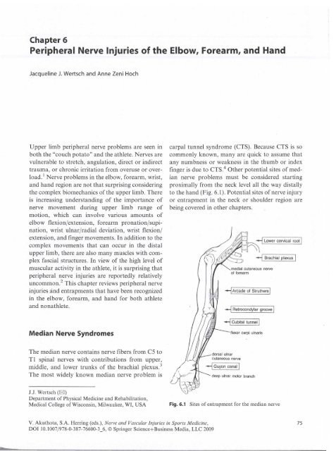
Chapter 6<strong>Peripheral</strong> <strong>Nerve</strong> <strong>Injuries</strong> <strong>of</strong> <strong>the</strong> <strong>Elbow</strong>, <strong>Forearm</strong>, <strong>and</strong> H<strong>and</strong>Jacqueline J. Wertsch <strong>and</strong> Anne Zeni HochUpper limb peripheral nerve problems are seen inboth <strong>the</strong> "couch potato" <strong>and</strong> <strong>the</strong> athlete. <strong>Nerve</strong>s arevulnerable to stretch, angulation, direct or indirecttrauma, or chronic irritation from overuse or overload.! <strong>Nerve</strong> problems in <strong>the</strong> elbow, forearm, wrist,<strong>and</strong> h<strong>and</strong> region are not that surprising considering<strong>the</strong> complex biomechanics <strong>of</strong> <strong>the</strong> upper limb. Thereis increasing underst<strong>and</strong>ing <strong>of</strong> <strong>the</strong> importance <strong>of</strong>nerve movement during upper limb range <strong>of</strong>motion, which can involve various amounts <strong>of</strong>elbow flexion/extension, forearm pronation/supination,wrist ulnar/radial deviation, wrist flexion/extension, <strong>and</strong> finger movements. In addition to <strong>the</strong>complex movements that can occur in <strong>the</strong> distalupper limb, <strong>the</strong>re are also many muscles with complexfascial structures. In view <strong>of</strong> <strong>the</strong> high level <strong>of</strong>muscular activity in <strong>the</strong> athlete, it is surprising thatperipheral nerve injuries are reportedly relativelyuncommon. 2 This chapter reviews peripheral nerveinjuries <strong>and</strong> entrapments that have been recognizedin <strong>the</strong> elbow, forearm, <strong>and</strong> h<strong>and</strong> for both athlete<strong>and</strong> nonathlete.Median <strong>Nerve</strong> Syndromescarpal tunnel syndrome (CTS). Because CTS is socommonly known, many are quick to assume thatany numbness or weakness in <strong>the</strong> thumb or indexfinger is due to CTS.4 O<strong>the</strong>r potential sites <strong>of</strong> mediannerve problems must be considered startingproximally from <strong>the</strong> neck level all <strong>the</strong> way distallyto <strong>the</strong> h<strong>and</strong> (Fig. 6.1). Potential sites <strong>of</strong> nerve injuryor entrapment in <strong>the</strong> neck or shoulder region arebeing covered in o<strong>the</strong>r chapters.medial cutaneous nerve<strong>of</strong> forearm--1 Arcade <strong>of</strong> Stru<strong>the</strong>rs'--1 Retrocondylar groove'--1 Cubital tunnel'flexor carpi ulnaris--1 Lower cervical root '--1 Brachial plexus'The median nerve contains nerve fibers from C5 toTl spinal nerves with contributions from upper,middle, <strong>and</strong> lower trunks <strong>of</strong> <strong>the</strong> brachial plexus. 3The most widely known median nerve problem isdeep ulnar motor branchJ.J. Wertsch (IBJ)Department <strong>of</strong> Physical Medicine <strong>and</strong> Rehabilitation,<strong>Medical</strong> College <strong>of</strong> Wisconsin, Milwaukee, WI, USAFig.6.1 Sites <strong>of</strong> entrapment for <strong>the</strong> median nerveV. Akuthota, S.A. Herring (eds.), <strong>Nerve</strong> <strong>and</strong> Vascular <strong>Injuries</strong> in Sports Medicine,DOI 10.1007/978-0-387-76600-3_6, © Springer Science+Business Media, LLC 200975
76In <strong>the</strong> antecubital region <strong>the</strong> most widely recognizedanatomic sites <strong>of</strong> potential median nerveentrapment include <strong>the</strong> ligament <strong>of</strong> Stru<strong>the</strong>rs, lacertusfibrosus, pronator teres muscle, <strong>and</strong> sublimisarch <strong>of</strong> <strong>the</strong> flexor digitorum superficialis muscles. 4Ligament <strong>of</strong> Stru<strong>the</strong>rsThe ligament <strong>of</strong> Stru<strong>the</strong>rs is a rare anatomic structure,being noted in only I % <strong>of</strong> upper limbs. 3 Thisligament has been described as extending from ananomalous humeral bony spur to <strong>the</strong> medial epicondyle.The bony spur cannot always be seen onradiographs. s Most individuals who have a ligament<strong>of</strong> Stru<strong>the</strong>rs are asymptomatic, <strong>and</strong> mediannerve compromise occurs only with some o<strong>the</strong>r contributingfactor or trauma. 3 . S - 9 Diagnosis can bedifficult. The patient may complain <strong>of</strong> pain in <strong>the</strong>wrist or medial forearm <strong>and</strong> pares<strong>the</strong>sias in <strong>the</strong>index or long finger.S, IO,11 The pain may be exacerbatedwith full extension <strong>of</strong> <strong>the</strong> elbow or supination<strong>of</strong> <strong>the</strong> forearm such as <strong>the</strong> follow-through phase <strong>of</strong>pitching. 9 , II Because <strong>the</strong> brachial artery accompanies<strong>the</strong> median nerve, attenuation <strong>of</strong> <strong>the</strong> pulse withprovocative maneuvers may be clinically observed. 4Needle electromyography (EMG) may help defineabnormalities in median-innervated muscles in <strong>the</strong>forearm. Because a diagnosis <strong>of</strong> a median nerveentrapment by a ligament <strong>of</strong> Stru<strong>the</strong>rs would berare, consultation with a specialist in upper limbperipheral nerves is recommended early in <strong>the</strong> consideration<strong>of</strong> this diagnosis.J.J. Wertsch <strong>and</strong> A.Z. Hochmedian nerve distribution. 13 Unfortunately, variationsin symptoms <strong>and</strong> signs are common, 14 <strong>and</strong>surgical outcomes are not convincing that <strong>the</strong> anatomicproblem is <strong>the</strong> static relation <strong>of</strong> <strong>the</strong> mediannerve <strong>and</strong> <strong>the</strong> pronator teres musc le. i S' 16 0 t h erstructures in <strong>the</strong> area (e.g., lacertus fibrosus <strong>and</strong>sublimis arch <strong>of</strong> <strong>the</strong> flexor digitorum superficialismuscles) need to also be considered in <strong>the</strong> presence<strong>of</strong> antecubital median nerve problems. 16 Even anaberrant fibrous component <strong>of</strong> <strong>the</strong> flexor carpiradialis originating from <strong>the</strong> ulna can compromise<strong>the</strong> median nerve at this level. ? Physical examinationmaneuvers to detect <strong>the</strong> pronator syndromedescribed by Spinner are discussed in a previouschapter.Axonal changes should be evident on needleEMG if <strong>the</strong>re is median nerve entrapment. A cleardiagnosis <strong>of</strong> proximal forearm median nerveentrapment would <strong>the</strong>n be preferred over <strong>the</strong> termnonspecific pronator syndrome. O<strong>the</strong>r causes <strong>of</strong>proximal forearm symptoms (e.g. , focal muscle orfascia injuries) should be specifically diagnosed <strong>and</strong>not lumped toge<strong>the</strong>r as pronator syndrome. Use <strong>of</strong><strong>the</strong> term pronator syndrome is discouraged in favor<strong>of</strong> a specific diagnosis including possible dynamicconditions in which <strong>the</strong> nerve is compressed onlyduring sports activities. Clarity <strong>of</strong> diagnosis helps toguide management. In <strong>the</strong> athlete with possibledynamic proximal forearm median nerve compression,technique modification may be helpful alongwith rest <strong>and</strong> splinting. 13 This syndrome is seen insports that require repetitive, forceful pronation<strong>and</strong> gripping. I ?, 18 Most commonly seen in throwers,pronator syndrome has been reported in weightliftmg,. arc h ery, tenms, . arm wrest l' mg, an d rowmg. . 19 ,- ?0Pronator SyndromeIn 1951 , Seyffarth l 2 described median nerve entrapmentby <strong>the</strong> pronator teres muscle. In <strong>the</strong> antecubitalarea <strong>the</strong> median nerve passes between <strong>the</strong> twoheads <strong>of</strong> <strong>the</strong> pronator teres muscle <strong>and</strong> <strong>the</strong>n under<strong>the</strong> edge <strong>of</strong> <strong>the</strong> flexor digitorum sublimis muscle.The nerve can be trapped by ei<strong>the</strong>r <strong>of</strong> <strong>the</strong>se musclesor compromised by <strong>the</strong> overlying lacertus fibrosusfrom <strong>the</strong> biceps tendon. The term "pronator syndrome"has been used by many for a clinical picture<strong>of</strong> diffuse forearm pain <strong>and</strong> pares<strong>the</strong>sia in <strong>the</strong> distalAnterior Interosseous <strong>Nerve</strong>The anterior interosseous nerve CAIN), <strong>the</strong> largestbranch <strong>of</strong> <strong>the</strong> median nerve, arises from <strong>the</strong> mediannerve soon after it passes between <strong>the</strong> two heads <strong>of</strong><strong>the</strong> pronator teres <strong>and</strong> under <strong>the</strong> flexor digitorumsublimis muscle. The AIN innervates three forearmmuscles: <strong>the</strong> flexor pollicis longus, <strong>the</strong> radial part <strong>of</strong><strong>the</strong> flexor digitorum pr<strong>of</strong>undus, <strong>and</strong> <strong>the</strong> pronatorquadratus. Because <strong>the</strong> AIN does not have anycutaneous representation, it is <strong>of</strong>ten considered to
6 <strong>Peripheral</strong> <strong>Nerve</strong> <strong>Injuries</strong> <strong>of</strong> <strong>the</strong> <strong>Elbow</strong>, <strong>Forearm</strong>, <strong>and</strong> H<strong>and</strong>be a pure motor nerve. This is technically not truebecause sensory fibers from <strong>the</strong> wrist radiocarpal,radioulnar, intercarpal, <strong>and</strong> carpometacarpal jointstravel in <strong>the</strong> AIN. 2 1 ,22 Injury to <strong>the</strong> terminal branch<strong>of</strong> <strong>the</strong> AIN can be <strong>the</strong> source <strong>of</strong> persistent, dullaching volar wrist pain, which may be diagnosedby local block <strong>of</strong> <strong>the</strong> terminal AIN branch.As early as 1918, Tinel recognized an isolated"neuritis" <strong>of</strong> <strong>the</strong> AIN branch <strong>of</strong> <strong>the</strong> mediannerve.23 Anterior interosseous nerve syndrome(AINS) was described in 1952 by Kiloh <strong>and</strong>Nevin? 4 They reported two cases that were spontaneous<strong>and</strong> recovered without treatment. Fearn <strong>and</strong>Goodfellow 25 reported <strong>the</strong> first case <strong>of</strong> surgicallytreated AIN with discovery <strong>of</strong> a fibrous b<strong>and</strong> compressing<strong>the</strong> AIN in <strong>the</strong> region <strong>of</strong> <strong>the</strong> pronator teres.Although <strong>of</strong>ten described in earlier literature asidiopathic <strong>and</strong> spontaneous, AINS can have manycauses including bone injuries, antecubital vascularprocedures, metastasis, accessory muscles, <strong>and</strong>fibrous b<strong>and</strong>s.23 Consideration should always begiven to <strong>the</strong> possibility <strong>of</strong> neuralgic amyotrophy orParsonage Turner syndrome presenting with AINweakness. 26 ,27 Careful clinical examination isneeded to ensure <strong>the</strong>re are no o<strong>the</strong>r sites <strong>of</strong> muscleweakness, such as serratus anterior scapularwmgmg.With AINS, <strong>the</strong> typical history is acute onset <strong>of</strong>thumb <strong>and</strong> index finger weakness. The patient mayhave noted difficulty using a fork, tearing out acheck, or picking up a cup. There are no sensorycomplaints. Clinical examination reveals an inabilityto flex <strong>the</strong> terminal phalanges <strong>of</strong> <strong>the</strong> thumb <strong>and</strong>index finger. Because <strong>of</strong> this weakness, <strong>the</strong> patientis unable to form an "0" (Fig. 6.2), also called <strong>the</strong>OK sign. This abnormal pinch is quite constant<strong>and</strong> characteristic <strong>of</strong> <strong>the</strong> anterior interosseousnerve syndrome. 23 AINS can be confirmed onEMG examination. Needle EMG reveals abnormalitiesin <strong>the</strong> three muscles supplied by <strong>the</strong> AINwith normal examination results <strong>of</strong> o<strong>the</strong>r muscles.Sensory nerve conduction studies are normal.Clinical management depends on <strong>the</strong> etiology <strong>of</strong><strong>the</strong> AINS.Internal topography is an important issue for<strong>the</strong> AIN. Fascicular groups remain localizedwithin <strong>the</strong> median nerve for long distances?8,29Because <strong>the</strong>se fascicles are on <strong>the</strong> posterior aspect,a humeral supracondylar fracture may damage <strong>the</strong>AIN fibers <strong>of</strong> <strong>the</strong> median nerve. 3D Thus, this much77Fig. 6.2 Typical pinch sign with flattening <strong>of</strong> index pulp <strong>and</strong> classic palsy <strong>of</strong> <strong>the</strong> anterior interosseus nerve. From Planche ret al. 50 Copyright Elsevier 1996. Used with permission
78more proximal partial median nerve injury maypresent just like an AIN problem with weaknessconfined to anterior interosseous innervated muscles3 ! ,32 A partial median nerve injury at <strong>the</strong> antecubitallevel presenting as pseudo-AINS has beenreported. 33Carpal Tunnel SyndromeThe most frequently diagnosed peripheral nerveentrapment in <strong>the</strong> general population is carpal tunnelsyndrome (CTS). In <strong>the</strong> athletic population, somebelieve that "<strong>the</strong> proximal forearm is <strong>the</strong> more usualanatomic location <strong>of</strong> median nerve impairment."!Sports involving repetitive wrist flexion <strong>and</strong> extensionsuch as gymnastics or prolonged grip withcycling may cause more cases <strong>of</strong> CTS than o<strong>the</strong>rsports. !8,34 CTS is frequently noted in wheelchairathletes. 2 ! Krivickas <strong>and</strong> Wilbourn,2 in a study <strong>of</strong>333 athletes, found that 24 <strong>of</strong> <strong>the</strong> 28 median neuropathycases were CTS. It is <strong>of</strong> interest that only 6 <strong>of</strong><strong>the</strong>ir 24 patients with CTS were symptomatic. Collinset al. 35 reported CTS frequently in rowers <strong>and</strong> bicyclistssecondary to h<strong>and</strong> position. In a separate retrospectivereview, Krivickas <strong>and</strong> Wilbourn 36 studied180 athletes from 27 different sports. They found43 athletes to have median neuropathies. 25 out <strong>of</strong><strong>the</strong>se 43 athletes were asymptomatic.The carpal tunnel is an osse<strong>of</strong>ibrous structure.The lunate, capitate, <strong>and</strong> trapezoid are <strong>the</strong> floor; <strong>the</strong>pisiform <strong>and</strong> hamate are medial; <strong>and</strong> <strong>the</strong> navicular<strong>and</strong> trapezium are lateral. The ro<strong>of</strong> is <strong>the</strong> transversecarpal ligament, a portion <strong>of</strong> <strong>the</strong> volar carpalligament(flexor retinaculum). Ten structures passthrough <strong>the</strong> carpal tunnel: four flexor digitorumtendons, four flexor superficialis (or sublimes) tendons,<strong>the</strong> flexor pollicis longus tendon, <strong>and</strong> <strong>the</strong>median nerve. 3 CTS can be caused by anythingthat ei<strong>the</strong>r decreases <strong>the</strong> size <strong>of</strong> <strong>the</strong> canal (e.g.,thickening <strong>of</strong> <strong>the</strong> flexor retinaculum) or increases<strong>the</strong> volume <strong>of</strong> <strong>the</strong> contents (e.g., tendonitis).Carpal tunnel syndrome is <strong>of</strong>ten described with<strong>the</strong> classic presentation <strong>of</strong>pares<strong>the</strong>sias in <strong>the</strong> thumb<strong>and</strong> index <strong>and</strong> long fingers that increases with activity.It is important to note if <strong>the</strong> pares<strong>the</strong>sias areepisodic or constant, <strong>the</strong> exacerbating or remittingactivities, <strong>and</strong> if <strong>the</strong>re is nocturnal worsening. It isJ.j. Wertsch <strong>and</strong> A.Z. Hochimportant to distinguish <strong>the</strong> individual describingnightly burning intense pares<strong>the</strong>sias (dyses<strong>the</strong>sias)from <strong>the</strong> individual reporting episodic h<strong>and</strong> tingling.The time course can also be revealing, withan acute presentation perhaps preceded by years <strong>of</strong>episodic symptoms. The patient may report that <strong>the</strong>h<strong>and</strong> feels clumsy, especially noting difficulty withfine motor activities such as buttoning a shirt. Proximalsymptoms with discomfort <strong>and</strong> aching may bereported, especially in <strong>the</strong> extensor mass region.Physical examination may reveal sensorychanges in <strong>the</strong> median innervated digits. Themotor examination may reveal weakness <strong>of</strong> palmarabduction, although this requires attention to commonextrinsic substitution patterns that would beexpected. During a careful clinical examination,abductor pollicis brevis weakness is quite sensitivein those with CTS 3 A Phalen test can be quite helpfulwith details <strong>of</strong> how long <strong>the</strong> pares<strong>the</strong>sia lasts(less than 20 seconds would be expected) <strong>and</strong> whatdigit develops <strong>the</strong> pares<strong>the</strong>sias (most commonly <strong>the</strong>long finger). Tapping over <strong>the</strong> median nerve at <strong>the</strong>wrist to elicit a Tinel sign is commonly done.Because <strong>the</strong> carpal tunnel is tightest at its distalend, tapping in <strong>the</strong> palm at <strong>the</strong> distal end <strong>of</strong> <strong>the</strong>transverse ligament may increase <strong>the</strong> yield <strong>of</strong> <strong>the</strong>Tinel testing for CTS. 3Unfortunately, CTS has come to be associatedwith any h<strong>and</strong> numbness, <strong>and</strong> o<strong>the</strong>r causes <strong>of</strong> h<strong>and</strong>numbness may not be considered. Ano<strong>the</strong>r unfortunatereality is that many physicians still believe that<strong>the</strong>re is a high false-negative rate for nerve conductionstudies in CTS. This was perhaps true in <strong>the</strong>early days before careful temperature control <strong>and</strong>short segment transcarpal studies?9 Now, nerveconduction studies, including paired transcarpalstudies, are considered by many to be <strong>the</strong> gold st<strong>and</strong>ardfor CTS diagnosis. <strong>Nerve</strong> conduction studiesfor CTS would include both median <strong>and</strong> ulnar studiesto make sure <strong>the</strong>re is no diffuse slowing. Bothsensory <strong>and</strong> motor studies should be done. Theshort segment paired trans carpal study would beconsidered <strong>the</strong> most sensitive test. 8 Paired digitalsensory <strong>and</strong> paired motor studies would also bedone. "Mapping" should be done when <strong>the</strong> motorstudies are inconsistent with <strong>the</strong> sensory studies.Mapping simply implies moving <strong>the</strong> E I (previouslyreferred to as <strong>the</strong> active electrode) location <strong>and</strong> canbe important for avoiding false-positive median
6 <strong>Peripheral</strong> <strong>Nerve</strong> <strong>Injuries</strong> <strong>of</strong> <strong>the</strong> <strong>Elbow</strong>, <strong>Forearm</strong>, <strong>and</strong> H<strong>and</strong>motor studies. 37 Hsu et al. reported an isolatedmedian mononeuropathy in <strong>the</strong> palm <strong>of</strong> an amateurgolfer. 38Radial <strong>Nerve</strong>The radial nerve is <strong>the</strong> largest peripheral branch <strong>of</strong><strong>the</strong> brachial plexus. It receives contributions from<strong>the</strong> cervical roots C5- 8 <strong>and</strong> is derived from <strong>the</strong>posterior cord <strong>of</strong> <strong>the</strong> brachial plexus. In <strong>the</strong> axilla,<strong>the</strong> radial nerve is posterior to <strong>the</strong> axillary artery<strong>and</strong> superficial to <strong>the</strong> subscapularis muscle <strong>and</strong> tendons<strong>of</strong> <strong>the</strong> latissimus dorsi <strong>and</strong> teres major. Theradial nerve travels between <strong>the</strong> long <strong>and</strong> medialheads <strong>of</strong> <strong>the</strong> triceps, continuing to <strong>the</strong> spiral groove<strong>of</strong> <strong>the</strong> humerus.Proximal Radial <strong>Nerve</strong> EntrapmentThe term "high radial nerve lesions" has been usedin <strong>the</strong> past for radial nerve lesions proximal to <strong>the</strong>division in <strong>the</strong> forearm into <strong>the</strong> posterior interosseousnerve <strong>and</strong> superficial radial sensory branch. 39The classic "honeymooners" spiral groove radialneuropathies have wrist <strong>and</strong> finger extension weakness.Distal interphalangeal (DIP) joint extension isusually preserved owing to <strong>the</strong> mechanical effect <strong>of</strong><strong>the</strong> intact median <strong>and</strong> ulnar innervated intrinsicmuscles. 39 Compression <strong>of</strong> <strong>the</strong> radial nerve by <strong>the</strong>lateral head <strong>of</strong> <strong>the</strong> triceps muscles following strenuousmuscular effort has been described. I ,21 ,32,34This site is distal to <strong>the</strong> branches innervating <strong>the</strong>triceps <strong>and</strong> anconeus muscles, both <strong>of</strong> which wouldbe expected to be spared clinically <strong>and</strong> on needleEMG studies. Compression <strong>of</strong> <strong>the</strong> radial nerve by<strong>the</strong> long head <strong>of</strong> <strong>the</strong> triceps has also beenreported. 22 ,32,34 Compression or injury in <strong>the</strong> spiralgroove may be seen in activities that require forcefuladduction <strong>of</strong> <strong>the</strong> shoulder, as seen in gymnastics 17<strong>and</strong> wrestling. 4o Activities that require forcefulextension <strong>of</strong> <strong>the</strong> elbow against resistance, such asthrowing (e.g., discus, javelin, baseball) are associatedwith high radial nerve entrapment. 18, 19,41"Runner's radial palsy," an unusual <strong>and</strong> distinctentity that presents with numbness in <strong>the</strong> distalforearm <strong>and</strong> dorsum <strong>of</strong> <strong>the</strong> h<strong>and</strong> <strong>of</strong> runners whokeep <strong>the</strong>ir elbow acutely flexed while running, isano<strong>the</strong>r example <strong>of</strong> a high radial nerve lesion. Thenerve in this injury is compressed between <strong>the</strong>humerus <strong>and</strong> triceps. Treatment is altering <strong>the</strong> runningposition, resulting in complete resolution <strong>of</strong>symptoms. 42<strong>Forearm</strong> Radial <strong>Nerve</strong> EntrapmentThe radial nerve innervates <strong>the</strong> brachioradialis <strong>and</strong>extensor carpi radialis longus distal to <strong>the</strong> spiralgroove. In <strong>the</strong> lateral antecubital region, <strong>the</strong> radialnerve lies between <strong>the</strong> brachialis <strong>and</strong> brachioradialismuscles <strong>and</strong> passes over <strong>the</strong> annular ligament <strong>of</strong> <strong>the</strong>radius. In <strong>the</strong> proximal forearm, <strong>the</strong> radial nervebifurcates into a sensory branch <strong>and</strong> a motorbranch, <strong>the</strong> posterior interosseous nerve (PIN).The PIN usually innervates <strong>the</strong> extensor carpi radialisbrevis <strong>and</strong> sends several branches to <strong>the</strong> supinatorprior to going through <strong>the</strong> arcade <strong>of</strong> Frohse. As<strong>the</strong> PIN emerges from <strong>the</strong> supinator, it supplies <strong>the</strong>muscles in <strong>the</strong> extensor forearm, including <strong>the</strong>extensor digitorum communis, extensor digitiquinti, extensor carpi ulnaris <strong>and</strong> <strong>the</strong> deeper muscles(abductor pollicis longus, extensor pollicislongus <strong>and</strong> brevis, extensor indicis proprius). Similarto <strong>the</strong> AIN, <strong>the</strong> PIN terminates at <strong>the</strong> wrist, withseveral sensory branches innervating <strong>the</strong> carpaljoints. 43Historically, radial tunnel syndrome <strong>and</strong> supinatorsyndrome have both been described. 44 Theradial tunnel syndrome remains a somewhat controversialdisorder, while <strong>the</strong> supinator syndrome iscurrently more clearly called posterior interosseousnerve syndrome (PINS). They differ in <strong>the</strong>ir clinicalpresentations, with <strong>the</strong> radial tunnel syndrome<strong>of</strong>ten described as "resistant tennis elbow" becauseit presents with pain as its primary feature whereasPINS characteristically has painless weakness.Radial Tunnel SyndromeIn 1972, Roles <strong>and</strong> Maudslel 5 introduced <strong>the</strong> term"radial tunnel syndrome." The radial tunnel was79
80described as extending from where <strong>the</strong> radial nervepierces <strong>the</strong> lateral intermuscular septum to where<strong>the</strong> PIN enters <strong>the</strong> supinator muscle. Radial tunnelsyndrome presents with lateral elbow pain. Objectivemotor or sensory loss in <strong>the</strong> distribution <strong>of</strong> <strong>the</strong>radial nerve would not be expected.When evaluating a patient for radial tunnel syndrome,<strong>the</strong> main differential diagnosis to consider islateral epicondylitis, or "tennis elbow." Physicalexamination findings may help differentiate <strong>the</strong>two. With radial tunnel syndrome <strong>the</strong>re is painupon palpation <strong>of</strong> <strong>the</strong> extensor forearm musclemass a few centimeters distal to <strong>the</strong> lateral epicondyle,whereas with lateral epicondylitis <strong>the</strong> point <strong>of</strong>maximum tenderness is at <strong>the</strong> lateral epicondyle. 10Resisted supination is more painful with radial tunnelsyndrome. lo Three characteristic findings aredescribed for radial tunnel syndrome: pain uponpalpation <strong>of</strong> <strong>the</strong> extensor forearm muscle massabout <strong>the</strong> radial head; pain in <strong>the</strong> forearm uponresisted supination; <strong>and</strong> lateral elbow pain withresisted elbow, wrist, <strong>and</strong> long finger extension. 39The initial treatment is rest for both radial tunnel<strong>and</strong> lateral epicondylitis, followed by stretching <strong>and</strong>streng<strong>the</strong>ning. 46Radial tunnel syndrome is <strong>of</strong>ten seen in thoseathletes who repetitively pronate <strong>and</strong> supinate <strong>and</strong>in those who are stressed in wrist extension, as inweightlifting, bowling, rowing, discus, racquetsports, swimming, <strong>and</strong> golf. 18,19,47 Roles <strong>and</strong>Maudsley thought that radial tunnel syndromemimics <strong>and</strong> may coexist with lateral epicondylitisin up to 10% <strong>of</strong> patients. 45 Werner found an 8%coincident lesion in 203 patients. 48Posterior Interosseous <strong>Nerve</strong> SyndromeThe classic presentation <strong>of</strong> PINS is painless weakness<strong>of</strong> <strong>the</strong> wrist <strong>and</strong> finger extensors without sensoryimpairment. Wrist extension weakness generallyis not complete owing to sparing <strong>of</strong> <strong>the</strong> extensorcarpi radialis longus (ECRL), which receives innervationfrom <strong>the</strong> main radial nerve before <strong>the</strong> PINbranch. The characteristic radial deviation duringattempted wrist extension is explained by <strong>the</strong> imbalance<strong>of</strong> a strong direct radial-innervated ECRLwith weak PIN-innervated extensor carpi ulnarisJ.J. Wertsch <strong>and</strong> A.l. Hoch(ECU). Weakness <strong>of</strong> finger extension would alsobe expected. Weakness <strong>of</strong> <strong>the</strong> brachioradialis <strong>and</strong>superficial radial sensory impairment would notsupport a PINS but, instead, suggests a higherradial nerve lesion. Electrodiagnostic studies shouldbe done to confirm <strong>and</strong> grade <strong>the</strong> PINS. Denervation,motor unit changes, <strong>and</strong> recruitment abnormalitieswould be expected proportional to <strong>the</strong> clinicalweakness. Surgical treatment IS <strong>of</strong>tennecessa ry. 39Cheiralgia pares<strong>the</strong>tica refers to isolated injury <strong>of</strong><strong>the</strong> superficial sensory branch <strong>of</strong> <strong>the</strong> radial nerve.Numerous etiologies have been described, includingdirect trauma, fibrosis from hemorrhage, h<strong>and</strong>cuffs,<strong>and</strong> pressure (from tight watchb<strong>and</strong>s, a cast, tightwristb<strong>and</strong>s, tape, archery guards, gloves). 17,49 Directtrauma may be responsible for this syndrome in contactsports such as hockey, football, <strong>and</strong> lacrosse. 50Ulnar <strong>Nerve</strong>The ulnar nerve receives contributions from C8 <strong>and</strong>Tl nerve roots <strong>and</strong> is <strong>the</strong> distal continuation <strong>of</strong> <strong>the</strong>medial cord <strong>of</strong> <strong>the</strong> brachial plexus (Fig. 6.3). Nobranches arise from <strong>the</strong> ulnar nerve in <strong>the</strong> arm. Theulnar nerve goes under <strong>the</strong> arcade <strong>of</strong> Stru<strong>the</strong>rs,which is <strong>the</strong> mid-arm b<strong>and</strong> <strong>of</strong> fascia connecting <strong>the</strong>deep investing fascia <strong>of</strong> <strong>the</strong> anterior <strong>and</strong> posteriorcompartments <strong>of</strong> <strong>the</strong> arm. Proximal to <strong>the</strong> arcade <strong>of</strong>Stru<strong>the</strong>rs <strong>the</strong> nerve is freely mobile. In <strong>the</strong> area <strong>of</strong><strong>the</strong> arcade, <strong>the</strong> nerve travels from <strong>the</strong> anterior toposterior compartments <strong>of</strong> <strong>the</strong> arm.At <strong>the</strong> elbow, <strong>the</strong> ulnar nerve travels between <strong>the</strong>medial epicondyle <strong>and</strong> <strong>the</strong> olecranon in <strong>the</strong> retrocondylargroove. The superior edge <strong>of</strong> <strong>the</strong> medialepicondyle is an interesting site where <strong>the</strong> ulnarnerve has a fairly tight curve as it comes posteriorlytoward <strong>the</strong> retrocondylar groove. This site shouldbe carefully examined with inching nerve conductionstudies when evaluating idiopathic ulnar neuropathies.Continuing distally, <strong>the</strong> nerve passesbetween <strong>the</strong> two heads <strong>of</strong> <strong>the</strong> flexor carpi ulnaris(FCU), passing under <strong>the</strong> humeroulnar aponeuroticarcade, which along with <strong>the</strong> underlying bone <strong>and</strong>ligaments form <strong>the</strong> cubital tunnel. The ulnar nervecontinues in <strong>the</strong> forearm resting on <strong>the</strong> flexor digitorum pr<strong>of</strong>undus (FDP) <strong>and</strong> covered by <strong>the</strong> FCU.
6 <strong>Peripheral</strong> <strong>Nerve</strong> <strong>Injuries</strong> <strong>of</strong> <strong>the</strong> <strong>Elbow</strong>, <strong>Forearm</strong>, <strong>and</strong> H<strong>and</strong>biceps brachi muscleproximal end <strong>of</strong>pronator teres muscle--l Lacertus fibrosus 1--l Pronator teres I--1 Sublimis arch Iflexor digitorum sublimis muscleanterior interosseous nervemain median nerve--1 Median sensory digital branches IFig.6.3 Sites <strong>of</strong> entrapment <strong>of</strong> <strong>the</strong> ulnar nerveAt <strong>the</strong> wrist, <strong>the</strong> ulnar nerve enters <strong>the</strong> h<strong>and</strong>through Guyon's canal, which is bound mediallyby <strong>the</strong> pisiform <strong>and</strong> laterally by <strong>the</strong> hook <strong>of</strong> <strong>the</strong>hamate. In <strong>the</strong> h<strong>and</strong>, <strong>the</strong> ulnar nerve divides into asuperficial branch <strong>and</strong> a deep branch. The superficialbranch innervates <strong>the</strong> palmaris brevis <strong>and</strong>divides into <strong>the</strong> palmar digital sensory branches.The deep ulnar motor branch supplies all o<strong>the</strong>rulnar innervated h<strong>and</strong> muscles. The dorsal ulnarcutaneous (DUC) nerve branch comes <strong>of</strong>f <strong>the</strong>main ulnar nerve 5- 10 cm proximal to <strong>the</strong> wrist,but <strong>the</strong> DUe can be identified as an independentbundle for some distance proximal. 28 This anatomicfeature explains <strong>the</strong> clinical observation that <strong>the</strong>DUe nerve is quite sensitive to proximal ulnarproblems.The ulnar nerve exists in a dynamic relation withanatomic structures in <strong>the</strong> elbow region. 5t Wi<strong>the</strong>xtension, <strong>the</strong> nerve becomes redundant within <strong>the</strong>retrocondylar groove; with flexion, <strong>the</strong> medialligament <strong>of</strong> <strong>the</strong> elbow bulges into <strong>the</strong> floor <strong>of</strong> <strong>the</strong>retrocondylar groove. <strong>Elbow</strong> flexion tightens <strong>the</strong>humeral ulnar arcuate ligament over <strong>the</strong> ulnarnerve, decreases <strong>the</strong> volume in <strong>the</strong> cubital tunnel,<strong>and</strong> elongates <strong>the</strong> ulnar nerve from its previouslyslack position. With extreme elbow flexion, <strong>the</strong>medial head <strong>of</strong> <strong>the</strong> triceps can exert a posteriorforce on <strong>the</strong> nerve. Partial or complete subluxation<strong>of</strong> <strong>the</strong> ulnar nerve occurs in up to 16% <strong>of</strong> <strong>the</strong> population.52 A subluxing ulnar nerve is rarely <strong>of</strong> anyclinical significance.Proximal Ulnar NeuropathiesEpisodic pares<strong>the</strong>sias in <strong>the</strong> ring <strong>and</strong> little fingersare a common human experience, similar to legpares<strong>the</strong>sia noted with leg crossing. The dynamicnature <strong>of</strong> ulnar pares<strong>the</strong>sias is vastly underappreciated<strong>and</strong> in need <strong>of</strong> much more research. Thepatient with a proximal ulnar neuropathy has apersistent sense <strong>of</strong> decreased sensation in <strong>the</strong> ulnardigits, hypo<strong>the</strong>nar region, <strong>and</strong> dorsal ulnar h<strong>and</strong>.The forearm should not have any numbness. Thereis no elbow pain. There may be a sense <strong>of</strong> weaknessin <strong>the</strong> h<strong>and</strong>. Patients may report difficulty gettingobjects out <strong>of</strong> <strong>the</strong>ir pockets because <strong>the</strong>ir little fingergets caught on <strong>the</strong> pocket edge (Wartenberg sign).On clinical examination <strong>the</strong>re may be impairedsensation in <strong>the</strong> ulnar nerve distribution. The motorexamination may reveal ulnar intrinsic muscleweakness with preserved median intrinsic musclestrength. The ulnar innervated palmaris brevis musclecan be important to examine by looking for <strong>the</strong>presence or absence <strong>of</strong> palmaris brevis hypo<strong>the</strong>narskin wrinkling. ThiS is a fairly reliable physical findingin significant ulnar neuropathies <strong>and</strong> shouldalways be included as ei<strong>the</strong>r a pertinent positive ornegative finding. A positive Froment sign is seenwith ulnar neuropathies owing to <strong>the</strong> median innervatedflexor pollicis longus attempting to compensatefor <strong>the</strong> weak ulnar intrinsic muscles. The Wartenbergsign with <strong>the</strong> little finger hanging out in anabducted position can also been seen in ulnar neuropathies,reflecting <strong>the</strong> misbalance <strong>of</strong> <strong>the</strong> weakulnar interossei muscles <strong>and</strong> <strong>the</strong> intact radial innervatedextensor muscles. 53 Although both <strong>the</strong> Fromentsign <strong>and</strong> <strong>the</strong> Wartenberg sign are indicative <strong>of</strong>ulnar neuropathy, nei<strong>the</strong>r identifies <strong>the</strong> site <strong>of</strong> <strong>the</strong>lesion. 54 The Tinel sign over <strong>the</strong> ulnar nerve should81
82be interpreted with caution because it has beennoted to be present in more than 80% <strong>of</strong> asymptomaticnormal people. 6 ,54Although an ulnar neuropathy at <strong>the</strong> elbow isfrequently considered first in patients presentingwith "ulnar" seJ?sory symptoms, a broad differentialdiagnosis must be considered, including cervicalradiculopathy, medial cord brachial plexopathy,or even vascular or visceral problems. 55 It is widelyrecognized clinically that even patients with CTSmay present with "ulnar" sensory symptoms. 56The role <strong>of</strong> electrophysiologic data in <strong>the</strong> evaluation<strong>of</strong> suspected ulnar neuropathies has been <strong>the</strong>subject <strong>of</strong> many publications. Parameters that havebeen utilized have included motor, mixed nerve, <strong>and</strong>sensory conduction velocity <strong>of</strong> <strong>the</strong> elbow segment;comparison <strong>of</strong> <strong>the</strong> elbow segment velocity to that <strong>of</strong>an adjacent nerve segment; inching; change in <strong>the</strong>size or configuration <strong>of</strong> <strong>the</strong> compound muscleaction potential or sensory nerve action potentials;<strong>and</strong> <strong>the</strong> pattern <strong>of</strong> needle examination abnormalitiesin ulnar-supplied muscles. 57 When interpretingulnar nerve conduction studies, it should be rememberedthat <strong>the</strong> exact site where <strong>the</strong> cubital begins canbe extremely variable. In a patient in whom <strong>the</strong>ulnar amplitude may appear to drop at <strong>the</strong> "belowelbow" site, this may simply reflect a proximal origin<strong>of</strong> <strong>the</strong> cubital tunnel <strong>and</strong> should not be misinterpretedas a conduction block. Fur<strong>the</strong>r work isneeded on all ulnar techniques to examine repeatability,correlation to clinical features, <strong>and</strong> outcomes.Current research efforts are exploringrefinements <strong>of</strong> inching, <strong>the</strong> usefulness <strong>of</strong> including<strong>the</strong> dorsal ulnar cutaneous study, <strong>and</strong> <strong>the</strong> role <strong>of</strong> E2(previously referred to as <strong>the</strong> referenceelectrode).57,58The act <strong>of</strong> throwing is <strong>of</strong>ten responsible for ulnarnerve involvement at <strong>the</strong> elbow in athletes. Tremendousforces develop at <strong>the</strong> elbow with angular velocitiesup to 7000° Is. 18 Studies have shown that <strong>the</strong>position <strong>of</strong> elbow flexion <strong>and</strong> wrist extension createsa threefold increase in ulnar nerve pressure in <strong>the</strong>cubital tunnel; when <strong>the</strong> upper extremity is placed in<strong>the</strong> cocking position <strong>of</strong> a throw, <strong>the</strong> pressureincreases to six times <strong>the</strong> resting pressure. 59 Biomechanically,several processes cause an elevation <strong>of</strong>pressure. The arcuate ligament leng<strong>the</strong>ns wi<strong>the</strong>lbow flexion until <strong>the</strong> proximal edge becomes tautat 90° <strong>of</strong> flexion. At <strong>the</strong> same time, <strong>the</strong> ulnarJ.J. Wertsch <strong>and</strong> A.Z. Hochcollateral ligament relaxes <strong>and</strong> may bulge inward,potentially decreasing <strong>the</strong> space available to <strong>the</strong>nerve. 60 During elbow flexion, <strong>the</strong> ulnar nerve hasbeen shown to elongate an average <strong>of</strong> 4.7 mm. I ,51All <strong>of</strong> <strong>the</strong>se factors can contribute to athletes' symptoms.lobe <strong>and</strong> colleagues believe that throwersdevelop a progressive flexion valgus deformitywith attenuation <strong>of</strong> <strong>the</strong> ulnar collateral ligament. 6 IThis exacerbates <strong>the</strong> athletes' symptoms by increasing<strong>the</strong> magnitude <strong>of</strong> <strong>the</strong> tensile forces already atwork.Cubital tunnel syndrome is found more <strong>of</strong>ten in<strong>the</strong> throwing athlete but can be seen in <strong>the</strong> recreationalskier, weightlifter, <strong>and</strong> racquet sport enthusiast.62 There has been a case report <strong>of</strong> cubital tunnelsyndrome involving a cross-country skier. Themechanism <strong>of</strong> injury was <strong>the</strong> extension <strong>and</strong> pronationrequired for poling. 63Distal Ulnar NeuropathiesUlnar neuropathy at <strong>the</strong> wrist <strong>and</strong> h<strong>and</strong> is uncommon.31 ,64,65 Reported etiologies have includedganglion cysts, ulnar artery thrombosis, carpal fractures(especially <strong>the</strong> hamate), anomalous muscles,<strong>and</strong> repetitive trauma or direct pressure. 1,65 In <strong>the</strong>athlete, <strong>the</strong> most widely known cause is pressurefrom h<strong>and</strong>lebars seen in bicyclists (Fig. 6.4). 1,66The Guyon canal, or ulnar tunnel, is <strong>the</strong> depressionbetween <strong>the</strong> pisiform <strong>and</strong> <strong>the</strong> hook <strong>of</strong> <strong>the</strong>hamate. It is different from <strong>the</strong> carpal tunnel inthat <strong>the</strong> ro<strong>of</strong> is quite thin, <strong>the</strong>re are no tendons in<strong>the</strong> Guyon canal, <strong>and</strong> <strong>the</strong>re are vascular structuresincluding <strong>the</strong> ulnar artery <strong>and</strong> its associated veins 67Problems that can occur with <strong>the</strong> ulnar nerve distallyin <strong>the</strong> wrist have been classified by Shea amdMcClain. 31 Type I has involvement <strong>of</strong> all parts <strong>of</strong><strong>the</strong> nerve including <strong>the</strong> hypo<strong>the</strong>nar muscles, <strong>the</strong>deep ulnar motor branch to <strong>the</strong> ulnar intrinsic muscles,<strong>and</strong> <strong>the</strong> ulnar digital sensory fibers. Type IIinvolves only <strong>the</strong> deep ulnar motor branch. Type IIIinvolves only <strong>the</strong> superficial ulnar sensory branches.Unfortunately, this classification scheme does notinclude <strong>the</strong> branch to <strong>the</strong> palmaris brevis muscle.This motor branch initially travels with <strong>the</strong> ulnardigital sensory fibers as <strong>the</strong> superficial branch <strong>of</strong> <strong>the</strong>ulnar nerve. Clinicians are increasingly aware <strong>of</strong> this
6 <strong>Peripheral</strong> <strong>Nerve</strong> <strong>Injuries</strong> <strong>of</strong> <strong>the</strong> <strong>Elbow</strong>, <strong>Forearm</strong>, <strong>and</strong> H<strong>and</strong> 83Fig.6.4 Mechanism <strong>of</strong> injury for cyclist's palsy. From Plancher et al. so Copyright Elsevier 1996. Used with permissionunusual anatomy <strong>and</strong> finding it useful to check forpalmaris brevis wrinkling with little finger abductionin all cases <strong>of</strong> suspected ulnar neuropathl 5To evaluate possible distal ulnar nerve problemsbeyond a detailed history <strong>and</strong> physical examination,electro diagnostic studies can be helpful. An initialpaired transcarpal study should be done to rule out apossible CTS presentation with ulnar sensory symptoms.56 Ulnar digital sensory studies should be doneboth orthodromically <strong>and</strong> antidromically to avoid misinterpretation<strong>of</strong> an interosseous far field motorresponse as a prolonged digital sensory response.Ulnar motor studies can be done by recording over<strong>the</strong> hypo<strong>the</strong>nar muscles or any <strong>of</strong> <strong>the</strong> ulnar intrinsicmuscles. 57 Caution is urged during interpretation <strong>of</strong>ulnar motor studies, however, as <strong>the</strong> E2 is always active<strong>and</strong> is a significant factor that can affect not onlyamplitude <strong>and</strong> waveform shape but even latency. 58Needle EMG <strong>of</strong> many ulnar intrinsic muscles, includingboth volar <strong>and</strong> dorsal interossei, are <strong>of</strong>ten needed tolocalize a true distal ulnar neuropathy.65Isolated entrapment <strong>of</strong> <strong>the</strong> DUC nerve has beenreported 68 <strong>and</strong> should be considered in any patientpresenting with ulnar h<strong>and</strong> sensory symptoms. TheDUC passes between <strong>the</strong> flexor carpi ulnaris <strong>and</strong> <strong>the</strong>ulna to pierce <strong>the</strong> fascia in <strong>the</strong> distal forearm. Itappears vulnerable to irritation by fast repetitiveforearm pronation <strong>and</strong> concomitant wrist flexion .Isolated entrapment <strong>of</strong> <strong>the</strong> DUC was initiallyreported in a grocery store clerk who had to passobjects over a static universal product code (UPC)reader; <strong>the</strong> disorder was thus named "pricer palsy. "68The technology improvement to w<strong>and</strong>-type UPCreaders makes this less likely to occur currently, butisolated DUC entrapment should still be consideredin those involved in sports activities with a lot <strong>of</strong>forearm/wrist repetitive movements.Digital <strong>Nerve</strong>sInjury to <strong>the</strong> digital nerves is <strong>of</strong>ten <strong>the</strong> result <strong>of</strong>repetitive trauma over <strong>the</strong> palm or digits. Themost common example, described by Dobynset a1. 69 in 1972, is <strong>the</strong> bowler's thumb. The termbowler's thumb describes an injury to <strong>the</strong> ulnardigital sensory nerve to <strong>the</strong> thumb caused by direct
84J.J. Wertsch <strong>and</strong> A.Z. Hochchain elements ra<strong>the</strong>r than <strong>the</strong> vulnerable elbow.Although <strong>the</strong> evidence is unclear, ergonomic correctionsin <strong>the</strong> individuals engaged in desk work mayalso help reduce pressures within <strong>the</strong> carpal tunnel<strong>and</strong> around <strong>the</strong> elbow.Special EquipmentFig.6.S Compression <strong>of</strong> <strong>the</strong> ulnar digital nerve <strong>of</strong> <strong>the</strong> thumbin bowler's thumbtrauma from <strong>the</strong> edge <strong>of</strong> <strong>the</strong> thumb hole <strong>of</strong> a bowlingball (Fig. 6.5). The radial digital nerve <strong>of</strong> <strong>the</strong>index finger is similarly at risk in racquet sports.?O.?1Digital nerve symptoms can be seen in baseball,h<strong>and</strong>ball, gymnastics, <strong>and</strong> <strong>the</strong> martial arts. I? Thechronic nature <strong>of</strong> <strong>the</strong> injury was demonstrated byBuckhout <strong>and</strong> Warner, who showed that digitalnerve involvement in h<strong>and</strong>ball players occur inthose with more than 2 years or 200 hours <strong>of</strong>play.72TreatmentFocal neuropathies <strong>of</strong> <strong>the</strong> elbow, forearm, <strong>and</strong> h<strong>and</strong>can all be treated similarly. After diagnosis <strong>and</strong>severity <strong>of</strong> neuropathy is elucidated <strong>and</strong> underlyingconditions are addressed, a typical treatment algorithmcan be applied.Activity ModificationFirst, activity modification should be considered.For example, in bicyclists experiencing ulnar neuropathyat <strong>the</strong> wrist or carpal tunnel syndrome, <strong>the</strong>h<strong>and</strong>/wrist position should be modified so excessivepressure does not occur. Reducing repetitive loadson nerves at <strong>the</strong>ir te<strong>the</strong>ring points has also beenadvised. Overhead athletes can be rehabilitated togenerate kinetic forces through proximal kineticEquipment can sometimes be used to reduce external/internalpres~ures <strong>and</strong> splint joints so nerves arenot stretched. For ulnar neuropathies at <strong>the</strong> elbow,pads (e.g. , hockey elbow pads) or splints can be usedto prevent <strong>the</strong> elbow joint from fully flexing <strong>and</strong>avoid maximum stretch <strong>of</strong> <strong>the</strong> ulnar nerve. Paddedbicycling gloves are also thought to reduce externalpressures on ulnar <strong>and</strong> median nerves at <strong>the</strong> wrist.Wrist splints (carpal tunnel braces), particularly in<strong>the</strong> neutral position, have also been shown to helpup to 30% <strong>of</strong> individuals with CTS achieve significantsymptom reduction.?3MedicationMedications have been used empirically to treat variousneuropathies. Antiinflammatory drugs, neuropathicmedications, diuretics, <strong>and</strong> various analgesicshave been used with mixed success. There is no goodliterature on <strong>the</strong> use <strong>of</strong> nonsteroidal antiinflammatoriesas mono<strong>the</strong>rapy for CTS <strong>and</strong> little literaturefor o<strong>the</strong>r neuropathies. Vitamin B6 (pyridoxine) hasbeen used for CTS, but <strong>the</strong>oretically it should onlyhelp individuals with concomitant peripheral neuropathy.Injected corticosteroids are relatively frequentlyused for CTS but not o<strong>the</strong>r neuropathies.Bl<strong>and</strong> concluded that 70% <strong>of</strong> CTS patients significantlyimprove with carpal tunnel corticosteroidinjection, but 50% relapse within 1 year. 73SurgerySurgical release can be utilized to treat various neuropathies.Carpal tunnel release surgery is common<strong>and</strong> appears to "cure" CTS in at least 75% <strong>of</strong> individuals.73 Open <strong>and</strong> endoscopic carpal tunnel release
6 <strong>Peripheral</strong> <strong>Nerve</strong> <strong>Injuries</strong> <strong>of</strong> <strong>the</strong> <strong>Elbow</strong>, <strong>Forearm</strong>, <strong>and</strong> H<strong>and</strong>can be performed for recalcitrant <strong>and</strong> severe CTScases. Endoscopic surgery may be appealing to athletesas it <strong>of</strong>fers a faster recovery time. However,individuals should be cautioned that endoscopicmethods are not as well studied as o<strong>the</strong>rs, <strong>and</strong> complicationpr<strong>of</strong>iles are not as clear owing to incompleteexposure <strong>and</strong> transection <strong>of</strong> <strong>the</strong> transverse carpalligament.Ulnar nerve surgery about <strong>the</strong> elbow is als<strong>of</strong>requently performed for recalcitrant ulnar neuropathies.Surgery may include release <strong>and</strong> decompression(e.g. , in cases where <strong>the</strong> ulnar nerve appears to beentrapped distal to <strong>the</strong> elbow at <strong>the</strong> humeroulnaraponeurotic arcade) or an anterior transposition <strong>and</strong>medial epicondylectomy (e.g., in cases where ulnarneuropathy occurs at <strong>the</strong> ulnar groove). Accordingto a study by Bartels et a!., ulnar nerve surgery at <strong>the</strong>elbow works about two-thirds <strong>of</strong> <strong>the</strong> time using ei<strong>the</strong>rtechnique. 74 The authors advocated for simpledecompression ra<strong>the</strong>r than transposition, as decompressionengendered less cost <strong>and</strong> fewer complications.In athletes, ulnar nerve surgery at <strong>the</strong> elbow is<strong>of</strong>ten accompanied by repair <strong>of</strong> an injured ulnar collateralligament (via palmaris longus tendon transfer).References1. Weinstein SM, Herring SA. <strong>Nerve</strong> problems <strong>and</strong> compartmentsyndromes in <strong>the</strong> h<strong>and</strong>, wrist, <strong>and</strong> forearm .Clin Sports Med 1992;11(1):161-88.2. Krivickas LS, Wilbourn AJ. Sports <strong>and</strong> peripheral nerveinjuries: report <strong>of</strong> 190 injuries evaluated in a single electromyographylaboratory. Muscle <strong>Nerve</strong> 1998;21(8):1092-94.3. Wertsch JJ, Melvin J. Median nerve anatomy <strong>and</strong>entrapment syndromes: a review. Arch Phys Med Rehabil1982;63( 12):623- 7.4. Wertsch JJ. Median neuropathies at <strong>the</strong> elbow. In:American Association <strong>of</strong> Electrodiagnostic Medicine(AAEM) Course: Unusual Mononeuropathies. Rochester,MN: AAEM; 2000:7-10.5. Laha RK, Dujovny M , DeCastro Sc. Entrapment <strong>of</strong>median nerve by supracondylar process <strong>of</strong> <strong>the</strong> humerus:case report. J Neurosurg 1977;46(2):252-5.6. Alfonso MI, Rioja LN, Wang S, et al. Tinel's sign <strong>of</strong> <strong>the</strong>ulnar nerve at <strong>the</strong> elbow. Muscle <strong>Nerve</strong> 1998;21:1571.7. Johnson RK, Spinner M , Shrewsbury MM. Mediannerve entrapment syndrome in <strong>the</strong> proximal forearm . JH<strong>and</strong> Surg [Am] 1979;4(1):48-51.8. Mills KR. Orthodromic sensory action potentials frompalmar stimulation in <strong>the</strong> diagnosis <strong>of</strong> carpal tunnel syndrome.J Neurol Neurosurg Psychiatry 1985;48(3):250-5.9. Symeonides PP. The humerus supracondylar processsyndrome. Clin Orthop 1972;82:141-3.10. Kessel L, Rang M. Supracondylar spur <strong>of</strong> <strong>the</strong> humerus. JBone Joint Surg Br 1966;48(4):765-9.II. Smith RV, Fisher RG. Stru<strong>the</strong>rs ligament: a source <strong>of</strong>median nerve compression above <strong>the</strong> elbow: case report.J Neurosurg 1973;38(6):778-9.12. Seyffarth H. Primary myoses in <strong>the</strong> M. pronator teres ascause <strong>of</strong>lesion <strong>of</strong> <strong>the</strong> N. medianus (<strong>the</strong> pronator syndrome).Acta Psychiatr Neurol Sc<strong>and</strong> Suppl 1951 ;74:251-4.13. Rettig AC, Shelbourne KD, McCarroll JR, et al. Thenatural history <strong>and</strong> treatment <strong>of</strong> delayed union stressfractures <strong>of</strong> <strong>the</strong> anterior cortex <strong>of</strong> <strong>the</strong> tibia. Am J SportsMed 1988;16(3):250-5.14. Eversmann WW Jr. Compression <strong>and</strong> entrapment neuropathies<strong>of</strong> <strong>the</strong> upper extremity. J H<strong>and</strong> Surg [Am]1983;8(5 Pt 2):759- 66.15. Werner CO, Rosen I, Thorngren KG. Clinical <strong>and</strong> neurophysiologiccharacteristics <strong>of</strong> <strong>the</strong> pronator syndrome.Clin Orthop 1985;( 197):231 -6.16. Mackinnon SE, Dellon AL, ed. Surgery <strong>of</strong> <strong>the</strong> <strong>Peripheral</strong><strong>Nerve</strong>. New York: Thieme; 1988.17. Long RR. <strong>Nerve</strong> anatomy <strong>and</strong> diagnostic principles. In:Pappas M , editor. Upper Extremity <strong>Injuries</strong> in <strong>the</strong> Athlete.New York: Churchill Livingstone; 1995:53-4.18. Sicuranza MJ, McCue FC III. Compressive neuropathies in<strong>the</strong> upper extremity <strong>of</strong> athletes. H<strong>and</strong> Clin 1992;8(2):263-73.19. Cabrera JM, McCue FC III. Nonosseous athletic injuries<strong>of</strong> <strong>the</strong> elbow, forearm, <strong>and</strong> h<strong>and</strong>. Clin Sports Med1986;5(4):681-700.20. Whitaker JH. Compressive neuropathies. In: Strickl<strong>and</strong>JW, Rettig AC, eds. H<strong>and</strong> <strong>Injuries</strong> in Athletes. Philadelphia:Saunders; 1992:209-28.21. Burnham RS, Steadward RD. Upper extremity peripheralnerve entrapments among wheelchair athletes: prevalence,location, <strong>and</strong> risk factors. Arch Phys Med Rehabil1994;75(5):519-24.22. Wertsch JJ, Sanger J. Thumb extension in radial nerveinjuries. Arch Phys Med Rehabil 1986;67:678-9.23. Wertsch JJ. AAEM case report # 25: anterior interosseousnerve syndrome. Muscle <strong>Nerve</strong> 1992; 15(9):977-83.24. Kiloh LG, Nevin S. Isolated neuritis <strong>of</strong> <strong>the</strong> anteriorinterosseous nerve. BMJ 1952; 1(4763):850-1.25. Fearn CB, Goodfellow JW. Anterior interosseous nervepalsy. J Bone Joint Surg Br 1965;47:91-3.26. Rennels GD, Ochoa J. Neuralgic amyotrophy manifestingas anterior interosseous nerve palsy. Muscle <strong>Nerve</strong>1980;3(2): 160-4. .27. Engl<strong>and</strong> JD, Sumner AJ. Neuralgic amyotrophy: anincreasingly diverse entity. Muscle <strong>Nerve</strong> 1987; I 0(1):60-8.28. Jabaley ME, Wallace WH, Heckler FR. Internal topography<strong>of</strong> major nerves <strong>of</strong> <strong>the</strong> forearm <strong>and</strong> h<strong>and</strong>: acurrent view. J H<strong>and</strong> Surg [Am] 1980;5(1):1-18.29. Watchmaker GP, Gumucio CA, Cr<strong>and</strong>all RE, et al. Fasciculartopography <strong>of</strong> <strong>the</strong> median nerve: a computerbased study to identify branching patterns. J H<strong>and</strong>Surg [Am] 1991 ;16(1):53- 9.30. Jones ET, Louis DS. Median nerve injuries associatedwith supracondylar fractures <strong>of</strong> <strong>the</strong> humerus in children.Clin Orthop 1980;(150): 18 1- 6.31 . Shea JD, McClain EJ. Ulnar-nerve compression syndromesat <strong>and</strong> below <strong>the</strong> wrist. J Bone Joint Surg Am1969;51(6): 1095-103.85
8632. Steward JD. Anatomy <strong>of</strong> nerve fascicles <strong>and</strong> <strong>the</strong>ir relevancein focal peripheral neuropathies. In: AmericanAssociation <strong>of</strong> Electrodiagnostic Medicine (AAEM)Course D: Focal <strong>Peripheral</strong> Neuropathies. Rochester,MN: AAEM; 1991:43-8.33. Wertsch JJ, Sanger JR, Matloub HS. Pseudo-anteriorinterosseous nerve syndrome. Muscle <strong>Nerve</strong> 1985;8:68- 70.34. Szabo RM, Madison M. Carpal tunnel syndrome.Orthop Clin North Am 1992;23 (1):103- 9.35. Collins K, Storey M, Peterson K. <strong>Nerve</strong> injuries in athletes.Physician Sports Med 1988; 16:92-6.36. Krivickas LS, Wilbourn AJ. <strong>Peripheral</strong> nerve injuries inathletes: a case series <strong>of</strong> over 200 injuries. Semin Neurol2000;20(2):225-32.37. Ferdjallah M, Wertsch JJ, Ahad MA, et al. <strong>Nerve</strong> conductiontopography in geriatric h<strong>and</strong> assessment. JRehabil Res Dev 2005;42(6):821-8.38. Hsu WC, Chen WH, Oware A, et al. Unusual entrapmentneuropathy in a golf playe r. Neurology 2002;59(4):646-7.39. Dawson EM, Hallett M, Wilbourn AJ, eds. EntrapmentNeuropathies, 3rd ed. Philadelphia: Lippincott-Raven;1999.40. Mackinnon SE, Dellon AL. The overlap pattern <strong>of</strong> <strong>the</strong> lateralantebrachial cutaneous nerve <strong>and</strong> <strong>the</strong> superficial branch <strong>of</strong> <strong>the</strong>radial nerve. J H<strong>and</strong> Surg [Am] 1985; 10(4):522-6.41. Mitsunga MM, Nakano K. High radial nerve palsy followingstrenuous muscular activity. Clin Orthop1988;234:39.42. Pickering TG. Runner's radial palsy. N Engl J Med1981 ;305( 13):768.43. Spinner M, ed. <strong>Injuries</strong> to <strong>the</strong> Major Branches <strong>of</strong> <strong>the</strong><strong>Peripheral</strong> <strong>Nerve</strong>s <strong>of</strong> <strong>the</strong> <strong>Forearm</strong>. 2nd ed. Philadelphia:Saunders; 1978.44. Khanna P, Zeni AI, Niedfeldt M, et al. Scapular wingingin a pr<strong>of</strong>essional ballet dancer. Med Sci Sports Exerc1999;31 (5):S98.45 . Roles NC, Maudsley RH. Radial tunnel syndrome: resistanttennis elbow as a nerve entrapment. J Bone JointSurg Br 1972;54(3):499- 508.46. Simmons BP, Wyman ET. Occupational injuries <strong>of</strong> <strong>the</strong>elbow. In: Millender LH, Louis DS, Simmons BP, eds.Occupational Disorders <strong>of</strong> <strong>the</strong> Upper Extremity. NewYork: Churchill Livingstone; 1992:157-60.47. Regan WD, Morrey BF. Entrapment neuropathies above<strong>the</strong> elbow. In: DeLee JC, Drez D Jr, eds. OrthopaedicSports Medicine. Philadelphia: Saunders; 1994:844-59.48. Werner CO. Lateral elbow pain <strong>and</strong> posterior interosseousnerve entrapment. Acta Orthop Sc<strong>and</strong> Supp11979; 174: 1-62.49 . Spinner M. The arcade <strong>of</strong> Frohse <strong>and</strong> its relationship toposterior interosseus nerve paralys is. J Bone Joint SurgBr 1968;50:809-12.50. Plancher KD, Peterson RK, Steichen JB . Compressiveneuropathies <strong>and</strong> tendinopathies in <strong>the</strong> athletic elbow<strong>and</strong> wrist. Clin Sports Med 1996; 15(2):33 1- 71.51. Apfelberg DB, Larson SJ. Dynamic anatomy <strong>of</strong> <strong>the</strong> ulnarnerve at <strong>the</strong> elbow. Plast Reconstr Surg 1973;51(1):79- 81.52. Childress HM. Recurrent ulnar-nerve dislocation at <strong>the</strong>elbow. Clin Orthop 1975 May;(l08):168- 73.53. Wartenberg R. A sign <strong>of</strong> ulnar palsy. JAMA 1939;1 12:1688.J.J. Wertsch <strong>and</strong> A.Z. Hoch54. Mackinnon SE, Dellon AL, eds. Surgery <strong>of</strong> <strong>the</strong> <strong>Peripheral</strong><strong>Nerve</strong>. New York: Thieme; 1988.55. Wertsch JJ. H<strong>and</strong> numbness: ulnar. In: American Association<strong>of</strong> Electrodiagnostic Medicine (AAEM) CourseA. Rochester, MN: AAEM; 1997:27-33.56. Wertsch JJ, Oswald TA, Vennix MJ, et al. Ulnar pares<strong>the</strong>siasas a presenting symptom <strong>of</strong> carpal tunnel "syndrome."Muscle <strong>Nerve</strong> 1994;17:1082.57. Wertsch JJ, Oswald TA, Kincaid J. Ulnar techniques(workshop h<strong>and</strong>out). Rochester, MN: American Association<strong>of</strong> Electrodiagnostic Medicine, 1994.58. Phongsamart G, Wertsch JJ, Ferdjallah M, et al. Effect<strong>of</strong> reference electrode position on <strong>the</strong> compound muscleaction potential (CMAP) onset latency. Muscle <strong>Nerve</strong>2002;25(6):816-21 .59. Pechan J, Julis I. The pressure measurement in <strong>the</strong> ulnarnerve: a contribution to <strong>the</strong> pathophysiology <strong>of</strong> <strong>the</strong> cubitaltunnel syndrome. J Biomech 1975;8( 1):75- 9.60. Butters KP, Singer KM. <strong>Nerve</strong> lesions <strong>of</strong> <strong>the</strong> arm <strong>and</strong>elbow. In: Deter JC, Drez DJ, eds. Orthopaedic SportsMedicine: Principles <strong>and</strong> Practice. Philadelphia: Saunders;1994:802- 7 1.6 1. Jobe FW, Stark H, Lombardo SJ. Reconstruction <strong>of</strong> <strong>the</strong>ulnar collateral ligament in athletes. J Bone Joint SurgAm 1986;68(8): 1158-63.62 . G lousman RE. Ulnar nerve problems in <strong>the</strong> athlete'selbow. Clin Sports Med 1990;9(2):365- 77.63 . Fulkerson JP. Transient ulnar neuropathy from Nordicskiing. Clin Orthop 1980;(153):230- 1.64. Olney RK, Hanson M. AAEE case report # 15: ulnarneuropathy at or distal to <strong>the</strong> wrist. Muscle <strong>Nerve</strong>1988; II (8):828- 32.65. Wertsch JJ. Neuropathies at <strong>the</strong> wrist <strong>and</strong> h<strong>and</strong>. In:American Association <strong>of</strong> Electrodiagnostic Medicine(AAEM) Course: Unusual Mononeuropathies. Rochester,MN: AAEM; 2000:11-6.66. Burke ER. Ulnar neuropathy in bicyclists. PhysicianSportsmed 1981;9:53-6.67 . Sunderl<strong>and</strong> S. Ulnar <strong>Nerve</strong> Lesions. <strong>Nerve</strong> <strong>and</strong> <strong>Nerve</strong><strong>Injuries</strong>. New York: Churchill Livingstone; 1978:760-72.68. Wertsch JJ. Pricer palsy. N Engl J Med 1985;3 12(25):1645.69. Dobyns JH, O'Brien ET, Linscheid RL, et al. Bowler'sthumb: diagnosis <strong>and</strong> treatment; a review <strong>of</strong> seventeencases. J Bone Joint Surg Am 1972;54(4):75 1- 5.70. Dobyns JH, Sim FH, Linscheid RL. Sports stress syndromes<strong>of</strong> <strong>the</strong> h<strong>and</strong> <strong>and</strong> wrist. Am J Sports Med1978;6(5):236-54.7 1. Osterman AL, Moskow L, Low DW. S<strong>of</strong>t-tissue injuries<strong>of</strong> <strong>the</strong> h<strong>and</strong> <strong>and</strong> wrist in racquet sports. Clin Sports Med1988;7(2):329-48.72. Buckhout BC, Warner MA. Digital perfusion <strong>of</strong> h<strong>and</strong>ballplayers: effects <strong>of</strong> repeated ball impact on structures<strong>of</strong> <strong>the</strong> h<strong>and</strong>. Am J Sports Med 1980;8(3):206-7.73. Bl<strong>and</strong> JD. Treatment <strong>of</strong> carpal tunnel syndrome. Muscle<strong>Nerve</strong> 2007;36: 161-71.74. Bartels R, Verhagen W, van der Wilt GJ, et al. Prospectiver<strong>and</strong>omized controlled study comparing simple decompressionversus anterior subcutaneous transposition foridiopathic neuropathy <strong>of</strong> <strong>the</strong> ulnar nerve at <strong>the</strong> el bow:Part I. Neurosurgery 2005;56:522-30.