Considerations When Optimizing Coating Protocols for Corning ...
Considerations When Optimizing Coating Protocols for Corning ...
Considerations When Optimizing Coating Protocols for Corning ...

You also want an ePaper? Increase the reach of your titles
YUMPU automatically turns print PDFs into web optimized ePapers that Google loves.
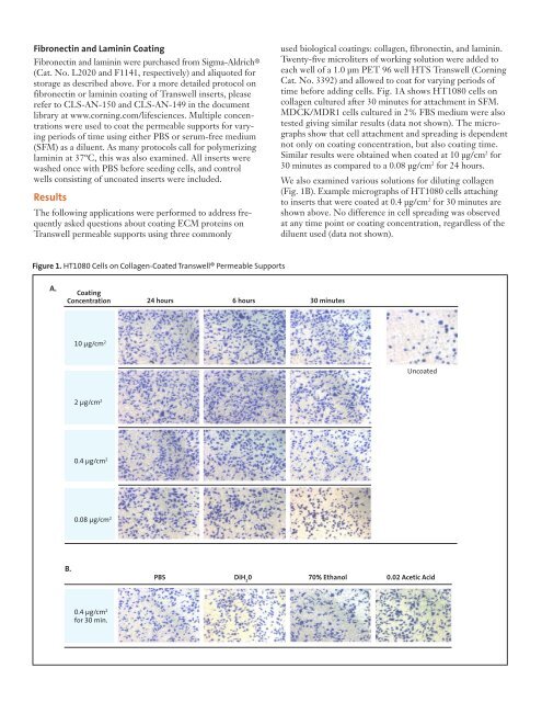
Fibronectin and Laminin <strong>Coating</strong>Fibronectin and laminin were purchased from Sigma-Aldrich ®(Cat. No. L2020 and F1141, respectively) and aliquoted <strong>for</strong>storage as described above. For a more detailed protocol onfibronectin or laminin coating of Transwell inserts, pleaserefer to CLS-AN-150 and CLS-AN-149 in the documentlibrary at www.corning.com/lifesciences. Multiple concentrationswere used to coat the permeable supports <strong>for</strong> varyingperiods of time using either PBS or serum-free medium(SFM) as a diluent. As many protocols call <strong>for</strong> polymerizinglaminin at 37ºC, this was also examined. All inserts werewashed once with PBS be<strong>for</strong>e seeding cells, and controlwells consisting of uncoated inserts were included.ResultsThe following applications were per<strong>for</strong>med to address frequentlyasked questions about coating ECM proteins onTranswell permeable supports using three commonlyused biological coatings: collagen, fibronectin, and laminin.Twenty-five microliters of working solution were added toeach well of a 1.0 µm PET 96 well HTS Transwell (<strong>Corning</strong>Cat. No. 3392) and allowed to coat <strong>for</strong> varying periods oftime be<strong>for</strong>e adding cells. Fig. 1A shows HT1080 cells oncollagen cultured after 30 minutes <strong>for</strong> attachment in SFM.MDCK/MDR1 cells cultured in 2% FBS medium were alsotested giving similar results (data not shown). The micrographsshow that cell attachment and spreading is dependentnot only on coating concentration, but also coating time.Similar results were obtained when coated at 10 µg/cm 2 <strong>for</strong>30 minutes as compared to a 0.08 µg/cm 2 <strong>for</strong> 24 hours.We also examined various solutions <strong>for</strong> diluting collagen(Fig. 1B). Example micrographs of HT1080 cells attachingto inserts that were coated at 0.4 µg/cm 2 <strong>for</strong> 30 minutes areshown above. No difference in cell spreading was observedat any time point or coating concentration, regardless of thediluent used (data not shown).Figure 1. HT1080 Cells on Collagen-Coated Transwell® Permeable SupportsA.<strong>Coating</strong>Concentration24 hours 6 hours 30 minutes10 µg/cm 2Uncoated2 µg/cm 20.4 µg/cm 20.08 µg/cm 2B.PBS DiH 20 70% Ethanol 0.02 Acetic Acid0.4 µg/cm 2<strong>for</strong> 30 min.