Monosaccharide inhibition of adherence by Pseudomonas ...
Monosaccharide inhibition of adherence by Pseudomonas ...
Monosaccharide inhibition of adherence by Pseudomonas ...

Create successful ePaper yourself
Turn your PDF publications into a flip-book with our unique Google optimized e-Paper software.
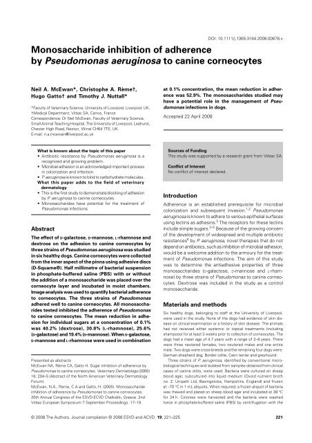
DOI: 10.1111/j.1365-3164.2008.00678.x<strong>Monosaccharide</strong> <strong>inhibition</strong> <strong>of</strong> <strong>adherence</strong>Blackwell Publishing Ltd<strong>by</strong> <strong>Pseudomonas</strong> aeruginosa to canine corneocytesNeil A. McEwan*, Christophe A. Rème†,Hugo Gatto† and Timothy J. Nuttall**Faculty <strong>of</strong> Veterinary Science, University <strong>of</strong> Liverpool, Liverpool, UK,†Medical Department, Virbac SA, Carros, FranceCorrespondence: Dr Neil McEwan, Faculty <strong>of</strong> Veterinary Science,Small Animal Teaching Hospital, The University <strong>of</strong> Liverpool, Leahurst,Chester High Road, Neston, Wirral CH64 7TE, UK.E-mail: n.a.mcewan@liverpool.ac.ukat 0.1% concentration, the mean reduction in <strong>adherence</strong>was 52.9%. The monosaccharides studied mayhave a potential role in the management <strong>of</strong> <strong>Pseudomonas</strong>infections in dogs.Accepted 22 April 2008What is known about the topic <strong>of</strong> this paper• Antibiotic resistance <strong>by</strong> <strong>Pseudomonas</strong> aeruginosa is arecognized and growing problem.• Microbial adhesion is an acknowledged important processin colonization and infection.• P. aeruginosa is known to bind to carbohydrate molecules.What this paper adds to the field <strong>of</strong> veterinarydermatology• This is the first study to demonstrate blocking <strong>of</strong> adhesion<strong>by</strong> P. aeruginosa to canine corneocytes.• <strong>Monosaccharide</strong>s have potential for the treatment <strong>of</strong><strong>Pseudomonas</strong> infections.AbstractThe effect <strong>of</strong> D-galactose, D-mannose, L-rhamnose anddextrose on the adhesion to canine corneocytes <strong>by</strong>three strains <strong>of</strong> <strong>Pseudomonas</strong> aeruginosa was studiedin six healthy dogs. Canine corneocytes were collectedfrom the inner aspect <strong>of</strong> the pinna using adhesive discs(D-Squame®). Half millimetre <strong>of</strong> bacterial suspensionin phosphate-buffered saline (PBS) with or withoutthe addition <strong>of</strong> a monosaccharide was placed over thecorneocyte layer and incubated in moist chambers.Image analysis was used to quantify bacterial <strong>adherence</strong>to corneocytes. The three strains <strong>of</strong> <strong>Pseudomonas</strong>adhered well to canine corneocytes. All monosaccharidestested inhibited the <strong>adherence</strong> <strong>of</strong> <strong>Pseudomonas</strong>to canine corneocytes. The mean reduction in adhesionfor individual sugars at a concentration <strong>of</strong> 0.1%was 40.2% (dextrose), 30.8% (L-rhamnose), 25.6%(D-galactose) and 19.4% (D-mannose). When D-galactose,D-mannose and L-rhamnose were used in combinationPresented as abstractsMcEwan NA, Rème CA, Gatto H. Sugar <strong>inhibition</strong> <strong>of</strong> <strong>adherence</strong> <strong>by</strong><strong>Pseudomonas</strong> to canine corneocytes. Veterinary Dermatology (2005)16; 204–5 (Abstract <strong>of</strong> the North American Veterinary DermatologyForum).McEwan, N.A., Rème, C.A and Gatto, H. (2005). <strong>Monosaccharide</strong><strong>inhibition</strong> <strong>of</strong> <strong>adherence</strong> <strong>by</strong> <strong>Pseudomonas</strong> to canine corneocytes.20th Annual Congress <strong>of</strong> the ESVD-ECVD Chalkidiki, Greece. 2ndVirbac European Symposium 7 September Proceedings. 17–19.Sources <strong>of</strong> FundingThis study was supported <strong>by</strong> a research grant from Virbac SA.Conflict <strong>of</strong> InterestNo conflict <strong>of</strong> interest declared.IntroductionAdherence is an established prerequisite for microbialcolonization and subsequent invasion. 1,2 <strong>Pseudomonas</strong>aeruginosa is known to adhere to various epithelial surfacesusing lectins as adhesins. 3 The receptors for these lectinsinclude simple sugars. 3–5 Because <strong>of</strong> the growing concern<strong>of</strong> the development <strong>of</strong> widespread and multiple antibioticresistances 6 <strong>by</strong> P. aeruginosa, novel therapies that do notdepend on antibiotics, such as <strong>inhibition</strong> <strong>of</strong> microbial adhesion,would be a welcome addition to the armoury for the treatment<strong>of</strong> <strong>Pseudomonas</strong> infections. The aim <strong>of</strong> this studywas to determine the antiadhesive properties <strong>of</strong> threemonosaccharides (D-galactose, D-mannose and L-rhamnose)<strong>by</strong> three strains <strong>of</strong> <strong>Pseudomonas</strong> to canine corneocytes.Dextrose was included in the study as a controlmonosaccharide.Materials and methodsSix healthy dogs, belonging to staff at the University <strong>of</strong> Liverpool,were used in the study. None <strong>of</strong> the dogs had evidence <strong>of</strong> skin diseaseon clinical examination or a history <strong>of</strong> skin disease. The animalshad not received either systemic or topical treatments (includingshampoos) for at least 3 weeks prior to collection <strong>of</strong> corneocytes. Thedogs had a mean age <strong>of</strong> 4.7 years with a range <strong>of</strong> 2–8 years. Therewere three neutered females, two neutered males and one entiremale. Two dogs were cross-breeds and the remaining four dogs were:German shepherd dog, Border collie, Cairn terrier and greyhound.Three strains <strong>of</strong> P. aeruginosa, identified <strong>by</strong> conventional microbiologicaltechniques and isolated from samples obtained from clinicalcases <strong>of</strong> canine otitis, were used. Bacteria were cultured on sheepblood agar, subcultured into liquid medium (Oxoid nutrient brothno. 2. Unipath Ltd, Basingstoke, Hampshire, England) and frozenat –70 °C in 1 mL aliquots. When required, a frozen aliquot <strong>of</strong> bacteriawas thawed and plated on sheep blood agar and incubated at 38 °Cfor 24 h. Colonies were harvested and the bacteria were washedtwice in phosphate-buffered saline (PBS) <strong>by</strong> centrifugation with the© 2008 The Authors. Journal compilation © 2008 ESVD and ACVD. 19; 221–225 221
McEwan et al.Table 1. Effects <strong>of</strong> individual sugars used at concentrations <strong>of</strong> 0.05% and 0.1% on <strong>adherence</strong> <strong>by</strong> three strains <strong>of</strong> <strong>Pseudomonas</strong> aeruginosa tocanine corneocytesStrain <strong>of</strong> <strong>Pseudomonas</strong> aeruginosaP1 P2 P3<strong>Monosaccharide</strong>sDog 1 Dog 2 Dog 1 Dog 2 Dog 1 Dog 2 Values for all dogs (n = 6)Dextrose 0.05% 63.6 46.7 37.9 51.2 68.6 72.8 56.8 13.7 43.20.10% 58.9 55.9 55.1 58.2 42.6 88.0 59.8 15.0 40.2D-galactose 0.05% 76.7 62.0 99.8 83.1 78.0 104.6 84.0 15.8 16.00.10% 42.5 71.5 95.7 75.8 88.3 72.9 74.5 18.3 25.6D-mannose 0.05% 59.3 73.5 88.1 81.1 87.5 97.5 81.2 13.4 18.80.10% 40.5 84.7 87.5 100.6 85.4 85.0 80.6 20.5 19.4L-rhamnose 0.05% 78.4 74.5 78.3 86.9 89.6 48.5 76.0 14.7 24.00.10% 48.6 70.9 71.9 68.1 68.5 87.1 69.2 12.3 30.8Each cell represents the percentage <strong>of</strong> bacterial adhesion compared to control (<strong>Pseudomonas</strong> aeruginosa added without sugar).MeanSDMean reductionin <strong>adherence</strong>Table 2. Effects <strong>of</strong> combined D-galactose, D-mannose and L-rhamnose used at concentration <strong>of</strong> 0.1% on <strong>adherence</strong> <strong>by</strong> three strains <strong>of</strong><strong>Pseudomonas</strong> aeruginosa to canine corneocytesDogMean reductionStrain <strong>of</strong> <strong>Pseudomonas</strong> aeruginosa 1 2 3 4 5 6 Mean SD in <strong>adherence</strong>P1 28.3 37.0 73.3 28.3 37.0 73.3 46.2 21.3 53.8P2 38.6 50.1 53.5 35.9 48.4 52.4 46.5 7.4 53.5P3 56.3 40.1 50.8 61.8 30.4 52.3 48.6 11.5 51.4Combined results 47.1 13.8 52.9Each cell represents the percentage <strong>of</strong> bacterial adhesion compared to control (<strong>Pseudomonas</strong> aeruginosa added without sugar).resulting suspension adjusted to an optical density <strong>of</strong> approximately0.15 at 570 nm (OD 570).A modification <strong>of</strong> an adhesion assay previously developed andvalidated 7 was used to quantify adhesion <strong>by</strong> <strong>Pseudomonas</strong> bacteria tocorneocytes. Prior to the study, repeatability <strong>of</strong> the counting methodand the optimal bacterial concentration was determined. 7 Briefly, corneocyteswere collected from the inner aspect <strong>of</strong> the pinna <strong>by</strong> usinga 22-mm-diameter adhesive disc (D-Squame®, CuDerm Corporation,Dallas, TX, USA). Prior to sampling, surface debris was removed fromthe collection site <strong>by</strong> applying five successive adhesive tape strips(Sellotape® Original, Henkel Consumer Adhesives, Winsford, Cheshire,UK). The adhesive disc was placed over the collection site and gentlypressed before being carefully removed. Where multiple sampleswere required from the same animal, discs were applied sequentiallyto the same area <strong>of</strong> skin. Half millimetre <strong>of</strong> bacterial suspension inPBS with or without the addition <strong>of</strong> a monosaccharide (D-galactose,D-mannose, L-rhamnose or dextrose) was placed over the corneocytelayer and incubated for 45 min in moist chambers. All monosaccharideswere obtained in pure form from Sigma-Aldrich (Gillingham, Dorset,UK). After incubation, the corneocytes were washed and stained.Adherent <strong>Pseudomonas</strong> bacteria were quantified using image analysis<strong>by</strong> calculating the corneocyte surface area covered <strong>by</strong> bacteria.From each D-Squame® disc 10 areas were selected and saved asblack and white TIFF files at ×630 magnification. From each TIFFfile, the surface area covered <strong>by</strong> bacteria in a 400 × 400 pixel box wascalculated. The analysis gave a final value <strong>of</strong> the bacteria adheringover a surface area equivalent to 100 corneocytes. The operator wasblinded to the identity <strong>of</strong> individual samples during the image collectionand the counting process.In the first part <strong>of</strong> the study, three P. aeruginosa strains were eachtested in two dogs at sugar concentrations <strong>of</strong> 0.05% and 0.1%. In thesecond part <strong>of</strong> the study, a combination <strong>of</strong> D-galactose, D-mannoseand L-rhamnose in 0.1% solution was tested in the same six dogsusing the same three strains <strong>of</strong> P. aeruginosa. Bacteria incubated withPBS without sugar was used as a positive control and PBS alone wasused as a negative control. Dextrose was included as a controlmonosaccharide. The antiadhesive effect <strong>of</strong> the monosaccharideunder test was calculated as a percentage <strong>of</strong> the <strong>adherence</strong> shown <strong>by</strong>the positive control.Statistical analysisData were tested for normality before statistical analysis. One-wayanalysis <strong>of</strong> variance (ANOVA) with Tukey post-test was used to analysethe appropriate data. Significance was set at P < 0.05. All analyseswere performed using Graphpad Prism version 4.00 (Graphpad Inc.,San Diego, CA, USA [www.graphpad.com]).ResultsAll three test sugars and dextrose inhibited <strong>adherence</strong><strong>by</strong> <strong>Pseudomonas</strong> to corneocytes. The mean <strong>adherence</strong>compared to the positive control for individual sugars at0.05% was 56.8% (dextrose), 76.0% (L-rhamnose), 81.2%(D-mannose) and 84.0% (D-galactose). Incubation withdextrose significantly reduced <strong>adherence</strong> compared toD-galactose and D-mannose (Tukey post-test, P < 0.05).The mean <strong>adherence</strong> using individual sugars at 0.1% was59.8% (dextrose), 69.2% (L-rhamnose), 74.5% (D-galactose)and 80.6% (D-mannose). There were no significant differencesamong the sugars at this concentration. When thethree sugars were used in combination at 0.1% concentration,the mean <strong>adherence</strong> was 47.1%, which gave areduction in <strong>adherence</strong> <strong>of</strong> 52.9%. The mixture <strong>of</strong> thethree sugars gave a significantly lower adhesion comparedto D-galactose and D-mannose (Tukey post-test, P < 0.01),L-rhamnose (Tukey post-test, P < 0.05) but not dextrose.Tables 1 and 2 and Figs 1 and 2 summarize all data.DiscussionAll three strains <strong>of</strong> P. aeruginosa were shown to adherestrongly to canine corneocytes which supports a similarfinding <strong>by</strong> Forsythe et al. 8 This study is the first to documentsugar <strong>inhibition</strong> <strong>of</strong> <strong>adherence</strong> <strong>by</strong> P. aeruginosa to222 © 2008 The Authors. Journal compilation © 2008 ESVD and ACVD.
<strong>Monosaccharide</strong> <strong>inhibition</strong> <strong>of</strong> <strong>adherence</strong>Figure 1. The effect <strong>of</strong> monosaccharides on adhesion <strong>by</strong> <strong>Pseudomonas</strong>aeruginosa to canine corneocytes. All sugars at 0.1% concentration.Combined sugars consist <strong>of</strong> D-galactose, D-mannose andL-rhamnose. Bars show mean percentage <strong>adherence</strong> with standarderror bars compared with control (bacteria without sugar added).*P < 0.01 compared to combined sugars.†P < 0.05 compared to combined sugars.Figure 2. (a) Control. <strong>Pseudomonas</strong> aeruginosa in phosphatebufferedsaline incubated with canine corneocytes. (b) Adherenceto canine corneocytes <strong>by</strong> P. aeruginosa after addition <strong>of</strong> combinedD-galactose, D-mannose and L-rhamnose in a 0.1% solution. Originalmagnification ×630. Crystal violet staining.canine corneocytes. All sugars studied produced decreased<strong>adherence</strong> but the combination <strong>of</strong> D-galactose, D-mannoseand L-rhamnose proved particularly effective. Dextrosewas surprisingly effective in preventing the adhesion tocanine corneocytes <strong>by</strong> P. aeruginosa but should be considereda poor choice for a therapeutic agent as it is aready energy and carbohydrate source and, as such, mayhave the effect <strong>of</strong> enhancing microbial growth.Sugars have been known to have the potential to blockbacterial adhesion to animal cells for over two decades 9and a major goal <strong>of</strong> many current studies has been the<strong>inhibition</strong> <strong>of</strong> bacterial <strong>adherence</strong>. Few studies have beenconducted in the veterinary field. One study 5 demonstratedthat mannose and N-acetyl-D-galactosamine played a rolein inhibiting the adhesion <strong>of</strong> Streptococcus zooepidemicus,P. aeruginosa and Escherichia coli to equine endometrialepithelial cells. Other studies have shown that <strong>Pseudomonas</strong><strong>adherence</strong> to various substrates could be blocked <strong>by</strong> thefollowing sugars: D-galactose (human buccal epithelial cells), 10D-galactose and D-mannose (collagen type I molecules), 11D-galactose (erythrocytes) 12 and D-galactose (rodent trachealepithelial cells). 13 In a human study <strong>of</strong> P. aeruginosa otitis,patients treated with a combination <strong>of</strong> galactose, mannoseand N-acetylneuraminic acid recovered more rapidly thana control group. As bacteria typically employ several differentadhesins, it has been proposed that using mixtures <strong>of</strong>sugars which block more than one bacterial adhesin arelikely to be more effective than using a single sugar. 3Results <strong>of</strong> our study support this hypothesis.<strong>Pseudomonas</strong> aeruginosa produces a number <strong>of</strong> adhesivemolecules, including sialic acid-binding, gangliosidebindingand hydrophobic adhesins. 3 Lectins are commonlyused <strong>by</strong> bacteria as adhesins and are proteins capable <strong>of</strong>binding saccharide structures with high specificity andaffinity (reviewed in 3,14–16 ). Two soluble lectins, PA-IL 17and PA-IIL, 18 have been identified from P. aeruginosa andthese adhesive molecules are considered to be virulencefactors playing important roles in human infection andtissue damage. 19,20 Sugar <strong>inhibition</strong> experiments demonstratethat D-galactose and L-fucose inhibit both lectins, 21although clear preferences <strong>of</strong> D-galactose for PA-IL and <strong>of</strong>L-fucose for PA-IIL exist. The PA-IIL lectin will also recognizeother monosaccharides including D-mannose. 22 The ability<strong>of</strong> D-galactose and D-mannose to inhibit P. aeruginosa<strong>adherence</strong> in this study may be due to blocking <strong>of</strong> thePA-IL and PA-IIL lectins. L-rhamnose can inhibit bacterial<strong>adherence</strong> 23 but no studies have been specifically conductedfor P. aeruginosa. L-rhamnose is chemically similarto L-fucose both being deoxy sugars which may suggestthat L-rhamnose may also block the PA-IIL lectin.Microbial <strong>adherence</strong> can be thought <strong>of</strong> a multistep processwith initial nonspecific or reversible binding followed<strong>by</strong> specific and irreversible binding. Bi<strong>of</strong>ilms, produced <strong>by</strong>some bacteria including P. aeruginosa, may also contributeto the adhesive process. EPS (extracellular polymer substances)is used as a collective term for the sugar components<strong>of</strong> microbial bi<strong>of</strong>ilms. <strong>Monosaccharide</strong>s, includingthose used in this study, <strong>of</strong>ten form part <strong>of</strong> microbial bi<strong>of</strong>ilms.The PA-IL lectin has been shown to contribute tobi<strong>of</strong>ilm development where it would be important incross-linking bacteria through galactosides to form microcoloniesthen mature bi<strong>of</strong>ilms. 24 Competitive <strong>inhibition</strong> <strong>of</strong>the PA-IL lectin results in reduced bi<strong>of</strong>ilm production. 24Although bi<strong>of</strong>ilm development is typically a later phenomenon,it is conceivable that the inhibitory effects <strong>of</strong> thesugars in this study are related to bi<strong>of</strong>ilm activity. Lastly,sugars may influence adhesion <strong>by</strong> binding to keratinocytes.14 Human keratinocytes are known to have specificsugar receptors on their cell surface 25 some <strong>of</strong> whichrecognize L-fucose and L-rhamnose. 26© 2008 The Authors. Journal compilation © 2008 ESVD and ACVD. 223
McEwan et al.Antiadhesive therapy <strong>of</strong> bacterial disease is an attractiveprospect particularly as drug-resistant bacteria are increasing.As bacteria utilize several adhesins, treatments thattarget multiple adhesins are likely to be more efficienttherapeutic agents. 15,16In conclusion, this study demonstrates that P. aeruginosaadheres strongly to canine corneocytes. The monosaccharidestested resulted in reduced <strong>adherence</strong> <strong>by</strong> P. aeruginosato canine corneocytes. The combination <strong>of</strong> the three monosaccharides,D-galactose, D-mannose and L-rhamnose, wasmore effective than individual monosaccharides.AcknowledgementsThe authors would like to acknowledge the help andsupport freely given <strong>by</strong> staff <strong>of</strong> the Connective Tissue, andMicrobiology and Infectious Disease Research Groups atThe University <strong>of</strong> Liverpool Faculty <strong>of</strong> Veterinary Science.References1. Feingold DS. Bacterial <strong>adherence</strong>, colonization, and pathogenicity.Archives <strong>of</strong> Dermatology 1986; 122: 161–3.2. Patti JM, Allen BL, McGavin MJ, Hook M. MSCRAMM-mediated<strong>adherence</strong> <strong>of</strong> microorganisms to host tissues. Annual Review <strong>of</strong>Microbiology 1994; 48: 585–617.3. Ofek I, Hasty DL, Doyle RJ, Ofek I. Bacterial Adhesion to AnimalCells and Tissues. Washington, DC: ASM Press, 2003.4. Acord J, Maskell J, Sefton A. A rapid microplate method for quantifying<strong>inhibition</strong> <strong>of</strong> bacterial adhesion to eukaryotic cells. Journal<strong>of</strong> Microbiological Methods 2005; 60: 55–62.5. King SS, Young DA, Nequin LG, Carnevale EM. Use <strong>of</strong> specificsugars to inhibit bacterial <strong>adherence</strong> to equine endometrium invitro. American Journal <strong>of</strong> Veterinary Research 2000; 61: 446–9.6. Petersen AD, Walker RD, Bowman MM, Schott HC 2nd, Rosser EJ Jr.Frequency <strong>of</strong> isolation and antimicrobial susceptibility patterns <strong>of</strong>Staphylococcus intermedius and <strong>Pseudomonas</strong> aeruginosa isolatesfrom canine skin and ear samples over a 6-year period (1992–97).Journal <strong>of</strong> the American Animal Hospital Association 2002; 38:407–13.7. Lu YF, McEwan NA. Staphylococcal and micrococcal <strong>adherence</strong>to canine and feline corneocytes: quantification using a simpleadhesion assay. Veterinary Dermatology 2007; 18: 29–35.8. Forsythe PJ, Hill PB, Thoday KL, Brown J. Use <strong>of</strong> computerized imageanalysis to quantify staphylococcal adhesion to canine corneocytes:does breed and body site have any relevance to the pathogenesis<strong>of</strong> pyoderma? Veterinary Dermatology 2002; 13: 29–36.9. Aronson M, Medalia O, Schori L, Mirelman D, Sharon N, Ofek I.Prevention <strong>of</strong> colonization <strong>of</strong> the urinary tract <strong>of</strong> mice with Escherichiacoli <strong>by</strong> blocking <strong>of</strong> bacterial <strong>adherence</strong> with methyl alpha-Dmannopyranoside.Journal <strong>of</strong> Infectious Diseases 1979; 139: 329–32.10. Wolska K, Bednarz B, Jakubczak A. Adherence <strong>of</strong> <strong>Pseudomonas</strong>aeruginosa to human buccal epithelial cells. Acta MicrobiologicaPolonica 2003; 52: 419–23.11. Stepinska M, Trafny EA. Modulation <strong>of</strong> <strong>Pseudomonas</strong> aeruginosa<strong>adherence</strong> to collagen type I and type II <strong>by</strong> carbohydrates. FEMSImmunology and Medical Microbiology 1995; 12: 187–94.12. Gilboa-Garber N, Sudakevitz D, Sheffi M, Sela R, Levene C. PA-Iand PA-II lectin interactions with the ABO(H) and P blood groupglycosphingolipid antigens may contribute to the broad spectrum<strong>adherence</strong> <strong>of</strong> <strong>Pseudomonas</strong> aeruginosa to human tissues insecondary infections. Glycoconjugate Journal 1994; 11: 414–7.13. Marcus H, Austria A, Baker NR. Adherence <strong>of</strong> <strong>Pseudomonas</strong>aeruginosa to tracheal epithelium. Infection and Immunity 1989;57: 1050–3.14. Lloyd DH, Viac J, Werling D, Reme CA, Gatto H. Role <strong>of</strong> sugarsin surface microbe–host interactions and immune reactionmodulation. Veterinary Dermatology 2007; 18: 197–204.15. Ofek I, Hasty DL, Sharon N. Anti-adhesion therapy <strong>of</strong> bacterialdiseases: prospects and problems. FEMS Immunology andMedical Microbiology 2003; 38: 181–91.16. Sharon N, Ofek I. Safe as mother’s milk: carbohydrates as futureanti-adhesion drugs for bacterial diseases. Glycoconjugate Journal2000; 17: 659–64.17. Gilboa-Garber N. Purification and properties <strong>of</strong> hemagglutininfrom <strong>Pseudomonas</strong> aeruginosa and its reaction with human bloodcells. Biochimica et Biophysica Acta 1972; 273: 165–73.18. Gilboa-Garber N, Mizrahi L, Garber N. Mannose-binding hemagglutininsin extracts <strong>of</strong> <strong>Pseudomonas</strong> aeruginosa. Canadian Journal<strong>of</strong> Biochemistry 1977; 55: 975–81.19. Mitchell E, Houles C, Sudakevitz D et al. Structural basis foroligosaccharide-mediated adhesion <strong>of</strong> <strong>Pseudomonas</strong> aeruginosain the lungs <strong>of</strong> cystic fibrosis patients. Nature Structural Biology2002; 9: 918–21.20. Wu AM, Wu JH, Singh T, Liu JH, Tsai MS, Gilboa-Garber N. Interactions<strong>of</strong> the fucose-specific <strong>Pseudomonas</strong> aeruginosa lectin, PA-IIL, with mammalian glycoconjugates bearing polyvalent Lewis(a)and ABH blood group glycotopes. Biochimie 2006; 88: 1479–92.21. Mewe M, Tielker D, Schonberg R, Schachner M, Jaeger KE,Schumacher U. <strong>Pseudomonas</strong> aeruginosa lectins I and II and theirinteraction with human airway cilia. Journal <strong>of</strong> Laryngology andOtology 2005; 119: 595–9.22. Glick J, Garber N. The intracellular localization <strong>of</strong> <strong>Pseudomonas</strong>aeruginosa lectins. Journal <strong>of</strong> General Microbiology 1983; 129:3085–90.23. Naess V, Johannessen C, H<strong>of</strong>stad T. Adherence <strong>of</strong> Campylobacterjejuni and Campylobacter coli to porcine intestinal brush bordermembranes. APMIS 1988; 96: 681–7.24. Diggle SP, Stacey RE, Dodd C, Camara M, Williams P, Winzer K.The galactophilic lectin, LecA, contributes to bi<strong>of</strong>ilm developmentin <strong>Pseudomonas</strong> aeruginosa. Environmental Microbiology 2006;8: 1095–104.25. Palacio S, Viac J, Vinche A, Huband JC, Gatto H, Schmitt D. Suppressiveeffect <strong>of</strong> monosaccharides on ICAM-1/CD54 expressionin human keratinocytes. Archives <strong>of</strong> Dermatological Research1997; 289: 234–7.26. Cerdan D, Grillon C, Monsigny M, Redziniak G, Kieda C. Humankeratinocyte membrane lectins: characterization and modulation<strong>of</strong> their expression <strong>by</strong> cytokines. Biology <strong>of</strong> the Cell 1991; 73: 35–42.Résumé Cette étude a évalué les effets du D-galactose, du D-mannose, du L-rhamnose et du dextrosesur l’adhésion aux cornéocytes canins de trois souches de <strong>Pseudomonas</strong> aeruginosa chez 6 chiens sains.Les cornéocytes ont été récoltés sur la face interne des pavillons auriculaires en utilisant des disquesadhésifs (D-Squame®). Un demi millimètre d’une suspension bactérienne dans du phosphate bufferedsaline (PBS) avec ou sans addition de monosaccharides a été placé sur la couche de cornéocytes et mis àl’incubation. Une analyse d’image a été réalisée pour quantifier l’adhérence bactérienne. Les trois souchesde <strong>Pseudomonas</strong> adhéraient bien aux cornéocytes. Tous les monosaccharides testés ont inhibé l’adhérencede <strong>Pseudomonas</strong> aux cornéocytes. La réduction moyenne de l’adhésion pour une concentration de 0.1%était de 40.2% (dextrose), 30.8% (L-rhamnose), 25.6% (D-galactose), et 19.4% (D-mannose). Lorsque leD-galactose, le D-mannose et le L-rhamnose ont été combinés à la concentration de 0.1% la réductionmoyenne d’adhérence était de 52.9%. les monosaccharides étudiés ici pourraient avoir un potentiel pourle traitement des infections à <strong>Pseudomonas</strong> chez le chien.224 © 2008 The Authors. Journal compilation © 2008 ESVD and ACVD.
<strong>Monosaccharide</strong> <strong>inhibition</strong> <strong>of</strong> <strong>adherence</strong>Resumen Se estudiaron en seis perros sanos los efectos de la D-galactosa, D-manosa, L-ramnosa y dextrosaen la adherencia a corneocitos caninos de cepas de <strong>Pseudomonas</strong> aeruginosa. Los corneocitos setomaron de la parte interna del oido externo usando discos adhesivos (D-Squame®). Se colocaron sobrelos cultivos de corneocitos medio mililitro de suspensión bacteriana en solución tamponada de fosfatos.cono sin la adición de monosacáridos y se incubaron en cámaras húmedas. Se utilizó análisis de imagen paracuantificar la adherencia bacteriana a los corneocitos. Las tres cepas de <strong>Pseudomonas</strong> se adhirieron biena los corneocitos caninos. Todos los monosacáridos probados inhibieron la adherencia de <strong>Pseudomonas</strong> alos corneocitos. La reducción media de la adherencia para azúcares individuales a la concentración de 0.1%fue de 40.2% (dextrosa), 30.8% (L-ramnosa), 25.6% (D-galactosa) y 19.4% (D-manosa). Cuando se utilizaronen combinación al 0.1% D-galactosa, D-manosa y L-ramnosa la reducción en la adherencia fue del 52.9%.Los monosacáridos estudiados pueden tener potencial en el control de la infección por <strong>Pseudomonas</strong> enperros.Zusammenfassung Die Wirkung von D-Galaktose, D-Mannose, L-Rhamnose und Dextrose auf dieAdhäsion von drei <strong>Pseudomonas</strong> aeruginosa Stämmen an canine Corneozyten wurde bei sechs gesundenHunden untersucht. Canine Corneozyten wurden von der Innenseite der Ohrmuschel mittels Klebe-Disks(D-Squame®) entnommen. Ein halber Millimeter bakterieller Suspension in Phosphatbuffer (PBS) wurdemit oder ohne die Zugabe eines Monosaccharids über eine Lage von Corneozyten gegeben und diesein feuchten Kammern inkubiert. Die Bildanalyse wurde verwendet, um die bakterielle Anhaftung anden Corneozyten quantitativ zu bestimmen. Die drei <strong>Pseudomonas</strong> Stämme hafteten gut an den caninenCorneozyten. Alle getesteten <strong>Monosaccharide</strong> verhinderten die Anhaftung der <strong>Pseudomonas</strong> an die caninenCorneozyten. Die durchschnittliche Verminderung der Adhäsion für die einzelnen Zucker in einer Konzentrationvon 0.1% lag bei 40.2% (Dextrose), bei 30.8% (L-Rhamnose), bei 25.6% (D-Galaktose) und bei 19.4%(D-Mannose). Wenn D-Galaktose, D-Mannose und L-Rhamnose in einer Konzentration von 0.1% kombiniertwurden, lag die durchschnittliche Verminderung der Adhäsion bei 52.9%. Die untersuchten <strong>Monosaccharide</strong>könnten eine mögliche Rolle beim Management von <strong>Pseudomonas</strong> Infektionen des Hundes spielen.© 2008 The Authors. Journal compilation © 2008 ESVD and ACVD. 225