Visual Field Defects in Deformational Posterior Plagiocephaly
Visual Field Defects in Deformational Posterior Plagiocephaly
Visual Field Defects in Deformational Posterior Plagiocephaly
- No tags were found...

Create successful ePaper yourself
Turn your PDF publications into a flip-book with our unique Google optimized e-Paper software.
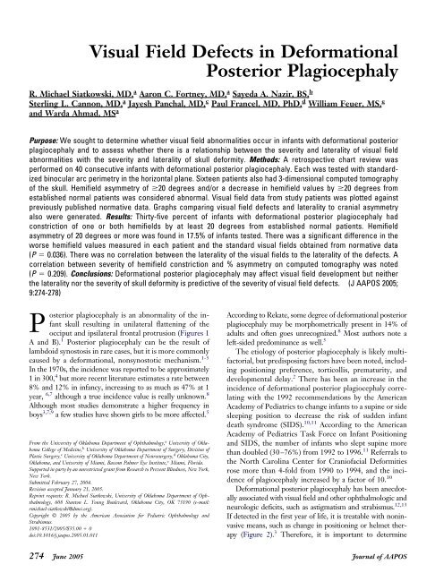
<strong>Visual</strong> <strong>Field</strong> <strong>Defects</strong> <strong>in</strong> <strong>Deformational</strong><strong>Posterior</strong> <strong>Plagiocephaly</strong>R. Michael Siatkowski, MD, a Aaron C. Fortney, MD, a Sayeda A. Nazir, BS, bSterl<strong>in</strong>g L. Cannon, MD, a Jayesh Panchal, MD, c Paul Francel, MD, PhD, d William Feuer, MS, eand Warda Ahmad, MS aPurpose: We sought to determ<strong>in</strong>e whether visual field abnormalities occur <strong>in</strong> <strong>in</strong>fants with deformational posteriorplagiocephaly and to assess whether there is a relationship between the severity and laterality of visual fieldabnormalities with the severity and laterality of skull deformity. Methods: A retrospective chart review wasperformed on 40 consecutive <strong>in</strong>fants with deformational posterior plagiocephaly. Each was tested with standardizedb<strong>in</strong>ocular arc perimetry <strong>in</strong> the horizontal plane. Sixteen patients also had 3-dimensional computed tomographyof the skull. Hemifield asymmetry of 20 degrees and/or a decrease <strong>in</strong> hemifield values by 20 degrees fromestablished normal patients was considered abnormal. <strong>Visual</strong> field data from study patients was plotted aga<strong>in</strong>stpreviously published normative data. Graphs compar<strong>in</strong>g visual field defects and laterality to cranial asymmetryalso were generated. Results: Thirty-five percent of <strong>in</strong>fants with deformational posterior plagiocephaly hadconstriction of one or both hemifields by at least 20 degrees from established normal patients. Hemifieldasymmetry of 20 degrees or more was found <strong>in</strong> 17.5% of <strong>in</strong>fants tested. There was a significant difference <strong>in</strong> theworse hemifield values measured <strong>in</strong> each patient and the standard visual fields obta<strong>in</strong>ed from normative data(P 0.036). There was no correlation between the laterality of the visual fields to the laterality of the defects. Acorrelation between severity of hemifield constriction and % asymmetry on computed tomography was noted(P 0.209). Conclusions: <strong>Deformational</strong> posterior plagiocephaly may affect visual field development but neitherthe laterality nor the severity of skull deformity is predictive of the severity of visual field defects. (J AAPOS 2005;9:274-278)<strong>Posterior</strong> plagiocephaly is an abnormality of the <strong>in</strong>fantskull result<strong>in</strong>g <strong>in</strong> unilateral flatten<strong>in</strong>g of theocciput and ipsilateral frontal protrusion (Figures 1A and B). 1 <strong>Posterior</strong> plagiocephaly can be the result oflambdoid synostosis <strong>in</strong> rare cases, but it is more commonlycaused by a deformational, nonsynostotic mechanism. 1-5In the 1970s, the <strong>in</strong>cidence was reported to be approximately1 <strong>in</strong> 300, 4 but more recent literature estimates a rate between8% and 12% <strong>in</strong> <strong>in</strong>fancy, <strong>in</strong>creas<strong>in</strong>g to as much as 47% at 1year, 6,7 although a true <strong>in</strong>cidence value is really unknown. 8Although most studies demonstrate a higher frequency <strong>in</strong>boys 3,7,9 a few studies have shown girls to be more affected. 5From the University of Oklahoma Department of Ophthalmology, a University of OklahomaCollege of Medic<strong>in</strong>e, b University of Oklahoma Department of Surgery, Division ofPlastic Surgery, c University of Oklahoma Department of Neurosurgery, d Oklahoma City,Oklahoma, and University of Miami, Bascom Palmer Eye Institute, e Miami, Florida.Supported <strong>in</strong> party by an unrestricted grant from Research to Prevent Bl<strong>in</strong>dness, New York,New York.Submitted February 27, 2004.Revision accepted January 21, 2005.Repr<strong>in</strong>t requests: R. Michael Siatkowski, University of Oklahoma Department of Ophthalmology,608 Stanton L. Young Boulevard, Oklahoma City, OK 73190 (e-mail:rmichael-siatkowski@dmei.org).Copyright © 2005 by the American Association for Pediatric Ophthalmology andStrabismus.1091-8531/2005/$35.00 0doi:10.1016/j.jaapos.2005.01.011Accord<strong>in</strong>g to Rekate, some degree of deformational posteriorplagiocephaly may be morphometrically present <strong>in</strong> 14% ofadults and often goes unrecognized. 8 Most authors note aleft-sided predom<strong>in</strong>ance as well. 5The etiology of posterior plagiocephaly is likely multifactorial,but predispos<strong>in</strong>g factors have been noted, <strong>in</strong>clud<strong>in</strong>gposition<strong>in</strong>g preference, torticollis, prematurity, anddevelopmental delay. 2 There has been an <strong>in</strong>crease <strong>in</strong> the<strong>in</strong>cidence of deformational posterior plagiocephaly correlat<strong>in</strong>gwith the 1992 recommendations by the AmericanAcademy of Pediatrics to change <strong>in</strong>fants to a sup<strong>in</strong>e or sidesleep<strong>in</strong>g position to decrease the risk of sudden <strong>in</strong>fantdeath syndrome (SIDS). 10,11 Accord<strong>in</strong>g to the AmericanAcademy of Pediatrics Task Force on Infant Position<strong>in</strong>gand SIDS, the number of <strong>in</strong>fants who slept sup<strong>in</strong>e morethan doubled (30–76%) from 1992 to 1996. 11 Referrals tothe North Carol<strong>in</strong>a Center for Craniofacial Deformitiesrose more than 4-fold from 1990 to 1994, and the <strong>in</strong>cidenceof plagiocephaly <strong>in</strong>creased by a factor of 10. 10<strong>Deformational</strong> posterior plagiocephaly has been anecdotallyassociated with visual field and other ophthalmologic andneurologic deficits, such as astigmatism and strabismus. 12,13If detected <strong>in</strong> the first year of life, it is treatable with non<strong>in</strong>vasivemeans, such as change <strong>in</strong> position<strong>in</strong>g or helmet therapy(Figure 2). 3 Therefore, it is important to determ<strong>in</strong>e274 June 2005Journal of AAPOS
Journal of AAPOSVolume 9 Number 3 June 2005 Siatkowski et al 275FIG 2. Photograph of an <strong>in</strong>fant with deformational posterior plagiocephalywear<strong>in</strong>g an orthotic helmet.FIG 1. Head CT (bone w<strong>in</strong>dows) show<strong>in</strong>g posterior plagiocephaly. A,Axial view. B, Sagittal view.whether any quantifiable, cl<strong>in</strong>ically relevant neurological deficitsare associated with posterior plagiocephaly, and if treatmentimproves these deficits. <strong>Visual</strong> field test<strong>in</strong>g has beensuggested as a means of detect<strong>in</strong>g abnormalities <strong>in</strong> corticalpathway maturation <strong>in</strong> <strong>in</strong>fants. 14,15 Us<strong>in</strong>g the normative dataobta<strong>in</strong>ed by a previous study 14 and attempt<strong>in</strong>g to duplicatethe test<strong>in</strong>g method and environment, visual field test<strong>in</strong>gwas performed <strong>in</strong> a cohort of patients with posterior plagiocephalyto document the presence and severity of visualfield abnormalities <strong>in</strong> this group.METHODSAfter approval from the University of Oklahoma HealthSciences Center Institutional Review Board, a retrospectivechart review of 40 consecutive <strong>in</strong>fants, ages 19-53weeks, with a diagnosis of deformational posterior plagiocephalyand otherwise-normal ophthalmologic exam<strong>in</strong>ationwas performed dur<strong>in</strong>g a 3-year period. Patients were<strong>in</strong>cluded if they had no prior helmet therapy, were able tocooperate for visual field test<strong>in</strong>g, had normal Teller acuities(Vistech Consultants, Inc., Seattle, WA), and had nostrabismus, amblyopia, or ret<strong>in</strong>al or optic nerve abnormalities;on these bases, they were considered likely to havenormal b<strong>in</strong>ocular visual function for age. Standardizedb<strong>in</strong>ocular arc perimetry (Ferree-Rand Simplified Perimeter,Bausch and Lomb, Rochester, NY) <strong>in</strong> the horizontalplane, as previously described, 12 was performed on eachchild by 2 separate exam<strong>in</strong>ers masked to each other, measur<strong>in</strong>gthe temporal right and left hemifields us<strong>in</strong>g a preferentiallook<strong>in</strong>g technique with a target subtend<strong>in</strong>g anangle of 6 degrees. The visual field measurement wasrepeated up to 3 times by each exam<strong>in</strong>er, and the mean ofeach exam<strong>in</strong>er’s result was recorded. If there was a discrepancyof 10 degrees between the 2 exam<strong>in</strong>ers’ measurements,both exam<strong>in</strong>ers performed the field test until aconsensus was reached. Hemifield asymmetry of 20 degreeswas considered abnormal. In addition, a decrease <strong>in</strong>hemifield values of 20 degrees from established normalpatients also was considered abnormal.Graphs were generated plott<strong>in</strong>g the visual field dataobta<strong>in</strong>ed from the patients with plagiocephaly aga<strong>in</strong>st nor-
276 Siatkowski et alJournal of AAPOSVolume 9 Number 3 June 2005FIG 3. Graph of temporal visual darker field (degrees) versus squareroot transformation of age (weeks). Black, Normative visual fielddata; white, worse hemifield data; lighter black, better hemifielddata; and gray, average visual field data.mative data extracted from the study by Mohn and VanHof-van Du<strong>in</strong> 14 (Figure 3). The square-root transformationof the age was graphed aga<strong>in</strong>st visual field <strong>in</strong> degreesto establish a normative l<strong>in</strong>e for visual field development.We then compared the heights of the regression l<strong>in</strong>esgenerated by the data obta<strong>in</strong>ed <strong>in</strong> our study to the normativel<strong>in</strong>e us<strong>in</strong>g a one-way analysis of covariance.Three-dimensional computed tomography (CT) of theskull was performed <strong>in</strong> 16 of the study patients to obta<strong>in</strong>objective data on the severity of plagiocephaly. CT imageswere received from the radiology department on CD-ROM and transferred to the Panchal Imag<strong>in</strong>g Lab, Universityof Oklahoma College of Medic<strong>in</strong>e, Division ofPlastic Surgery. The data were then copied to the harddrive of the imag<strong>in</strong>g workstation (DELL Precision P-530with dual 1.5 GHz processors, 500 MB RAM and 100 GBRAID 1 mirrored storage). The raw slices (2.5-mm thick)were imported <strong>in</strong> to Analyze 4.0 (Biomedical Imag<strong>in</strong>gResource, Mayo Cl<strong>in</strong>ic Rochester, MN) to be prepared forlandmark<strong>in</strong>g. The empty outer marg<strong>in</strong>s of the images werecropped to reduce rendered volume file size, and the volumewas forced to cubic voxel dimensions. Proximal portionsof the mandible and vertebral column were segmentedout (when necessary) to allow visualization ofexocranial base landmarks. Rema<strong>in</strong><strong>in</strong>g artifacts of the CTscan gantry image were removed and the rendered volumesaved <strong>in</strong> Analyze Image 7.5 format. The Analyze Image 7.5volume was opened, and the bone threshold level for eachimage was determ<strong>in</strong>ed empirically. A standard coord<strong>in</strong>atetemplate file was opened, renamed, and saved under thepatients’ data archive with an identifier, date, and thresholdlevel for future reference.Collection of CT and Magnetic Resonance ImagesCT Scans of the Head. Each CT scan was opened <strong>in</strong>exploratory 2-/3-dimensional image process<strong>in</strong>g system (etdips)software. This imag<strong>in</strong>g software allows the simultaneousvisualization of the reconstructed volume <strong>in</strong> 3 orthogonalviews, thereby allow<strong>in</strong>g accurate placement ofFIG 4. CT reconstructed image measur<strong>in</strong>g the distance between theanterior supraorbital notch and the contralateral occipital bone.biologically relevant landmarks. Landmarks were chosenat the supraorbital notch anteriorly on each side. At thesame CT slice level, the most prom<strong>in</strong>ent part on theoccipital bone was chosen on the right and the left occipitalbone (Figure 4). The right oblique diameter measuredthe distance from the right supraorbital notch to the contralateralmost prom<strong>in</strong>ent po<strong>in</strong>t on the occipital lobe asdescribed. The left oblique diameter was measured viceversa, ie, from the left supraorbital notch to the mostprom<strong>in</strong>ent po<strong>in</strong>t on the right occiput. The absolute differencebetween the right and left oblique diameter gavethe extent of asymmetry caused by plagiocephaly. Graphswere generated compar<strong>in</strong>g visual field defect and lateralityto cranial asymmetry to determ<strong>in</strong>e if a correlation existedbetween visual field defect and cranial asymmetry.RESULTS<strong>Visual</strong> <strong>Field</strong> ConstrictionConstriction of one or both hemifields by at least 20degrees compared with established normal patients occurred<strong>in</strong> 35% (14/40) of patients. Four (10%) patients(4/40) had constriction of both hemifields whereas 10(25%) patients (10/40) had constriction of one hemifield.There was a significant difference between the worsehemifield measured (white l<strong>in</strong>e) <strong>in</strong> each patient and thestandard visual field calculated from the normative data(darker black l<strong>in</strong>e) (P 0.004). (Figure 3)Hemifield F<strong>in</strong>d<strong>in</strong>gsHemifield asymmetry of 20 degrees or more (<strong>in</strong>trapatientright vs. left hemifield difference) was found <strong>in</strong> 17.5%
Journal of AAPOSVolume 9 Number 3 June 2005 Siatkowski et al 277FIG 6. Degree of CT asymmetry (%) versus amount of visual fieldconstriction (degrees).FIG 5. Skull measurement asymmetry (mm) versus visual field asymmetry(degrees).(7/40) <strong>in</strong>fants tested, with a range of 20 to 30 degrees. Asimilar plot of the better hemifield was not statisticallydifferent from the normative curve (P 0.390) (Figure 3,lighter black l<strong>in</strong>e). A plot of the mean of the 2 hemifieldmeasurements for each patient versus age was statisticallydifferent from standard visual field (P 0.036; Figure 3,darker black l<strong>in</strong>e).<strong>Visual</strong> <strong>Field</strong> DevelopmentAll data from the study population showed a seem<strong>in</strong>glydelayed progression of visual field compared with the standardcurve, ie, the slopes of all cohort data curves wereshallower than the slope of the standard data group. Althoughthis difference was not statistically significant (P 0.147, analysis of covariance), it may <strong>in</strong>dicate a trend ofdelay <strong>in</strong> visual field maturation <strong>in</strong> patients with posteriorplagiocephaly.<strong>Visual</strong> <strong>Field</strong> <strong>Defects</strong> Versus Severity or Laterality ofDeformityNo correlation between laterality of visual field defect andlaterality of plagiocephaly could be made on the basis ofobjective data obta<strong>in</strong>ed from 3D-CT (Figure 5). CTs from3 of 16 patients showed visual field defects contralateral tothe flattened side whereas 9 of 16 showed ipsilateral fielddefects. The rema<strong>in</strong><strong>in</strong>g 4 patients demonstrated no visualfield defects. A correlation between <strong>in</strong>creased constrictionof the worse hemifield from normal visual field for age and<strong>in</strong>creased asymmetry on CT was noted (Figure 6).Developmental DelayOn the basis of parental responses, 12% (5/40) childrenhad some type of developmental delay or did not appropriatelymeet expected milestones for age. Formal datafrom pediatricians or neurologists were not available <strong>in</strong>this regard.DISCUSSIONRisk factors for deformational posterior plagiocephaly <strong>in</strong>cludemale sex, first-born, prematurity, and sleep<strong>in</strong>g <strong>in</strong> thesup<strong>in</strong>e position only. These children often are perceived tobe less active, have developmental delay, and a preferredhead orientation by 6 weeks of age. 9 Localized cranialflatten<strong>in</strong>g (which occurs <strong>in</strong> 13% of healthy newborns) atbirth may predispose to deformational posterior plagiocephaly.6 Mulliken et al 3 proposed that the fetal headbecomes distorted as the result of passage through thematernal bony pelvis, which would account for the higher<strong>in</strong>cidence of right parietal and left anterior plagiocephalyas expected with the left occipital anterior passage throughthe birth canal.A retrospective review of 100 patients with positionalposterior plagiocephaly found that 64% had sternocleidomastoidimbalance <strong>in</strong> muscle mass, muscle strength, orboth, and that 12% had torticollis. 16 Such sternocleidomastoiddysfunction or weakness may also predispose childrento deformational posterior plagiocephaly. As a result,these children develop flatten<strong>in</strong>g of the parieto-occipitalarea and an anteriorly shifted ear contralateral to theshortened sternocleidomastoid or ipsilateral to the weaksternocleidomastoid. The patients with sternocleidomastoidimbalance also frequently have an <strong>in</strong>termittent head tilt andfavor rotat<strong>in</strong>g the head to one side. In patients with sternocleidomastoidimbalance caused by weakness, the proneposition<strong>in</strong>g may actually encourage strengthen<strong>in</strong>g andstretch<strong>in</strong>g of neck muscles through frequent head lift<strong>in</strong>g. 16Alternatively, a child could conceivably develop sternocleidomastoidcontracture from nonmuscular plagiocephaly, althoughthis process may take weeks or months to occur.Although it was previously thought that posterior plagiocephalyposed only cosmetic problems, Miller andClarren 17 showed that 39.7% of patients with deformationalposterior plagiocephaly required an Individual EducationPlan, with services such as special education,speech therapy, physical therapy, and occupation therapy.These results show that <strong>in</strong>fants with deformational posteriorplagiocephaly are at a higher risk for future developmentaldifficulties, with subtle problems of cerebral dysfunctionsuch as learn<strong>in</strong>g disabilities, language disorders,visual–perceptual problems, motor delays, and problemswith attention span. At the <strong>in</strong>itial evaluation of these patients,13% had a history of concerns of developmental
278 Siatkowski et alJournal of AAPOSVolume 9 Number 3 June 2005delay. Panchal et al 18 evaluated the <strong>in</strong>cidence of neurodevelopmentaldelay <strong>in</strong> children with deformational posteriorplagiocephaly and found that, depend<strong>in</strong>g on the testused, 20-48% of children had mild developmental delayand 9-13% had severe developmental delay.Few longitud<strong>in</strong>al studies exist that evaluate the prevalenceof positional preference <strong>in</strong> children. A study of 623 <strong>in</strong>fants <strong>in</strong>the Netherlands showed a prevalence of 8.2% at 16 weeks ofage. At the first follow-up study at 7-14 months, 47% hadasymmetric flatten<strong>in</strong>g of the occiput, decreas<strong>in</strong>g only to 45%at 2 to 3 years of life. 7This study demonstrates that deformational posteriorplagiocephaly can affect visual development <strong>in</strong> a quantifiablemanner. On the basis of our study, there is a notable<strong>in</strong>cidence of visual field constriction <strong>in</strong> patients with deformationalposterior plagiocephaly. Another hypothesis isthat patients with deformational posterior plagiocephalymay have delayed progression of visual field development,whether attributable to the plagiocephaly or to someglobal developmental delay. This study also demonstratesthat posterior plagiocephaly can be quantifiably measuredbut neither the laterality nor the severity of skull deformityis predictive of the severity of visual field defects.The expected result of unilateral flatten<strong>in</strong>g would be acontralateral hemifield defect. Interest<strong>in</strong>gly, the lateralityof the skull flatten<strong>in</strong>g and the laterality of the visual fielddefects were not predictive of one another. We are unableto expla<strong>in</strong> this f<strong>in</strong>d<strong>in</strong>g. Invok<strong>in</strong>g the “coup contre coup”forces that occur with head trauma as play<strong>in</strong>g a role <strong>in</strong> thiscondition seems unlikely and difficult to conceptualize.Limitations of this study <strong>in</strong>clude those <strong>in</strong>herent <strong>in</strong> perform<strong>in</strong>gvisual field test<strong>in</strong>g <strong>in</strong> <strong>in</strong>fants because of cooperation.Also, we relied on established normative data fromthe literature rather than repeat<strong>in</strong>g these studies <strong>in</strong> ourcl<strong>in</strong>ic. Additionally, there is a possible referral bias, withonly the more severe cases of deformational posteriorplagiocephaly be<strong>in</strong>g sent for evaluation at a major academiccenter. Further studies are needed to determ<strong>in</strong>e thenatural history of visual field development <strong>in</strong> posteriorplagiocephaly, as well as to evaluate any effect helmettherapy may have <strong>in</strong> this regard. It is possible that spontaneousnormalization occurs <strong>in</strong> most cases; such improvement<strong>in</strong> gross motor milestones <strong>in</strong> children who sleepsup<strong>in</strong>e has been seen by 18 months of life. 19 Nevertheless,our data lend credence to the concept that posterior plagiocephalymay be associated with cl<strong>in</strong>ically measurableneurologic sequelae. These f<strong>in</strong>d<strong>in</strong>gs may be due to directeffects of the plagiocephaly itself, or related to a moreglobal developmental delay.References1. Dias MS, Kle<strong>in</strong> DM. Occipital plagiocephaly: deformation or lambdoidsynostosis? II. A unify<strong>in</strong>g theory regard<strong>in</strong>g pathogenesis. PediatrNeurosurg 1996;2:69-73.2. Pollack IF, Losken HW, Fasick P. Diagnosis and management ofposterior plagiocephaly. Pediatrics 1997;99:80-5.3. Mulliken JB, Vander Woulde DL, Hansen M, LaBrie RA, Scott RM.Analysis of posterior plagiocephaly: deformational versus synostotic.Plastic Reconstr Surg 1999;103:371-80.4. Clarren, SK. <strong>Plagiocephaly</strong> and torticollis: etiology, natural history,and helmet therapy. J Pediatr 1981;98:92-5.5. Bruneteau RJ, Mulliken J. Frontal plagiocephaly: synostotic, compensational,or deformational. Plast Reconstr Surg 1992;89:21-31.6. Peitsch WK, Keefer CH, LaBrie RA, Mulliken JB. Incidence ofcranial asymmetry <strong>in</strong> healthy newborns. Pediatrics 2002;110:e72.7. Boere-Boonekamp MM, van der L<strong>in</strong>den-Kniper LT. Positionalprevalence <strong>in</strong> <strong>in</strong>fants and follow-up after two years. Pediatrics 2001;107:339-43.8. Rekate, HL. Occipital plagiocephaly: a critical review of the literature.J Neurosurg 1998;89:24-30.9. Hutchison BL, Thompson J, Mitchell EA. Determ<strong>in</strong>ants of nonsynostoticplagiocephaly: a case-control study. Pediatrics 2003;112:e316.10. Argenta LC, David LR, Wilson JA, Bell WO. An <strong>in</strong>crease <strong>in</strong> <strong>in</strong>fantcranial deformity with sup<strong>in</strong>e sleep<strong>in</strong>g position. J Craniofacial Surg1996;7:5-11.11. American Academy of Pediatrics AAP. Task Force on Infant Position<strong>in</strong>gand SIDS. Pediatrics 1992;89:1120-6.12. Denis D, Genitori L, Conrath J, Lena G, Choux M. Ocular f<strong>in</strong>d<strong>in</strong>gs<strong>in</strong> children operated on for plagiocephaly and trigonocephaly. ChildsNervous System 1996;12:683-9.13. Robb RM, Boger WP. Vertical strabismus associated with plagiocephaly.J Pediatr Ophthalmol Strabismus 1983;20:58-62.14. Mohn G, Van Hof-van Du<strong>in</strong> J. Development of the b<strong>in</strong>ocular andmonocular visual fields of human <strong>in</strong>fants dur<strong>in</strong>g the first year of life,Cl<strong>in</strong>. Vision Sci 1986;1:51-6415. Dobson, V, Brown AM, Harvey EM, Narter DB. <strong>Visual</strong> filed extend<strong>in</strong> children 3.5-30 months of age tested with a double-arc LEDperimeter. Vision Res 1998;38:2743-60.16. Golden KA, Beals SP, Littlefield TR, Pomatto JK. Sternocleidomastoidimbalance versus congenital muscular torticollis: their relationshipto postitional plagiocephaly. Cleft Palate-Craniofacial J 1999;36:256-61.17. Miller RI, Clarren SK. Long-term developmental outcomes <strong>in</strong> patientswith deformational plagiocephaly. Pediatrics 2000;105:e26.18. Panchal J, Amirsheybani H, Gurwitch R, et al. Neurodevelopment Ichildren with s<strong>in</strong>gle suture craniosynostosis and plagiocephaly withoutsynostosis. J Plast Reconstr Surg 2001;108:1492-8.19. Jantz JW, Blosser CD, Fruecht<strong>in</strong>g LA. A motor milestone changenoted with a change <strong>in</strong> sleep position. Arch Pediatr Adolescent Med151:565.