nerve conduction studies: essentials and pitfalls in practice
nerve conduction studies: essentials and pitfalls in practice
nerve conduction studies: essentials and pitfalls in practice
- No tags were found...

You also want an ePaper? Increase the reach of your titles
YUMPU automatically turns print PDFs into web optimized ePapers that Google loves.
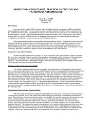
R M E D I A N - A P B1 .21 .31 .41 .51 .61 .71 .81 .91 .1 05 0 m s 5 0 0 µ VDownloaded from jnnp.bmj.com on June 5, 2012 - Published by group.bmj.comNEUROLOGY IN PRACTICEMii26CellbodyAxonF waveStimulusMMuscleFFigure 2 Median orthodromic sensory study. The <strong>in</strong>dex f<strong>in</strong>ger digital<strong>nerve</strong>s are stimulated via r<strong>in</strong>g electrodes <strong>and</strong> the response recordedover the median <strong>nerve</strong> at the wrist.CMAPs of similar shape <strong>and</strong> amplitude because the samemotor axons <strong>in</strong>nervate the muscle fibres mak<strong>in</strong>g up theresponse. However, the latency will be greater for elbowstimulation compared with wrist stimulation because of thelonger distance between the stimulat<strong>in</strong>g <strong>and</strong> record<strong>in</strong>gelectrodes (fig 1B). The difference <strong>in</strong> latency represents thetime taken for the fastest <strong>nerve</strong> fibres to conduct between thetwo stimulation po<strong>in</strong>ts as all other factors <strong>in</strong>volv<strong>in</strong>gneuromuscular transmission <strong>and</strong> muscle activation arecommon to both stimulation sites. If one measures thedistance between the two sites then the fastest motor <strong>nerve</strong><strong>conduction</strong> velocity can be calculated as follows: FMNCV(m/s) = distance between stimulation site 1 <strong>and</strong> site 2(mm)/[latency site 2 – latency site 1 (ms)].Sensory <strong>conduction</strong> <strong>studies</strong>The sensory <strong>nerve</strong> action potential (SNAP) is obta<strong>in</strong>ed byelectrically stimulat<strong>in</strong>g sensory fibres <strong>and</strong> record<strong>in</strong>g the <strong>nerve</strong>action potential at a po<strong>in</strong>t further along that <strong>nerve</strong>. Onceaga<strong>in</strong> the stimulus must be supramaximal.Record<strong>in</strong>g the SNAP orthodromically refers to distal <strong>nerve</strong>stimulation <strong>and</strong> record<strong>in</strong>g more proximally (the direction <strong>in</strong>which physiological sensory <strong>conduction</strong> occurs). Antidromictest<strong>in</strong>g is the reverse. Different laboratories prefer antidromicor orthodromic methods for test<strong>in</strong>g different <strong>nerve</strong>s. Anorthodromic median sensory study is shown <strong>in</strong> fig 2. Thesensory latency <strong>and</strong> the peak to peak amplitude of the SNAPare measured. The velocity correlates directly with thesensory latency <strong>and</strong> therefore either the result may beexpressed as a latency over a st<strong>and</strong>ard distance or a velocity.Only the 20% largest diameter <strong>and</strong> fastest conduct<strong>in</strong>gsensory fibres are tested us<strong>in</strong>g conventional sensory <strong>studies</strong>functionally supply<strong>in</strong>g f<strong>in</strong>e touch, vibration, <strong>and</strong> positionsense. Predom<strong>in</strong>antly small fibre neuropathies affect<strong>in</strong>g theother 80% of fibres exist usually with prom<strong>in</strong>ent symptoms ofpa<strong>in</strong> <strong>and</strong> conventional sensory <strong>studies</strong> may be normal. Insuch cases quantitative sensory test<strong>in</strong>g <strong>and</strong> autonomictest<strong>in</strong>g will be required, which are beyond the scope of thisarticle (see Interpretation <strong>pitfalls</strong>).FwavesF waves (F for foot where they were first described) are a typeof late motor response. When a motor <strong>nerve</strong> axon iselectrically stimulated at any po<strong>in</strong>t an action potential ispropagated <strong>in</strong> both directions away from the <strong>in</strong>itial stimulationsite. The distally propagated impulse gives rise to theMFFigure 3 Schematic representation of the early M response from thedistally propagated action potential <strong>and</strong> the later F wave from theproximally propagated action potential. The latter depolarises the axonhillock caus<strong>in</strong>g it to backfire. Actual F wave responses are shown <strong>in</strong> thelower trace. F waves vary <strong>in</strong> latency <strong>and</strong> shape due to differentpopulations of axons backfir<strong>in</strong>g each time.CMAP. However, an impulse also conducts proximally to theanterior horn cell, depolaris<strong>in</strong>g the axon hillock <strong>and</strong> caus<strong>in</strong>gthe axon to backfire. This leads to a small additional muscledepolarisation (F wave) at a longer latency. Only about 2% ofaxons backfire with each stimulus. Unlike the M response(fig 3), F waves vary <strong>in</strong> latency <strong>and</strong> shape because differentpopulations of neurones normally backfire with eachstimulus. The most reliable measure of the F wave is them<strong>in</strong>imum latency of 10–20 fir<strong>in</strong>gs.Why are F waves useful?F waves allow test<strong>in</strong>g of proximal segments of <strong>nerve</strong>s thatwould otherwise be <strong>in</strong>accessible to rout<strong>in</strong>e <strong>nerve</strong> <strong>conduction</strong><strong>studies</strong>. F waves test long lengths of <strong>nerve</strong>s whereas motor<strong>studies</strong> test shorter segments. Therefore F wave abnormalitiescan be a sensitive <strong>in</strong>dicator of peripheral <strong>nerve</strong> pathology,particularly if sited proximally. The F wave ratio whichcompares the <strong>conduction</strong> <strong>in</strong> the proximal half of the totalpathway with the distal may be used to determ<strong>in</strong>e the site of<strong>conduction</strong> slow<strong>in</strong>g—for example, to dist<strong>in</strong>guish a root lesionfrom a patient with a distal generalised neuropathy.ErrorsThe ma<strong>in</strong> sources of non-biological error <strong>in</strong> NCS measurementsare the identification <strong>and</strong> measurement of waveformonset <strong>and</strong> the measurement of the length of the <strong>nerve</strong>segment on the limb. Calculations have shown that <strong>in</strong> a <strong>nerve</strong>with a <strong>conduction</strong> velocity of 50 m/s, the 26SD experimentalerror for velocity is 14 m/s over 10 cm <strong>and</strong> 4.7 m/s over25 cm. Of the error, time measurement is 92.3% <strong>and</strong> distance7.7%, so the use of the measur<strong>in</strong>g tape is quite adequate <strong>in</strong>conventional NCS.www.jnnp.com