Crayfish Dissection - Mr.E Science
Crayfish Dissection - Mr.E Science
Crayfish Dissection - Mr.E Science
- No tags were found...

You also want an ePaper? Increase the reach of your titles
YUMPU automatically turns print PDFs into web optimized ePapers that Google loves.
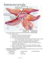
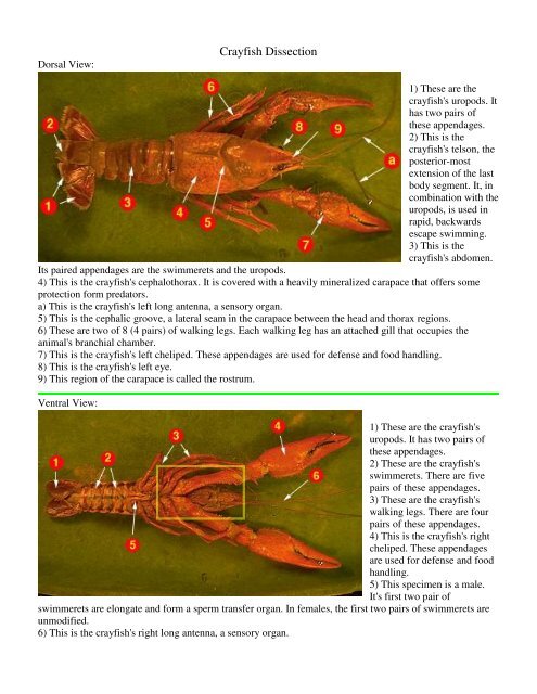
Dorsal View:<strong>Crayfish</strong> <strong>Dissection</strong>1) These are thecrayfish's uropods. Ithas two pairs ofthese appendages.2) This is thecrayfish's telson, theposterior-mostextension of the lastbody segment. It, incombination with theuropods, is used inrapid, backwardsescape swimming.3) This is thecrayfish's abdomen.Its paired appendages are the swimmerets and the uropods.4) This is the crayfish's cephalothorax. It is covered with a heavily mineralized carapace that offers someprotection form predators.a) This is the crayfish's left long antenna, a sensory organ.5) This is the cephalic groove, a lateral seam in the carapace between the head and thorax regions.6) These are two of 8 (4 pairs) of walking legs. Each walking leg has an attached gill that occupies theanimal's branchial chamber.7) This is the crayfish's left cheliped. These appendages are used for defense and food handling.8) This is the crayfish's left eye.9) This region of the carapace is called the rostrum.Ventral View:1) These are the crayfish'suropods. It has two pairs ofthese appendages.2) These are the crayfish'sswimmerets. There are fivepairs of these appendages.3) These are the crayfish'swalking legs. There are fourpairs of these appendages.4) This is the crayfish's rightcheliped. These appendagesare used for defense and foodhandling.5) This specimen is a male.It's first two pair ofswimmerets are elongate and form a sperm transfer organ. In females, the first two pairs of swimmerets areunmodified.6) This is the crayfish's right long antenna, a sensory organ.
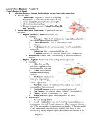
Branchial Chamber:1) The arrows show where the gills are attached toeach of the five pairs of walking legs.2) The carapace covering the right branchialchamber has been removed showing the gills.Water is drawn into the posterior end of thischamber, flows across the gills, and leaves via theanterior end.3) This is the cephalic groove, a lateral seam in thecarapace between the head and thorax regions.4) The arrow points to the right third maxillilped.5) This is the crayfish's right cheliped. Theseappendages are used for defense and foodhandling.6) This is the crayfish's right eye.7) This region of the carapace is called the rostrum.8) This is the crayfish's right long antenna, a sensory organ.Abdomen (Dorsal View):crayfish's right branchial chamber.1) These are the crayfish's uropods. It has two pairs ofthese appendages.2) This is the crayfish's telson, the posterior-mostextension of the last body segment. It, in combinationwith the uropods, is used in rapid, backwards escapeswimming.3) These are the crayfish's abdominal flexor muscles.They provide the major force for rapid backwardsswimming by flexing the tail.4) These are the crayfish's intestine.5) The arrow points to the posterior portion of theCephalothorax (dorsal)1) The arrow points to a gill within the exposedbranchial cavity.2) The arrow points to the crayfish's heart.3) The arrow points to a portion of the crayfish'shepatopancreas gland.4) The arrow points to the mandibular adductormuscle. It was attached to the inner surface ofthe carapace. It's contraction causes themandibles to come together.5) The arrow points to the gastric stomach, theportion of the digestive system containing thegastric mill, a chitinous arrangement of teeth,files, and sieves used to grind up the food.
6) The arrow points to the crayfish's right eye.Cephalathorax (lateral view)The arrows point to the cut bases of walkinglegs 3, 4, and 5.This is the right branchial chamber. The gillfilaments have been trimmed to revealdeeper structures.The arrow points to the heart. Click withinthe yellow rectangle to see an isolatedcrayfish heart.4) The arrow points to a portion of thecrayfish's hepatopancreas gland.5) The arrow points to the stomach. Clickwithin the yellow rectangle to see adissection of the gastric mill within thecardiac portion of the stomach.6) The arrow points to a green gland. These osmoregularoty organs secrete excess water and ammonia. Thefiltrate exits via the renal pore.<strong>Dissection</strong> ProcedureObtain a preserved crayfish and place it in your dissecting pan. The body is divided into an anteriorcephalothorax (fused head and thorax) and a posterior abdomen (see Figure 6). The chitinous exoskeletonprotects the crayfish from predators. The carapace is a saddlelike covering over the cephalothorax. Atransverse groove separates the fused head from the thoracic region. The rostrum is an anterior, pointedextension of the head. The abdomen consists of several segments and is terminated by the telson. Examinethe appendages. The appendages are modified to serve a variety of functions: feeding, walking andswimming. Although male and female crayfish have an equal number of appendages, in male crayfish theappendages joining the thorax have been modified. They are elongate and can be brought together to form atroughlike channel, used for the transfer of sperm from the male to the seminal receptacles of the female. UseFigure 6 to assist you in determining if your specimen is a male or a female.Position the crayfish dorsal side up. Using a pair of scissors, remove the carapace from the thorax by cuttingalong the mid-dorsal line anteriorly to the rostrum (refer to Figure 6). Carefully cut the carapace away alongthe side of the thorax; do not disturb any of the underlying organs.The organs of the digestive system are illustrated in Figure 6b (internal anatomy of the crayfish) below. Themouth (not visible at this time) leads via a short esophagus to the large stomach which occupies most of theanterior region of the cephalothorax. Open the stomach and locate the chitinous teeth that form the gastricmill which is used to grind food. The intestine runs from the stomach through the abdomen ending at theanus. The large yellowish digestive gland, which secretes enzymes and stores food, occupies much of thearea lateral and posterior to the stomach.The gonads (testes or ovaries) are lateral to the proximal portion of the intestine. Testes are generally whiteand ovaries orange. Sperm from the testes exit the body through a pore at the base of the fifth pair of walkinglegs, and eggs are released at the base of the third pair of walking legs. During reproduction, males andfemales copulate and sperm cells are passed to the genital openings of the female. Fertilization is internal,and fertilized eggs are extruded and retained for maturation of the swimmerets (the appendages associatedwith the abdomen) of the female.