Novocastratm Origintm Mouse Monoclonal Antibody Melan A ...
Novocastratm Origintm Mouse Monoclonal Antibody Melan A ...
Novocastratm Origintm Mouse Monoclonal Antibody Melan A ...
You also want an ePaper? Increase the reach of your titles
YUMPU automatically turns print PDFs into web optimized ePapers that Google loves.
Warnings and Precautions<br />
Inspect vial on receipt – if damaged, do not use.<br />
This reagent is a biological product; reasonable care should be taken when handling it. ProClin® 300 may cause irritation to skin, eyes,<br />
mucus membranes and upper respiratory tract. The concentration of ProClin® 300 in this product is 0.1% and so does not meet the OSHA<br />
criteria for a hazardous substance.<br />
Consult federal, state or local regulations for disposal of any potentially toxic components.<br />
Specimens, before and after fixation and all materials exposed to them, should be handled as if capable of transmitting infection and<br />
disposed of with proper precautions2 . Never pipette reagents by mouth and avoid contacting the skin and mucus membranes with reagents<br />
and specimens. If reagents or specimens come in contact with sensitive areas, wash with copious amounts of water. Seek medical<br />
advice.<br />
Minimize microbial contamination of reagents or an increase in non-specific staining may occur.<br />
Incubation times or temperatures, other than those specified, may give erroneous results. Any such changes must be validated by the<br />
user.<br />
Recommendations On Use<br />
Prior to undertaking this methodology, users must be trained in Immunohistochemical techniques on Ventana® Medical Systems<br />
Automated Immunostainers.<br />
Origin Antibodies were developed for use with the Ventana® Medical Systems, NexES® and BenchMark Immunohistochemistry<br />
Staining Systems in combination with Ventana® Detection Kits and Ventana® Prep Kit Dispensers. Novocastra has titered and quality<br />
controlled this antibody to ensure consistent and reliable performance. Before use Origin <strong>Antibody</strong> must be loaded into a Ventana®<br />
Prep Kit Dispenser – see the instructions supplied with Ventana® Prep Kit Dispenser. Apply the expiry date as indicted on the Origin<br />
<strong>Antibody</strong> vial label.<br />
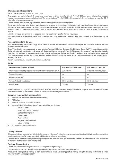
Table 1 summarises the requirements for Immunostaining.<br />
Table 1<br />
Requirements for FFPE Tissues Specification - BenchMark Specification - NexES®<br />
HIER (Heat Induced Epitope Retrieval) on NexES® or BenchMark CC1 solution To be determined by user<br />
Enzyme Digestion n/a n/a<br />
Enzyme Incubation n/a n/a<br />
Primary <strong>Antibody</strong> Incubation 16–32 minutes 16–32 minutes<br />
Amplification Optional Optional<br />
The combination of Origin <strong>Antibody</strong> incubation time and optimum conditions for epitope retrieval, together with the detection system<br />
should be validated by the user on a series of known positive and negative controls.<br />
Materials required but not supplied:<br />
1. Standard solvents used in Immunohistochemistry<br />
2. PBS buffer<br />
3. Retrieval solutions (if needed for HIER)<br />
4. Ventana® NexES® or BenchMark Automated Staining Systems<br />
Bar code labels<br />
Ventana® Prep Kit Dispenser<br />
Cell conditioning fluids<br />
Detection kit<br />
Wash solution<br />
Negative control reagent<br />
Liquid coverslip<br />
5. Mounting solution and coverslips<br />
Quality Control<br />
Differences in tissue processing and technical procedures in the user’s laboratory may produce significant variability in results, necessitating<br />
regular performance of in-house controls in addition to the following procedures.<br />
Controls should be fresh autopsy/biopsy/surgical specimens, formalin-fixed, processed and paraffin wax-embedded as soon as possible<br />
in the same manner as the patient sample(s).<br />
Positive Tissue Control<br />
Used to indicate correctly prepared tissues and proper staining techniques.<br />
One positive tissue control should be included for each set of test conditions in each staining run.<br />
A tissue with weak positive staining is more suitable than a tissue with strong positive staining for optimal quality control and to detect<br />
minor levels of reagent degradation. 3<br />
ORG- 8953/28/01/09


