Plenary Oral Presentations - Macquarie University Hospital
Plenary Oral Presentations - Macquarie University Hospital
Plenary Oral Presentations - Macquarie University Hospital
Create successful ePaper yourself
Turn your PDF publications into a flip-book with our unique Google optimized e-Paper software.
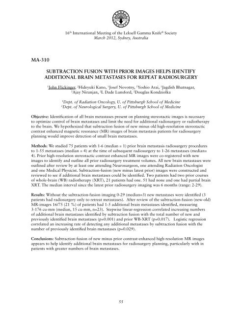
16 th International Meeting of the Leksell Gamma Knife ® SocietyMarch 2012, Sydney, AustraliaMA-310Subtraction Fusion With Prior Images Helps IdentifyAdditional Brain Metastases for Repeat Radiosurgery1John Flickinger, 2 Hideyuki Kano, 1 Josef Novotny, 1 Yoshio Arai, 1 Jagdish Bhatnagar,2Ajay Niranjan, 2 L Dade Lunsford, 2 Douglas Kondziolka1Dept. of Radiation Oncology, U. of Pittsburgh School of Medicine2Dept. of Neurological Surgery, U. of Pittsburgh School of MedicineObjective: Identification of all brain metastases present on planning stereotactic images is necessaryto optimize control of brain metastases and limit the need for additional radiosurgery or radiotherapyto the brain. We hypothesized that subtraction fusion of new minus old high-resolution stereotacticcontrast enhanced magnetic resonance (MR) images of brain metastasis patients for radiosurgeryplanning would improve detection of small brain metastases.Methods: We studied 75 patients with 1-6 (median = 1) prior brain metastasis radiosurgery proceduresto 1-55 metastases (median = 4) at the time of subsequent radiosurgery to 1-26 metastases (median=4). Prior high-resolution stereotactic contrast enhanced MR images were co-registered with newimages to identify and outline all prior radiosurgery treatment volumes. All new brain metastases wereoutlined after review by at least one attending Neurosurgeon, one attending Radiation Oncologistand one Medical Physicist. Subtraction-fusion (new minus latest prior) images were constructed andreviewed to see if additional brain metastases could be identified. Two patients had two prior coursesof whole-brain (WB) radiotherapy (XRT), 21 patients had one. 51 had none and one had partial brainXRT. The median interval since the latest prior radiosurgery imaging was 6 months (range: 2-29).Results: Without the subtraction-fusion imaging 0-29 (median=3) new metastases were identified (3patients had radiosurgery only to retreat metastases). After review of the subtraction-fusion (new-old)MR-images 16/75 (21 %) of patients had 1-5 additional brain metastases identified, measuring3-176 cu-mm (median, 15 cu-mm, n=23). Stepwise linear-regression correlated increasing numbersof additional brain metastases identified by subtraction fusion with the total number of new andpreviously identified brain metastases (p=0.001) and prior WB-XRT (p=0.017). Logistic regressioncorrelated an increasing rate of detecting any additional metastases by subtraction fusion with thenumber of previously identified brain metastases (p=0.029).Conclusions: Subtraction-fusion of new minus prior contrast-enhanced high-resolution MR imagesappears to help identify additional brain metastases for radiosurgery planning, particularly with inpatients with greater numbers of brain metastases.55


