AxyPrep Multisource Genomic DNA Miniprep Kit
AxyPrep Multisource Genomic DNA Miniprep Kit
AxyPrep Multisource Genomic DNA Miniprep Kit
You also want an ePaper? Increase the reach of your titles
YUMPU automatically turns print PDFs into web optimized ePapers that Google loves.
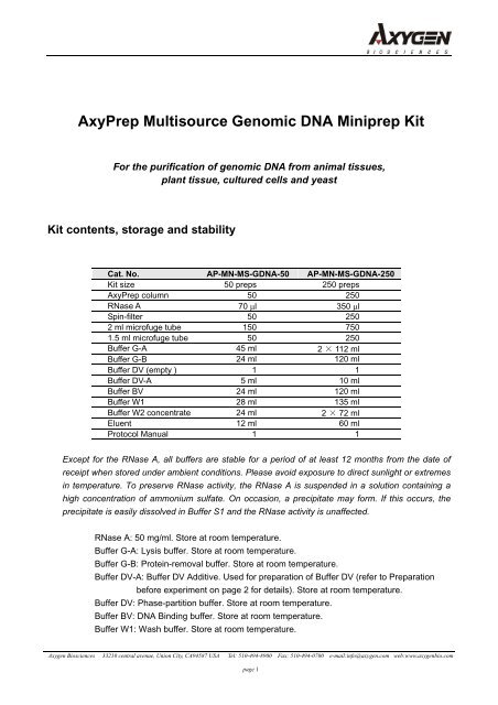
<strong>AxyPrep</strong> <strong>Multisource</strong> <strong>Genomic</strong> <strong>DNA</strong> <strong>Miniprep</strong> <strong>Kit</strong>For the purification of genomic <strong>DNA</strong> from animal tissues,plant tissue, cultured cells and yeast<strong>Kit</strong> contents, storage and stabilityCat. No. AP-MN-MS-G<strong>DNA</strong>-50 AP-MN-MS-G<strong>DNA</strong>-250<strong>Kit</strong> size 50 preps 250 preps<strong>AxyPrep</strong> column 50 250RNase A 70 μl 350 μlSpin-filter 50 2502 ml microfuge tube 150 7501.5 ml microfuge tube 50 250Buffer G-A 45 ml 2 × 112 mlBuffer G-B 24 ml 120 mlBuffer DV (empty ) 1 1Buffer DV-A 5 ml 10 mlBuffer BV 24 ml 120 mlBuffer W1 28 ml 135 mlBuffer W2 concentrate 24 ml 2 × 72 mlEluent 12 ml 60 mlProtocol Manual 1 1Except for the RNase A, all buffers are stable for a period of at least 12 months from the date ofreceipt when stored under ambient conditions. Please avoid exposure to direct sunlight or extremesin temperature. To preserve RNase activity, the RNase A is suspended in a solution containing ahigh concentration of ammonium sulfate. On occasion, a precipitate may form. If this occurs, theprecipitate is easily dissolved in Buffer S1 and the RNase activity is unaffected.RNase A: 50 mg/ml. Store at room temperature.Buffer G-A: Lysis buffer. Store at room temperature.Buffer G-B: Protein-removal buffer. Store at room temperature.Buffer DV-A: Buffer DV Additive. Used for preparation of Buffer DV (refer to Preparationbefore experiment on page 2 for details). Store at room temperature.Buffer DV: Phase-partition buffer. Store at room temperature.Buffer BV: <strong>DNA</strong> Binding buffer. Store at room temperature.Buffer W1: Wash buffer. Store at room temperature.Axygen Biosciences 33210 central avenue, Union City, CA94587 USA Tel: 510-494-8900 Fax: 510-494-0700 e-mail:info@axygen.com web:www.axygenbio.compage 1
Buffer W2 concentrate: Desalting buffer. Before using the kit, add the amount of ethanolspecified on the bottle label to the Buffer W2 concentrate. Either 100% or 95%denatured ethanol can be used. Store at room temperature.Eluent: 2.5 mM Tris-HCl, pH 8.5. Store at room temperature.IntroductionThe <strong>AxyPrep</strong> <strong>Multisource</strong> <strong>Genomic</strong> <strong>DNA</strong> <strong>Miniprep</strong> <strong>Kit</strong> is designed to purify genomic <strong>DNA</strong> from animaland plant tissues, cultured cells and yeast. This system employs a special lysis buffer, G-A toefficiently release genomic <strong>DNA</strong> from the biologic starting material. Proteins, pigments, carbohydratesand lipids are then efficiently segregated from the genomic <strong>DNA</strong> by a unique two-phase partition. Thelower phase is aspirated off and free genomic <strong>DNA</strong> is bound to an <strong>AxyPrep</strong> <strong>Genomic</strong> <strong>DNA</strong> column,where residual impurities and salt are removed. The purified <strong>DNA</strong> is then eluted in either a Tris bufferor deionized water. <strong>Genomic</strong> <strong>DNA</strong> prepared by this method is approximately 30 Kb in length and issuitable for a variety of applications, such as PCR amplification, Southern blot analysis, RAPD, AFLPand RFLP, etc. Each <strong>AxyPrep</strong> column will bind and purify up to 20 μg of genomic <strong>DNA</strong>.For the purification of genomic <strong>DNA</strong> from whole blood, we recommend the <strong>AxyPrep</strong> Whole Blood<strong>Genomic</strong> <strong>DNA</strong> <strong>Kit</strong>s: Mini #AP-MN-BL-50/250; Midi (#AP-MD-BL-G<strong>DNA</strong>-10 or –25); (Maxi #AP-MX-BL-G<strong>DNA</strong>-10 or –25). For the purification of bacterial genomic <strong>DNA</strong>, please refer to the <strong>AxyPrep</strong>Bacterial <strong>Genomic</strong> <strong>DNA</strong> <strong>Miniprep</strong> <strong>Kit</strong> (#AP-MN-BT-G<strong>DNA</strong>/50 or –250).CautionBuffer G-A, Buffer G-B, Buffer BV and Buffer W1 contain chemical irritants. When working with thebuffers, always wear suitable protective clothing such as safety glasses, laboratory coat and gloves.Be careful and avoid contact with eyes and skin. In the case of such contact, wash immediately withwater. If necessary, seek for medical assistance.Equipment and consumables required• Microcentrifuge capable of 12,000 × g• Mortar and pestle• Heated water bath• Vacuum manifold (#AP-VAC)• Vacuum regulator• Vacuum source (-25-30 inches Hg required)• Isobutanol and isopropanol.Preparation before experiment1) Before using the kit, add the amount of ethanol specified on the Buffer W2 label and mix well. Either100% or 95% (denatured) ethanol can be used.Axygen Biosciences 33210 central avenue, Union City, CA94587 USA Tel: 510-494-8900 Fax: 510-494-0700 e-mail:info@axygen.com web:www.axygenbio.compage 2
2) Prepare Buffer DV: Add 2 ml of Buffer DV-A, 125 ml of isopropanol and 75 ml of isobutanol to the 250ml bottle provided with kit and mix well.3) Chill Buffer DV at 4°C before proceeding.4) Adjust a water bath to 65°C.5) Check Buffer G-A and Buffer G-B for precipitation before each use. If precipitation occurs, incubate at65°C to dissolve the precipitate.6) Pre-warming the Eluent at 65°C will improve elution efficiency.ProtocolsI. Purification of <strong>Genomic</strong> <strong>DNA</strong> from Animal TissuesII. Purification of <strong>Genomic</strong> <strong>DNA</strong> from Plant TissuesIII. Purification of <strong>Genomic</strong> <strong>DNA</strong> from Cultured CellsIV. Purification of <strong>Genomic</strong> <strong>DNA</strong> from YeastEach type of starting material has different requirements for the method(s) used to achieve efficientlysis/homogenization. This necessitates that the protocol be organized into groups of steps, with eachgroup associated with a particular type of function or process within the overall protocol. Beforestarting the protocol, carefully read the entire procedure, including the various “Notes” in each step.Determine the correct protocol path in advance and prepare all required reagents, buffers andequipments.Groups:Steps1-3Lysis and homogenization of sampleSteps 4-5 Phase-partitioning to remove proteins and other impuritiesSteps 6-7 Spin-filter clarification of the aqueous phaseSteps 8-12 Binding, washing and elution on the <strong>AxyPrep</strong> columnA. Vacuum procedureB. Centrifuge procedureI. Purification of <strong>Genomic</strong> <strong>DNA</strong> from Animal Tissues[Animal Tissue vacuum protocol]This procedure requires the use of Axygen’s vacuum manifold or other manifold withcomplimentary luer fittings which can accommodate the <strong>AxyPrep</strong> columns. The use of a vacuumregulator is also recommended. A negative pressure of –25-30 inches Hg is required. This isequivalent to approximately -850-1,000 mbar and -12-15 psi.Lysis and homogenization of sampleUsing a mortar and pestle for homogenization1. Select 1-20 mg of tissue from animal or human and transfer to a mortar, pre-chilled on ice.Grind rapidly and vigorously to form a homogenate.Note: the following tissue-types should be completely frozen in liquid nitrogen before grinding:• DNase-rich tissues, such as pancreas, thymus, lymphoid tissue, etc.• Collagen-rich tissues, such as skin, connective tissue, etc.Axygen Biosciences 33210 central avenue, Union City, CA94587 USA Tel: 510-494-8900 Fax: 510-494-0700 e-mail:info@axygen.com web:www.axygenbio.compage 3
• Keratoprotein-rich tissues or hard tissues, such as bone.2. Add 650 μl of Buffer G-A and 0.9 μl of RNase A. Gently grind for 30 seconds to homogenouslymix the Buffer G-A with the ground tissue.Note: For those tissue types in Step 2A (above) requiring freezing in liquid nitrogen, please perform thefollowing steps after pulverization: Add 650 μl of Buffer G-A and 0.9 μl of RNase A. Warm the mortar to65°C in water bath until the Buffer G-A just melts. Gently grind for 1 minute. Then proceed to Step 3,below.3. Collect 650 μl of the homogenate and transfer to a 2 ml microfuge tube (provided). If thevolume of the homogenate is less than 650 μl, make it up to 650 μl with Buffer G-A. Incubatefor 5 min at 65°C in a water bath.Phase-partition to removal of protein and other impurities4. Add 400 μl of Buffer G-B and 1 ml of Buffer DV (pre-chilled to 4°C) in that order. Mix vigorouslyand centrifuge at 12,000 × g for 2 minutes.Note: Please refer to “Preparation before experiment” on page 3 to prepare Buffer DV.5. Discard the upper-phase as much as possible, keep the interface precipitate and the lowerphasein the tube. Add 1 ml of Buffer DV pre-chilled at 4°C, mix well and centrifuge at 12,000× g for 2 minutes.Spin-filter clarification of the aqueous phase6. Discard the upper phase, transfer the lower phase to a Spin-filter (placed in a 2 ml microfugetube), and centrifuge at 12,000 × g for 1 minute.Note: When recovering the lower phase, complete removal of the upper phase is not necessary. Any of theupper phase which is carried over will rapidly shift to the top of the pipette tip and can easily be removed(Refer to figure below).Note: If the lower phase is transferred without any contaminating interphase debris, Step 6 can be omitted.Pipettelower phasePhase partitioninside TipTransfer lowerphase into Spin-filterDiscardupper phaseNote: Avoid carryover of upper phase liquid. This will cause inhibition of genomic <strong>DNA</strong> binding to the <strong>AxyPrep</strong>column (Step 8, below).7. Discard the spin-filter. Add 400 μl of Buffer BV to the flow-through and mix well by brisk,repeated inversion.Axygen Biosciences 33210 central avenue, Union City, CA94587 USA Tel: 510-494-8900 Fax: 510-494-0700 e-mail:info@axygen.com web:www.axygenbio.compage 4
Binding, washing and elution on the <strong>AxyPrep</strong> column8. Attach the vacuum manifold base to a vacuum source. Firmly position the <strong>AxyPrep</strong> column(s)into the complimentary fittings on the manifold top. Transfer the binding mix from Step 7 to the<strong>AxyPrep</strong> column. Turn on the vacuum source and adjust to -25 inches Hg. Continue to applythe vacuum until no solution remains in the <strong>AxyPrep</strong> column.9. Add 500 μl of Buffer W1 and draw all of the solution through the column.10. Add 700 μl of Buffer W2 along the wall of <strong>AxyPrep</strong> column to wash off residual Buffer W1 anddraw all of the solution through the column. Repeat this wash with a second 700 μl aliquot ofBuffer W2.Note: Make sure that ethanol has been added into Buffer W2 concentrate.Note: Add Buffer W2 along the tube wall to wash off any residual salt.Note: Two washes with Buffer W2 are used to ensure the complete removal of salt, eliminating potentialproblems in subsequent enzymatic reactions.11. Transfer the <strong>AxyPrep</strong> column to a 2 ml microfuge tube and centrifuge at 12,000 × g for 1minute.12. Transfer the <strong>AxyPrep</strong> column into a clean 1.5 ml microfuge tube. To elute the genomic <strong>DNA</strong>,add 100-200 μl of Eluent (or deionized water) to the center of the membrane. Let it stand for 1minute at room temperature. Centrifuge at 12,000 × g for 1 minute.Note: Pre-warming water or Eluent at 65°C will often improve elution efficiency.[Animal Tissue spin protocol]Lysis and homogenization of sampleUsing a mortar and pestle for homogenization1. Select 1-20 mg of tissue from animal or human and transfer to a mortar, pre-chilled on ice.Grind rapidly and vigorously to form a homogenate.Note: the following tissue-types should be completely frozen in liquid nitrogen before grinding:• DNase-rich tissues, such as pancreas, thymus, lymphoid tissue, etc.• Collagen-rich tissues, such as skin, connective tissue, etc.• Keratoprotein-rich tissues or hard tissues, such as bone.2. Add 650 μl of Buffer G-A and 0.9 μl of RNase A. Gently grind for 30 seconds to homogenouslymix the Buffer G-A with the ground tissue.Note: For those tissue types in Step 2A (above) requiring freezing in liquid nitrogen, please perform thefollowing steps after pulverization: Add 650 μl of Buffer G-A and 0.9 μl of RNase A. Warm the mortar to65°C in water bath until the Buffer G-A just melts. Gently grind for 1 minute. Then proceed to Step 3,below.3. Collect 650 μl of the homogenate and transfer to a 2 ml microfuge tube (provided). If thevolume of the homogenate is less than 650 μl, make it up to 650 μl with Buffer G-A. Incubatefor 5 min at 65°C in a water bath.Phase-partition to removal of protein and other impuritiesAxygen Biosciences 33210 central avenue, Union City, CA94587 USA Tel: 510-494-8900 Fax: 510-494-0700 e-mail:info@axygen.com web:www.axygenbio.compage 5
4. Add 400 μl of Buffer G-B and 1 ml of Buffer DV (pre-chilled to 4°C) in that order. Mix vigorouslyand centrifuge at 12,000 × g for 2 minutes.Note: Please refer to “Preparation before experiment” on page 3 to prepare Buffer DV.5. Discard the upper-phase as much as possible, keep the interface precipitate and the lowerphasein the tube. Add 1 ml of Buffer DV pre-chilled at 4°C, mix well and centrifuge at 12,000× g for 2 minutes.Spin-filter clarification of the aqueous phase6. Discard the upper phase, transfer the lower phase to a Spin-filter (placed in a 2 ml microfugetube), and centrifuge at 12,000 × g for 1 minute.Note: When recovering the lower phase, complete removal of the upper phase is not necessary. Any of theupper phase which is carried over will rapidly shift to the top of the pipette tip and can easily be removed(Refer to figure below).Note: If the lower phase is transferred without any contaminating interphase debris, Step 6 can be omitted.Pipettelower phasePhase partitioninside TipTransfer lowerphase into Spin-filterDiscardupper phaseNote: Avoid carryover of upper phase liquid. This will cause inhibition of genomic <strong>DNA</strong> binding to the <strong>AxyPrep</strong>column (Step 8, below).7. Discard the spin-filter. Add 400 μl of Buffer BV to the flow-through and mix well by brisk,repeated inversion.Binding, washing and elution on the <strong>AxyPrep</strong> column8. Place a <strong>AxyPrep</strong> column to a 2 ml microfuge tube (provided). Transfer the binding mix fromStep 7 to the <strong>AxyPrep</strong> column. Centrifuge at 12,000 × g for 1 minute.9. Discard the filtrate in the 2 ml microfuge tube. Place the <strong>AxyPrep</strong> column back to the 2 mlmicrofuge tube. Add 500 μl of Buffer W1 to the <strong>AxyPrep</strong> column and centrifuge at 12,000 × gfor 1 minute.10. Discard the filtrate and place the <strong>AxyPrep</strong> column back to the 2 ml microfuge tube. Add 700 μlof Buffer W2 and centrifuge at 12,000 × g for 1 minute.Note: Make sure that ethanol has been added into Buffer W2 concentrate.11. Optional Step: Discard the filtrate from the 2 ml microfuge tube. Place the <strong>AxyPrep</strong> columnback into the 2 ml microfuge tube. Add 700 μl of Buffer W2 to the <strong>AxyPrep</strong> column andcentrifuge at 12,000 × g for 1 minute.Axygen Biosciences 33210 central avenue, Union City, CA94587 USA Tel: 510-494-8900 Fax: 510-494-0700 e-mail:info@axygen.com web:www.axygenbio.compage 6
Note: Two washes with Buffer W2 are used to ensure the complete removal of salt, eliminating potentialproblems in subsequent enzymatic reactions.12. Discard filtrate from the 2 ml microfuge tube. Place the <strong>AxyPrep</strong> column back into the 2 mlmicrofuge tube. Centrifuge at 12,000 × g for 1 minute.13. Transfer the <strong>AxyPrep</strong> column into a clean 1.5 ml microfuge tube (provide). To elute thegenomic <strong>DNA</strong>, add 100-200 μl of Eluent (or deionized water) to the center of the membrane.Let it stand for 1 minute at room temperature. Centrifuge at 12,000 × g for 1 minute.Note: Pre-warming water or Eluent at 65°C will often improve elution efficiency.II. Purification of <strong>Genomic</strong> <strong>DNA</strong> from Plant Tissues[Plant Tissue Vacuum Protocol]This procedure requires the use of Axygen’s vacuum manifold or other manifold withcomplimentary luer fittings which can accommodate the <strong>AxyPrep</strong> columns. The use of a vacuumregulator is also recommended. A negative pressure of –25-30 inches Hg is required. This isequivalent to approximately -850-1,000 mbar and -12-15 psi.Lysis and homogenization of sampleUsing a mortar and pestle for homogenization1. Using Table 1(below), weigh out the appropriate amount of fresh plant tissue (the amountshould be reduced by half if lyophilized, dehydrated, or dry tissues are used) and transfer to themortar. Carefully add liquid nitrogen directly to the sample until it is completely frozen. Use thepestle to pulverize it quickly and vigorously until it is reduced to a fine powder.Table 1. Types of fresh plant tissues used for genomic <strong>DNA</strong> preparationFlower or leavesPlant stemPlant rootPlant seed10-100 mg≤ 240 mg≤ 240 mg≤ 240 mgNote: If cultured plant cells are used, collect 2 × 10 3 ~ 1 × 10 7 plant cells and spin for 1 minute at 10,000 × gto pellet the cells. Resuspend the plant cells in 150 μl of deionized water and transfer to the mortar.Carefully add liquid nitrogen directly to the sample until it is completely frozen. Use the pestle topulverize it quickly and vigorously until it is reduced to a fine powder. Add liquid nitrogen as required toprevent the material from thawing during pulverization. After pulverization, warm the mortar at 65°C in awater bath until the pulverized material just melts. Proceed to Step 2, below.2. Add 700 μl of Buffer G-A and 1.2 μl of RNase A. Quickly grind the sample for 30 seconds.Note: Incomplete grinding will reduce the yield of genomic <strong>DNA</strong>.Note: When the weight of the fresh plant tissue is >120 mg or the dried plant tissue is >60 mg, add 1.3 ml ofBuffer G-A. After Step 2 has been completed, divide the sample evenly between two 2 ml microfugetubes. Steps 4 -7 will proceed in two parallel 2 ml microfuge tubes. In Step 8, the contents of the twotubes will be consolidated into a single <strong>AxyPrep</strong> column.Axygen Biosciences 33210 central avenue, Union City, CA94587 USA Tel: 510-494-8900 Fax: 510-494-0700 e-mail:info@axygen.com web:www.axygenbio.compage 7
3. Transfer the tissue homogenate into a 2 ml microfuge tube. Determine the approximate volume.If the volume of the homogenate is less than 650 μl, add additional Buffer G-A up to 650 μl.Incubate it for 15 minutes at 65°C in a water bath.Note: If fibrous samples such as plant stem and root, or starch- and protein-rich samples such as seeds areused, increase the incubation time to 60 minutes in the water bath.Phase-partition to removal of protein and other impurities4. Add 400 μl of Buffer G-B and 1 ml of Buffer DV (pre-chilled to 4°C) in that order. Mix vigorouslyand centrifuge at 12,000 × g for 2 minutes.Note: Please refer to “Preparation before experiment” on page 3 to prepare Buffer DV.5. Discard the upper-phase as much as possible, keep the interface precipitate and the lowerphasein the tube. Add 1 ml of Buffer DV pre-chilled at 4°C, mix well and centrifuge at 12,000× g for 2 minutes.Spin-filter clarification of the aqueous phase6. Discard the upper phase, transfer the lower phase to a Spin-filter (placed in a 2 ml microfugetube), and centrifuge at 12,000 × g for 1 minute.Note: When recovering the lower phase, complete removal of the upper phase is not necessary. Any of theupper phase which is carried over will rapidly shift to the top of the pipette tip and can easily be removed(Refer to figure below).Note: If the lower phase is transferred without any contaminating interphase debris, Step 6 can be omitted.Pipettelower phasePhase partitioninside TipTransfer lowerphase into Spin-filterDiscardupper phaseNote: Avoid carryover of upper phase liquid. This will cause inhibition of genomic <strong>DNA</strong> binding to the <strong>AxyPrep</strong>column (Step 8, below).7. Discard the spin-filter. Add 400 μl of Buffer BV to the flow-through and mix well by brisk,repeated inversion.Binding, washing and elution on the <strong>AxyPrep</strong> column8. Attach the vacuum manifold base to a vacuum source. Firmly position the <strong>AxyPrep</strong> column(s)into the complimentary fittings on the manifold top. Transfer the binding mix from Step 7 to the<strong>AxyPrep</strong> column. Turn on the vacuum source and adjust to -25 inches Hg. Continue to applythe vacuum until no solution remains in the <strong>AxyPrep</strong> column.Axygen Biosciences 33210 central avenue, Union City, CA94587 USA Tel: 510-494-8900 Fax: 510-494-0700 e-mail:info@axygen.com web:www.axygenbio.compage 8
9. Add 500 μl of Buffer W1. Draw all of the solution through the column.10. Add 700 μl of Buffer W2 along the wall of <strong>AxyPrep</strong> column to wash off residual Buffer W1,draw all of the solution through the column. Repeat this wash with a second 700 μl aliquot ofBuffer W2.Note: Make sure that ethanol has been added into Buffer W2 concentrate.Note: Add Buffer W2 along the tube wall to wash off any residual salt.Note: Two washes with Buffer W2 are used to ensure the complete removal of salt, eliminating potentialproblems in subsequent enzymatic reactions.11. Transfer the <strong>AxyPrep</strong> column to a 2 ml microfuge tube (provided) and centrifuge at 12,000 ×g for 1 minute.12. Transfer the <strong>AxyPrep</strong> column into a clean 1.5 ml microfuge tube (provided). To elute thegenomic <strong>DNA</strong>, add 100-200 μl of Eluent (or deionized water) to the center of the membrane.Let it stand for 1 minute at room temperature. Centrifuge at 12,000 × g for 1 minute.Note: Pre-warming water or Eluent at 65°C will often improve elution efficiency.[Plant Tissue Spin Protocol]Lysis and homogenization of sampleUsing a mortar and pestle for homogenization1. Using Table 1(below), weigh out the appropriate amount of fresh plant tissue (the amountshould be reduced by half if lyophilized, dehydrated, or dry tissues are used) and transfer to themortar. Carefully add liquid nitrogen directly to the sample until it is completely frozen. Use thepestle to pulverize it quickly and vigorously until it is reduced to a fine powder.Table 1. Types of fresh plant tissues used for genomic <strong>DNA</strong> preparationFlower or leavesPlant stemPlant rootPlant seed10-100 mg≤ 240 mg≤ 240 mg≤ 240 mgNote: If cultured plant cells are used, collect 2 × 10 3 ~ 1 × 10 7 plant cells and spin for 1 minute at 10,000 × gto pellet the cells. Resuspend the plant cells in 150 μl of deionized water and transfer to the mortar.Carefully add liquid nitrogen directly to the sample until it is completely frozen. Use the pestle topulverize it quickly and vigorously until it is reduced to a fine powder. Add liquid nitrogen as required toprevent the material from thawing during pulverization. After pulverization, warm the mortar at 65°C in awater bath until the pulverized material just melts. Proceed to Step 2, below.2. Add 700 μl of Buffer G-A and 1.2 μl of RNase A. Quickly grind the sample for 30 seconds.Note: Incomplete grinding will reduce the yield of genomic <strong>DNA</strong>.Note: When the weight of the fresh plant tissue is >120 mg or the dried plant tissue is >60 mg, add 1.3 ml ofBuffer G-A. After Step 2 has been completed, divide the sample evenly between two 2 ml microfugetubes. Steps 4 -7 will proceed in two parallel 2 ml microfuge tubes. In Step 8, the contents of the twotubes will be consolidated into a single <strong>AxyPrep</strong> column.Axygen Biosciences 33210 central avenue, Union City, CA94587 USA Tel: 510-494-8900 Fax: 510-494-0700 e-mail:info@axygen.com web:www.axygenbio.compage 9
3. Transfer the tissue homogenate into a 2 ml microfuge tube. Determine the approximate volume.If the volume of the homogenate is less than 650 μl, add additional Buffer G-A up to 650 μl.Incubate it for 15 minutes at 65°C in a water bath.Note: If fibrous samples such as plant stem and root, or starch- and protein-rich samples such as seeds areused, increase the incubation time to 60 minutes in the water bath.Phase-partition to removal of protein and other impurities4. Add 400 μl of Buffer G-B and 1 ml of Buffer DV (pre-chilled to 4°C) in that order. Mix vigorouslyand centrifuge at 12,000 × g for 2 minutes.Note: Please refer to “Preparation before experiment” on page 3 to prepare Buffer DV.5. Discard the upper-phase as much as possible, keep the interface precipitate and the lowerphasein the tube. Add 1 ml of Buffer DV pre-chilled at 4°C, mix well and centrifuge at 12,000 ×g for 2 minutes.Spin-filter clarification of the aqueous phase6. Discard the upper phase, transfer the lower phase to a Spin-filter (placed in a 2 ml microfugetube), and centrifuge at 12,000 × g for 1 minute.Note: When recovering the lower phase, complete removal of the upper phase is not necessary. Any of theupper phase which is carried over will rapidly shift to the top of the pipette tip and can easily be removed(Refer to figure below).Note: If the lower phase is transferred without any contaminating interphase debris, Step 6 can be omitted.Pipettelower phasePhase partitioninside TipTransfer lowerphase into Spin-filterDiscardupper phaseNote: Avoid carryover of upper phase liquid. This will cause inhibition of genomic <strong>DNA</strong> binding to the <strong>AxyPrep</strong>column (Step 8, below).7. Discard the spin-filter. Add 400 μl of Buffer BV to the flow-through and mix well by brisk,repeated inversion.Binding, washing and elution on the <strong>AxyPrep</strong> column8. Place a <strong>AxyPrep</strong> column to a 2 ml microfuge tube (provided). Transfer the binding mix fromStep 7 to the <strong>AxyPrep</strong> column. Centrifuge at 12,000 × g for 1 minute.Axygen Biosciences 33210 central avenue, Union City, CA94587 USA Tel: 510-494-8900 Fax: 510-494-0700 e-mail:info@axygen.com web:www.axygenbio.compage 10
9. Discard the filtrate in the 2 ml microfuge tube. Place the <strong>AxyPrep</strong> column back to the 2 mlmicrofuge tube. Add 500 μl of Buffer W1 to the <strong>AxyPrep</strong> column and centrifuge at 12,000 × gfor 1 minute.10. Discard the filtrate and place the <strong>AxyPrep</strong> column back to the 2 ml microfuge tube. Add 700 μlof Buffer W2 and centrifuge at 12,000 × g for 1 minute.Note: Make sure that ethanol has been added into Buffer W2 concentrate.11. Optional Step: Discard the filtrate from the 2 ml microfuge tube. Place the <strong>AxyPrep</strong> columnback into the 2 ml microfuge tube. Add 700 μl of Buffer W2 to the <strong>AxyPrep</strong> column andcentrifuge at 12,000 × g for 1 minute.Note: Two washes with Buffer W2 are used to ensure the complete removal of salt, eliminating potentialproblems in subsequent enzymatic reactions.12. Discard filtrate from the 2 ml microfuge tube. Place the <strong>AxyPrep</strong> column back into the 2 mlmicrofuge tube. Centrifuge at 12,000 × g for 1 minute.13. Transfer the <strong>AxyPrep</strong> column into a clean 1.5 ml microfuge tube (provide). To elute thegenomic <strong>DNA</strong>, add 100-200 μl of Eluent (or deionized water) to the center of the membrane.Let it stand for 1 minute at room temperature. Centrifuge at 12,000 × g for 1 minute.Note: Pre-warming water or Eluent at 65°C will often improve elution efficiency.III. Purification of <strong>Genomic</strong> <strong>DNA</strong> from Cultured Animal Cells[Cultured Animal Cells Vacuum Protocol]This procedure requires the use of Axygen’s vacuum manifold or other manifold withcomplimentary luer fittings which can accommodate the <strong>AxyPrep</strong> columns. The use of a vacuumregulator is also recommended. A negative pressure of –25-30 inches Hg is required. This isequivalent to approximately -850-1,000 mbar and -12-15 psi.Lysis and homogenization of sampleSelect homogenization method A or B, depending upon the type of cultured animal cell used. Ifgenomic <strong>DNA</strong> is extracted from plant cells, please follow the previous protocol “Purification of<strong>Genomic</strong> <strong>DNA</strong> from Plant Tissues” (above) to homogenize the plant cells.A. Cells grown in suspension or cell suspension freshly-isolated from animal or humantissues:1A. Collect 1 × 10 3 - 2 × 10 6 cells in suspension and transfer into a 2 ml microfuge tube.Centrifuge at 2,000 × g for 5 minutes to pellet the cells. Discard the supernatant.2A. Add 150 μl of deionized water or PBS to resuspend the cells and then add 500 μl ofBuffer G-A. Let the tube stand for 1 minute at room temperature.Proceed to Step 3, below.Axygen Biosciences 33210 central avenue, Union City, CA94587 USA Tel: 510-494-8900 Fax: 510-494-0700 e-mail:info@axygen.com web:www.axygenbio.compage 11
B. Cells grown in a monolayer in a 96-well, 24-well, 12-well or 6-well plate:1B. Discard as much of the supernatant as possible, then add 650 μl of Buffer G-A into eachwell. Let the plate stand for 1 minute at room temperature.2B. Pipette up and down several times, then transfer 650 μl of the cell homogenate into a 2ml microfuge tube.3. Add 0.8 μl of RNase A. Vortex for 15 seconds and let the tube stand for 1 minute at roomtemperature.Phase-partition to removal of protein and other impurities4. Add 400 μl of Buffer G-B and 1 ml of Buffer DV (pre-chilled to 4°C) in that order. Mix vigorouslyand centrifuge at 12,000 × g for 2 minutes.Note: Please refer to “Preparation before experiment” on page 3 to prepare Buffer DV.5. Discard the upper-phase as much as possible, keep the interface precipitate and the lowerphasein the tube. Add 1 ml of Buffer DV pre-chilled at 4°C, mix well and centrifuge at 12,000× g for 2 minutes.Spin-filter clarification of the aqueous phase6. Discard the upper phase, transfer the lower phase to a Spin-filter (placed in a 2 ml microfugetube), and centrifuge at 12,000 × g for 1 minute.Note: When recovering the lower phase, complete removal of the upper phase is not necessary. Any of theupper phase which is carried over will rapidly shift to the top of the pipette tip and can easily be removed(Refer to figure below).Note: If the lower phase is transferred without any contaminating interphase debris, Step 6 can be omitted.Pipettelower phasePhase partitioninside TipTransfer lowerphase into Spin-filterDiscardupper phaseNote: Avoid carryover of upper phase liquid. This will cause inhibition of genomic <strong>DNA</strong> binding to the <strong>AxyPrep</strong>column (Step 8, below).7. Discard the spin-filter. Add 400 μl of Buffer BV to the flow-through and mix well by brisk,repeated inversion.Binding, washing and elution on the <strong>AxyPrep</strong> column8. Attach the vacuum manifold base to a vacuum source. Firmly position the <strong>AxyPrep</strong> column(s)into the complimentary fittings on the manifold top. Transfer the binding mix from Step 7 to theAxygen Biosciences 33210 central avenue, Union City, CA94587 USA Tel: 510-494-8900 Fax: 510-494-0700 e-mail:info@axygen.com web:www.axygenbio.compage 12
<strong>AxyPrep</strong> column. Turn on the vacuum source and adjust to -25 inches Hg. Continue to applythe vacuum until no solution remains in the <strong>AxyPrep</strong> column.9. Add 500 μl of Buffer W1. Draw all of the solution through the column.10. Add 700 μl of Buffer W2 along the wall of <strong>AxyPrep</strong> column to wash off residual Buffer W1,draw all of the solution through the column. Repeat this wash with a second 700 μl aliquot ofBuffer W2.Note: Make sure that ethanol has been added into Buffer W2 concentrate.Note: Add Buffer W2 along the tube wall to wash off any residual salt.Note: Two washes with Buffer W2 are used to ensure the complete removal of salt, eliminating potentialproblems in subsequent enzymatic reactions.11. Transfer the <strong>AxyPrep</strong> column to a 2 ml microfuge tube (provided) and centrifuge at 12,000 ×g for 1 minute.12. Transfer the <strong>AxyPrep</strong> column into a clean 1.5 ml microfuge tube. To elute the genomic <strong>DNA</strong>,add 100-200 μl of Eluent (or deionized water) to the center of the membrane. Let it stand for 1minute at room temperature. Centrifuge at 12,000 × g for 1 minute.Note: Pre-warming water or Eluent at 65°C will often improve elution efficiency.[Cultured Animal Cells Spin Protocol]Lysis and homogenization of sampleSelect homogenization method A or B, depending upon the type of cultured animal cell used. Ifgenomic <strong>DNA</strong> is extracted from plant cells, please follow the previous protocol “Purification of<strong>Genomic</strong> <strong>DNA</strong> from Plant Tissues” (above) to homogenize the plant cells.A. Cells grown in suspension or cell suspension freshly-isolated from animal or humantissues:1A. Collect 1 × 10 3 - 2 × 10 6 cells in suspension and transfer into a 2 ml microfuge tube.Centrifuge at 2,000 × g for 5 minutes to pellet the cells. Discard the supernatant.2A. Add 150 μl of deionized water or PBS to resuspend the cells and then add 500 μl ofBuffer G-A. Let the tube stand for 1 minute at room temperature.Proceed to Step 3, below.B. Cells grown in a monolayer in a 96-well, 24-well, 12-well or 6-well plate:1B. Discard as much of the supernatant as possible, then add 650 μl of Buffer G-A into eachwell. Let the plate stand for 1 minute at room temperature.2B. Pipette up and down several times, then transfer 650 μl of the cell homogenate into a 2ml microfuge tube.3. Add 0.8 μl of RNase A. Vortex for 15 seconds and let the tube stand for 1 minute at roomtemperature.Phase-partition to removal of protein and other impuritiesAxygen Biosciences 33210 central avenue, Union City, CA94587 USA Tel: 510-494-8900 Fax: 510-494-0700 e-mail:info@axygen.com web:www.axygenbio.compage 13
4. Add 400 μl of Buffer G-B and 1 ml of Buffer DV (pre-chilled to 4°C) in that order. Mix vigorouslyand centrifuge at 12,000 × g for 2 minutes.Note: Please refer to “Preparation before experiment” on page 3 to prepare Buffer DV.5. Discard the upper-phase as much as possible, keep the interface precipitate and the lowerphasein the tube. Add 1 ml of Buffer DV pre-chilled at 4°C, mix well and centrifuge at 12,000× g for 2 minutes.Spin-filter clarification of the aqueous phase6. Discard the upper phase, transfer the lower phase to a Spin-filter (placed in a 2 ml microfugetube), and centrifuge at 12,000 × g for 1 minute.Note: When recovering the lower phase, complete removal of the upper phase is not necessary. Any of theupper phase which is carried over will rapidly shift to the top of the pipette tip and can easily be removed(Refer to figure below).Note: If the lower phase is transferred without any contaminating interphase debris, Step 6 can be omitted.Pipettelower phasePhase partitioninside TipTransfer lowerphase into Spin-filterDiscardupper phaseNote: Avoid carryover of upper phase liquid. This will cause inhibition of genomic <strong>DNA</strong> binding to the <strong>AxyPrep</strong>column (Step 8, below).7. Discard the spin-filter. Add 400 μl of Buffer BV to the flow-through and mix well by brisk,repeated inversion.Binding, washing and elution on the <strong>AxyPrep</strong> column8. Place a <strong>AxyPrep</strong> column to a 2 ml microfuge tube (provided). Transfer the binding mix fromStep 7 to the <strong>AxyPrep</strong> column. Centrifuge at 12,000 × g for 1 minute.9. Discard the filtrate in the 2 ml microfuge tube. Place the <strong>AxyPrep</strong> column back to the 2 mlmicrofuge tube. Add 500 μl of Buffer W1 to the <strong>AxyPrep</strong> column and centrifuge at 12,000 × gfor 1 minute.10. Discard the filtrate and place the <strong>AxyPrep</strong> column back to the 2 ml microfuge tube. Add 700 μlof Buffer W2 and centrifuge at 12,000 × g for 1 minute.Note: Make sure that ethanol has been added into Buffer W2 concentrate.11. Optional Step: Discard the filtrate from the 2 ml microfuge tube. Place the <strong>AxyPrep</strong> columnback into the 2 ml microfuge tube. Add 700 μl of Buffer W2 to the <strong>AxyPrep</strong> column andcentrifuge at 12,000 × g for 1 minute.Axygen Biosciences 33210 central avenue, Union City, CA94587 USA Tel: 510-494-8900 Fax: 510-494-0700 e-mail:info@axygen.com web:www.axygenbio.compage 14
Note: Two washes with Buffer W2 are used to ensure the complete removal of salt, eliminating potentialproblems in subsequent enzymatic reactions.12. Discard filtrate from the 2 ml microfuge tube. Place the <strong>AxyPrep</strong> column back into the 2 mlmicrofuge tube. Centrifuge at 12,000 × g for 1 minute.13. Transfer the <strong>AxyPrep</strong> column into a clean 1.5 ml microfuge tube (provide). To elute thegenomic <strong>DNA</strong>, add 100-200 μl of Eluent (or deionized water) to the center of the membrane.Let it stand for 1 minute at room temperature. Centrifuge at 12,000 × g for 1 minute.Note: Pre-warming water or Eluent at 65°C will often improve elution efficiency.IV. Purification of <strong>Genomic</strong> <strong>DNA</strong> from Yeast[Yeast Vacuum Protocol]This procedure requires the use of Axygen’s vacuum manifold or other manifold with complimentaryluer fittings which can accommodate the <strong>AxyPrep</strong> columns. The use of a vacuum regulator is alsorecommended. A negative pressure of –25-30 inches Hg is required. This is equivalent toapproximately -850-1,000 mbar and -12-15 psi.Lysis and homogenization of sampleUsing a mortar and pestle for homogenization1. Collect 2 × 10 6 - 5 × 10 7 yeast cells and centrifuge for 1 minute at 10,000 × g to pellet thecells. Resuspend the yeast cells in 150 μl of water and transfer to a mortar.Note: For yeast, an OD 600 of 1 ≅ 3 × 10 7 cells/ml.2. Gradually add liquid nitrogen until the yeast suspension is completely frozen. Using the pestle,quickly and forcefully reduce it to a fine powder. Add liquid nitrogen to prevent the sample fromthawing during pulverization. After grinding is complete, warm the mortar at 65°C in water bathuntil it just begins to melt.3. Add 600 μl of Buffer G-A and 1.2 μl of RNase A. Quickly grind the sample for 30 seconds.Transfer 650 μl of the yeast homogenate into a 2 ml microfuge tube. If the volume of thehomogenate is less than 650 μl, add additional Buffer G-A up to 650 μl. Incubate it for 10minutes at 65°C in a water bath.Proceed to Step 4 (below)Phase-partition to removal of protein and other impurities4. Add 400 μl of Buffer G-B and 1 ml of Buffer DV (pre-chilled to 4°C) in that order. Mix vigorouslyand centrifuge at 12,000 × g for 2 minutes.Note: Please refer to “Preparation before experiment” on page 3 to prepare Buffer DV.5. Discard the upper-phase as much as possible, keep the interface precipitate and the lowerphasein the tube. Add 1 ml of Buffer DV pre-chilled at 4°C, mix well and centrifuge at 12,000× g for 2 minutes.Axygen Biosciences 33210 central avenue, Union City, CA94587 USA Tel: 510-494-8900 Fax: 510-494-0700 e-mail:info@axygen.com web:www.axygenbio.compage 15
Spin-filter clarification of the aqueous phase6. Discard the upper phase, transfer the lower phase to a Spin-filter (placed in a 2 ml microfugetube), and centrifuge at 12,000 × g for 1 minute.Note: When recovering the lower phase, complete removal of the upper phase is not necessary. Any of theupper phase which is carried over will rapidly shift to the top of the pipette tip and can easily be removed(Refer to figure below).Note: If the lower phase is transferred without any contaminating interphase debris, Step 6 can be omitted.Pipettelower phasePhase partitioninside TipTransfer lowerphase into Spin-filterDiscardupper phaseNote: Avoid carryover of upper phase liquid. This will cause inhibition of genomic <strong>DNA</strong> binding to the <strong>AxyPrep</strong>column (Step 8, below).7. Discard the spin-filter. Add 400 μl of Buffer BV to the flow-through and mix well by brisk,repeated inversion.Binding, washing and elution on the <strong>AxyPrep</strong> column8. Attach the vacuum manifold base to a vacuum source. Firmly position the <strong>AxyPrep</strong> column(s)into the complimentary fittings on the manifold top. Transfer the binding mix from Step 7 to the<strong>AxyPrep</strong> column. Turn on the vacuum source and adjust to -25 inches Hg. Continue to applythe vacuum until no solution remains in the <strong>AxyPrep</strong> column.9. Add 500 μl of Buffer W1. Draw all of the solution through the column.10. Add 700 μl of Buffer W2 along the wall of <strong>AxyPrep</strong> column to wash off residual Buffer W1,draw all of the solution through the column. Repeat this wash with a second 700 μl aliquot ofBuffer W2.Note: Make sure that ethanol has been added into Buffer W2 concentrate.Note: Add Buffer W2 along the tube wall to wash off any residual salt.Note: Two washes with Buffer W2 are used to ensure the complete removal of salt, eliminating potentialproblems in subsequent enzymatic reactions.11. Transfer the <strong>AxyPrep</strong> column to a 2 ml microfuge tube (provided) and centrifuge at 12,000 ×g for 1 minute.12. Transfer the <strong>AxyPrep</strong> column into a clean 1.5 ml microfuge tube. To elute the genomic <strong>DNA</strong>,add 100-200 μl of Eluent (or deionized water) to the center of the membrane. Let it stand for 1minute at room temperature. Centrifuge at 12,000 × g for 1 minute.Note: Pre-warming water or Eluent at 65°C will often improve elution efficiency.Axygen Biosciences 33210 central avenue, Union City, CA94587 USA Tel: 510-494-8900 Fax: 510-494-0700 e-mail:info@axygen.com web:www.axygenbio.compage 16
[Yeast Spin Protocol]Lysis and homogenization of sampleUsing a mortar and pestle for homogenization1. Collect 2 × 10 6 - 5 × 10 7 yeast cells and centrifuge for 1 minute at 10,000 × g to pellet thecells. Resuspend the yeast cells in 150 μl of water and transfer to a mortar.Note: For yeast, an OD 600 of 1 ≅ 3 × 10 7 cells/ml.2. Gradually add liquid nitrogen until the yeast suspension is completely frozen. Using the pestle,quickly and forcefully reduce it to a fine powder. Add liquid nitrogen to prevent the sample fromthawing during pulverization. After grinding is complete, warm the mortar at 65°C in water bathuntil it just begins to melt.3. Add 600 μl of Buffer G-A and 1.2 μl of RNase A. Quickly grind the sample for 30 seconds.Transfer 650 μl of the yeast homogenate into a 2 ml microfuge tube. If the volume of thehomogenate is less than 650 μl, add additional Buffer G-A up to 650 μl. Incubate it for 10minutes at 65°C in a water bath.Proceed to Step 4 (below)Phase-partition to removal of protein and other impurities4. Add 400 μl of Buffer G-B and 1 ml of Buffer DV (pre-chilled to 4°C) in that order. Mix vigorouslyand centrifuge at 12,000 × g for 2 minutes.Note: Please refer to “Preparation before experiment” on page 3 to prepare Buffer DV.5. Discard the upper-phase as much as possible, keep the interface precipitate and the lowerphasein the tube. Add 1 ml of Buffer DV pre-chilled at 4°C, mix well and centrifuge at 12,000× g for 2 minutes.Spin-filter clarification of the aqueous phase6. Discard the upper phase, transfer the lower phase to a Spin-filter (placed in a 2 ml microfugetube), and centrifuge at 12,000 × g for 1 minute.Note: When recovering the lower phase, complete removal of the upper phase is not necessary. Any of theupper phase which is carried over will rapidly shift to the top of the pipette tip and can easily be removed(Refer to figure below).Note: If the lower phase is transferred without any contaminating interphase debris, Step 6 can be omitted.Pipettelower phasePhase partitioninside TipTransfer lowerphase into Spin-filterDiscardupper phaseAxygen Biosciences 33210 central avenue, Union City, CA94587 USA Tel: 510-494-8900 Fax: 510-494-0700 e-mail:info@axygen.com web:www.axygenbio.compage 17
Note: Avoid carryover of upper phase liquid. This will cause inhibition of genomic <strong>DNA</strong> binding to the <strong>AxyPrep</strong>column (Step 8, below).7. Discard the spin-filter. Add 400 μl of Buffer BV to the flow-through and mix well by brisk,repeated inversion.Binding, washing and elution on the <strong>AxyPrep</strong> column8.Place a <strong>AxyPrep</strong> column to a 2 ml microfuge tube (provided). Transfer the binding mix fromStep 7 to the <strong>AxyPrep</strong> column. Centrifuge at 12,000 × g for 1 minute.9. Discard the filtrate in the 2 ml microfuge tube. Place the <strong>AxyPrep</strong> column back to the 2 mlmicrofuge tube. Add 500 μl of Buffer W1 to the <strong>AxyPrep</strong> column and centrifuge at 12,000 × gfor 1 minute.10. Discard the filtrate and place the <strong>AxyPrep</strong> column back to the 2 ml microfuge tube. Add 700 μlof Buffer W2 and centrifuge at 12,000 × g for 1 minute.Note: Make sure that ethanol has been added into Buffer W2 concentrate.11. Optional Step: Discard the filtrate from the 2 ml Microfuge tube. Place the <strong>AxyPrep</strong> columnback into the 2 ml microfuge tube. Add 700 μl of Buffer W2 to the <strong>AxyPrep</strong> column andcentrifuge at 12,000 × g for 1 minute.Note: Two washes with Buffer W2 are used to ensure the complete removal of salt, eliminating potentialproblems in subsequent enzymatic reactions.12. Discard filtrate from the 2 ml microfuge tube. Place the <strong>AxyPrep</strong> column back into the 2 mlmicrofuge tube. Centrifuge at 12,000 × g for 1 minute.13. Transfer the <strong>AxyPrep</strong> column into a clean 1.5 ml microfuge tube (provide). To elute thegenomic <strong>DNA</strong>, add 100-200 μl of Eluent (or deionized water) to the center of the membrane.Let it stand for 1 minute at room temperature. Centrifuge at 12,000 × g for 1 minute.Note: Pre-warming water or Eluent at 65°C will often improve elution efficiency.Axygen Biosciences 33210 central avenue, Union City, CA94587 USA Tel: 510-494-8900 Fax: 510-494-0700 e-mail:info@axygen.com web:www.axygenbio.compage 18
OverviewMake up to 650 μl with Buffer G-AAdd RNase A and heatLysisAdd 400 μl of Buffer G-BAdd 1 ml of Buffer DVRepeat extraction with Buffer DVPhasepartitionAdd 400 μl of Buffer BVFiltrationAdd 500 μl of Buffer W1Add 700 μl of Buffer W2Repeat wash with Buffer W2BindingWashingAdd 100-200 μl of water or theEluentElutionTroubleshooting:1. Low or no yield• Insufficient bacteria processed• Inefficient lysis• Bring the upper phase liquid into the lower phase.• <strong>DNA</strong> not efficiently eluted• <strong>AxyPrep</strong> column membrane overdried during vacuum removal of W22. Low A 260/280• Too many material processed• Inefficient lysis• Contamination with interphase material3. RNA present (elevated A 260/280 )• Failure to add RNase A to Buffer S• Buffer S datedAxygen Biosciences 33210 central avenue, Union City, CA94587 USA Tel: 510-494-8900 Fax: 510-494-0700 e-mail:info@axygen.com web:www.axygenbio.compage 19
4. <strong>Genomic</strong> <strong>DNA</strong> appears to be degradedDepending upon the completeness of degradation, the genomic <strong>DNA</strong> will either appear as a smearor as a smear trailing in front of a high molecular weight band on an agarose gel. Since no physicalmeasure used during the purification process is sufficient to cause any visually discernabledegradation, the most likely source is enzymatic. Many strains of bacteria exhibit high levels ofendonuclease activity. These endA+ strains must be lysed rapidly and completely to preventsubstantial enzymatic degradation of the intrinsic genomic <strong>DNA</strong>.5. <strong>Genomic</strong> <strong>DNA</strong> performs poorly in enzymatic reactions• Low <strong>DNA</strong> concentration• Salt contamination: insufficient Buffer W1 removal• Ethanol contamination: insufficient centrifugation to remove residual Buffer W26. Clogged spin-filter• Too much material processed• Inefficient lysis7. Clogged <strong>AxyPrep</strong> column• Too many bacteria processed• Inefficient lysisAxygen Biosciences 33210 central avenue, Union City, CA94587 USA Tel: 510-494-8900 Fax: 510-494-0700 e-mail:info@axygen.com web:www.axygenbio.compage 20


